Nicu Survival Guide for Residents
26
1 of 26 NICU SURVIVAL GUIDE (revised June 2011 by Dr. S.Kadiwala & Dr. N.Sharma) This manual, revised for the first time, is designed for use by the pediatric residents and interns created by residents for the UF-Jacksonville Pediatric Residency Program. The recommendations in this manual are specific for the practices in this program. Please understand that this is not a mini-textbook or outline of general newborn care. The purpose of this manual is to assist pediatric house officers by: a) Providing a guideline for management of patients that require immediate attention b) Reminders to help them in their daily work There is little discussion of pathophysiology, pharmacology and infectious disease processes. Certain important and common problems are not covered at all. If at any time you are unsure about the contents provided in this guide, please refer to more comprehensive texts, or contact the on call attending.
Transcript of Nicu Survival Guide for Residents

1 of 26
NICU SURVIVAL GUIDE
(revised June 2011 by Dr. S.Kadiwala & Dr. N.Sharma)
This manual, revised for the first time, is designed for use by the pediatric residents and interns created by residents for the UF-Jacksonville Pediatric Residency Program. The recommendations in this manual are specific for the practices in this program. Please understand that this is not a mini-textbook or outline of general newborn care. The purpose of this manual is to assist pediatric house officers by:
a) Providing a guideline for management of patients that require immediate attention b) Reminders to help them in their daily work
There is little discussion of pathophysiology, pharmacology and infectious disease processes. Certain important and common problems are not covered at all. If at any time you are unsure about the contents provided in this guide, please refer to more comprehensive texts, or contact the on call attending.

2 of 26
Phone Numbers
Dr. Dial 306-4734 Dr. Sharma 306-3979
Dr. Garrison 306-3981 Dr. Banadera 393-5959
Dr. Alviedo 393-4800 Dr. Huddleston 306-3976
NICU 244-5100 Dr. D. Cuevas 306-3974
3N 244-6110 Dr. Tan 498-5350
Step down 244-3330 Dr. Driscoll 393-4414
L&D 244-6127 UF ID 393-6299
Lab 244-6040 Genetics 393-1180
Nemours 697-3600 Dr. Spierre (Rehab) 633-0926
Attending call room 244-3348 Radiology 244-6084
NP/PA call room 244-5109
Schedule
0700 Sign-out from Night call/ on-call PA
1000 Rounds (can be later, attending dependant)
1200 Noon Conference
1400 Radiology Rounds (excluding Thursday)
1900 Sign-out to Night call/ on call PA
Expectations
If you are on overnight, please double check that your name & pager number are on the white board located by the NICU Secretary and in the Step down unit.
All orders and notes must have a DATE, TIME, SIGNATURE and DOCTOR NUMBER.
Be familiar with the standing orders
Attend afternoon clinic
If you are called about an infant, evaluate/examine and document a note (why you were called, your examination, your findings, and what actions were taken).
Document any procedures, updates with family, etc. in Neodata (print & place in chart)
NICU POLICIES & PROCEDURES
Infection Control
Scrub for 3 minutes to elbow when first coming into the NICU and Step-Down units
No watches or jewelry, except for a plain wedding band while handling patients
Please keep sleeves at or above your elbows when handling patients
Wash hands or use foam before and after handling patients
Must gown and glove (in addition to hand washing) when handling isolated patients

3 of 26
Patients with NEC should be considered as having a communicable infection with use of isolation technique above until proven Rotazyme negative
NO FOOD/DRINKS are allowed anywhere except the nursing/staff lounge – this includes keeping residents’ computer station clear of food/drinks
Charts, including nursing charting material, must remain at the patient’s bedside
Please follow sterile techniques when introducing central lines/procedures Chart Entries
All orders and notes must have a DATE, TIME, SIGNATURE and DOCTOR NUMBER.
Verbal orders are prohibited except during an emergency such as a resuscitation or when gowned for or performing a sterile procedure.
All verbal and telephone orders must be signed before leaving for the day.
All read back orders written by the nurse must be co-signed
Check all flagged chart pages for need for signature
Orders “flagged” in chart should be placed face down to comply with HIPPA Other
Any baby transferred to normal nursery must have an interim summary on the chart before transfer and either the nursery attending or Patty Willams ANRP notified by telephone prior to transfer
Patients returning from surgery must have all orders re-written
Patients transferred to the Step Down unit must have orders re-written
All medication orders must contain the method of dose calculation ie scheduled does and, mg/kg; or for infusions: concentration, mcg/kg/min and mL/kg/min.
TPN, Lipids and large fluid volume infusions should be ordered using the TPN program o To access, password is FEEDME
Remember not to use banned abbreviations, especially the use of “q.d” and trailing zeros on medication or fluid orders (E.g “5” and not 5.0mg )
All parental consents must be signed on admissions or as close to admission as possible

4 of 26
PULMONOLOGY Ventilator Terminology
Peak inspiratory pressure (PIP)
Maximum pressure measured during the delivery of gas during conventional mechanical
ventilation.
PIP reflects the effects of the amount of gas delivered to the lungs in a given breath (tidal volume)
PEEP
PEEP helps to maintain functional residual capacity (FRC). At the end of expiration, the PEEP exerts pressure to oppose passive emptying of the lung and to keep the airway pressure above the atmospheric pressure. The presence of PEEP opens up collapsed or unstable alveoli and increases the FRC and surface area for gas exchange, thus reducing the size of the shunt
Rate
Reflects how often a volume of gas in the system is delivered to the infant. Inspiratory/Expiratory Ratio
I/E ratio reflects the relationship between time spent in inspiration and time spent in expriation.
If the I/E ratio is 1:2 with a rate of 60 and the total respiratory cycle is 1 second, inspiration is 0.33 second and expriation is 0.66 second.
Prolonged inspiration may be associated with more efficient ventilation, optimal arterial oxygenation, a higher risk of air leak, and impending of venous return.
Prolonged expriation also improves oxygenation, especially in air-trapping conditions
Mean Airway Pressure (MAP)
Amount of pressure transmitted to the airway throughout an entire respiratory cycle.
Ways to increase MAP o Increase PEEP, or PIP, or Ti, or RR, or Flow
Minute Ventilation (Vt)
Determines rate of carbon dioxide removal
Minute ventilation= TVxRR Amplitude
Amount of pressure oscillation that occurs around he MAP.
Increasing the amplitude will increase the TV and therefore decrease PCO2.
Decreasing the amplitude will decrease the TV and therefore increase PCO2.

5 of 26
Mechanical Ventilation and Respiratory Support
Types of Vents Synchronized Intermittent Mandatory Ventilation (SIMV)
Delivers a set number of breaths with a certain amount of pressure each minute, synchronizes with babies’ inspiration attempts
When ordering SIMV- write for PIP, PEEP, Rate, IT, and FiO2 SIMV/VG
VG- Volume guarantees a certain tidal volume, usually 6ml/kg per assisted breath. Target tidal volume maintained by the ventilator as the pressure limit varies inversely with lung compliance.
You still set PEEP and rate Assist Control
Not used much in our NICU, you still must set the PIP and PEEP
The vent assists every breath the baby takes, even if the baby breathes 80 times/min
Must set a minimum backup rate in case the baby does not breathe at all.
Reserved for very ill neonates who require very high support Pressure Support Ventilation (PSV)
Pressure increases in proportion to inspiratory volume. High-Frequency Ventilation
Goal of HFOV is to reduce barotrauma
Delivers a very fast in and out oxygen supply- the baby does not take breaths but there is a continous in and out motion multiple times per minute
Gas exchange with a kind of double spiral effect- there is a pulse of O2 going centrally down the airway with pulses of CO2 going out up the sides of the airway.
Main paratemeters are amplitude which affects volume in and out with each oscillation. Mean Airway pressure (MAP) affects expansion and oxgenation of the lungs and frequency of oscillation (Hz) affecting how many times per minute the exchange occurs
Oscillators vibrate columns of air and have active exhalation cycles. Typically set at 10-15Hz (600-900 breaths/min)
JET Ventilator
Used if the infant has a pneumothorax, PIE, over/hyperexpanded lungs, pneumatoceles, and severe meconium aspiration/PPHN
Allows longer exhalation time vs oscillator (passive exhalation)
Change: PIP to change CO2, PEEP (oxygenation & expansion), and rate (expansion)
Back up breaths: to treat and correct atelectasis (can increase oxygenation in RDS)
Starting Pressures for beginning ventilator support
FiO2: min. to keep Sa02 88%-92% (for premies) and > 95% (for term infants)
PEEP: 4-6cm water
PIP: 14-20cm water (14-16 for premature infants, especially <1000 grams, and 16-18 for term infants)
Rate: 40-60
I/E ratio: 1:1-1:2
I-time: 0.35-0.4

6 of 26
Changing Vent Settings
What affects Tidal Volume?- PIP, PEEP, and AMP Factors TV CO2
Inc PIP Increases Decreases
Dec PEEP Increases Decreases
Inc AMP No change Decreases
What affects respiratory Rate? o Vent Rate increase in rate will decrease CO2 o I: E ratio decrease I time will Increase E time which will decrease CO2
What affects airway pressure/expansion? Factors Volume O2
Inc PIP Increases Increases
Inc PEEP Increases Increases
Inc MAP Increases Increases
What affects O2 concentration? o FiO2 o Change oxygenation by either changing FiO2 or PIP o Monitor oxygenation one of three ways
SaO2 ABG PaO2 (only accurate on ABG, on VBG or CBG tells you essentially
nothing) CXR- count at least 8 ribs
How to change your PaCO2?
Variable Rate PIP PEEP IT FiO2
Increase PaCO2 Decrease Decrease NA NA NA
Decrease PaCO2 Increase Increase NA NA NA
Increase PaO2 NA Increase Increase Increase Increase
Decrease PaCO2 NA Decrease Decrease NA Decrease
Formula o Remember AMP changes CO2 (increase AMP dec CO2) o MAP changes O2 o Extubate MAP <7 and FIO2<35%
Formula OI= FIO2 x MAP x 100 PaO2
OI= oxygen Index, indicative of severe resp distress, >20 need for Nitric Oxide & >30 ECMO MAP = (Ti x PIP) + (Te x PEEP)/ Ti + Te or [(RR x Ti)/60] x [PIP-PEEP] + PEEP Blood Gases
Type pH PCO2 HCO3 Compensation
Metabolic Acidosis <7.4 <40 low -BE (hypervent)

7 of 26
Respiratory Acidosis <7.4 >40 high +BE (hypovent)
Metabolic Alkalosis >7.4 >40 high +BE (hypovent)
Respiratory Alkalosis >7.4 <40 low -BE (hypervent)
Sodium Bicarbonate: investigate cause of metabolic acidosis, e.g. hypovolemia or sepsis, etc., and treat accordingly. (Refer to Pediatrics - Sodium bicarbonate: basically useless therapy. Aschner JL, Poland RL. Pediatrics. 2008 Oct;122(4):831-5.) Give if adequate ventilation; very seldom indicated Goal BE < -10 Give not more than 1 meq/kg/hr, followed by ABG after 1hr Formula mEq Na HCO3= BEx 0.3 x wt (kg) (¼ correction) 2 mEq NaHCO3= BE x 0.6 x wt (kg) ( ½ correction) 2 Metabolic Alkalosis CO2 Oxygen DDx
Low High Over ventilation, air bubbles, hyperventilation
High Normal/High Obstructed ET, ET down right main stem bronchus, pneumothorax, PDA, permissive hypercapnea
High Low Pneumothorax, improper ET position, PDA, atelectasis, lung disease
Normal Low Agitation, pneumothorax, improper ETT position, atelectasis, pulmonary HTN, pulmonary edema
Criteria for use of Palivizumab (Synagis) Indication
Indication for the prevention of serious lower respiratory tract disease caused by RSV
Potential Candidates for Palivizumab
Patient Group Age at Start of RSV Season
Premature, no chronic lung disease or congenital heart disease
< 28weeks gestation age
29-32 weeks gestational age
32-35 weeks gestational age
< 12months < 6months with 1 additional risk factor < 6months with 2 additional risk factor
Chronic Lung Disease < 2years

8 of 26
Hemodynamically significant congenital heart disease < 2years
Other high-risk children with pulmonary or immune function < 2years
risk factors: child care, school-age siblings, exposure to environmental air pollution, congenital abnormalities of the airways, or severe neuromuscular disease
Dosage
15mg/kg IM
Given every month during RSV season Apnea
>20seconds breathing cessation o Central- no diaphragm activity o Obstructive- upper airway obstruction with diaphragm activity
Treatment: Caffeine (see dosing in pharmacology guide) Bronchopulmonary Dysplasia (BPD)
Due to arrested lung development resulting from interference with alveolarization and vascularization
>36wk, still requiring oxygen at 28days
If O2 sats <92% and if hypoxia develops chronically, it can cause pulmonary HTN (cor pulmonale)
Keep CO2>30 on AGB, if less then PVL (periventricular leukomalacia) may develop Chronic Lung Disease (CLD)/ Bronchopulmonary Dysplasia (BPD)
Supplemental oxygen requirement at 28-30 days of life or 36 weeks postmenstrual age.
Occurs due to chronic and constant lung injury with ongoing repair and healing of the injury.
Also caused by lung immaturity, oxygen toxicity, and barotrauma/volutrauma (high PIP and PEEP, pneumothorax, and PIE)
Meconium Aspiration Syndrome (MAS)
Typically occurs in a post term infant who becomes hypoxic in utero. Fetal asphyxia causes the anal sphincter to relax and colonic peristalsis ensues expelling meconium into the amniotic fluid. A second episode of asphyxia occurs, during which the infant makes gasping respiratory movements, causing meconium to enter into the oropharynx and lung. o Etiology
Atelectasis: due to blockage of smaller airway causing air trapping and alveolar collapse
Chemical pneumonitis: inflammatory response to meconium

9 of 26
Inhibits surfactant function Increases pulmonary vascular resistance
o Diagnosis History CXR: air trapping, hyperexpansion, and hyperinflation; bilateral diffuse, coarse,
patchy infiltrates o Treatment
During labor: Amnioinfusion Delivery: intubate and tracheal suction in depressed infants (do not stimulate
baby prior to this!)
Anytime there is a history of thick meconium at delivery with respiratory distress, obtain histopathology on the placenta. Meconium staining of placental can determine length of time (how long prior to delivery) the baby released meconium and was in distress.
Air leaks
Types: pneumomediastinum, pneumothorax, pneumopericardium, or subcutaneous emphysema
Pulmonary Interstitial Emphysema (PIE) o Occurs when free air is released from ruptured alveoli o Diagnosis
CXR Fiberoptic probe may reveal hyperlucency of affected side
o Treatment Elevate head 30-40 degrees (this decrease the work of breathing) Give 100% oxygen Needle aspiration and chest tube placement
Lateral approach- 4th or 6th intercostals space
Superior approach- 2nd-3rd IC space or just lateral to midclavicular line Acute Respiratory Distress Syndrome (RDS)
Defined- disease of immature lung anatomy and physiology.
Pathology- o Anatomically the preterm lung is unable to support oxygenation and ventilation due
to insufficiently developed alveolar saccules, which causes a deficient surface area for gas exchange. The volume of surfactant is insufficient to prevent collapse of unstable alveoli. Because the alveoli collapse with each breath, normal FRC is not established.
o Increase WOB due to poorly compliant lung and insufficient oxygenation and ventilation requiring increased energy output. Increased WOB results in hypoxemia and academia which cause constriction of pulmonary vascular (arterial) musculature, severely limiting pulmonary capillary blood flow. Without adequate pulmonary capillary blood flow, the type II pneumocytes become deficient in the precursor material necessary for production of surfactant.
o Diagnosis CXR- show reduced lung volumes, air bronchograms (visible outlines of air-filled
bronchi due atelectasis), reticulogranularity, and lung opacification. o Treatment
Surfactant Correct academia

10 of 26
Reduce hypoxemia- by maintaining BP and Hct, maintain neutral thermal environment.
Transient Tachypnea of Newborn (TTN)
Etiology- seen in term or near term infants born via C-section
TTN is due to lack of gradual compression of the chest that eliminates some fluid during a normal vaginal delivery. This causes a delay in reabsorption of normal lung fluid and lung fluid accumulates in peribronchilar lymphatics and the bronchovascular spaces, causing an “obstructive” lung disease. Accumulation of interstitial fluid interferes with the forces that hold the bronchioli open, causing collapse and air trapping.
Dx: CXR- show hyperexpansion with streaky infiltrates radiating from the hilum
Tx: supplemental oxygen
Nitric Oxide Treatment Protocol for
Improvement of Survival Without BPD
Eligibility o birth weigth/GA groups o 500-799 grams: on NCPAP or ventilator o 800-1000 grams: if on ventilator o 1001-1250 grams: if on ventilator and GA< 28 0/7weeks o postnatal age 7-14days o Respiratory severity score (RSS=MAPx FiO2) >2
Exclusions o lethal congenital anomalies o intent to withdraw care o bilateral grad 4 IVH o Target SpO2 o recommend 88-94%
Treatment o initiate iNO at 20ppm for 96hrs; obtain methemoglobin level at 24hrs o wean iNO to 10ppm for 168 hours (7 days) o wean iNO to 5ppm for 168 hours (7 days) o wean iNO to 2ppm for 168 hours (7days) o discontinue iNO o Note: treatment continues even if on nasal CPAP or nasal cannula
Note: fluid in fissure

11 of 26
NUTRITION
Total Parenteral Nutrition (TPN)
Source of nutrition for those who cannot eat adequately o Contains a mixture of dextrose, protein, electrolytes, vitamins, and minerals. o Usually the yellow IV bag hanging at the bedside
Route o Peripheral IV limits
Dextrose: 12.5% (12.5 g/100 mL) Calcium: 12meq/L or 300mg/100ml Potassium: 60 meq/L Protein: 2-3%
o Central IV (PICC, Broviac/Hickman Port) Dextrose: maximum of 25%
Labs (general guidelines –always subject to change based on individual cases) o Prior to starting TPN, check a triglyceride level
Goal is to have triglycerides < 150mg/dL o Daily chemistry (BMP) o Check magnesium, phosphate, triglycerides q Monday &Thursday
Formulas o Determine Kcal/kg/day
Kcal in 24hr period = Kcal/kg/24hr Weight (kg)
Weight/Age Fluids Labs
>1500grs/ Term DOL 0- 80-90mL/kg/d DOL 1- 100-120 mL/kg/d DOL 2- 140-150 mL/kg/d
q AM BMP DOL 2 or 3 Tbili/Dbili
1000-1500gm DOL 0 -80-100mL/kg/day DOL 1 -100-200 mL/kg/d DOL2 -120-160 mL/kg/d
At 12hrs of life: BMP DOL 1 Tbili/Dbili q AM BMP, Hct
800-1000gm DOL0- 100mL/kg/d DOL 1 -130 mL/kg/d DOL 2 -150 mL/kg/d
BMP q8-12hrs x 48hrs, then DOL1 Tbili/Dbili q AM BMP, Tbili
600-800gm DOL0-120 mL/kg/d DOL 1 -130-50 mL/kg/d DOL 2- 150-170 mL/kg/d
BMP q8hrs x 48hrs, then q AM BMP, Hct, Tbili
<600gm DOL0- 120mL/kg/d DOL 1- 150mL/kg/d DOL 2 -180-200mL/kg/d
BMP q8hrs x 48hrs, then q AM BMP, Hct, Tbili
Note, it takes 50-75kcal/kg/day to maintain weight in a thermo-neutral environment GOAL: 120kcal/kg/day

12 of 26
Standard Electrolytes
Starting Advance Weight/Age Based Max
Carbohydrate 5-8 mg/kg/day 1-2 mg/kg/day
ELBW6-8 mg/kg/min Preterm 4-6 mg/kg/min Term 4mg/kg/min
Glucose concentration, no more than +/- 2%
Protein (watch BUN, Cr)
2-3 gr/kg/day 0.5-1 g/kg/day Peripheral line- 30g/L Central line- 4g/L
Fats (20% IL) (watch triglycerides)
Term: 2 g/kg/day Preterm: 1 g/kg/day
0.5-1g/kg/day 4 g/kg/day
Sodium 0.8 mEq/kg/day Start if Na <135 Goal 2-4mEq/kg/day
1-2mg/kg/day
Potassium (watch UOP)
DOL 2 when K<3.5 1-4mEq/kg/day
1g/kg/day 4mEq/kg/day
Calcium DOL 1 with 2.5mEq/kg/day Goal 1.5-2.5mEq/kg/day
Mg 0.25-0.5 mEq/kg/day
Phosphate 0.5-1.5 mEq/kg/day
Fluid Requirements
First 24hrs of life o Start 10W or D7.5 with AA & calcium at 80 kg/day in term infants and 100 mL/kg/day
in preterm infants o D10W= 10g/100mL, therefore 1mL D10W= 100mg Glucose (1g Dextrose=3.4kcal)
Max Volume o 150-160 mL/kg/day (>150mL/kg/day is associated with CLD and PDA) o 130mL/kg/day for infant’s with CLD o VLBW infant may need more fluids due to larger surface area, therefore more
insensible loss To wean off TPN (general guidelines)
Begin enteral feeds on DOL 3 if stable at 20ml/kg/day (20% total volume) q 3hrs o Please note: this is a GENERAL rule of thumb, more mature infants can be started
on enteral feeds on DOL 1
Advance enteral by 20-30 mL/kg/day
Decrease TPN rate accordingly to maintain overall fluid balance Enteral Feeds
Criteria for oral (PO) feeds o > 1500 grams o Suck/swallow reflex (> 32-34wk) o Regular stooling pattern, good bowel sounds

13 of 26
o No UAC, no NG with bile, no presssors
Start feeds o Use breast milk if available (may need to be fortified to meet caloric needs) o Formula should be 20kcal with iron o 10-20 mL/kg/day q 3hrs
Increase Feeds o Advance PO feeds by 20% per day.
Be cautious, aggressive feeding can result in NEC Goal is usually between 150-160 mL/kg/day
o Breast fed infants need to suck well for 10 mins prior to advancing o Advance PO attempts as follows: 1PO attempt/day to 1 PO attempt/shift to 2-3 PO
attempt/day, PO every other feed, to PO every feed (again, this is a general guideline)
Weight Gain o If gaining weight well on 24kcal formula, consider changing to a 22cal formula 3-4
days prior to discharge to ensure continued weight gain
Human Milk Fortifier (HMF) o May add when tolerating > 100mL/kg/day o Add 1pkt/50mL to 22kcal/oz or 1pkt/25mL to 24kcal/oz o HMF increases Ca so if patient has hypercalcemic, add MCT oil to increase calories o MCT oil can also be used for patients with CLD
Supplements o Once on full formula feeds, add:
2-4mg/kg/day of Iron BID Multivitamin 1mL daily
Formula
o After tolerating 100-140mL/kg/day, can change to 24kcal/oz formula o Remember 24kcal/oz formula is ISOtonic and ISOosmolar like 20kcal/oz o 27kcal/oz formula is HYPERosmolar, so use with caution
Residuals o Often re-feeding regimen is attending dependent o Good rule of thumb
If < 10% total feeds reefed and continue feeding If 10-30% subtract and refeed If >30% hold feeds and evaluate infant (always consider NEC)
o If >20mL worry about intestinal obstruction, if >60mL almost definite intestinal obstruction
o If the infant has continued large residuals, look at how the baby is positioned (R side down will decrease emptying, while L side down will incease emptying)
20kcal/oz 180mL/kg/day= 120kcal/kg/day 30mL= 0.67kcal
24kcal/oz 150ml/kg/day= 120kcal/kg/day 30mL= 0.8kcal

14 of 26
GASTEROENTEROLOGY
Gastrochisis vs Omphalocele
Gastroschisis Omphalocele
Location Right of umbilicus Central
Umbilical Cord Intact Umbilical cord elements
surround the sac and join
above the extrusion
Protective Sac Full-thickness defect, no sac Protective sac is preserved
Liver and Spleen Remain within the abdominal
cavity
Partially extruded in large
defects
Associated Congenital
Anomalies
Rare Common
Treatment
Fluid/Temperature regulation- monitor for hypothermia
Wrap abdomen in wet dressing to protect and cellophane
Place NG tube to decompress the stomach
Intubate to reduce abdominal pressure
Start broad spectrum antibiotics
Obtain stat KUB
Immediate Surgical Consult
Duodenal Obstruction
Present- vomiting and abdominal distension, may have history of polyhydraminos
Dx: KUB- double bubble sign (x ray to the left)
Tx: NPO, IVF, NG decompression, and surgical repair
Proximal GI Obstruction
Vomiting is prominent and distention less prominent
Dx: KUB and cross table, UGI with SB follow through
Tx: NPO, IVF, NG decompression, surgical repair
Distal GI Obstruction
Present- abdominal distention, absence or limited passage of stool, feeding intolerance
Dx: KUB with cross table reveal multiple loops of dilated bowel, contrast enema may reveal colonic atresia, microcolon, or meconium plug
Tx: depends on cause

15 of 26
o Ileal or colonic atresia requires surgical repair o Meconium plug- rectal stim Q6hrs, glycerin chips, o Hirschsprung’s- rectal biopsy, may need full thickness biopsy and ostomy.
Meconium Ileus
Present- abdominal distension, feed intolerance- vomiting, passage of few (inspissated meconium) or no stool
Dx: KUB with cross table. Barium enema reveal microcolon
Tx: Enemas, may need surgery
Necrotizing Enterocolitis (NEC)
Present- feeding intolerance with increased gastric residuals, abdominal distention, heme + stools, abdominal wall discoloration. Hypotension, hypothermia
Labs- thrombocytopenia
Classification o IA- suspected NEC o IB/IIA- suspected Definite, mildly ill NEC o IIB- definite moderately ill o IIA- advanced severely ill, intact bowel o IIIB- advanced, severely ill, perforated bowel
Tx: NPO, NG decompression, KUB, draw a CBC, test stool for occult blood, serial abdominal exams, Surgical consult
Diagnostic Category Suspected NEC Definite NEC Advanced NEC
Systemic illness Temperature & glucose
instability, increased
episodes of apena/brady
Mild to moderate
symptoms
Shock like picture with
possible hypotension
Abdominal symptoms Increased gastric residuals,
abdominal distension,
occult or grossly bloody
stools
Abdominal distension
& tenderness, absent
bowel sounds
Marked distension,
peritoneal signs
Labs Metabolic acidosis,
thrombocytopenia
Metabolic & respiratory
acidosis, DIC
Imaging Normal to mild distention;
bowel wall thickening;
possibly a fixed dilated loop
Pneumatosis
intestinalis; portal
venous gas
Pneumoperitoneum if
perforated
Pneumatosis intestinalis

16 of 26
HEMATOLOGY
Polycythemia
Defined as Hct >65%, that is seen in neonates exposed to chronic hypoxia in utero, or a delay in cord clamping
Risks- hyperviscosity, sludging
Present- hypoglycemia, ruddy complexion, irritable
Confirm peripheral stick value with central blood sample
Treatment is by partial exchange transfusion Transfusions
When to Transfuse o Hct < 25, asymptomatic with a retic <1% o Hct < 30, with:
< 35% on Oxy hood, CPAP/Mech vent with MAP<6 Significant episodes of apnea/bradycardia (>9 in 12hrs or >2 that require more
than moderate stimulation to recover) Persistent HR>180 or RR >80 over 1day <10g/d weight gain over 4days when receiving >100kcal/kg/day Pre-op
o Hct < 35 >35% on oxy hood CPAP/Mech vent with MAP > 6-8
o Hct< 40 On ventilator
PRBC o 15-20mL/kg over 3-4hrs o Draw post-transfusion H/H 4hrs after transfusion (especially if no labs scheduled for
the morning, or if infant is extremely unstable/critical) o If the infant is < 1kg, order “DESIGNATED UNIT” on pink slip held for 35days from
1st draw date o 2mL/kg PRBC will increase Hgb by 1g/dL
Anemia of prematurity
Nadir is usually seen at 4-6wks of life o Supplement with iron when on full feeds
Some attendings may choose to start Epopoetin Taper epo dose if Retic >7, and discontinue when infant is > 1800gm
o Check Hct and Retic q Monday Platelets
Plateletphoresis of 15-20mL/kg IV (platelets “drop” in, therefore no time requirement needed on orders)
Check 4hr post transfusion level (especially if no labs scheduled for the morning, or if infant is extremely unstable/critical)
Transfuse if <20k or <50k with active bleeding
Pre-op

17 of 26
OPHTHALMOLOGY Retinopathy of Prematurity (ROP)
Occurs when there is disruption of the normal progression of retinal vascular development in the preterm infant which results in abnormal proliferation of blood vessels in the developing retina (neovascularization). ROP develops in 84% of preterm infants < 28wks.
Etiology o Pregnancy complications (HTN, DM, smoking) o Supplemental oxygen (oxygen radicals produced) o Anemia: decrease in oxygen-carrying capacity, results in increased FiO2 to maintain
adequate oxygenation, thus exposing the lung/retina to more oxygen/oxygen toxicity. o Apnea/Bradycardia: causes repeated cycles of hypoxia/hyperoxia and
hypocapnia/hypercapnia exposing retinal vessels to alternating ischemia and hyperoxic toxicity.
Degree of retinal vascularizaiton at birth determines an infant’s susceptibility to ROP
Screen for ROP if the infant has one of the following: o Birth weight < 1500 g or <30wk o Birth weight between 1500-2000 g with unstable clinical course o Exam shall be done at 32 weeks post conception on average
If gestational age at birth is between 22-27 weeks, schedule the first ROP exam at 31 weeks
If gestational age at birth is 28 weeks, schedule the first ROP exam at 32 weeks If gestational age at birth is 29 weeks, schedule the first ROP exam at 33 weeks If gestational age at birth is 30 weeks, schedule the first ROP exam at 34 weeks If gestational age at birth is 31 weeks, schedule the first ROP exam at 35 weeks If gestational age at birth is 32 weeks, schedule the first ROP exam at 36 weeks
Pre-exam meds o Cyclomydril (cyclopentolate/phenylephrine) 1drop OU Q5min x3, give 1hr prior to
exam o 0.5% Tetracine at bedside
∙Follow up schedule
Follow up 1wk or less Stage I or II with Zone I Stage 3 ROP Zone II
1-2wk Follow up Immature Vascularization, Zone I, no ROP Stage 2 ROP- Zone II Regressing ROP, Zone I
2wk Follow up Stage I ROP, Zone II Regressing ROP, Zone II
2-3wk Follow up Immature vascularization, Zone II, no ROP Stage 1 or 2 ROP, Zone III Regressing ROP,Zone III
Zones o Plus Disease - degree of dilation and tortusoity of the posterior retinal blood vessels

18 of 26
o Prethreshold- zones 1-2 any stage
o Threshold- blindness approximately 50%
Stages of ROP Figure from J Bernbaum. Preterm Infants in Primary Care: A Guide to Office Management, 2000.

19 of 26
CARDIOLOGY
Fetal Circulation and Birth
In the fetus, pulmonary vascular resistance is high, and pulmonary artery blood pressure > systemic blood pressure, causing blood flow from the main pulmonary artery to travel through the open ductus arteriosus to the descending aorta. A second right-to-left shunt occurs across the foramen ovale in the fetus.
Pulmonary vascular resistance is high in the normal fetal “hypoxemic” state, which means that pulmonary vascular resistance and pulmonary artery blood pressure decrease as the PaO2 of the neonate increases. At birth, the ductus arteriosus actively constricts in response to the increase in PaO2, eliminating blood flow across the ductus and completing the transition to neonatal circulation.
During hypoxemia, the pulmonary vasculature vasoconstricts raising pulmonary vascular resistance, thus relaxing the vasculature of the ductus arteriosus and allowing blood flow from pulmonary artery to the descending aorta.
Patent Ductus Arteriosis (PDA)
Allows blood from right ventricle (RV) and pulmonary arterial system to flow into descending aorta (thereby bypassing the pulmonary circulation). The PDA closes quickly after birth (hours to several days).
Physical Exam Findings: o Holosystolic murmur heard at 2nd or 3rd intercostals space at left sternal boarder o Bounding peripheral pulses o Hypotension- decreased mean arterial pressure (MAP) o Respiratory deterioration- Tachypnea
Diagnosis o ECHO (Gold Standard)
Perform pre and post treatment of PDA o CXR may show cardiomegaly with increased pulmonary vascularity in large shunts.
Treatment o Indomethacin or Ibuprofen
Monitor urine output (UOP) during treatment o Respiratory support (may need to be intubated) o Fluid restriction o A Hct > 40% will decrease L to R shunting
Coarctation of the Aorta
Localized constriction of the aorta that usually occurs at the junction of the transverse aortic arch and the descending

20 of 26
aorta in the vicinity of the ductus arteriosus. Coarctation can occur anywhere in the aorta from above the aortic valve to the abdominal aorta.
Diagnosis o Blood pressures in all four extremities. o SBP > 15mmHg in upper extremities vs lower o Typically no murmur on exam
Treatment o Keep the PDA open with prostaglandin (PG). o Stat Cardiology consult o Closely monitor pulses, and any signs of CHF such as increased work of breathing
(WOB), UOP, and HTN
Transposition of the Great Arteries
Most common cyanotic congenital heart lesion in the newborn period; cyanosis will present within hours after birth
Aorta arises from the morphologic right ventricle and the pulmonary artery arises from the morphologic left ventricle which means that the two circulations are in parallel (ie independent)
o Aorta is positioned anterior and to the right of the pulmonary artery
Symptoms on presentation will be tachypnea, tachycardia and a second heart sound that is greater in intensity o Murmur not always heard unless another lesion
is present
EKG finding will be RVH (non-specific) Diagnosis
o CXR: increased pulmonary vascular markings and a narrow mediastinal shadow secondary to a
small thymus “egg on side” or “apple on a string” Echocardiogram
Treatment o Keep the PDA open with prostaglandin E1 (PG). o Metabolic acidosis should be corrected o Mechanical ventilation may be necessary if pulmonary edema develops in concert
with severe hypoxemia o Stat Cardiology consult
Tetralogy of Fallot
Common cyanotic heart lesion, 7-10% of congenital heart disease
Comprised of o RV outflow tract obstruction o VSD o Overriding aorta (detropoitioning) o RVH

21 of 26
Cyanotic due to systemic venous blood shunting across the VSD into the aorta
Tet spell- acute reduction in pulmonary BF, a drop in systemic afterload, and worsening R-L shunt. Squatting allow increase systemic arterial resistance, increase pulmonary blood flow
Murmur- systolic murmur at middle or LLSB (RV outflow obstruction)
CXR- “boot shaped heart”
EKG- RAD, RVH Presents
o Infants with mild degrees of right ventricular outflow obstruction may initially be seen with heart failure caused by a ventricular-level left-to-right shunt.
o Often, cyanosis is not present at birth, but with increasing hypertrophy of the right ventricular infundibulum and patient growth, cyanosis occurs later in the 1st yr of life. It is most prominent in the mucous membranes of the lips and mouth and in the finger-nails and toenails.
o In infants with severe degrees of right ventricular outflow obstruction, neonatal cyanosis is noted
immediately. In these infants, pulmonary blood flow may be dependent on flow through the ductus arteriosus. When the ductus begins to close in the 1st few hours or days of life, severe cyanosis and circulatory collapse may occur.
Treatment o PGE1 helpful in neonate with pulmonary outflow obstruction to alleviate cyanosis,
surg by 6-12mo, o The modified Blalock-Taussig shunt is currently the most common aortopulmonary
shunt procedure and consists of a Gore-Tex conduit anastomosed side to side from the subclavian artery to the homolateral branch of the pulmonary artery
Mx: Hct 50-55%, give Beta Blocker until surgery

22 of 26
NEUROLOGY Intraventricular Hemorrhage (IVH)
90% of bleeding events occur in first 72hrs of life. Most commonly seen in premature infants < 1500gm
Etiology o Asphyxia, severe RDS, pneumothorax, hypoglycemia, shock, acidosis o Tend to occur in first few hours or days of life
Classification o 0- No bleeding o I- Germinal matrix
only. Confined to subependymal area
o II- Hemorrhage filling 50% of lateral (as seen on image)
o III- Ventriculmegaly and more than 50% of occlusion of lateral ventricles
o IV- Intraventricular and perenchymal bleeding (other than germinal matrix)
Symptoms o Apnea and bradycardia o Pale or cyanosis o Weak suck o High-pitched cry
o Seizures o Swelling/bulging fontanelle o Anemia
Treatment o Indomethacin 0.1mg/kg/dose PO q 24hr x 3days
Mechanism of Action: cause cerebral vasoconstriction Decreases PG production which influences platelet function and improves
gut/renal perfusion o Starting criteria
Good urine output (>1mg/kg/hr) with Cr <1.8 and BUN < 30 No evidence of active bleeding with ulcer Hct >40 No NEC or guiac +stools
o Repeat HUS 3 days post treatment o Depending on HUS, infant may need an MRI prior to discharge
Seizures
Management: o Review birth and FHx o Obtain Chem7, ionized Ca, Mg, pH, Blood Cx, HUS, Lumbar puncture with CSF
studies o Give phenobarbital bolus to stop seizure

23 of 26
INFECTIOUS DISEASES Please see Red Book for full discussion and management of ID topics Congenital Syphilis
Present- snuffles, band keratitis, polymorphic skin rashes, PNA alba, perositis
Adequate Treatment o Mom is adequately treated if she received 3 doses of PCN greater than 30 days
before delivery PLUS demonstrates a 4-fold drop in titer, AND baby’s titer cannot be higher than Mom’s.
Inadequate treatment o Check serum RPR, CSF for cell count and VDRL, long bone radiographs, and CBC. o Start Penicillin G (50,000 U/kg/dose IV q 12hrs for 7 days then IV Q8hr for 3 days) o If maternal status is reactive, infant RPR lab must be drawn in 2cc red top tube
Chlamydia
Conjunctivitis 5-14days, PNA by 4-8wks
Begin treatment with Erythromycin (PO/IV) Gonorrhea
Conjunctivitis (2-5day of life) can cause blindness, sepsis, meningtitis
Full septic work up if mom is positive and untx
Treatment o Rocephin 125mg IV/IM x 1 dose o Preterm/LBW Infant: 25-50mg/kg/dose IV/IM x 1 (maximum of 125mg/dose)
Hepatitis B Status
If maternal status is unknown, infant needs to receive HBV and HBIg prior to discharge
If maternal status is positive, infant needs to receive HBV and HBIg prior to discharge
Note o Must give HBV within 12hrs of delivery o HBIg can be given up to a week of age or at discharge
CMV
SGA, HSM, jaundice, cerebral calcifications, microcephaly
Send Urine Cx
Treatment: Acyclovir +/- CMV Ig Herpes
Swab eye, nsopharynx, rectum, CSF, Blood
Treatment: Acyclovir

24 of 26
OPERATING NEODATA
1. You must first have a username and password setup 2. Double click on the icon that says “Neodata”. 3. With the screen opened, look at the top of the right column. Click on the drop down menu
that says “practitioner” and select “newborn”. Next, click on the drop down menu that says “NICU” and select “Nsy”. Finally, click on “service” and this should pull up just the newborn nursery babies.
4. Click on Procedures and select “Clone”. This will give you a drop down menu from which to choose from. Select “Clone all patients”. This will allow you to enter patient information for that day, otherwise you will get a window asking you if you want to “continue or clone”.
5. Once you have the list in front of you. Look at the box that says “All Forms” and click on the drop down menu. Select the type of form you will be currently filling in for the day. Ex “progress note, admission, or discharge”.
6. Now look at the left column on the page and notice that depending on the type of form you have selected your list of entries will also change.
7. Click on each folder and enter the respective data (including all labs). NOTE: when entering vital signs enter ranges whenever available.
8. Make sure to go through each folder, so that you don’t forget to enter any information. 9. Most boxes within each folder contains drop down menus from which you can choose your
entries. If your physical exam entry or any other entry is not found on the drop down menu, type it in. At times there will be a window that pops up and asks if you would like to override the entry. Enter “yes”.
10. When you have completed entering all of your information for the day, go to File and select Print. This will open another drop down menu from which to select the type of form you are working with. Please remember if you are doing a progress note, it must be printed out on progress note paper. Admission and discharges can be printed on plain white paper. Please place the admission papers in the “H&P” section of the binder and the discharge papers at the end of the “progress notes”.
Special Instructions for Admissions:
1. When entering an admission click on “Procedures” and look at the drop down menu. Please select “New Patient”. Here you will enter the babies’ last name, their first name (gender), MRN number, and account number.
2. Once you have entered the baby, you may proceed by choosing the “Admission Form.” 3. Again, go down the list of folders and enter all information (including labs). 4. You must enter your diagnoses. You may look at other baby’s charts to help you enter
information and to see what kind of information needs to be entered. 5. You must fill in as complete as possible the “Pregnancy”, “Birth”, and “Admit” folders.
This is like writing up a delivery note. Again you may look at another baby’s chart to get an idea.
Special Instructions for Discharge:
1. Click on “Procedures” and select “Discharge Form” 2. Enter your daily information. This includes a complete physical and all labs. 3. You must enter your updated assessment for the day under the respective diagnosis box. 4. Diagnoses must be consolidated and finalized before completion of the form. 5. You must enter completion of the anticipatory guidance under the folder that says “Daily”.
You may have to go under “progress notes” or “all forms” in order to pull up this folder. At the bottom, where it says “supplemental notes” is where you will enter it.

25 of 26
6. Under diagnosis, in the middle of the page on the left hand side click on the circle that says “Final Comments”. Click on the “Add” button and it will add the entry in the box into the final comments. These comments should be as concise as possible and a synopsis of all of the assessments done during the hospital stay, including the diagnoses that have been resolved. To see the resolved diagnoses, click on the box on the right top corner that says inactive diagnosis ( the same for any meds or procedures you may be inquiring about). This is what prints out in the discharge report. NOTE: You must remove all entries under “plan” if the diagnosis has been resolved.
7. Click on the “Discharge” folder and click on the gray “extract” box to the left lower hand corner.
8. Then look at right half of the screen and right click on the box that says “Discharge addendum”. Click on “Macros” and select “Disch Documentation 2”. There will be a line for you to enter the amount of time it took to prepare discharge (> 30 mins).
9. Then click on the folder that says “Summary” and once again click on the gray box in the middle of the screen. This will extract all of your information.
10. If all of your information has been entered including (length and head circumference), you may now print. Depending on the parent, you may print two copies. One to give to the parents for their records or to present to the PCP, so they may make a copy, and the other copy is for the babies’ chart.
Special Instructions for Progress Notes:
1. You must enter your diagnoses. You may look at other babies charts to help you enter information and to see what kind of information needs to be entered.
2. None of the folders should be left without information being entered into it. 3. Please enter you assessment for the day on each diagnoses listed in the box available. 4. You must add any meds that were started for the day under the respective diagnosis. 5. Make sure to keep your entries updated.
26 of 26
SPEECH PATHOLOGY IN THE NICU AT SHANDS JAX
Speech Referrals:
A’s, B’s, and/ or desats associated with po feeding
Minimal interest with po offers
Oxygen dependency when initiating nipple feeding
Absent or weak gag reflex and/ or cry
Excessive gagging
Inspiratory or expiratory stridor
Oral aversions/ defensivenss
New onset of feeding difficulty
Nasal regurgiatation
Cleft lip and/ or palate
Craniofacial anomalies
Infants born before 32 weeks gestation at birth, birth weight less than 1000 grams, and/ or present IVH are the most at risk for prolonged transitions to full oral feedings (Shaker & Woida, 2007)
Speech/ Feeding Therapy in the NICU:
Appropriateness for po feedings
Non-nutritive suck techniques
Oral sensory stimulation
Nipple suggestions/ modifications
Feeding positions
Techniques to encourage latching, coordination, and rhythmic patterns
Patent/ caregiver education and demonstration
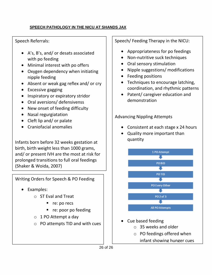
Advancing Nippling Attempts
Consistent at each stage x 24 hours
Quality more important than quantity
Cue based feeding o 35 weeks and older
o PO feedings offered when
infant showing hunger cues
Writing Orders for Speech & PO Feeding
Examples:
o ST Eval and Treat
re: po recs
re: poor po feeding
o 1 PO Attempt a day
o PO attempts TID and with cues