Nicolas Fawzi - Residue-by-Residue View of the In Vitro FUS Granules
21
Nicolas Lux Fawzi Department of Molecular Pharmacology, Physiology and Biotechnology Brown University Residue-by-Residue View of In Vitro FUS granules that bind the C-terminal domain of RNA polymerase II Alzforum Webinar October 30, 2015
Transcript of Nicolas Fawzi - Residue-by-Residue View of the In Vitro FUS Granules
Nicolas Lux Fawzi
Department of Molecular Pharmacology, Physiology and Biotechnology
Brown University
Residue-by-Residue View of In Vitro FUS granules
that bind the C-terminal domain
of RNA polymerase II
Alzforum Webinar
October 30, 2015
Fused in Sarcoma (FUS)
• Aggregation-prone Low complexity (LC
aka SYGQ-rich) essential for aggregation
in ALS
• Mutations in NLS and LC regions have
been associated with amyotrophic lateral
sclerosis, leading to protein aggregation
• RNA binding
domains
• Low complexity
putatively
disordered domains
• Nuclear/cytoplasm
shuttling
• Cellular [FUS]
> 2 μM *
*Alberti and coworkers
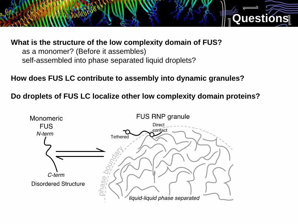
Questions
What is the structure of the low complexity domain of FUS?
as a monomer? (Before it assembles)
self-assembled into phase separated liquid droplets?
How does FUS LC contribute to assembly into dynamic granules?
Do droplets of FUS LC localize other low complexity domain proteins?
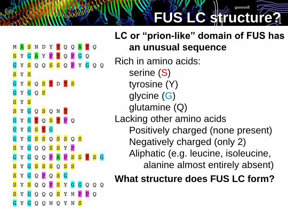
FUS LC structure?LC or “prion-like” domain of FUS has
an unusual sequence
Rich in amino acids:
serine (S)
tyrosine (Y)
glycine (G)
glutamine (Q)
Lacking other amino acids
Positively charged (none present)
Negatively charged (only 2)
Aliphatic (e.g. leucine, isoleucine,
alanine almost entirely absent)
What structure does FUS LC form?
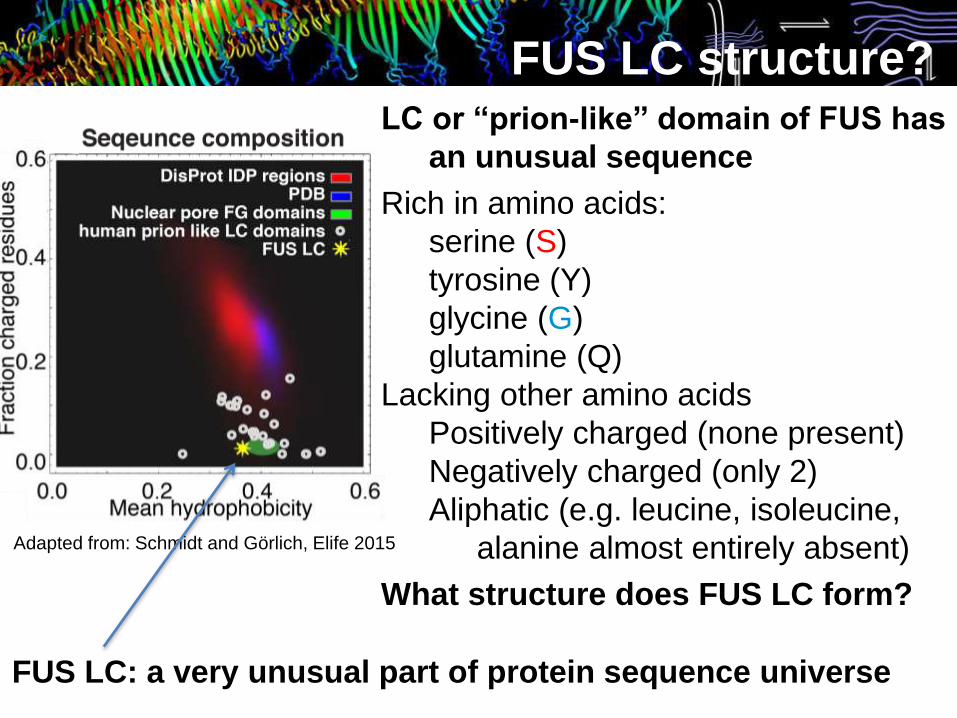
FUS LC structure?
Adapted from: Schmidt and Görlich, Elife 2015
FUS LC: a very unusual part of protein sequence universe
LC or “prion-like” domain of FUS has
an unusual sequence
Rich in amino acids:
serine (S)
tyrosine (Y)
glycine (G)
glutamine (Q)
Lacking other amino acids
Positively charged (none present)
Negatively charged (only 2)
Aliphatic (e.g. leucine, isoleucine,
alanine almost entirely absent)
What structure does FUS LC form?

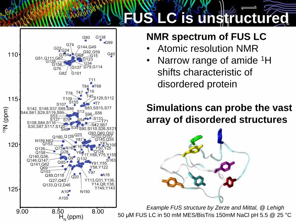
FUS LC is unstructuredNMR spectrum of FUS LC
• Atomic resolution NMR
• Narrow range of amide 1H
shifts characteristic of
disordered protein
Translation
Each dot here is a “peak” that
represents a signal from a
particular position in the FUS LC
protein sequence
From the distribution of peak
positions, we see that FUS LC
does not fold into one structure50 μM FUS LC in 50 mM MES/BisTris 150mM NaCl pH 5.5 @ 25 °C
FUS LC is unstructuredNMR spectrum of FUS LC
• Atomic resolution NMR
• Narrow range of amide 1H
shifts characteristic of
disordered protein
Simulations can probe the vast
array of disordered structures
50 μM FUS LC in 50 mM MES/BisTris 150mM NaCl pH 5.5 @ 25 °C
Example FUS structure by Zerze and Mittal, @ Lehigh
277K
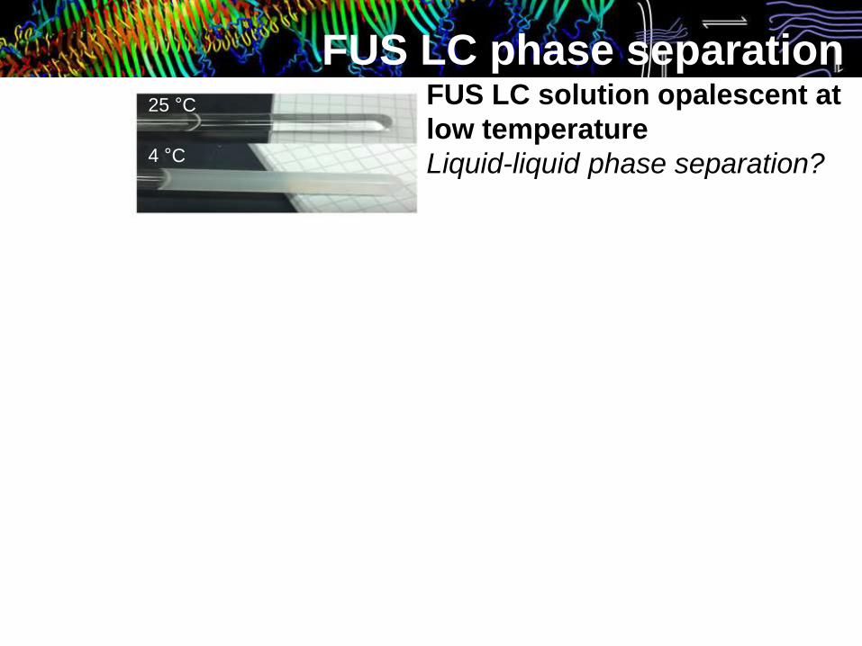
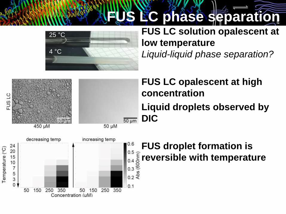
FUS LC phase separation25 °C
4 °C
FUS LC solution opalescent at
low temperature
Liquid-liquid phase separation?
277K
FUS LC phase separation25 °C
4 °C
FUS LC solution opalescent at
low temperature
Liquid-liquid phase separation?
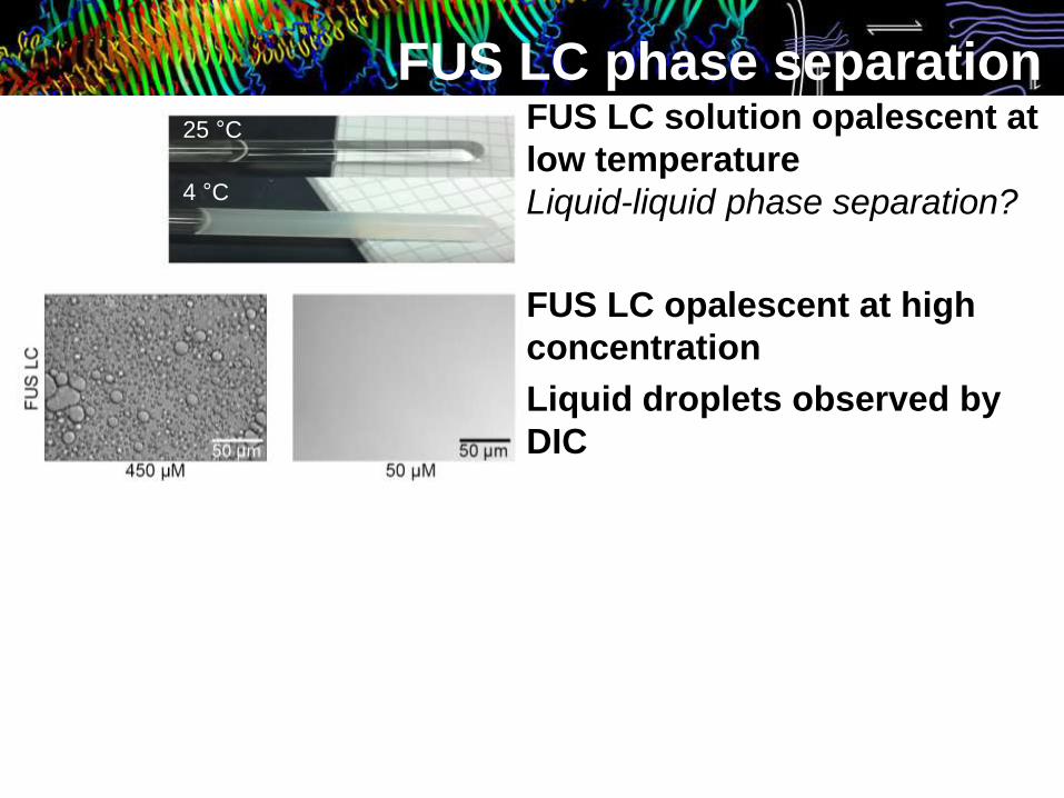
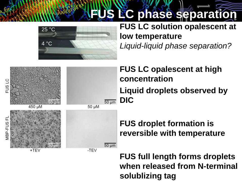
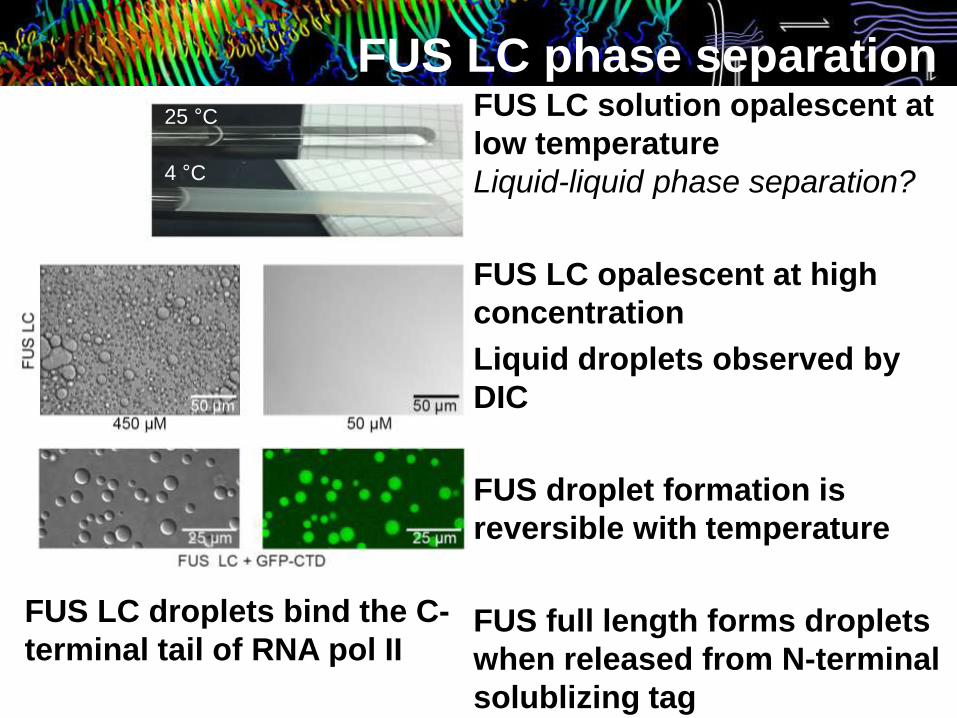
FUS LC opalescent at high
concentration
Liquid droplets observed by
DIC
277K
FUS LC phase separation25 °C
4 °C
FUS LC solution opalescent at
low temperature
Liquid-liquid phase separation?
FUS LC opalescent at high
concentration
Liquid droplets observed by
DIC
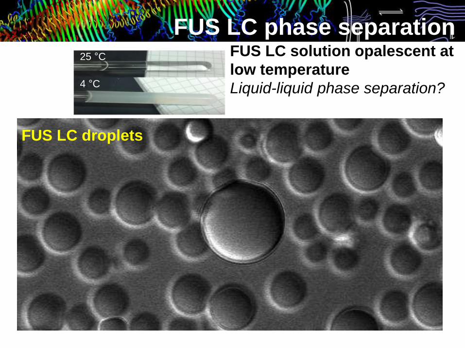
FUS LC droplets
277K
FUS LC phase separation25 °C
4 °C
FUS LC solution opalescent at
low temperature
Liquid-liquid phase separation?
FUS LC opalescent at high
concentration
Liquid droplets observed by
DIC
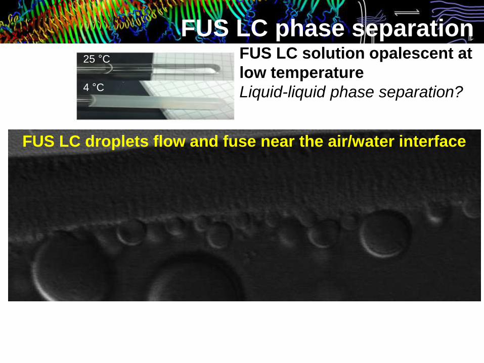
FUS LC droplets flow and fuse near the air/water interface
277K
FUS LC phase separation25 °C
4 °C
FUS LC solution opalescent at
low temperature
Liquid-liquid phase separation?
FUS LC opalescent at high
concentration
Liquid droplets observed by
DIC
FUS droplet formation is
reversible with temperature
277K
FUS LC phase separation25 °C
4 °C
FUS LC solution opalescent at
low temperature
Liquid-liquid phase separation?
FUS LC opalescent at high
concentration
Liquid droplets observed by
DIC
FUS droplet formation is
reversible with temperature
FUS full length forms droplets
when released from N-terminal
solublizing tag
FUS LC droplets bind the C-
terminal tail of RNA pol II
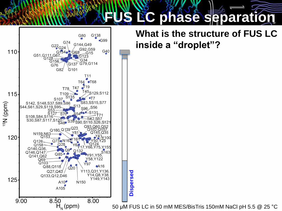
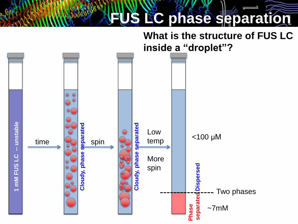
FUS LC phase separationWhat is the structure of FUS LC
inside a “droplet”?
50 μM FUS LC in 50 mM MES/BisTris 150mM NaCl pH 5.5 @ 25 °C
Dis
pe
rse
d

FUS LC phase separation
Clo
ud
y, p
ha
se
se
pa
rate
d
1 m
MF
US
LC
--
un
sta
ble
time
What is the structure of FUS LC
inside a “droplet”?
FUS LC phase separation
Clo
ud
y, p
ha
se
se
pa
rate
d
1 m
MF
US
LC
--
un
sta
ble
Clo
ud
y, p
ha
se
se
pa
rate
dtime spin
Low
temp
More
spin
Two phasesP
ha
se
se
pa
rate
d D
isp
ers
ed
~7mM
<100 μM
What is the structure of FUS LC
inside a “droplet”?

277K
Ph
as
e s
ep
ara
ted
Dis
pe
rse
d
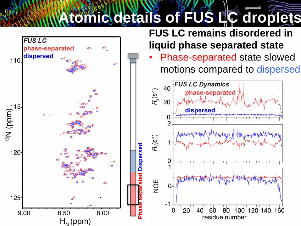
Atomic details of FUS LC droplets
~7mM FUS LC at 25 °C derived from a original sample:
1 mM FUS LC in 50 mM MES/BisTris 150mM NaCl pH 5.5
NMR Observation volume
FUS LC remains disordered in
liquid phase separated state
• Phase-separated state
spectrum overlays with
dispersed
Translation
In liquid droplets, the majority of
FUS LC retains a disordered
structure very similar to the
dispersed state
277K
Ph
as
e s
ep
ara
ted
Dis
pe
rse
d
Atomic details of FUS LC droplets FUS LC remains disordered in
liquid phase separated state
• Phase-separated state slowed
motions compared to dispersed
277K
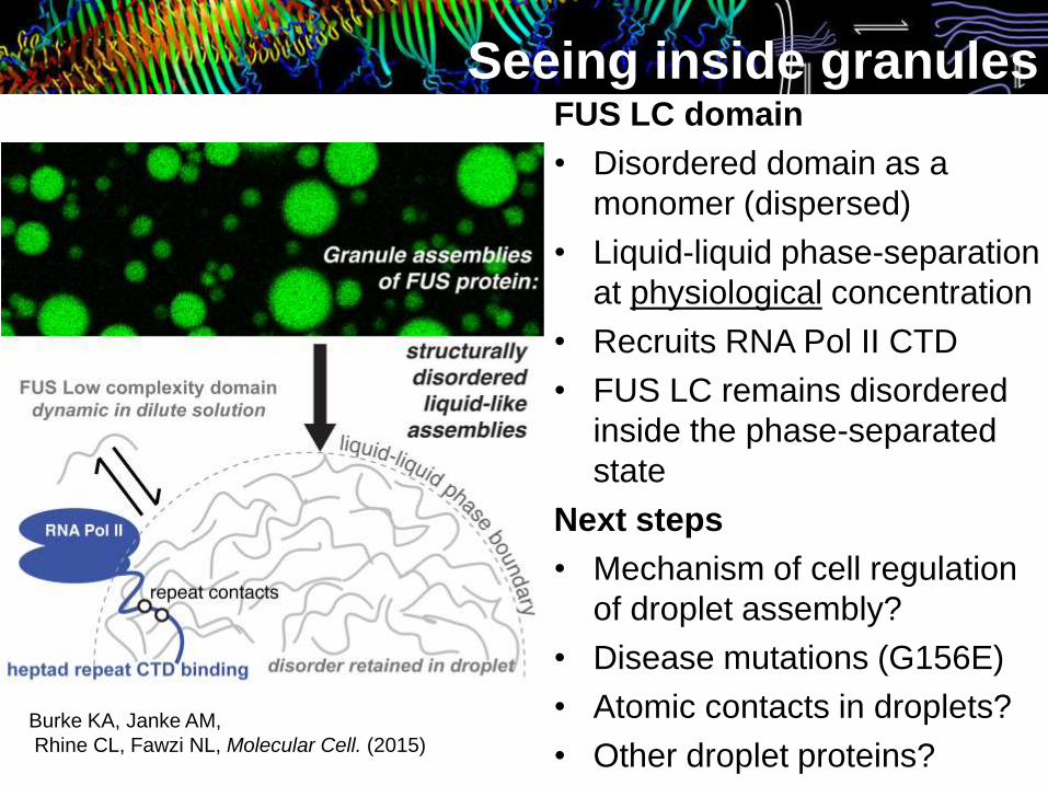
Seeing inside granulesFUS LC domain
• Disordered domain as a
monomer (dispersed)
• Liquid-liquid phase-separation
at physiological concentration
• Recruits RNA Pol II CTD
• FUS LC remains disordered
inside the phase-separated
state
Next steps
• Mechanism of cell regulation
of droplet assembly?
• Disease mutations (G156E)
• Atomic contacts in droplets?
• Other droplet proteins?
Burke KA, Janke AM,
Rhine CL, Fawzi NL, Molecular Cell. (2015)
Fawzi group:
Dr. Kathleen Burke
Alex Conicella
Veronica Ryan
Charlene Chabata
Abigail Janke
Daniel Ramirez Montero
Dahee Seo
Joshua Amaya
Collaborators:
Frank Shewmaker - USUHS
Yuh Min Chook and Mike Rosen - UT Southwestern
Steve McKnight - UT Southwestern
Jeetain Mittal – Lehigh
Tony Hyman – MPI Dresden
Acknowledgements:
Michael “Sparky” Clarkson and Geoff Williams
Structural biology at Brown: Wolfgang Peti, Rebecca Page,
Alexandra Deaconescu, Gerwald Jogl and their groups
Funding:
Acknowledgements
P20GM104937
P20GM103430