Next-Generation GHz NMR Technology - Bruker GHz NMR Technology Focus on Structural Biology and IDPs...
6
Next-Generation GHz NMR Technology Focus on Structural Biology and IDPs NMR Innovation with Integrity
Transcript of Next-Generation GHz NMR Technology - Bruker GHz NMR Technology Focus on Structural Biology and IDPs...

Next-Generation GHz NMR TechnologyFocus on Structural Biology and IDPs
NMRInnovation with Integrity

Improving our Understanding of the Dark ProteomeBruker’s next-generation of GHz-class NMR technology is a combination of major method and instrumentation advances, which enable even more advanced scientific and translational research in structural biology, drug discovery and the study of macromolecular complexes.
The focus of Bruker’s unique, next-generation GigaHertz NMR spectroscopy technology is to enable breakthrough fundamental research in molecular and cell biology on Intrinsically Disordered Proteins (IDPs). In particular, ultra-high field NMR, in combination with other experimental and computational methods, has recently been shown to enable more and more detailed studies of important functions of Intrinsically Disordered Proteins[1-6]. Due to the scarcity of understanding of the molecular functions for the vast majority of IDPs, they are sometimes also referred to as the ‘Dark Proteome’.

Direct C-13 and N-15 Detection in Protein NMRDirect detection of hetero-nuclei, such as C-13 and N-15 is becoming increasingly popular in studies of large biomolecules, paramagnetic proteins and intrinsically disordered proteins [7,8]. This is due to the significantly larger chemical shift dispersion in the spectra of hetero-nuclei as compared to proton NMR spectra that are prone to overlap especially for unstructured protein segments. Furthermore, hetero-nuclear direct detection is particularly useful in situations where proton signals are severely broadened, sometimes even beyond detection – as in NMR of paramagnetic proteins or protein sequences undergoing conformational exchange.
The recent advances in CryoProbe technology have dramatically increased the sensitivity of the direct detection experiments (see Table 1) making the use of the direct detection methods in the studies of complex bio-molecular systems a common practice.
CryoProbe TXI TCI TXO DCH
H-1 8500:1 8500:1 4500:1 4500:1
C13 700:1 1400:1 2800:1 3000:1
Table 1.
Sensitivity comparison of H-1 and C-13 in various CryoProbes at 600 MHz
Fig. 1 Comparison of sensitivities of the C-13 detected part of H,C’-N HSQC spectra of the FABS protein (1 mM in D2O/H2O, 1:9).
The sensitivity of the direct detection becomes particularly important in parallel NMR experiments [9] which record several multi-dimensional experiments simultaneously. This is shown in Figure 1 that compares the sensitivity improvement of the C-13 detected part of parallel H,C’-N HSQC spectra recorded for chicken fatty acid binding protein (FABS).
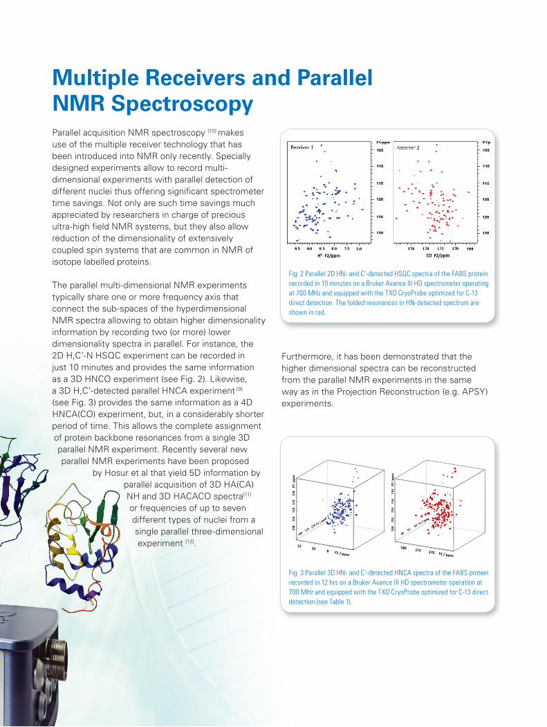
Fig. 2 Parallel 2D HN- and C’-detected HSQC spectra of the FABS protein recorded in 10 minutes on a Bruker Avance III HD spectrometer operating at 700 MHz and equipped with the TXO CryoProbe optimized for C-13 direct detection. The folded resonances in HN-detected spectrum are shown in red.
Parallel acquisition NMR spectroscopy [10] makes use of the multiple receiver technology that has been introduced into NMR only recently. Specially designed experiments allow to record multi-dimensional experiments with parallel detection of different nuclei thus offering significant spectrometer time savings. Not only are such time savings much appreciated by researchers in charge of precious ultra-high field NMR systems, but they also allow reduction of the dimensionality of extensively coupled spin systems that are common in NMR of isotope labelled proteins. The parallel multi-dimensional NMR experiments typically share one or more frequency axis that connect the sub-spaces of the hyperdimensional NMR spectra allowing to obtain higher dimensionality information by recording two (or more) lower dimensionality spectra in parallel. For instance, the 2D H,C’-N HSQC experiment can be recorded in just 10 minutes and provides the same information as a 3D HNCO experiment (see Fig. 2). Likewise, a 3D H,C’-detected parallel HNCA experiment [9] (see Fig. 3) provides the same information as a 4D HNCA(CO) experiment, but, in a considerably shorter period of time. This allows the complete assignment of protein backbone resonances from a single 3D parallel NMR experiment. Recently several new parallel NMR experiments have been proposed
by Hosur et al that yield 5D information by parallel acquisition of 3D HA(CA)NH and 3D HACACO spectra[11]
or frequencies of up to seven different types of nuclei from a single parallel three-dimensional experiment [12].
Multiple Receivers and Parallel NMR Spectroscopy
Furthermore, it has been demonstrated that the higher dimensional spectra can be reconstructed from the parallel NMR experiments in the same way as in the Projection Reconstruction (e.g. APSY) experiments.
Fig. 3 Parallel 3D HN- and C’-detected HNCA spectra of the FABS protein recorded in 12 hrs on a Bruker Avance III HD spectrometer operation at 700 MHz and equipped with the TXO CryoProbe optimized for C-13 direct detection (see Table 1).
5D, 6D and 7D NMR and the Emerging GHz NMR Technologies
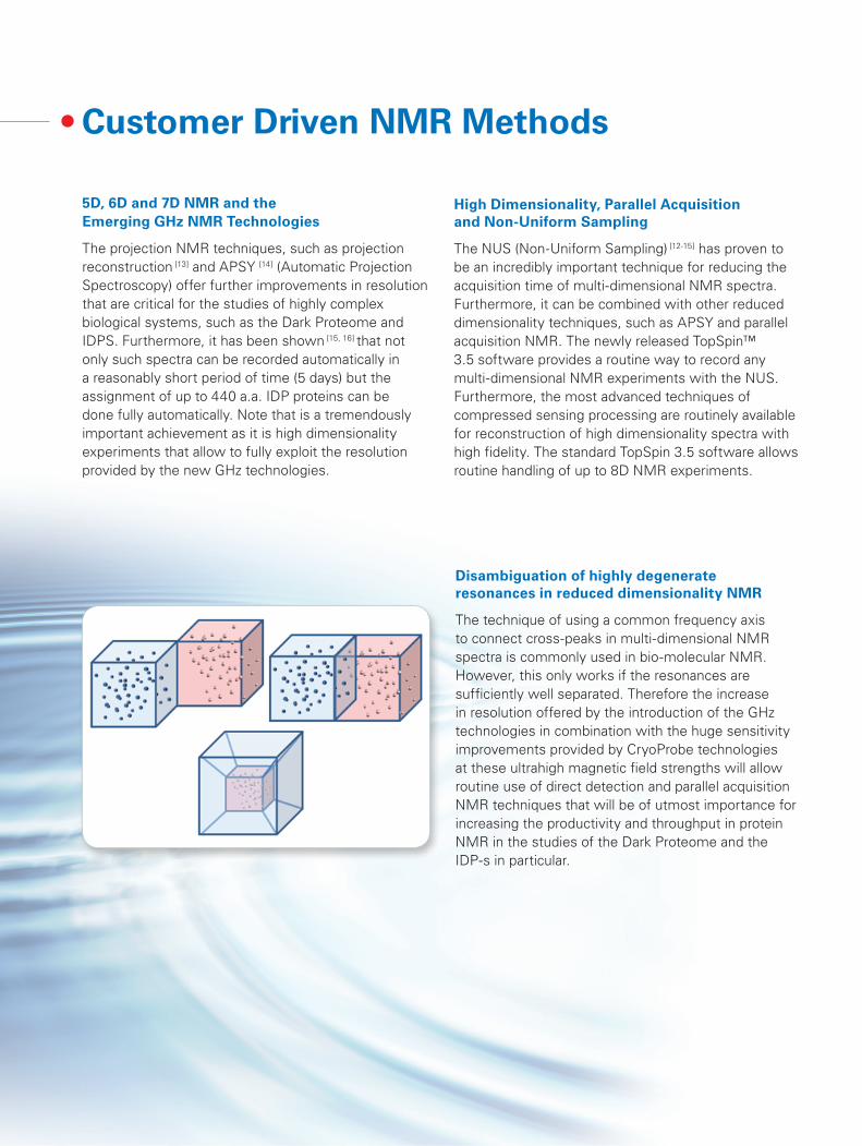
The projection NMR techniques, such as projection reconstruction [13] and APSY [14] (Automatic Projection Spectroscopy) offer further improvements in resolution that are critical for the studies of highly complex biological systems, such as the Dark Proteome and IDPS. Furthermore, it has been shown [15, 16] that not only such spectra can be recorded automatically in a reasonably short period of time (5 days) but the assignment of up to 440 a.a. IDP proteins can be done fully automatically. Note that is a tremendously important achievement as it is high dimensionality experiments that allow to fully exploit the resolution provided by the new GHz technologies.
Disambiguation of highly degenerate resonances in reduced dimensionality NMR
The technique of using a common frequency axis to connect cross-peaks in multi-dimensional NMR spectra is commonly used in bio-molecular NMR. However, this only works if the resonances are sufficiently well separated. Therefore the increase in resolution offered by the introduction of the GHz technologies in combination with the huge sensitivity improvements provided by CryoProbe technologies at these ultrahigh magnetic field strengths will allow routine use of direct detection and parallel acquisition NMR techniques that will be of utmost importance for increasing the productivity and throughput in protein NMR in the studies of the Dark Proteome and the IDP-s in particular.
High Dimensionality, Parallel Acquisition and Non-Uniform Sampling
The NUS (Non-Uniform Sampling) [12-15] has proven to be an incredibly important technique for reducing the acquisition time of multi-dimensional NMR spectra. Furthermore, it can be combined with other reduced dimensionality techniques, such as APSY and parallel acquisition NMR. The newly released TopSpin™ 3.5 software provides a routine way to record any multi-dimensional NMR experiments with the NUS. Furthermore, the most advanced techniques of compressed sensing processing are routinely available for reconstruction of high dimensionality spectra with high fidelity. The standard TopSpin 3.5 software allows routine handling of up to 8D NMR experiments.
Customer Driven NMR Methods

Bruker BioSpin
[email protected] www.bruker.com/IDP
New Techniques – an integral part of the New GHz TechnologiesThe NMR techniques described will benefit enormously from the incredibly high resolution and sensitivity offered by the emerging Giga-Hertz technologies and continuous developments in the CryoProbe technologies. Such improvements will lead to new ways of recording the multi-dimensional experiments that will benefit not only the fields of protein NMR, RNA research and the Human Dark Proteome Initiative (HDPI), but will inevitably spread to the world of small molecule research, pharma and metabonomics NMR [22]. Most of the experiments briefly mentioned here are available either from our standard libraries in TopSpin 3.5 or the online Bruker User Library. TopSpin provides an excellent platform for development of new experiments and exploring new avenues in parallel experiment design for the studies of complex biomolecular systems.
© B
ruke
r B
ioS
pin
04/1
5 T1
5470
0
References1. Uversky VN, Oldfield CJ, Dunker AK, Annu. Rev. Biophys. 2008. 37:215–462. Delgado AP, Brandao P, Narayanan R, MOJ Proteomics Bioinform 2014, 1:203. Delgado AP, Narayanan R, Current Cancer Therapy Reviews 01/2013; 9).4. Ward JJ, Sodhi JS, McGuffin LJ, Buxton BF, Jones, DT, (2004). J.Mol. Biol. 337: 635–45.5. Dunker AK, Kriwacki RW, Scientific American Mar 22, 2011.6. Wright PE, & Dyson J, Nature Reviews Molecular Cell Biology 16, (2015) 18–29.7. Bermel W, Bertini I, Chill JH, Felli IC, Kumar VMV, Haba N, Pierattelli R (2012a) Chem Bio Chem 13:2425–2432.8. Takeuchi K., Heffron G., Sun Z.Y., Frueh D.P., Wagner G., J Biomol NMR. 2010, 47, 271.9. E. Kupce and L. E. Kay, J. Biomol. NMR, 2012, 54, 1.10. E. Kupce, Topics in Current Chemistry, 2013, 335, 71.11. J. G. Reddy and R.V. Hosur, J. Biomol. NMR, 56, 77-84 (2013).12. J. G. Reddy and R.V. Hosur, J. Struct. Funct. Genomics, 2014, 32, 15-25.13. E. Kupce and R. Freeman, J. Am. Chem. Soc., 2004, 126, 6429.14. S. Hiller, F. Fiorito, K. Wüthrich, G. Wider, Proc. Natl. Acad. Sci. USA, 102, 10876–10881 (2005).15. R. L. Narayanan, U. H. N. Dürr, S. Bibow, J. Biernat, E. Mandelkow and M. Zweckstetter, J. Am.
Chem. Soc., 132, 11906-11907 (2010).16. X. Yao, S. Becker and M. Zweckstetter, J Biomol NMR (2014) 60, 231–24017. M. Zweckstetter, private communication.18. J. C. Hoch, M. W. Maciejewski, M. Mobli, A. D. Schuyler, A. S. Stern, Acc. Chem. Res.47, 708-717 (2014).19. K. Kazimierczuk and V. Y. Orekhov, Angew. Chem. Int. Ed. Engl., 50, 5556–5559 (2011)20. S. G. Hyberts, H. Arthanari and G. Wagner, Top. Curr. Chem., 316, 125 (2012).21. A. Piai, T. Hošek, L. Gonnelli, A. Zawadzka-Kazimierczuk, W. Kozminski, B. Brutscher, W. Bermel,
R. Pierattelli, I. C. Felli, J. Biomol. NMR, 60, 209-218, (2014). 22. S. M. Pudakalakatti, A. Dubey, G. Jaipuria, U. Shubhashree, S. K. Adiga, D. Moskau and H. S. Atreya,
J. Biomol. NMR, 2014, 58, 165.