News No.24 - BONDBONE
24 ® BONDBONE ® a Biphasic Calcium Sulfate: A Preliminary Study in Socket Therapy News No. 24 July 2010
-
Upload
mis-implants-technologies -
Category
Documents
-
view
213 -
download
0
description
BondBone™ a Biphasic Calcium Sulfate: A Preliminary Study in Socket Therapy
Transcript of News No.24 - BONDBONE
24© MIS Corporation. All rights Reserved
®
M IS Imp lan t s Techno log ies L td .
®
www.mis-implants.com
5 News 24, July 2010
MC
-N2410 R
ev.3
BONDBONE® a Biphasic Calcium Sulfate: A Preliminary Study in Socket Therapy
ConclusionsThis technique of extraction and simultaneous graft and barrier placement is predictable for restoring the alveolar ridge volume. BONDBONE® can be safely left partially exposed to the oral environment. In this 4-month prospective study, the predictable formation of vital bone in treated extraction sockets has led to 100% success rate in implant placement and loading. Additionally, the bone has maintained its integrity radiographically and enabled support of keratinized tissue with no dimensional alterations over the experimental period. BONDBONE® is simple and effective to use in treating extraction defects before dental implant placement. Within the limits of the presented case, it is suggested that BONDBONE® is bio-compatible and osteoconductive and allows for newly-formed bone. Although the data are base on a single case, BONDBONE® appears to be an accepted material in socket therapy.
References
1. Iasella JM, Greenwell H, Miller RL, Hill M, Drisko C, Bohra AA, Scheetz JP. Ridge preservation with freeze- dried bone allograft and a collagen membrane compared to extraction alone for implant site development: a clinical and histologic study in humans. J Periodontol 2003;74:990-999.
2. Araujo M, Linder E, Lindhe J. Effect of a xenograft on early bone formation in extraction sockets: an experimental study in dog. Clin Oral Implants Res 2009;20(1): 1-6.
3. Artzi Z, Givol N, Rohrer MD, Nemcovsky CE, Prasad HS, Tal H. Qualitative and quantitative expression of bovine bone mineral in experimental bone defects. Part 2: Morphometric analysis. J Periodontol 2003;74(8):1153-1160.
4. Babbush CA. Histologic evaluation of human biopsies after dental augmentation with a demineralized bone matrix putty. Implant Dent 2003;12(4):325-332.
5. Piattelli A, Scarano A, Piattelli M. Microscopic and histochemical evaluation of demineralized freeze-dried bone allograft in association with implant placement: a case report. Int J Periodontics Restorative Dent 1998;18(4):355-361.
6. Horowitz RA, Mazor Z, Miller RJ, Krauser J, Prasad HS, Rohrer MD. Clinical evaluation alveolar ridge preservation with a beta-tricalcium phosphate socket graft. Compend Contin Educ Dent 2009;30(9):588-590, 592, 594 passim; quiz 604, 606.
7. Vance GS, Greenwell H, Miller RL, Hill M, Johnston H, Scheetz JP. Comparison of an allograft in an experimental putty carrier and a bovine-derived xenograft used in ridge preservation: a clinical and histologic study in humans. Int J Oral Maxillofac Implants 2004;19(4):491-497.
8. Anson D. Using calcium sulfate in guided tissue regeneration: a recipe for success. Compend Contin Educ Dent 2000;21(5):365-370, 372-3, 376; quiz 378.
9. Sottosanti JS. Aesthetic extractions with calcium sulfate and the principles of guided tissue regeneration. Pract Periodontics Aesthet Dent 1993;5(5):61-69; quiz 69.
10. Horowitz RA. Extraction Environment EnhancementTM - Critical evaluation of early socket healing in long-term barrier protected extraction sockets. Compend Contin Educ Dent 2005;26(10):619-630.
11. Bartee B. The use of high-density polytetrafluorethylene membrane to treat osseous defects: clinical reports. Implant Dent 1995;4(1):21-26.
12. Hoffmann O, Bartee BK, Beaumont C, Kasaj A, Deli G, Zafiropoulos GG. Alveolar bone preservation in extraction sockets using non-resorbable dPTFE membranes: a retrospective non-randomized study. J Periodontol 2008;79(8):1355-1369.
13. Donath K, Breuner G. A method for the study of undecalcified bones and teeth with the attached soft tissues: the Sage Schliff (sawing and grinding) technique. J Oral Pathol 1982;11:318-326.
14. Rohrer, MD, Schubert, CC. The cutting-grinding technique for histological preparation of undecalcified bone and bone-
anchored implants: Improvement in instrumentation and procedures. Oral Surg Oral Med Oral Pathol 1992;74:73-78.
15. Dreesmann H: Ueber Knochenplombierung. Bietr Klin Chir 1892;9:804-810.
16. Peltier LF. The use of plaster of paris to fill large defects in bone. Am J Surg 1959; 97(3):311-315.
17. Yoshikawa G, Murashima Y, Wadachi R, Sawada N, Suda H. Guided bone regeneration (GBR) using membranes and calcium sulfate after apicectomy: a comparative histomorphometrical study. Int Endod J 2002;35:255-263.
18. Pecora GE, De Leonardis D, Della Rocca C, et al. Short-term healing following the use of calcium sulfate as a grafting material for sinus augmentation. A clinical report. Int J Oral Maxillofac Implants 1998;13:866-887.
19. Coetzee AS. Regeneration of bone in the presence of calcium sulfate. Arch Otolaryngol 1980;106(7):405-409.
20. Silveira RL, Machado RA, Silveira CR, Oliveira RB. Bone repair process in calvarial defects using bioactive glass and calcium sulfate barrier. Acta Cirurgica Brasileira 2008;23(4):322-328.
21. Bahn SL. Plaster: a bone substitute. Oral Surg, Oral Med, Oral Pathol 1966;21(5): 672-681.
22. Tay BK. Patel VV. Bradford DS. Calcium sulfate- and calcium phosphate-based bone substitutes. Mimicry of the mineral phase of bone. [Review] Orthop Clin North Am 1999;30(4):615-623.
23. Ricci JL, Alexander H, et al. Biological mechanisms of calcium sulfate replacement by bone. In: Davies JE, ed. Bone engineering. Toronto, Ontario: EM Squared Inc., 2000: 332-344.
News No. 24 July 2010
MIS’s Quality System complies with international quality control standards: ISO 13485:2003 - Quality Management System for Medical Devices, ISO 9001: 2008 – Quality Management System and CE Directive for Medical Devices 93/42/EEC. MIS’s products are cleared for marketing in the USA and CE approved.
Ziv Mazor, DMD1; Michael D. Rohrer, DDS, MS2; Hari S. Prasad, BS, MDT3; Nick Tovar, PhD4; Robert A. Horowitz, DDS5.
IntroductionClinical studies have shown significant bone resorption and volume loss in the first 6 months after tooth extraction (1). Socket augmentation has been advocated to eliminate the need for a secondary reconstructive procedure (2). Several types of graft material have been used to prevent bone resorption and volume loss, including FDBA (1), ABBM (3), DFDBA
(4,5), alloplastic materials (6), and mixtures of allograft materials with calcium sulfate (7-9), as well as only dense PTFE barriers to protect a blood clot in the extraction socket. This enables vital bone formation in the site (10-12). The purpose of this study was to evaluate an innovative, biphasic calcium sulfate (BCS), BONDBONE® (MIS, Israel), to be used as a graft material at the time of tooth extraction.
The ability of BONDBONE® to preserve and augment socket volume and resorb in the desired time period between extraction and implant placement was evaluated clinically and histologically.
1 Private Practice Periodontics and Implant Dentistry, Ra’anana, Israel. 2Professor and Director, Division of Oral and Maxillofacial Pathology, Director Hard Tissue Research Lab, University of Minnesota, School of Dentistry, Minneapolis, Minnesota. 3Senior Researcher, Department of Hard Tissue Research, University of Minnesota, Minneapolis, Minnesota. 4Senior Researcher, NYU College of Dentistry, Department of Biomaterials and Biomimetics, New York, NY. 5Private Practice Periodontics and Implant Dentistry, Scarsdale, NY and NYU College of Dentistry, Departments of Periodontics and Implant Dentistry, Oral Surgery, New York, NY.
BONDBONE® a Biphasic Calcium Sulfate: A preliminary study in socket therapy
4 News 24, July 20103 News 24, July 20102 News 24, July 2010
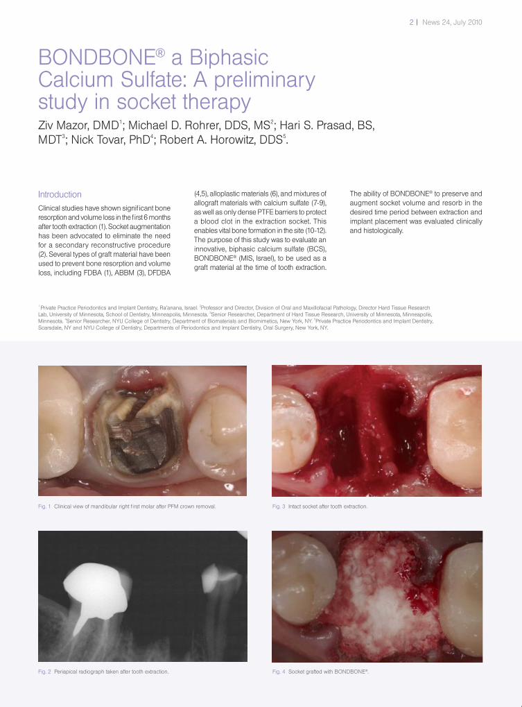
Case DescriptionA 39-year-old woman presented with a failing mandibular right first molar under a fixed PFM prosthesis. The patient was in good health and had no medical contra-indications that would prevent routine dento-alveolar surgery.
Pre-operative photographs and periapical radiographs were taken of the site. After bridge removal, the tooth was deemed hopeless. It was sectioned and extracted in an atraumatic manner using periotomes and luxators (Figs. 1-3).
The site was thoroughly debrided by mechanical means to remove granulated tissue. BCS, packaged in a sterile syringe, was grafted to the level of the gingival margin, and the BCS powder was whetted with sterile saline before placing in the socket. Excess liquid was expressed into sterile gauze and the BCS injected into the site. After the site filled to ideal contour, dry gauze was
applied and lightly compressed on top of the BCS. Working time was approximately 2 minutes (Fig. 4).
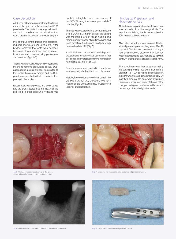
The site was covered with a collagen fleece (Fig. 5). Over a 3-month period, the patient was monitored for soft tissue healing and radiographic evidence of graft resorption and bone formation. A radiograph was taken which revealed a defect fill (Fig. 6).
A full thickness mucoperiosteal flap was elevated and a trephine was used as the first bur for osteotomy preparation in the mandibular right first molar site (Figs. 7,8).
A dental implant was inserted in dense bone which was fully stable at the time of placement.
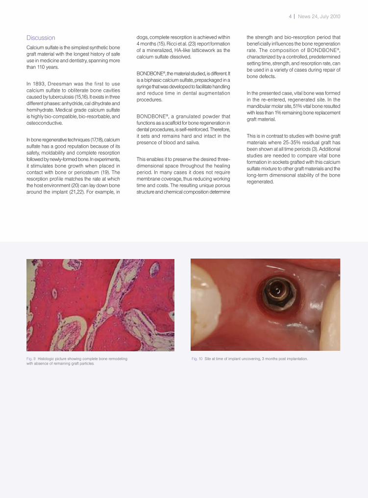
Histologic evaluation showed vital bone in the site (Fig. 9), which was allowed to heal for 3 months before uncovering (Fig. 10), prosthetic loading, and restoration.
Histological Preparation and HistomorphometryAt the time of implant placement, bone core was harvested from the surgical site. The trephine containing the bone was fixed in 10% neutral buffered formalin.
After dehydration, the specimen was infiltrated with a light-curing embedding resin. After 20 days of infiltration with constant shaking at normal atmospheric pressure, the specimen was embedded and polymerized by 450 nm light with a temperature of no more than 40°C.
The specimen was then prepared using the cutting/grinding method of Donath and Breuner (13,14). After histologic preparation, the core was evaluated morphometrically. At least two slides of the core were evaluated. Parameters evaluated were total area of the core, percentage of newly-formed bone, and percentage of residual graft material.
DiscussionCalcium sulfate is the simplest synthetic bone graft material with the longest history of safe use in medicine and dentistry, spanning more than 110 years.
In 1893, Dreesman was the first to use calcium sulfate to obliterate bone cavities caused by tuberculosis (15,16). It exists in three different phases: anhydride, cal dihydrate and hemihydrate. Medical grade calcium sulfate is highly bio-compatible, bio-resorbable, and osteoconductive.
In bone regenerative techniques (17,18), calcium sulfate has a good reputation because of its safety, moldability and complete resorption followed by newly-formed bone. In experiments, it stimulates bone growth when placed in contact with bone or periosteum (19). The resorption profile matches the rate at which the host environment (20) can lay down bone around the implant (21,22). For example, in
dogs, complete resorption is achieved within 4 months (15). Ricci et al. (23) report formation of a mineralized, HA-like latticework as the calcium sulfate dissolved.
BONDBONE®, the material studied, is different. It is a biphasic calcium sulfate, prepackaged in a syringe that was developed to facilitate handling and reduce time in dental augmentation procedures.
BONDBONE®, a granulated powder that functions as a scaffold for bone regeneration in dental procedures, is self-reinforced. Therefore, it sets and remains hard and intact in the presence of blood and saliva.
This enables it to preserve the desired three-dimensional space throughout the healing period. In many cases it does not require membrane coverage, thus reducing working time and costs. The resulting unique porous structure and chemical composition determine
the strength and bio-resorption period that beneficially influences the bone regeneration rate. The composition of BONDBONE®, characterized by a controlled, predetermined setting time, strength, and resorption rate, can be used in a variety of cases during repair of bone defects.
In the presented case, vital bone was formed in the re-entered, regenerated site. In the mandibular molar site, 51% vital bone resulted with less than 1% remaining bone replacement graft material.
This is in contrast to studies with bovine graft materials where 25-35% residual graft has been shown at all time periods (3). Additional studies are needed to compare vital bone formation in sockets grafted with this calcium sulfate mixture to other graft materials and the long-term dimensional stability of the bone regenerated.
Fig. 9 Histologic picture showing complete bone remodeling with absence of remaining graft particles.
Fig. 10 Site at time of implant uncovering, 3 months post implantation.
Fig. 6 Periapical radiograph taken 3 months post-socket augmentation.
Fig. 5 Collagen fleece placed on top of the grafted socket with partial coverage of the extraction site.
Fig. 7 Biopsy of the bone core. Note complete ridge reconstruction.
Fig. 8 Trephined core from the augmented socket.
Fig. 1 Clinical view of mandibular right first molar after PFM crown removal.
Fig. 2 Periapical radiograph taken after tooth extraction.
Fig. 3 Intact socket after tooth extraction.
Fig. 4 Socket grafted with BONDBONE®.
Ziv Mazor, DMD1; Michael D. Rohrer, DDS, MS2; Hari S. Prasad, BS, MDT3; Nick Tovar, PhD4; Robert A. Horowitz, DDS5.
IntroductionClinical studies have shown significant bone resorption and volume loss in the first 6 months after tooth extraction (1). Socket augmentation has been advocated to eliminate the need for a secondary reconstructive procedure (2). Several types of graft material have been used to prevent bone resorption and volume loss, including FDBA (1), ABBM (3), DFDBA
(4,5), alloplastic materials (6), and mixtures of allograft materials with calcium sulfate (7-9), as well as only dense PTFE barriers to protect a blood clot in the extraction socket. This enables vital bone formation in the site (10-12). The purpose of this study was to evaluate an innovative, biphasic calcium sulfate (BCS), BONDBONE® (MIS, Israel), to be used as a graft material at the time of tooth extraction.
The ability of BONDBONE® to preserve and augment socket volume and resorb in the desired time period between extraction and implant placement was evaluated clinically and histologically.
1 Private Practice Periodontics and Implant Dentistry, Ra’anana, Israel. 2Professor and Director, Division of Oral and Maxillofacial Pathology, Director Hard Tissue Research Lab, University of Minnesota, School of Dentistry, Minneapolis, Minnesota. 3Senior Researcher, Department of Hard Tissue Research, University of Minnesota, Minneapolis, Minnesota. 4Senior Researcher, NYU College of Dentistry, Department of Biomaterials and Biomimetics, New York, NY. 5Private Practice Periodontics and Implant Dentistry, Scarsdale, NY and NYU College of Dentistry, Departments of Periodontics and Implant Dentistry, Oral Surgery, New York, NY.
BONDBONE® a Biphasic Calcium Sulfate: A preliminary study in socket therapy
4 News 24, July 20103 News 24, July 20102 News 24, July 2010
Case DescriptionA 39-year-old woman presented with a failing mandibular right first molar under a fixed PFM prosthesis. The patient was in good health and had no medical contra-indications that would prevent routine dento-alveolar surgery.
Pre-operative photographs and periapical radiographs were taken of the site. After bridge removal, the tooth was deemed hopeless. It was sectioned and extracted in an atraumatic manner using periotomes and luxators (Figs. 1-3).
The site was thoroughly debrided by mechanical means to remove granulated tissue. BCS, packaged in a sterile syringe, was grafted to the level of the gingival margin, and the BCS powder was whetted with sterile saline before placing in the socket. Excess liquid was expressed into sterile gauze and the BCS injected into the site. After the site filled to ideal contour, dry gauze was
applied and lightly compressed on top of the BCS. Working time was approximately 2 minutes (Fig. 4).
The site was covered with a collagen fleece (Fig. 5). Over a 3-month period, the patient was monitored for soft tissue healing and radiographic evidence of graft resorption and bone formation. A radiograph was taken which revealed a defect fill (Fig. 6).
A full thickness mucoperiosteal flap was elevated and a trephine was used as the first bur for osteotomy preparation in the mandibular right first molar site (Figs. 7,8).
A dental implant was inserted in dense bone which was fully stable at the time of placement.
Histologic evaluation showed vital bone in the site (Fig. 9), which was allowed to heal for 3 months before uncovering (Fig. 10), prosthetic loading, and restoration.
Histological Preparation and HistomorphometryAt the time of implant placement, bone core was harvested from the surgical site. The trephine containing the bone was fixed in 10% neutral buffered formalin.
After dehydration, the specimen was infiltrated with a light-curing embedding resin. After 20 days of infiltration with constant shaking at normal atmospheric pressure, the specimen was embedded and polymerized by 450 nm light with a temperature of no more than 40°C.
The specimen was then prepared using the cutting/grinding method of Donath and Breuner (13,14). After histologic preparation, the core was evaluated morphometrically. At least two slides of the core were evaluated. Parameters evaluated were total area of the core, percentage of newly-formed bone, and percentage of residual graft material.
DiscussionCalcium sulfate is the simplest synthetic bone graft material with the longest history of safe use in medicine and dentistry, spanning more than 110 years.
In 1893, Dreesman was the first to use calcium sulfate to obliterate bone cavities caused by tuberculosis (15,16). It exists in three different phases: anhydride, cal dihydrate and hemihydrate. Medical grade calcium sulfate is highly bio-compatible, bio-resorbable, and osteoconductive.
In bone regenerative techniques (17,18), calcium sulfate has a good reputation because of its safety, moldability and complete resorption followed by newly-formed bone. In experiments, it stimulates bone growth when placed in contact with bone or periosteum (19). The resorption profile matches the rate at which the host environment (20) can lay down bone around the implant (21,22). For example, in
dogs, complete resorption is achieved within 4 months (15). Ricci et al. (23) report formation of a mineralized, HA-like latticework as the calcium sulfate dissolved.
BONDBONE®, the material studied, is different. It is a biphasic calcium sulfate, prepackaged in a syringe that was developed to facilitate handling and reduce time in dental augmentation procedures.
BONDBONE®, a granulated powder that functions as a scaffold for bone regeneration in dental procedures, is self-reinforced. Therefore, it sets and remains hard and intact in the presence of blood and saliva.
This enables it to preserve the desired three-dimensional space throughout the healing period. In many cases it does not require membrane coverage, thus reducing working time and costs. The resulting unique porous structure and chemical composition determine
the strength and bio-resorption period that beneficially influences the bone regeneration rate. The composition of BONDBONE®, characterized by a controlled, predetermined setting time, strength, and resorption rate, can be used in a variety of cases during repair of bone defects.
In the presented case, vital bone was formed in the re-entered, regenerated site. In the mandibular molar site, 51% vital bone resulted with less than 1% remaining bone replacement graft material.
This is in contrast to studies with bovine graft materials where 25-35% residual graft has been shown at all time periods (3). Additional studies are needed to compare vital bone formation in sockets grafted with this calcium sulfate mixture to other graft materials and the long-term dimensional stability of the bone regenerated.
Fig. 9 Histologic picture showing complete bone remodeling with absence of remaining graft particles.
Fig. 10 Site at time of implant uncovering, 3 months post implantation.
Fig. 6 Periapical radiograph taken 3 months post-socket augmentation.
Fig. 5 Collagen fleece placed on top of the grafted socket with partial coverage of the extraction site.
Fig. 7 Biopsy of the bone core. Note complete ridge reconstruction.
Fig. 8 Trephined core from the augmented socket.
Fig. 1 Clinical view of mandibular right first molar after PFM crown removal.
Fig. 2 Periapical radiograph taken after tooth extraction.
Fig. 3 Intact socket after tooth extraction.
Fig. 4 Socket grafted with BONDBONE®.
24© MIS Corporation. All rights Reserved
®
M IS Imp lan t s Techno log ies L td .
®
www.mis-implants.com
5 News 24, July 2010
MC
-N2410 R
ev.3
BONDBONE® a Biphasic Calcium Sulfate: A Preliminary Study in Socket Therapy
ConclusionsThis technique of extraction and simultaneous graft and barrier placement is predictable for restoring the alveolar ridge volume. BONDBONE® can be safely left partially exposed to the oral environment. In this 4-month prospective study, the predictable formation of vital bone in treated extraction sockets has led to 100% success rate in implant placement and loading. Additionally, the bone has maintained its integrity radiographically and enabled support of keratinized tissue with no dimensional alterations over the experimental period. BONDBONE® is simple and effective to use in treating extraction defects before dental implant placement. Within the limits of the presented case, it is suggested that BONDBONE® is bio-compatible and osteoconductive and allows for newly-formed bone. Although the data are base on a single case, BONDBONE® appears to be an accepted material in socket therapy.
References
1. Iasella JM, Greenwell H, Miller RL, Hill M, Drisko C, Bohra AA, Scheetz JP. Ridge preservation with freeze- dried bone allograft and a collagen membrane compared to extraction alone for implant site development: a clinical and histologic study in humans. J Periodontol 2003;74:990-999.
2. Araujo M, Linder E, Lindhe J. Effect of a xenograft on early bone formation in extraction sockets: an experimental study in dog. Clin Oral Implants Res 2009;20(1): 1-6.
3. Artzi Z, Givol N, Rohrer MD, Nemcovsky CE, Prasad HS, Tal H. Qualitative and quantitative expression of bovine bone mineral in experimental bone defects. Part 2: Morphometric analysis. J Periodontol 2003;74(8):1153-1160.
4. Babbush CA. Histologic evaluation of human biopsies after dental augmentation with a demineralized bone matrix putty. Implant Dent 2003;12(4):325-332.
5. Piattelli A, Scarano A, Piattelli M. Microscopic and histochemical evaluation of demineralized freeze-dried bone allograft in association with implant placement: a case report. Int J Periodontics Restorative Dent 1998;18(4):355-361.
6. Horowitz RA, Mazor Z, Miller RJ, Krauser J, Prasad HS, Rohrer MD. Clinical evaluation alveolar ridge preservation with a beta-tricalcium phosphate socket graft. Compend Contin Educ Dent 2009;30(9):588-590, 592, 594 passim; quiz 604, 606.
7. Vance GS, Greenwell H, Miller RL, Hill M, Johnston H, Scheetz JP. Comparison of an allograft in an experimental putty carrier and a bovine-derived xenograft used in ridge preservation: a clinical and histologic study in humans. Int J Oral Maxillofac Implants 2004;19(4):491-497.
8. Anson D. Using calcium sulfate in guided tissue regeneration: a recipe for success. Compend Contin Educ Dent 2000;21(5):365-370, 372-3, 376; quiz 378.
9. Sottosanti JS. Aesthetic extractions with calcium sulfate and the principles of guided tissue regeneration. Pract Periodontics Aesthet Dent 1993;5(5):61-69; quiz 69.
10. Horowitz RA. Extraction Environment EnhancementTM - Critical evaluation of early socket healing in long-term barrier protected extraction sockets. Compend Contin Educ Dent 2005;26(10):619-630.
11. Bartee B. The use of high-density polytetrafluorethylene membrane to treat osseous defects: clinical reports. Implant Dent 1995;4(1):21-26.
12. Hoffmann O, Bartee BK, Beaumont C, Kasaj A, Deli G, Zafiropoulos GG. Alveolar bone preservation in extraction sockets using non-resorbable dPTFE membranes: a retrospective non-randomized study. J Periodontol 2008;79(8):1355-1369.
13. Donath K, Breuner G. A method for the study of undecalcified bones and teeth with the attached soft tissues: the Sage Schliff (sawing and grinding) technique. J Oral Pathol 1982;11:318-326.
14. Rohrer, MD, Schubert, CC. The cutting-grinding technique for histological preparation of undecalcified bone and bone-
anchored implants: Improvement in instrumentation and procedures. Oral Surg Oral Med Oral Pathol 1992;74:73-78.
15. Dreesmann H: Ueber Knochenplombierung. Bietr Klin Chir 1892;9:804-810.
16. Peltier LF. The use of plaster of paris to fill large defects in bone. Am J Surg 1959; 97(3):311-315.
17. Yoshikawa G, Murashima Y, Wadachi R, Sawada N, Suda H. Guided bone regeneration (GBR) using membranes and calcium sulfate after apicectomy: a comparative histomorphometrical study. Int Endod J 2002;35:255-263.
18. Pecora GE, De Leonardis D, Della Rocca C, et al. Short-term healing following the use of calcium sulfate as a grafting material for sinus augmentation. A clinical report. Int J Oral Maxillofac Implants 1998;13:866-887.
19. Coetzee AS. Regeneration of bone in the presence of calcium sulfate. Arch Otolaryngol 1980;106(7):405-409.
20. Silveira RL, Machado RA, Silveira CR, Oliveira RB. Bone repair process in calvarial defects using bioactive glass and calcium sulfate barrier. Acta Cirurgica Brasileira 2008;23(4):322-328.
21. Bahn SL. Plaster: a bone substitute. Oral Surg, Oral Med, Oral Pathol 1966;21(5): 672-681.
22. Tay BK. Patel VV. Bradford DS. Calcium sulfate- and calcium phosphate-based bone substitutes. Mimicry of the mineral phase of bone. [Review] Orthop Clin North Am 1999;30(4):615-623.
23. Ricci JL, Alexander H, et al. Biological mechanisms of calcium sulfate replacement by bone. In: Davies JE, ed. Bone engineering. Toronto, Ontario: EM Squared Inc., 2000: 332-344.
News No. 24 July 2010
MIS’s Quality System complies with international quality control standards: ISO 13485:2003 - Quality Management System for Medical Devices, ISO 9001: 2008 – Quality Management System and CE Directive for Medical Devices 93/42/EEC. MIS’s products are cleared for marketing in the USA and CE approved.