NEWS FROM THE DEPARTMENT OF OTOLARYNGOLOGY AT HARVARD MEDICAL SCHOOL ...€¦ · NEWS FROM THE...
32
SPRING 2018 VOL. 15, NO. 1 HARVARD Otolaryngology NEWS FROM THE DEPARTMENT OF OTOLARYNGOLOGY AT HARVARD MEDICAL SCHOOL Otolaryngology Mapping the Landscape of Head and Neck Cancer Head and neck surgeons develop atlas that may help predict the likelihood of cancer spread (page 16)
Transcript of NEWS FROM THE DEPARTMENT OF OTOLARYNGOLOGY AT HARVARD MEDICAL SCHOOL ...€¦ · NEWS FROM THE...

SPRI
NG
201
8
VO
L. 1
5, N
O. 1
HARVARDOtolaryngology
NEWS FROM THE DEPARTMENT OF OTOLARYNGOLOGY AT HARVARD MEDICAL SCHOOL
Otolaryngology
Mapping the Landscape of Head and Neck Cancer Head and neck surgeons
develop atlas that may help predict the likelihood of cancer spread (page 16)

News from the Department of Otolaryngology at Harvard Medical School
Published twice per year.
Please send comments, requests for additional copies, and other inquiries regarding this issue to:
Mary YaegerCommunications ManagerDepartment of OtolaryngologyMassachusetts Eye and Ear243 Charles Street, Boston, MA 02114Ph: 617-573-3656 | [email protected]
Contributors Editor-in-ChiefD. Bradley Welling, MD, PhD, FACS Walter Augustus Lecompte Professor and Chair Department of Otolaryngology Harvard Medical School
Chief of OtolaryngologyMassachusetts Eye and EarMassachusetts General Hospital
Managing Editor/Writer Mary Yaeger
Design/Layout/Photography Garyfallia Pagonis
HARVARDOtolaryngology
Contents
1 Letter from the Chair D. Bradley Welling, MD, PhD, FACS
2 Beth Israel Deaconess Medical Center Appoints New Chief of Otolaryngology
3 Brigham and Women’s Hospital Appoints New Chief of Otolaryngology
4 Two Endowed Chairs Named at Massachusetts Eye and Ear
6 Reza Rahbar, DMD, MD, and Mark A. Varvares, MD, FACS, Promoted to Professors of Otolaryngology at Harvard Medical School
8 New Findings in Dystonia Researchers investigate the mechanisms of focal laryngeal dystonia, seeking diagnostic and therapeutic opportunities
12 Corneal Reinnervation Surgeons develop new procedure to circumvent corneal nerve damage
16 Mapping the Landscape of Head and Neck Cancer Head and neck surgeons develop atlas that may help predict the likelihood of cancer spread
20 New Trainees
21 Alumni Giving Society
22 Alumni Profiles Ernest A. Weymuller, Jr., MD, Class of 1973 Sridhar Kalluri, PhD, Class of 2000
24 Highlights
26 Research Advances
Cover art by Garyfallia Pagonis
Spring 2018 Vol. 15, No. 1
©2018, Massachusetts Eye and Ear
Otolaryngology
Massachusetts Eye and EarBeth Israel Deaconess Medical CenterBoston Children’s HospitalBrigham and Women’s HospitalMassachusetts General Hospital

1
Letter from the C
hair
Dear colleagues and friends,
The underlying mechanisms of a disease can play a crucial role in understanding why and how it occurs. Having this level of knowledge can allow for the identification and investigation of potential targets and pathways for treatment.
A team of head and neck surgeons in our Department recently developed a detailed map of head and neck cancer that can be used to characterize the mechanisms of head and neck tumors. This tool has promise in offering insights as to how head and neck cancers metastasize and could help physicians better predict poor prognosis. In our cover story starting on page 16, we delve into this work and outline an important discovery that has already resulted from use of this atlas.
In our newly established Dystonia and Speech Motor Control Laboratory, investigators are looking into the mechanisms and patho-physiology of focal dystonia, focusing on the neural mechanisms of fine motor control during speech. In a story starting on page 8, we discuss their recent findings and how they may help guide the way for new diagnostic and treatment opportunities for patients with movement disorders.
Switching gears, we also highlight a new procedure in this issue that was developed through a collaboration between the Otolaryngology and Ophthalmology Departments at Massachusetts Eye and Ear. It uses reinnervation techniques to combat corneal nerve damage in patients with neurotrophic keratitis, a debilitating disorder caused by damage to the trigeminal nerve.
We’re excited to share with you more about our research advances and current progress across the field. Thank you for your interest in and support of the Department’s activities.
Sincerely,
D. Bradley Welling, MD, PhD, FACS
Walter Augustus Lecompte Professor and Chair Department of Otolaryngology Harvard Medical School
Chief of Otolaryngology Massachusetts Eye and Ear Massachusetts General Hospital

2 HARVARD Otolaryngology
NE
WS
S charukh M. Jalisi, MD, MA, FACS, Associate Professor of Otolaryngology at Boston University, recently joined the Beth Israel Deaconess Medical Center (BIDMC) team
as Chief of Otolaryngology (ENT)/Head and Neck Surgery in the Department of Surgery.
Joining BIDMC/Harvard Medical School from Boston Medical Center (BMC), which is the teaching affiliate of the Boston University School of Medicine (BUSM), Dr. Jalisi is known for his entrepreneurial spirit and contributions to the field of head and neck oncology both nationally and internationally.
He earned his medical degree from BUSM, where he also completed his surgical internship and residency in otolaryn-gology–head and neck surgery. He later completed his fellowship training in head and neck surgical oncology, skull base surgery, and microvascular surgery at Vanderbilt University before returning to Boston to join the faculty at BUSM.
At BUSM, Dr. Jalisi was instrumental in the development of their head and neck cancer surgery program, which focuses on delivering care to underprivileged patients. Through this work, Dr. Jalisi became Director of the Head and Neck Cancer Center of Excellence at BMC and the Director of Head and Neck Surgical Oncology and Skull Base Surgery at BUSM. Under his leadership, he established a head and neck oncology fellowship program, forged strategic partnerships across
disciplines, and expanded inpatient care with a focus on highly complex patients.
During this time, Dr. Jalisi was also involved in the launch of the first robotic surgery program for head and neck cancer in New England. Under his direction, BMC became the first hospital in Massachusetts to use the da Vinci® transoral robot in surgery, which allows surgeons to remove tumors of the throat through the mouth without any big incisions. For patients, this can offer better treatment and quality of life outcomes.
Dr. Jalisi has authored more than 60 original papers, case reports, and reviews in peer-reviewed journals, as well as book chapters and textbooks throughout his career. He is the author of three otolaryngology and robotic surgery textbooks, and a reviewer for several industry journals. He is also a past President of the Massachusetts Society of Otolaryngology and a fellow of the Triological Society.
As Chief, Dr. Jalisi hopes to grow his department into a compre-hensive otolaryngology program by recruiting more faculty, expanding clinical offerings, and establishing a profound research program. He also plans to develop a fellowship program in head and neck oncology and microvascular reconstruction at BIDMC. Ultimately, he hopes to establish impactful systems that change human lives and with the vision he has for BIDMC, he is well on his way to accomplishing this. l
Appoints New Chief of OtolaryngologyBeth Israel Deaconess Medical Center

3HARVARD Otolaryngology
R avindra Uppaluri, MD, PhD, FACS, Associate Professor of Otolaryngology at Harvard Medical School and Director of Head and Neck Surgical Oncology at
Brigham and Women’s Hospital and Dana-Farber Cancer Institute, was recently appointed Chief of the Division of Otolaryngology at Brigham and Women’s Hospital (BWH).
Dr. Uppaluri joined BWH/Harvard Medical School in 2016 after serving as Chief of Otolaryngology at the John Cochran VA Medical Center in St. Louis for five years and as a tenured Associate Professor of Otolaryngology–Head and Neck Surgery at Washington University in St. Louis for 16 years.
A graduate of Carleton College, Dr. Uppaluri received his medical degree and doctorate in genetics from the University of Minnesota before completing his surgical internship and residency in otolaryngology at Washington University School of Medicine/Barnes-Jewish Hospital. Following his training, he joined the Washington University School of Medicine faculty.
In medical school, Dr. Uppaluri developed an interest in head and neck oncology and has since dedicated his career to the pursuit of innovative approaches that advance outcomes for head and neck cancer patients. His surgical interests encompass minimally invasive approaches to head and neck cancers, including transoral access to reduce morbidity and novel surgical approaches of the anterior skull base.
His research focus has been on high fidelity preclinical modeling of head and neck cancers and directly studying patients afflicted with this disease. His laboratory work, which is funded by the National Institutes of Health, has leveraged genomic approaches, novel model systems, and leading-edge therapeutics to more clearly define oral squamous cell head and neck cancer depend-encies. Through this work, he has developed preclinical models that are widely used in the field.
A major area of his translational efforts has been in immunotherapy, specifically identifying the determinants of immunotherapy success in patients with head and neck cancers. Immunotherapy is a type of treatment that uses the body’s own immune system to fight a disease such as cancer. He has been the primary investigator of clinical trials that integrate small molecule and immunotherapeutics in the surgical management of head and neck cancers.
As Chief, Dr. Uppaluri hopes to add to the foundation built by Jo Shapiro, MD, former Chief of Otolaryngology at BWH, and continue to provide excellent otolaryngologic care, support faculty development, and foster a vibrant educational environment for residents and medical students. He also plans to expand the hospital’s services within the Brigham community to emphasize all aspects of otolaryngology care. l
Appoints New Chief of OtolaryngologyBrigham and Women’s Hospital

4 HARVARD Otolaryngology
NE
WS
Konstantina M. Stankovic, MD, PhD, FACS, Associate Professor of Otolaryngology at Harvard Medical School, has been named the inaugural incumbent of the Sheldon and Dorothea Buckler Chair in Otolaryngology at Massachusetts Eye and Ear.
Sheldon (Shelly) and Dorothea Buckler have a long history of commitment and service to Mass. Eye and Ear, including Shelly’s leadership as Chairman of the Board from 1996 to 2002
and Trustee for the past 34 years. When the Bucklers learned that an anonymous donor had seeded a Chair in otolaryngology, they were eager to bring it to fruition. Thanks to their remarkable generosity, a Chair in their name was established, and to their delight, Dr. Stankovic, whom they had long admired for her innovative research and “bench-to-bedside” approach, was chosen as the first recipient.
“Working alongside Edward Land at Polaroid for many years, my passion was innovation and bringing new ideas and new products to the marketplace,” said Shelly. “When I became involved with Mass. Eye and Ear, I was struck by both the opportunity and promise of medical research to improve outcomes for patients.”
Dr. Stankovic wholly exemplifies Shelly’s belief in the power of innovation to improve lives. Through her work, she aims to
improve the diagnostics, prognostics, and therapeutics for sensorineural hearing loss, the world’s most common sensory deficit for which cures do not yet exist.
Bringing together expertise in physics, molecular biology, auditory neuroscience, systems electrophysiology, and otologic surgery, Dr. Stankovic takes a cross-disciplinary approach to address the unmet needs of her hearing-impaired patients. She shapes her research through clinical insights, allowing her clinical work and research to drive and reinforce one another. Ultimately, she aims to enable personalized cellular-level diagnosis of and precise treatments for sensorineural hearing loss.
As a permanent endowed fund at Mass. Eye and Ear, the income from the Buckler Chair will support Dr. Stankovic as she furthers her research and pioneers new
technologies to treat hearing loss.
“When considering the opportunity to fund a Chair, it was important to Shelly and Dorothea that the first incumbent be someone with a passion and commitment for innovation,” said D. Bradley Welling, MD, PhD, FACS, the Walter Augustus Lecompte Professor and Chair of Otolaryngology at Harvard Medical School. “Dr. Stankovic is just that person. I have no doubt that she will achieve truly remarkable breakthroughs in medical care in the years ahead.” l
Top photo: Dr. Tina Stankovic with Dr. Brad Welling (left) and Mass. Eye and Ear President John Fernandez (right).
Bottom photo: Dorothea and Shelly Buckler with Dr. Tina Stankovic.
Two Endowed Chairs Named at Massachusetts Eye and Ear
Photos by Pierce Harman

5HARVARD Otolaryngology
Daniel B. Polley, PhD, Associate Professor of Otolaryngology at Harvard Medical School and Associate Director of the Eaton-Peabody Laboratories (EPL) at Massachusetts Eye and Ear/Harvard Medical School, has been named the inaugural recipient of the Amelia Peabody Chair in Otolaryngology at Mass. Eye and Ear.
Made possible by the generosity of an anonymous donor and the Amelia Peabody Charitable Fund, this Chair honors the legacy of renowned Massachusetts philanthropist Amelia Peabody. It was Miss Peabody’s vision that helped establish the EPL 60 years ago, forging a partnership that has advanced hearing research for decades. In fact, it was the Amelia Peabody Charitable Fund that enabled the recruit-ment of Dr. Polley to Mass. Eye and Ear in 2009.
“Amelia Peabody enjoyed a life-long love of science, believing that groundbreaking research was the key to disease prevention and better medical care,” said D. Bradley Welling, MD, PhD, FACS, the Walter Augustus Lecompte Professor and Chair of Otolaryngology at Harvard Medical School. “Understand- ing her desire to support such research is why it seems truly fitting that Dr. Polley, the first Amelia Peabody Scientist, is now the first incumbent of this Chair.”
Embodying Miss Peabody’s love for bold science, Dr. Polley’s work is focused on the mechanisms and therapeutic potential of brain plasticity, or the brain’s ability to modify its own structure, physiology, and chemistry. He is pioneering new approaches to understand how brain plasticity supports normal sound perception as well as debilitating perceptual disorders such as hyperacusis and tinnitus. His work combines state-of-the-art cellular imaging and electrophysiological approaches in animal models and in human subjects to assess these changes.
He leverages the insights gained from his plasticity studies to develop novel approaches to auditory rehabilitation in persons who struggle to understand speech in noise or who suffer from tinnitus and hyperacusis.
The Amelia Peabody Chair, which is now a permanently endowed fund at Mass. Eye and Ear, will help ensure that this work continues by investing in Dr. Polley’s research, as well as his successors’, for generations to come.
“Of all of the people studying the auditory cortex, Dr. Polley stands out for thinking about the thera- peutic benefit of his work,” said M. Charles Liberman, PhD, Harold F. Schuknecht Professor of Otolaryngology at Harvard Medical School and Director of the EPL. “His work could transform our understanding of auditory function and thereby lead us to treatments. It was this translational approach that brought him to Mass. Eye and Ear, and it is why he is uniquely deserving of this honor.” l
Top photo: Dr. Dan Polley with Dr. Brad Welling (left) and Mass. Eye and Ear President John Fernandez (right).
Bottom photo: From left to right: Dr. Brad Welling, Amelia Peabody Charitable Fund Executive Director Bethany Kendall, Amelia Peabody Charitable Fund Trustees Katherine Babson, Bob Bannish, and Caleb Loring, Dr. Dan Polley, Amelia Peabody Charitable Fund Trustee Meg Morton, and John Fernandez.
Photos by Pierce Harman
Name a $2M Chair in Otolaryngology with a gift of $1M.
The Chairs have already been seeded with $1M and there are just two opportunities left!
Contact Melissa Paul at 617-573-4168 or [email protected] for details.

6 HARVARD Otolaryngology
NE
WS
Reza Rahbar, DMD, MD, and Mark A. Varvares, MD, FACS,
Promoted to Professors of Otolaryngology at Harvard Medical School
T he Department recently celebrated the promotion of Reza Rahbar, DMD, MD, Associate Otolaryngologist-in-Chief and McGill Chair in Pediatric Otolaryngology at Boston
Children’s Hospital (BCH), to Professor of Otolaryngology at Harvard Medical School.
Dr. Rahbar first joined BCH/Harvard Medical School as a pediatric otolaryngology fellow in 1998. He completed his undergraduate degree in biology at Boston University before attending Tufts University for both his dental and medical degrees. He remained at Tufts for his general surgery internship and residency in otolaryngology. During that time, he had a rotation at BCH, which inspired him to later pursue fellowship training there.
Co-directing both the Center for Airway Disorders and the Head, Neck, and Skull Base Surgery Program at BCH, Dr. Rahbar has become nationally and internationally known for his expertise in these areas over the past 20 years. His efforts in minimally invasive, endoscopic approaches to managing airway disorders, as well as in pediatric head and neck tumors, have resulted in more than 150 peer-reviewed publications and book chapters.
Early on, he focused on the utilization of adjuvant treatment modalities to decrease scar/restenosis in airway surgery, which led to protocols on the efficacy and limitation of Mitomycin (a type of chemotherapy). He later expanded his work to advance minimally invasive airway surgery in the pediatric population, which led him to become a leader in laryngeal cleft surgery and grow BCH’s laryngeal cleft program into one of the world’s largest.
In 2003, Dr. Rahbar helped establish the Head, Neck, and Skull Base Surgery Program, in collaboration with Dana-Farber Cancer Institute. This program initially provided an avenue for multidisciplinary management of pediatric patients with head and neck tumors and has since led to the development of many publications and protocols in the evaluation and medical and surgical management of these patients.
Teaching and mentoring students has always been a significant part of Dr. Rahbar’s career. Serving as Chair of the Education Committee at BCH, he also directs their pediatric otolaryngology fellowship program. In recognition of his dedication to teaching, he received the Department of Otolaryngology Teaching Award in 2012 and 2015.
Dr. Rahbar has additionally led and served his profession through a number of external positions. At a regional level, he is a former President of the New England Otolaryngological Society. Nationally, he’s held positions with the American Society of Pediatric Otolaryngology and the American Broncho-Esophagological Association, for which he currently serves as Chair of the Liaison Committee. Internationally, he is a co-founder of the International Pediatric Otolaryngology Group, which provides management protocol and consensus on rare and complex otolaryngologic disorders worldwide.
“This honor is a testament to Dr. Rahbar’s many contributions to the Department, Boston Children’s Hospital, and the overall field of pediatric otolaryngology,” said Michael J. Cunningham, MD, FACS, Otolaryngologist-in-Chief and the Gerald B. Healy Chair in Pediatric Otolaryngology at BCH. “The respect our national and international colleagues hold for Dr. Rahbar is reflected by his leadership in many of our programs and involvement throughout the field.” l
Dr. Reza Rahbar with Dr. Michael Cunningham (left) and Dr. Brad Welling (right).

7HARVARD Otolaryngology
T he Department also recently celebrated the promotion of Mark A. Varvares, MD, FACS, Associate Chair of the Department of Otolaryngology at Harvard Medical School
and Massachusetts Eye and Ear, to Professor of Otolaryngology at Harvard Medical School.
A leader in head and neck ablative and reconstructive surgery, Dr. Varvares attended medical school at Saint Louis University School of Medicine before completing his general surgery internship at Northwestern Memorial Hospital. He then completed his residency and clinical fellowships in head and neck reconstructive surgery at Mass. Eye and Ear/Harvard Medical School.
Dr. Varvares initially joined the Harvard Medical School faculty in 1992 for two years before taking a faculty position in St. Louis. He returned to Boston in 1996 and practiced at Mass. Eye and Ear until 2003, when he accepted a Department Chairmanship at Saint Louis University School of Medicine, where he served with great distinction for more than a decade. As Chair, two of his most notable achievements were establishing a multidisciplinary program in head and neck oncology and developing a more academic and scholarly direction for his department.
Returning to Mass. Eye and Ear/Harvard Medical School in 2015, Dr. Varvares now oversees clinical and quality program development across the Department. He also conducts a productive research program in head and neck oncology, specifically studying
how surgical pathology of head and neck tumors impacts survival.
Over the course of 30 years, Dr. Varvares has made many significant contributions to the way head and neck cancer patients are treated. In collaboration with William W. Montgomery, MD, former Harvard Medical School Professor of Otolaryn-gology, he helped to popularize a procedure to simplify the surgical management of voice rehabilitation following vocal cord paralysis. This system is now one of the most accepted “prefabricated” thyroplasty implant
systems in the world. He was also the principal investigator of a study group that first reported the combined use of the salivary bypass tube and radial forearm free flap as an approach to reduce the incidence of post-operative pharyngocutaneous fistulae formation following tumor resection and reconstruction of the hypopharynx and cervical esophagus.
Within the last few years, he has also begun to focus on tongue cancer resections. His goal is to promote a broad change in the surgical approach to early oral tongue cancer. With ongoing studies to achieve higher rates of complete resection and improved local tumor control, his work has the potential to become a model for future clinical trials and build the foundation for an eventual head and neck cancer consortium at Harvard.
“Dr. Varvares’ recognition as a Professor at Harvard Medical School is a tremendous affirmation of his accumulated academic accomplishment,” said D. Bradley Welling, MD, PhD, FACS, the Walter Augustus Lecompte Professor and Chair of Otolaryngology at Harvard Medical School. “This honor is eclipsed only by the compassionate, dedicated, and kind human being that he is. I am so grateful to have him in our Department and appreciate his devoted contribution to his patients, to our residents and faculty, and to all of human kind.” l
Dr. Mark Varvares (left) with Dr. Brad Welling (right).

FEA
TU
RE
Researchers investigate the mechanisms of focal laryngeal dystonia, seeking diagnostic and therapeutic opportunities
New Findings in
8
Dystonia

9HARVARD Otolaryngology
A ffecting the muscles of the vocal cords, spasmodic dysphonia has historically been somewhat of a mystery to the medical world.
This disorder, which is also known as laryngeal dystonia, causes involuntary spasms of the muscles that power the vocal cords and selectively impacts patients during speech production.
Previously, spasmodic dysphonia was thought to be a form of psychiatric disorder until researchers began to find similarities with other forms of focal dystonia, such as writer’s cramp or cervical dystonia. Like these other movement disorders that affect certain muscle groups, spasmodic dysphonia is a neurological condition—one that impairs the larynx.
For years, researchers have been working to identify the neural mechanisms causing these spasms, hoping to develop new strategies for enhanced clinical care. This includes the Dystonia and Speech Motor Control Laboratory at Massachusetts Eye and Ear, which is led by Kristina Simonyan, MD, PhD, Dr. med., who is also the Director of Laryngology Research at Mass. Eye and Ear/Harvard Medical School.
Since there are no objective markers for the diagnosis of spasmodic dysphonia and treatments are currently limited to symptom management with botulinum toxin (Botox®) injections, the Laboratory’s efforts are aimed at unraveling the neural aspects of this disorder to bring patients better diagnostic and therapeutic options.
A potential pathway to treatmentIt has been suggested that focal dystonias (including
spasmodic dysphonia) involve abnormal functioning of the basal ganglia, a deep structure in the brain impacting movement control and learning. More specifically, an imbalance between the direct and indirect basal ganglia pathways is thought to be one of the primary drivers of this disorder.
Motivated by this idea, Dr. Simonyan and her team began to study neurotransmission within the basal ganglia that is relevant to the development of dystonia. She demonstrated that the indirect pathway, which turns down motor activity, is abnormal in focal dystonia due to a loss of D2 receptors and a low release of dopamine, a chemical in the brain that carries signals between brain cells, during speech production.
Through recent investigations, she further found that the function of the direct pathway, which turns up motor activity, is abnormally increased in these patients due to an increase of D1 receptors. Reported in the Journal of Neuroscience and Brain, these findings provide a comprehensive insight into the pathophysiological mechanisms of focal dystonia, one that opens a window into novel explorations of how these pathways can be modulated in order to normalize the dystonic behavior.
“Having a clear understanding of dopaminergic abnormalities in patients with dystonia, we can start thinking about specific drugs that may target and
continued on page 10
Organization of dopaminergic transmission in the basal ganglia (putamen and caudate nucleus) in healthy subjects and patients with spasmodic dysphonia. The three measures of dopaminergic function are depicted: D1 receptor binding, D2 receptor binding, and phasic dopamine release during speech production (obtained using position emission tomography with specialized radioligands). Healthy subjects have a great degree of overlap between all three measures as well as smaller regions of distinct receptor distribution. This organization is reversed in spasmodic dysphonia, where the overlap is largely diminished. (Figure modified from Simonyan et al., Brain, 2017.)

modulate abnormal activity to improve symptoms,” said Dr. Simonyan. “We may also be able to target these abnormalities neurosurgically with deep brain stimula-tion, which has currently a limited, if any, application in focal dystonias, including spasmodic dysphonia.”
“Altogether, this knowledge offers the opportunity for more personalized treatment. At the same time, it is also critical to consider that dystonia is a multifaceted disorder, and we need to take an integrated approach to fully understand its underlying mechanisms. This will result in more treatment options to offer patients,” she continued.
An unexpected connectionFocusing on such brain abnormalities led Dr. Simonyan
to another, unexpected discovery. After noticing several of her study participants with spasmodic dysphonia and voice tremor mention that a glass of wine helps their symptoms, she began to wonder if there was a connection. She started looking into possible mechanisms of alcohol’s action and a drug that mimics the effects of alcohol as a treatment for this disorder.
Mainly used for the treatment of cataplexy and extreme daytime sleepiness associated with narcolepsy, sodium oxybate is the sodium salt of gamma hydroxybutyric acid, which is a precursor of neuro-transmitter GABA. Being the brain’s most abundant inhibitory (or “calming”) neurotransmitter, GABA is significantly deficient in patients with spasmodic dysphonia and other focal dystonias. In an open-label study published in The Laryngoscope, Dr. Simonyan demonstrated that sodium oxybate reduces voice symp-toms in 82.2 percent of patients with spasmodic dysphonia and voice tremor whose symptoms respond to alcohol.
“It appears that sodium oxybate could be a potent oral drug for the treatment of spasmodic dysphonia and voice tremor,” said Dr. Simonyan. “Further exploring this idea, we are now conducting a double-blind placebo-controlled study of sodium oxybate in patients with spasmodic dysphonia and voice tremor. If our initial results are confirmed, we will be able to make specific recommendations for the treatment of these disorders with this drug as one of the first oral medications specifically targeting the pathophysiology of dystonia.”
10 HARVARD Otolaryngology
Hierarchical organization of the central control of voice production. Investigations of neural bases of normal voice control are important not only for the understanding of basic principles of speaking, but also for continuous elucidation of brain mechanisms that fail and lead to the development of neurological voice and speech disorders. (Figure modified from Simonyan and Horwitz, The Neuroscientist, 2011.)
FEA
TU
RE
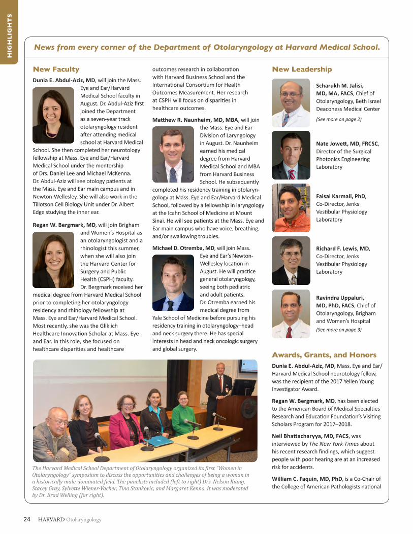
“Continuation of this research is vital in determining the exact causes and mechanisms of this disorder. This knowledge will be crucial for the future development of novel diagnostic procedures and advanced treatments, which will offer hope to so many people worldwide.” —Dr. Simonyan
11
More to come While these studies hold potential
for the development of new thera-peutic options to treat these disorders, there is still more to be understood about what happens in patients with focal dystonia.
For Dr. Simonyan and her team, they have other ongoing projects that examine the potential of brain imag-ing coupled with machine learning algorithms to derive much-needed neural biomarkers of spasmodic dysphonia for objective diagnosis. They are also looking into possible genetic causes and risk factors of this disorder that may explain how it develops. This could open up avenues for predicting the hereditary risk of dystonia in families.
The largest collection of donated postmortem brains of patients with spasmodic dysphonia is also located in Dr. Simonyan’s laboratory. Using this collection, her team conducts detailed examinations of rare brain tissue and draws parallels between neuropathological and neuroimaging findings. They also have ongoing studies that use advanced tools for computational analysis of multi-modal neuroimaging data in order to establish shared and disorder-characteristic alterations across different forms of focal dystonia.
Spasmodic dysphonia might have once been a mystery—but with recent findings and those to come, we might soon have better answers for patients with this and other forms of focal dystonia.
“We have come a long way to understand how the brain functions and how it is disorganized when an individual is experiencing dystonic symptoms,” said Dr. Simonyan. “Continuation of this research is vital in determining the exact causes and mechanisms of this disorder. This knowledge will be crucial for the future development of novel diagnostic procedures and advanced treatments, which will offer hope to so many people worldwide.” l
Dr. Simonyan’s research has been generously supported by the National Institute on Deafness and Other Communication Disorders, the National Institute of Neurological Disorders and Stroke, the Bachmann-Strauss Dystonia & Parkinson Foundation, Jazz Pharmaceuticals, and Amazon Web Services.
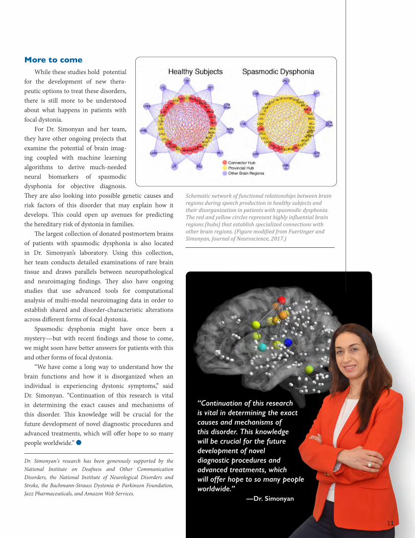
Schematic network of functional relationships between brain regions during speech production in healthy subjects and their disorganization in patients with spasmodic dysphonia. The red and yellow circles represent highly influential brain regions (hubs) that establish specialized connections with other brain regions. (Figure modified from Fuertinger and Simonyan, Journal of Neuroscience, 2017.)

FEA
TU
RE
A rare disorder known as neurotrophic keratitis can leave patients at risk of losing their eyesight. This degenerative disease results from impaired sensory innervation to the
cornea due to injury to the nerve that powers it, the trigeminal nerve. The cornea is the most sensitive structure in the eye and relies
on sensory input to function. If this input is lost, the eye’s surface becomes unhealthy and natural protective reflexes (such as blinking) fail. When this happens, foreign objects that enter the eye aren’t detected and could cause erosions and/or perforations—trauma that can significantly impair vision.
“One must be able to sense danger to protect against it,” said Nate Jowett, MD, FRCSC, a specialist in the Division of Facial Plastic and Reconstructive Surgery at Massachusetts Eye and Ear/Harvard Medical School. “With loss of sensory input to the cornea, patients may experience worsening vision, infection, and ultimately blindness.”
Historically, treatment options for these patients have been limited. Without sensory input, most interventions to restore vision, including corneal transplants, aren’t likely to succeed. Unless the loss of this sensation is addressed with treatment, the cornea cannot heal properly and damage may persist.
Surgeons develop new procedure to circumvent corneal nerve damage
12 HARVARD Otolaryngology

13HARVARD Otolaryngology
Dr. Jowett, an expert in head and neck reconstruc-tion and reinnervation, envisioned using a novel nerve transfer technique to restore sensory input to the cornea. Similar to how a less-critical motor nerve may be used to reanimate a smile in patients with facial palsy, this technique involves transfer of a less-critical regional sensory nerve to restore sensory input to the cornea.
At Mass. Eye and Ear, Dr. Jowett combined his efforts with ophthalmologist Roberto Pineda, II, MD, who specializes in corneal disorders, to pioneer a similar surgery, one that transfers the great auricular nerve to the cornea. The goal is to restore sensory input to the cornea through an ingrowth of fibers that originally supplied sensation to the earlobe. With sensation reestablished, the cornea may begin to heal and restora-tion of sensory feedback can prevent further damage.
“Neurotrophic keratitis can be extremely difficult to manage,” said Dr. Pineda. “Up until now, we’ve had limited options to rehabilitate affected eyes. With this procedure, we address the underlying problem—the lack of innervation to the cornea. This allows for rehab-
continued on page 15
ilitation and reestablishment of a healthy enough ocular surface that patients can either stabilize or become candidates for other procedures, such as corneal transplantation, giving them the opportunity to get their vision back.”
The development of a new technique
The idea of using reinnervation techniques on patients with neurotrophic keratitis isn’t completely new. Similar surgeries have been developed using transfer of sensory branches of the contralateral trigeminal nerve, which provides sensation to the forehead. Although successful, these approaches require taking away more sensation from a face already suffering from sensory loss.
Seeking a different approach, Dr. Jowett and Dr. Pineda considered transfer of the ipsilateral great auricular nerve, which is commonly harvested as a “donor nerve” for repair of more important nerves such as the facial nerve. Because having sensation in the ear-lobe is not that critical, the morbidity from redirecting
Drs. Nate Jowett (left) and Roberto Pineda, II (right), in the Joseph B. Nadol, Jr., MD, Otolaryngology Surgical Training Laboratory at Mass. Eye and Ear.
“With loss of sensory input to the cornea, patients may experience worsening vision, infection, and ultimately blindness.” —Dr. Jowett
“Neurotrophic keratitis can be extremely difficult to manage. Up until now, we’ve had limited options to rehabilitate affected eyes. With this procedure, we address the underlying problem—the lack of innervation to the cornea.” —Dr. Pineda

14 HARVARD Otolaryngology
FEA
TU
RE
| Corneal Reinnervation | continued
Case 1: Pre-op 5 months post-op 7 months post-op
Pre- and five-month post-operative corneal tomography on patient above (green and light-blue represent normal values). Top row: Axial curvature map demonstrating dramatic improvement in astigmatism at five months. Bottom row: Corneal pachymetry demonstrating normalization in corneal thickness at five months.Jowett N, Pineda RII. Corneal neurotization by ipsilateral great auricular nerve transfer and circumferential corneal scleral tunnel incisions for neurotrophic keratopathy. American Association for Peripheral Nerve Annual Meeting. January 12–14, 2018. Phoenix, Arizona.
Pre- and post-operative corneal photography of the right eye. Pre-op: A prominent ulcer is demonstrated in the right inferior quadrant. Five and seven months post-op: Progressive resolution of the ulcer is demonstrated, with tips of five nerve fascicles (*) visible within the anterior corneal stroma circumferentially about the limbus.Jowett N, Pineda RII. Corneal neurotization by ipsilateral great auricular nerve transfer and circumferential corneal scleral tunnel incisions for neurotrophic keratopathy. American Association for Peripheral Nerve Annual Meeting. January 12–14, 2018. Phoenix, Arizona.

15HARVARD Otolaryngology
that nerve is minimal. Additionally, its use avoids the need for skin incisions near the eyebrows.
“Head and neck surgeons are very familiar with the anatomy of this nerve and the low donor site morbidity associated with its harvest,” said Dr. Jowett. “Comparatively speaking, it’s a larger caliber nerve with more fibers than the supratrochlear nerve, which carries promise of more robust sensory reinnervation. It also provides ipsilateral referred sensation, which might be easier for patients to interpret.”
The surgeons started in the Joseph B. Nadol, Jr., MD, Otolaryngology Surgical Training Laboratory at Mass. Eye and Ear. A critical question they sought to answer was how close great auricular nerve branches could be mobilized towards the eye. Using cadaver models, they discovered the nerve could reach within a few centi-meters of the lower lid, rendering interposition grafting of the remaining gap likely to succeed.
The procedure takes approximately three hours and begins with harvesting the sural nerve from the lower
leg to be used as a bridge between the dissected great auricular nerve and the cornea. Concurrently, the great auricular nerve and its distal branches are mobilized, transected, and transposed towards the lower eyelid. The sural nerve graft is then tunneled from the eye to the cheek and nerve graft fascicles are carefully separated from each other and tunneled into the mid-portion of the cornea using a fine microblade. The opposite end of the sural nerve graft is then coapted to branches of the great auricular nerve. Once this is complete, incisions are closed and the healing process begins.
“I think this procedure is a really innovative approach to dealing with a serious condition that has a big upswing for patients,” said Dr. Pineda. “We modified a technique that was described ten years ago to be better in terms of sensory connections. It isn’t going to fix everything for
the patient, but it’s the primary procedure that will open up options for them.”
Patients beginning to see a differenceTo date, Dr. Jowett and Dr. Pineda have performed
this surgery on three patients, with more expected in the next year. The first patient was operated on in June of 2017, the second in October of 2017, and the third in April of 2018. The first two patients are reporting referred sensation from the cornea to the earlobe and one has documented evidence of healing of a chronic corneal scar. Overall, both of these patients have had better results than predicted by this time point.
Eight weeks post-op, the first patient reported sub-jective vision improvement in the affected eye and by nine weeks post-op, they reported earlobe irritation relieved by ocular irrigation. The second patient reported earlobe paresthesia with ocular irrigation at 11 weeks post-op and objective improvement in visual acuity from 20/300 pre-op to 20/100 five months post-op. A sizeable objective
reduction of astigmatism was also noted in one patient thus far.
“We know that the corneal nerves take a long time to grow and given this fact, healing will take some time,” said Dr. Pineda. “Our thought was that somewhere between six months to a year would be the window where we’d have a sense of whether or not
the procedure worked. So, having our first two patients seeing and feeling a difference already is promising.”
The goal of the procedure is to protect the cornea from further damage and to allow the cornea to heal. This healing then allows for vision restoration in patients with corneal blinding. Dr. Jowett and Dr. Pineda are eager to offer this procedure, carrying hope of vision restoration to others suffering from this debilitating disorder.
“Contrary to patients with long-standing facial palsy, there is no time limit on when patients who’ve lost corneal sensation can undergo such reinnervation,” said Dr. Jowett. “Patients who lost corneal sensation many years ago could potentially benefit, even if they’re already blind in the affected eye due to corneal perforation. This procedure might be new, but there are many patients who could greatly benefit from it.” l
“Contrary to patients with long-standing facial palsy, there is no time limit on when patients who’ve lost corneal sensation can undergo such reinnervation. Patients who lost corneal sensation many years ago could potentially benefit, even if they’re already blind in the affected eye due to corneal perforation.” —Dr. Jowett

FEA
TU
RE
Mapping the Landscape of Head and Neck Cancer
16
Head and neck surgeons develop atlas that may help predict the likelihood of cancer spread
Reprinted from Cell, 171(7), Puram SV, Tirosh I, Parikh AS, Patel AP, Yizhak K, Gillespie S, Rodman C, Luo CL, Mroz EA, Emerick KS, Deschler DG, Varvares MA, Mylvaganam R, Rozenblatt-Rosen O, Rocco JW, Faquin WC, Lin DT, Regev A, Bernstein BE, Single-Cell Transcriptomic Analysis of Primary and Metastatic Tumor Ecosystems in Head and Neck Cancer, 1611–1624, Copyright 2017, with permission from Elsevier.

A s the sixth leading cause of cancer-related illness and death, head and neck tumors are widespread throughout the United States.
Found in the tongue, cheek, lips, or throat, prognosis for these tumors often depends on the presence of metastases (secondary malignant growths), which significantly reduce survival rates.
While not all head and neck tumors will spread, it isn’t well known why or how certain ones do. For years, head and neck oncologists have been studying the mechanisms of these tumors, hoping to find a way to kill the cancer cells and stop them from spreading and/or recurring. Unfortunately, most of the approaches developed have relied on extracting genetic material from the tumor for detailed studies without having a sense of which specific cells that material came from.
This approach can be problematic—without knowing which genes arose from particular cells, drugs or biologic agents targeting these genes for treatment might actually be harmful. For instance, a gene could be expressed by immune cells trying to fight the cancer and thus, making a drug against that expressed gene could weaken the immune response and potentially accelerate cancer growth.
“These tumors are not simply a homogeneous ball of cancer cells,” said Derrick T. Lin, MD, FACS, Director of the Head and Neck Oncology Division at Massachusetts Eye and Ear and the Daniel Miller Associate Professor of Otolaryngology at Harvard Medical School. “Instead, they represent an ecosystem of cancer cells that form complex interactions with other cells such as immune cells, blood vessels, and support cells. This is why we must understand what these cells consist of to properly target them.”
Wanting to discern why and how genes are expressed in head and neck tumors led Dr. Lin and Harvard Medical School otolaryngology residents Sid V. Puram, MD, PhD, and Anuraag Parikh, MD, to apply innovative single-cell RNA-sequencing technology, which can separate tumors into individual cells to study gene expression, to head and neck cancer tumors. As the pioneering group to do this type of analysis, the investigators created an atlas of head and neck cancer cells.
Done in collaboration with Bradley E. Bernstein, MD, PhD, at Massachusetts General Hospital (MGH),
and Itay Tirosh, PhD, and Aviv Regev, PhD, at the Broad Institute of MIT and Harvard, this detailed map reveals the different kinds of cells, cancerous and non-cancerous, in primary head and neck tumors and their metastases. First reported in Cell, this offers important clues as to how head and neck cancers metastasize and may have implications for other common cancers as well.
“Our goal is to understand the heterogeneity of tumors in a global way,” said Dr. Puram, who is a chief resident in otolaryngology at Mass. Eye and Ear/Harvard Medical School. “We have often wondered whether unique subpopulations of cells could explain why some tumors spread to the neck and beyond, while others do not. Here, we cataloged thousands of cells found in head and neck tumors to see what answers we could find.”
A structural transition that might cause metastasis
Using single-cell RNA-sequencing, the researchers analyzed more than 6,000 individual cells from 18 head and neck squamous cell carcinomas, the most commonly diagnosed head and neck tumor. Through their analysis, the investigators gained new insights into the different cell subtypes present in head and neck cancer, including an association between tumors with cells expressing partial epithelial-to-mesenchymal programs and a progression of disease with metastasis.
17HARVARD Otolaryngology
“We have often wondered whether unique subpopulations of cells could explain why some tumors spread to the neck and beyond, while others do not. Here, we cataloged thousands of cells found in head and neck tumors to see what answers we could find.” — Dr. Puram
continued on page 18

18 HARVARD Otolaryngology
FEA
TU
RE
Partial epithelial-to-mesenchymal transition, or p-EMT, is a process that transforms cells from neatly organized blocks into irregular structures that extend into the surrounding environment. In an embryo’s development, the process is completely normal. However, in head and neck cancer cells, the researchers believe that when p-EMT is expressed, cells might be more likely to invade and spread to other parts of the body.
It is thought that the tumors temporarily “borrow” the process used in normal embryonic development to invade nearby tissues and spread—a process that could be relevant in other solid tumors as well, such as breast, colon, prostate, and/or lung cancers.
“This is the clearest picture we’ve had of this kind of structural transition in a human tumor,” said Dr. Bernstein of the MGH Cancer Center, who is also a Professor of Pathology at Harvard Medical School and a member of the Broad Institute. “For years, it’s been known that cells can lose their connections to the surrounding tissue and become more mobile, but when, how, and where this occurs in human cancer has been long debated.”
With knowledge on the role p-EMT may play in metastasis, the investigators envision a future in which identifying tumors undergoing this transition may influence the way cancer patients are clinically managed. Having this information might allow surgeons to make decisions based on the biology of the tumor rather than its size and predict its likelihood of spreading to other parts of the body.
“Being able to understand what cell(s) are driving this and other processes is relevant not only for thinking about how we approach surgery, but also thinking about drugs that could potentially attack that cell and help us avoid surgery altogether,” said Dr. Lin.
“With our atlas and findings so far, we may be able to predict poor prognosis with better accuracy than our current staging system.” —Dr. Lin
(A) Head and neck squamous cell carcinoma tumors from the oral cavity and neck were separated into individual cells and sequenced using single cell RNA-sequencing. (B) Based on sequencing, cells were categorized into cancer (malignant) cells and non-cancerous support cells based on whether they had the presence or absence of chromosomal deletions (blue) or amplifications (red). (C) Cancerous and non-cancerous cells were also distinguished using epithelial marker genes. (D) Plot shows the tight association of cells categorized as malignant or non-malignant based on chromosomal deletion or amplification with cells separated based on epithelial marker expression, suggesting cells can be robustly distinguished.Reprinted from Cell, 171(7), Puram SV, Tirosh I, Parikh AS, Patel AP, Yizhak K, Gillespie S, Rodman C, Luo CL, Mroz EA, Emerick KS, Deschler DG, Varvares MA, Mylvaganam R, Rozenblatt-Rosen O, Rocco JW, Faquin WC, Lin DT, Regev A, Bernstein BE, Single-Cell Transcriptomic Analysis of Primary and Metastatic Tumor Ecosystems in Head and Neck Cancer, 1611–1624, Copyright 2017, with permission from Elsevier.
| Mapping the Landscape of Head and Neck Cancer | continued

19HARVARD Otolaryngology
From left to right: Drs. Derrick Lin, Anuraag Parikh, Brad Bernstein, and Sid Puram.
More discoveries on the horizonFor years, clinicians have faced the daunting
possibility of tumor-spread and remained unsure how to look for metastasis and decide whether surgery is necessary. With the head and neck cancer atlas, these are questions they might soon have answers to.
Further studies assessing additional tumors will determine if p-EMT expression can correctly predict whether certain tumors develop metastasis. Being able to predict this could lead to the development of new therapeutics that fight against specific gene expression programs and the opportunity for more personalized treatments for patients.
Moving forward, the investigators also plan to continue to use the map to see what other observations they have. They are turning to their peers to see what they will be able to find with it as well.
“As we continue our research, we have also passed the atlas to the community to investigate the dataset and make their own observations. We hope for this to become an access point for a huge array of follow-up studies. Just imagine the power of combining all of our efforts,” said Dr. Puram.
With all of the data presented through this new tool, many important discoveries are expected be on the horizon—not just with head and neck cancer, but for other similar cancers as well.
“With our atlas and findings so far, we may be able to predict poor prognosis with better accuracy than our current staging system,” said Dr. Lin. “Because metastasis is one of the hardest problems to treat and one of the biggest reasons cancer patients do not survive, these findings open up whole new areas of research in our field.” l

20 HARVARD Otolaryngology
The Otolaryngology Residency Program at Harvard Medical SchoolWe would like to recognize our five PGY-1 residents, who started with us last summer: Eric R. Barbarite, MD, Adeeb Derakhshan, MD, Krupa R. Patel, MD, Tiffany V. Wang, MD, and Phoebe Kuo Yu, MD.
We are also pleased to announce the five medical students who matched in the Harvard Otolaryngology Residency Program this year. We would like to welcome Ciersten Burks, MD, Christopher McHugh, MD, Lauren Miller, MD, Tara E. Mokhtari, MD, and Alan D. Workman, MD, who will be officially part of our team this June.
Meet Our PGY-1 ResidentsEric R. Barbarite, MD, grew up in South Florida and graduated summa cum laude
from the University of Miami (UM) as a part of the combined BS/MD Honors Program in Medicine. Following his undergraduate work, he matriculated at the UM Miller School of Medicine, where he was
inducted into the Alpha Omega Alpha and Gold Humanism honor societies and awarded the Adam Teitler Award for Constancy in Unselfish Service. While at the Miller School of Medicine, Dr. Barbarite worked with Dr. Michael Hoffer under a grant funded by the National Football League to study the effects of traumatic brain injuries on athletes using portable, three-dimensional goggle technology. Dr. Barbarite’s current interests include the use of biomedical devices to enhance treatment strategies as well as skull base surgery.
Adeeb Derakhshan, MD, grew up in Charleston, West Virginia, and attended West
Virginia University for his undergraduate studies. He graduated summa cum laude with a degree in chemistry and mathematics. He subsequently matriculated at the Cleveland Clinic
Lerner College of Medicine/Case Western Reserve University. While in medical school, Dr. Derakhshan spent a year as a fellow in the Medical Research Scholars Program at
New Trainees
the National Institutes of Health, during which time he investigated targeted therapies for the treatment of head and neck squamous cell carcinoma. Dr. Derakhshan has participated in several different types of clinical and basic science research. His primary research interests include the use of precision medicine in cancer patients, nerve regeneration, and surgical outcomes.
Originally from Rockaway, New Jersey, Krupa R. Patel, MD, graduated cum laude
from Dartmouth College with a major in English literature. She subsequently attended Weill Cornell Medical College, where she was awarded the Rockefeller University
pilot project grant from the National Institutes of Health Clinical and Translational Science Award program to investigate the risk factors, therapeutic management, and survival outcomes for squamous cell carcinoma in Fanconi anemia patients under the mentorship of Drs. Agata Smogorzewska and David Kutler. During medical school, she also conducted several research studies related to head and neck and skull base oncologic outcomes under the guidance of Dr. Marc Cohen at Memorial Sloan Kettering Cancer Center and was awarded the Arthur Palmer Prize for Efficiency in Otorhinolaryngology.
Tiffany V. Wang, MD, grew up in Southern California and attended the University of
California, San Diego, where she received her degree in human biology. She then went on to earn her medical degree from the University of Southern California. During medical
school, she worked with Dr. Niels Kokot on studying the impact of socioeconomic factors on disease outcomes for oropharyngeal squamous cell carcinoma patients. Under the guidance of Dr. Jon-Paul Pepper, she investigated the therapeutic potential of human induced pluripotent stem cells to improve outcomes after peripheral motor nerve injury. Her current interests include
head and neck surgery outcomes research as well as improving otolaryngology residency training.
Hailing from Montgomery, New Jersey, Phoebe Kuo Yu, MD, is a graduate of Harvard College, where she completed
her studies in cellular biology and health policy. Her undergraduate thesis was on muscle regeneration in muscular dystrophy, and she also completed clinical
research studies establishing guidelines for patient transfers between hospitals for hand surgery. Subsequently, she went on to receive her medical degree from Yale School of Medicine and complete a thesis on head and neck cancer outcomes and surgical quality measures under the mentorship of Dr. Benjamin Judson. Her research interests relate to clinical outcomes, epidemiology, and quality improvement.
OTO
LARY
NG
OLO
GY
EDU
CAT
ION
Upcoming Harvard CME Courses
July 20–22, 2018 Snoring and Sleep Disordered Breathing – Evaluation and Management Mass. Eye and Ear, Boston, Massachusetts
September 21–23, 2018 Temporal Bone Dissection Course Mass. Eye and Ear, Boston, Massachusetts
November 9–10, 2018 Endoscopic Ear Surgery: A 2-Day Dissection Course Mass. Eye and Ear, Boston, Massachusetts
November 9–10, 2018 Surgery of the Thyroid and Parathyroid Glands Boston Marriott Long Wharf, Boston, Massachusetts

Otolaryngology
Alumni Giving Society LeadershipD. Bradley Welling, MD, PhD, FACS Walter Augustus Lecompte Professor and Chair of Otolaryngology, Harvard Medical School Chief of Otolaryngology, Mass. Eye and Ear/ Massachusetts General Hospital
Michael B. Rho, MD, FACS, ’05 President, Harvard Otolaryngology Alumni Society Medical Director, Otolaryngology, Mass. Eye and Ear, Stoneham
Alumni Giving Society
Current Alumni Giving Society members for fiscal year 2018 from October 1, 2017, to May 10, 2018,
are listed below. With your gift of $1,000 or more, you will be included in the 2018 Alumni Giving Society.
The Alumni Giving Society of the Department of Otolaryngology at Harvard Medical School
The Department of Otolaryngology at Massachusetts Eye and Ear/Harvard Medical School established the Alumni Giving Society in 2015 to recognize faculty and alumni who make gifts of $1,000 or more during the fiscal year (October 1–September 30). Participation is a way to stay connected and to help deliver the finest teaching experience for today’s otolaryngology trainees.
Our alumni know from firsthand experience that support of the vital work of our students and faculty in the Department of Otolaryngology helps drive continued achievement across all areas of education, research, and patient care. To date, we have 28 members whom we thank for their generosity and for partnering with us to achieve our department goals and institutional mission.
If you are not a member, please consider joining your colleagues today by making a gift with the enclosed envelope. As a member, you may designate your gift in the way that is most meaningful to you.
To learn more, please contact Julie Dutcher in the Development Office at 617-573-3350.
Alumni LeadersDaniel G. Deschler, MD, FACS Richard E. Gliklich, MD, ’93, ’94 Donald G. Keamy, Jr., MD, MPH Paul M. Konowitz, MD, FACS John B. Lazor, MD, MBA, FACS, ’95, ’96 Jon B. Liland, MD, ’72 Derrick T. Lin, MD, FACS, ’98, ’02 Leila A. Mankarious, MD William W. McClerkin, MD, ’73 Ralph B. Metson, MD, ’87 Michael M. Paparella, MD Herbert Silverstein, MD, FACS, ’66
CHAMPION: Gifts of $25,000 or more Ralph B. Metson, MD Michael B. Rho, MD, FACS
INNOVATOR: Gifts of $5,000 to $9,999 Nicolas Y. BuSaba, MD, FACS Michael S. Cohen, MD Stacey T. Gray, MD Thomas J. Kereiakes, MD John B. Lazor, MD, MBA, FACS Daniel J. Lee, MD, FACS
PIONEER: Gifts of $2,500 to $4,999 Wade W. Han, MD, FACS Jon B. Liland, MD Leila A. Mankarious, MD Cliff A. Megerian, MD Jonathan Y. M. Ting, MD, MS, MBA D. Bradley Welling, MD, PhD, FACS
FRIEND: Gifts of $1,000 to $2,499 Megan E. Abbott, MD Samir M. Bhatt, MD Sarah N. Bowe, MD Daniel G. Deschler, MD, FACS Ruth Anne Eatock, PhD Peter N. Friedensohn, MD Paul E. Hammerschlag, MD, FACS Lukas Landegger, MD, PhD Eugene N. Myers, MD, FACS, FRCS Edin (Hon) Brian J. Park, MD, MPH, FACS Sunil Puria, PhD Mark F. Rounds, MD Herbert Silverstein, MD, FACS Mark A. Varvares, MD, FACS
From left to right: Drs. Kiran Kakarala, Kevin Emerick, John Lazor, Brad Welling, Margo Benoit, Josh Meier, Andrew Scott, and Josh Silverman.
21HARVARD Otolaryngology

22 HARVARD Otolaryngology
Alumni Profile
Ernest A. Weymuller, Jr., MD, Otolaryngology Resident, Class of 1973
Encouraging outcomes research
Son of a prominent New York City otolaryngologist, Ernest A. Weymuller, Jr., MD, enrolled at Dartmouth College
as a “pre-med” student, but initially chose a major in history instead.
“At the outset, I resisted the thought of a career in medicine,” said Dr. Weymuller, a Professor and Emeritus Chair in the Department of Otolaryngology–Head and Neck Surgery at the University of Washington (UW). “It wasn’t until my junior year that I realized medicine was my future.”
After deciding to pursue medicine, he continued at the Geisel School of Medicine at Dartmouth for two years before transferring to Harvard Medical School, where he finished his medical degree.
It was his anatomy class that piqued his interests in otolaryngology—being particularly intrigued by the complexity of head and neck anatomy. Later, during his surgical training at Vanderbilt University Hospital, he decided to become a head and neck surgeon.
While at Vanderbilt, Dr. Weymuller met his wife, Alice, and applied for residency training at Massachusetts Eye and Ear/Harvard Medical School. The inter- view process included a meeting with Dr.
William W. Montgomery, former Professor of Otolaryngology at Harvard Medical School, and upon meeting him, he knew it was the program for him. After serving as a Captain in the United States Air Force Medical Corps in Greece for two years, he began his residency at Mass. Eye and Ear.
“The opportunity to learn from Dr. Montgomery, as well as Drs. Richard R. Gacek and Harold F. Schuknecht, was life-changing,” he said. “They were outstanding surgeons and teachers. I’ve always felt that they were the best teachers I’ve been exposed to throughout my academic career.”
Following his residency, Dr. Weymuller joined head and neck oncologist and long-time friend Dr. Charles W. Cummings at Boston Ear, Nose, and Throat Associates. Once Dr. Cummings became Chairman at the UW, Dr. Weymuller joined him in Seattle as Vice Chair, a position he held until becoming Chairman himself in 1991.
“At UW, we emphasized the Mass. Eye and Ear tradition of hands-on teaching, and the program started to flourish with a significant expansion of required resident research based on a training grant from the National Institute on Deafness and Other Communication Disorders,” he said. “I also had the great benefit of learning the nuances of local, regional, and national academic life from Dr. Cummings.”
As Chair, Dr. Weymuller developed a unique outcomes research-training program, from which eleven of his residents graduated with Master’s degrees in public health.
“Outcomes research is critical to identifying the best treatments for patients,” he said. “You have to do good outcomes research to know what to recommend to patients, so
I wanted this to become an essential part of our department ethos.”
While he encouraged outcomes reporting in all sub-specialties, Dr. Weymuller had a specific interest in quality of life measures for head and neck cancer patients. Notably, he developed the UW Head and Neck Quality of Life Instrument, which has been translated and validated in 21 languages and continues to be widely used.
Dr. Weymuller’s other investigative areas have included intubation injury, quality control, and results reporting in head and neck national protocols, complications in head and neck surgery, and complications in endoscopic sinus surgery.
As Director of the UW Medical Center Operating Room Committee, Dr. Weymuller led an intensive review of retained foreign bodies and a subsequent four-year project to systematize operating room protocols, which dramatically reduced perioperative complications.
He has published 125 peer-reviewed papers, co-authored two surgical atlases, contributed to 30 textbook chapters, and served on many editorial review boards. He was also heavily involved in national activities, including being President of the American Head and Neck Society and Conference Chair of the Society’s 7th International Conference on Head and Neck Cancer. He’s also been an invited lecturer throughout the world.
Dr. Weymuller’s non-medical pursuits include golf, fly-fishing, and art, but his family is his greatest joy. “My wife Alice and I recently celebrated our 50th anniversary with our two sons, their wives, and our four wonderful grandchildren,” he said. “We are mighty fortunate people.” l

23HARVARD Otolaryngology
Sridhar Kalluri, PhD, joined the Speech and Hearing Biosciences and Technology (SHBT) graduate program
at MIT/Harvard Medical School by chance. He knew he wanted to combine his biology and electrical engineering backgrounds, but it wasn’t until he was introduced to the SHBT program that he knew how.
“I didn’t find the speech and hearing program; it actually found me,” said Dr. Kalluri, who is Director of the Starkey Hearing Research Center, which is part of the research arm of Starkey Hearing Technologies, a leading hearing aid manufacturer. “When applying to MIT’s electrical engineering graduate program, I mentioned my interest in biological systems and, the next thing I knew, I was recommended for the SHBT program.”
“It was exciting. The program was so interesting. I wasn’t just eager to study
Sridhar Kalluri, PhD, Eaton-Peabody Laboratories of Massachusetts Eye and Ear/Harvard Medical School, 1992–2000
Transitioning from academia to industry
new topics as an electrical engineer, I was excited to be exposed to all of these new ways of thinking, especially in relation to hearing,” he continued.
A native of Lowell, Massachusetts, Dr. Kalluri studied electrical engineering at the University of Massachusetts Lowell before joining the SHBT program. It was during this time that he realized he wanted to combine his interests in biology with engineering.
Once at MIT/Harvard, he began research- ing the auditory brainstem response with Jennifer R. Melcher, PhD, in the Eaton-Peabody Laboratories (EPL) at Massachusetts Eye and Ear. He later worked with Bertrand Delgutte, PhD, also of the EPL, on computational principles underlying brainstem auditory processing, which became the basis for his doctoral thesis.
“My years working in the EPL were really formative for me,” said Dr. Kalluri. “I still draw upon my experiences there quite often. I learned a lot from the seasoned researchers and my fellow students about how to think and operate in an interdisciplinary environment. The researchers there remain an inspiration.”
Following his graduate work, he went to the University of Maryland for a post-doctoral fellowship in computational neuroscience. There, he studied the cortical neural mechanisms of auditory perception, with a focus on the mechanisms underlying the perception of harmonic sounds.
In 2004, he was offered the opportunity to move from academia to industry. He made the switch and became a research scientist for the Starkey Hearing Research Center. He
has since grown to become the Director of the Center, now leading a team of 12 to 15 researchers dedicated to improving hearing aids through an understanding of hearing impairments.
“My research now is aimed at generating practical solutions for improving hearing care,” he said. “Over the years, I’ve become increasingly interested for my research to have a tangible impact on people, especially those with hearing difficulties. Improving hearing aids is a good way to accomplish that.”
During his time at Starkey, Dr. Kalluri has seen many achievements. He was part of a team that first demonstrated that hearing technology can reduce the effort of listening even when it does not change speech understanding. This work has been quite influential; it opened up a new way to understand and assess the consequence of using hearing technology.
He has also developed outcome measurements that better predict the daily-life experience of hearing aid users. This work, as well as his other investigations, has led to multiple feature additions in his company’s product line.
Throughout his career, he would ultimately like to have an impact on the difficulty hearing-impaired listeners experience when using hearing aids in noise.
“The biggest complaint we receive from hearing aid users is the difficulty they have listening in noisy environments,” said Dr. Kalluri. “If I could be meaningfully involved in a suitable solution to this, that would be a fantastic accomplishment.” l
Alumni Profile
24 HARVARD Otolaryngology
News from every corner of the Department of Otolaryngology at Harvard Medical School.
New FacultyDunia E. Abdul-Aziz, MD, will join the Mass.
Eye and Ear/Harvard Medical School faculty in August. Dr. Abdul-Aziz first joined the Department as a seven-year track otolaryngology resident after attending medical school at Harvard Medical
School. She then completed her neurotology fellowship at Mass. Eye and Ear/Harvard Medical School under the mentorship of Drs. Daniel Lee and Michael McKenna. Dr. Abdul-Aziz will see otology patients at the Mass. Eye and Ear main campus and in Newton-Wellesley. She will also work in the Tillotson Cell Biology Unit under Dr. Albert Edge studying the inner ear.
Regan W. Bergmark, MD, will join Brigham and Women’s Hospital as an otolaryngologist and a rhinologist this summer, when she will also join the Harvard Center for Surgery and Public Health (CSPH) faculty. Dr. Bergmark received her
medical degree from Harvard Medical School prior to completing her otolaryngology residency and rhinology fellowship at Mass. Eye and Ear/Harvard Medical School. Most recently, she was the Gliklich Healthcare Innovation Scholar at Mass. Eye and Ear. In this role, she focused on healthcare disparities and healthcare
outcomes research in collaboration with Harvard Business School and the International Consortium for Health Outcomes Measurement. Her research at CSPH will focus on disparities in healthcare outcomes.
Matthew R. Naunheim, MD, MBA, will join the Mass. Eye and Ear Division of Laryngology in August. Dr. Naunheim earned his medical degree from Harvard Medical School and MBA from Harvard Business School. He subsequently
completed his residency training in otolaryn-gology at Mass. Eye and Ear/Harvard Medical School, followed by a fellowship in laryngology at the Icahn School of Medicine at Mount Sinai. He will see patients at the Mass. Eye and Ear main campus who have voice, breathing, and/or swallowing troubles.
Michael D. Otremba, MD, will join Mass. Eye and Ear’s Newton-Wellesley location in August. He will practice general otolaryngology, seeing both pediatric and adult patients. Dr. Otremba earned his medical degree from
Yale School of Medicine before pursuing his residency training in otolaryngology–head and neck surgery there. He has special interests in head and neck oncologic surgery and global surgery.
New Leadership Scharukh M. Jalisi, MD, MA, FACS, Chief of Otolaryngology, Beth Israel Deaconess Medical Center
(See more on page 2)
Nate Jowett, MD, FRCSC, Director of the Surgical Photonics Engineering Laboratory
Faisal Karmali, PhD, Co-Director, Jenks Vestibular Physiology Laboratory
Richard F. Lewis, MD, Co-Director, Jenks Vestibular Physiology Laboratory
Ravindra Uppaluri, MD, PhD, FACS, Chief of Otolaryngology, Brigham and Women’s Hospital(See more on page 3)
Awards, Grants, and HonorsDunia E. Abdul-Aziz, MD, Mass. Eye and Ear/Harvard Medical School neurotology fellow, was the recipient of the 2017 Yellen Young Investigator Award.
Regan W. Bergmark, MD, has been elected to the American Board of Medical Specialties Research and Education Foundation’s Visiting Scholars Program for 2017–2018.
Neil Bhattacharyya, MD, FACS, was interviewed by The New York Times about his recent research findings, which suggest people with poor hearing are at an increased risk for accidents.
William C. Faquin, MD, PhD, is a Co-Chair of the College of American Pathologists national
HIG
HLI
GH
TS
The Harvard Medical School Department of Otolaryngology organized its first “Women in Otolaryngology” symposium to discuss the opportunities and challenges of being a woman in a historically male-dominated field. The panelists included (left to right) Drs. Nelson Kiang, Stacey Gray, Sylvette Wiener-Vacher, Tina Stankovic, and Margaret Kenna. It was moderated by Dr. Brad Welling (far right).

25HARVARD Otolaryngology
HIG
HLIG
HT
S
HMS Promotions
Stacey T. Gray, MD, Associate Professor of Otolaryngology
Alicia M. Quesnel, MD, Assistant Professor of Otolaryngology
Reza Rahbar, DMD, MD, Professor of Otolaryngology (See more on page 6)
Ravindra Uppaluri, MD, PhD, FACS, Associate Professor of Otolaryngology
Mark A. Varvares, MD, FACS, Professor of Otolaryngology (See more on page 7)
guidelines for testing for HPV in cancers of the head and neck and a Co-Editor of the new Milan System for Reporting Salivary Gland Cytopathology.
Allen L. Feng, MD, and Sid Puram, MD, PhD, Mass. Eye and Ear/Harvard Medical School residents, are the co-recipients of the 2018 Triological Society Eastern Section’s William W. Montgomery, MD Resident Research Award for their abstract, “Intraoperative recurrent laryngeal nerve monitoring during thyroid surgery: Trends among otolaryngologists and general surgeons.”
Stacey T. Gray, MD, is now on the editorial board for International Forum of Allergy & Rhinology.
Tessa A. Hadlock, MD, has been re-elected as President of the Sir Charles Bell Society.
Eric H. Holbrook, MD, has joined the American Rhinologic Society Board of Directors. His term began in 2017 and will run until 2020.
Judith S. Kempfle, MD, post-doc in the Tillotson Cell Biology Unit, won the poster blitz contest at the 2018 Association for Research in Otolaryngology annual meeting. She was also awarded the American Otological Society grant for her proposal, “Virus based direct reprogramming of reactive glial cells into functional neurons in a mouse model of auditory neuropathy.”
Elliott D. Kozin, MD, Mass. Eye and Ear/Harvard Medical School chief resident, has been named the Neskey-Coghlan Fellow in Neurotology. He also recently received a grant from the Hearing Health Foundation for his work, “Evaluation of hearing loss and quality of life in patients with mild traumatic brain injury.”
Claire M. Lawlor, MD, Boston Children’s Hospital/Harvard Medical School pediatric otolaryngology fellow, was awarded the Charles Ferguson award for her clinical manuscript, “Innovative management of severe tracheobronchomalacia using anterior and posterior tracheobronchopexy,” at the 2018 COSM.
Gi Soo Lee, MD, EdM, was awarded first place for his clinical poster presentation, “Risk factors for obstructive sleep apnea among morbidly obese adolescents under-going evaluation for bariatric surgery,” at the 2018 COSM.
Katie M. Phillips, MD, Mass. Eye and Ear/Harvard Medical School resident, won the European Rhinologic Society Juniors/Naso Sano Grant for Best Research at the Congress of the European Otorhinolaryngology–Head and Neck Surgery in Barcelona for her abstract, “Chronic rhinosinusitis exacerbations are associated with loss of productivity in asthmatics.”
Daniel B. Polley, PhD, received a new R01 grant from the National Institute on Deafness and Other Communication Disorders titled, “Corticofugal circuits for active listening.”
Gregory W. Randolph, MD, FACS, FACE, has joined the Internal Liaison Committee at ENT & Audiology News. He has also been appointed for another term to the Board of Directors for the American Association of Endocrinology.
Aaron K. Remenschneider, MD, MPH, was awarded the Hood Foundation Grant through the Charles H. Hood Foundation Child Health Research Awards Program. Dr. Remenschneider will receive $150,000 over two years for his work on pediatric tympanoplasty using 3-D printed graft materials.
George A. Scangas, MD, Mass. Eye and Ear/Harvard Medical School rhinology fellow, was awarded the Patient Centered Outcomes Research Institute Graduate Trainee Scholarship Grant.
Ahmad R. Sedaghat, MD, PhD, has been named the recipient of the 2018 Eleanor and Miles Shore Fellowship Program Award for Scholars in Medicine.
David A. Shaye, MD, FACS, runs an ongoing Surgical Sherpa Program in Nepal, where lay people are taught how to identify surgical disease in the remote areas of the Himalayas.
This work is funded by a Harvard Medical School-Dubai Cooperative Research Award in Global Health Delivery Grant and was featured on CNN’s Vital Signs with Sanjay Gupta.
Mark G. Shrime, MD, MPH, PhD, FACS, has become the Academic Editor of PLOS One as well as an editorial reviewer for several other publications.
Konstantina M. Stankovic, MD, PhD, FACS, gave an invited lecture, “Vestibular schwannoma: From biology to clinical trials,” at the Shanghai Forum on the Frontier of Hearing Research, which was held at Jiao Tong University School of Medicine in Shanghai, China. She also gave the Guest of Honor lecture at the 151st meeting of the American Otological Society at the 2018 COSM.
Ravindra Uppaluri, MD, PhD, FACS, received an R01 from the National Institute of Dental and Craniofacial Research of the National Institutes of Health for his project, “Immunotherapeutic responses to neoantigens in head and neck cancer.”

26 HARVARD Otolaryngology
The following are select research advances from the Department of Otolaryngology at Harvard Medical School.
Basic Science
Cochlear amplification and tuning depend on the cellular arrangement within the organ of CortiThe field of cochlear mechanics has been undergoing a revolution due to recent findings made possible by advancements in
measurement techniques. While it has long been assumed that basilar-membrane (BM) motion is the most important determinant of sound transduction by the
inner hair cells (IHCs), a recent study led by Sunil Puria, PhD, and Hamid Motallebzadeh, PhD, of Mass. Eye and Ear/Harvard Medical School, with
Joris A. M. Soons, PhD, of the University of Antwerp in Belgium, explained why other parts of the sensory epithelium close to the IHCs, such as the reticular lamina (RL), move with significantly greater amplitude for weaker sounds. With a computational model of the mouse cochlea, the researchers showed that individual outer hair cells (OHCs) can work together to produce high hearing sensitivity and frequency selectivity because of the overlapping asymmetrical Y-shaped structures that they form with the Deiters’ cells (DCs) and phalangeal processes (PhPs). Altering the geometry and material properties of these structures reveals that all three components have a profound effect on BM and reticular-lamina amplification and tuning. These results imply that the spatial arrangement of OHCs as well as the DCs and PhPs must be properly accounted for in emerging OHC regeneration therapies.
Motallebzadeh H, Soons JAM, Puria S. Cochlear amplification and tuning depend on the cellular arrangement within the organ of Corti. PNAS. 2018 May 14. DOI: 10.1073/pnas.1720979115.
CRISPR therapy preserves hearing in progressive deafness modelIn collaboration with the Broad Institute of MIT and Harvard, Harvard University, and Howard Hughes Medical Institute, Zheng-Yi Chen, DPhil, of Mass. Eye and Ear/Harvard Medical School, developed a CRISPR-Cas9
genome editing therapy to prevent hearing loss in a mouse model of human genetic progressive deafness. The therapy delivers a CRISPR-Cas9 gene editing protein complex directly into the sound-sensing hair cells via lipid rafts to disrupt an autosomal dominant
mutation that would otherwise cause the cells to die.
The researchers tested the method in a mouse model of progressive
hearing loss with a mutated Tmc1 gene. The team injected the gene editing mix into the cochlea of newborn mice genetically destined for profound hearing loss. The treated mice maintained a substantial amount of their hearing compared to the untreated mice. At four weeks, the untreated mice had a measurable response in their brainstem to sound starting at roughly 80 decibels, the volume of a garbage disposal or a loud radio. But the treated mice responded to sound starting around 65 decibels—approximately the same volume as a typical spoken conver-sation. Physiological measurements showed that the hair cells survived at a higher rate in the treated cochlea. At eight weeks, treated mice also retained their instinctive physical “startle” response to sudden loud sound, while the untreated mice did not respond.
This work represents a significant step toward genome editing to halt progression of genetic hearing loss. Delivering the Cas9 protein itself locally, instead of DNA elements that the cell can use to build Cas9, improved the DNA specificity and potential safety of the treatment.
Gao X, Tao Y, Lamas V, Huang M, Yeh WH, Pan B, Hu YJ, Hu JH, Thompson DB, Shu Y, Li Y, Wang H, Yang S, Xu Q, Polley DB, Liberman MC, Kong WJ, Holt JR, Chen ZY, Liu DR. Treatment of autosomal dominant hearing loss by in vivo delivery of genome editing agents. Nature. 2018 Jan 11;553(7687):217–221.
Bayesian optimal adaptation explains age-related human sensorimotor changesWhile correlations between age-dependent decline in anatomical structures and behavior have been demonstrated in many neural systems, no conceptual framework has explained these relationships. Building on
studies that support the “Bayesian brain” notion, a team of researchers including Faisal Karmali, PhD, Richard F. Lewis, MD, and Gregory T. Whitman, MD, of Mass. Eye and Ear/Harvard Medical School, showed that age-dependent changes in a sensorimotor reflex, the vestibulo-ocular reflex, are explained by a Bayesian-optimal adaptation in the brain that occurs in response to age-dependent sensory anatomical changes. Specifically, they found that the temporal dynamics of the reflex
as a function of age emerge from a Kalman filter model that determines the optimal behavioral output when the sensory signal-to-noise characteristics are degraded by death of the transducers. These findings have substantial implications for understanding and treating the balance disorders caused by aging, and more generally, provide significant insight into the brain’s response to changes in bodily structures that occur as part of normal aging.
Karmali F, Whitman GT, Lewis RF. Bayesian optimal adaptation explains age-related human sensorimotor changes. J Neurophysiol. 2018 Feb 1;119(2):509–520.
Clinical Practice
Periostin as a biomarker for nasal polyps in chronic rhinosinusitis Serum periostin levels may be an easily accessible novel biomarker for the presence of nasal polyps in patients with chronic rhinosinusitis (CRS). A team of researchers from Brigham and Women’s Hospital, Mass. Eye and Ear, and Harvard Medical School, including Alice Z. Maxfield, MD, and Ralph B. Metson, MD, set out to identify a biomarker that could help better categorize the various subtypes of CRS. The findings were published in Otolaryngology–Head and Neck Surgery.
RE
SEA
RC
H A
DV
AN
CE
S

27HARVARD Otolaryngology
The research team measured serum periostin levels from blood samples collected from 71 patients undergoing sinus surgery for CRS (subdivided into patients with and without polyps and asthma). The results were compared with assays performed on 62 control subjects. Results showed that mean serum periostin levels were significantly elevated in patients with CRS versus
controls. In addition, mean periostin levels were significantly higher in CRS patients with nasal polyps as compared to those without polyps. The highest periostin levels were found in patients with nasal polyps and asthma.
The results of this study indicate that periostin may be a useful biomarker in identifying the subtypes of CRS and could potentially serve as a target for future therapeutic interventions in treatment of nasal polyp disease.
Maxfield AZ, Landegger LD, Brook CD, Lehmann AE, Campbell AP, Bergmark RW, Stankovic KM, Metson R. Periostin as a biomarker for nasal polyps in chronic rhinosinusitis. Otolaryngol Head Neck Surg. 2018 Jan;158(1):181–186.
Otolaryngologic manifestations of Klippel-Feil syndrome There is limited information on the otolaryngologic manifestations of patients
with Klippel-Feil syndrome (KFS). Researchers from Boston Children’s Hospital/Harvard Medical School sought to define otolaryngologic diagnoses and procedures in the largest series of KFS patients published to date. A team including Margaret A. Kenna, MD, MPH, FACS, FAAP, and Michael J. Cunningham, MD, FACS, identified 95 pediatric patients
with KFS. Each KFS patient averaged eight otolaryngology office visits and five otolaryn-gologic diagnoses. The most common diagnosis was conductive hearing loss, followed by sensorineural hearing loss and dysphagia. Sixty-five percent of patients underwent otolaryngologic procedures, with
RE
SEA
RC
H A
DV
AN
CE
S
the most common being tympanostomy tube placement and office flexible endoscopy. Twelve patients underwent direct laryngoscopy with almost half having a compromised laryngeal view (Grade 2, 3, or 4) by Cormack-Lehane classification. Three patients required tracheotomies for airway stabilization purposes; each had severe upper airway obstruction leading to respiratory failure.
In summary, although hearing loss is the most common diagnosis in KFS patients, airway issues related to cervical spine fusion are the most challenging and require formulation of an appropriate airway care plan prior to even routine otolaryngology procedures.
Kenna MA, Irace AL, Strychowsky JE, Kawai K, Barrett D, Manganella J, Cunningham MJ. Otolaryngologic manifestations of Klippel-Feil syndrome in children. JAMA Otolaryngol Head Neck Surg. 2018 Mar 1;144(3):238–243.
Mifepristone may halt growth of intracranial tumor that causes hearing lossA Mass. Eye and Ear/Harvard Medical School team led by Konstantina M. Stankovic,
MD, PhD, FACS, has identified mifepristone, an FDA-approved drug for chemical abortion, as a promising candidate for the treatment of vestibular schwannoma (also known as acoustic
neuroma), a sometimes-lethal tumor associated with neurofibromatosis 2 (NF2) or occurring sporadically.
Currently, there are no FDA-approved drugs for vestibular schwannomas or the associated hearing loss. Therefore, patients with symptomatic or growing vestibular schwannomas often undergo surgical resection (through craniotomy) or radiation therapy, but both come with significant risks.
In this study, the researchers performed the largest meta-analysis of the vestibular schwannoma transcriptome by studying 80 tumors from patients with sporadic and NF2-associated vestibular schwannomas. Then, through a survey of more than 1,100 drug candidates (all FDA-approved), they identified a short list of drugs likely to convert the abnormal transcriptional signature of a tumor to a more normal one. They tested eight of the most promising candidate drugs against
cells grown from other human vestibular schwannomas and found mifepristone to be the most effective. Treatment of vestibular schwannoma cells with mifepristone reduced their proliferation rate by 80 percent.
This represents the first application of algorithm-based repositioning of FDA-approved drugs for this tumor class. The study authors are cautiously optimistic about the therapeutic potential of mifepristone for patients with vestibular schwannoma and hope to begin a phase II clinical trial soon to determine efficacy of the drug for this indication.
Sagers JE, Brown AS, Vasilijic S, Lewis RM, Sahin MI, Landegger LD, Perlis RH, Kohane IS, Welling DB, Patel CJ, Stankovic KM. Computational repositioning and preclinical validation of mifepristone for human vestibular schwannoma. Sci Rep. 2018 Apr 3;8(1):5437.
Autologous platelet-rich plasma (PRP) for treatment of androgenic alopecia
Platelet-rich plasma (PRP) is a minimally invasive, office-based procedure that uses autologous growth factors from a standard venipuncture to stimulate hair growth in patients with androgenic alopecia (AGA). Although PRP is commonly used to treat orthopedic issues and burn injuries, it is still a relatively novel technique for treatment of androgenic alopecia. A team of researchers from Mass. Eye and Ear/Harvard Medical School, led by facial plastic surgeon Linda N. Lee, MD, and including otolaryngology residents
Jenny X. Chen, MD, and Natalie S. Justicz, MD, performed a systematic review of the literature to examine the use and efficacy of PRP for AGA. The results of the study demonstrated PRP is a low-risk intervention that is associated with high patient satisfaction and objective improvements in multiple outcome measurements. The study revealed a range of techniques and protocols, highlighting the need for further research to standardize delivery methods and clinical outcome measurements.
continued on page 28

28 HARVARD Otolaryngology
GoalRaise $200 million
Achieved$177 million in gifts committed
ImpactAccelerate cures for blindness, deafness,
and diseases of the head and neck
Join UsLearn how you can make a difference:
MassEyeAndEar.org/BoldScience
BOLD SCIENCE. LIFE-CHANGING CURES.THE CAMPAIGN FOR MASS. EYE AND EAR
receiving surgical care. Noting this, a team of researchers, including David A. Shaye, MD, FACS, of Mass. Eye and Ear/Harvard
Medical School, tested the effectiveness of a short message service (SMS) text message in expanding awareness. Using an automated system, the team sent SMS text messages
to multiple phone numbers one week before arriving in Zimbabwe to encourage enrollment with their visiting surgical team. Sent by the largest cell service provider in the country, which covers 25 percent of its subscribers, the message provided information about the surgical clinic and its location.
During the visit, 53 patients showed up. Of those, 37 patients (70 percent) had heard about the surgical team through the text message. The patients ranged from newborns to 46 years old. The average patient was around nine years old. The patients traveled an average distance of 100 miles to get to the surgical team site. Those who were informed by text messages, however, traveled shorter distances.
Shaye DA, Muchemwa FC, Gonga A, Tollefson TT. Blast SMS text messaging to facilitate enrollment for cleft lip and palate surgery in Zimbabwe. JAMA Facial Plast Surg. 2017 Nov 16.
microvascular anastomoses with and without the REMS. Videos of these trials, blindly graded by seven different microvascular
surgeons, demonstrated a significantly lower microvascular tremor score for all robotically-assisted trials compared to their free-hand counterparts, while no significant difference in time completion of surgery for robotic and free-hand trials was seen. Future studies will include the incorporation of the REMS platform
into various procedures across all sub-specialties of otolaryngology.
Feng AL, Razavi CR, Lakshminarayanan P, Ashai Z, Olds K, Balicki M, Gooi Z, Day AT, Taylor RH, Richmon JD. The robotic ENT microsurgery system: A novel robotic platform for microvascular surgery. Laryngoscope. 2017 Nov;127(11):2495–2500.
Global Surgery
Blast SMS text messaging facilitates enrollment for cleft lip and palate surgery in ZimbabweFor a majority of patients with cleft lip and/or cleft palate in Sub-Saharan Africa, lack of awareness is one of the largest barriers to
RE
SEA
RC
H A
DV
AN
CE
S
Mass. Eye and Ear is currently one of the only academic centers in the Northeast to perform and study this technique, both as a stand-alone procedure for androgenic alopecia and as an adjunct to hair transplantation surgeries. Currently, additional prospective studies for PRP are ongoing.
Chen JX, Justicz N, Lee LN. Platelet-rich plasma for the treatment of androgenic alopecia: A systematic review. Facial Plastic Surgery. Accepted. 2018.
A novel robotic platform for microvascular surgery A team of researchers, including Mass. Eye and Ear/Harvard Medical School faculty member Jeremy D. Richmon, MD, and otolaryngology resident Allen L. Feng, MD, recently published their findings on the development of a novel surgical platform for use in microvascular surgery. The Robotic ENT Microsurgery System (REMS) was developed as an adjunct platform for various high precision surgeries within otolaryngology, allowing increased precision and fine motor dexterity while eliminating tremor and maintaining haptic feedback.
Recently highlighted on the cover of The Laryngoscope (November 2017), this study focused on the applicability of the REMS to microvascular surgery. Six microvascular naïve medical students and one micro-vascular expert were tasked to perform

In a report released by U.S. News & World Report and the physician network Doximity, the Department of Otolaryngology at Mass. Eye and Ear/Massachusetts General Hospital was ranked #2 in the nation for otolaryngology care.
Massachusetts Eye and Ear is top-ranked in the nation for otolaryngology care by U.S. News & World Report.
Phot
o by
Mar
y Le
ach
Upcoming EventsMassEyeAndEar.org/ENTCalendarPlease visit the online calendar for updated information about upcoming events in the Harvard Medical School Department of Otolaryngology.

Nonprofit Org.U.S. Postage
PAIDHolliston, MAPermit NO. 20
News from the Department of Otolaryngology at Harvard Medical School 243 Charles Street, Boston, MA 02114
HARVARDOtolaryngology
Otolaryngology
Massachusetts Eye and EarBeth Israel Deaconess Medical CenterBoston Children’s HospitalBrigham and Women’s HospitalMassachusetts General Hospital