
NewandEmergingYeastPathogenscmr.asm.org/content/8/4/462.full.pdf · NewandEmergingYeastPathogens...
17
CLINICAL MICROBIOLOGY REVIEWS, Oct. 1995, p. 462–478 Vol. 8, No. 4 0893-8512/95/$04.0010 Copyright q 1995, American Society for Microbiology New and Emerging Yeast Pathogens KEVIN C. HAZEN* Division of Clinical Microbiology, Department of Pathology, University of Virginia Health Sciences Center, Charlottesville, Virginia 22908 INTRODUCTION .......................................................................................................................................................462 DEFINITION OF NEW OR EMERGING YEAST PATHOGENS ......................................................................462 WHICH YEASTS ARE NEW OR EMERGING PATHOGENS? .........................................................................463 ANATOMIC SITES ATTACKED BY YEASTS.......................................................................................................464 HISTOPATHOLOGY .................................................................................................................................................466 TREATMENT OF INFECTIONS DUE TO UNUSUAL YEASTS .......................................................................466 Catheter Removal ...................................................................................................................................................466 Antifungal Therapy .................................................................................................................................................469 MICROBIOLOGICAL IDENTIFICATION ............................................................................................................469 Taxonomy .................................................................................................................................................................469 Significant Laboratory Characteristics ................................................................................................................469 Yeast morphology on standard media .............................................................................................................471 Pigment production ............................................................................................................................................471 Assimilation .........................................................................................................................................................471 Fermentation .......................................................................................................................................................475 Urease production...............................................................................................................................................475 Morphology on cornmeal agar with and without Tween 80 .........................................................................475 Nitrate assimilation ............................................................................................................................................475 CHX resistance ...................................................................................................................................................475 Growth at 37&C....................................................................................................................................................475 Pellicle formation ................................................................................................................................................475 Ascospore production .........................................................................................................................................475 Other methods.....................................................................................................................................................475 CONCLUSIONS .........................................................................................................................................................475 REFERENCES ............................................................................................................................................................475 INTRODUCTION In the fifth century B.C., Hippocrates described thrush (from reference 3) and, by doing so, was the first to describe a yeast infection. Microscopic detection of yeast cells in thrush did not occur until 1839 with the studies of Langenbeck and was subsequently confirmed by Berg and Gruby (from refer- ence 3). Since then, the primary etiologic agent of thrush, Candida albicans, has been demonstrated to cause many forms of disease, some of which are life-threatening. C. albicans is the most frequently isolated yeast associated with human infec- tions. Despite recognition of Candida species as agents of disease, little medical or scientific concern was given to them, in contrast to the many serious and highly prevalent bacterial infections recognized in the late 1800s. By 1963, approximately five medically important species of Candida had been de- scribed. The species were C. albicans, C. stellatoidea (which is now considered synonymous with C. albicans), C. parapsilosis, C. tropicalis, and C. guilliermondii (42). However, the advent in the 1960s of new modalities to treat cancer, increasing use of central venous catheters, an explosion in new antibacterial agents, increases in average life expectancy, and other devel- opments in medicine soon paved the way for innocuous yeasts to cause serious infections. There are now at least 17 species of Candida that have been shown to cause disease in humans (127). With further developments in medical intervention and with the increasing population of patients who have immuno- deficiencies or undergo transient or long-term immunosup- pression, the list of yeasts that can cause disease continues to grow. This review is intended to summarize the clinical and microbiological information about these new and emerging yeast pathogens. DEFINITION OF NEW OR EMERGING YEAST PATHOGENS Operationally defining a yeast species as a new or emerging pathogen is a subjective endeavor. Numerous problems affect how this decision is made. Yeast infections are not notifiable diseases, and therefore, no database in the United States or other country exists which allows comparisons of specific yeast isolations from year to year. Case reports in the medical liter- ature are an indication of emerging yeast infections, but the propensity to publish such reports is affected by the desire of investigators to write the reports, to confirm the species iden- tification, and to submit reports on species for which one or two previous reports from other investigators have already been published. It is likely that the incidence of isolations and infections associated with unusual yeasts is significantly under- reported. Further complicating the evaluation of the medical significance of unusual yeasts is the consideration that single reports describing several cases of infection by a novel yeast species do not necessarily indicate that a new yeast infection is emerging. A yeast species may be unusually abundant at the reporting institution, or the institution may have changed to a * Mailing address: Department of Pathology, Box 214, University of Virginia Health Sciences Center, Charlottesville, VA. Phone: (804) 924- 8967. Fax: (804) 924-8060. Electronic mail address: [email protected]. 462 on June 13, 2018 by guest http://cmr.asm.org/ Downloaded from
-
Upload
truongtram -
Category
Documents
-
view
217 -
download
2
Transcript of NewandEmergingYeastPathogenscmr.asm.org/content/8/4/462.full.pdf · NewandEmergingYeastPathogens...

CLINICAL MICROBIOLOGY REVIEWS, Oct. 1995, p. 462–478 Vol. 8, No. 40893-8512/95/$04.0010Copyright q 1995, American Society for Microbiology
New and Emerging Yeast PathogensKEVIN C. HAZEN*
Division of Clinical Microbiology, Department of Pathology, University ofVirginia Health Sciences Center, Charlottesville, Virginia 22908
INTRODUCTION .......................................................................................................................................................462DEFINITION OF NEW OR EMERGING YEAST PATHOGENS ......................................................................462WHICH YEASTS ARE NEW OR EMERGING PATHOGENS? .........................................................................463ANATOMIC SITES ATTACKED BY YEASTS.......................................................................................................464HISTOPATHOLOGY .................................................................................................................................................466TREATMENT OF INFECTIONS DUE TO UNUSUAL YEASTS .......................................................................466Catheter Removal ...................................................................................................................................................466Antifungal Therapy.................................................................................................................................................469
MICROBIOLOGICAL IDENTIFICATION ............................................................................................................469Taxonomy .................................................................................................................................................................469Significant Laboratory Characteristics................................................................................................................469Yeast morphology on standard media .............................................................................................................471Pigment production ............................................................................................................................................471Assimilation .........................................................................................................................................................471Fermentation .......................................................................................................................................................475Urease production...............................................................................................................................................475Morphology on cornmeal agar with and without Tween 80 .........................................................................475Nitrate assimilation............................................................................................................................................475CHX resistance ...................................................................................................................................................475Growth at 37&C....................................................................................................................................................475Pellicle formation................................................................................................................................................475Ascospore production .........................................................................................................................................475Other methods.....................................................................................................................................................475
CONCLUSIONS .........................................................................................................................................................475REFERENCES ............................................................................................................................................................475
INTRODUCTION
In the fifth century B.C., Hippocrates described thrush(from reference 3) and, by doing so, was the first to describe ayeast infection. Microscopic detection of yeast cells in thrushdid not occur until 1839 with the studies of Langenbeck andwas subsequently confirmed by Berg and Gruby (from refer-ence 3). Since then, the primary etiologic agent of thrush,Candida albicans, has been demonstrated to cause many formsof disease, some of which are life-threatening. C. albicans is themost frequently isolated yeast associated with human infec-tions. Despite recognition of Candida species as agents ofdisease, little medical or scientific concern was given to them,in contrast to the many serious and highly prevalent bacterialinfections recognized in the late 1800s. By 1963, approximatelyfive medically important species of Candida had been de-scribed. The species were C. albicans, C. stellatoidea (which isnow considered synonymous with C. albicans), C. parapsilosis,C. tropicalis, and C. guilliermondii (42). However, the advent inthe 1960s of new modalities to treat cancer, increasing use ofcentral venous catheters, an explosion in new antibacterialagents, increases in average life expectancy, and other devel-opments in medicine soon paved the way for innocuous yeaststo cause serious infections. There are now at least 17 species ofCandida that have been shown to cause disease in humans
(127). With further developments in medical intervention andwith the increasing population of patients who have immuno-deficiencies or undergo transient or long-term immunosup-pression, the list of yeasts that can cause disease continues togrow. This review is intended to summarize the clinical andmicrobiological information about these new and emergingyeast pathogens.
DEFINITION OF NEW OR EMERGINGYEAST PATHOGENS
Operationally defining a yeast species as a new or emergingpathogen is a subjective endeavor. Numerous problems affecthow this decision is made. Yeast infections are not notifiablediseases, and therefore, no database in the United States orother country exists which allows comparisons of specific yeastisolations from year to year. Case reports in the medical liter-ature are an indication of emerging yeast infections, but thepropensity to publish such reports is affected by the desire ofinvestigators to write the reports, to confirm the species iden-tification, and to submit reports on species for which one ortwo previous reports from other investigators have alreadybeen published. It is likely that the incidence of isolations andinfections associated with unusual yeasts is significantly under-reported. Further complicating the evaluation of the medicalsignificance of unusual yeasts is the consideration that singlereports describing several cases of infection by a novel yeastspecies do not necessarily indicate that a new yeast infection isemerging. A yeast species may be unusually abundant at thereporting institution, or the institution may have changed to a
* Mailing address: Department of Pathology, Box 214, University ofVirginia Health Sciences Center, Charlottesville, VA. Phone: (804) 924-8967. Fax: (804) 924-8060. Electronic mail address: [email protected].
462
on June 13, 2018 by guesthttp://cm
r.asm.org/
Dow
nloaded from
new identification system that distinguishes the species. Singlereports that compare yeast isolates obtained during two de-fined time spans must be assessed carefully. Yeast isolationand identification during the two time spans may be affected bylaboratory (procedural and technical changes) and environ-mental factors. A ‘‘novel’’ yeast species may also be synony-mous with a more common pathogenic yeast species (e.g.,Candida claussenii and Candida langeron are considered syn-onymous with C. albicans [82, 164]).Despite these concerns, it is clear that yeast infections are
increasing. Nosocomial fungal infections rose from 2.0 to 3.8infections per 1,000 discharges between 1980 and 1990 in theUnited States (16). Candida species had become one of themost common causes of nosocomial bloodstream infections by1990 (15). The increase in fungal infections can be ascribed tomany factors, such as immunosuppressive therapeutic regi-mens, long-term catheterization, broad-spectrum antibioticuse, and longer survival of immunologically compromised in-dividuals. Accompanying the increase in fungal infections isthe recognition that yeasts previously thought innocuous arecapable of damaging the human body. Organisms that wereonce relegated to plant pathology or industrial use are nowincluded as potential agents of disease. Yeasts previously rec-ognized to cause disease rarely or only under specific condi-tions are now reported with increasing frequency. This reviewwill focus primarily on the newer, previously rare, or innocuousorganisms. Organisms such as Candida parapsilosis and Cryp-tococcus neoformans which were well established to cause dis-ease in humans decades ago will not be extensively described.Several other emerging yeast species, including Blastoschi-
zomyces capitatus, Candida tropicalis, Malassezia furfur, Tricho-sporon beigelii, and Phaeoannelomyces elegans (its mold synana-morph is Exophiala jeanselmei), will be mentioned only briefly,as several excellent reviews have been published recently aboutthem (51, 65, 72, 86, 88, 143, 149, 154, 155, 159). When appro-priate, information comparing these organisms with other newand emerging yeasts will be presented. This review is basedprimarily on literature reports during the past decade. Non-English reports were generally not included.Yeast-like organisms are also becoming recognized as emerg-
ing pathogens. These include the algae Prototheca spp. and themold Penicillium marneffei (67, 146, 147). Both organisms pro-duce yeast-like cells in the host, but neither is a yeast, and theywill be excluded from this review. Geotrichum spp. (61), whichcan be confused with Trichosporon spp., are also not includedin this review because they are molds and do not produceblastoconidial cells.
WHICH YEASTS ARE NEW OR EMERGINGPATHOGENS?
Several investigators have attempted to determine thechanging incidence of yeast infections in the hospital setting,particularly at tertiary-care hospitals. All of these investiga-tions are based on retrospective review of laboratory isolateswith or without correlation of clinical data during a particulartime frame. In numerous instances, a review of isolates iden-tified in the clinical laboratory has led to the suggestion thatparticular isolates are now emerging as potential pathogenswithout any evidence that such organisms have caused infec-tion. When infection is considered, the term is usually notdefined, leading to confusion about the significance of a yeastisolate. This problem is particularly true with fungemia. Sev-eral of the new and emerging yeasts have been obtained byblood culture. While the organism may be detected in blood,evidence supporting its involvement in a pathogenic process is
not provided. In some cases, the basis for considering an or-ganism as causing an infectious process is based on resolutionof fever accompanied by sterility of blood without evidence ofclearance of an infectious focus. Such evidence is suggestivethat the blood isolate was the etiologic agent of fever, but it isnot definitive.Despite the problems with retrospective reviews of labora-
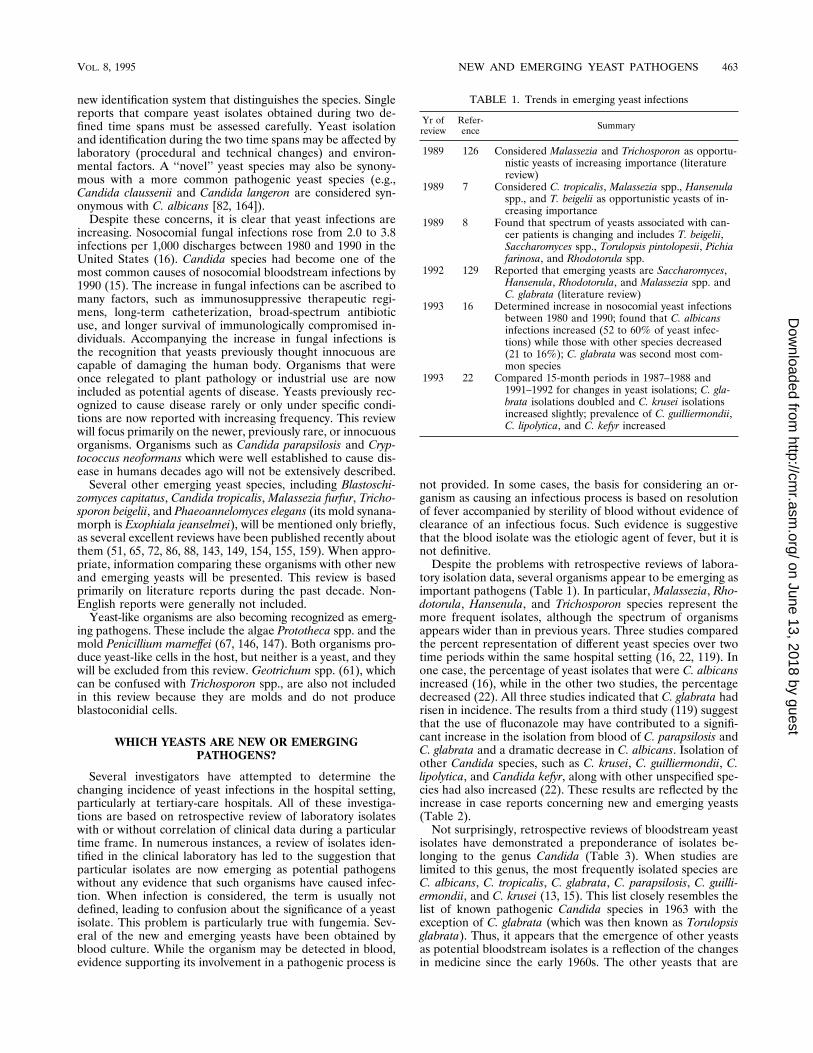
tory isolation data, several organisms appear to be emerging asimportant pathogens (Table 1). In particular, Malassezia, Rho-dotorula, Hansenula, and Trichosporon species represent themore frequent isolates, although the spectrum of organismsappears wider than in previous years. Three studies comparedthe percent representation of different yeast species over twotime periods within the same hospital setting (16, 22, 119). Inone case, the percentage of yeast isolates that were C. albicansincreased (16), while in the other two studies, the percentagedecreased (22). All three studies indicated that C. glabrata hadrisen in incidence. The results from a third study (119) suggestthat the use of fluconazole may have contributed to a signifi-cant increase in the isolation from blood of C. parapsilosis andC. glabrata and a dramatic decrease in C. albicans. Isolation ofother Candida species, such as C. krusei, C. guilliermondii, C.lipolytica, and Candida kefyr, along with other unspecified spe-cies had also increased (22). These results are reflected by theincrease in case reports concerning new and emerging yeasts(Table 2).Not surprisingly, retrospective reviews of bloodstream yeast
isolates have demonstrated a preponderance of isolates be-longing to the genus Candida (Table 3). When studies arelimited to this genus, the most frequently isolated species areC. albicans, C. tropicalis, C. glabrata, C. parapsilosis, C. guilli-ermondii, and C. krusei (13, 15). This list closely resembles thelist of known pathogenic Candida species in 1963 with theexception of C. glabrata (which was then known as Torulopsisglabrata). Thus, it appears that the emergence of other yeastsas potential bloodstream isolates is a reflection of the changesin medicine since the early 1960s. The other yeasts that are
TABLE 1. Trends in emerging yeast infections
Yr ofreview
Refer-ence Summary
1989 126 Considered Malassezia and Trichosporon as opportu-nistic yeasts of increasing importance (literaturereview)
1989 7 Considered C. tropicalis, Malassezia spp., Hansenulaspp., and T. beigelii as opportunistic yeasts of in-creasing importance
1989 8 Found that spectrum of yeasts associated with can-cer patients is changing and includes T. beigelii,Saccharomyces spp., Torulopsis pintolopesii, Pichiafarinosa, and Rhodotorula spp.
1992 129 Reported that emerging yeasts are Saccharomyces,Hansenula, Rhodotorula, and Malassezia spp. andC. glabrata (literature review)
1993 16 Determined increase in nosocomial yeast infectionsbetween 1980 and 1990; found that C. albicansinfections increased (52 to 60% of yeast infec-tions) while those with other species decreased(21 to 16%); C. glabrata was second most com-mon species
1993 22 Compared 15-month periods in 1987–1988 and1991–1992 for changes in yeast isolations; C. gla-brata isolations doubled and C. krusei isolationsincreased slightly; prevalence of C. guilliermondii,C. lipolytica, and C. kefyr increased
VOL. 8, 1995 NEW AND EMERGING YEAST PATHOGENS 463
on June 13, 2018 by guesthttp://cm
r.asm.org/
Dow
nloaded from

becoming more frequently recognized as etiologic agents ofbloodstream infections includeHansenula anomala, Blastoschi-zomyces capitatus, Rhodotorula spp., and Trichosporon beigelii.The change in frequency of isolation may also reflect the abilityof clinicians and technologists to recognize a non-C. albicansisolate as an important opportunistic pathogen and the abilityof contemporary blood culture systems and procedures to sup-port the growth of the unusual yeast isolates (see below). Therecent reviews demonstrate that the important yeasts in blood-stream infection remain Candida species.
ANATOMIC SITES ATTACKED BY YEASTS
C. albicans can attack nearly every organ in the body andcause a wide spectrum of clinical manifestations. C. albicans isthe most common yeast species isolated from blood. For manyof the new and emerging yeasts, bacteremia or catheter-asso-ciated infection is the primary or only manifestation of disease(Table 4). However, several species appear to cause diseaseprimarily at sites other than blood. For example, Candida ci-ferrii and Candida pulcherrima are associated with nail infec-tions, and Candida zeylanoides has been obtained from skinand nails as well as blood.It is striking how commonly the bloodstream is involved in
the new and emerging yeast infections, as is the common as-sociation of hematogenous and solid malignancies with theappearance of the unusual yeasts. In numerous cases, the or-ganism was obtained on repeat blood culture, suggesting aconstant shedding of organisms into the blood. What is thenidus for the organisms? Unfortunately, insufficient studies are
available to assess the possible origin of the unusual yeasts. Forthe species that have been isolated rarely, risk factor analysis isnot possible. From an epidemiologic standpoint, all of the newand emerging yeasts can be found in the environment (109,153), and many of the Candida species and Saccharomycescerevisiae can be isolated from human mucosal sites, especiallythe gastrointestinal tract and vagina (109, 138). If risk factoranalysis for common candidemias can be extended to the otherCandida species and non-Candida yeasts, then several factorsappear to be particularly involved. These factors includebroad-spectrum antibiotic use, antineoplastic agent use, ad-ministration of vancomycin, intravenous catheterization, andneutropenia and other immunodeficiencies (7, 68, 125). Thesefactors then result in further alterations in innate and specificimmunity. Catheterization results in disruption of the integrityof the cutaneous barrier, antineoplastic agents cause thinningof the protective mucous barrier of the gastrointestinal tractand further attenuation of immune cell function, and broad-spectrum antibiotics can lead to proliferation of yeast growthon mucosal surfaces.When considered together, these factors suggest that disrup-
tion of the gastrointestinal tract may be the most importantpredisposing factor leading to the development of infection,particularly fungemia, by the unusual (and usual) yeasts. Ba-denhorst et al. (13) noted that two factors, broad-spectrumantimicrobial therapy and abdominal disorders, including lap-arotomy, appeared to be most often involved (47 and 94%,respectively) in the development of fungemia. Surveillance sur-veys typically may not demonstrate the presence of the unusualyeasts on skin, except in association with nails (4, 109, 153).
TABLE 2. Examples of clinical reports or reviews on the newand emerging yeasts and yeast-like organismsa
Organism References
Blastoschizomyces capitatus .....................................33, 88, 117, 154, 155Candida chiropterum................................................48Candida ciferrii..........................................................34, 48Candida famata ........................................................120, 141Candida glabrata.......................................................52, 73Candida guilliermondii .............................................21, 36, 56Candida haemulonii .................................................50Candida humicola ....................................................4Candida kefyr ............................................................99Candida krusei ..........................................................10, 54Candida lipolytica .....................................................162Candida lusitaniae ....................................................14, 57, 130Candida norvegensis .................................................107Candida pintolopesii .................................................8, 78Candida parapsilosis .................................................32, 115, 131Candida pulcherrima ................................................118Candida rugosa .........................................................38, 144Candida tropicalis .....................................................11, 20, 51, 66Candida utilis ............................................................5, 23Candida viswanathii .................................................132Candida zeylanoides .................................................31, 84, 158Exophiala jeanselmei ................................................53, 65, 85, 143Hansenula anomala..................................................6, 59, 106Penicillium marneffei ................................................63, 112, 146, 147Pichia farinosa...........................................................8Prototheca wickerhamii ............................................62, 67, 133Rhodotorula rubra.....................................................26, 70Saccharomyces cerevisiae .........................................29, 30, 43, 110, 138Sporobolomyces sp. ..................................................17, 100Trichosporon beigelii .................................................9, 47, 154, 155a This table is intended to provide some examples of clinical reports and
reviews on emerging yeast infections from the past 10 years. It is not a completelist of all such reports and does not include non-English language reports. TABLE 3. Trends in bloodstream infections caused by yeasts
Yr ofreview
Refer-ence Summary and comments
1985 66 Studied only candidemia; frequency of specieswas C. albicans . C. tropicalis . C. parapsi-losis . C. glabrata . C. krusei . other species
1986 102 Concerned with Virginia hospitals; found in-crease in Candida infections between 1978 and1984 from 0.1 to 1.5 cases/10,000 patient dis-charges
1989 113 Ranked nosocomial bloodstream infections; from1984 to 1988, Candida species changed fromeighth to fourth most common agent of infec-tion; genera of gram-negative bacilli are con-sidered as individual categories
1991 15 Candida species are fifth leading cause of blood-stream infection in 1989 (up from sixth in1980) if gram-negative bacilli are consideredone group
1991 13 Survey of fungemia for 1989 in one hospital inSouth Africa found that 2.1% of blood cul-tures contained yeasts; these included C. albi-cans (42%), C. tropicalis (26%), C. parapsilosis(20%), C. glabrata (7%), Hansenula spp. (2%),C. guilliermondii (1%), and C. krusei (1%)
1992 28 Retrospective study in Indian teaching hospital;compared 5-yr periods 1980–1985 and 1986–1990; found 11-fold increase in candidemia;most common species isolated were C. albi-cans, C. tropicalis, C. parapsilosis, and C. guilli-ermondii
1992 93 Fungemia isolates in one hospital between 1984and 1990; C. albicans . C. tropicalis 5 C. gla-brata . C. krusei . C. parapsilosis 5 C. guilli-ermondii
464 HAZEN CLIN. MICROBIOL. REV.
on June 13, 2018 by guesthttp://cm
r.asm.org/
Dow
nloaded from

However, a survey of hospital personnel demonstrated thatgreater than 70% of nurses and nonnursing hospital personnelharbored yeasts on their hands (142). The most frequentlyisolated organisms were Rhodotorula spp. and C. parapsilosis.
The latter species has also been shown to frequently colonizethe skin, particularly the subungual space (160). Whether suchcolonization contributes to the increased isolation of C. para-psilosis in nosocomial candidal infections and contamination of
TABLE 4. Clinical information associated with the emerging and new yeasts
Organism Site infected or affected Underlying conditions of patientsa Predisposing factorsb Reference(s)
Candida ciferrii Nails, ear Otomycosis, onychomycosis None stated 48Nails NIDDM, vasculopathies, valvulo-
pathyNail trophisms 34
Candida famata Catheter,c blood CML (BMT) Long-term catheter 141Uvea Cataracts Cataract extraction with implanta-
tion of intraocular lens120
Candida glabrata Various, especially urinary tract,mucosal areas, lungs
DM, solid tumors; rarely hemato-logic malignancies; malnutri-tion; neonate
Cannulas, valve grafts, catheters,vascular surgery, mechanical ven-tilation, gastric perforations
52, 73, 101,137
Candida haemulonii Toe skin, nails, blood Diabetes or not indicated Unknown 50
Candida kefyr Blood, spleen, kidneys Adenocarcinoma Radiation chemotherapy 99
Candida krusei Blood HIV, leukemia, lymphoma, BMT Neutropenia, immunosuppression,prophylactic fluconazole
54, 140, 165
Candida lusitaniae Blood, catheter, central venouscannula, urinary tract
Leukemia, myeloma, BMT,cystitis
Immunosuppression, antibiotictherapy
19, 57, 92
Candida parapsilosis Blood, intravenous catheter, Foleycatheter, peritoneum
Low birth weight, ESRD, immuno-deficiency
TPN, antibiotic use 39, 115, 139
Candida norvegensis Blood, peritoneal fluid ESRD (renal transplant) Antibiotic use, immunosuppression 107
Candida rugosa Blood, burn wounds, catheter Leukemia, granulocytopenia, burns Topical nystatin, antibiotic use 12, 38, 144
Candida pulcherrima Nails 118
Candida zeylanoides Groin None None implicated 158Nails Papillomavirus infection or none Estrogen cream (?), nail plate
separation31
Blood, right knee Scleroderma, gastrointestinal mal-absorption, IDDM
Kidney-pancreas transplant, hemo-dialysis, TPN
18, 84
Hansenula anomala Blood, cannula insertion site Low birth weight TPN 104Endocardium (aortic valve) Drug addiction IVDA, alcohol abuse 108Blood AIDS, carcinoma, MS, pancreati-
tis, AML, MVAPN, CVC, tracheostomy, IVDA,antibiotic use, gastrointestinalbleeding
6, 64, 71,106, 128
Rhodotorula rubra Blood, catheter site ALL, AML, aplastic anemia, lym-phoma, sarcoma
CVC 70
Peritoneum ESRD, renal dysplasia CAPD 40Alveoli (allergic alveolitis) None Long-term environmental exposure 41
Sporobolomycessalmonicolor
Lymph node, bone marrow AIDS IVDA, PCP (?) 100, 116
Saccharomycescerevisiae
Vagina None Recurrent vaginal candidiasis, topi-cal antimycotics, urinary tractinfection, multiple antibiotics
138
Blood AML, anemia (myelodysplasticsyndrome)
Granulocytopenia or not stated 8, 110
a Abbreviations: ALL, acute lymphoblastic leukemia; AML, acute myelogenous leukemia; BMT, bone marrow transplant; CAPD, continuous ambulatory peritonealdialysis; CML, chronic myelogenous leukemia; CVC, central venous catheter; DM, diabetes mellitus; ESRD, end-stage renal disease; HIV, human immunodeficiencyvirus; IDDM, insulin-dependent diabetes mellitus; IVDA, intravenous drug abuse; MS, multiple sclerosis; MVA, motor vehicle accident; NIDDM, non-insulin-dependent diabetes mellitus; PCP, Pneumocystis carinii pneumonia; PN, parenteral nutrition; TPN, total parenteral nutrition.b In some cases, particularly in patients with cancer, the predisposing factors leading to yeast infection can be numerous and manifold. Only a few of the predisposing
factors are provided in these cases. Interested readers may find several reviews concerning predisposing factors of yeast infections in the literature (8, 16, 129, 152, 159).c Unless otherwise specified, catheter refers to a long-term indwelling intravenous catheter.
VOL. 8, 1995 NEW AND EMERGING YEAST PATHOGENS 465
on June 13, 2018 by guesthttp://cm
r.asm.org/
Dow
nloaded from

irrigation fluids, hyperalimentation solutions, and catheters re-quires further study (25, 131, 160). When a catheter is shown tobe contaminated with yeasts, peripheral blood cultures arenegative, and no other nidus is apparent, then skin may be apossible source for the implicated yeast.If these observations are corroborated by other studies, the
importance of surveillance cultures of gastrointestinal sitesmay be significant. That is, it may be useful to survey the yeaststrains associated with the gastrointestinal tract of an immu-nocompromised patient in order to predict the most likelyagent that could cause subsequent infection. Preliminaryscreening may also help to determine the effective antifungalagent prior to development of serious infection. The imple-mentation of surveillance cultures and subsequent microbio-logical work-up may be too expensive to perform except for asubset of patient populations.
HISTOPATHOLOGY
A limited number of studies have described the fungal cellmorphology and the histopathologic appearance associatedwith the infections caused by the unusual yeasts. In many ofthese studies, the histopathologic appearance is described onlyin broad terms. For the descriptions that are available, theirusefulness must also be judged with the understanding thathost immunologic status will influence the histopathologic pic-ture. Diagnosis of unusual yeast infections would be helped bysystematic studies of the histopathology associated with themin humans. On the basis of the information presented in Table5, several generalizations can be made.Unlike active C. albicans infection, which is typified histo-
logically by the presence of yeasts and pseudohyphae (at leastat later stages of infection), several of the unusual Candidaspecies appear to produce only yeast forms during infection(Table 5). Species that attack the nails, a site that has a tem-perature lower than 378C, appear to produce yeasts andpseudohyphae. This observation suggests that pseudohyphalproduction does not require a temperature of 378C. C. glabrataappears to produce only yeast forms, which is consistent withits previous designation as T. glabrata. Candida famata (previ-ously designated Torulopsis candida) also produced only yeastforms, but the significance of this finding is limited becauseonly one study is involved (120).It is evident from Table 5 that the appearance of yeasts and
pseudohyphae does not imply that the etiologic agent is C.albicans. C. ciferrii and several other uncommon species ofCandida produce similar tissue forms. It is notable that T.beigelii and B. capitatus also produce blastoconidial cells alongwith hyphae (97, 156). These organisms can be differentiatedfrom Candida species by the predominance of hyphae pro-duced in comparison with pseudohyphae and the paucity ofblastoconidial forms. The presence of arthroconidia providesfurther evidence that the etiologic agent is a Trichosporon orBlastoschizomyces sp. and not a Candida sp.A significant range of histopathology is associated with the
unusual yeasts (Table 5). Low-virulence organisms do not nec-essarily stimulate poor immune responses. A mild cellular re-action seen in response to a low-virulence organism, such as C.guilliermondii, may not indicate that the organism does notprovide immune stimulation. In one case of infection with thisorganism (36), the patient suffered from aplastic anemia. Herhematocrit was low (15.6%), and her leukocyte count was only2,400 cells per mm3. Most significant was the observation thather bone marrow was hypocellular, lacking leukocyte precur-sors. The mild reaction to the organism is easily explained bythe host’s attenuated cellular immune capacity. The poor host
response also explains how this low-virulence organism wasable to establish infection. Another low-virulence organism, C.parapsilosis, can induce acute and chronic inflammatory re-sponses. D’Antonio et al. (32) have noted that hepatic andsplenic microabscesses are frequently associated with C. para-psilosis fungemia in patients with hematologic malignancies.These observations demonstrate that organisms which are in-frequent agents of infection and typically cause only mild dis-ease in most patient populations can, under appropriate con-ditions, cause serious infection.In most cases of infection by the unusual yeasts, the his-
topathologic response is characterized as inflammatory or asan abscess. However, granulomatous responses may also beseen. This is true with C. glabrata, E. jeanselmei, and T. beigelii(Table 5). Because of the limited number of investigations, it isnot clear that other yeasts (Table 2) can also elicit a granulo-matous response in an immunocompetent patient. Surgicalpathologists and cytopathologists should be alerted to the pos-sibility that many yeast species may cause a granulomatousresponse, and thus this characteristic does not help narrow thedifferential diagnosis.
TREATMENT OF INFECTIONS DUE TOUNUSUAL YEASTS
Infections by the unusual and emerging yeasts are, as men-tioned above, diseases of the immunocompromised. Improve-ment of the host immunological status is perhaps the mostimportant method by which to prevent the development andbring about the resolution of yeast infections. Unfortunately,in many cases, intervention of this nature is not possible, ne-cessitating alternative therapeutic modalities for an ongoinginfection.In the case of fungemia, it is necessary to determine the
origin of the organisms that are being shed into the blood-stream. Whenever possible, amelioration of a gastrointestinalproblem would likely lead to significant improvement in thepatient suffering from candidemia, particularly if the offendingorganism has a low virulence potential and the patient canmount an immune response. While helpful, gastrointestinalremediation provides only one element of therapy when theyeast has seeded other organs. In this case, antifungal therapyand other steps must be considered.
Catheter Removal
When a catheter is an infectious nidus, the choice of imple-menting antifungal therapy in light of the possible toxicitiesassociated with such therapy must be weighed against possiblepatient outcome with no therapy and/or removal of the cath-eter. Surgically implanted central venous catheters are expen-sive to remove and replace. For many patients, the catheter isneeded to provide venous access for delivery of various agents,such as parenteral nutrition. It is therefore desirable to treatthe patient with antifungal agents while keeping the catheter inplace. However, this management strategy is inappropriate.Table 6 summarizes the results of treatment in patients withfungemia due to unusual yeasts. In approximately 31% of theindicated cases, the patients died before the infection resolved.At least 74 of the 104 patients (71%) had catheters in place. Inseveral cases, information about the use of a catheter was notprovided, but it is likely that many of the patients had cathetersin place, given their underlying illnesses. Among the 40 casesfor which indications about the treatment outcome were in-cluded, 32 (80%) were successfully resolved by removing thecatheter. In 28 of these cases, treatment with antifungal agents,
466 HAZEN CLIN. MICROBIOL. REV.
on June 13, 2018 by guesthttp://cm
r.asm.org/
Dow
nloaded from

usually amphotericin B, was included. In one case of infec-tion with C. zeylanoides (84), amphotericin B therapy alone didnot prevent subsequent infections. Only upon removal of a con-taminated Hickman catheter was complete resolution obtained.While these results indicate that catheter removal is a fre-
quent practice, whether it is necessary in all catheter-associ-ated yeast infections is unclear. In two cases, one involving H.anomala (103) and the other C. lipolytica (157), the catheterwas removed but evidence of infection continued. Addition ofantifungal therapy was necessary to resolve the infections. Be-
fore general conclusions about the efficacy of catheter removalor ‘‘treating through the catheter’’ can be made, more exten-sive studies are needed. A recent multicenter trial suggests thathost factors, including the presence of intravenous catheters,may be more important than the MICs of antifungal agents forpredicting patient outcome (123, 124). However, it is likely thateach catheter-associated yeast infection will need to be ap-proached as an individual management problem.For at least one unusual yeast, Rhodotorula rubra, the im-
portance of catheter removal is being addressed. Kiehn et al.
TABLE 5. Tissue morphology and histopathology associated with emerging yeastsa
Organism Site affected or studied Histopathology Fungal cell tissue morphology Reference
Blastoschizomycescapitatus
Endocardium (prosthetic mi-tral valve)
NS Septate hyphae with some dichoto-mously branching, occasional yeastcells; arthroconidial cells alsopresent
117
Vertebral body NS Septate hyphae 33
Candida ciferrii Nail NS Pseudohyphae, yeasts in clusters 34Skin (tinea pedis) NS Pseudohyphae, yeasts in clusters 34
Candida famata Uvea Histiocytes, epitheloid giant cellscontaining melanin pigment, andfew lymphocytes on posterior sur-face of posterior lens capsule
Yeast forms 120
Candida glabrata Endocardium Fibrinopurulent exudate Yeast cell clusters 27Various Mild chronic infiltrate with lympho-
cytes, macrophages to frank granu-lomatous reaction
Yeast forms 11, 137
Candida guilliermondii Various organs but not lung Little tissue reaction Yeast forms 36
Candida kefyr Kidney, spleen NS Hyphae and budding yeasts typical ofCandida species
99
Candida lusitaniae Kidney Mononuclear infiltrate, neutrophilssparse
Organisms eosinophilic (H&E stain)with clear haloes, budding cells
57
Candida parapsilosis Synovium Acute and chronic inflammatorychanges
NS 69
Candida zeylanoides Nail NS Mycelial elements and yeast cells,some pseudohyphae
31
Exophiala jeanselmei Soft tissue of forearm Inflammatory infiltrate with plasmacells, lymphocytes, macrophages,multinucleated giant cells
Lightly pigmented brown hyphae onH&E
143
Lung aspirate (also containedStaphylococcus aureus andHaemophilus influenzae)
Acute inflammation Hyphae 85
Cervical lymph node Granulomatous changes withmultinucleated cells, histiocytes,neutrophils, and loci of large num-bers of eosinophils
Pigmented pale brown, septate hy-phae; single yeast-like cells, chainsof fungal cells
65
Hansenula anomala Intravenous cannula insertionsite
Abscess (microscopy not described) Yeasts 104
Aortic valve vegetation NS Yeasts, many intracellular 108
Sporobolomycesholsaticus
Skin NS Yeasts with single buds 17
Trichosporon beigelii Skin nodule Granulomatous inflammation Hyphae and blastoconidia 97Maculonodular skin lesion(patient with ALL)
NS Arthroconidia, blastoconidia,pseudohyphae
156
a Abbreviations: ALL, acute lymphocytic leukemia; NS, not stated; H&E, hematoxylin and eosin stain.
VOL. 8, 1995 NEW AND EMERGING YEAST PATHOGENS 467
on June 13, 2018 by guesthttp://cm
r.asm.org/
Dow
nloaded from

(70) described 22 patients who had evidence of fungemia dueto R. rubra. All of the patients had catheters in place. Only twopatients had illness complicated by neutropenia. Regardless ofwhether the catheter was removed, the patients recoveredfrom the episode of fungemia. All patients from whom thecatheter was not removed received antifungal therapy. Wheth-er antifungal treatment alone would have been sufficient for allof the patients is not clear. This study highlights several con-siderations. The particular yeast in this study is a skin and
environmental saprobe with low virulence. The authors notethat previous reports document only one death associated withculture-proven Rhodotorula fungemia. The source of the or-ganism was likely the skin, as opposed to the gastrointestinaltract for many candidemias. Only one patient had a positiveperipheral blood culture. All positive blood cultures were other-wise obtained through the catheter. R. rubra is also susceptibleto amphotericin B (see below). These results suggest that afirm conclusion about the utility of catheter removal for treat-
TABLE 6. Summary of outcome of fungemia caused by various emerging yeasts
Organism No. ofcases
No. of deathsbefore reso-lution of in-fection
No. withlong-termcathetersa
No. of cases with resolution of infection
Patient charac-teristicsb
Refer-enceCatheter not
removed
Catheter removed,no antifungaltherapy
Catheter re-moved, antifun-gal therapy
Candida famata 1 0 1 1 BMT 141
Candida glabrata 1 0 1 Vascular surgery 1012 1 0 Neonate 52
Candida guilliermondii 1 1 NS Aplastic anemia 36
Candida kefyr 1 1 0 Adenocarcinoma 99
Candida krusei 4 3 2 (4?) 1 BMT, leukemia, lymphoma 5410 0 NSc BMT, leukemia 165
Candida lusitaniae 1 1 0 Multiple myeloma 1111 0 1 1 Preterm newborn 1682 0 1 (2?) 1 1? Leukemia 19
Candida norvegensis 1 1 NS ESRD 107
Candida lipolytica 1 0 1 1d Cholecystectomy 157
Candida parapsilosis 22 15 22 NS NS NS Cancer 115
Candida rugosa 1 0 1 1 Hypotension 1229 4 NS Burn patients 381 0 1 (Port-a-Cath) 1 CF 12
Candida utilis 1 0 1 1 Hemophilia, neutropenia 51 0 0 Alzheimer’s 23
Candida zeylanoides 1 0 1 1 GI bleeding 841 0 1 1 IDDM, renal Tx 18
Hansenula anomala 11 2 8 NS NS NS Newborn, AML 591 0 1 1 Lung carcinoma 641 1 1 Acute pancreatitis 1061 0 1 1 TPN 371 0 1 1 MVA 61 0 1 1e Leukemia 1032 0 2 1 1 Carcinoma 71
Rhodotorula rubra 22 0 22 5 17 Cancer, others 700 0 1 1 TPN 26
Saccharomyces cerevisiae 1 0 NS AIDS 1351 1 1 Renal failure 301 1 1 Myelodysplastic syndrome 110
a The specific catheter types (Hickman, Broviac, etc.) are usually not indicated in the references. The typical descriptions were central venous catheter or intravenouscatheter. NS, not stated.b Abbreviations: AML, acute myelogenous leukemia; BMT, bone marrow transplant; CF, cystic fibrosis; ESRD, end-stage renal disease; GI, gastrointestinal; IDDM,
insulin-dependent diabetes mellitus; MVA, motor vehicle accident; TPN, total parenteral nutrition; Tx, transplant.c Although not specifically stated, it is likely that many of the cases of severe disease (e.g., BMT patients) had long-term venous catheters in place during their hospital stay.d In this patient, the central venous catheter was removed but fungemia persisted for another 11 days. The patient’s blood became sterile after resolution of
thrombophlebitis and fever.e The catheter was removed, but the infection did not resolve. Amphotericin B was then administered, and the infection resolved.
468 HAZEN CLIN. MICROBIOL. REV.
on June 13, 2018 by guesthttp://cm
r.asm.org/
Dow
nloaded from

ment of R. rubra infection is difficult to draw. It is possible that,for this organism, either antifungal therapy or catheter re-moval may be sufficient. Such a limited approach may not beappropriate for other unusual yeasts.
Antifungal Therapy
The antifungal agents that are available for treatment ofyeast infections at sites other than the skin, nails, or vagina aregenerally limited to polyenes, primarily amphotericin B, 5-fluo-rocytosine (5-FC), and the azoles, namely, fluconazole, itra-conazole, and ketoconazole. New agents with different mech-anisms of action are under development (60). The efficacy ofthese agents for treating the unusual yeasts is unknown be-cause insufficient cases have been reported to provide usefulguidelines. Attempts to obtain some indication of the appro-priate therapy for an ongoing infection must be judged on thein vitro susceptibility of the organism.Yeast susceptibility testing has not been standardized. In
1992, the National Committee for Clinical Laboratory Stan-dards published a proposed standard for yeast susceptibilitytesting (105). The proposed method involves broth dilution ineither small (,150 ml) or large (,1 ml) volumes. Further workis needed before the proposed standard becomes finalized. Inmany cases, the MICs of a particular antifungal agent forunusual yeasts have been evaluated either by the proposedstandard or by the agar dilution methods (Table 7). When theagar dilution method was performed by different laboratories,it involved different media, making comparison of the datafrom laboratory to laboratory difficult: this problem argues forthe need for a standardized method.Despite the use of different methods, several important sus-
ceptibility patterns are emerging for the unusual yeasts. Am-photericin B may not be the agent of choice for infectionscaused by C. lusitaniae, C. parapsilosis, and C. kefyr, while itappears to be satisfactory for the other organisms. C. tropicalis,C. rugosa, and T. beigelii may display higher MICs while re-maining susceptible. Two organisms, C. guilliermondii and C.lusitaniae, have been reported to develop resistance with treat-ment with amphotericin B (2, 36). C. parapsilosis may showtolerance to amphotericin B. Thus, its MIC of amphotericin Bwould suggest that it is susceptible, but the minimal fungicidalconcentration, as evidenced by growth of organisms on stan-dard solid media after exposure to the drug in the MIC test,may be more than 32 times higher than the MIC (134). C.rugosa exhibits differential susceptibility to the polyenes, am-photericin B, and nystatin. Dube et al. (38) noted that C.rugosa became the most common agent of fungemia in theirburn patients following the use of topical nystatin ointment forprophylactic treatment of burn wounds. The overall incidenceof fungemia decreased, however. Upon susceptibility testing, itwas noted that the C. rugosa isolates were generally resistant tonystatin (MIC, .18.5 mg/ml) but remained susceptible to am-photericin B (MIC, generally #1.16 mg/ml) and to fluconazole(MIC, #5 mg/ml). These data show that the mechanism ofresistance to nystatin may differ from that for amphotericin Bdespite shared mechanisms of action (24, 58, 91).The susceptibility of the emerging and unusual yeasts to
azole antifungal agents is variable (Table 7). The bistriazolefluconazole appears by in vitro tests to be ineffective or mar-ginally effective against C. krusei, C. guilliermondii,H. anomala,and R. rubra. Variable efficacy is evident with C. glabrata, C.parapsilosis, C. rugosa, C. tropicalis, S. cerevisiae, and T. beigelii.C. krusei also appears to be clinically resistant to fluconazole.The MIC patterns for itraconazole, a recently approved tria-zole, did not parallel the patterns obtained with fluconazole. In
many cases, organisms that were generally resistant to flucon-azole were more susceptible to itraconazole (e.g., C. krusei, C.guilliermondii, R. rubra, and S. cerevisiae) but at MIC levels thatwere higher than those for C. albicans. Two exceptions are C.parapsilosis and C. tropicalis. These results indicate that in vitrosusceptibility testing should include both triazoles, i.e., onetriazole cannot be used to predict the efficacy of a secondtriazole. Interestingly, many of the unusual yeasts appear to besusceptible to ketoconazole. Variable susceptibility, however,was obtained with C. glabrata, C. parapsilosis, and B. capitatus.De Gentile et al. (34) noted that an isolate of C. ciferrii whichhad been obtained from a case of toenail onyxis demonstratedvariable susceptibilities to different azoles when tested by dif-fusion methods. The organism was susceptible to clotrimazole,ketoconazole, and econazole but resistant to itraconazole, flu-conazole, miconazole, and bifoconazole. Additional studies todetermine if this species characteristically displays differentialsusceptibilities are needed. Such information could be valuablefor identifying the organism by using an antibiogram.Many of the unusual yeasts appear to be susceptible to 5-FC,
suggesting that the combination of amphotericin B and 5-FCmay provide effective therapeutic management regimen. T.beigelii and B. capitatus appear generally resistant to 5-FC.Candida norvegensis appears to be susceptible to 5-FC but atlevels higher than those of most of the susceptible yeasts.
MICROBIOLOGICAL IDENTIFICATION
Taxonomy
Binomial epithets for the unusual (and usual) yeasts changefrequently as more definitive methods (vis a vis molecularmethods) are developed to differentiate the organisms andbecause there is no official organization that approves thecorrect taxonomic description and classification of fungal or-ganisms. Taxonomic affiliation and appropriate binomial epi-thets are decided by consensus, resulting in the use of multiplenames for the same organism (see Table 8) when differentauthors disagree about an organism’s taxonomic status (82,89). Further complicating this problem is the use of anamor-phic epithets for organisms that have a known teleomorph(e.g., C. krusei [anamorph] and Issatchenkia orientalis [teleo-morph]). This practice will likely persist because many timesthe teleomorph of a yeast is not evident, making its anamor-phic name seem more appropriate. Also, as many clinical my-cologists are aware, fewer inquiries about the implications ofan organism are likely when a well-known anamorphic genusepithet (e.g., Candida) is reported than when a seemingly ar-cane teleomorphic epithet (e.g., Clavispora) is used. For theclinician, name changes appear to serve no useful purpose.Many of the unusual yeasts are affiliated with the division
Ascomycotina and are heterothallic (Table 8), that is, matingrequires the union of thalli of opposite mating types. Thenotable exception is the teleomorph Sporidiobolus salmoni-color (anamorph, Sporobolomyces), which belongs to the Ba-sidiomycotina. The number of different teleomorphic generarepresented by the various Candida species is striking andhelps demonstrate how uninformative a form genus designa-tion can be for those hoping to understand an organism on thebasis of its taxonomic nomenclature.
Significant Laboratory Characteristics
The mycology laboratory is challenged by the identificationof clinically significant unusual yeasts. Many laboratories nowuse some form of rapid multiple biochemical test system (e.g.,
VOL. 8, 1995 NEW AND EMERGING YEAST PATHOGENS 469
on June 13, 2018 by guesthttp://cm
r.asm.org/
Dow
nloaded from

TABLE 7. Antifungal antibiograms of new and emerging yeastsa
Organism No. of iso-lates studied
MIC (mg/ml)
Method Refer-ence(s)Amphoter-
icin B Fluconazole Intraconazole Ketoconazole 5-FC
Candida ciferrii 1 S R R S (also clotri-mazole)
R ‘‘Diffusion’’ 34
Candida famata 3 Generally R(.12.5)b
Multiple meth-ods BD
121
1 0.8 0.78 0.2 AD or BD 1411 1.56 6.25b 0.2 AD 1661 ,20 49
Candida glabrata 25 0.06–1.0 0.05–12.5 0.05–0.2 0.0037–0.12 0.12–4.0 AD 87NS 0.1–0.4 1–64b 0.05–1.56 BD 13663 0.5–20b 1.0–512b 0.06–2.0 CBD 114
Candida guilliermondii 1 ‘‘Develop R’’ 0.25 0.2 AD 365 0.06–0.12 3.2–25b ,0.05–0.2 0.0075–0.06 0.24 AD 8710 0.25–2.0 4.0–64b 0.06–1.0 CBD 114
Candida kefyr 1 0.4 BD 134.100 0.09–.6.25b Various 90
Candida krusei 24 0.5–4.0 8.0–.512b 0.12–64b CBD 1149 0.5–2.0 10–40b BD or AD 16543 0.06–2 0.05–.100b ,0.05–3.2 0.0037–0.25 0.12–4 AD 87
Candida lipolytica 9 0.313–1.25 0.078–0.313 BD or AD 157
Candida lusitaniae 7 0.39–6.3 0.18–6.3 0.05–0.18 BD 2(developing R)
6 0.1–20b ,0.03–.160b NS 111, 1688 0.12–1 ,0.05–1.6 ,0.05–6.4 0.0037–0.12 0.12–1 AD 87
Candida norvegensis 2 0.39 0.78 25b BD 21 0.4–0.8 0.4–1.0 3.2–12.5b AD 107
(over 10 days) (over 10 days)
Candida parapsilosis 105 0.5–.2.0b 0.25–.512b 0.12–256b CBD 11433 0.025–.6.25b 0.063–.128b ,0.125–.64b ,0.025–.100b Various 9019 0.12–1 ,0.05–50b ,0.05 0.0037–0.12 0.25–0.5 AD 87
Candida rugosa 10 0.5–2.0 2.0–32.0b 0.12–2.0 CBD 1144 0.25–4.0b ,0.046–12.5 ,0.078–6.25 BD 14410 0.58–1.16 2.5–20b 0.1–0.8 ,10–.323b BD 38
Candida tropicalis 86 0.25–4.0b 2.0–.512b 0.12–.512b CBD 11474 0.12–2 0.05–.100b ,0.05–100b 0.0037–8 0.12–4 AD 87
Candida utilis 1 0.52 BD 51 0.04 4 BD 23
Candida zeylanoides 3 4–8.0b 0.12–1.0 ,0.13 (oneisolate, .128)
BD 84
1 S S S DD 311 S S S DD 18
Blastoschizomycescapitatus
15 0.15–0.62 0.04–50b 0.04–.100b BD 881 0.78 25 1.56 100b BD 33
Hansenula anomala 4 0.78–1.56 1.56–12.5b 0.2–0.78 BD and AD 1661 (MIC99) 3.13 6.25 12.5b AD 64Various 0.039–1 0.08 0.015–.100b BD 71
(4 isolates) (1 isolate) (5 isolates)1 1.56 12.5 0.39 0.1 BD 59
Rhodotorula rubra 9 0.8–1.6 6.4–.100b 0.8–12.8b 0.4–0.8 ,0.1 BD 70
Sporobolomyces salmonicolor 1 ,0.14 ,1.25 0.2 BD 100
Saccharomyces cerevisiae 20 (MIC90) 0.2 40b 1.56 0.78 0.31 BD 1384 0.156–0.312 0.09–0.78 0.04–0.78 0.04–0.35 BD 12
Trichosporon beigelii 4 2.0 4 0.5 32–.32b NS 471 0.08 10 0.15 .100b BD 150
a Abbreviations: AD, agar dilution assay; BD, broth micro- or macrodilution assay; CBD, colorimetric microbroth dilution assay; DD, disk diffusion assay; NS, notstated; R, resistant; S, susceptible; MIC90, MIC for 90% of isolates; MIC99, MIC for 99% of isolates.b These ranges suggest that some isolates of the species may be resistant to the antifungal agent.
470 HAZEN CLIN. MICROBIOL. REV.
on June 13, 2018 by guesthttp://cm
r.asm.org/
Dow
nloaded from

Vitek Yeast Biochemical Card [BioMerieux Vitek Inc., Hazel-wood, Mo.], API 20C [BioMerieux Vitek], Uni-Yeast Tek [Re-mel Laboratories, Lenexa, Kans.], ID 32C [BioMerieux Vitek],MicroScan Yeast Identification Panel [MicroScan, West Sac-ramento, Calif.], and others). These systems provide a conve-nient method for identifying many yeasts, but the databases forall of these systems have insufficient test strains of the unusualyeasts or lack the unusual yeasts altogether (C. utilis is a com-mon problem). This difficulty is particularly evident when thespecificity and sensitivity of the systems are tested with themore unusual yeasts. These difficulties are not simple to alle-viate because manufacturers must anticipate which speciesmay arise as new opportunistic pathogens following the com-mercial release of their system. Such prognostication is obvi-ously impossible. However, the clinical laboratory can performsome relatively simple tests that can provide useful clues toidentification (Table 9).Yeast morphology on standard media.While it is frequently
true that cellular morphology is not a useful clue for yeastidentification because many yeasts look similar when grown onstandard mycologic media (e.g., Sabouraud dextrose agar andpotato dextrose agar), this characteristic of a yeast should notbe ignored. S. salmonicolor produces an elongated cell, withthe spore produced at the end of a denticle (Sporobolomycesholsaticus is shaped similarly but lacks the denticle). Kloeckeraspecies produce a distinctive apiculated yeast form. C. glabratayeast cells are generally smaller than those of C. albicans (andcan be confused with yeast forms of Histoplasma capsulatum orM. furfur). Blastoschizomyces and Trichosporon spp. form pre-
dominantly hyphal cells. A compendium on yeast identifica-tions, such as that of Kreger-van Rij (75), should be consultedfor other helpful cellular morphologies.Pigment production. Perhaps the most obvious clue to spe-
cies identification is colony color. Sporobolomyces and Rhodo-torula spp. produce carotenoid pigments, although some spe-cies of these genera may not produce pigments (103). R. rubraproduces the carotenoid torularhodin. With carotenoid pro-duction, S. salmonicolor appears salmon, S. holsaticus is peachto salmon, and R. rubra is salmon or pink. Microscopic mor-phology could then sort out which species is likely involved.Assimilation. Standard assimilation reactions may be suffi-
cient to differentiate many of the unusual yeasts but may some-times lead to equivocal results for the emerging yeasts. C.lusitaniae, C. tropicalis, C. parapsilosis, and S. cerevisiae mayappear similar by assimilation reactions. If rhamnose assimila-tion is positive, then the result is indicative of C. lusitaniae. Apositive raffinose result suggests S. cerevisiae (57). C. ciferrii isnot easily differentiated from Candida edax or Candida chiro-pterum. However, C. chiropterum does not assimilate melibi-ose. C. edax assimilates nitrate, while neither C. ciferrii nor C.chiropterum is positive for this assimilation.Additional assimilation reactions may be useful, particularly
when a commercial assimilation system profile index indicateslow selectivity or low specificity and other standard identifica-tion tests do not match the system’s first choice. This problemwas noted by Walsh et al. (157) for a case of fungemia. Initialtesting suggested that the offending yeast was Candida ingens.Upon further testing, it was identified as C. lipolytica. C. ciferrii
TABLE 8. Taxonomic nomenclature and classification of new and emerging yeastsa
Anamorph (previous common ormerged synonym)
Teleomorph(alternative epithet)
Homo- orheterothallic
Teleomorphtaxonomic affinity
Blastoschizomyces capitatus (Geotrichum capita-tum, Trichosporon capitatum)
Endomyces spp. (?)b Ascomycetes
Candida ciferrii Stephanomyces ciferrii Hetero AscomycetesCandida famata (Torulopsis candida) Debaryomyces hansenii Homo AscomycetesCandida glabrata Not knownCandida guilliermondii var. guilliermondii Pichia guilliermondii (Yamadazyma guilli-
ermondii)Hetero Ascomycetes
Candida guilliermondii var. membranaefaciens Pichia ohmeri Hetero AscomycetesCandida haemulonii Not knownCandida kefyr (Candida pseudotropicalis, Can-dida macedoniensis)
Kluyveromyces marxianus var. marxianus Hetero Ascomycetes
Candida krusei Issatchenkia orientalis Hetero AscomycetesCandida lipolytica Saccharomycopsis lipolytica (Yarrowia
lipolytica)Hetero Ascomycetes
Candida lusitaniae (Candida obtusa, Candidaparapsilosis var. obtusa)
Clavispora lusitaniae Hetero Ascomycetes
Candida norvegensis Pichia norvegensis Homo AscomycetesCandida pintolopesii (Candida slooffii) Saccharomyces telluris Homo AscomycetesCandida parapsilosis Not knownCandida pelliculosa Hansenula anomala Hetero AscomycetesCandida pulcherrima Metschnikowia pulcherrima Hetero AscomycetesCandida rugosa Not knownCandida tropicalis Not knownCandida utilis Hansenula jadinii (Pichia jadinii) Homo AscomycetesCandida viswanathii Not knownCandida zeylanoides Not knownPenicillium marneffei Not knownRhodotorula rubra Not known—(no anamorph) Saccharomyces cerevisiae Homo AscomycetesSporobolomyces sp. Sporidiobolus salmonicolor Hetero BasidiomycetesTrichosporon beigelii (Trichosporon cutaneum) Not known
a Data are from references 74, 82, 94, 127, 161, and 167.b Questionable classification.
VOL. 8, 1995 NEW AND EMERGING YEAST PATHOGENS 471
on June 13, 2018 by guesthttp://cm
r.asm.org/
Dow
nloaded from
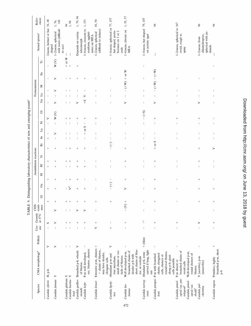
TABLE9.Distinguishinglaboratorycharacteristicsofnewandemergingyeastsa
Species
CMAmorphologyb
Pellicle
Ure-
aseGrowth
at37
8C
CHX
resist-
ance
Assimilationreactions
Fermentation
Sexualsporesc
Refer-
ences
Gl
Ga
MRf
Tr
Ri
InLa
Ni
Gl
GaLa
MSu
Tr
CandidaciferriiH,p-h
V2
S1
11
11
11
22
V2
22
22
2/ascus,helmetorhat
shaped
34,48
CandidafamataY
V2
VV
11
11
11
2V
2W(V)
V2
VV
W(V)1–2/ascus,spherical
withwarts(difficult
tosee)
1,76,
141
CandidaglabrataY
22
12
12
22
12
22
21
22
22
1orW—
94Candidahaemu-
lonii
Y2
21
21
Vd
12
11
22
21
22
21
1—
1,94
Candidaguillier-
mondii
Branchedp-h,whorls
ofblastoc.
V2
1V
11
11
11
22
21
V2
21
1Dependsonvariety
teleomorph
1,79,94
Candidakefyr
P-mwelldeveloped,
occ.blastoc.clusters
22
1V
11
21
21
21orS
21
1SV
21
1–4/ascus,crescentto
reniform,aggluti-
natesonMEA
1,151
Candidakrusei
Extensivep-m,clusters
1chainsofblastoc.,
mayhaveslender,
elongatecells
1V
12
12
22
22
22
21
22
22
1–2/ascus,spherical
(difficulttoinduce)81,94
Candidalipoly-
tica
Abundantp-mand
truemycelium,
smallchainsorver-
ticilsofblastoc.
V1
21
2(1)
22
22(1)
22
22
22
22
1–4/ascus,sphericalor
hatshaped,protu-
beranceon1or2
ends
77,157
Candidalus-
itaniae
Welldevelopedp-m,
branchedchainsof
slenderp-hwith
shortchainsofblas-
toc.atverticils
22
12(V)
1V
12
11
22
21
V2
2(1W)
1orW
1–4/ascus,clavateon
MEA
1,35,57
Candidanorveg-
ensis
Abundantp-h,some
curvediflong,light
tan
1(thin)
22e
22
22
22
22
22
2(1S)
22
22
21–4/ascus,hatshaped
onacetateagar
79,107
Candidaparapsi-
losis
P-mwithbranched
chains,elongated
cells,clustersof
round/ovalblastoc.
alongp-h;giant
cellspossible
22
12
11
12
11orS
22
21
V2
2(1W)
2(1W)
—94
Candidapintol-
opesiivar.pin-
tolopesiif
P-mabsentor
branchedchainsof
ovoidcells
22
12
22
22
22
22
12
22
21–2/ascus,sphericalto
ovoid,roughto
spiny
167
Candidapintol-
opesiivar.
sloofiif
Well-developedp-m,
roundclustersof
blastoc.
Candidapul-
cherrima
Y(aerobic),p-m
(anaerobic)
22
V1
11
21
12
22
1V
22
21–2/ascus,from
chlamydospore,
sphericalwithpe-
duncle
96
Candidarugosa
Primitive,highly
branchedp-m,short
p-h
12
12
11
22
2V
22
22
22
22
2—
94
472
on June 13, 2018 by guesthttp://cm
r.asm.org/
Dow
nloaded from

Candidatropica-
lisP-m,abundant,long,
branchedp-hwith
blastoc.assingles,
shortchains,or
clusters;truemyce-
liumpossible
V2
11V
11
12
12
22
21
12
1V
1S
—94
Candidautilis
Primitivep-m,short,
coarsep-h,ovoid
cells
2(if
1,
thenthin)
21
2(or
1)
21
11(or
2)
22
21
1(orW)
22
21
1–4/ascus,hatshaped
onMEA
5,79
Candida
viswanathii
P-hlong,wavy,irregu-
larlybranched,
chainsofovoidblas-
toc.
22
11
11
21
12
22
11W
21
1Sor
21S
94
Candidazeyl-
anoides
P-m,curvingp-h,
sphericaltoelon-
gateblastoc.,single
orclusters
22
22
1V
22
1V
22
22(or
1W)
22
22
1Sor
2—
94
Blastoschizo-
mycescapita-
tus
Truemycelium,annel-
locon.percurren
(arthrocon.)
21(upto
458C)
11
12
22
22
22
22
22
22
—33,148
Hansenula
anomala
Yorabundant
branchedp-h
2V
1V
1V
1V
22
11
V2
V1W
1–4/ascus,hatshaped
80
Rhodotorula
rubra(salmon
topink)
Yorrudimentaryp-m,
occ.well-developed
p-mortruemyce-
lium
21
V1V
1V
V1
11(or
2)
22
22
22
22
2—
45
Sporobolomyces
salmonicolor
Variable(nonetotrue
hyphae)
1V
12(or
1)
1V
2(or
1)V
11
22
12
22
22
2Reniformballisto-
spores
46
Sporobolomyces
holsaticus
Truehyphae,sparsely
septate,ballisto-
sporesatterminus
12
11S
1V
11
22
12
22
22
2Obovoid,pyriform,
andreniformballis-
tospores
17,44
Saccharomyces
cerevisiae
Ytorudimentaryp-h
21
21
VV
VV
22
22
1V
2V
V1–4/ascus,sphericalor
shortellipsoidal
(acetateagar)
167
Trichosporon
beigelii
Truemycelium,ar-
throcon.abundant
andvariableinsize,
fewblastoc.
11
11(few 2)
1(few 2)
1(2)
VV
V1
12
22
22
22
—74
aAbbreviations:annellocon.,annelloconidia;arthrocon.,arthroconidia;blastoc.,blastoconidia;CMA,cornmealagar;Ga,galactose;Gl,glucose;h,hyphae;In,inositol;La,lactose;M,maltose;MEA,maltextractagar;Ni,nitrate;
occ.,occasional;p-h,pseudohyphae;p-m,pseudomycelium;Ri,ribitol;Rf,raffinose;Tr,trehalose;S,slow;Su,sucrose;V,variable;W,weak;Y,yeast;1S,positive,maybeslow;1W,positive,maybeweak;
1V,typicallypositive,
seldom
negative.
bMorphologyisassessedbyDalmautechniqueonCMAwithoutTween80.Insomecases,typicalmorphologiesrequire7to14daystodevelop.
cOnV-8agarforascomycetes.
dTwotypeshavebeenidentified(seereference83fordistinguishingcharacteristics).TypeIvariablyassimilatesmaltose;typeIIassimilatesmaltose.
eFirstisolatedfrom
humanvaginalspecimens,suggestingthattheorganismmaybeatleasttolerantto37
8C.
fThetwovarietiesofthisspeciesdifferintheirrequirementforinositol.C.pintolopesiivar.slooffiirequiresinositolforgrowth,whileC.pintolopesiivar.pintolopesiidoesnot.
473
on June 13, 2018 by guesthttp://cm
r.asm.org/
Dow
nloaded from

FIG. 1. Morphology of various unusual yeasts on cornmeal-Tween 80 agar: (a) C. lusitaniae (3250); (b) C. kefyr (3250); (c) C. lipolytica (3100); (d) C. zeylanoides(3250); (e) B. capitatus (3250); and (f) S. salmonicolor (3500). Photographs were generously provided by Davise Larone.
474 HAZEN CLIN. MICROBIOL. REV.
on June 13, 2018 by guesthttp://cm
r.asm.org/
Dow
nloaded from

is unusual in that it can assimilate allantoin, inositol, adenine,and xanthine (95). Trichosporon adeninovorans and Tricho-sporon terrestre also assimilate adenine and xanthine but notallantoin, and T. beigelii assimilates inositol but is allantoinnegative (74, 95). A recent report provides further differenti-ating characteristics for the Trichosporon species (55).An organism that gives negative results on all assimilation
tests may either grow too slowly for identification by the rapidassimilation systems or have a vitamin requirement. Candidapintolopesii var. slooffii requires inositol for growth, while C.pintolopesii var. pintolopesii does not. Vitamin requirementsmay also serve as important distinguishing characteristics forsome yeasts; e.g., C. lusitaniaemay be differentiated from atyp-ical C. tropicalis by its vitamin requirements (145).It is important to note that the assimilation profiles indicated
in Table 9 are based on the results of the Wickerham assimi-lation method (163). It is possible that some of the reactions maynot occur with the rapid commercial assimilation test systems.Fermentation. Fermentation reactions are not usually tested
in the clinical mycology laboratory, with the occasional excep-tion of one or two sugars. These tests are helpful for identifyingunusual yeasts and should be included along with assimilationreactions whenever possible. Fermentation reactions are usu-ally slower than assimilation reactions and, for this reason, donot lend themselves to the rapid turnaround times that aredesired by clinical laboratorians and physicians. Molina et al. (98)have developed a ‘‘rapid’’ (4-day) microfermentation system thatcould potentially find its way into the clinical laboratory.Urease production. A positive urease test can provide a
significant clue to the identity of an organism. Few nonbasid-iomycetous organisms are urease positive. C. krusei strains varyin urease production, indicating that this species may actuallybe a complex of subspecies. T. beigelii is also urease positive,suggesting that it may have a basidiomycetous affinity.Morphology on cornmeal agar with and without Tween 80.
Depending on the species, a yeast will produce a number offorms when grown on cornmeal agar, especially if the mediumis supplemented with Tween 80 (Fig. 1). True hyphae, pseudo-hyphae, arthroconidia, chlamydoconidia, and yeasts may all beformed. Among the unusual pathogenic yeasts, the productionof true hyphae is characteristic of only a few organisms (Table9). Many species produce pseudomycelium along with blasto-conidia that emanate either from the junctions of catenatedpseudohyphal cells or on the side of the pseudohyphal cells.The appearance of these structures at low magnification(1003) can be distinctive (e.g., a feather-like appearance for C.zeylanoides).Nitrate assimilation. The very useful and rapid test for the
presence of nitrate reductase is commercially available (Ni-trate Swab-Rapid Test; Remel Laboratories). Only a fewyeasts are able to assimilate nitrate (Table 9). Thus, a positivetest provides significant information about the possible identityof an isolate and helps to rule out other organisms (e.g., Cryp-tococcus albidus versus Cryptococcus neoformans). If a false-negative result is suspected, a nitrate broth test should be used. Ofthe non-pigment-producing species listed in Table 9,H. anomalaand C. utilis are the only nitrate-assimilating organisms.CHX resistance. Resistance to cycloheximide (CHX) is, like
the urease test, an extremely useful test for distinguishing yeastspecies. It can be easily determined by subculturing an isolateonto Mycosel (Difco Laboratories, Detroit, Mich.) or equiva-lent agar containing 400 to 500 mg of CHX per ml. However,some laboratories conduct the test with media containing 1,000mg of CHX per ml (55). While the common Candida speciesthat are isolated from patients are resistant to CHX, this char-acteristic is not shared by many of the unusual yeasts. The
inability to grow in the presence of CHX implies that many ofthese unusual organisms may be missed by routine cultureconditions. Infections at sites that are normally contaminatedby other microbiota require that the laboratory use antibiotic-containing media in order to inhibit growth of bacteria. If theonly antibiotic-containing medium used by the clinical labora-tory has CHX, then the unusual yeast will not be isolated.Growth at 37&C. All of the organisms discussed in this review
have been associated with human infection, indicating thatthey are capable of at least tolerating and growing slowly at ornear 378C. As indicated in Table 9, several species do not growwell or do not grow at all at 378C when tested under standardmycological test conditions.Pellicle formation. Pellicle formation is easily evaluated by
inoculating a glucose or other appropriate sugar assimilationtube with the yeast and checking for pellicle formation during thesubsequent 7 to 10 days. Pellicle formation is not a rapid test butis useful when yeasts that are difficult to identify are isolated.Ascospore production. A hallmark feature of fungi is the
appearance and organization of their sexual structures. Theteleomorphs of most of the unusual yeasts belong to the hemi-ascomycetes, indicating that no fruiting structure is made. Theasci are considered naked. The size and appearance of the asciand the number and arrangement of the ascospores containedwithin them could provide substantial clues to the identity ofthe organism. A convenient medium for inducing sexual sporeformation is V8 juice agar. Exceptions to this are indicated inTable 9.Other methods. Various nontraditional methods are under
development for the identification of yeasts. These methodsrequire specialized and often relatively expensive equipment.Fatty acid analysis, rRNA sequence fingerprints, isoenzymeprofiles, and random amplified polymorphic DNA assay pro-files are examples of such methods. The next few years shouldprove interesting as these methods are investigated further.
CONCLUSIONS
C. albicans was once considered the only important yeastspecies associated with human infection. Modern medical ther-apy and improved methods for detecting and differentiatingthe yeasts have now shown that many other species are clini-cally important. During the past decade, it has also becomeapparent that species once considered only of industrial im-portance or innocuous inhabitants of the environment are ca-pable of attacking the human host. These organisms can varygreatly in their susceptibility to the current antifungal agents,causing significant patient management problems. As morecases are reported and further developments in yeast identifi-cation procedures occur, the clinical microbiology laboratoryand physicians will be better prepared to identify and treatinfections with these unusual yeasts.
REFERENCES
1. Ahearn, D. G. 1988. Candidiasis, p. 588. In A. Balows, W. J. Hausler, Jr., M.Ohashi, and A. Turano (ed.), Laboratory diagnosis of infectious diseases:principles and practice, vol. 1. Springer-Verlag, New York.
2. Ahearn, D. G., and M. S. McGlohn. 1984. In vitro susceptibilities of sucrose-negative Candida tropicalis, Candida lusitaniae, and Candida norvegensis toamphotericin B, 5-fluorocytosine, miconazole, and ketoconazole. J. Clin.Microbiol. 19:412–416.
3. Ainsworth, G. C. 1986. History of medical and veterinary mycology, p.43–47. Cambridge University Press, Cambridge.
4. Al-Hedaithy, S. S. A. 1992. The medically important yeasts present inclinical specimens. Ann. Saudi Med. 12:57–62.
5. Alsina, A., M. Mason, R. A. Uphoff, W. S. Riggsby, J. M. Becker, and D.Murphy. 1986. Catheter-associated Candida utilis fungemia in a patientwith acquired immunodeficiency syndrome: species verification with a mo-lecular probe. J. Clin. Microbiol. 26:621–624.
VOL. 8, 1995 NEW AND EMERGING YEAST PATHOGENS 475
on June 13, 2018 by guesthttp://cm
r.asm.org/
Dow
nloaded from

6. Alter, S. J., and J. Farley. 1994. Development of Hansenula anomala in-fection in a child receiving fluconazole therapy. Pediatr. Infect. Dis. J. 13:158–159.
7. Anaissie, E., and G. P. Bodey. 1989. Nosocomial fungal infections: oldproblems and new challenges. Infect. Dis. Clin. North Am. 3:867–882.
8. Anaissie, E., G. P. Bodey, H. Kantarjian, J. Ro, S. E. Vartivarian, R.Hopfer, J. Hoy, and K. Rolston. 1989. New spectrum of fungal infections inpatients with cancer. Rev. Infect. Dis. 11:369–378.
9. Anaissie, E., A. Gokaslan, R. Hachem, R. Rubin, G. Griffin, R. Robinson, J.Sobel, and G. Bodey. 1992. Azole therapy for trichosporonosis: clinicalevaluation of eight patients, experimental therapy for murine infection, andreview. Clin. Infect. Dis. 15:781–787.
10. Anaissie, E., R. Hachem, C. K-Tin-U, L. C. Stephens, and G. P. Bodey.1993. Experimental hematogenous candidiasis caused by Candida kruseiand Candida albicans: species differences in pathogenicity. Infect. Immun.61:1268–1271.
11. Anaissie, E., and H. Pinczowski. 1993. Invasive candidiasis during granu-locytopenia. Recent Results Cancer Res. 132:137–145.
12. Arisoy, E. S., A. Correa, D. K. Seilheimer, and S. L. Kaplan. 1993. Candidarugosa central venous catheter infection in a child. Pediatr. Infect. Dis. J.12:961–963.
13. Badenhorst, L., P. L. Botha, and M. N. Janse Van Rensburg. 1991. Theincidence of hospital fungal infections—yeast fungaemia. S. Afr. Med. J. 79:302–303.
14. Baker, J. G., H. L. Nadler, P. Forgacs, and S. R. Kurtz. 1984. Candidalusitaniae: a new opportunistic pathogen of the urinary tract. Diagn. Mi-crobiol. Infect. Dis. 2:145–149.
15. Banerjee, S. N., T. G. Emori, D. H. Culver, R. P. Gaynes, W. R. Jarvis, T.Horan, J. R. Edwards, J. Tolson, T. Henderson, W. J. Marton, and theNational Nosocomial Infections Surveillance System. 1991. Secular trendsin nosocomial primary bloodstream infections in the United States, 1980–1989. Am. J. Med. 91(Suppl. 3B):86S–89S.
16. Beck-Sague, C. M., W. R. Jarvis, and the National Nosocomial InfectionsSurveillance System. 1993. Secular trends in the epidemiology of nosoco-mial fungal infections in the United States, 1980–1990. J. Infect. Dis. 167:1247–1251.
17. Bergman, A. G., and C. A. Kauffman. 1984. Dermatitis due to Sporobolo-myces infection. Arch. Dermatol. 120:1059–1060.
18. Bisbe, J., J. Vilardell, M. Valls, A. Moreno, M. Brancos, and J. Andreu.1987. Transient fungemia and candida arthritis due to Candida zeylanoides.Eur. J. Clin. Microbiol. 6:668–669.
19. Blinkhorn, R. J., D. Adelstein, and P. J. Spagnuolo. 1989. Emergence of anew opportunistic pathogen, Candida lusitaniae. J. Clin. Microbiol. 27:236–240.
20. Bodey, G. P. 1993. What’s new in fungal infection in leukemic patients.Leuk. Lymphoma 11(Suppl. 2):127–135.
21. Booth, L. V., A. L. Collins, J. A. Lowes, and M. Radford. 1988. Skin rashassociated with Candida guilliermondii. Med. Pediatr. Oncol. 16:295–296.
22. Borg von-Zepelin, M., H. Eiffert, M. Kann, and R. Ruchel. 1993. Changesin the spectrum of fungal isolates: results from clinical specimens gatheredin 1987/1988 compared with those in 1991/1992 in the University Hospital,Gottingen, Germany. Mycoses 36:247–253.
23. Bougnoux, M., E. Gueho, and A. Potocka. 1993. Resolutive Candida utilisfungemia in a nonneutropenic patient. J. Clin. Microbiol. 31:1644–1645.
24. Brajtburg, J., W. G. Powderly, G. S. Kobayashi, and G. Medoff. 1990.Amphotericin B: current understanding of mechanisms of action. Antimi-crob. Agents Chemother. 34:183–188.
25. Branchini, M. L., M. A. Pfaller, J. Rhine-Chalberg, T. Frempong, and H. D.Isenberg. 1994. Genotypic variation and slime production among blood andcatheter isolates of Candida parapsilosis. J. Clin. Microbiol. 32:452–456.
26. Braun, D. K., and C. A. Kauffman. 1992. Rhodotorula fungaemia: a life-threatening complication of indwelling central venous catheters. Mycoses35:305–308.
27. Carmody, T. J., and K. K. Kane. 1986. Torulopsis (Candida) glabrata en-docarditis involving a bovine pericardial xenograft heart valve. Heart Lung15:40–42.
28. Chakrabarti, A., J. Chander, P. Kasturi, and D. Panigrahi. 1992. Candi-daemia: a 10-year study in an Indian teaching hospital. Mycoses 35:47–51.
29. Chertow, G. M., E. R. Marcantonio, and R. G. Wells. 1991. Saccharomycescerevisiae empyema in a patient with esophago-pleural fistula complicatingvariceal sclerotherapy. Chest 99:1518–1519.
30. Cimolai, N., M. J. Gill, and D. Church. 1987. Saccharomyces cerevisiaefungemia: case report and review of the literature. Diagn. Microbiol. Infect.Dis. 8:113–117.
31. Crozier, W. J. 1993. Two cases of onychomycosis due to Candida zeyl-anoides. Aust. J. Dermatol. 34:23–25.
32. D’Antonio, D., G. Fioritoni, A. Iacone, S. Betti, P. Fazii, M. Dell’Isola, R. DiGianfilippo, A. Di Silverio, S. Ubezio, C. Zeccara, M. Mosca, and G.Torlontano. 1992. Hepatosplenic infection caused by Candida parapsilosisin patients with acute leukaemia. Mycoses 35:311–313.
33. D’Antonio, D., R. Piccolomini, G. Fioritoni, A. Iacone, S. Betti, P. Fazii, andA. Mazzoni. 1994. Osteomyelitis and intervertebral discitis caused by Blas-
toschizomyces capitatus in a patient with acute leukemia. J. Clin. Microbiol.32:224–227.
34. De Gentile, L., J. P. Bouchara, B. Cimon, and D. Chabasse. 1991. Candidaciferrii: clinical and microbiological features of an emerging pathogen. My-coses 34:125–128.
35. De Miranda, L. R. 1984. Clavispora Rodrigues de Miranda, p. 120–122. InN. J. W. Kreger-van Rij (ed.), The yeasts: a taxonomic study. ElsevierScience Publishers, Amsterdam.
36. Dick, J. D., B. R. Rosengard, W. G. Merz, R. K. Stuart, G. M. Hutchins, andR. Saral. 1985. Fatal disseminated candidiasis due to amphotericin-B-re-sistant Candida guilliermondii. Ann. Intern. Med. 102:67–68.
37. Dickensheets, D. L. 1989. Hansenula anomala infection. Rev. Infect. Dis.11:507–508.
38. Dube, M. P., P. N. R. Heseltine, M. G. Rinaldi, S. Evans, and B. Zawacki.1994. Fungemia and colonization with nystatin-resistant Candida rugosa ina burn unit. Clin. Infect. Dis. 18:77–82.
39. Dyess, D. L., R. N. Garrison, and D. E. Fry. 1985. Candida sepsis. Arch.Surg. 120:345–348.
40. Eisenberg, E. S., B. E. Alpert, R. A. Weiss, N. Mittman, and R. Soeiro. 1983.Rhodotorula rubra peritonitis in patients undergoing continuous ambulatoryperitoneal dialysis. Am. J. Med. 75:349–352.
41. Eisenberg, E. S., I. Leviton, and R. Soeiro. 1986. Fungal peritonitis inpatients receiving peritoneal dialysis: experience with 11 patients and re-view of the literature. Rev. Infect. Dis. 8:309–321.
42. Emmons, C. W., C. H. Binford, and J. P. Utz. 1963. Medical mycology. Lea& Febiger, Philadelphia.
43. Eng, R. H. K., R. Drehmel, S. M. Smith, and E. J. C. Goldstein. 1984.Saccharomyces cerevisiae infections in man. Sabouraudia: J. Med. Vet. My-col. 22:403–407.
44. Fell, J. W., and A. S. Tallman. 1984. Sporobolomyces Kluyver et van Niel, p.911–920. In N. J. W. Kreger-van Rij (ed.), The yeasts: a taxonomic study.Elsevier Science Publishers, Amsterdam.
45. Fell, J. W., A. S. Tallman, and D. G. Ahearn. 1984. Rhodotorula Harrison,p. 893–905. In N. J. W. Kreger-van Rij (ed.), The yeasts: a taxonomic study.Elsevier Science Publishers, Amsterdam.
46. Fell, J. W., and A. T. Tallman. 1984. Sporidiobolus Nyland, p. 532–540. InN. J. W. Kreger-van Rij (ed.), The yeasts: a taxonomic study. ElsevierScience Publishers, Amsterdam.
47. Fisher, D. J., C. Christy, P. Spafford, W. M. Maniscalco, D. J. Hardy, andP. S. Graman. 1993. Neonatal Trichosporon beigelii infection: report of acluster of cases in a neonatal intensive care unit. Pediatr. Infect. Dis. J.12:149–155.
48. Furman, R. M., and D. G. Ahearn. 1983. Candida ciferrii and Candidachiropterum isolated from clinical specimens. J. Clin. Microbiol.18:1252–1255.
49. Gabal, M. A. 1986. Antifungal activity of ketoconazole with emphasis onzoophilic fungal pathogens. Am. J. Vet. Res. 47:1229–1234.
50. Gargeya, I. B., W. R. Pruitt, S. A. Meyer, and D. G. Ahearn. 1991. Candidahaemulonii from clinical specimens in the USA. J. Med. Vet. Mycol. 29:335–338.
51. Gelfand, M. S. 1989. Candida tropicalis. Infect. Control Hosp. Epidemiol.10:280–283.
52. Glick, C., G. R. Graves, and S. Feldman. 1993. Torulopsis glabrata in aneonate: an emerging fungal pathogen. South. Med. J. 86:969–970.
53. Gold, W. L., H. Vellend, I. E. Salit, I. Campbell, R. Summerbell, M. Rinaldi,and A. E. Simor. 1994. Successful treatment of systemic and local infectionsdue to Exophiala species. Clin. Infect. Dis. 19:339–341.
54. Goldman, M., J. C. Pottage, and D. C. Weaver. 1993. Candida krusei fun-gemia. Medicine (Baltimore) 72:143–150.
55. Gueho, E., L. Improvisi, G. S. de Hoog, and B. Dupont. 1994. Trichosporonon humans: a practical account. Mycoses 37:3–10.
56. Gugnani, H. C., B. C. Okafor, F. Nzelibe, and A. N. Njoku-Obi. 1989.Etiological agents of otomycosis in Nigeria. Mycoses 32:224–229.
57. Hadfield, T. L., M. B. Smith, R. E. Winn, M. G. Rinaldi, and C. Guerra.1987. Mycoses caused by Candida lusitaniae. Rev. Infect. Dis. 9:1006–1012.
58. Hamilton-Miller, J. M. T. 1973. The effect of pH and temperature on thestability and bioactivity of nystatin and amphotericin B. J. Pharm. Pharma-col. 25:401–407.
59. Haron, E., E. Anaissie, F. Dumphy, K. McCredie, and V. Fainstein. 1988.Hansenula anomala fungemia. Rev. Infect. Dis. 10:1182–1186.
60. Hector, R. F. 1993. Compounds active against cell walls of medically im-portant fungi. Clin. Microbiol. Rev. 6:1–21.
61. Heinic, G. S., D. Greenspan, L. A. MacPhail, and J. S. Greenspan. 1992.Oral Geotrichum candidum infection associated with HIV infection. OralSurg. Oral Med. Oral Pathol. 73:726–728.
62. Heney, C., M. Greef, and V. Davis. 1991. Hickman catheter-related proto-thecal algaemia in an immunocompromised child (letter). J. Infect. Dis.163:930–931.
63. Hilmarsdottir, I., A. Coutellier, J. Elbaz, J. M. Klein, A. Datry, E. Gueho,and S. Herson. 1994. A French case of laboratory-acquired disseminatedPenicillium marneffei infection in a patient with AIDS (letter). Clin. Infect.Dis. 19:357–358.
64. Hirasaki, S., T. Ijichi, N. Fujita, S. Araki, H. Gotoh, and M. Nakagawa.
476 HAZEN CLIN. MICROBIOL. REV.
on June 13, 2018 by guesthttp://cm
r.asm.org/
Dow
nloaded from

1992. Fungemia caused by Hansenula anomala: successful treatment withfluconazole. Intern. Med. 31:622–624.
65. Hiruma, M., A. Kawada, H. Ohata, Y. Ohnishi, H. Takahashi, M.Yamazaki, A. Ishibashi, K. Hatsuse, M. Kakihara, and M. Yoshida. 1993.Systemic phaeohyphomycosis caused by Exophiala dermatitidis. Mycoses36:1–7.
66. Horn, R., B. Wong, T. E. Kiehn, and D. Armstrong. 1985. Fungemia in acancer hospital: changing frequency, earlier onset, and results of therapy.Rev. Infect. Dis. 7:646–655.
67. Kaminiski, Z. C., R. Kapila, L. R. Sharer, P. Kloser, and L. Kaufman. 1992.Meningitis due to Prototheca wickerhamii in a patient with AIDS. Clin.Infect. Dis. 15:704–706.
68. Karabinis, A., C. Hill, B. Leclercq, C. Tancrede, D. Baume, and A. Andre-mont. 1988. Risk factors for candidemia in cancer patients: a case-controlstudy. J. Clin. Microbiol. 26:429–432.
69. Katzenstein, D. 1985. Isolated Candida arthritis: report of a case anddefinition of a distinct clinical syndrome. Arthritis Rheum. 28:1421–1424.
70. Kiehn, T. E., E. Gorey, A. E. Brown, F. F. Edwards, and D. Armstrong.1992. Sepsis due to Rhodotorula related to use of indwelling central venouscatheters. Clin. Infect. Dis. 14:841–846.
71. Klein, A. S., G. T. Tortora, R. Malowitz, and W. H. Green. 1988. Hansenulaanomala: a new fungal pathogen. Ann. Intern. Med. 148:1210–1213.
72. Klotz, S. A. 1989. Malassezia furfur. Infect. Dis. Clin. North Am. 3:53–64.73. Komshian, S. V., A. K. Uwaydah, J. D. Sobel, and L. R. Crane. 1989.
Fungemia caused by Candida species and Torulopsis glabrata in the hospi-talized patient: frequency, characteristics, and evaluation of factors influ-encing outcome. Rev. Infect. Dis. 11:379–390.
74. Kreger-van Rij, N. J. W. 1984. Trichosporon Behrend, p. 933–962. InN. J. W. Kreger-van Rij (ed.), The yeasts: a taxonomic study. ElsevierScience Publishers, Amsterdam.
75. Kreger-van Rij, N. J. W. (ed.). 1984. The yeasts: a taxonomic study. ElsevierScience Publishers, New York.
76. Kreger-van Rij, N. J. W. 1984. Debaryomyces Lodder et Kreger-van Rijnom. cons, p. 130–145. In N. J. W. Kreger-van Rij (ed.), The yeasts: ataxonomic study. Elsevier Science Publishers, Amsterdam.
77. Kreger-van Rij, N. J. W. 1984. Saccharomycopsis Schionning, p. 399–413. InN. J. W. Kreger-van Rij (ed.), The yeasts: a taxonomic study. ElsevierScience Publishers, Amsterdam.
78. Krogh, P., P. Holmstrup, J. J. Thorn, P. Vedtofte, and J. J. Pindborg. 1987.Yeast species and biotypes associated with oral leukoplakia and lichenplanus. Oral Surg. Oral Med. Oral Pathol. 63:48–54.
79. Kurtzman, C. P. 1984. Pichia Hansen, p. 295–378. In N. J. W. Kreger-vanRij (ed.), The yeasts: a taxonomic study. Elsevier Science Publishers, Am-sterdam.
80. Kurtzman, C. P. 1984. Hansenula H. et P. Sydow, p. 165–213. In N. J. W.Kreger-van Rij (ed.), The yeasts: a taxonomic study. Elsevier Science Pub-lishers, Amsterdam.
81. Kurtzman, C. P. 1984. Issatchenkia Kudriavzev, p. 214–223. In N. J. W.Kreger-van Rij (ed.), The yeasts: a taxonomic study. Elsevier Science Pub-lishers, Amsterdam.
82. Kwon-Chung, K. J., and J. E. Bennet. 1992. Medical mycology. Lea &Febiger, Philadelphia.
83. Lehmann, P. F., L. Wu, W. R. Pruitt, S. A. Meyer, and D. G. Ahearn. 1993.Unrelatedness of groups of yeasts within the Candida haemulonii complex.J. Clin. Microbiol. 31:1683–1687.
84. Levenson, D., M. A. Pfaller, M. A. Smith, R. Hollis, T. Gerarden, C. B.Tucci, and H. D. Isenberg. 1991. Candida zeylandoides: another opportu-nistic yeast. J. Clin. Microbiol. 29:1689–1692.
85. Mannian, F. A., and M. J. Brischetto. 1993. Pulmonary infection due toExophiala jeanselmei: successful treatment with ketoconazole. Clin. Infect.Dis. 16:445–446.
86. Marcon, M. J., and D. A. Powell. 1992. Human infections due toMalasseziaspp. Clin. Microbiol. Rev. 5:101–119.
87. Martin, E., P. Parras, and M. Del Carmen Lozano. 1992. In vitro suscep-tibility of 245 yeast isolates to amphotericin B, 5-fluorocytosine, ketocon-azole, fluconazole and intraconazole. Chemotherapy 38:335–339.
88. Martino, P., M. Venditti, A. Micozzi, G. Morace, L. Polonelli, M. P. Man-tovani, M. C. Petti, V. L. Burgio, C. Santini, and P. Serra. 1990. Blasto-schizomyces capitatus: an emerging cause of invasive fungal disease inleukemia patients. Rev. Infect. Dis. 12:570–582.
89. McGinnis, M. R., L. Ajello, E. S. Beneke, E. Drouhet, N. L. Goodman, C. J.Halde, L. D. Haley, J. Kane, G. A. Land, A. A. Padhye, D. H. Pincus, M. G.Rinaldi, A. L. Rogers, I. F. Salkin, W. A. Schell, and I. Weitzman. 1984.Taxonomic and nomenclatural evaluation of the genera Candida and Toru-lopsis. J. Clin. Microbiol. 20:813–814.
90. McGinnis, M. R., and M. G. Rinaldi. 1991. Antifungal drugs: mechanismsof action, drug resistance, susceptibility testing, and assays of activity inbiological fluids, p. 198–257. In V. Lorian (ed.), Antibiotics in laboratorymedicine. Williams & Wilkins, Baltimore.
91. Medoff, G., J. Brajtburg, and G. S. Kobayashi. 1983. Antifungal agentsuseful in therapy of systemic fungal infections. Annu. Rev. Pharmacol.Toxicol. 23:303–330.
92. Merz, W. G. 1984. Candida lusitaniae: frequence of recovery, colonization,infection, and amphotericin B resistance. J. Clin. Microbiol. 20:1194–1195.
93. Meunier, F., M. Aoun, and N. Bitar. 1992. Candidemia in immunocompro-mised patients. Clin. Infect. Dis. 14:S120–S125.
94. Meyer, S. A., D. G. Ahearn, and D. Yarrow. 1984. Candida Berkhout, p.585–844. In N. J. W. Kreger-van Rij (ed.), The yeasts: a taxonomic study.Elsevier Science Publishers, Amsterdam.
95. Middlehoven, W. J., H. De Kievit, and A. L. Biesbroek. 1985. Yeast speciesutilizing uric acid, adenine, n-alkylamines or diamines as sole source ofcarbon and energy. Antonie van Leeuwenhoek J. Microbiol. Serol. 51:289–301.
96. Miller, M. W., and H. J. Phaff. 1984.Metschnikowia Kamienski, p. 266–278.In N. J. W. Kreger-van Rij (ed.), The yeasts: a taxonomic study. ElsevierScience Publishers, Amsterdam.
97. Mizra, S. H. 1993. Disseminated Trichosporon beigelii infection causing skinlesions in a renal transplant patient. J. Infect. 27:67–70.
98. Molina, T. C., S. K. Mishra, and D. L. Pierson. 1992. A microfermentationtest for the rapid identification of yeasts. J. Med. Vet. Mycol. 30:323–326.
99. Morgan, M. A., C. J. Wilkowske, and G. D. Roberts. 1984. Candida pseudo-tropicalis fungemia and invasive disease in an immunocompromised patient.J. Clin. Microbiol. 20:1006–1007.
100. Morris, J. T., M. Beckius, and C. K. McAllister. 1991. Sporobolomycesinfection in an AIDS patient. J. Infect. Dis. 164:623–624.
101. Morris, J. T., and C. K. McAllister. 1993. Fungemia due to Torulopsisglabrata. South. Med. J. 86:356–357.
102. Morrison, A. J., Jr., C. V. Freer, M. A. Searcy, S. M. Landry, and R. P.Wenzel. 1986. Nosocomial bloodstream infections: secular trends in a state-wide surveillance program in Virginia. Infect. Control 7:550–553.
103. Munoz, P., M.-E. G. Leoni, J. Berenguer, J. C. L. Bernaldo de Quiros, andE. Bouza. 1989. Catheter-related fungemia by Hansenula anomala. Arch.Intern. Med. 149:709–712.
104. Murphy, N., V. Damjanovic, C. A. Hart, C. R. Buchanan, R. Whitaker, andR. W. I. Cooke. 1986. Infection and colonisation of neonates by Hansenulaanomala. Lancet i:291–293.
105. National Committee for Clinical Laboratory Standards. 1992. Referencemethod for broth dilution antifungal susceptibility testing of yeasts: pro-posed standard. M27-P. National Committee for Clinical Laboratory Stan-dards, Villanova, Pa.
106. Neumeister, B., M. Rockemann, and R. Marre. 1992. Fungaemia due toCandida pelliculosa in a case of acute pancreatitis. Mycoses 35:309–310.
107. Nielsen, H., J. Stenderup, B. Bruun, and J. Ladefoged. 1990. Candidanorvegensis peritonitis and invasive disease in a patient on continuous am-bulatory peritoneal dialysis. J. Clin. Microbiol. 28:1664–1665.
108. Nohinek, B., C.-S. Zee-Cheng, W. G. Barnes, L. Dall, and H. R. Gibbs. 1987.Infective endocarditis of a bicuspid aortic valve caused by Hansenulaanomala. Am. J. Med. 82:165–168.
109. Odds, F. C. 1988. Candida and candidosis, 2nd ed. Bailliere Tindall, Lon-don.
110. Oriol, A., J.-M. Ribera, J. Arnal, F. Milla, M. Batlle, and E. Filiu. 1993.Saccharomyces cerevisiae septicemia in a patient with myelodysplastic syn-drome. Am. J. Hematol. 43:325–326.
111. Pappagianis, D., M. S. Collins, R. Hector, and J. Remington. 1979. Devel-opment of resistance to amphotericin B in Candida lusitaniae infecting ahuman. Antimicrob. Agents Chemother. 16:123–126.
112. Pautler, K. B., A. A. Padhye, and L. Ajello. 1984. Imported penicilliosismarneffei in the United States: report of a second human infection. Sab-ouraudia: J. Med. Vet. Mycol. 22:433–438.
113. Pfaller, M. A. 1989. Infection control: opportunistic fungal infections—theincreasing importance of Candida species. Infect. Control Hosp. Epidemiol.10:270–273.
114. Pfaller, M. A., and A. L. Barry. 1994. Evaluation of a novel colorimetricbroth microdilution method for antifungal susceptibility testing of yeastisolates. J. Clin. Microbiol. 32:1992–1996.
115. Piper, J. P., M. G. Rinaldi, and R. E. Winn. 1988. Candida parapsilosis: anemerging problem. Infect. Dis. Newslett. 7:49–50, 55–56.
116. Plazas, J., J. Portilla, V. Boix, and M. Perez-Mateo. 1994. Sporobolomycessalmonicolor lymphadenitis in an AIDS patient: pathogen or passenger?AIDS 8:387–398.
117. Polacheck, I., I. F. Salkin, R. Kitzes-Cohen, and R. Raz. 1992. Endocarditiscaused by Blastoschizomyces capitatus and taxonomic review of the genus. J.Clin. Microbiol. 30:2318–2322.
118. Pospisil, L. 1989. The significance of Candida pulcherrima findings in hu-man clinical specimens. Mycoses 32:581–583.
119. Price, M. F., M. T. LaRocco, and L. O. Gentry. 1994. Fluconazole suscep-tibilities of Candida species and distribution of species recovered fromblood cultures over a 5-year period. Antimicrob. Agents Chemother. 38:1422–1424.
120. Rao, N. A., A. V. Nerenberg, and D. J. Forster. 1991. Torulopsis candida(Candida famata) endophthalmitis simulating Propionibacterium acnes syn-drome. Arch. Ophthalmol. 109:1718–1721.
121. Regli, P., A. Blancard, M. Goudard, J. Moulin-Traffort, J. M. Sarzier, andM. Quilici. 1992. Evaluation de la sensibilite in vitro au fluconazole de
VOL. 8, 1995 NEW AND EMERGING YEAST PATHOGENS 477
on June 13, 2018 by guesthttp://cm
r.asm.org/
Dow
nloaded from

differentes especes de levures rencontrees en pathologie. Pathol. Biol. 40:500–506.
122. Reinhardt, J. F., P. J. Ruane, L. J. Walker, and W. L. George. 1985.Intravenous catheter-associated fungemia due to Candida rugosa. J. Clin.Microbiol. 22:1056–1057.
123. Rex, J. H., J. E. Bennett, A. M. Sugar, P. G. Pappas, C. M. van der Horst,J. E. Edwards, R. G. Washburn, W. M. Scheld, A. W. Karchmer, A. P. Dine,M. J. Levenstein, and C. D. Webb. 1994. A randomized trial comparingfluconazole with amphotericin B for the treatment of candidemia in pa-tients without neutropenia. N. Engl. J. Med. 331:1325–1330.
124. Rex, J. H., M. A. Pfaller, A. L. Barry, P. W. Nelson, and C. D. Webb. 1995.Antifungal susceptibility testing of isolates from a randomized, multicentertrial of fluconazole versus amphotericin B as treatment of nonneutropenicpatients with candidemia. Antimicrob. Agents Chemother. 39:40–44.
125. Richet, H. M., A. Andremont, C. Tancrede, J. L. Pico, and W. R. Jarvis.1991. Risk factors for candidemia in patients with acute lymphocytic leu-kemia. Rev. Infect. Dis. 13:211–215.
126. Rinaldi, M. G. 1989. Emerging opportunists. Infect. Dis. Clin. North Am.3:65–76.
127. Rinaldi, M. G. 1993. Biology and pathogenicity of Candida species, p. 1–20.In G. P. Bodey (ed.), Candidiasis: pathogenesis, diagnosis and treatment.Raven Press, New York.
128. Salesa, R., A. Burgos, C. Fernandez-Mazarrasa, G. Quindos, and J. Pon-ton. 1991. Transient fungaemia due to Candida pelliculosa in a patient withAIDS. Mycoses 34:327–329.
129. Samonis, G., and D. Bafaloukos. 1992. Fungal infections in cancer patients:an escalating problem. In Vivo 6:183–194.
130. Sanchez, V., J. A. Vazquez, D. Barth-Jones, L. Dembry, J. D. Sobel, andM. J. Zervos. 1992. Epidemiology of nosocomial acquisition of Candidalusitaniae. J. Clin. Microbiol. 30:3005–3008.
131. Sanchez, V., J. A. Vazquez, D. Barth-Jones, L. Dembry, J. D. Sobel, andM. J. Zervos. 1993. Nosocomial acquisition of Candida parapsilosis: anepidemiologic study. Am. J. Med. 94:577–582.
132. Sandhu, D. K., R. S. Sandhu, and V. C. Misra. 1976. Isolation of Candidaviswanathii from cerebrospinal fluid. Sabouraudia 14:251–254.
133. Sands, M., D. Poppel, and R. Brown. 1991. Peritonitis due to Protothecawickerhamii in a patient undergoing chronic ambulatory peritoneal dialysis.Rev. Infect. Dis. 13:376–378.
134. Seidenfeld, S. M., B. H. Cooper, J. W. Smith, J. P. Luby, and P. A. Mack-owiak. 1983. Amphotericin B tolerance: a characteristic of Candida para-psilosis not shared by other Candida species. J. Infect. Dis. 147:116–119.
135. Sethi, N., and W. Mandell. 1988. Saccharomyces fungemia in a patient withAIDS. N. Y. State J. Med. 15:278–279.
136. Shadomy, S., and M. A. Pfaller. 1991. Laboratory studies with antifungalagents: susceptibility tests and quantitation in body fluids, p. 1173–1183. InA. Balows, W. J. Hausler, Jr., K. L. Herrmann, H. D. Isenberg, and H. J.Shadomy (ed.), Manual of clinical microbiology, 5th ed. American Societyfor Microbiology, Washington, D.C.
137. Sinnott, J. T., J. P. Cullison, and M. P. Sweeney. 1987. Candida (Torulop-sis) glabrata. Infect. Control 8:334–336.
138. Sobel, J. D., J. Vazquez, M. Lynch, C. Meriwether, and M. J. Zervos. 1993.Vaginitis due to Saccharomyces cerevisiae: epidemiology, clinical aspects,and therapy. Clin. Infect. Dis. 16:93–99.
139. Solomon, S. L., R. F. Khabbaz, R. H. Parker, R. L. Anderson, M. A.Geraghty, R. M. Furman, and W. J. Martone. 1984. An outbreak of Can-dida parapsilosis bloodstream infections in patients receiving parenteralnutrition. J. Infect. Dis. 149:98–102.
140. Stellbrink, H. J., H. Albrecht, S. Fenske, and K. Koperski. 1992. Candidakrusei sepsis in HIV infection. AIDS 6:746–748.
141. St.-Germain, G., and M. Laverdiere. 1986. Torulopsis candida, a new op-portunistic pathogen. J. Clin. Microbiol. 24:884–885.
142. Strausbaugh, L. J., D. L. Sewell, T. T. Ward, M. A. Pfaller, T. Heitzman,and R. Tjoelker. 1994. High frequence of yeast carriage on hands of hos-pital personnel. J. Clin. Microbiol. 32:2299–2300.
143. Sudduth, E. J., A. J. Crumbley III, and W. E. Farrar. 1992. Phaeohypho-mycosis due to Exophiala species: clinical spectrum of disease in humans.Clin. Infect. Dis. 15:639–644.
144. Sugar, A. M., and D. A. Stevens. 1985. Candida rugosa in immunocompro-mised infection. Cancer 56:318–320.
145. Summerbell, R. C. 1992. Candida lusitaniae confirmed by a simple test(abstr. F-90, p. 513). Abstr. 92nd Gen. Meet. Am. Soc. Microbiol. 1992.American Society for Microbiology, Washington, D.C.
146. Supparatpinyo, K., S. Chiewchanvit, P. Hirunsri, C. Uthammachai, K. E.Nelson, and T. Sirisanthana. 1992. Penicillium marneffei infection in pa-tients infected with human immunodeficiency virus. Clin. Infect. Dis. 14:871–874.
147. Supparatpinyo, K., and T. Sirisanthana. 1994. Disseminated Penicilliummarneffei infection diagnosed on examination of a peripheral blood smearof a patient with human immunodeficiency virus infection. Clin. Infect. Dis.18:246–247.
148. Takamiya, H., A. Vogt, S. Batsford, E. S. Kuttin, and J. Muller. 1984.Further studies on the immunoelectronmicroscopic localization of polysac-charide antigens on ultra-thin sections of Candida albicans. Mykosen 28:17–32.
149. Teglia, O., P. E. Schoch, and B. A. Cunha. 1991.Malassezia furfur infections.Infect. Control Hosp. Epidemiol. 12:676–681.
150. Ujhelyi, M. R., R. H. Raasch, C. M. van der Horst, and W. D. Mattern.1990. Treatment of peritonitis due to Curvularia and Trichosporon withamphotericin B. Rev. Infect. Dis. 12:621–627.
151. Van der Walt, J. P., and E. Johannsen. 1984. Kluyveromyces van der Waltemend. van der Walt, p. 224–251. In N. J. W. Kreger-van Rij (ed.), Theyeasts: a taxonomic study. Elsevier Science Publishers, Amsterdam.
152. Vartivarian, S., and C. B. Smith. 1993. Pathogenesis, host resistance, andpredisposing factors, p. 59–84. In G. P. Bodey (ed.), Candidiasis: pathogen-esis, diagnosis and treatment. Raven Press, New York.
153. Wade, J. C. 1993. Epidemiology of Candida infections, p. 85–107. In G. P.Bodey (ed.), Candidiasis: pathogenesis, diagnosis and treatment. RavenPress, New York.
154. Walsh, T. J. 1989. Trichosporonosis. Infect. Dis. Clin. North Am. 3:43–52.155. Walsh, T. J., G. P. Melcher, J. W. Lee, and P. A. Pizzo. 1993. Infections due
to Trichosporon species: new concepts in mycology, pathogenesis, diagnosisand treatment. Curr. Top. Med. Mycol. 5:79–113.
156. Walsh, T. J., G. P. Melcher, M. G. Rinaldi, J. Lecciones, E. A. McGough, P.Kelly, J. Lee, D. Callender, M. Rubin, and P. A. Pizzo. 1990. Trichosporonbeigelii, an emerging pathogen resistant to amphotericin B. J. Clin. Micro-biol. 28:1616–1622.
157. Walsh, T. J., I. F. Salkin, D. M. Dixon, and N. J. Hurd. 1989. Clinical,microbiological, and experimental animal studies of Candida lipolytica. J.Clin. Microbiol. 27:927–931.
158. Wan-qing, L., L. Zhi-gang, G. Ming, and Z. Ji-zhong. 1993. Candida zeyl-anoides causing candidiasis as tinea cruris. Chin. Med. J. 106:542–545.
159. Warnock, D. W., and E. M. Johnson. 1991. Clinical manifestations andmanagement of hyalohyphomycosis, phaeohyphomycosis and other uncom-mon forms of fungal infection in the compromised patient, p. 247–310. InD. W. Warnock and M. D. Richardson (ed.), Fungal infection in thecompromised patient. John Wiley & Sons, New York.
160. Weems, J. J., Jr. 1992. Candida parapsilosis: epidemiology, pathogenicity,clinical manifestations, and antimicrobial susceptibility. Clin. Infect. Dis.14:756–766.
161. Wey, S. B., M. Mori, M. A. Pfaller, R. F. Woolson, and R. P. Wenzel. 1988.Hospital acquired candidemia: the attributable mortality and excess lengthof stay. Arch. Intern. Med. 148:2642–2645.
162. Wherspann, P., and U. Fullbrandt. 1985. Yarrowia lipolytica (Wickerham etal.) van der Walt and von Arx isolated from a blood culture. Mykosen28:217–222.
163. Wickerham, L. J., and K. A. Burton. 1948. Carbon assimilation tests for theclassification of yeasts. J. Bacteriol. 56:363–371.
164. Wickes, B. L., J. B. Hicks, W. G. Merz, and K. J. Kwon-Chung. 1992. Themolecular analysis of synonymy among medically important yeasts withinthe genus Candida. J. Gen. Microbiol. 138:901–907.
165. Wingard, J. R., W. G. Merz, M. G. Rinaldi, T. R. Johnson, J. E. Karp, andR. Saral. 1991. Increase in Candida krusei infection among patients withbone marrow transplantation and neutropenia treated prophylactically withfluconazole. N. Engl. J. Med. 325:1274–1277.
166. Yamaguchi, H., and K. Uchida. 1993. In vitro susceptibility to fluconazoleof fungal strains freshly isolated from child patients with deep-seated my-coses. Jpn. J. Antibiot. 46:647–653. (In Japanese.)
167. Yarrow, D. 1984. Saccharomyces Meyen ex Reess, p. 379–395. In N. J. W.Kreger-van Rij (ed.), The yeasts: a taxonomic study. Elsevier Science Pub-lishers, Amsterdam.
168. Yinnon, A. M., K. A. Woodin, and K. R. Powell. 1992. Candida lusitaniaeinfection in the newborn: case report and review of the literature. Pediatr.Infect. Dis. J. 11:878–880.
478 HAZEN CLIN. MICROBIOL. REV.
on June 13, 2018 by guesthttp://cm
r.asm.org/
Dow
nloaded from


![Research Paper A Novel Theranostic Combination of Near ...previously [22]. For ex vivo imaging, each organ (heart, lung, liver, spleen, kidneys, pancreas and gastrointestinal tract)](https://static.fdocuments.us/doc/165x107/5e2b2bb3fde8f3583f633845/research-paper-a-novel-theranostic-combination-of-near-previously-22-for.jpg)


