New tools for studying microglia in the mouse and human CNS · of a lack of tools to distinguish...
9
New tools for studying microglia in the mouse and human CNS Mariko L. Bennett a,1 , F. Chris Bennett a,b , Shane A. Liddelow a,c , Bahareh Ajami d , Jennifer L. Zamanian a , Nathaniel B. Fernhoff e,f,g,h , Sara B. Mulinyawe a , Christopher J. Bohlen a , Aykezar Adil a , Andrew Tucker a , Irving L. Weissman e,f,g,h , Edward F. Chang i , Gordon Li j , Gerald A. Grant j , Melanie G. Hayden Gephart j , and Ben A. Barres a,1 a Department of Neurobiology, Stanford University School of Medicine, Stanford, CA 94305; b Department of Psychiatry and Behavioral Sciences, Stanford University School of Medicine, Stanford, CA 94305; c Department of Pharmacology and Therapeutics, University of Melbourne, Melbourne, VIC, Australia, 3010; d Department of Neurology, Stanford University School of Medicine, Stanford, CA 94305; e Institute for Stem Cell Biology and Regenerative Medicine, Stanford University School of Medicine, Stanford, CA 94305; f Ludwig Center for Cancer Stem Cell Research and Medicine, Stanford University School of Medicine, Stanford, CA 94305; g Stanford Cancer Institute, Stanford University School of Medicine, Stanford, CA 94305; h Department of Pathology, Stanford University School of Medicine, Stanford, CA 94305; i University of California, San Francisco Epilepsy Center, University of California, San Francisco, CA 94143; and j Department of Neurosurgery, Stanford University School of Medicine, Stanford, CA 94305 Contributed by Ben A. Barres, January 12, 2016 (sent for review November 8, 2015; reviewed by Roman J. Giger and Beth Stevens) The specific function of microglia, the tissue resident macrophages of the brain and spinal cord, has been difficult to ascertain because of a lack of tools to distinguish microglia from other immune cells, thereby limiting specific immunostaining, purification, and manipu- lation. Because of their unique developmental origins and predicted functions, the distinction of microglia from other myeloid cells is critically important for understanding brain development and dis- ease; better tools would greatly facilitate studies of microglia function in the developing, adult, and injured CNS. Here, we identify trans- membrane protein 119 (Tmem119), a cell-surface protein of unknown function, as a highly expressed microglia-specific marker in both mouse and human. We developed monoclonal antibodies to its intracellular and extracellular domains that enable the immuno- staining of microglia in histological sections in healthy and diseased brains, as well as isolation of pure nonactivated microglia by FACS. Using our antibodies, we provide, to our knowledge, the first RNAseq profiles of highly pure mouse microglia during develop- ment and after an immune challenge. We used these to demon- strate that mouse microglia mature by the second postnatal week and to predict novel microglial functions. Together, we antici- pate these resources will be valuable for the future study and understanding of microglia in health and disease. microglia | glia | developmental neuroscience | RNAseq | macrophage M icroglia are the resident parenchymal myeloid cells of the CNS, with important roles in development, homeostasis, disease, and injury (1). A major limitation to dissecting microglia- specific contributions to these processes is an inability to distin- guish microglia from related cells, such as macrophages. The importance of this distinction is increasingly clear, because micro- glia not only have unique origins and developmental transcriptional programs, but are self-renewing and function differently than in- filtrating macrophages in CNS disease and injury (2–4). Until recently, methods to distinguish microglia from other CNS cells relied on morphological distinctions (ramified vs. amoeboid), relative marker expression by flow cytometry (the common leukocyte antigen CD45 hi/lo ) (5), or generating bone marrow (BM) chimeras (6, 7). Unfortunately, these approaches have inherent limitations. First, parameters such as morphology or CD45 expression may change with disease or injury. Second, chimeric mouse generation leads to partial chimerism, causes inflammatory damage, and can take many months (8, 9). More recently, tools based on microglial expression of the fractalkine receptor, Cx3cr1, overcame some of these limitations. The gen- eration of knockin Cx3cr1-GFP (10) and Cx3cr1-CreERT (11, 12) mice advanced the specificity and sophistication with which to study microglial function. Cx3cr1, however, is also highly expressed by circulating monocytes (Ly6C lo ) and other tissue resident macrophages (10, 13, 14). In addition, no widely available and well-validated antibodies to known antigens yet exist to specifically and stably identify microglia. Therefore, we sought a molecular marker that would allow for the identification, isolation, and study of microglia across many applications. Whereas several studies have proposed potential microglia-specific markers (15–17), none have systematically validated these candidates, elucidated whether the markers identified all microglia, or developed microglia-specific anti- bodies for use by the larger neuroscience community. Here we identify and describe transmembrane protein 119 (Tmem119) as a microglia-specific marker in both mouse and human CNS. We developed rabbit monoclonal antibodies against the intracellular and extracellular domains of mouse Tmem119 for use in im- munohistochemical identification and FACS isolation of microglia, respectively. We adapted existing isolation methods to generate what are, to our knowledge, the first ever RNA sequencing profiles of pure, nonactivated microglia during development Significance Microglia are the tissue resident macrophages of the brain and spinal cord, implicated in important developmental, homeo- static, and disease processes, although our understanding of their roles is complicated by an inability to distinguish micro- glia from related cell types. Although they share many features with other macrophages, microglia have distinct developmental origins and functions. Here we validate a stable and robustly expressed microglial marker for both mouse and human, trans- membrane protein 119 (Tmem119). We use custom-made anti- bodies against Tmem119 to perform deep RNA sequencing of developing microglia, and demonstrate that microglia mature by the second postnatal week in mice. The antibodies, cell isolation methods, and RNAseq profiles presented here will greatly facilitate our understanding of microglial function in health and disease. Author contributions: M.L.B., F.C.B., and B.A.B. designed research; M.L.B., F.C.B., S.A.L., B.A., N.B.F., S.B.M., C.J.B., A.A., and A.T. performed research; M.L.B., J.L.Z., I.L.W., E.F.C., G.L., G.A.G., and M.G.H.G. contributed new reagents/analytic tools; M.L.B., F.C.B., B.A., and B.A.B. analyzed data; M.L.B. and B.A.B. wrote the paper; and I.L.W. performed BMT exper- iments in his laboratory. Reviewers: R.J.G., University of Michigan; and B.S., Harvard Medical School Children’s Hospital. The authors declare no conflict of interest. Freely available online through the PNAS open access option. Data deposition: The sequence reported in this paper has been deposited in the NCBI BioProject, www.ncbi.nlm.nih.gov/bioproject (accession no. PRJNA307271). See Commentary on page 3130. 1 To whom correspondence may be addressed. Email: [email protected] or [email protected]. This article contains supporting information online at www.pnas.org/lookup/suppl/doi:10. 1073/pnas.1525528113/-/DCSupplemental. E1738–E1746 | PNAS | Published online February 16, 2016 www.pnas.org/cgi/doi/10.1073/pnas.1525528113
Transcript of New tools for studying microglia in the mouse and human CNS · of a lack of tools to distinguish...

New tools for studying microglia in the mouse andhuman CNSMariko L. Bennetta,1, F. Chris Bennetta,b, Shane A. Liddelowa,c, Bahareh Ajamid, Jennifer L. Zamaniana, Nathaniel B. Fernhoffe,f,g,h,Sara B. Mulinyawea, Christopher J. Bohlena, Aykezar Adila, Andrew Tuckera, Irving L. Weissmane,f,g,h, Edward F. Changi,Gordon Lij, Gerald A. Grantj, Melanie G. Hayden Gephartj, and Ben A. Barresa,1
aDepartment of Neurobiology, Stanford University School of Medicine, Stanford, CA 94305; bDepartment of Psychiatry and Behavioral Sciences, StanfordUniversity School of Medicine, Stanford, CA 94305; cDepartment of Pharmacology and Therapeutics, University of Melbourne, Melbourne, VIC, Australia,3010; dDepartment of Neurology, Stanford University School of Medicine, Stanford, CA 94305; eInstitute for Stem Cell Biology and Regenerative Medicine,Stanford University School of Medicine, Stanford, CA 94305; fLudwig Center for Cancer Stem Cell Research and Medicine, Stanford University School ofMedicine, Stanford, CA 94305; gStanford Cancer Institute, Stanford University School of Medicine, Stanford, CA 94305; hDepartment of Pathology, StanfordUniversity School of Medicine, Stanford, CA 94305; iUniversity of California, San Francisco Epilepsy Center, University of California, San Francisco, CA 94143;and jDepartment of Neurosurgery, Stanford University School of Medicine, Stanford, CA 94305
Contributed by Ben A. Barres, January 12, 2016 (sent for review November 8, 2015; reviewed by Roman J. Giger and Beth Stevens)
The specific function of microglia, the tissue resident macrophagesof the brain and spinal cord, has been difficult to ascertain becauseof a lack of tools to distinguish microglia from other immune cells,thereby limiting specific immunostaining, purification, and manipu-lation. Because of their unique developmental origins and predictedfunctions, the distinction of microglia from other myeloid cells iscritically important for understanding brain development and dis-ease; better tools would greatly facilitate studies of microglia functionin the developing, adult, and injured CNS. Here, we identify trans-membrane protein 119 (Tmem119), a cell-surface protein of unknownfunction, as a highly expressed microglia-specific marker in bothmouse and human. We developed monoclonal antibodies to itsintracellular and extracellular domains that enable the immuno-staining of microglia in histological sections in healthy and diseasedbrains, as well as isolation of pure nonactivated microglia by FACS.Using our antibodies, we provide, to our knowledge, the firstRNAseq profiles of highly pure mouse microglia during develop-ment and after an immune challenge. We used these to demon-strate that mouse microglia mature by the second postnatal weekand to predict novel microglial functions. Together, we antici-pate these resources will be valuable for the future study andunderstanding of microglia in health and disease.
microglia | glia | developmental neuroscience | RNAseq | macrophage
Microglia are the resident parenchymal myeloid cells of theCNS, with important roles in development, homeostasis,
disease, and injury (1). A major limitation to dissecting microglia-specific contributions to these processes is an inability to distin-guish microglia from related cells, such as macrophages. Theimportance of this distinction is increasingly clear, because micro-glia not only have unique origins and developmental transcriptionalprograms, but are self-renewing and function differently than in-filtrating macrophages in CNS disease and injury (2–4).Until recently, methods to distinguish microglia from other CNS
cells relied on morphological distinctions (ramified vs. amoeboid),relative marker expression by flow cytometry (the commonleukocyte antigen CD45hi/lo) (5), or generating bone marrow(BM) chimeras (6, 7). Unfortunately, these approaches haveinherent limitations. First, parameters such as morphology orCD45 expression may change with disease or injury. Second,chimeric mouse generation leads to partial chimerism, causesinflammatory damage, and can take many months (8, 9). Morerecently, tools based on microglial expression of the fractalkinereceptor, Cx3cr1, overcame some of these limitations. The gen-eration of knockin Cx3cr1-GFP (10) and Cx3cr1-CreERT (11, 12)mice advanced the specificity and sophistication with which tostudy microglial function. Cx3cr1, however, is also highlyexpressed by circulating monocytes (Ly6Clo) and other tissueresident macrophages (10, 13, 14). In addition, no widely available
and well-validated antibodies to known antigens yet exist tospecifically and stably identify microglia.Therefore, we sought a molecular marker that would allow for
the identification, isolation, and study of microglia across manyapplications. Whereas several studies have proposed potentialmicroglia-specific markers (15–17), none have systematicallyvalidated these candidates, elucidated whether the markersidentified all microglia, or developed microglia-specific anti-bodies for use by the larger neuroscience community. Here weidentify and describe transmembrane protein 119 (Tmem119) asa microglia-specific marker in both mouse and human CNS. Wedeveloped rabbit monoclonal antibodies against the intracellularand extracellular domains of mouse Tmem119 for use in im-munohistochemical identification and FACS isolation ofmicroglia, respectively. We adapted existing isolation methods togenerate what are, to our knowledge, the first ever RNA sequencingprofiles of pure, nonactivated microglia during development
Significance
Microglia are the tissue resident macrophages of the brain andspinal cord, implicated in important developmental, homeo-static, and disease processes, although our understanding oftheir roles is complicated by an inability to distinguish micro-glia from related cell types. Although they share many featureswith other macrophages, microglia have distinct developmentalorigins and functions. Here we validate a stable and robustlyexpressed microglial marker for both mouse and human, trans-membrane protein 119 (Tmem119). We use custom-made anti-bodies against Tmem119 to perform deep RNA sequencing ofdeveloping microglia, and demonstrate that microglia mature bythe second postnatal week in mice. The antibodies, cell isolationmethods, and RNAseq profiles presented here will greatly facilitateour understanding of microglial function in health and disease.
Author contributions: M.L.B., F.C.B., and B.A.B. designed research; M.L.B., F.C.B., S.A.L.,B.A., N.B.F., S.B.M., C.J.B., A.A., and A.T. performed research; M.L.B., J.L.Z., I.L.W., E.F.C., G.L.,G.A.G., and M.G.H.G. contributed new reagents/analytic tools; M.L.B., F.C.B., B.A., andB.A.B. analyzed data; M.L.B. and B.A.B. wrote the paper; and I.L.W. performed BMT exper-iments in his laboratory.
Reviewers: R.J.G., University of Michigan; and B.S., Harvard Medical School Children’sHospital.
The authors declare no conflict of interest.
Freely available online through the PNAS open access option.
Data deposition: The sequence reported in this paper has been deposited in the NCBIBioProject, www.ncbi.nlm.nih.gov/bioproject (accession no. PRJNA307271).
See Commentary on page 3130.1To whom correspondence may be addressed. Email: [email protected] [email protected].
This article contains supporting information online at www.pnas.org/lookup/suppl/doi:10.1073/pnas.1525528113/-/DCSupplemental.
E1738–E1746 | PNAS | Published online February 16, 2016 www.pnas.org/cgi/doi/10.1073/pnas.1525528113
and activated microglia following systemic inflammation. Weadded these data to a user-friendly website (www.BrainRNAseq.org). For human study, we identified and validated an anti-humanTMEM119 rabbit polyclonal antiserum that specifically stainsmicroglia in postmortem and surgical human brain sections. To-gether, the new microglial tools we developed have the potential tobroadly enable studies of microglia function in health and disease.
ResultsTmem119 mRNA Expression Is Highly Enriched in the CNS and Specificto Most or all Microglia. To identify a microglia-specific marker,we generated a microglia-enriched gene expression profile ofCD45+ immunopanned brain leukocytes from adult mice (18).Comparing these data with our datasets of highly pure CNS cellsand profiles of acutely purified immune cells (18–22), we iden-tified seven highly expressed and enriched candidates: Tmem119,Fcrls, P2ry12, P2ry13, Gpr34, Gpr84, Il1a (SI Appendix, Fig. S1A).By in situ hybridization, we found only Tmem119 was expressedby all parenchymal myeloid cells (Fig. 1 and SI Appendix, Fig. S1B–F) but not C1q+ choroid plexus or meningeal macrophages(Fig. 1 D and E), or Cd163+ perivascular cells (Fig. 1F).Tmem119+ cells constituted 93 ± 4% (mean ± SEM) of all C1q+
cells in brain sections, whereas all Tmem119+ cells were C1q+.Tmem119−C1q+ cells, with rare exception (Fig. 1C), were locatedoutside the CNS parenchyma. By quantitative PCR (qPCR),Tmem119 was highly expressed by CNS CD45+ cells but not BM,spleen, liver, or blood (Fig. 1G; blood data not shown for clarity).Taken together, these data demonstrate Tmem119 is highlyexpressed by and limited to microglia.
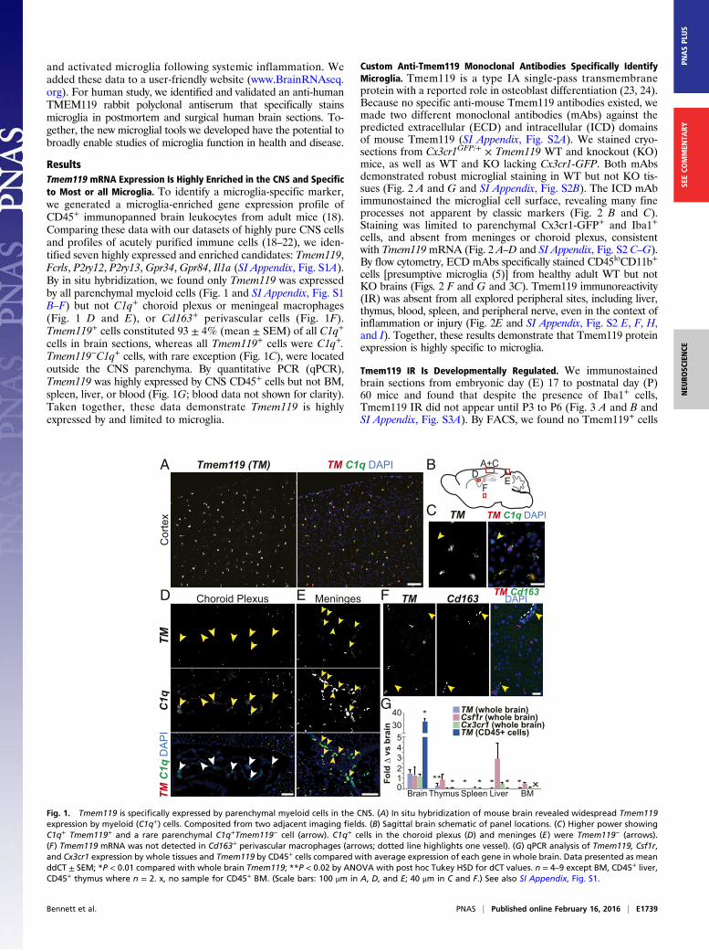
Custom Anti-Tmem119 Monoclonal Antibodies Specifically IdentifyMicroglia. Tmem119 is a type IA single-pass transmembraneprotein with a reported role in osteoblast differentiation (23, 24).Because no specific anti-mouse Tmem119 antibodies existed, wemade two different monoclonal antibodies (mAbs) against thepredicted extracellular (ECD) and intracellular (ICD) domainsof mouse Tmem119 (SI Appendix, Fig. S2A). We stained cryo-sections from Cx3cr1GFP/+ × Tmem119 WT and knockout (KO)mice, as well as WT and KO lacking Cx3cr1-GFP. Both mAbsdemonstrated robust microglial staining in WT but not KO tis-sues (Fig. 2 A and G and SI Appendix, Fig. S2B). The ICD mAbimmunostained the microglial cell surface, revealing many fineprocesses not apparent by classic markers (Fig. 2 B and C).Staining was limited to parenchymal Cx3cr1-GFP+ and Iba1+
cells, and absent from meninges or choroid plexus, consistentwith Tmem119mRNA (Fig. 2 A–D and SI Appendix, Fig. S2 C–G).By flow cytometry, ECD mAbs specifically stained CD45loCD11b+
cells [presumptive microglia (5)] from healthy adult WT but notKO brains (Figs. 2 F and G and 3C). Tmem119 immunoreactivity(IR) was absent from all explored peripheral sites, including liver,thymus, blood, spleen, and peripheral nerve, even in the context ofinflammation or injury (Fig. 2E and SI Appendix, Fig. S2 E, F, H,and I). Together, these results demonstrate that Tmem119 proteinexpression is highly specific to microglia.
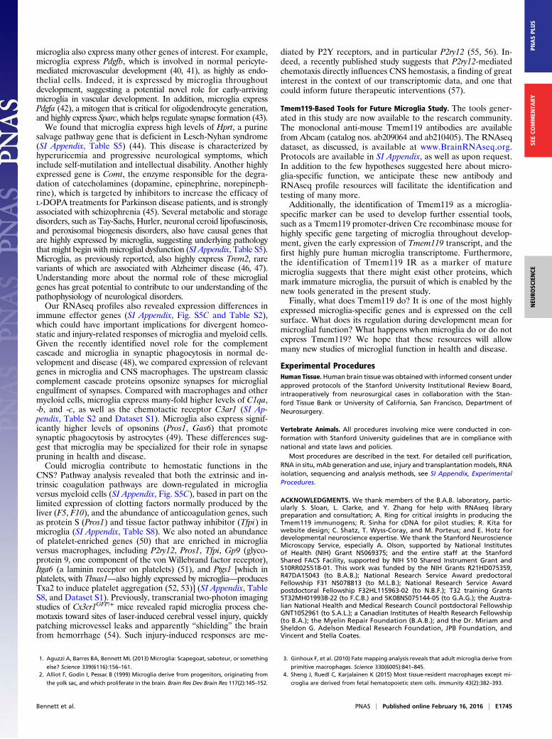
Tmem119 IR Is Developmentally Regulated. We immunostainedbrain sections from embryonic day (E) 17 to postnatal day (P)60 mice and found that despite the presence of Iba1+ cells,Tmem119 IR did not appear until P3 to P6 (Fig. 3 A and B andSI Appendix, Fig. S3A). By FACS, we found no Tmem119+ cells
*
x
G
Fold
∆ v
s br
ain
40305
34
12
0Brain Thymus BMSpleen Liver
TM (CD45+ cells)
TM (whole brain)Csf1r (whole brain)Cx3cr1 (whole brain)
** * * * * * *
A+CD
EF
TMC
1q
Choroid Plexus
TM C
1q D
AP
I
TM C1q DAPI
Cor
tex
Tmem119 (TM)
Cd163TM
A
Meninges
B
TM TM C1q DAPI
TM Cd163DAPI
C
D E F
Fig. 1. Tmem119 is specifically expressed by parenchymal myeloid cells in the CNS. (A) In situ hybridization of mouse brain revealed widespread Tmem119expression by myeloid (C1q+) cells. Composited from two adjacent imaging fields. (B) Sagittal brain schematic of panel locations. (C) Higher power showingC1q+ Tmem119+ and a rare parenchymal C1q+Tmem119− cell (arrow). C1q+ cells in the choroid plexus (D) and meninges (E) were Tmem119− (arrows).(F) Tmem119 mRNA was not detected in Cd163+ perivascular macrophages (arrows; dotted line highlights one vessel). (G) qPCR analysis of Tmem119, Csf1r,and Cx3cr1 expression by whole tissues and Tmem119 by CD45+ cells compared with average expression of each gene in whole brain. Data presented as meanddCT ± SEM; *P < 0.01 compared with whole brain Tmem119; **P < 0.02 by ANOVA with post hoc Tukey HSD for dCT values. n = 4–9 except BM, CD45+ liver,CD45+ thymus where n = 2. x, no sample for CD45+ BM. (Scale bars: 100 μm in A, D, and E; 40 μm in C and F.) See also SI Appendix, Fig. S1.
Bennett et al. PNAS | Published online February 16, 2016 | E1739
NEU
ROSC
IENCE
PNASPL
US
SEECO
MMEN
TARY

at E17, whereas by P7 ∼25% of CD45loCD11b+ cells wereTmem119+ (Fig. 3C). Between P10 and P14, the number ofTmem119+ microglia increased to adult (P60) levels (P60: 98.1 ±0.6%, mean ± SEM). We found no mean fluorescence intensity(MFI) difference between young and adult Tmem119+ microglia,suggesting that once IR is detected, Tmem119 protein is expressedat adult levels. Together, these studies demonstrate that by P14 allmicroglia are Tmem119+.
Tmem119 IR Distinguishes Microglia from Resident and InfiltratingMacrophages After CNS Inflammation and Injury. To assess theutility of Tmem119 as a stable microglia marker, we selected threemouse models of injury and disease: sciatic nerve injury-inducedmicroglial activation, lipopolysaccharide (LPS)-induced systemic
inflammation, and optic nerve crush injury. Despite significantlyincreased Iba1+ IR, we noted no loss of Tmem119+ microgliaproximal to the dorsal root entry zone 4 d postsciatic nerve crushinjury (SI Appendix, Fig. S2 E and F). For systemic inflammation-induced microglia activation, we injected adult WT mice withPBS or LPS (5 mg/kg, i.p.) (25), and analyzed two to three mice pergroup at 1 d and 3 d postinjection. As expected, LPS caused in-creased Iba1 IR and process hypertrophy (Fig. 3D and SI Appendix,Fig. S3B). In addition, all Iba1+ parenchymal cells remainedTmem119+ and staining intensity did not qualitatively decrease ateither time point (Fig. 3D) (for 3 d). Separately, we quantifiedTmem119 IR by FACS 1 d after LPS or PBS. We found no differ-ence in the number or MFI of Tmem119+ cells between groups (Fig.3C) (P = 0.39 for MFI, pairwise t test with Bonferroni correction).
TM CX DAPI
xetroC T
W
Tmem119 (TM) Cx3cr1-GFP (CX)
3D re
nder
ing
TM CX TM CX
CD11b BV421
CD
45 P
E-C
y7
-103
103 104 1050
0
103
104
105
Live cellsTM+ cells
Live
r
Iba1CXTMTM DAPICX Iba1
TM CX TMCX DAPI
Hig
h P
ower
Epi
xetr oC
OK
B C
D E
A
Cho
roid
Ple
xus
TM Iba1TM Iba1DAPI
G
Tmem119 FITC
WildtypeKnockout
-103 103 104 1050
Live
cel
ls (C
ount
)
CD11b BV421
CD
45 P
E-C
y7
-103
103 104 1050
0
103
104
105
F
MacrophagesLymphocytes
Microglia
Fig. 2. Monoclonal antibodies reveal microglia-specific Tmem119 IR. (A) ICD Tmem119 mAbs stained all Cx3cr1-GFP+ cells except in meninges (arrow andInset) in Tmem119 WT but not KO brain. [Scale bars, 100 μm in A (50 μm in Inset).] Tmem119 protein localized to microglia cell surface, by epifluorescence (B)and confocal microscopy (C). (Scale bars, 10 μm in B, 7 μm in C.) (D) Tmem119 IR is limited to myeloid cells in CNS parenchyma and was not detected in choroidplexus (outlined with dotted line) Iba1+ macrophages or liver (E). (Scale bars, 50 μm in D and E.) (F) FACS plot schema showing cell populations revealed byCD11b and CD45 expression at P60 (Left). Tmem119 (TM+) is restricted to CD45loCD11b+ microglia (blue cell population, Right). (G) ECD mAbs are specific, asrevealed by staining in WT but not KO brains (G). See also SI Appendix, Fig. S2.
E1740 | www.pnas.org/cgi/doi/10.1073/pnas.1525528113 Bennett et al.

Finally, we investigated whether Tmem119 distinguishes micro-glia from infiltrating macrophages following optic nerve crush(ONC), a well-established CNS traumatic injury model withmonocyte influx and local blood–brain barrier disruption (26,27). We performed unilateral retro-orbital ONCs in 30 and 120 dCCR2RFP/+ mice, which express red fluorescent protein (RFP)only in infiltrating monocytes (n = 3, each) (13). We harvestednerves 7 d post-ONC, and processed sections with Tmem119ICD and anti-Iba1 antibodies. At the crush site, some Iba1+ cellswere Tmem119− (SI Appendix, Fig. S3D, “x” marks). Iba1+
Tmem119− cells, with rare exception (SI Appendix, Fig. S3D,asterisks), were RFP+, indicating these cells infiltrated from theperiphery. Although many Iba1+ cells were Tmem119+, these wereRFP−, demonstrating that Tmem119 labels resident optic nervemicroglia and not infiltrating macrophages. Perhaps most notably,RFP+ cells were never Tmem119+ (Fig. 3E and SI Appendix, Fig.S3 D and E), suggesting that even after nerve injury, Tmem119 is astable marker of resident microglia and not infiltrating macro-phages. In summary, in peripheral injury, systemic inflammation(LPS) and traumatic CNS injury (ONC), Tmem119 specificallylabeled only resident microglia, allowing for the visualization ofmicroglia after inflammation and injury and providing a cleardistinction between resident and infiltrating myeloid cells.
BM-Derived Cells in the Adult CNS Do Not Express Tmem119.Microglia normally arise from yolk-sac progenitors (3). Aftersome forms of brain injury, BM macrophages can take residencein the brain (8). Because of the lack of markers to distinguishmicroglia from macrophages, it was unclear if engrafted BMmacrophages transform into microglia. We performed a modified
BM transplant conditioning regimen with GFP+ donor BM tobolster CNS engraftment (SI Appendix, Experimental Procedures)We euthanized mice 3 and 6 mo (n = 2 and 3 animals, respectively,for conditioned; n = 1 and 1 for naïve) after transplant and ex-amined if engrafted BM cells in the brain were Tmem119+. Atboth time points, we observed GFP+ cells along the meninges, inblood vessels, and in parenchyma of conditioned but not un-conditioned mice. We noted more GFP+ cells with complex,ramified morphology in the CNS parenchyma at 6 vs. 3 mo, al-though regionally, engraftment rates were never >15% of Iba1+
cells. All GFP+ cells were Iba1+ (SI Appendix, Fig. S3F). At bothtime points, however, we observed no or extremely lowTmem119 IR in GFP+ cells, despite robust Tmem119 IR ofGFP−Iba1+ cells (Fig. 3F and SI Appendix, Fig. S3 F and G).These data indicate that the adult CNS does not induceTmem119 IR in BM cells and that engrafted BM myeloid cellsretain their nonmicroglial identity, even after 6 mo in the CNS.
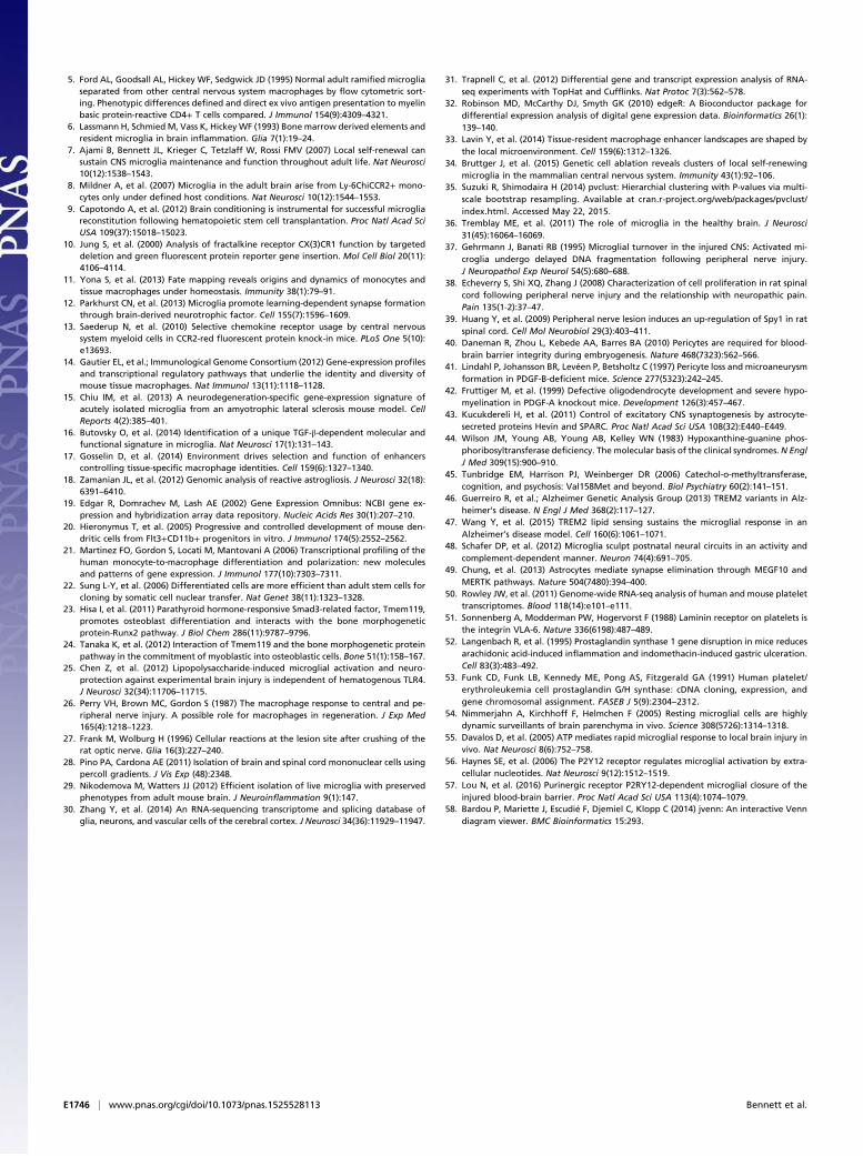
Elucidation of Developing, Adult, and Activated MicroglialTranscriptomes from Tmem119-Purified Microglia. Given the speci-ficity and developmental regulation of Tmem119 in microglia,we wanted to generate a highly pure RNAseq profiling resourceto better understand microglial development, maturation, andactivation. We modified existing microglia isolation protocols(28, 29) to formulate a method that resulted in the most non-activated and pure microglia profiles possible (Fig. 4A). Wedounce-homogenized brains, performed MACS-based myelindepletion, and sorted cells with Tmem119 ECD mAbs. Wedouble-sorted to obtain >99% purity as assessed by flowcytometry and RNAseq expression of cell-type–specific markers
Location of B,except for
P3-6 6-9 9-12
P6 P6* P9 P12 P60
TMIb
a1D
AP
I
A B
*
**
n.s. 100806040200%
Mic
rogl
iaTM
+
E17 P7
P10
P14
P21
PB
S-P
60LP
S-P
60
*
Age
TM Donor cellsTM DAPI
Donor CellsD TM Iba1 TM Iba1 DAPI
PB
S
F
Tmem119+(TM+) cell debut
*
*
TM
C E
Opt
ic n
erve
crus
h si
te (7
d)
Auto DAPIT/C/D-AutoT/C/D
CCR2RFPTM
LPS
Fig. 3. Tmem119 is a developmentally regulated but stable microglia marker. (A) Schematic showing timing of Tmem119 IR in microglia. (Scale bars, 50 μm.)(B) Tmem119 and Iba1 IR in brain sections from different ages (maximum intensity projection, MIP). (C) Percent Tmem119+ cells by age and post-LPStreatment, using ECD mAb. Bars show mean± SEM *P < 0.01 P14–P60; **P < 0.01 all ages; n.s., no significant differences by ANOVA with Tukey HSD. n = 2experiments of two animals (P10 and older) or one litter (under P10) for each age/condition, except for P7 (n = 3), P21 (n = 3), P60 (n = 8, 3 PBS injected pooledwith naïve). (D) Tmem119 and Iba1 IHC 3 d post-PBS or LPS reveals no gross reduction in Tmem119 IR (MIP). (Scale bars, 20 μm.) (E) Representative MIP of opticnerve crush site in 120 d CCR2-RFP+/− mouse. Tmem119 (T) IR (white arrows) and CCR2-RFP (C, yellow arrows) detected at the crush site. There are noTmem119+RFP+ cells. Autofluorescence channel (auto) is digitally subtracted from merge (T/C/D) to differentiate microglia (green) from infiltrating cells (red)in debris-filled crush site. (Scale bars, 25 μm.) (F) GFP+ donor BM cells in the CNS 6 mo posttransplant are Tmem119− in the parenchyma (yellow) and out(white) despite the presence of Tmem119+ microglia. (Scale bars, 20 μm.) See also SI Appendix, Fig. S3.
Bennett et al. PNAS | Published online February 16, 2016 | E1741
NEU
ROSC
IENCE
PNASPL
US
SEECO
MMEN
TARY

(Fig. 2F and SI Appendix, Fig. S4A). We generated two to threereplicates for the following microglia samples: E17 (CD45lo
CD11b+Tmem119−), P7− (CD45loCD11b+Tmem119−), P7+
(Tmem119+), P14 (Tmem119+), P21 (Tmem119+), and P60(Tmem119+). We also collected Tmem119+ cells from P60mice 24 h after 5 mg/kg, i.p. LPS (n = 3). We generated tworeplicates each of P60 and P21 nonmicroglia myeloid cells(CD45hiCD11b+Tmem119−), one sample each for myeloid cellsfrom E17 brain, E17 liver, P7 liver (hereafter “myeloid cells” forclarity) and whole brain for each age (n = 1 for E17–P21, n = 2 forP60) (Fig. 4B). We generated cDNA libraries and performedRNAsEq. (30), obtaining 27.6 ± 7.1 million paired-end 75-bp readsper replicate, and mapped and analyzed these data using the Tux-edo suite of tools (31) and edgeR (32) (see SI Appendix for details).The full Cufflinks-generated fragments per kilobase per mil-
lion (FPKM) dataset and edgeR differential expression analysesare available as Dataset S1, and on a custom website to view incomparison with our laboratory’s other CNS cell type datasets:www.BrainRNAseq.org. Raw sequencing files are available fromNational Center for Biotechnology Information (NCBI)BioProject (accession no. PRJNA30727).
RNAseq Profiles Quality and Purity: Lower Expression of ActivationMarker Genes. We assessed the quality and purity of our RNAseqprofiles by mapping quality and the expression of cell-type–specificand activation-associated genes. We mapped >58% of reads for
all, and >70% of reads for most (n = 30) samples. As expected,myeloid-specific genes were highly enriched, with very lowexpression of other cell-type–specific genes (SI Appendix, Fig.S4A). These results reflect purity and that Tmem119 IR is suf-ficient to isolate microglia by P14. We calculated percentileranks for several canonical activation genes for our naïve andLPS-stimulated samples, as well as five published datasets (15,17, 30, 33, 34); these datasets were generated using CD45 orCx3cr1 rather than a specific microglia marker, such asTmem119, and used enzymatic digestion or Percoll for dissociationand myelin depletion. Compared with published profiles, canonicalactivation marker expression (Tnf, Il1b, Nfkb2) was significantlyreduced in our naïve microglia, providing evidence that our pro-cedure limits microglial activation (SI Appendix, Fig. S4B).
Microglial Maturation Occurs by P14 and Correlates with Tmem119Gene Expression. The precise onset of Tmem119 IR made uswonder about differences between microglia before and afterP14. First, we compared microglia, myeloid cell, and whole-brainRNA expression profiles and found that microglia were highlysimilar throughout development, with more variability betweenbiological replicates at P60 (Fig. 4B). We clustered the averagedsamples (35) and identified two distinct branches for microgliaand CNS myeloid cells (SI Appendix, Fig. S4 C and D), high-lighting differences between these cell types.
A
P21P14
E17P7+8
887
175
04
2623
576
30
B
Dissociatebrain with dounce
All on ice or at 4°C
Removebrain
Filter70µm
Spin30s
10k xg
+anti-MyelinMACS abs
ResuspendMACS buffer
+DNAse+RNAse inhibitor
Apply toMACS column
Stain
+FACS abs& viability dye
FACSinto Trizol LS
Time: 0 hrs Time: 4 hrs
•TM+•TM- CD45hi•TM- CD45lo
P14to P7
4 109 176103
61024
P21to P7
P60to P7
23
6
29
Spe
arm
an C
orre
latio
n
0.8
0.0
1.0
0.2
P60LPS
P7
P60
P21
P14P7 TM+ P7 TM-
E17 TM-
P60 P21E17SZ CD45+
E17
Bra
inLi
vP60 P21P14E17P7
P60
LPS P7
P60
P21
P14
P7
TM+
P7
TM-
E17
TM
-
MICROGLIA
P60
P21E17
SZ
CD
45+
E17
Brain Liv
P60
P21
P14
E17 P
7
WHOLEBRAIN
TM-MYELOID
D
C
MIC
RO
GLI
AW
HO
LEB
RA
INTM
-M
YE
LOID
Fig. 4. Isolation and RNAseq profiling of microglia using Tmem119 IR. (A) Schematic showing streamlined microglia isolation procedure for minimizingbaseline microglia reactivity and death. Abbreviations: abs, antibodies; CV, column volumes; TM, Tmem119. (B) Heatmap of Spearman correlation betweenindividual microglia, myeloid, and whole-tissue RNAseq replicates for genes expressed FPKM > 5. (C) Four-way diagram demonstrating top 100 expressedgenes by microglia (Tmem119+ except for E17) at each age. (D) Venn diagram showing number of differentially expressed genes between P7 Tmem119+ andmature microglia (P14, P21, P60). Black numbers outside diagram represent the few differentially expressed genes between these time points. Diagramsadapted from jVenn (58). See also SI Appendix, Fig. S4 and Dataset S1.
E1742 | www.pnas.org/cgi/doi/10.1073/pnas.1525528113 Bennett et al.

Microglia clustered by age with relatively small heights betweenbranches, indicative of their highly correlated gene expression (SIAppendix, Fig. S4C). Indeed, 57 of the 100 most highly expressedgenes were shared from E17–P21 (Fig. 4C). These include knownmicroglia-enriched genes, such as Cx3cr1, P2ry12, Csf1r, and Fcrls.After P14, few genes were differentially expressed (Fig. 4D),whereas most differences between P14, P21, and P60, each com-pared with P7 Tmem119+ microglia, were shared. In contrast toyounger ages, most differentially expressed genes at and after P14were lowly expressed at all ages (FPKM < 10) or a consequence oflow-level synaptic contamination in some P60 replicates (Gad2,Snap25, Oprm1, Vsnl1, Pclo, Bdnf), evidenced by RNAseq reads insome but not all replicates. Given the small number of gene ex-pression differences between microglia over P14, we concludedthat microglia mature by P14.Despite a lack of Tmem119 IR in 100% of E17 and 75% of
P7 microglia (Fig. 3C), our RNAseq profiles revealed robustTmem119 expression at these ages, with an FPKM of 168 and390, respectively (Fig. 5A and Dataset S1). By P14, Tmem119expression increased 6.8-fold to adult levels. Surprisingly, therewas no difference in Tmem119 expression between P7+ and P7−
microglia, possibly suggesting an uncoupling of transcription andtranslation and that Tmem119-driven gene expression could be arobust microglial marker before IR.
With Maturity, Microglia-Enriched Gene Expression Increases, WhileProliferation Decreases. Although microglial gene expression ishighly correlated throughout development, we were interested ingenetic changes that may underlie development-specific functions.First, we examined the top up- and down-regulated genes betweenE17 and “adult” (P21 and P60) microglia (SI Appendix, Table S1).A cassette of microglia-enriched genes were up-regulated,including Tmem119, P2ry13, and Olfml3. We generated a list ofgenes expressed by Tmem119+microglia versus Tmem119−myeloidcells and found that 37 of 100 top microglia-enriched genes are up-regulated from E17 to adulthood (Fig. 5A and SI Appendix, TableS2). We validated a selection of these genes by microfluidics-basedqPCR and in situ (SI Appendix, Fig. S5 E, G, and H). Although notall microglia-enriched genes demonstrated this trend, many reachedtheir mature levels by P14. Given our interest in Tmem119expression patterns and microglial maturity, we performedunsupervised hierarchical clustering of our microglial and pub-lished RNAseq datasets (30) to find other similarly behavedgenes (SI Appendix, Table S3 and Dataset S1, “clusterID” col-umn). The gene cluster of Tmem119 not only contained manymicroglia-enriched genes, but a number of genes associated withleukocyte development, differentiation, and homeostasis: Blnk,Inpp5d, Rgs10, Mef2c, Cysltr1, Orai1, Ifngr1, Pnp, Casp8, andTgfbr1 (SI Appendix, Table S3, Ingenuity Pathway Analysis).Cell cycle-associated genes were among the top down-regulated
genes we identified during microglia development (Fig. 5B and SIAppendix, Fig. S5A and Table S1). Given the inverse correlationbetween cell-cycle gene expression and the postnatal appearance ofTmem119 IR, we asked if Tmem119 IR, as a proxy for microglialmaturity, corresponded with the end of microglia proliferation. Westained brain cryosections from E14, E17, P6, P12, and P60 mice(n = 2 each) with anti-Ki67, Tmem119 ICD, and Iba1 antibodies(Fig. 5C). We quantified the relative percentages of proliferating(Ki67+) and nonproliferating (Ki67−) Iba1+ cells and theirTmem119 IR (Fig. 5D). Proliferating Iba1+ cells were seen at E14and E17, but only rarely >P6. No proliferating Tmem119+Iba1+
cells were detected at any age, and <5% of Iba1+ cells at P6 wereTmem119+. By P12, Tmem119−Iba1+ cells were still present, de-spite the end of microglial proliferation more than a week ear-lier. Together, these data show that although immature microgliabefore P6 are more proliferative in vivo, Tmem119 IR is not di-rectly related to the termination of proliferation.
Other Analyses: Microglial Activation, Ligands/Receptors/TranscriptionFactors, and Platelets. Given our low baseline activation ofTmem119-sorted microglia, we wanted to determine the tran-scriptome of “classically” activated microglia. As discussed, wealso sequenced microglia 24 h post-LPS injection. LPS-treatedmicroglia up-regulated the expression of many myeloid cell in-flammation-associated transcripts, including Lcn2, Ccl3, Cxcl10,Ccl5, Il1b, Tnf, and Tlr2 (SI Appendix, Table S4). Several microglia-enriched genes were also down-regulated, including P2ry12 andTmem119, Fcrls, Olfml3, Ltc4s, and Adora3 (SI Appendix, Table S4and Dataset S1). We validated several of these by qPCR (SI Ap-pendix, Fig. S5F). Pathway analysis (Qiagen) revealed increasedToll-like receptor (TLR), vitamin D3-receptor/RXR activation,and acute phase response signaling pathways (SI Appendix, Fig. S5B)between naïve and LPS-treated microglia. Together, these resultssuggest that microglia respond to peripheral LPS with a classic-typeactivation profile and that some microglia-enriched transcripts aredown-regulated. The notion that expression of microglial markersis sensitive to activation state is intriguing, as is the stability ofTmem119 IR despite decreased mRNA expression (Fig. 3 C andD).Among the top genes expressed by E17 versus adult microglia
are several classes of genes associated with myeloid cell activa-tion. Given the attributed functions of these enriched genes, andpublished hypotheses that young microglia, which are highlyphagocytic and less ramified, might be activated (36), we com-pared our young microglia with LPS-treated microglia. Several
FPK
M
100
375
****
Ccr5
***
Fcrls
1250
***
s
100
******
Slc2a5
B
A
1000 **
** **
Olfml3
600
n.d.Itgam
FPK
M
C
300
***
Ltc4s
FPK
M Cx3cr1
**
1500
#
2000
**
P2ry12
***
5
300
**
* *
Plxdc2
1000
FPK
M
0**
* ***
Tmem119
E17
P7-
P7+
P14
P21
P60
MY
P7 (TM+)P7 (TM-) E17 (TM-)
P60P21P14
P60 Myeloid (TM-)
D Maturity
6050403020100
E14 E17 P6P12 P60
Ki6
7+/Ib
a1+
(%)
Proliferation*
n.d.
E14
Cor
tex
Iba1 Ki67 Auto
P60
Cor
tex
E E17 P60 (+LPS)
S100
a8D
API
** ******
Selplg
750
FPK
M
B
E P P P P
Mki67
150
**
*
100
P P
7*
Pcna **
Ccnd1
50
*
**
Iba1 Ki67 TM
E14 E17 P6P12 P60
6040200
80100 *****
TM
+/Ib
a1+
(%)
Fig. 5. Developmental changes in microglial gene expression. Bar plots ofRNAseq data showing FPKM expression of selected microglia-enriched (A)and proliferation marker genes (B) by developing microglia and myeloidcells. Error bars depict mean ± SEM **P < 0.01, *P < 0.05 for values com-pared with each P60, P21, and P14 microglia; #P < 0.01 compared with P60.(C) Representative Ki67, Tmem119, and Iba1 epifluorescence image quan-tified in D, showing Iba1+Ki67+ (white arrowheads), Iba1+only (cyan), andKi67+only (yellow) cells. Auto: autofluorescence. (D) Box-and-whisker plotsdepicting percentage of Iba1+ cells that were Ki67+ (Left) and Tmem119+
(Right). n.d., no difference; *P < 0.01 compared each with P6, P12, and P60;**P < 0.01 compared with P6; ***P < 0.01 compared with P6 and P12; n = 2mice each, quantified four to eight fields from two to three sections. Box/whiskers represent standard values. All P values calculated by one-wayANOVA with Tukey HSD and verified by pairwise t test with Bonferronicorrection. (E) In situ of S100a8 at E17 and P60, the latter 24 h after LPSinjection, revealing bright, S100a8-expressing cells not detected in naïve P60brain (not shown). (Scale bars, 50 μm.) See also SI Appendix, Fig. S5 andTables S2–S8.
Bennett et al. PNAS | Published online February 16, 2016 | E1743
NEU
ROSC
IENCE
PNASPL
US
SEECO
MMEN
TARY

top LPS-induced genes were expressed more highly by E17 thanadult microglia (Fig. 5E for S100a8 in situ, and SI Appendix,Table S4, bold genes). Despite these similarities, we found moredifferences than similarities between young and activatedmicroglia (SI Appendix, Fig. S5D).We also generated a list of known transcription factors, ligands,
and receptors enriched in microglia over CNS myeloid cells, wholebrain, and other CNS cell types (SI Appendix, Table S5). We cu-rated a list of disease-associated genes (SI Appendix, Table S6),transcription factors (SI Appendix, Table S7), and platelet-enrichedgenes highly enriched in microglia (SI Appendix, Table S8).
Tmem119 Is also a Specific Marker of Human Microglia. Tmem119 ishighly conserved, with 73% sequence homology between mouseand humans. To test the specificity of TMEM119 expressionto human microglia, we quickly harvested CD11B+ cells sortedfrom intraoperatively obtained, normal-appearing temporal lobecortical tissue (n = 2; 47 and 8 y), and compared TMEM119expression by qPCR with unpurified whole brain ( n = 2; pooledadult and 45 y) and peripheral blood leukocytes (n = 2; pooledadult and 66 y) (Fig. 6A). We also examined the expression ofCX3CR1 and CD11B as positive controls. TMEM119 expressionwas highly enriched in CD11b+ cells over whole brain, and barelydetectable in peripheral blood leukocytes, despite the presenceof CD11B. We tested several commercially available anti-TMEM119 antibodies on human surgical brain specimens,identifying a rabbit polyclonal anti-human TMEM119 anti-body that stained only CNS parenchymal Iba1+ cells (Fig. 6B).TMEM119 antisera stained the plasma membrane of most or allparenchymal Iba1+ cells (microglia) in both gray and surround-ing white matter from rapidly fixed surgical tissue from multiplepatients (n = 4; ages 8, 47, 51, and 71 y). Despite the sequencehomology between mouse and human Tmem119, neither theanti-mouse mAbs nor the anti-human polyclonal antisera workedto stain microglia in human or mouse, respectively. Together,these findings demonstrate that Tmem119 is a microglia markerin human CNS tissue as well as in mouse.
DiscussionTmem119 Is a Specific and Stable Marker of Microglia in Mouse andHuman. In this study, we show that Tmem119, a transmembraneprotein of unknown function, is a developmentally regulated andhighly specific cell-surface marker of most or all microglia that isnot expressed by macrophages or other immune or neural celltypes. Tmem119 expression is abundant in prenatal microglia butIR—detected by two custom monoclonal antibodies—correlateswith microglial maturity postnatally. These antibodies (Abcam,
catalog nos. ab209064 ICD and ab210405 ECD) stably identifymicroglia, even after injury and inflammation. In addition, weoptimized a method for isolating pure, nonactivated microgliausing these antibodies. Tmem119 is also a specific markerof human microglia, for which we identified a polyclonal anti-body to stain microglia in human brain cryosections (Abcam,catalog no. ab185333). These antibodies provide a powerful toolfor specifically identifying microglia in future studies of the rolesof microglia in CNS health and disease. In addition, the ECDmAb to mouse Tmem119 enabled us to develop a FACS methodto prospectively isolate highly pure microglial cells. These puri-fied cells can be used for biochemical studies, culture, or RNAisolation for gene expression studies.
RNAseq Profiles of Developing Microglia Reveal That MicrogliaMature by P14. Using the Tmem119 mAbs we generated here,we produced, to our knowledge, the first highly pure RNAseqprofiles of microglia during development. These data are publiclyaccessible through www.BrainRNAseq.org. This resource will helpfoster the formation and testing of new hypotheses about micro-glial development and function. Using these profiles, we madeseveral novel observations about microglial function during de-velopment. First, mouse microglia mature in vivo by P14, with fewgenetic differences between P14, P21, and P60 microglia. BothTmem119 IR and microglia-enriched gene expression increasethroughout development until P14, when they stabilize. Theseobservations raise an important question for future studies: whatsignals induce microglial maturation? For example, is microgliamaturation induced at P14 by a systemic endocrine signal or doesCNS brain maturation induce a signal that acts on microglia?In addition, microglial proliferation ceases days before matu-
ration at P14. What is the relationship of microglial proliferationto maturity, and then to activation? In peripheral nerve injury,such as that in SI Appendix, Fig. S2F, microglia proliferate in thespinal cord by 2 d postinjury (37–39). We showed that all Iba1+
cells in spinal cord are Tmem119+ even after injury, suggestingthat mature, Tmem119+ microglia are able to proliferate afterinjury. Are the signals regulating developmental and activation-induced microglial proliferation different? What are the disease-related implications of these different signals?
Microglia Transcriptome Suggests Potential Novel Functions forMicroglia in Health and Disease. Our microglia RNAseq tran-scriptomes revealed the expression of ligands, receptors, anddisease-associated genes, which strongly suggest new microglialfunctions in development and disease. In addition to knownchemokines, Toll-like signaling, and phagocytosis mediators,
TMEM119 Iba1TMEM119 Iba1 DAPI
300
200
25150
250
15
20
10
5
0
CX3CR1 CD11B TMEM119
Fold
Diff
eren
ce (v
. WB
)
CD11B+ WB PBLTissue
qPCR Human
**
*
A B+Auto
Fig. 6. Tmem119 is also highly enriched in and specific to human microglia. (A) Relative expression of TMEM119, CD11B, and CX3CR1 mRNA in humanperipheral blood leukocytes (PBL), whole brain (WB), and CD11B+ brain cells *P < 0.01 by ANOVA with Tukey HSD. Error bars show SEM, for n = 2 independentsamples per tissue type. (B) Representative confocal images of 51-y-old normal appearing cortical tissue showing localization of TMEM119 (green) to highlyramified, Iba1+ cells (red) at low (Upper) and high (Lower) magnification. Autofluorescence channel (red) overlays green to highlight nonspecific fluorescencefrom lipofuscin (arrows), which are absent in Lower because of Sudan Black treatment. (Scale bars, 30 μm.)
E1744 | www.pnas.org/cgi/doi/10.1073/pnas.1525528113 Bennett et al.
microglia also express many other genes of interest. For example,microglia express Pdgfb, which is involved in normal pericyte-mediated microvascular development (40, 41), as highly as endo-thelial cells. Indeed, it is expressed by microglia throughoutdevelopment, suggesting a potential novel role for early-arrivingmicroglia in vascular development. In addition, microglia expressPdgfa (42), a mitogen that is critical for oligodendrocyte generation,and highly express Sparc, which helps regulate synapse formation (43).We found that microglia express high levels of Hprt, a purine
salvage pathway gene that is deficient in Lesch-Nyhan syndrome(SI Appendix, Table S5) (44). This disease is characterized byhyperuricemia and progressive neurological symptoms, whichinclude self-mutilation and intellectual disability. Another highlyexpressed gene is Comt, the enzyme responsible for the degra-dation of catecholamines (dopamine, epinephrine, norepineph-rine), which is targeted by inhibitors to increase the efficacy ofL-DOPA treatments for Parkinson disease patients, and is stronglyassociated with schizophrenia (45). Several metabolic and storagedisorders, such as Tay-Sachs, Hurler, neuronal ceroid lipofuscinosis,and peroxisomal biogenesis disorders, also have causal genes thatare highly expressed by microglia, suggesting underlying pathologythat might begin with microglial dysfunction (SI Appendix, Table S5).Microglia, as previously reported, also highly express Trem2, rarevariants of which are associated with Alzheimer disease (46, 47).Understanding more about the normal role of these microglialgenes has great potential to contribute to our understanding of thepathophysiology of neurological disorders.Our RNAseq profiles also revealed expression differences in
immune effector genes (SI Appendix, Fig. S5C and Table S2),which could have important implications for divergent homeo-static and injury-related responses of microglia and myeloid cells.Given the recently identified novel role for the complementcascade and microglia in synaptic phagocytosis in normal de-velopment and disease (48), we compared expression of relevantgenes in microglia and CNS macrophages. The upstream classiccomplement cascade proteins opsonize synapses for microglialengulfment of synapses. Compared with macrophages and othermyeloid cells, microglia express many-fold higher levels of C1qa,-b, and -c, as well as the chemotactic receptor C3ar1 (SI Ap-pendix, Table S2 and Dataset S1). Microglia also express signif-icantly higher levels of opsonins (Pros1, Gas6) that promotesynaptic phagocytosis by astrocytes (49). These differences sug-gest that microglia may be specialized for their role in synapsepruning in health and disease.Could microglia contribute to hemostatic functions in the
CNS? Pathway analysis revealed that both the extrinsic and in-trinsic coagulation pathways are down-regulated in microgliaversus myeloid cells (SI Appendix, Fig. S5C), based in part on thelimited expression of clotting factors normally produced by theliver (F5, F10), and the abundance of anticoagulation genes, suchas protein S (Pros1) and tissue factor pathway inhibitor (Tfpi) inmicroglia (SI Appendix, Table S8). We also noted an abundanceof platelet-enriched genes (50) that are enriched in microgliaversus macrophages, including P2ry12, Pros1, Tfpi, Gp9 (glyco-protein 9, one component of the von Willebrand factor receptor),Itga6 (a laminin receptor on platelets) (51), and Ptgs1 [which inplatelets, with Tbxas1—also highly expressed by microglia—producesTxa2 to induce platelet aggregation (52, 53)] (SI Appendix, TableS8, and Dataset S1). Previously, transcranial two-photon imagingstudies of Cx3cr1GFP/+ mice revealed rapid microglia process che-motaxis toward sites of laser-induced cerebral vessel injury, quicklypatching microvessel leaks and apparently “shielding” the brainfrom hemorrhage (54). Such injury-induced responses are me-
diated by P2Y receptors, and in particular P2ry12 (55, 56). In-deed, a recently published study suggests that P2ry12-mediatedchemotaxis directly influences CNS hemostasis, a finding of greatinterest in the context of our transcriptomic data, and one thatcould inform future therapeutic interventions (57).
Tmem119-Based Tools for Future Microglia Study. The tools gener-ated in this study are now available to the research community.The monoclonal anti-mouse Tmem119 antibodies are availablefrom Abcam (catalog nos. ab209064 and ab210405). The RNAseqdataset, as discussed, is available at www.BrainRNAseq.org.Protocols are available in SI Appendix, as well as upon request.In addition to the few hypotheses suggested here about micro-glia-specific function, we anticipate these new antibody andRNAseq profile resources will facilitate the identification andtesting of many more.Additionally, the identification of Tmem119 as a microglia-
specific marker can be used to develop further essential tools,such as a Tmem119 promoter-driven Cre recombinase mouse forhighly specific gene targeting of microglia throughout develop-ment, given the early expression of Tmem119 transcript, and thefirst highly pure human microglia transcriptome. Furthermore,the identification of Tmem119 IR as a marker of maturemicroglia suggests that there might exist other proteins, whichmark immature microglia, the pursuit of which is enabled by thenew tools generated in the present study.Finally, what does Tmem119 do? It is one of the most highly
expressed microglia-specific genes and is expressed on the cellsurface. What does its regulation during development mean formicroglial function? What happens when microglia do or do notexpress Tmem119? We hope that these resources will allowmany new studies of microglial function in health and disease.
Experimental ProceduresHuman Tissue.Human brain tissue was obtained with informed consent underapproved protocols of the Stanford University Institutional Review Board,intraoperatively from neurosurgical cases in collaboration with the Stan-ford Tissue Bank or University of California, San Francisco, Department ofNeurosurgery.
Vertebrate Animals. All procedures involving mice were conducted in con-formation with Stanford University guidelines that are in compliance withnational and state laws and policies.
Most procedures are described in the text. For detailed cell purification,RNA in situ, mAb generation and use, injury and transplantationmodels, RNAisolation, sequencing and analysis methods, see SI Appendix, ExperimentalProcedures.
ACKNOWLEDGMENTS. We thank members of the B.A.B. laboratory, partic-ularly S. Sloan, L. Clarke, and Y. Zhang for help with RNAseq librarypreparation and consultation; A. Ring for critical insights in producing theTmem119 immunogens; R. Sinha for cDNA for pilot studies; R. Kita forwebsite design; C. Shatz, T. Wyss-Coray, and M. Porteus; and E. Hotz fordevelopmental neuroscience expertise. We thank the Stanford NeuroscienceMicroscopy Service, especially A. Olson, supported by National Institutesof Health (NIH) Grant NS069375; and the entire staff at the StanfordShared FACS Facility, supported by NIH S10 Shared Instrument Grant andS10RR025518-01. This work was funded by the NIH Grants R21HD075359,R47DA15043 (to B.A.B.); National Research Service Award predoctoralFellowship F31 NS078813 (to M.L.B.); National Research Service Awardpostdoctoral Fellowship F32HL115963-02 (to N.B.F.); T32 training Grants5T32MH019938-22 (to F.C.B.) and 5K08NS075144-05 (to G.A.G.); the Austra-lian National Health and Medical Research Council postdoctoral FellowshipGNT1052961 (to S.A.L.); a Canadian Institutes of Health Research Fellowship(to B.A.); the Myelin Repair Foundation (B.A.B.); and the Dr. Miriam andSheldon G. Adelson Medical Research Foundation, JPB Foundation, andVincent and Stella Coates.
1. Aguzzi A, Barres BA, Bennett ML (2013) Microglia: Scapegoat, saboteur, or something
else? Science 339(6116):156–161.2. Alliot F, Godin I, Pessac B (1999) Microglia derive from progenitors, originating from
the yolk sac, and which proliferate in the brain. Brain Res Dev Brain Res 117(2):145–152.
3. Ginhoux F, et al. (2010) Fate mapping analysis reveals that adult microglia derive from
primitive macrophages. Science 330(6005):841–845.4. Sheng J, Ruedl C, Karjalainen K (2015) Most tissue-resident macrophages except mi-
croglia are derived from fetal hematopoietic stem cells. Immunity 43(2):382–393.
Bennett et al. PNAS | Published online February 16, 2016 | E1745
NEU
ROSC
IENCE
PNASPL
US
SEECO
MMEN
TARY
5. Ford AL, Goodsall AL, Hickey WF, Sedgwick JD (1995) Normal adult ramified microgliaseparated from other central nervous system macrophages by flow cytometric sort-ing. Phenotypic differences defined and direct ex vivo antigen presentation to myelinbasic protein-reactive CD4+ T cells compared. J Immunol 154(9):4309–4321.
6. Lassmann H, Schmied M, Vass K, HickeyWF (1993) Bone marrow derived elements andresident microglia in brain inflammation. Glia 7(1):19–24.
7. Ajami B, Bennett JL, Krieger C, Tetzlaff W, Rossi FMV (2007) Local self-renewal cansustain CNS microglia maintenance and function throughout adult life. Nat Neurosci10(12):1538–1543.
8. Mildner A, et al. (2007) Microglia in the adult brain arise from Ly-6ChiCCR2+ mono-cytes only under defined host conditions. Nat Neurosci 10(12):1544–1553.
9. Capotondo A, et al. (2012) Brain conditioning is instrumental for successful microgliareconstitution following hematopoietic stem cell transplantation. Proc Natl Acad SciUSA 109(37):15018–15023.
10. Jung S, et al. (2000) Analysis of fractalkine receptor CX(3)CR1 function by targeteddeletion and green fluorescent protein reporter gene insertion. Mol Cell Biol 20(11):4106–4114.
11. Yona S, et al. (2013) Fate mapping reveals origins and dynamics of monocytes andtissue macrophages under homeostasis. Immunity 38(1):79–91.
12. Parkhurst CN, et al. (2013) Microglia promote learning-dependent synapse formationthrough brain-derived neurotrophic factor. Cell 155(7):1596–1609.
13. Saederup N, et al. (2010) Selective chemokine receptor usage by central nervoussystem myeloid cells in CCR2-red fluorescent protein knock-in mice. PLoS One 5(10):e13693.
14. Gautier EL, et al.; Immunological Genome Consortium (2012) Gene-expression profilesand transcriptional regulatory pathways that underlie the identity and diversity ofmouse tissue macrophages. Nat Immunol 13(11):1118–1128.
15. Chiu IM, et al. (2013) A neurodegeneration-specific gene-expression signature ofacutely isolated microglia from an amyotrophic lateral sclerosis mouse model. CellReports 4(2):385–401.
16. Butovsky O, et al. (2014) Identification of a unique TGF-β-dependent molecular andfunctional signature in microglia. Nat Neurosci 17(1):131–143.
17. Gosselin D, et al. (2014) Environment drives selection and function of enhancerscontrolling tissue-specific macrophage identities. Cell 159(6):1327–1340.
18. Zamanian JL, et al. (2012) Genomic analysis of reactive astrogliosis. J Neurosci 32(18):6391–6410.
19. Edgar R, Domrachev M, Lash AE (2002) Gene Expression Omnibus: NCBI gene ex-pression and hybridization array data repository. Nucleic Acids Res 30(1):207–210.
20. Hieronymus T, et al. (2005) Progressive and controlled development of mouse den-dritic cells from Flt3+CD11b+ progenitors in vitro. J Immunol 174(5):2552–2562.
21. Martinez FO, Gordon S, Locati M, Mantovani A (2006) Transcriptional profiling of thehuman monocyte-to-macrophage differentiation and polarization: new moleculesand patterns of gene expression. J Immunol 177(10):7303–7311.
22. Sung L-Y, et al. (2006) Differentiated cells are more efficient than adult stem cells forcloning by somatic cell nuclear transfer. Nat Genet 38(11):1323–1328.
23. Hisa I, et al. (2011) Parathyroid hormone-responsive Smad3-related factor, Tmem119,promotes osteoblast differentiation and interacts with the bone morphogeneticprotein-Runx2 pathway. J Biol Chem 286(11):9787–9796.
24. Tanaka K, et al. (2012) Interaction of Tmem119 and the bone morphogenetic proteinpathway in the commitment of myoblastic into osteoblastic cells. Bone 51(1):158–167.
25. Chen Z, et al. (2012) Lipopolysaccharide-induced microglial activation and neuro-protection against experimental brain injury is independent of hematogenous TLR4.J Neurosci 32(34):11706–11715.
26. Perry VH, Brown MC, Gordon S (1987) The macrophage response to central and pe-ripheral nerve injury. A possible role for macrophages in regeneration. J Exp Med165(4):1218–1223.
27. Frank M, Wolburg H (1996) Cellular reactions at the lesion site after crushing of therat optic nerve. Glia 16(3):227–240.
28. Pino PA, Cardona AE (2011) Isolation of brain and spinal cord mononuclear cells usingpercoll gradients. J Vis Exp (48):2348.
29. Nikodemova M, Watters JJ (2012) Efficient isolation of live microglia with preservedphenotypes from adult mouse brain. J Neuroinflammation 9(1):147.
30. Zhang Y, et al. (2014) An RNA-sequencing transcriptome and splicing database ofglia, neurons, and vascular cells of the cerebral cortex. J Neurosci 34(36):11929–11947.
31. Trapnell C, et al. (2012) Differential gene and transcript expression analysis of RNA-seq experiments with TopHat and Cufflinks. Nat Protoc 7(3):562–578.
32. Robinson MD, McCarthy DJ, Smyth GK (2010) edgeR: A Bioconductor package fordifferential expression analysis of digital gene expression data. Bioinformatics 26(1):139–140.
33. Lavin Y, et al. (2014) Tissue-resident macrophage enhancer landscapes are shaped bythe local microenvironment. Cell 159(6):1312–1326.
34. Bruttger J, et al. (2015) Genetic cell ablation reveals clusters of local self-renewingmicroglia in the mammalian central nervous system. Immunity 43(1):92–106.
35. Suzuki R, Shimodaira H (2014) pvclust: Hierarchial clustering with P-values via multi-scale bootstrap resampling. Available at cran.r-project.org/web/packages/pvclust/index.html. Accessed May 22, 2015.
36. Tremblay ME, et al. (2011) The role of microglia in the healthy brain. J Neurosci31(45):16064–16069.
37. Gehrmann J, Banati RB (1995) Microglial turnover in the injured CNS: Activated mi-croglia undergo delayed DNA fragmentation following peripheral nerve injury.J Neuropathol Exp Neurol 54(5):680–688.
38. Echeverry S, Shi XQ, Zhang J (2008) Characterization of cell proliferation in rat spinalcord following peripheral nerve injury and the relationship with neuropathic pain.Pain 135(1-2):37–47.
39. Huang Y, et al. (2009) Peripheral nerve lesion induces an up-regulation of Spy1 in ratspinal cord. Cell Mol Neurobiol 29(3):403–411.
40. Daneman R, Zhou L, Kebede AA, Barres BA (2010) Pericytes are required for blood-brain barrier integrity during embryogenesis. Nature 468(7323):562–566.
41. Lindahl P, Johansson BR, Levéen P, Betsholtz C (1997) Pericyte loss and microaneurysmformation in PDGF-B-deficient mice. Science 277(5323):242–245.
42. Fruttiger M, et al. (1999) Defective oligodendrocyte development and severe hypo-myelination in PDGF-A knockout mice. Development 126(3):457–467.
43. Kucukdereli H, et al. (2011) Control of excitatory CNS synaptogenesis by astrocyte-secreted proteins Hevin and SPARC. Proc Natl Acad Sci USA 108(32):E440–E449.
44. Wilson JM, Young AB, Young AB, Kelley WN (1983) Hypoxanthine-guanine phos-phoribosyltransferase deficiency. The molecular basis of the clinical syndromes. N EnglJ Med 309(15):900–910.
45. Tunbridge EM, Harrison PJ, Weinberger DR (2006) Catechol-o-methyltransferase,cognition, and psychosis: Val158Met and beyond. Biol Psychiatry 60(2):141–151.
46. Guerreiro R, et al.; Alzheimer Genetic Analysis Group (2013) TREM2 variants in Alz-heimer’s disease. N Engl J Med 368(2):117–127.
47. Wang Y, et al. (2015) TREM2 lipid sensing sustains the microglial response in anAlzheimer’s disease model. Cell 160(6):1061–1071.
48. Schafer DP, et al. (2012) Microglia sculpt postnatal neural circuits in an activity andcomplement-dependent manner. Neuron 74(4):691–705.
49. Chung, et al. (2013) Astrocytes mediate synapse elimination through MEGF10 andMERTK pathways. Nature 504(7480):394–400.
50. Rowley JW, et al. (2011) Genome-wide RNA-seq analysis of human andmouse platelettranscriptomes. Blood 118(14):e101–e111.
51. Sonnenberg A, Modderman PW, Hogervorst F (1988) Laminin receptor on platelets isthe integrin VLA-6. Nature 336(6198):487–489.
52. Langenbach R, et al. (1995) Prostaglandin synthase 1 gene disruption in mice reducesarachidonic acid-induced inflammation and indomethacin-induced gastric ulceration.Cell 83(3):483–492.
53. Funk CD, Funk LB, Kennedy ME, Pong AS, Fitzgerald GA (1991) Human platelet/erythroleukemia cell prostaglandin G/H synthase: cDNA cloning, expression, andgene chromosomal assignment. FASEB J 5(9):2304–2312.
54. Nimmerjahn A, Kirchhoff F, Helmchen F (2005) Resting microglial cells are highlydynamic surveillants of brain parenchyma in vivo. Science 308(5726):1314–1318.
55. Davalos D, et al. (2005) ATP mediates rapid microglial response to local brain injury invivo. Nat Neurosci 8(6):752–758.
56. Haynes SE, et al. (2006) The P2Y12 receptor regulates microglial activation by extra-cellular nucleotides. Nat Neurosci 9(12):1512–1519.
57. Lou N, et al. (2016) Purinergic receptor P2RY12-dependent microglial closure of theinjured blood-brain barrier. Proc Natl Acad Sci USA 113(4):1074–1079.
58. Bardou P, Mariette J, Escudié F, Djemiel C, Klopp C (2014) jvenn: An interactive Venndiagram viewer. BMC Bioinformatics 15:293.
E1746 | www.pnas.org/cgi/doi/10.1073/pnas.1525528113 Bennett et al.