LC-MS/MS as an enabler for a broader application of microdose ...

Philips MicroDose mammography SI
New spectral benefits, proven low dose

Proven MicroDose mammography, new spectral insights
The foundation of MicroDose SI is Philips proven photon counting technology. In use since 2003 across hundreds of sites worldwide, photon counting eases the traditional trade-off between radiation dose and image quality, enabling users to capture outstanding images with significantly reduced radiation dose.
Spectral information within the MicroDose mammogramMicroDose SI adds a new advancement to this remarkable technology: non-invasive spectral imaging capability that holds promise of providing new insights into breast composition. Philips spectral imaging is unique, because photon counting makes it possible to acquire the spectral data in the same exposure as a regular, low-dose mammogram, and without the use of contrast media. This single-shot, non-invasive approach does not require adjustments to your current workflow, and no additional radiation dose is needed to acquire the spectral data. The first MicroDose SI spectral application, the Breast Density Measurement tool, can immediately make a difference for radiologists looking for an objective, quantitative method to assess breast density and refine risk assessment.
Philips MicroDose mammography SI with single-shot spectral imaging delivers proven dose efficiency, outstanding image quality, and non-invasive spectral applications in one fast and comfortable mammogram. Designed to delight physicians, patients, and technologists, it helps deliver high-quality, efficient, patient-focused care.
Transforming care, togetherPhilips commitment to transforming care together with clinicians is reflected in the MicroDose SI system.
Drive clinical performancePhoton counting and spectral technology are strong platforms for further advancements in mammography. Philips works closely with clinical partners to advance the art and science of mammography, and to explore the potential of spectral imaging.
Enhance Patient Experience Women appreciate the short examination time, the low radiation dose and the curved and warm breast support.
Ensure economic valueMicroDose SI is designed to enable the high throughput required for screening, while delivering excellent patient care.
High confidence, high potential
• Proven: Experience outstanding image quality at a very low X-ray dose
• Objective: Refine your breast density assessment with spectral, volumetric breast density measurement
• Non-invasive: Collect spectral data in a fast and comfortable mammogram
• Distinctive: Gain patients and referrals by offering unique, patient-focused technology
Philips MicroDose SI 3

Diagnostic Scan helps discern fine detailsAn exclusive optional feature of the Philips MicroDose SI, Diagnostic Scan helps discern fine details in spot compression images. Diagnostic Scan uses higher dose on a spot compression area. Then, because of the system’s high resolution, electronic magnification can be used on the review station to make it easier to see fine details at a significantly lower dose than geometric magnification. In addition, Diagnostic Scan does not require a magnification table on the patient support.
Diagnostic scan image of the left breast in MLO view
Experience imaging excellence at a proven low dose
At the heart of the MicroDose SI is photon counting, an amazing approach to breast imaging that recognizes the importance of minimizing radiation exposure in screening applications. MicroDose SI allows for the same low dose as on the previous MicroDose model and for an increase of up to 11% in technical image quality.* MicroDose SI provides industry-leading 50 µm in resolution and has shown great clinical performance, even in dense breasts. Also, MicroDose SI scanning technology offers 100 % pixel warranty by eliminating dead pixels and associated image data loss.
In addition, MicroDose SI features Smart AEC, which continuously adjusts the radiation exposure depending on the density of the breast throughout the scan. This provides correct exposure without the need for scout images.
MicroDose SI is a scanning system that uses direct digital, photon counting technology to provide low radiation dose without compromising image quality.
High dose efficiency, high image quality
MicroDose Mammography delivers high image quality at a low dose through:• High X-ray photon absorption• Immediate photon counting• Elimination of 97 % of scattered radiation• Virtually no electronic noise• 100 % pixel warranty• No ghost images• The smallest pixel size at 50 μm
“The exquisite image quality of the new MicroDose SI has allowed me to go down in dose compared to my MicroDose L30 by more than 30% and still achieve excellent image quality.”
J.-C. Piguet, MD, Groupe 3R, Switzerland
“MicroDose SI has allowed us to significantly reduce the radiation dose and provides excellent image quality. We are delighted by the ease-of-use of the system which has helped us to minimize examination time as well as increase our efficiency, all while providing enhanced patient experience.”
J. Wellwood, Clinical Director, BreastScreen Central, New Zealand
5Philips MicroDose SIPhilips MicroDose SI4 * As measured by the CDMAM phantom for 0,1 mm disk diameter

Isocentric rotation: no
need for height adjustments
between projections
Easy access to rotation
and height adjustment
in four different places
Motorized compression
with foot pedals
Dedicated shortcut
keypad to automatically
control your workflow
X-ray exposure
from the acquisition
workstation
Optional X-ray
exposure foot switch
for more flexibility
Dedicated compression
paddles to cover
different breast sizes
Deliver speed and comfort
A fast mammography exam benefits everyone involved. MicroDose SI offers a simplified and efficient workflow that contributes to high patient throughput, enabling as many as 15 four-minute examinations per hour. The ergonomic design, automated positioning, and operator-friendly features make it easy for users to focus on patients and put them at ease, and also may help to decrease operator risk of musculoskeletal injury, such as repetitive strain injury (RSI).
Easy positioningMicroDose SI is designed to provide women with a less stressful mammography experience. The breast can be easily positioned anywhere in the field of view (FOV). At 24 cm x 26 cm, the optimally-sized FOV allows imaging of over 99 % of breasts in a single view, without compromising imaging of the smaller breasts.5
Patients also benefit from a curved, warm support that increases comfort, as well as compression paddles fitted for different breast sizes.
In mammography screening, what is good for patients is good for business. Now you can offer your patients the speed and convenience of a low-dose mammogram that takes less than five minutes, increasing their comfort, as well as your productivity.
High efficiency, high exam comfort
• Single click, intuitive operations• Easy breast positioning• Warmed, curved breast support• No waiting time between exposures
leads to short exam times
5 Hoffmeister, S., 2009. A Comparison of Digital Mammography Field of View Detector Sizes and the Need for Extra Views. BSC DCR
“Philips Microdose SI mammography system allows a very low dose for optimized and relevant information: this technology paves the way for a dose-wise screening.”
Muriel Viala-Trentini, MD, Clinique Beau soleil, France
Optional
height-adjustable
workstation table
76 Philips MicroDose SI Philips MicroDose SI

Volumetric glandularity: 15%Glandular volume: 135 cm3
Breast volume: 899 cm3
MicroDose Density Score: II
Volumetric glandularity: 46%Glandular volume: 168 cm3
Breast volume: 366 cm3
MicroDose Density Score: III
Volumetric glandularity: 68%Glandular volume: 269 cm3
Breast volume: 395 cm3
MicroDose Density Score: IV
Volumetric glandularity: 8%Glandular volume: 146 cm3
Breast volume: 1825 cm3
MicroDose Density Score: I
Refine risk assessment Philips MicroDose SI brings the potential of non-invasive spectral imaging to clinical practice. With single-shot spectral imaging, the system takes advantage of the information gained from photon counting technology to obtain spectral data from the breast within the regular, low-dose mammogram.
Paving the way for personalized care
• Objective spectral volumetric breast density measurements
• Objective scoring correlated to BI-RADS breast composition score
• Automatic scoring can help reduce reading and reporting time
• Refined breast density assessment can potentially lead to personalized breast examinations
How it worksX-ray beams are made up of photons with different energies.
Because different tissue types and materials within the breast
absorb X-ray at different energies, it is possible to use this
information to ascertain valuable information about the tissue.
The Spectral Breast Density Measurement
tool uses spectral information obtained during
a regular, low-dose mammogram to:
• Calculate the volume and the volumetric
percentage of glandular tissue
• Assign the image a density score that is correlated
to BI-RADS® breast composition score, which
saves time in reading the images
• Store the breast density data in the DICOM header
and as a DICOM structured report, to make it easy
to display on the image on the workstation
“MicroDose SI delivers low dose mammograms with excellent image quality. This is specifically true for our country, Japan, where a larger number of women have dense breasts. I expect to use spectral breast density measurement for risk assessment and in the future to provide each patient with a personalized screening program.”
Mitsuhiro Tozaki, M.D. , Ph.D., Kyobashi Kameda Clinic , Kameda Medical Center, Tokyo, Japan
“This new method of Spectral Breast Density Measurement opens up a new research field on risk of breast cancer. In five years from now, I believe a low dose spectral mammogram with a Breast Density Measurement could be a starting point for a personalized screening plan with, for example, tailored screening interval and choice of imaging modality.”
Dr. Elena Cauzza, MD, xDonna Institute, Switzerland
With MicroDose SI, Philips introduces its first spectral imaging application: Spectral Breast Density Measurement. Rather than estimating density, Spectral Breast Density Measurement uses differences in the energy spectrum to differentiate between adipose and fibroglandular tissue to provide objective volumetric breast density measurement.6 This paves the way for refined risk assessment and personalized care.
6 Ding H, Molloi S., 2012. Quantification of breast density with spectral mammography based on a scanned multi-slit photon-counting detector: A feasibility study. Phys Med Biol. 57: 4719–4738.
“I am impressed with how easy it was to start working with Spectral Breast Density Measurement in our clinical practice. It is very exciting to get a true objective measurement of breast density. For example, volume assessment of the breast is quite an advantage in case presentation during multi-disciplinary meetings.”
Dr. J.-C. Piguet, MD, Groupe 3R, Switzerland
8 Philips MicroDose SI

Differentiate your servicesIn addition to providing state-of-the-art mammography, Philips offers a host of services and options designed to help you differentiate your services and target specific markets.
We are there for youAt Philips, we know that high system availability is crucial to the smooth operation of a busy screening clinic. Our "RightFit" service program makes it possible to create a tailored service and support package that fits your commercial and operational needs.
Our secure remote service tools have the capability to proactively monitor your MicroDose system, to predict and plan when support may be needed and allow us to act rapidly when intervention is necessary.
We aim to keep your MicroDose system performing at its peak, and to motivate and educate your staff, thus enabling you to deliver the best care possible.
Localize lesions before surgeryTo assist in localizing lesions before breast surgery, MicroDose SI offers a laser-based needle guidance system that provides localization of non-palpable lesions with a hookwire solution. The guidance system can be easily attached to the mammography system and does not require extra tools. The laser module is easy to handle and ensures that the needle is inserted at the correct location and angle. The Needle Examination Package also includes two types of compression paddles to accommodate different breast types and operator preference.
A single workspace for breast images and analysisIntelliSpace Breast* brings the breast images and information you need to make informed decisions to a single workspace. With a single log in, users can access patients’ mammography, ultrasound, and MRI images, including both current and prior studies. High-performance software helps boost productivity, while structured BI-RADS® reports aid in streamlining communications among care team members.
* Multi-modality options powered by IntelliSpace Clinical Applications
High versatility
• Ambient Experience delivers a unique, multi-sensory imaging environment
• Marketing kit supports your success• Mobile MicroDose brings
mammography to women• Needle Examination Package
facilitates lesion localization• IntelliSpace Breast* provides comprehensive,
multi-modality review and analysis
Philips MammoDiagnost AR 1112Philips MicroDose SI
Four reasons to choose this imaging center for your next mammogram!This clinic uses the Philips MicroDose Mammography digital mammography system, which is designed to provide outstanding comfort and excellent image quality at a low radiation dose.
Women appreciate it for the following reasons: 1. low radiation dose 2. anatomically curved breast support 3. warm breast support surface4. quick and effi cient process
References
1. Bray, F. et al., 2004. The changing global patterns of female breast cancer incidence and mortality. Breast cancer research, doi: 10.1186/bcr932, available at: www.ncbi.nlm.nih.gov/pmc/articles/PMC1064079/
2. Breast Cancer Facts and Figures 2011-2012, American Cancer Society, available at: www.cancer.org/Research/CancerFactsFigures/BreastCancerFactsFigures/breast-cancer-facts-and-fi gures-2011-2012
© 2012 Koninklijke Philips Electronics N.V. All rights are reserved. Philips Healthcare reserves the right to make changes in specifi cations and/or to discontinue any product at any time without notice or obligation and will not be liable for any consequences resulting from the use of this publication.
4522 962 84141 * APR 2012
For more information visit www.philips.com/microdose
Philips MicroDose Mammography
Revolutionizing mammography one small dose at a time
This clinic uses the Philips MicroDose Mammography system, which means your mammogram will be quick and at a low radiation dose. Now there’s no reason to miss your screening mammogram!
Place label/stamp Place label/stamp Place label/stamp
PHCSWE001-02 Patient flyer_99 x 210_7.indd 1-3 3-4-2012 9:09:39
Create an extraordinary environmentPhilips Ambient Experience can transform your imaging suite into a patient-customized experience. This optional, multi-sensory concept combines light, animation, sound technologies, and spatial design to create unique ambience that helps women relax.
Ambient Lighting solution
Spread the wordThe considerable patient benefits that MicroDose SI provides give your practice a strong competitive edge. Our customizable MicroDose marketing kit includes materials to help you inform community members and referrals about the benefits of low-dose mammography and promote your patient-focused practice.
Logical choice for mobile environmentsMicroDose SI’s humidity- and temperature-tolerant detector technology and system stability are well-suited for a mobile environment. The system can tolerate temperatures from -10°C to +50°C (14° to 122° F) while in transport, so mobile units can be used year-round, and no expensive temperature management is required while on the road.
“As our vans operate on generators, we were aware that the (MicroDose) detector is not so sensitive to temperature. The generators are switched off at night, so the detector must be capable of withstanding wide variations in temperature.”
Babs Arnold, Deputy Breast Services Manager, Beds & Herts NHS Trust, UK
11Philips MicroDose SI
Philips MicroDose Mammography© 2012 Koninklijke Philips Electronics N.V. All rights reserved. Philips Healthcare reserves the right to make changes in specifi cations and/or to discontinue any product at any time without notice or obligation and will not be liable for any consequences resulting from the use of this publication.
Printed in The Netherlands. 4522 962 84151 * MAR 2012
Philips Healthcare is part of Royal Philips Electronics
How to reach us: www.philips.com/healthcare • [email protected]: +49 7031 463 2254 Europe, Middle East, Africa: +49 7031 463 2254Latin America: +55 11 2125 0744 North America: +1 425 487 7000 or 800 285 5585 (toll free, US only)
Revolutionizing mammography one small dose at a time
Place label/stamp
Four reasons to choose this imaging center for your next mammogram!This clinic uses the Philips MicroDose Mammog-raphy digital mammography system, which is designed to provide superior comfort and excel-lent image quality at a low radiation dose.
Women appreciate it for the following reasons: 1. low radiation dose 2. anatomically curved breast support 3. warm breast support surface4. quick and effi cient process
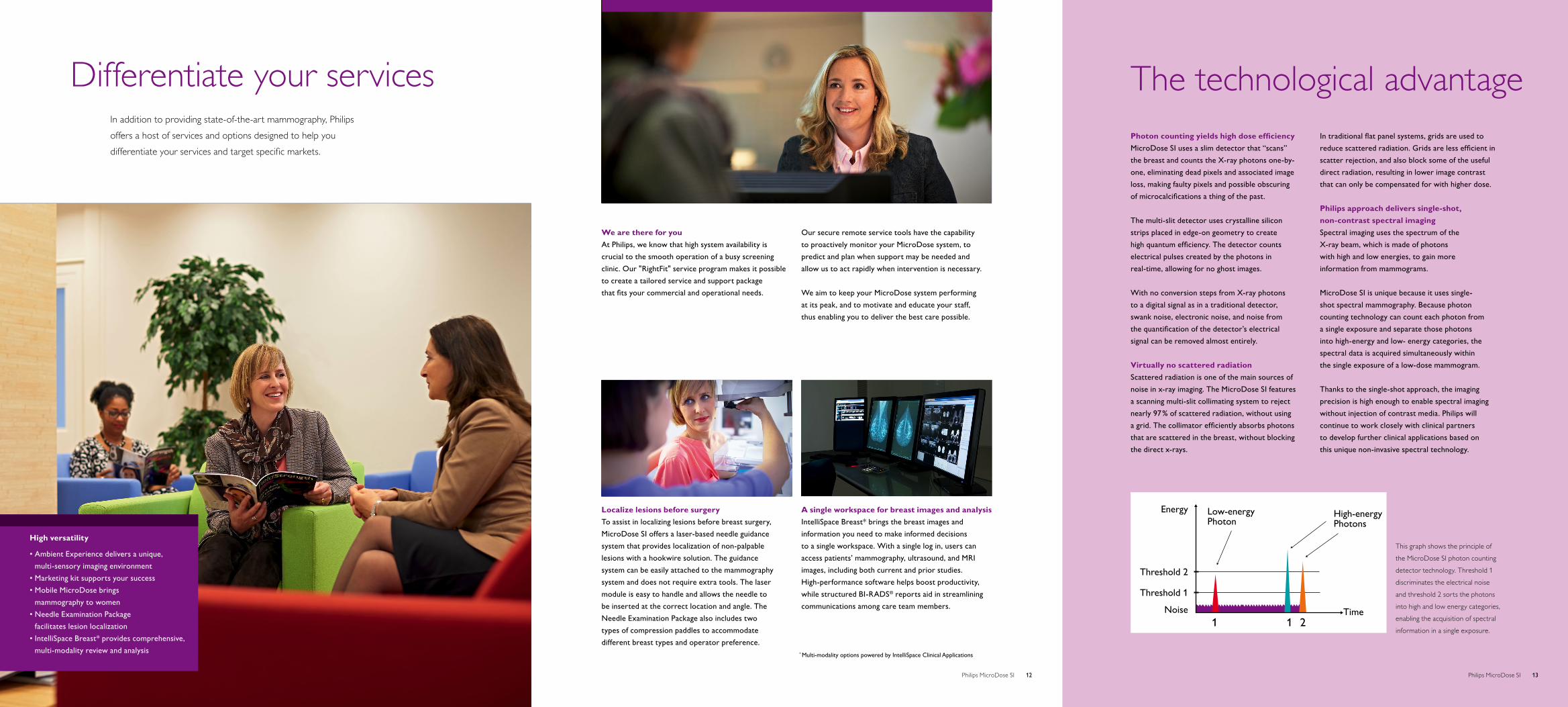
Energy Low-energyPhoton
High-energyPhotons
Time1 1 2
Threshold 2
Threshold 1
Noise
Differentiate your servicesIn addition to providing state-of-the-art mammography, Philips offers a host of services and options designed to help you differentiate your services and target specific markets.
We are there for youAt Philips, we know that high system availability is crucial to the smooth operation of a busy screening clinic. Our "RightFit" service program makes it possible to create a tailored service and support package that fits your commercial and operational needs.
Our secure remote service tools have the capability to proactively monitor your MicroDose system, to predict and plan when support may be needed and allow us to act rapidly when intervention is necessary.
We aim to keep your MicroDose system performing at its peak, and to motivate and educate your staff, thus enabling you to deliver the best care possible.
A single workspace for breast images and analysisIntelliSpace Breast* brings the breast images and information you need to make informed decisions to a single workspace. With a single log in, users can access patients’ mammography, ultrasound, and MRI images, including both current and prior studies. High-performance software helps boost productivity, while structured BI-RADS® reports aid in streamlining communications among care team members.
* Multi-modality options powered by IntelliSpace Clinical Applications
This graph shows the principle of
the MicroDose SI photon counting
detector technology. Threshold 1
discriminates the electrical noise
and threshold 2 sorts the photons
into high and low energy categories,
enabling the acquisition of spectral
information in a single exposure.
The technological advantage
High versatility
• Ambient Experience delivers a unique, multi-sensory imaging environment
• Marketing kit supports your success• Mobile MicroDose brings
mammography to women• Needle Examination Package
facilitates lesion localization• IntelliSpace Breast* provides comprehensive,
multi-modality review and analysis
Localize lesions before surgeryTo assist in localizing lesions before breast surgery, MicroDose SI offers a laser-based needle guidance system that provides localization of non-palpable lesions with a hookwire solution. The guidance system can be easily attached to the mammography system and does not require extra tools. The laser module is easy to handle and allows the needle to be inserted at the correct location and angle. The Needle Examination Package also includes two types of compression paddles to accommodate different breast types and operator preference.
Photon counting yields high dose efficiencyMicroDose SI uses a slim detector that “scans” the breast and counts the X-ray photons one-by-one, eliminating dead pixels and associated image loss, making faulty pixels and possible obscuring of microcalcifications a thing of the past.
The multi-slit detector uses crystalline silicon strips placed in edge-on geometry to create high quantum efficiency. The detector counts electrical pulses created by the photons in real-time, allowing for no ghost images.
With no conversion steps from X-ray photons to a digital signal as in a traditional detector, swank noise, electronic noise, and noise from the quantification of the detector’s electrical signal can be removed almost entirely.
Virtually no scattered radiation Scattered radiation is one of the main sources of noise in x-ray imaging. The MicroDose SI features a scanning multi-slit collimating system to reject nearly 97 % of scattered radiation, without using a grid. The collimator efficiently absorbs photons that are scattered in the breast, without blocking the direct x-rays.
In traditional flat panel systems, grids are used to reduce scattered radiation. Grids are less efficient in scatter rejection, and also block some of the useful direct radiation, resulting in lower image contrast that can only be compensated for with higher dose.
Philips approach delivers single-shot, non-contrast spectral imagingSpectral imaging uses the spectrum of the X-ray beam, which is made of photons with high and low energies, to gain more information from mammograms.
MicroDose SI is unique because it uses single-shot spectral mammography. Because photon counting technology can count each photon from a single exposure and separate those photons into high-energy and low- energy categories, the spectral data is acquired simultaneously within the single exposure of a low-dose mammogram.
Thanks to the single-shot approach, the imaging precision is high enough to enable spectral imaging without injection of contrast media. Philips will continue to work closely with clinical partners to develop further clinical applications based on this unique non-invasive spectral technology.
12Philips MicroDose SI 13Philips MicroDose SI

© 2013 Koninklijke Philips N.V. All rights reserved. Philips Healthcare reserves the right to make changes in specifications and/or to discontinue any product at any time without notice or obligation and will not be liable for any consequences resulting from the use of this publication.
Printed in The Netherlands.4522 962 98121 * DEC 2013
Philips Healthcare is part of Royal Philips
How to reach us:www.philips.com/[email protected]
Product information:www.philips.com/microdosesi


![A systematic study of microdosing psychedelics · grams, or a microdose of psilocybin might be .1 to .5 grams of dried mushrooms [3]. People microdose using a wide range of different](https://static.fdocuments.us/doc/165x107/5eb7f1e8051cc87bb330cdb2/a-systematic-study-of-microdosing-psychedelics-grams-or-a-microdose-of-psilocybin.jpg)