New Insights on Classification, Identification, and Clinical … · the stramenopiles despite the...
27
CLINICAL MICROBIOLOGY REVIEWS, Oct. 2008, p. 639–665 Vol. 21, No. 4 0893-8512/08/$08.000 doi:10.1128/CMR.00022-08 Copyright © 2008, American Society for Microbiology. All Rights Reserved. New Insights on Classification, Identification, and Clinical Relevance of Blastocystis spp. Kevin S. W. Tan* Laboratory of Molecular and Cellular Parasitology, Department of Microbiology, Yong Loo Lin School of Medicine, 5 Science Drive 2, Singapore 117597, Republic of Singapore INTRODUCTION .......................................................................................................................................................639 CLASSIFICATION .....................................................................................................................................................639 Genetic Diversity .....................................................................................................................................................639 Phylogenetic Studies and Identification of Isolates to the Species Level .......................................................640 BIOLOGY ....................................................................................................................................................................641 Morphological Forms .............................................................................................................................................641 Life Cycle .................................................................................................................................................................646 LABORATORY DIAGNOSIS ....................................................................................................................................646 Microscopy ...............................................................................................................................................................646 Laboratory Culture .................................................................................................................................................648 Serology ....................................................................................................................................................................649 Molecular Approaches............................................................................................................................................649 CLINICAL ASPECTS.................................................................................................................................................650 Epidemiology and Prevalence................................................................................................................................650 Infection and Disease .............................................................................................................................................653 Signs and symptoms ...........................................................................................................................................653 Pathogenic potential ...........................................................................................................................................654 Animal Studies ........................................................................................................................................................655 In Vitro Studies .......................................................................................................................................................656 Cytopathic effects on host cells .........................................................................................................................656 Cysteine proteases as virulence factors ...........................................................................................................656 Treatment .................................................................................................................................................................657 CONCLUSIONS .........................................................................................................................................................659 ACKNOWLEDGMENTS ...........................................................................................................................................659 REFERENCES ............................................................................................................................................................659 INTRODUCTION Blastocystis is an unusual enteric protozoan parasite of hu- mans and many animals (233, 250). It has a worldwide distri- bution and is often the most commonly isolated organism in parasitological surveys (6, 8, 21, 25, 179, 198). The parasite has been described since the early 1900s (12, 37), but only in the last decade or so have there been significant advances in our understanding of Blastocystis biology. However, the pleomor- phic nature of the parasite and the lack of standardization in techniques have led to confusion and, in some cases, misinter- pretation of data. This has hindered laboratory diagnosis and efforts to understand its mode of reproduction, life cycle, prev- alence, and pathogenesis. Accumulating epidemiological, in vivo, and in vitro data strongly suggest that Blastocystis is a pathogen. Many genotypes exist in nature, and recent obser- vations indicate that humans are in reality host to numerous zoonotic genotypes (1, 169). Such genetic diversity has led to a suggestion that previously conflicting observations on its pathogenesis are due to pathogenic and nonpathogenic geno- types (53). Recent epidemiological, animal infection, and in vitro host-Blastocystis interaction studies suggest that this may indeed be the case. This review will focus on such recent advances and also provide updates on laboratory and clinical aspects of Blastocystis spp. Excellent reviews on various topics in Blastocystis biology, including historical perspectives on par- asite biology, animal isolates, and pathogenesis, were reported elsewhere previously (33, 233, 250, 256, 319). CLASSIFICATION Genetic Diversity Blastocystis spp. from humans and animals have been re- ported to be morphologically similar. This is probably an over- simplification, as there have been reports describing distinct morphological differences among Blastocystis isolates (219, 236, 237, 303). However, it is nevertheless challenging to dif- ferentiate one isolate from another based on morphological criteria alone. Interestingly, extensive genetic variation has been observed among numerous isolates from both humans and animals. A number of molecular techniques to study the genetic diversity of Blastocystis spp. have been described. The techniques commonly employed are PCR-restriction fragment length polymorphism (RFLP) (2–4, 31, 53, 113, 202, 223, 271, * Mailing address: Laboratory of Molecular and Cellular Parasitol- ogy, Department of Microbiology, Yong Loo Lin School of Medicine, National University of Singapore, 5 Science Drive 2, Singapore 117597, Republic of Singapore. Phone: (65) 6516 6780. Fax: (65) 6776 6872. E-mail: [email protected]. 639 on December 3, 2018 by guest http://cmr.asm.org/ Downloaded from
Transcript of New Insights on Classification, Identification, and Clinical … · the stramenopiles despite the...

CLINICAL MICROBIOLOGY REVIEWS, Oct. 2008, p. 639–665 Vol. 21, No. 40893-8512/08/$08.00�0 doi:10.1128/CMR.00022-08Copyright © 2008, American Society for Microbiology. All Rights Reserved.
New Insights on Classification, Identification, and Clinical Relevanceof Blastocystis spp.
Kevin S. W. Tan*Laboratory of Molecular and Cellular Parasitology, Department of Microbiology, Yong Loo Lin School of Medicine,
5 Science Drive 2, Singapore 117597, Republic of Singapore
INTRODUCTION .......................................................................................................................................................639CLASSIFICATION .....................................................................................................................................................639
Genetic Diversity.....................................................................................................................................................639Phylogenetic Studies and Identification of Isolates to the Species Level .......................................................640
BIOLOGY ....................................................................................................................................................................641Morphological Forms .............................................................................................................................................641Life Cycle .................................................................................................................................................................646
LABORATORY DIAGNOSIS....................................................................................................................................646Microscopy ...............................................................................................................................................................646Laboratory Culture.................................................................................................................................................648Serology ....................................................................................................................................................................649Molecular Approaches............................................................................................................................................649
CLINICAL ASPECTS.................................................................................................................................................650Epidemiology and Prevalence................................................................................................................................650Infection and Disease .............................................................................................................................................653
Signs and symptoms...........................................................................................................................................653Pathogenic potential ...........................................................................................................................................654
Animal Studies ........................................................................................................................................................655In Vitro Studies.......................................................................................................................................................656
Cytopathic effects on host cells .........................................................................................................................656Cysteine proteases as virulence factors ...........................................................................................................656
Treatment.................................................................................................................................................................657CONCLUSIONS .........................................................................................................................................................659ACKNOWLEDGMENTS ...........................................................................................................................................659REFERENCES ............................................................................................................................................................659
INTRODUCTION
Blastocystis is an unusual enteric protozoan parasite of hu-mans and many animals (233, 250). It has a worldwide distri-bution and is often the most commonly isolated organism inparasitological surveys (6, 8, 21, 25, 179, 198). The parasite hasbeen described since the early 1900s (12, 37), but only in thelast decade or so have there been significant advances in ourunderstanding of Blastocystis biology. However, the pleomor-phic nature of the parasite and the lack of standardization intechniques have led to confusion and, in some cases, misinter-pretation of data. This has hindered laboratory diagnosis andefforts to understand its mode of reproduction, life cycle, prev-alence, and pathogenesis. Accumulating epidemiological, invivo, and in vitro data strongly suggest that Blastocystis is apathogen. Many genotypes exist in nature, and recent obser-vations indicate that humans are in reality host to numerouszoonotic genotypes (1, 169). Such genetic diversity has led to asuggestion that previously conflicting observations on itspathogenesis are due to pathogenic and nonpathogenic geno-
types (53). Recent epidemiological, animal infection, and invitro host-Blastocystis interaction studies suggest that this mayindeed be the case. This review will focus on such recentadvances and also provide updates on laboratory and clinicalaspects of Blastocystis spp. Excellent reviews on various topicsin Blastocystis biology, including historical perspectives on par-asite biology, animal isolates, and pathogenesis, were reportedelsewhere previously (33, 233, 250, 256, 319).
CLASSIFICATION
Genetic Diversity
Blastocystis spp. from humans and animals have been re-ported to be morphologically similar. This is probably an over-simplification, as there have been reports describing distinctmorphological differences among Blastocystis isolates (219,236, 237, 303). However, it is nevertheless challenging to dif-ferentiate one isolate from another based on morphologicalcriteria alone. Interestingly, extensive genetic variation hasbeen observed among numerous isolates from both humansand animals. A number of molecular techniques to study thegenetic diversity of Blastocystis spp. have been described. Thetechniques commonly employed are PCR-restriction fragmentlength polymorphism (RFLP) (2–4, 31, 53, 113, 202, 223, 271,
* Mailing address: Laboratory of Molecular and Cellular Parasitol-ogy, Department of Microbiology, Yong Loo Lin School of Medicine,National University of Singapore, 5 Science Drive 2, Singapore 117597,Republic of Singapore. Phone: (65) 6516 6780. Fax: (65) 6776 6872.E-mail: [email protected].
639
on Decem
ber 3, 2018 by guesthttp://cm
r.asm.org/
Dow
nloaded from

305), PCR followed by dideoxy sequencing (1, 17, 100, 169,170, 214, 218, 231, 271, 304), and PCR with subtype-specific(sequence-tagged site [STS]) primers (2–4, 117, 119–121, 124,128, 129). A few studies employed the use of arbitrary primedPCR (103, 125) or karyotyping (38, 99, 219, 276). Clark (52,53), by PCR-RFLP of the entire small-subunit rRNA (ssrRNA)gene, revealed a remarkable amount of genetic variation thatexisted among 30 randomly selected human isolates. TheseRFLP profiles (riboprints) could be grouped into seven distinctgenotypes (ribodemes). It was previously observed that therewas a 7% divergence between ribodemes 1 and 2, which isapproximately four times the genetic distance between homol-ogous genes of Entamoeba histolytica and Entamoeba dispar(53). In the most extensive phylogenetic study to date, Noel etal. (169) analyzed the ssrRNA genes of 12 Blastocystis isolatesfrom humans, rats, and reptiles together with 78 other Blasto-cystis sequences available in the GenBank database at the timeof the study. They showed that Blastocystis spp. could be un-ambiguously placed within seven distinct clades, with six of themajor groups comprising isolates from both humans and ani-mals. Those authors concluded that numerous zoonotic iso-lates existed, with frequent animal-to-human and human-to-animal transmissions, and that animals represent a largepotential reservoir for human infections. Thus, in the absenceof genotype information and due to the extreme genetic diver-sity among Blastocystis isolates, caution is warranted when in-terpreting data or when extrapolating observations of mor-phology, drug sensitivity, and pathogenesis from one isolate toanother.
Phylogenetic Studies and Identification ofIsolates to the Species Level
The taxonomic classification of Blastocystis spp. has provenchallenging and was only recently unambiguously placed withinthe stramenopiles despite the application of modern molecularphylogenetic approaches (18, 100, 218). The organism wasinitially classified as the cyst of a flagellate, vegetable, yeast,and fungus (319). It was subsequently reclassified as a protistby Zierdt and colleagues (319, 322) based on a number ofprotistan features, viz., one or more nuclei, smooth and roughendoplasmic reticulum, Golgi bodies, and mitochondrion-likeorganelles; it failed to grow on fungal media and was resistantto antifungal drugs but was sensitive to the antiprotozoal drugsmetronidazole (Flagyl) and emetine (319, 322). The subse-quent molecular analysis of Blastocystis ssrRNA and elonga-tion factor 1� (EF-1�) gene sequences resulted in disparateconclusions on its taxonomic and phylogenetic affiliations. Anearlier analysis of the ssRNA genes showed that Blastocystis sp.
is not monophyletic with the yeasts, fungi, sarcodines, or sporo-zoans (108) and should be placed among the stramenopiles(100, 218). In contrast, studies involving EF-1� suggested thatBlastocystis spp. diverged before the stramenopiles and may bea close relative of Entamoeba spp. (97, 158). This apparentdiscrepancy may be explained statistically by the low bootstrapvalue (58.1) used to group Blastocystis spp. with E. histolytica.Other possibilities for the variation in affinities exist. They maybe due to the choice of genes for analysis. The ssrRNA geneshave been known to possess drastic G�C content variationamong species (93), which may give rise to misleading trees,and hence, other genes (e.g., EF-1�, EF-2, and RNA polymer-ase III large subunit) with less extreme biases have been sug-gested to be better candidates for phylogenetic studies. Otherpossible factors contributing to the discrepancy include inad-equate species sampling, relatively few informative positions,mutational saturation, and long-branch attraction phenomena(186, 203, 204). A later study involving sequences from multi-ple conserved genes sought to resolve this discrepancy (18).The molecular analysis of Blastocystis ssrRNA, cytosolic-type70-kDa heat shock protein, translation elongation factor 2, andthe noncatalytic “B” subunit of vacuolar ATPase clearly dem-onstrated that Blastocystis is a stramenopile. The strameno-piles, synonymous with Heterokonta and Chromista (42), are acomplex collection of “botanical” protists comprising hetero-trophic and photosynthetic representatives. Molecular phylo-genetic studies indicated that Blastocystis sp. is most closelyrelated to Proteromonas lacertae (18, 100, 218), a flagellate ofthe hindgut of lizards and amphibians. Members of the stra-menopile group are characterized by possessing flagella withmastigonemes (hair-like projections that extend laterally fromthe flagellum). Interestingly, Blastocystis sp. does not possessflagella, is nonmotile, and is therefore placed in a newly cre-ated class, class Blastocystea, subphylum Opalinata, infraking-dom Heterokonta, subkingdom Chromobiota, kingdom Chrom-ista (41). A recent phylogenetic study showed that, based onthe genetic distance between homologous genes, Blastocystisspp. from humans and animals can be potentially divided into12 or more species (169). This and other studies have con-firmed that Blastocystis genotypes are prevalent throughout theanimal kingdom, with a number of genotypes comprising iso-lates from both humans and animals (1, 3, 4, 169, 214, 271, 295,300, 305). In this regard, the practice of assigning Blastocystisspecies according to host origin poses a problem and has prob-ably resulted in confusing reports regarding variations inpathogenesis and cell biology, since these differences could beattributed to distinct genotypes. Table 1 illustrates new desig-nations for some well-studied human and animal isolates basedon a recently published consensus terminology for Blastocystis
TABLE 1. Old and new classification of commonly studied Blastocystis isolates based on consensus terminologya
Species Isolate(s) Culture type Host New designation References
B. hominis Nand II Axenic Human Blastocystis sp. subtype 1 169, 218B. hominis Si Axenic Human Blastocystis sp. subtype 1 164, 169B. hominis B, C, E, G, H Axenic Human Blastocystis sp. subtype 7 98, 169B. ratti S1, WR1, WR2 Axenic Rat Blastocystis sp. subtype 4 45, 169Blastocystis sp. NIH:1295:1 Xenic Guinea pig Blastocystis sp. subtype 4 169, 306
a Terminology proposed by Stensvold et al. (229).
640 TAN CLIN. MICROBIOL. REV.
on Decem
ber 3, 2018 by guesthttp://cm
r.asm.org/
Dow
nloaded from
sp. (229). Humans can be host to Blastocystis spp. from variousmammals (subtype 1), primates and pigs (subtype 2), rodents(subtype 4), cattle and pigs (subtype 5), and birds (subtypes 6and 7) (169, 295). Subtype 3 is the most frequently isolatedgenotype in epidemiological surveys and is probably the onlygenotype of human origin (31, 113, 291, 304). By phylogeneticanalysis, subtypes 8 and 9 cluster most closely to subtypes 4 and6, respectively (169, 214). We have very little information onboth these subtypes except that subtype 8 has been reported inthree studies (1, 214, 228), from monkeys, a pheasant, andhumans, while subtype 9 was observed in 2 human isolates outof 102 isolates in an epidemiological study (304). These distri-butions of specific genotypes among various animal hostsshould be viewed as tentative and would, in due course, be-come more representative as additional studies are performed.For example, subtype 5 is currently accepted as being the mainBlastocystis genotype in pigs due the its high prevalence in thishost (1, 295, 297). However, other studies (163, 271) showedthat subtype 1 may dominates in pigs. Two recent reports (163,201) revealed that pigs may harbor subtype 2, which, untilthose studies, comprised isolates only from humans and pri-mates.
Blastocystis cells often possess one or two nuclei, and occa-sionally, quadrinucleate cells and cells possessing numerousnuclei have been reported (65, 142, 146, 319, 321). Whetherthese multinuclear states impact molecular and phylogenetic
analyses is currently not known. Karyogamy and the exchangeof genetic material have recently been demonstrated in thebinucleate enteric protozoan parasite Giardia intestinalis (189).It is unknown if Blastocystis also undergoes such a process, andif so, the implications of such a phenomenon for the construc-tion of phylogenetic trees should be ascertained. Future stud-ies should aim to elucidate the ploidy of the Blastocystis ge-nome and to understand if it is an asexual or sexual parasite,since such characteristics, which affect the extent to whichgenes evolve, can have major implications for how molecularphylogenetic data are interpreted (29, 174).
BIOLOGY
Morphological Forms
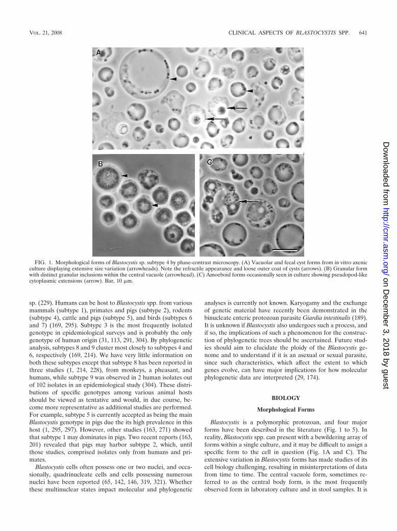
Blastocystis is a polymorphic protozoan, and four majorforms have been described in the literature (Fig. 1 to 5). Inreality, Blastocystis spp. can present with a bewildering array offorms within a single culture, and it may be difficult to assign aspecific form to the cell in question (Fig. 1A and C). Theextensive variation in Blastocystis forms has made studies of itscell biology challenging, resulting in misinterpretations of datafrom time to time. The central vacuole form, sometimes re-ferred to as the central body form, is the most frequentlyobserved form in laboratory culture and in stool samples. It is
FIG. 1. Morphological forms of Blastocystis sp. subtype 4 by phase-contrast microscopy. (A) Vacuolar and fecal cyst forms from in vitro axenicculture displaying extensive size variation (arrowheads). Note the refractile appearance and loose outer coat of cysts (arrows). (B) Granular formwith distinct granular inclusions within the central vacuole (arrowhead). (C) Amoeboid forms occasionally seen in culture showing pseudopod-likecytoplasmic extensions (arrow). Bar, 10 �m.
VOL. 21, 2008 CLINICAL ASPECTS OF BLASTOCYSTIS SPP. 641
on Decem
ber 3, 2018 by guesthttp://cm
r.asm.org/
Dow
nloaded from

spherical and may display large size variations, ranging from 2to 200 �m (average of 4 to 15 �m) (233). Extensive sizevariation can occur within and between isolates (65, 197).Dunn et al. (65) observed size variations of 4 to 63 �m among10 human isolates; the mean diameters between stocks alsovaried significantly, with overlap between some isolates. A
FIG. 2. Transmission electron micrographs of Blastocystis sp. sub-type 4. (A) Vacuolar form revealing large central vacuole (CV) result-ing in a thin band of peripheral cytoplasm. (B) Granular form reveal-ing electron-dense granules (arrowheads) occupying the entire centralvacuole. Note the surface coat surrounding the parasite. (C) Irregular-shaped amoeboid form with central vacuole (asterisk) and empty vac-uole (EV). M, mitochondrion-like organelle; N, nucleus. Bar, 1 �m.
FIG. 3. Phase-contrast microscopy of Blastocystis cysts. (A) Spher-ical cysts of subtype 4 from an in vitro axenic culture displaying a looseouter coat (arrows) among vacuolar forms (arrowheads). (B) Enrich-ment of subtype 4 cysts and loss of outer coat are apparent afterovernight incubation in distilled water. (C) Cysts from a human isolaterevealing ovoid morphology distinct from the spherical cysts of subtype4. Bar, 10 �m.
642 TAN CLIN. MICROBIOL. REV.
on Decem
ber 3, 2018 by guesthttp://cm
r.asm.org/
Dow
nloaded from

study of Blastocystis strains isolated from chickens revealedvacuolar forms ranging from 3 to 120 �m in diameter (132).The vacuolar form is characterized by a large central vacuolethat occupies approximately 90% of the cell’s volume (142,233) (Fig. 1A, 2A, and 5C to E). This relegates the cytoplasmand organelles into a thin peripheral rim, which may some-times be difficult to visualize under light microscopy (Fig. 5Aand C). Nuclei and mitochondrion-like organelles are usuallylocated within thickened cytoplasmic regions at opposite ends
of the cell (Fig. 5E). Certain amphibian isolates possess thickcytoplasmic rims, which are easily discernible by conventionallight microscopy (219). The central vacuole may appear emptyor may contain fine to flocculant material. It was reported tocontain carbohydrates, evidenced by positive staining with pe-riodic acid-Schiff and Alcian blue staining (299), or lipids,evidenced by Sudan black B and Nile blue staining (302),suggesting a storage role for the organelle. The vacuole hasalso been suggested to play a role in schizogony-like reproduc-tion (220, 241) by providing an environment for the develop-ment of minute parasite progeny. This is highly unlikelyconsidering that these progeny appear strikingly similar tometabolic granules described previously and are therefore sim-ply variants of the granular form (257, 290). Cytoplasmic con-tents, often containing organelles, may invaginate and depositfilament- or vesicle-like membrane-bound structures into thecentral vacuole (65, 159, 160, 181, 235, 239, 252). The exactsignificance of this process is unclear, although it has beenpostulated to be a mechanism of apoptotic body deposition inBlastocystis cells undergoing programmed cell death (254). Thecytoplasm contains organelles typically observed in eukaryotes.The features observable by transmission electron microscopy(TEM) include one or more nuclei, Golgi apparatus, endo-some-like vacuoles, microtubules, and mitochondrion-like or-ganelles (Fig. 2 and 4). The organism is often surrounded by asurface coat (Fig. 2B), sometimes referred to as the fibrillarlayer or capsule, of various thicknesses (65). The surface coatis often thicker in parasites freshly isolated from feces andgradually thins out during prolonged laboratory culture (40,235), and cells without surface coats have been observed invitro (65). The reason for this thinning out is unknown but maybe due to the postulated role of the coat in trapping bacteriafor nutritional purposes (311, 314), which is not possible duringaxenic culture, or may be unnecessary if nutrients provided inlaboratory culture are sufficient for growth. The surface coatcontains a variety of carbohydrates (127, 258) and has beenpostulated to play a role in trapping and degrading bacteria fornutrition (311, 314), protecting against osmotic shock (40), orto provide a mechanical barrier for functionally importantplasma membrane proteins from the immune system (259).
The granular form resembles the vacuolar form except thatgranules are present within the cytoplasm or, more commonly,within the central vacuole of the organism (Fig. 1B and 2B and5B). These are more frequently observed in nonaxenized,older, and antibiotic-treated cultures and have been describedas being vacuolar forms containing granules rather than asbeing a distinct parasite stage (33, 233). The intracellular gran-ules are heterogeneous and have been described as being my-elin-like inclusions, small vesicles, crystalline granules, andlipid droplets (65). Reproductive granules within the centralvacuole have been described and have been reported to func-tion in schizogony-like division (220, 241, 319). Other authorsargued that this cannot be accepted due to the lack of evidencethat these granules are indeed viable and develop into Blasto-cystis cells (33, 233, 257, 290).
The amoeboid form (Fig. 1C and 2C) of Blastocystis spp. israrely reported, and there are contradicting descriptions ofwhat constitutes this morphological type. An early report de-scribed numerous amoeba-like forms in the diarrheal fluid of apatient who died of aspiration pneumonia (326). These cells
FIG. 4. Transmission electron micrographs of Blastocystis sp. sub-type 4 cysts. (A) Mature cyst with a distinct double-layered cyst wall(arrow) and reduced glycogen mass (asterisk). Mitochondrion-like or-ganelles (M) contain faint saccate cristae (arrowhead). (B) Mature cystcontaining large vacuoles (V), a nucleus (N) with a dense chromatinmass (arrowhead), and mitochondrion-like organelles (M) with saccateand circular (arrow) cristae. Bar, 1 �m.
VOL. 21, 2008 CLINICAL ASPECTS OF BLASTOCYSTIS SPP. 643
on Decem
ber 3, 2018 by guesthttp://cm
r.asm.org/
Dow
nloaded from

were irregularly convoluted, and some cells possessed one ortwo large pseudopods. In another study, amoeboid forms fromin vitro culture were observed to be 10 to 15 �m, possessingfeatures typical of vacuolar forms, with the exception of one ortwo pseudopods (249). We have reported the presence of nu-merous amoeboid forms (Fig. 6) from Blastocystis coloniesgrown in soft agar (45, 252, 260, 261). Light microscopy andTEM showed cells with a central vacuole, a surface coat, andnumerous Golgi bodies and mitochondria within the cytoplas-mic extensions of pseudopods (252). Dunn et al. (65) previ-ously described amoeboid cells ranging from 2.6 to 7.8 �m withextended pseudopodia and lysosome-like compartments con-taining ingested bacteria. In contrast to our studies, theseforms lacked a central vacuole, a Golgi complex, a surface coat,and mitochondria (65). Considering the genetic diversity of theorganism, it is plausible that the differing descriptions are dueto genotypic variations among Blastocystis isolates. The pres-ence of bacteria and bacterial remnants within the amoeboidform suggests a nutritional role for this form. The amoeboidform has been postulated to play a role in pathogenesis (115,262, 264). However, the light and TEM micrographs in two ofthese reports (262, 264) were unconvincing for this form andappear more like irregularly shaped central vacuole forms, a
common artifact of TEM processing. Despite the observationof pseudopod-like cytoplasmic extensions, the amoeboid formappears to be nonmotile. The identification of stage-specificmolecular markers would be useful for studies of various de-velopmental forms of the parasite and would obviate the prob-lem of distinguishing the various forms by morphological cri-teria alone.
The cyst form (Fig. 1A and 3) is the most recently describedform of the parasite, and the late discovery is due to its smallsize (2 to 5 �m), which can result in confusion with fecal debris,and the observation that cysts are infrequently seen in labora-tory culture (46, 153, 154, 234, 312). The cysts are variable inshape but are mostly ovoid or spherical. The cyst is protectedby a multilayered cyst wall (Fig. 4), which may or may not becovered by a loose surface coat (153, 312, 313). The cytoplasmof the cyst may contain one to four nuclei, mitochondria, gly-cogen deposits, and small vacuoles (5, 46, 153, 312). One re-port (237) described the presence of large multinucleate cystsfrom the stools of Macaca monkeys, which were 15 �m in size,and this was suggested to be an indication of differences amongBlastocystis species. Blastocystis cysts were reportedly able tosurvive in water for up to 19 days at a normal temperature butare fragile at extreme temperatures and in common disinfec-
FIG. 5. Light microscopy of nonaxenic Blastocystis sp. subtype 1 cultured in Jones’ medium. (A to C) Unstained wet mounts of variousdiagnostic forms of Blastocystis. (A) Cells undergoing binary fission (arrow). (B) Granular forms commonly seen in laboratory culture. (C) Vac-uolar forms with virtually indiscernible thin cytoplasmic rims. (D) Iodine-stained wet mount revealing cells with distinct organelles, a cytoplasmicrim, and granular inclusions (arrowhead). (E) Giemsa-stained permanent smear of the large vacuolar form containing four nuclei (Nu) evenlydistributed around the cytoplasmic rim. (F and G) Trichrome-stained permanent smear of vacuolar forms. The central vacuole of some cells stainstrongly (arrow), while others stain less intensely; this may be due to the biochemical heterogeneity of the vacuolar contents. (G) Small vacuolarform (arrowhead) revealing organelles within the cytoplasmic rim. Bar, 20 �m.
644 TAN CLIN. MICROBIOL. REV.
on Decem
ber 3, 2018 by guesthttp://cm
r.asm.org/
Dow
nloaded from

tants (154). A later study (306) showed that cysts could surviveup to 1 month at 25°C and 2 months at 4°C. The contrastingviabilities among the studies may be due to isolate variations.Vacuolar and granular forms, in contrast, are sensitive to tem-perature changes, osmotic shock, and exposure to air (146,319). Experimental infectivity studies of BALB/c mice, Wistarrats, and a variety of bird species with the cyst form indicatethat this form is undoubtedly the transmissible form of theparasite. The formation of in vitro-derived cysts by the incu-bation of vacuolar forms in encystation medium was reportedpreviously (240, 241, 244, 282). These “cysts” appear to becuriously similar to the classical granular forms, and the gran-ules within the central vacuole were reported to be reproduc-tive in nature (240, 241, 243). It is likely that these are artifacts
of culture induced by the encystation medium, as they bear nomorphological similarity to the fecal cyst. Interestingly, invitro-derived cysts are able to infect Wistar rats (244) and wereapparently resistant to osmotic lysis (282).
Other forms have also been described, and these forms in-clude the avacuolar and multivacuolar forms. These formsreported from TEM studies of fresh stool samples were signif-icantly smaller (5 to 8 �m) than culture forms and were sug-gested to be the form that occurs in vivo (233, 235). However,others observed typical vacuolar forms from fresh fecal sam-ples (9, 64, 128, 132, 227, 232, 281, 318). The central vacuolewas absent in the avacuolar form, while the multivacuolarforms contained multiple small vacuoles. The small size anddistinct multivacuolar or avacuolar morphology may be due to
FIG. 6. Colonies of Blastocystis sp. subtype 4 after 12 days of culture on an agar plate containing 0.3% Bacto agar in Iscove’s modifiedDulbecco’s medium containing 10% horse serum. (A) Petri dish with numerous buff-colored colonies. (B and C) Magnified view of single colonies.(C) Occasionally, parasite cells spread out at the interface of the agar and the bottom surface of the plate. (D) Amoeboid cells isolated from acolony with large inclusions (arrows) within the central vacuole. Bar, 10 �m.
VOL. 21, 2008 CLINICAL ASPECTS OF BLASTOCYSTIS SPP. 645
on Decem
ber 3, 2018 by guesthttp://cm
r.asm.org/
Dow
nloaded from

strain variations, or they are possibly cells in various stagesof encystation or excystation, as similar morphologies weredescribed in TEM studies of cells undergoing excystation(46, 153).
Life Cycle
Numerous conflicting life cycles have been proposed (33,217, 220, 233, 250, 319, 321), and these discrepancies are duelargely to the belief that Blastocystis exhibits multiple repro-ductive processes (83, 220, 318). The suggestion that Blasto-cystis undergoes multiple fission has led to life cycles whereschizogony is one of the modes of reproduction (220, 319).This and other proposed modes such as plasmotomy (budding)(263), endodyogeny (318), and sac-like pouches (83, 242) aremore likely due to the pleomorphic nature of the organism andnot true modes of reproduction. A life cycle comprising thick-and thin-walled cysts from multiple fission was proposed (220).Those authors hypothesized that the thick-walled cysts areimportant for external transmission, while the thin-walled cystswere autoinfectious. There is little scientific evidence to sup-port such a proposal, although schizogony-like reproduction inBlastocystis has been perpetuated in a number of authoritativesources, including medical parasitology textbooks and theDPDx website of the Centers for Disease Control and Preven-tion (www.dpd.cdc.gov/dpdx/HTML/Blastocystis.htm). Untilproven otherwise, the only accepted mode of reproduction isbinary fission. The application of live-cell imaging technologyshould provide a better understanding of the modes of repro-duction of Blastocystis spp.
A revised life cycle (Fig. 7) must take into account the largereservoir of Blastocystis spp. among various animal populationsand that humans are potential hosts to numerous zoonoticgenotypes (subtypes). Upon ingestion of cysts, the parasiteundergoes excystation in the large intestines and develops intovacuolar forms. Encystation occurs during passage along thelarge intestines and is deposited in the feces (152). The fecalcysts may be covered by a fibrillar layer that is gradually lostduring cyst development (313).
Apart from a few studies, the transitions from one of theclassically described forms to another are not well understood.TEM studies of the development of cysts to vacuolar formswere elegantly demonstrated with a human isolate and a ratisolate (46, 153). In those reports, fecal cysts from both humansand rat develop similarly and dramatically into vacuolar formswithin 24 h of inoculation into growth medium. In one of thosestudies (153), cells undergoing excystation apparently devel-oped from cysts into granular forms before becoming vacuolarin morphology. Whether these granular forms are similar tothose from patient samples and laboratory culture is notknown. In a separate study (317), Blastocystis cysts enrichedfrom a patient sample were cultured in Jones’ medium andcharacterized by TEM at 24 h. The micrographs revealed thatcell division of vacuolar forms occurs while the parasite is stillwithin the cyst wall and that both granular and vacuolar formswere observed in the same sample. Because only one timepoint was performed, it is difficult to conclude the order inwhich these forms developed. Certain culture conditions werereported to induce the development of the granular form fromthe vacuolar form. These conditions include old cultures (250),
axenization (327), transfer to a different culture medium (235),and increases in serum concentrations in the culture medium(65, 130, 217, 321). Amoeboid forms probably arise from vac-uolar forms. Some evidence for this is seen when vacuolarforms are cultured in agar, and after incubation, the resultantcolonies contain numerous amoeboid forms (260, 261).
LABORATORY DIAGNOSIS
Microscopy
Blastocystis poses considerable challenges for the diagnosticlaboratory. Firstly, the uncertain pathogenesis of the parasitediscourages many clinicians from considering Blastocystis to bethe etiological agent of disease. Secondly, the polymorphicnature of the organism in wet mounts can result in confusionwith yeast, Cyclospora sp., or fat globules. The classical vacu-olar forms may not predominate in fresh fecal specimens (235),while the smaller fecal cyst, when present, may be difficult toidentify. Direct microscopy is usually done with stained spec-imens. Multiple stool specimens should be examined, since theparasite may exhibit irregular shedding (88, 281). Morpholog-ical features that may aid in the laboratory diagnosis of Blas-tocystis infection are summarized in Table 2. Wet mounts withLugol’s iodine (Fig. 5D) and permanent-stained smears withacid-fast, Giemsa (Fig. 5E), Field’s, and trichrome (Fig. 5F andG) stains have been described, with trichrome being the mostpopular stain employed (8, 171, 175, 179, 210, 227, 270, 289,307). Trichrome is a routinely employed stain in many clinicalmicrobiology laboratories, and studies have shown that it ismore sensitive for the detection of intestinal protozoa thaniodine-stained wet mounts (71, 78, 117), and this is also thecase for Blastocystis spp. (179, 270). However, short-term (24 to72 h) in vitro culture increases the sensitivity of detectioncompared to that of direct microscopy of fecal smears stainedwith Lugol’s iodine or trichrome (135, 227, 270), although onestudy (245) did not mentioned the use of any staining method.However, in the case of mixed infections, in vitro culture mayfavor the preferential amplification of one subtype over an-other (183, 295), although this was not seen in another studyinvolving a mixed infection (227). In contrast to in vitro cul-ture, a number of reports indicated that the formol ethyl ace-tate concentration technique (FECT) results in very poor sen-sitivity for parasite detection (227, 231, 245). Subtype 3 wasapparently associated with false-negative results associatedwith this method (227), although the reason for this bias iscurrently unknown. This may explain a study in Thailand, em-ploying only formol ethyl acetate concentration, revealing aprevalence of 0.19% for Blastocystis isolates in primary schoolchildren in central Thailand (211). Another study using a num-ber of diagnostic approaches (simple smear, formol ether con-centration, Boeck and Drbohlav’s Locke-egg-serum mediumculture, and modified acid-fast and modified trichrome stain-ing) showed the prevalence of Blastocystis isolation to be45.2% among Thai children in the same region (210). Present-day diagnostic laboratories should also include the fecal cyst asan indicator of infection. If necessary, these cysts can be selec-tively concentrated by density gradient approaches to increasesensitivity (309, 315). This enrichment approach may be morepractical in the research laboratory setting since it may be
646 TAN CLIN. MICROBIOL. REV.
on Decem
ber 3, 2018 by guesthttp://cm
r.asm.org/
Dow
nloaded from

FIG. 7. Proposed life cycle for Blastocystis cells taking into account recent studies (163, 169, 201, 295) suggesting the existence of zoonoticgenotypes (subtypes 1 to 7) with various host specificities. Humans and animals are infected by fecal cysts, which develop into vacuolar forms inthe large intestines. In humans, vacuolar forms divide by binary fission and may develop into amoeboid or granular forms. Vacuolar forms undergoencystation in the host intestines, and intermediate cyst forms may be surrounded by a thick fibrillar layer that is subsequently lost during passagein the external environment. Information on the transition from the amoeboid to the vacuolar form and from the vacuolar to the cyst form islacking. These hypothetical pathways are represented by dotted lines. Subtype 1 is cross-infective among mammalian and avian isolates; subtypes2, 3, 4, and 5 comprise primate/pig, human, cattle/pig, and rodent isolates, respectively; and subtypes 6 and 7 include avian isolates. The proposedscheme suggests that humans are potentially infected by seven or more species of Blastocystis and that certain animals represent reservoirs fortransmission to humans. (Adapted from reference 251 with permission from Taylor and Francis.)
VOL. 21, 2008 CLINICAL ASPECTS OF BLASTOCYSTIS SPP. 647
on Decem
ber 3, 2018 by guesthttp://cm
r.asm.org/
Dow
nloaded from

cumbersome to perform during routine diagnosis or in large-scale surveys. It was suggested that the intensity of infectionshould be reported, and the general criteria are whether five ormore parasites are seen in a high-powered field (�400) for wetmounts (77, 133, 166, 188, 194, 215, 319) or under oil immer-sion (�1,000) (175, 179, 307) if permanent-stained smears areused. Using this criterion and in comparison with reports thatdo not use this criterion, accumulating studies suggest a cor-relation between infection density and symptoms (51, 60, 67,82, 85, 111, 116, 155, 165, 167, 175, 177, 215, 307). There are,however, a number of studies that reported a lack of such acorrelation (63, 86, 131, 145, 180, 216). The reason for thisdiscrepancy is presently unclear but may be due to genotypedifferences among Blastocystis isolates or to host factors suchas age and genetic background variations in the populationsstudied.
Laboratory Culture
Xenic or monoxenic laboratory cultures of Blastocystis iso-lates, which are cultures of Blastocystis cells grown in associa-tion with nonstandardized or single known species of microor-ganisms, respectively, can be maintained in Jones’ (110) orBoeck and Drbohlav’s inspissated egg (30) medium. Jones’medium is the medium of choice in studies involving culture toidentify the parasite in patient samples (135, 183, 227, 231, 245,270, 291). There was one report (183) on the inability to growBlastocystis isolates from Australian marsupials in Jones’ me-dia, suggesting that this medium may not support fecal culturesfrom certain animal hosts. Other studies utilized diphasic agarslant medium, which was useful for the culture of Blastocystisisolates from cattle, pigs, and chickens (2–4, 109, 110). Ax-enized cultures, that is, cultures of Blastocystis cells not asso-ciated with any other living organism, display luxuriant growthin a variety of media such as Iscove’s modified Dulbecco’smedium, minimal essential medium, or biphasic inspissatedegg slant overlaid with Locke’s solution (98, 324). Cell densi-ties of up to 2.5 � 107 cells/ml can be attained for monophasicmedium (98), while slightly higher densities of approximately6.0 � 107 cells/ml were reported for cells cultured in biphasic
inspissated egg medium (324). The doubling time of axenicisolates can be variable, ranging from 6 to 23 h, depending onthe isolate, study, and type of medium used (33, 324). Doublingtimes of approximately 50 h can be deduced from growthcurves of Blastocystis isolates belonging to avian subtype 7 (98,169), and this may be due to the nonoptimal incubation tem-perature, as avian hosts, particularly chickens, generally havehigher body temperatures than mammals. The colony growthof Blastocystis cells can be established in soft agar (Fig. 6) usingthe pour plate method (260, 261, 277), and clonal growth wasachieved with the addition of sodium thioglycolate as a reduc-ing agent (260). The technique was useful as a step toward theaxenization of Blastocystis isolates by physically isolating para-site colonies from bacterial ones (45, 164) and for screeningsurface-reactive antibodies for cytotoxic activity (259). Blasto-cystis cells are also able to grow on solid medium, and parasiteclones appear to be macroscopically similar to bacterial colo-nies (255). These cultures were viable for up to 2 weeks andcould be further expanded in liquid or solid medium. Interest-ingly, for the same isolate grown in liquid medium (98), cul-tures reach maximal cell densities around 4 days postinocula-tion, enter death phase at day 5, and are subsequently difficultto subculture. This indicates that the growth characteristics ofthe same Blastocystis isolate in solid medium are markedlydifferent from those of the isolate in liquid medium.
Axenic cultures of Blastocystis isolates are important formolecular and biochemical studies. Axenization can beachieved by the addition of antibiotic cocktails to eliminatecontaminating bacteria and yeasts, and a variety of antibioticmixtures have been described, with various levels of success(45, 128, 164, 269, 319). The process is generally laborious andmay take weeks to months, and the successful elimination ofmicrobial contaminants is not guaranteed. It has been sug-gested that some isolates require the presence of bacteria tosurvive, and therefore, the removal of all bacteria results in thedeath of the parasite (319). Lanuza et al. (128) previouslydescribed an improved method for Blastocystis axenization andmanaged to axenize 25 out of 81 isolates using a combinationof Ficoll-metrozoic acid gradient and the addition of antibiot-
TABLE 2. Diagnostic features of Blastocystisa
Stage or form Size (�m) Presence of centralvacuole No. of nuclei Area(s) of
occurrence Description Reference(s)
Vacuolar 2–200 (usually 5–15) Present 1–4 (usually 1–2) Culture, feces Central vacuole occupies70–90% of cell vol;occasionally, giantcells with multiplenuclei are seen
142, 233
Granular 6.5–8 Present 1–4 Feces, culture Granules within centralvacuole
233
Multivacuolar 5–8 Present or absent 1–2 Feces, culture Rarely seen 233, 235Avacuolar �5 Absent 1–2 Intestine, feces Rarely seen in feces 233, 235Amoeboid 2.6–7.8 Present or absent 1–2 Feces, culture Rarely reported;
conflicting informationon morphology
65, 260, 326
Cyst 3–10 Absent 1–4 Feces, culture Rarely seen in culture;cyst wall present; maybe surrounded by afibrillar layer
233, 309, 310
a Adapted from reference 233 with permission.
648 TAN CLIN. MICROBIOL. REV.
on Decem
ber 3, 2018 by guesthttp://cm
r.asm.org/
Dow
nloaded from

ics. Cultures were initially treated with a basic antibiotic solu-tion comprising 0.4% ampicillin, 0.1% streptomycin, and0.0006% amphotericin B, and in subsequent subcultures, re-sistant bacteria were isolated and antimicrobial assays per-formed. The final stages of axenization involved the addition ofantibiotics against remaining bacteria with density gradientenrichment for Blastocystis spp. The time required for axeni-zation was approximately 3 weeks. In addition to antibiotictreatment, some authors noted improved success when physi-cal methods were employed during axenization to separateparasites from the bulk of the bacterial load. This includesdifferential centrifugation (269), density gradient separation(128), and colony growth (45, 164). Such methods enrich forthe parasite and provide a growth advantage. Chen et al. (45)noted that the axenization of Blastocystis would not have beenpossible without parasite enrichment via colony growth. Therewas a report on the use of micropipette manipulation to isolateBlastocystis clones from nonaxenic cultures of turkey cecal con-tents (95), and it would be interesting to investigate if thistechnique could be exploited as a step toward axenization.
Serology
Blastocystis infections lead to immunoglobulin G (IgG) andIgA responses, as detected by indirect fluorescent antibody(IFA) testing and enzyme-linked immunosorbent assay(ELISA) (104, 112, 143, 323, 328). ELISA titers ranged from1:50 to 1:1,600 (328), and previously reported studies revealedthat high titers are associated with symptomatic infections(104, 143, 323, 328). Early ELISA studies showed that serafrom patients harboring Blastocystis spp. had high IgG titersagainst parasite extracts (323, 328). In one of the studies (328),30 sera from 28 patients were tested: 3 were negative at the1/50 threshold dilution, 8 were positive at 1/50, 3 were positiveat 1/100, 2 were positive at 1/200, 3 were positive at 1/400, 6were positive at 1/800, and 5 were positive at 1/1,600. Normalsera (42 blood bank sera) were all negative at 1/50. Interest-ingly, IgA responses against Blastocystis spp. could not be de-tected in the symptomatic population. A recent study investi-gated the secretory IgA, serum IgA, and serum IgG levels inBlastocystis-positive individuals with and without symptoms byELISA (143). This study showed that only sera from symptom-atic patients had significantly higher Blastocystis-reactive secre-tory IgA, serum IgA, and serum IgG levels than did sera fromasymptomatic carriers and healthy controls. In contrast,Kaneda et al. (112) showed by IFA that asymptomatic individ-uals harboring Blastocystis spp. possessed serum antibodies tothe parasite, although antibody titers were very low, and serumdilutions greater than 1:60 failed to elicit a reaction. However,the strongest reaction was seen in an individual with chronicinfection. It was suggested that constant exposure to the par-asite was necessary to elicit a serological response. The 1/50ELISA cutoff used in studies of symptomatic patients reportedpreviously by Zierdt and colleagues (323, 328) is also ratherlow and suggests that the parasite induces a weak immuneresponse. However, considering the genotypic (53, 169) andantigenic (122, 126, 156, 258, 298) diversity among morpholog-ically identical isolates, the low values may be due to the choiceof isolate used as the coating antigen. Such antigenic variationsmay also explain the discrepant observations among studies
and must be taken into consideration should a serological testkit be developed. Monoclonal antibodies against Blastocystisspp. have been described (258, 298). The majority of antibodieswere IgM and localized to surface coat antigens. These anti-bodies exhibited limited cross-reactivity against different geno-types, indicating antigenic diversity among Blastocystis isolates(253, 258). Although currently unavailable, monoclonal anti-bodies specific for human-infective genotypes would be usefulfor antigen detection studies, as was previously described forEntamoeba histolytica/E. dispar (283). Similarly, the applicationof genotype-specific antigens in ELISA or immunofluores-cence formats should be useful for serological and epidemio-logical studies.
Blastocystis-associated symptoms are generally self-limitingand may last between 1 and 14 days (63, 131, 307, 320, 329).However, some infections persist for months if left untreated(90, 94, 112, 131, 165, 175), and it is currently unknown ifchronic infections influence seropositivity. A single study (112)of the serological response of Blastocystis-positive asymptom-atic individuals showed that the highest IFA titer was obtainedfrom a healthy individual infected with Blastocystis for 2 years.An ELISA study reported previously by Zierdt et al. (328)included two patients who provided sera within the first 2weeks postsymptom and subsequently another sample at con-valescence, about 6 weeks after onset. Comparison of acute-phase sera with convalescent-phase sera revealed an eightfoldincrease for one patient and a threefold increase for the otherpatient. Although large-scale studies are needed to validatethese observations, results of those studies suggested that Blas-tocystis sp. does elicit an immune response, and both chronicand acute infections can result in significantly higher antibodytiters than asymptomatic infections. Currently, considering ourlimited knowledge of the host immune response to Blastocystisspp. and the apparent antigenic diversity of the parasite, it isnot practical to include serology in the routine laboratory di-agnosis of Blastocystis, and it should be limited to epidemio-logical and serological studies.
Molecular Approaches
Molecular PCR-based diagnostic approaches for Blastocystisidentification have been described. Subtype-specific diagnosticprimers, also referred to as STS primers, were developed fromrandom amplification of polymorphic DNA analysis of Blasto-cystis isolates by Yoshikawa et al. (296, 297, 301), and theseapproaches amplified seven distinct subtypes, which corre-sponded to different clades inferred from ssrRNA. Such anapproach has been shown to be useful for epidemiologicalstudies, providing information on the distribution of variousgenotypes among human and animal populations (2, 4, 138,297, 304) and on the zoonotic nature of certain genotypes.
Other groups characterized isolates by PCR of ssrRNA fol-lowed by RFLP analysis (2–4, 31, 53, 202, 271, 291, 295, 305),dideoxy sequencing (183, 201, 227, 230, 231), or nested ampli-fication of intragenic regions (270). PCR-RFLP analysis of theBlastocystis ssrRNA gene is commonly employed for preva-lence studies, and a variety of primers for its amplification havebeen described (31, 53, 227, 291). However, major limitationsof this approach are the lack of standardization of the condi-tions and choice of primers, mutations at restriction sites, and
VOL. 21, 2008 CLINICAL ASPECTS OF BLASTOCYSTIS SPP. 649
on Decem
ber 3, 2018 by guesthttp://cm
r.asm.org/
Dow
nloaded from

the difficulty in interpreting RFLP profiles from mixed infec-tions. A high-throughput pyrosequencing technique for therapid sequencing of the Blastocystis ssrRNA gene was de-scribed (230). This approach detects nucleotide polymor-phisms in the gene and was able to genotype 48/48 Danishisolates in approximately 1 h but was unable to detect mixed-subtype infections. This approach would be extremely usefulfor large-scale epidemiological studies and for the rapid iden-tification of genotypes during outbreak situations (87, 114,123). A recent study compared the relative performances ofvarious diagnostic methods for the identification of Blastocystisisolates (227). The FECT, permanent trichrome staining offeces fixed in sodium acetate-acetic acid-formalin, in vitro cul-ture, and PCR approaches were compared using 107 samplesfrom 93 patients with suspected enteroparasitic disease. ThePCR approach was shown to be superior to the other ap-proaches, and detection of Blastocystis-specific DNA was assensitive as the culture method. This is in contrast to data froma study reported previously by Termmathurapoj et al. (270)that suggested that in vitro culture expansion was superior todirect PCR from stool samples. Another study (183) revealedthat direct PCR from stool samples was superior to the culturemethod, with PCR detecting Blastocystis spp. in 35% of thesamples, while the culture method detected 19%. The discrep-ancies observed in studies reported previously by Term-mathurapoj et al. (270) and Parkar et al. (183) could be attrib-uted to the fact that the latter involved the culture of fecesfrom various animals in Jones’ medium, which did not supportparasite growth, and that their DNA extraction method wasmore efficient. Other possibilities include the different speci-ficities of the PCR primers employed for these studies (227). Amethod for the detection of Blastocystis spp. directly fromunpreserved stool samples was described and provides a rapiddiagnostic tool for Blastocystis identification (231). Primersspecific for Blastocystis ssrRNA were able to detect greaterthan 32 parasites/200 mg stool artificially spiked with culturedparasites. In the evaluation of 43 clinical specimens, the PCRapproach was tested against FECT and a culture technique,proving 100% test specificity and a significantly higher sensi-tivity than FECT. In that study, there were instances whereculture-negative samples were PCR positive. This was attrib-uted to the degeneration of parasites in the stool or to lownumbers that prevented growth in vitro. Jones et al. (109)recently reported a method for the real-time PCR detection ofBlastocystis spp. from stools. Primers specific to an undefined152-bp region of the parasite genome was used for the assayand was able to amplify 11 laboratory-cultured isolates fromthe ATCC belonging to subtypes 1, 3, and 4. Results could beobtained within 3 h, with a detection limit of 760 cells per 100mg of stool. Oddly, only three clinical samples were used inthat study, and only one ATCC strain was spiked into Blasto-cystis-negative stool samples to determine sensitivity. For spec-ificity determinations, those authors excluded only cross-reac-tions with bacterial but not protozoal pathogens. The assay wasnot able to distinguish among Blastocystis subtypes.
In summary, a variety of methods for the laboratory diag-nosis of Blastocystis spp. exist. The FECT should be discour-aged due to low sensitivity. Trichrome staining of direct fecalsmears is sensitive, provides a permanent record of the speci-men, and should be supplemented with information on
whether five or more parasites are visible per oil immersion(�1,000) field. Other authors included more detailed reportingon parasite abundance (11, 85, 131, 179, 180) and quantifiedparasite abundance using terms such as rare (one to two par-asites in every 10 high-power fields), few to moderate (oneparasite in every one to five high-power fields), or abundant(five or more parasites per high-power field) (131). Such de-tailed reporting, beyond whether five or more parasites arepresent, is unnecessary since there is some controversy regard-ing correlation between infection density and disease. In in-stances of low parasite levels or when fecal cysts predominatein stool or environmental samples, in vitro culture is a usefulmethod for diagnosis (245, 246). Direct wet mounts stainedwith iodine do not seem to add additional value to the diag-nostic process, since trichrome-stained permanent smears havebeen shown to be more sensitive (179, 270). Considering cur-rent data, trichrome staining of direct smears coupled withstool culture in Jones’ medium, cost permitting, is the bestapproach for diagnosing Blastocystis infection in terms of spec-ificity and sensitivity. Future laboratory diagnosis may need toinclude genotype information once a link between genotypeand parasite pathogenesis is firmly established. For screeningand epidemiological studies, PCR amplification of BlastocystisDNA from fresh stools or stool cultures is a convenient alter-native to microscopy, and genotyping should also be includedin the analysis. The development of real-time PCR for thesensitive and rapid detection of Blastocystis spp. with the abilityto discriminate between multiple genotypes within a samplewould be similarly advantageous for screening and epidemio-logical studies.
CLINICAL ASPECTS
Epidemiology and Prevalence
Authors of early studies lamented the lack of epidemiolog-ical data on Blastocystis spp. (33, 233). However, recent yearshave shown a dramatic increase in prevalence studies, andthese studies have shed light on the parasite’s genotype distri-bution, mode of transmission, and pathogenesis.
Blastocystis is an extremely ubiquitous parasite with a world-wide distribution (107, 250). It is not uncommon for it to be themost frequently isolated parasite in epidemiological surveys(15, 21, 51, 72, 94, 185, 210, 248, 287, 289). Prevalence varieswidely from country to country and within various communitiesof the same country. In general, developing countries havehigher prevalences of the parasite than developed countries,and this has been linked to poor hygiene, exposure to animals,and consumption of contaminated food or water. Prevalencecan be low in countries such as Japan (0.5 to 1%) (96, 101) andSingapore (3.3%) (291) and high in developing nations includ-ing Argentina (27.2%) (25), Brazil (40.9%) (6), Cuba (38.5%)(70), Egypt (33.3%) (198), and Indonesia (60%) (185). In somecountries, the carriage rate can be rather variable, dependingon the subpopulation studied. Prevalence ranges of 1.9 to32.6%, 0.19 to 45.2%, and 1.04 to 18.3% in prevalence studiesfrom China (137), Thailand (210, 211), and Turkey (7, 56),respectively, have been reported. Such variations within thesame country could reflect true differences between communi-ties, especially if the same techniques were employed to iden-
650 TAN CLIN. MICROBIOL. REV.
on Decem
ber 3, 2018 by guesthttp://cm
r.asm.org/
Dow
nloaded from

tify the parasite. However, variations are also likely due to theuse of different diagnostic approaches and the inherent diffi-culty in identifying stages other than the vacuolar form.
Recent surveys incorporated genotype information by PCRof Blastocystis DNA from feces or from stool culture. Suchstudies are now shedding light on the distributions of geno-types among human populations (Table 3) and animal hostsand also provide information on transmission routes orsources. A study by Yoshikawa et al. (304) employed the use ofPCR-based genotype classification to study the distribution ofBlastocystis genotypes among isolates from Bangladesh, Ger-many, Japan, Pakistan, and Thailand. The most dominant sub-type among four populations except Thailand was subtype 3(41.7 to 92.3%), followed by either subtype 1 (7.7 to 25%) orsubtype 6 (10 to 22.9%). Similar genotype distributions inSingapore (78% subtype 3 and 22% subtype 1) (291), China(60.4% subtype 3 and 24.5% subtype 1) (137), Greece (60%subtype 3 and 20% subtype 1) (147), Germany (54% subtype 3and 21% subtype 1) (31), and Turkey (75.9% subtype 3) (180)were also reported. In most studies, other genotypes wereidentified at lower frequencies (Table 3). Collectively, thosestudies suggest that subtype 3 is the subtype of human originand that there is no correlation between Blastocystis geo-graphic origin and genotype. It may be worthwhile to note thatavian subtypes 6 and 7 may grow optimally at 40°C instead of37°C, as is the case for the avian protozoan flagellate Histo-monas meleagridis (84, 279). Isolates belonging to subtype 7have longer doubling times, about 50 h, when cultured at 37°C(98). In this case, these slow-growing subtypes may still bemissed during in vitro culture expansion of stool samples, re-sulting in an underrepresentation of such subtypes in epidemi-ological surveys. Although surveys revealed that the majority ofindividuals are host to a particular Blastocystis subtype, mixed
infections in a minority of individual have also frequently beenreported (31, 62, 137, 138, 180, 183, 227, 271, 294, 295). De-pending on the study, mixed subtypes have been seen among1.1 to 14.3% of samples surveyed. Most are coinfections withsubtype 1 and subtype 3 (137, 138, 271, 294) while subtype1/subtype 2 (137, 138), subtype 2/subtype 3 (137), and subtype3/subtype 5 (295) combinations were infrequently reported.Intra-subtype 1 and -subtype 2 variations in ssrRNA sequencewere also reported for single isolates (180, 227). It may bedifficult to ascertain the true distribution of mixed infections ina particular individual, as this depends on the method em-ployed to determine the Blastocystis subtype. Studies that ge-notype parasites after in vitro propagation (31, 62, 137, 138,271) risk underestimating mixed infections since certain sub-types may outgrow others, as was shown recently (183, 295).Hence, genotyping of Blastocystis DNA obtained directly fromstools may be more accurate for identifying mixed infections ifPCR conditions are optimal. PCR employing subtype-specificSTS primers is visually more discriminatory for mixed infec-tions than is PCR-RFLP or sequence analysis of a singlessrRNA amplicon. In the former approach, the presence ofbands corresponding to specific subtypes in agarose gels isimmediately indicative of a mixed infection (295, 304), while inthe latter two methods, mixed infections result in complicatedRFLP profiles or mixed peaks in sequencing chromatograms(31, 183), which may be difficult to interpret.
Accumulating recent (2003 to 2008) epidemiological andother studies suggest that Blastocystis is pathogenic or associ-ated with a variety of disorders (16, 22, 39, 47, 50, 67, 72, 85, 90,91, 134, 143, 149, 150, 161, 168, 179, 184, 191–193, 196, 205,264, 293, 308). This is in contrast to reports that suggestedotherwise (43, 131, 180, 274). Certain populations may besusceptible to Blastocystis-associated disorders, and risk factors
TABLE 3. Distribution of Blastocystis subtypes infecting humans in different geographic regions
Country/region and type ofisolates (no. of infected
individuals studied)
Subtype distribution (%)aNo. of positiveisolates/total
no. of isolates (%)bReference
1 2 3 4 5 6 7 8 9 Unknown/mixed
Bangladesh (26) 7.7 — 92.3 — — — — — — — NA 304Guangxi, China (35) 37.1 — 40 — — — 5.7 — — 17.2 NA 294Yunnan, China (78) 20.5 1.3 70.5 1.3 — — — — — 6.5 NA 138Denmark (29) 3.4 20.7 51.7 24.1 — — — — — — NA 231Denmark (28) 17.9 32.1 46.6 3.8 — — — — — — NA 227Egypt (44) 18.2 — 54.5 — — 18.2 9.1 — — — NA 105Germany (166) 21 1 66 7 — — — — — 5 NA 31Germany (12) 25 16.7 41.7 16.7 — — — — — — 12/67 (17.9) 304Greece (45) 20 13.3 60 2.2 — 2.2 2.2 — — — NA 147Japan (55) 20 21.8 43.6 10.9 — — — — — 3.6 NA 113Japan (50) 8 — 52 4 — 22 10 — 4 — 50/2,037 (2.45) 304Pakistan (10) 20 — 70 — — 10 — — — — NA 304Singapore (9) 22.2 — 77.8 — — — — — — — 9/276 (3.3) 291Thailand (153) 90.2 — 4.6 — — — 1.3 — — 3.9 334/924 (36.1)c 271Turkey (87) 9.2 13.8 75.9 — — — 1.1 — — — NA 180Turkey
Isolates from pediatricpatients (51)
21.6 19.6 52.9 — — — — — — 5.9 51/161 (31.7) 62
Isolates from adultpatients (41)
14.6 24.4 58.5 — — — — — — 2.4 41/125 (32.8) 62
a —, subtype not detected.b NA, not available.c Three hundred thirty-four Blastocystis-positive samples were obtained by in vitro culture of stool specimens. Out of these 334 isolates, only 153 were amenable to
PCR amplification.
VOL. 21, 2008 CLINICAL ASPECTS OF BLASTOCYSTIS SPP. 651
on Decem
ber 3, 2018 by guesthttp://cm
r.asm.org/
Dow
nloaded from

include immunocompromised health (36, 51, 79, 173, 176, 196,267, 308), poor hygiene practices (32, 55, 57, 85, 168, 171, 200,208, 225), immigrants from and travelers to developing tropicalcountries (48, 51, 107, 216, 224), and exposure to animals orconsumption of contaminated food or water (10, 25, 55, 57,111, 134, 138, 166, 167, 175, 183, 200, 207, 208, 211a, 248). Ina number of recent surveys, Blastocystis was reported to befound with higher incidences in immunocompetent individualssuffering from intestinal disorders than in the asymptomaticgroup (67, 85, 116, 134, 149, 150, 168, 293). Interestingly, arecent survey showed a high prevalence of Blastocystis isolationin patients with allergic skin disease (179). Patients infectedwith human immunodeficiency virus have a higher incidence ofharboring Blastocystis spp. (11, 36, 51, 72, 79, 91, 308). In anumber of those studies (36, 51, 79, 308), the presence of theparasite was linked to nonspecific symptoms including abdom-inal pain, diarrhea, and flatulence (51), although a report in-dicated that there was no correlation between Blastocystisinfection and disease in individuals infected with human im-munodeficiency virus/AIDS (11). Higher incidences of Blasto-cystis isolation were observed in individuals under immunosup-pressive therapy, such as renal transplant patients (176, 196)and children with nephrotic syndrome receiving corticosteroids(173). In a study of patients with hematological malignanciesundergoing chemotherapy-induced neutropenia, Blastocystiswas the most common parasite isolated, and infection waslinked to abdominal pain, diarrhea, and flatulence (267). Blas-tocystis infection is commonly seen in children from variousgeographical settings, and accumulating epidemiological andcase studies suggest that Blastocystis infection causes gastroin-testinal disease in this cohort (13, 16, 35, 59, 85, 145, 149, 167,221). Collectively, there is an increasing body of evidence sug-gesting that Blastocystis is pathogenic or is an opportunisticpathogen, with immunocompromised populations being moresusceptible to infection and its associated symptoms.
Blastocystis infections are common among certain occupa-tions that involve exposure to animals, again reinforcing thezoonotic nature of the organism. These include food handlers(14, 57, 119, 200, 208) and animal handlers such as zookeepersand abattoir workers (183, 211a).
Longitudinal epidemiological studies add an important char-acteristic to point prevalence studies by permitting the char-acterization of temporal changes in affected patients and indisease characteristics, such as the frequency, complications,and outcomes of a disease. There are only a few such studiesinvolving Blastocystis spp. (49, 94). An earlier study (49) in-volving young (10 to 28 months of age) Kenyan children overa 10-month period revealed a significant association betweenBlastocystis infection with unformed stools and diarrhea, whilea later study (94) of Peace Corps volunteers in Guatemala overa 2-year period showed no correlation between Blastocystisinfection and gastrointestinal symptoms. The discrepancy maybe attributed to the different age groups studied or to geo-graphical differences in Blastocystis genotypes.
An increasing number of prevalence studies have implicatedcontaminated water as being a source of Blastocystis infections(24, 25, 34, 68, 114, 118, 138, 162, 166, 167, 175, 248, 273, 285).This is not surprising since the transmissible form of the par-asite is the water-resistant cyst (154). In a study involving aThai army population, Blastocystis was found to be the most
common (21.9%) intestinal parasite (248). This high preva-lence among the soldier population was significantly linked tothe consumption of unfiltered or nonboiled water. A recentstudy (138) involving the use of STS primers on stool samplesof 238 randomly selected individuals from a village in Yunnanprovince, China, revealed high infection rates (32.6%). It wasobserved that the consumption of raw water plants was asso-ciated with subtype 1 infections, while drinking unboiled waterwas associated with subtype 3 infections. This was the firststudy to investigate the association between subtypes andtransmission routes, although more studies are needed beforeany firm associations can be made.
One of the key questions in Blastocystis biology is whetherdisease is genotype related. A few studies have been carriedout to address this issue, although the results have been equiv-ocal. A study by Kaneda et al. (113) employed PCR-RFLPribotyping on Blastocystis spp. isolated from asymptomatic in-dividuals and patients with gastrointestinal symptoms. Theirresults suggested that ribodemes I, III, and VI (subtypes 1, 4,and 2, respectively) were associated with symptoms, withcolonoscopic evidence of inflammation in patients harboringribodemes III and VI. Ribodeme II (subtype 3), which was themost commonly isolated genotype, was not associated withsymptoms. In a similar study, genotyping was carried out withisolates of 28 patients with gastrointestinal disorders and 16asymptomatic individuals (105). Subtype 1 was found exclu-sively in symptomatic patients, while subtypes 3 and 6 werefound in both groups. Subtype 7 was found only in asymptom-atic individuals. Those authors concluded that subtype 1 wasthe most virulent, while subtypes 3 and 6 consisted of patho-genic and nonpathogenic strains. A study of isolates fromChina revealed an association between subtype 1 and disease,while subtype 3 was isolated predominantly from asymptom-atic individuals (294). Tan et al. (265) employed arbitraryprimed PCR on Blastocystis DNA and were able to distinguishamong isolates obtained from eight symptomatic and eightasymptomatic isolates. In contrast, other studies indicated noassociation between disease and parasite genotype (31, 304).Bohm-Gloning et al. (31) analyzed 158 isolates by PCR-RFLPand determined that the study population was infected by fivesubgroups (genotypes), none of which was significantly corre-lated with intestinal disease. A study involving isolates fromasymptomatic and symptomatic individuals from Bangladeshrevealed no association between genotypes and disease, al-though only 26 samples were analyzed (304). In a recent casestudy, Blastocystis sp. subtype 8 was isolated from a Danishwoman suffering from diarrhea, abdominal pain, bloating, andflatulence. No other infectious cause was evident, and hersymptoms subsided after a course of trimethoprim-sulfameth-oxazole (TMP-SMX) therapy (228). A recent study amongBlastocystis isolates from a Turkish hospital revealed the pres-ence of subtypes 1, 2, and 3 among adult and pediatric patients(62). Only subtype 2 showed a statistically different distributionbetween asymptomatic and symptomatic patients, with agreater proportion within the asymptomatic group. One reasonfor the discrepant conclusions on subtype association with dis-ease is how the data were interpreted. Most authors seekstatistical differences in subtype distribution between asymp-tomatic and symptomatic groups (31, 62, 180, 294, 304), whileothers consider the possibility that pathogenic subtypes can be
652 TAN CLIN. MICROBIOL. REV.
on Decem
ber 3, 2018 by guesthttp://cm
r.asm.org/
Dow
nloaded from

present in approximately equal numbers in either group, pos-sibly due to intrasubtype variations (105) or the presence ofpathological evidence within the symptomatic group (113).Due to these complications, Dogruman-Al et al. (62) suggestedthat it is clearer to identify nonpathogenic subtypes since thesesubtypes should consistently be found in greater proportionswithin the asymptomatic group. Indeed, more studies withlarger sample sizes are needed before this issue is resolved.Genotyping of isolates during outbreak situations may providea valuable opportunity to identify pathogenic subtypes. Thepossibility of intrasubtype variation in pathogenesis should alsobe considered, as was suggested previously (105, 113). Collec-tively, studies suggest that at least subtype 1 is associated withdisease, while subtypes 2 and 3 may be nonpathogenic.
Infection and Disease
Signs and symptoms. An earlier report by Clark (53) indi-cated that there are about equal numbers of studies that eitherimplicate or exonerate Blastocystis spp. as a cause of disease.This balance has tipped dramatically over the last decade, withevidence accumulating from epidemiological, in vitro, and an-imal studies strongly suggesting the pathogenic potential of theparasite. Clinical features of illness that have been attributedto Blastocystis spp. are nonspecific and include nausea, an-orexia, abdominal pain, bloating, flatulence, and acute orchronic diarrhea. Of these features, the most commonly re-corded symptoms among patients are abdominal pain and di-arrhea (67, 116, 245, 267). Symptoms can be variable, rangingfrom mild diarrheal illness (90) and chronic diarrhea (205) toacute gastroenteritis (16, 136, 161). A number of studies sug-gested that the finding of greater than five parasites per high-power field (�400) or, less commonly, oil immersion (�1,000)objective is associated with the acute presentation of gastroin-testinal symptoms (51, 67, 82, 85, 111, 116, 155, 165, 167, 177,215), while one study described an association between infec-tion density and allergic cutaneous diseases (80). In a study ofBlastocystis-positive patients from a Turkish hospital, the cri-teria for selection were the absence of any other coinfectingpathogens and the presence of more than five parasites perhigh-power field (116). The symptoms from this group wereabdominal pain (76.9%), diarrhea (50%), distention (32.6%),and urticaria (5.7%), suggesting an association between para-site density and pathology. Other signs and symptoms associ-ated with Blastocystis infections include fecal leukocytes (60,89, 116, 206, 208, 280), eosinophilia (75, 124, 161, 215, 280,307), and cutaneous rashes, particularly urticaria (19, 28, 278).
There are no reports of Blastocystis-associated dysentery,and it appears that the parasite is generally noninvasive, asindicated by endoscopy (43, 76, 329, 330) and histology ofexperimentally infected animals (5, 152, 187). An interestingcase study described Blastocystis trophozoites present in theliver abscess aspirate of a 63-year-old man with a 5-day historyof fever and blood-tinged watery diarrhea (102). Blastocystiswas also present in stools, while E. histolytica could not bedetected, suggesting an invasive extraintestinal Blastocystis in-fection. This was subsequently ruled out, as serology indicatedhigh titers of E. histolytica antibodies, which subsided followingmetronidazole treatment, and because E. histolytica DNA wasdetected by PCR in the liver aspirate. Those authors concluded
that the patient suffered amebic dysentery and liver abscess,with the latter being coinfected with Blastocystis. The findingsof that study indicated that extraintestinal infections with Blas-tocystis spp. may occur, with invasion mediated by some otherpathogen. Fecal leukocytes, when present, are indicative ofinflammatory diarrhea, which may be due to infectious andnoninfectious etiologies such as inflammatory bowel diseaseand infections with Clostridium difficile, Salmonella spp., andShigella spp. (23, 172, 212, 213). Some studies reported thepresence of fecal leukocytes in symptomatic patients sufferingfrom Blastocystis-associated diarrhea (60, 89, 116, 206, 208,280), while others did not observe such an association (81, 275,329). There is currently little evidence to suggest that Blasto-cystis infection results in or is associated with inflammatorydiarrhea (157), and if fecal leukocytes are present, other pos-sible causes should be pursued. Two studies reported an asso-ciation between Blastocystis infection and the presence ofCharcot-Leyden crystals in stools (14, 161). Charcot-Leydencrystals are breakdown products of eosinophils, and their pres-ence in stools is traditionally associated with E. histolytica in-fections (73). The significance of Charcot-Leyden crystals inBlastocystis infections is presently unclear, although an unde-tected E. histolytica infection could have resulted in such anobservation, since mixed infections with Blastocystis and E.histolytica are not uncommon (102, 144, 171, 197, 215).
Accumulating reports also suggest an association betweenBlastocystis and irritable bowel syndrome (IBS), a functionalbowel disorder in which abdominal pain is associated with adefect or a change in bowel habits (82, 104, 144, 293). In twostudies (82, 293), Blastocystis was detected more frequently inIBS patients than in a control group consisting of IBS-negativepatients with gastrointestinal symptoms. A serological studyrevealed significantly higher IgG2 levels against Blastocystis inIBS patients who were both stool positive and negative for theparasite (104). Those authors suggested that carbohydrate an-tigens of the parasite were responsible for the increase in IgG2levels. In contrast, others found no correlation between Blas-tocystis infection in IBS patients and that in controls (274). Thecurrent data suggest an association between the parasite andIBS, although it cannot currently be concluded that it is theetiological agent of the disease, since an abnormal intestinalcondition may provide an environment in which the parasitecan thrive (275).
Parasites have been known to be associated with allergiccutaneous lesions, particularly urticaria. Helminths includingStrongyloides stercoralis, Ascaris lumbricoides, Anisakis simplex,and Trichuris trichiura have been reported to exhibit a directrelationship with chronic urticaria (80). Among the protozoa,there is some evidence linking G. intestinalis with urticaria (80).Interestingly, accumulating case reports also suggest a causallink between Blastocystis and cutaneous lesions (19, 28, 39, 90,115, 120, 148, 184, 278). In those studies, the presence of theparasite was seen concurrently in patients with acute or chronicurticaria (19, 28, 115, 278), delayed-pressure urticaria (39),angioedema (148), and palmoplantar pruritis (120). Treatmentof the parasite leads to the resolution of both the infection andcutaneous lesions. The concept of luminal protozoa as beingcausative agents of allergy-like cutaneous lesions is interestingand has been suggested to be linked to the activation, byparasite molecules, of certain specific immune cell subsets such
VOL. 21, 2008 CLINICAL ASPECTS OF BLASTOCYSTIS SPP. 653
on Decem
ber 3, 2018 by guesthttp://cm
r.asm.org/
Dow
nloaded from

as interleukin 3 (IL-3)-, IL-4-, IL-5-, or IL-13-secreting Th2cells, which mediate IgE allergic responses (184). It was alsosuggested that Blastocystis molecules may activate the comple-ment pathway with the generation of anaphylotoxins C3a andC5a. The interactions of these molecules with mast cells andbasophils induce histamine release and subsequent related skindisorders (278). Some studies suggested an association be-tween Blastocystis infection and the use of nonsteroidal anti-inflammatory medications, although the significance of thisassociation is unknown (28, 90). A recent study revealed thatacute urticaria was associated with amoeboid forms belongingto subtype 3 (115). Future studies should investigate the pres-ence of Blastocystis-specific IgE as an indication of parasite-specific allergic responses. The possibility of IgE-independentallergy mechanisms also exists and should be considered (278),since some studies showed that patients with Blastocystis-asso-ciated urticaria have IgE levels within the normal range (39, 80,115, 184, 278). Eosinophils were recently postulated to play adirect role in the pathology of urticaria (226), and it may beinteresting to investigate if there is any association betweenBlastocystis and eosinophil-mediated urticaria. Taken together,those studies suggested that Blastocystis can cause a variety ofdisorders not necessarily confined to the intestinal tract. In thisregard, the presence of Blastocystis infection in patients withurticaria indicates a causal role of the parasite, and appropri-ate chemotherapy should be considered.
Pathogenic potential. Among the studies that purported alack of association between Blastocystis infection and intestinaldisease, many of them focused on the distribution of the par-asite between symptomatic and asymptomatic individuals (11,43, 81, 94, 131, 216, 247, 275). Such studies indicated either nosignificant difference between the prevalences of the parasitein either group (11, 94, 131, 216, 275) or a higher prevalence inasymptomatic individuals (43, 81, 247). Leder et al. (131) con-ducted a case-controlled study in a cohort of 2,800 people andconcluded that there was no correlation between clinical symp-toms and Blastocystis infection in immunocompetent individu-als. This finding was based on the following observations. Inthe absence of any enteric pathogen, Blastocystis was detectedwithout significant differences between asymptomatic andsymptomatic groups. Symptoms did not correlate with parasiteabundance, and individuals were likely to harbor Blastocystisisolates during both symptomatic and asymptomatic periods.Such conclusions are based on the assumption that the parasiteis biologically homogenous, and therefore, if pathogenic, moresymptomatic individuals would harbor the parasite thanasymptomatic ones. However, these assumptions do not implythat Blastocystis is nonpathogenic since infections with otherestablished enteric protozoan pathogens do not always lead todisease. Most Giardia (approximately 60%) and E. histolytica(approximately 90%) infections are asymptomatic or mildlysymptomatic in immunocompetent individuals, and only a mi-nority of infections lead to intestinal disease (26, 103, 178).Furthermore, a number of asymptomatic E. histolytica carriersmay subsequently develop intestinal disease (121), and thismay also be true for Blastocystis. The asymptomatic state alsoappears to be common for Cryptosporidium infections based onthe high frequency of seroconversion compared to that ofclinically diagnosed disease (74, 125, 190). Studies of thesepathogens revealed that clinical outcome is multifactorial, in-
fluenced by host and parasite factors, and it may therefore bedifficult to predict pathogenic potential even from case-con-trolled studies. As such, studies that do not show a significantdifference between the occurrence of Blastocystis spp. inasymptomatic populations and the occurrence in symptomaticpopulations cannot be used as an argument that the parasite isnonpathogenic. In recent years, a number of studies focused onBlastocystis infections in symptomatic individuals in the ab-sence of coinfecting enteric pathogens (116, 140, 161, 165, 177,205). All those studies revealed the resolution of symptomsupon antiprotozoal treatment with the concomitant eradica-tion of the parasite (Table 4). Currently, there have been tworeports on the placebo-controlled treatment of Blastocystis-positive symptomatic patients, with both studies showing thatchemotherapy was successful in both clinical cure and eradi-cation of the parasite (165, 205). Nigro et al. (165) conducteda placebo-controlled treatment trial of Blastocystis infectionwith metronidazole to evaluate the drug’s efficacy in inducingclinical remission and parasite clearance in immunocompetentindividuals with Blastocystis infection as the only cause of di-arrhea. Out of 616 subjects with diarrhea, 76 patients wereinfected with Blastocystis spp. in the absence of other infectiousor noninfectious causes of diarrhea. Of these patients, 40 wereassigned to the treatment group and 36 were assigned to theplacebo group. After therapy, 35 of 40 (88%) patients in thetreatment group reported clinical cure, with parasitologicalclearance in 32 cases (80%), whereas only one case (3%) in theplacebo group achieved intestinal clearance of Blastocystis. Inthe other placebo-controlled study, Rossignol et al. (205) in-vestigated the efficacy of nitazoxanide for the treatment ofdiarrhea and enteritis with Blastocystis as the sole identifiedpathogen. The results indicated that in contrast to the placebogroup, the drug was effective in the resolution of symptoms,with parasite eradication in the nitazoxanide-treated group. Arecent study (116) investigated the effect of metronidazole on52 individuals infected with Blastocystis spp. in the absences ofother enteric parasites or bacterial pathogens. Intestinal symp-toms were evident in 46 of 52 (88.4%) of these patients. Out of41 individuals who submitted a second stool specimen aftermetronidazole therapy, 39 reported eradication of the parasite,with clinical cure in 36 of the 39 (92.3%) patients. Intestinalsymptoms persisted in the remaining two individuals, whofailed to respond to chemotherapy. A study by Ok et al. (177)investigated the effect of TMP-SMX on Blastocystis-positivepatients. Fifty-three symptomatic patients harboring Blastocys-tis spp. as the sole evident cause of diarrhea were treated withTMP-SMX for 7 days. The parasite was eradicated in 50 of 53(94.3%) patients, with clinical symptoms disappearing in 39(73.6%) patients and decreasing in 10 (18.9%) patients, and nochange was observed in one (1.9%) patient, whereas symptomspersisted in all three patients in whom Blastocystis infectioncould not be eradicated.
Studies of therapeutic improvement concomitant with par-asite clearance in symptomatic patients with Blastocystis infec-tion as the only evident cause of disease are compelling for apathogenic role of the organism. However, there are a numberof caveats to such a conclusion. The drugs used in these stud-ies, metronidazole, nitazoxanide, and TMP-SMX, are allbroad-spectrum antibiotics, and clinical cure may have beenattributed to some as-yet-unidentified, missed, or occult micro-
654 TAN CLIN. MICROBIOL. REV.
on Decem
ber 3, 2018 by guesthttp://cm
r.asm.org/
Dow
nloaded from

bial enteric pathogen. Secondly, some studies did not include acomprehensive exclusion of possible enteric pathogens, espe-cially viruses or bacterial toxins (116, 177, 205), which mayhave contributed to the reported symptoms. A major gap inour understanding of Blastocystis pathogenesis is the inabilityto fulfill Koch’s postulates. Recent studies suggested that ratsand chickens are good candidates as animal models (105, 106,266, 306), and future studies should focus on the pathology andtransmissibility of Blastocystis spp. in these hosts. The use ofBlastocystis-specific inhibitors such as cytotoxic monoclonal an-tibodies (253, 254) in these animal models would be an invalu-able strategy to investigate if there is a correlation betweeninfection and disease, as these would circumvent the broad-spectrum effects of drugs commonly administered. Taking intoaccount that there are no reports that unequivocally prove thatBlastocystis is nonpathogenic and that there are accumulatingstudies that indicate otherwise, it would be prudent to considerBlastocystis to be an emerging protozoan pathogen. Treatmentshould be administered for acute and chronic cases when allother possible infectious or noninfectious etiologies have beenexcluded.
Animal Studies
A major obstacle to our understanding of Blastocystis patho-biology is the absence of a good animal model in which to testKoch’s postulates. A variety of experimental infection studiesinvolving rats, mice, guinea pigs, and chickens have been de-scribed (5, 105, 106, 151, 152, 182, 187, 244, 266, 306). An early
study showed that germ-free guinea pigs were susceptible toBlastocystis infections via oral and intracecal inoculations(187). Heavy infections led to diarrhea and gross cecal hyper-emia. A later study involved the experimental infection ofyoung (less than 8 weeks old) BALB/c mice with Blastocystis(152). Infections were generally self-limiting, although somemice showed weight loss, and histological examination re-vealed inflammatory responses and mucosal sloughing. Themild and self-limiting nature of this infection may be due to thelow pathogenic potential of this particular isolate or the pos-sibility that mice in general may not be good hosts for Blasto-cystis. Surveys have shown that laboratory mice generally donot harbor Blastocystis, while rats and domestic fowl, particu-larly chickens, are often infected with the parasite (44, 132).Subsequent experimental infection studies focused on rats andchickens as hosts and potential animal models (105, 106, 266,306). Those studies showed that the cyst is the transmissibleform of the parasite and that as few as 10 to 100 cysts aresufficient to establish an infection (266, 306). Studies haveshown that isolates from a guinea pig and a laboratory rat caninfect Wistar rats (306) and that isolates from chickens, quails,and geese easily infect chicks (266). More recently, the infec-tivity of various zoonotic Blastocystis genotypes from humanswas tested in rats and chickens (106). There was variabilityin the infectivity of the isolates in relation to rodent subtype4 and avian subtype 6. Interestingly, subtype 3, a possiblehuman isolate, could not infect chickens and rats, whileavian subtype 7 could infect only chickens, suggesting thatthese subtypes have a restricted host range. In another
TABLE 4. Studies of treatment and outcome of symptomatic individuals harboring Blastocystis as the sole identified pathogen
Study type Drug administered, dosagea
Durationbetween
treatmentand clinicalevaluation
Outcome
Description ReferenceNo. of patients withclinical cure/total no.
of patients (%)
No. of patients withtotal parasite
eradication/totalnumber of patients
(%)
Placebo controlled,single blind
Metronidazole,1.5 g singledose/day for 10 days
1 mo Drug, 35/40 (88);placebo, 5/36 (14)
Drug, 32/40 (80);placebo, 1/36 (3)
Excluded Clostridiumdifficile toxin, pathogenicparasites, bacteria,viruses, andnoninfectious causes;follow-up at 6 mosuggested parasitologicalrelapses
165
6 mo Drug, 30/40 (75);placebo, 12/36 (33)
Drug, 19/40 (48);placebo, 5/36 (14)
Placebo controlled,double blind,randomized
Nitazoxanide, 500 mg bidfor 3 days
7 days Drug, 36/42 (86);placebo, 16/42 (38)
Drug, 36/42 (86);placebo, 5/42 (12)
Excluded parasitic andbacterial but not viralcauses of diarrhea
205
University-based survey 177Adults 320 mg TMP and 1,600 mg
SMX daily in two equaldoses for 7 days
7 days 8/15 (53)b 14/15 (93) Excluded parasitic andbacterial but not viralcauses of diarrhea
Children 6 mg/kg TMP and 30 mg/kgSMX daily in two equaldoses for 7 days
7 days 31/38 (82) 36/38 (95)
University-based survey Metronidazolec 14 days 36/41 (88) 39/41 (95) Excluded parasitic andbacterial but not viralcauses of diarrhea
116
a bid, twice a day.b Six out of 15 individuals in this group showed decreases in symptoms; collectively, 14 out of 15 individuals were cured or experienced alleviation of symptoms.c Information on the dose for metronidazole was not included in that study.
VOL. 21, 2008 CLINICAL ASPECTS OF BLASTOCYSTIS SPP. 655
on Decem
ber 3, 2018 by guesthttp://cm
r.asm.org/
Dow
nloaded from

study, isolates from asymptomatic and symptomatic patientswere genotyped and used for experimental infections in rats(105). Interestingly, isolates from symptomatic patients in-duced moderate to severe pathological changes in infectedrats, while isolates from asymptomatic individuals inducedmild pathology. Those authors concluded that subtype 1,which induced 25% mortality in rats, was pathogenic, whilepathogenic and nonpathogenic variants exist among sub-types 3 and 4. This is the first study to correlate Blastocystisgenotypes with virulence in a laboratory animal and suggeststhat rats are good animal models for the study of Blastocystispathobiology. However, the observation that variations inhost specificity may exist for different Blastocystis subtypesmay limit experimental infection studies using rats (106).Hence, the lack of pathology may reflect resistance to col-onization in the rat rather than a nonpathogenic role of theparasite. More studies are urgently needed to validate theroles of various genotypes in intestinal disease.
In Vitro Studies
Cytopathic effects on host cells. A few studies have sought toinvestigate the effects of Blastocystis spp. on mammalian cellcultures (139, 191, 192, 286). Cells and lysates of Blastocystisspp. isolated from asymptomatic carriers and symptomatic pa-tients induced cytopathic effects on Chinese hamster ovarycells but not in HT-29 colonic epithelial cells (286). The lack ofcytopathic effects on HT-29 was validated in a later study,which was also absent in T-84 cell lines exposed to Blastocystis(139). However, it was observed that 24 h of incubation ofthese colonic epithelial cells lines with Blastocystis induced theproduction of proinflammatory cytokines IL-8 and granulo-cyte-macrophage colony-stimulating factor. At 6 h of incuba-tion, Blastocystis cells did not induce IL-8 production but re-duced the Escherichia coli- or lipopolysaccharide-inducedsecretion of IL-8. Those authors suggested that Blastocystis isable to modulate the host immune response, and at initialstages of infection, the parasite may downregulate the hostimmune response to improve survival, as was demonstrated forToxoplasma gondii (58) and Cryptosporidium parvum (129).Our laboratory recently showed that Blastocystis ratti (subtype4) cysteine proteases induced IL-8 production from T-84 cellsin an NF-�B-dependent manner (191). Besides a proinflam-matory role, the upregulation of NF-�B can also be a mecha-nism to enhance replication, survival, and dissemination of theparasite within the host (268). Sustained NF-�B activation hasbeen shown to increase the expression of antiapoptotic mole-cules, hence preventing the death of host cells, which mayallow the pathogen to replicate (268). Whether this phenom-enon operates in Blastocystis-host interactions or if varioussubtypes modulate host responses differently is unknown, andthis should be investigated. The exposure of intestinal epithe-lial cells to Blastocystis sp. subtype 4 led to limited apoptosisnot associated with compromise in host cell barrier function(192), suggesting that the parasite may exert an antiapoptoticeffect on host cells. We recently reported a systematic in vitrostudy to investigate the effect of Blastocystis ratti (subtype 4) ona rat intestinal epithelial cell line (IEC-6) (192). The assayswere carried out using Millicell cell inserts, which facilitatedthe study of contact-independent effects on host cells via par-
asite secretions. The exposure of IEC-6 cells to Blastocystisinduced apoptosis in a contact-independent fashion. Host cellsalso revealed F-actin distribution, a loss of transepithelial re-sistance (TER), and increased permeability. The inhibition ofapoptosis did not rescue cells from TER loss and increasedpermeability, suggesting that apoptosis may not play a majorrole in barrier function compromise. Metronidazole was ableto abrogate TER loss and increased permeability, indicatingthat live parasites were required to mediate these adverseeffects and also suggesting the therapeutic role of the drug invivo. Those authors proposed that barrier function compro-mise, evidenced by TER and permeability assays, is related toBlastocystis-associated diarrhea. Taking into account the geno-typic and antigenic diversity of the parasite, more studiesshould be carried out on a range of Blastocystis subtypes andtheir effects on colonic cell lines.
Cysteine proteases as virulence factors. Cysteine proteasesof parasitic protozoa have been implicated in a number ofimportant biological functions including the invasion of hostcells, immune evasion, pathogenesis, and virulence (209). Likemost other protozoan parasites, Blastocystis contains predom-inantly cysteine proteases, evidenced by sensitivity to the in-hibitors iodoacetamide and E-64 in azocasein assays (191,222), and these localize to the parasite central vacuole (191).Parasitic lysates of Blastocystis isolate B (subtype 7) were foundto have high protease activity, and nine protease bands of low(20 to 33 kDa) and high (44 to 75 kDa) molecular masses werereported in sodium dodecyl sulfate-polyacrylamide gel electro-phoresis gelatin assays. Proteases were found to be pH depen-dent, and the highest proteolytic activity was observed at neu-tral pHs (222). Blastocystis lysates and spent medium were ableto degrade human secretory IgA, the predominant immuno-globulin defense at the mucosal surface (193). Two isolateswere investigated, and it was observed that B. hominis isolate B(subtype 7) and B. ratti (subtype 4) cleaved secretory IgA withcysteine and aspartic protease activities, respectively. This sug-gests that Blastocystis proteases are virulence factors and con-tribute to parasite survival in vivo by degrading neutralizingmucosal antibodies. The in vivo role of IgA in mucosal defenseagainst Blastocystis is unknown, although a serological study(143) showed significantly higher secretory IgA levels in symp-tomatic individuals with Blastocystis infections than in asymp-tomatic carriers and healthy individuals. Secretory IgA hasbeen shown to be important for the control and host eradica-tion of Giardia (66, 103), evidenced by the inability of IgA-deficient mice to clear experimental infections. Blastocystisdoes not readily colonize mice (44), and although rats can beexperimentally infected with Blastocystis, IgA-deficient ratshave not been described in the literature. However, IgA-defi-cient chickens have been described (141, 272), and it would beinteresting to investigate the role of IgA using chickens, sincechickens can be experimentally infected with Blastocystis (106).Additionally, longitudinal epidemiological studies focusing onthe persistence of Blastocystis infection in individuals sufferingfrom selective IgA deficiency may shed light on the pathogen-esis of the parasite. Cysteine proteases from live Blastocystisparasites and lysates were shown to mediate IL-8 secretionfrom T-84 colonic epithelial cells in an NF-�B-dependent man-ner. This was evidenced by the protease-induced nuclear trans-location of NF-�Bp50 and the transcription, expression, and
656 TAN CLIN. MICROBIOL. REV.
on Decem
ber 3, 2018 by guesthttp://cm
r.asm.org/
Dow
nloaded from

secretion of IL-8 (191). Those authors proposed that in vivo,Blastocystis-mediated intestinal epithelial cell production ofIL-8 causes an influx of inflammatory cells into the intestinalmucosa with resultant tissue damage and gastrointestinal dis-turbances. It was reported that the invasion of the intestinalepithelium by pathogens is not necessary for the induction ofinflammation (27), and since Blastocystis is a noninvasive par-asite, secreted products from the parasite might initiate theinflammatory process by activating cell surface receptors.Pathogenic E. histolytica cells produce the extracellular releaseof 10- to 1,000-fold more cysteine proteases than the noninva-sive E. dispar (199). It would be worthwhile to study the rela-tive protease levels among the various Blastocystis subtypes andto investigate if there is a similar correlation between proteaseactivity and virulence. Most parasite cysteine proteases thathave been characterized fall into clan CA (papain-like). Theseare assigned to various clans by a number of characteristicsincluding sequence homology directly spanning the catalyticcysteine and histidine (209). The cysteine protease of Blasto-
cystis should accordingly be further classified into their respec-tive clans and families to obtain a clearer picture of theirbiological functions.
Current in vitro studies support a pathogenic role for Blas-tocystis. Parasite secretory components, such as cysteine pro-teases, may exert a variety of detrimental effects on host cells,resulting in cytopathic effects, barrier compromise, and theproduction of proinflammatory cytokines (Fig. 8).
Treatment
The need to treat individuals infected with Blastocystis isequivocal, considering the controversial pathogenesis of theorganism and the apparent self-limiting nature of symptoms(11, 20, 63, 131, 238, 284, 307, 320, 329). In instances wheretreatment is warranted, metronidazole is the most frequentlyprescribed antibiotic (39, 155, 161, 165, 267, 278). Various drugregimens for metronidazole have been prescribed, rangingfrom 250 to 750 mg three times a day for 10 days (155, 161,
FIG. 8. Model for pathogenesis of Blastocystis spp. Blastocystis infection may result in a variety of pathological outcomes such as secretory IgAdegradation, barrier function compromise, host cell apoptosis, and induction of proinflammatory cytokines. IgA degradation and barrier disruptionmay promote the growth and invasion of neighboring pathogens. GM-CSF, granulocyte-macrophage colony-stimulating factor. (Adapted fromreference 251 with permission from Taylor and Francis.)
VOL. 21, 2008 CLINICAL ASPECTS OF BLASTOCYSTIS SPP. 657
on Decem
ber 3, 2018 by guesthttp://cm
r.asm.org/
Dow
nloaded from

278) to 1.5 mg/day for 10 days (39, 165, 267), or used incombination with other drugs such as paromomycin (184) orcotrimoxazole (TMP-SMX) (16). A placebo-controlled treat-ment trial was carried out in order to evaluate the efficacy ofmetronidazole treatment in inducing clinical remission andparasitological eradication in immunocompetent individualswith Blastocystis infection as the only evident cause of diarrhea(165). The data suggested that Blastocystis induced intestinaldisease and responded to metronidazole treatment. In anotherstudy involving 28 individuals severely infected with Blastocystisspp. (155), 12 were administered metronidazole at 250 to 750mg three times/day for 10 days, while 9 patients were admin-istered one tablet of cotrimoxazole (TMP-SMX) three times/day for 10 days. Eradication was observed in 4 of the 12 and 2of the 9 patients treated with metronidazole and cotrimox-azole, respectively, indicating treatment failure in some pa-tients with severe Blastocystis infections, possibly due to drugresistance. Studies have shown that Blastocystis strains isolatedfrom patients may exhibit differences in sensitivity to metroni-dazole (92, 292, 316), ranging from 0.01 to 5 mg/ml, dependingon the study. A report of isolates from IBS patients revealed60% resistance to 0.1 mg/ml of metronidazole (292). Anotherstudy of isolates from different geographical locations reportedan Indonesian isolate showing resistance at 1.0 mg/ml metro-nidazole (92). The cyst form has been shown to be resistant (upto 5 mg/ml) to the cytotoxic effect of the drug (316). Thoseobservations, together with the extensive genetic heterogeneityof the organism, may offer explanations for the variability indrug susceptibilities and treatment failures.
Studies to investigate the usefulness of cotrimoxazole inBlastocystis infections have been carried out (155, 177). Ok etal. (177) observed that the drug eradicated Blastocystis infec-tions and resolved or decreased symptoms in over 90% of thecohort. In contrast, a study by Moghaddam et al. (155) showed
only 22% (two of nine patients) eradication of the parasite inBlastocystis-infected individuals. Nitazoxanide, a 5-nitrothia-zole broad-spectrum antiparasitic agent, has been reported tobe effective against Blastocystis (50, 61, 205). A placebo-con-trolled study revealed that 36 (86%) of the 42 Blastocystis-infected patients who received nitazoxanide showed resolutionof symptoms, compared with 16 (38%) of 42 patients whoreceived placebo, with a concurrent absence of the parasite instool samples.
Paromomycin, a broad-spectrum antibiotic indicated foracute and chronic intestinal amoebiasis, was shown to success-fully treat Blastocystis infections associated with cutaneous le-sions, predominantly urticaria (19, 120, 184, 278). Treatmentfailure was suspected for one of three patients in one study,who was subsequently successfully treated with metronidazole(278). Curiously, an early in vitro study to investigate the sus-ceptibilities of four axenic strains of Blastocystis to a variety ofdrugs revealed that paromomycin was not inhibitory to theparasites (325). This suggests that the drug acts by destroyingbacterial flora necessary for the parasite’s growth. Alterna-tively, this discrepancy may reflect genotype variations of Blas-tocystis in the drug’s killing effects, and there is therefore aneed to reevaluate all in vitro sensitivity studies, case studies,and treatment trials in relation to the actual genotype exposedto the drug.
In summary, a variety of drug treatment options are avail-able for Blastocystis infections (Table 5), and metronidazoleappears to be the most effective drug for Blastocystis chemo-therapy despite some evidence for treatment failures (75, 155,288). In such circumstances, cotrimoxazole and nitazoxanidemay be considered as second-choice drugs. Treatment shouldbe considered if diarrhea is persistent and no other pathogenapart from Blastocystis is identified in fecal specimens. Futurestudies should investigate if there is an association between
TABLE 5. Treatment options and regimens for Blastocystis infections
Drug Dosagea Side effects Reference(s)
Metronidazole 750 mg tid for 10 days Metallic taste, transient nausea 90, 115, 161Adult 500 mg tid for 10 days 140, 267
1.5-g single dose/day for 10 days 39, 165Pediatric 15 mg/kg/day for 7 days 307
20–30 mg/kg/day for 10 days 175
TMP-SMXAdult 320 mg TMP and 1,600 mg SMX daily in 2 equal
doses for 7 daysHives, lack or loss of appetite,
nausea, skin rash, vomiting177
Pediatric 6 mg/kg TMP and 30 mg/kg SMX daily in 2 equaldoses for 7 days
177
Nitazoxanide 205Adult 500 mg bid for 3 days Yellowish urine, abdominal
pain, headache, nauseaPediatric
1–3 yr 100 mg bid for 3 days4–11 yr 200 mg bid for 3 days
Paromomycin 25 mg/kg tid for 10 days Nausea, abdominal cramps,diarrhea
278
Paromomycin-metronidazolecombination
1,000 mg paromomycin bid for 10 days and 750 mgmetronidazole tid for 10 days
184
a tid, three times a day; bid, twice a day.
658 TAN CLIN. MICROBIOL. REV.
on Decem
ber 3, 2018 by guesthttp://cm
r.asm.org/
Dow
nloaded from
genotype and variations in drug sensitivity and should alsofocus on the mechanism(s) of action of and resistance to metro-nidazole.
CONCLUSIONS
The authors of the last extensive review on Blastocystis,which was published in 1996 (233), wrote in their conclusion,“Our current knowledge of Blastocystis hominis and the puta-tive disease it causes is insufficient to determine the signifi-cance of the parasite in humans.” In the 12 years since, ourknowledge of this interesting parasite has increased tremen-dously. The term B. hominis is no longer applicable to allhuman isolates, since we now know that humans can be in-fected by numerous genotypes, many of which are zoonotic.Hence, laboratories should report the presence of the parasitefrom patient samples as being Blastocystis sp. instead of B.hominis and, in addition, include details on whether five ormore parasites are observed per oil immersion (�1,000) field.A standardization of Blastocystis terminology to improve com-munication and correlate research results has been proposed.All mammalian and avian isolates are designated Blastocystissp. and assigned to one of nine subtypes (229). Current datasuggest that subtype 3 is the only subtype of human origin and,therefore, is the true B. hominis. Laboratory rats or chicksappear to be good candidates for the development of an ani-mal model, and major efforts should be directed at this en-deavor. Once this is achieved, issues pertaining to pathogene-sis, life cycle, and roles of various stages can be betteraddressed. Our understanding of Blastocystis biology will ac-celerate once genome information is available, as has been thecase for other parasitic protozoa (54, 69). There are limitedstudies of the molecular and cell biology of Blastocystis. De-spite accumulating evidence indicating that the parasite ispathogenic and that proteases are involved in pathogenesis,not a single virulence factor gene has been identified, cloned,and characterized. As more studies are conducted, the roles ofeach Blastocystis subtype in disease will become clearer.Greater collaboration among research groups is needed. A keyresearch priority is to elucidate the pathogenic potential of theparasite and to understand its pathogenesis. Once these areclarified, there will be a surge in interest in other relevantaspects of Blastocystis biology, including diagnosis and treat-ment.
ACKNOWLEDGMENTS
I am especially grateful to Ng Geok Choo, N. P. Ramachandran,Eileen Chen, and Manoj Kumar for research input and laboratorysupport pertaining to our work mentioned in this review and SylvieAlonso, Cynthia He, Georges Snounou, and Mark Taylor for helpfuldiscussions and comments on various sections of this review.
Research from K. S. W. Tan’s laboratory has been supported bygenerous grants from the Academic Research Fund, National MedicalResearch Council, and Biomedical Research Council.
REFERENCES
1. Abe, N. 2004. Molecular and phylogenetic analysis of Blastocystis isolatesfrom various hosts. Vet. Parasitol. 120:235–242.
2. Abe, N., Z. Wu, and H. Yoshikawa. 2003. Molecular characterization ofBlastocystis isolates from birds by PCR with diagnostic primers and restric-tion fragment length polymorphism analysis of the small subunit ribosomalRNA gene. Parasitol. Res. 89:393–396.
3. Abe, N., Z. Wu, and H. Yoshikawa. 2003. Molecular characterization ofBlastocystis isolates from primates. Vet. Parasitol. 113:321–325.
4. Abe, N., Z. Wu, and H. Yoshikawa. 2003. Zoonotic genotypes of Blastocystishominis detected in cattle and pigs by PCR with diagnostic primers andrestriction fragment length polymorphism analysis of the small subunitribosomal RNA gene. Parasitol. Res. 90:124–128.
5. Abou El Naga, I. F., and A. Y. Negm. 2001. Morphology, histochemistry andinfectivity of Blastocystis hominis cyst. J. Egypt. Soc. Parasitol. 31:627–635.
6. Aguiar, J. I., A. Q. Goncalves, F. C. Sodre, R. Pereira Sdos, M. N. Boia,E. R. de Lemos, and R. R. Daher. 2007. Intestinal protozoa and helminthsamong Terena Indians in the State of Mato Grosso do Sul: high prevalenceof Blastocystis hominis. Rev. Soc. Bras. Med. Trop. 40:631–634.
7. Akdemir, C., and R. Helvaci. 2007. Evaluation of parasitology laboratoryresults of a group of people older than 15 years of age in Kutahya. TurkiyeParazitol. Derg. 31:129–132. (In Turkish.)
8. Aksoy, U., C. Akisu, S. Bayram-Delibas, S. Ozkoc, S. Sahin, and S. Usluca.2007. Demographic status and prevalence of intestinal parasitic infectionsin schoolchildren in Izmir, Turkey. Turk. J. Pediatr. 49:278–282.
9. Al, F. D., and M. Hokelek. 2007. Is Blastocystis hominis an opportunistagent? Turkiye Parazitol. Derg. 31:28–36. (In Turkish.)
10. Al-Binali, A. M., C. S. Bello, K. El-Shewy, and S. E. Abdulla. 2006. Theprevalence of parasites in commonly used leafy vegetables in South West-ern, Saudi Arabia. Saudi Med. J. 27:613–616.
11. Albrecht, H., H. J. Stellbrink, K. Koperski, and H. Greten. 1995. Blasto-cystis hominis in human immunodeficiency virus-related diarrhea. Scand. J.Gastroenterol. 30:909–914.
12. Alexeieff, A. 1911. Sur la nature des formations dites kystes de Trichomonasintestinalis. C. R. Soc. Biol. 71:296–298.
13. al-Tawil, Y. S., M. A. Gilger, G. S. Gopalakrishna, C. Langston, and K. E.Bommer. 1994. Invasive Blastocystis hominis infection in a child. Arch.Pediatr. Adolesc. Med. 148:882–885.
14. Amin, A. M. 1997. Blastocystis hominis among apparently healthy foodhandlers in Jeddah, Saudi Arabia. J. Egypt. Soc. Parasitol. 27:817–823.
15. Amin, O. M. 2002. Seasonal prevalence of intestinal parasites in the UnitedStates during 2000. Am. J. Trop. Med. Hyg. 66:799–803.
16. Andiran, N., Z. C. Acikgoz, S. Turkay, and F. Andiran. 2006. Blastocystishominis—an emerging and imitating cause of acute abdomen in children.J. Pediatr. Surg. 41:1489–1491.
17. Arisue, N., T. Hashimoto, and H. Yoshikawa. 2003. Sequence heterogeneityof the small subunit ribosomal RNA genes among Blastocystis isolates.Parasitology 126:1–9.
18. Arisue, N., T. Hashimoto, H. Yoshikawa, Y. Nakamura, G. Nakamura, F.Nakamura, T. A. Yano, and M. Hasegawa. 2002. Phylogenetic position ofBlastocystis hominis and of stramenopiles inferred from multiple molecularsequence data. J. Eukaryot. Microbiol. 49:42–53.
19. Armentia, A., J. Mendez, A. Gomez, E. Sanchis, A. Fernandez, R. de laFuente, and P. Sanchez. 1993. Urticaria by Blastocystis hominis. Successfultreatment with paromomycin. Allergol. Immunopathol. (Madrid) 21:149–151.
20. Babb, R. R., and S. Wagener. 1989. Blastocystis hominis—a potential intes-tinal pathogen. West. J. Med. 151:518–519.
21. Baldo, E. T., V. Y. Belizario, W. U. De Leon, H. H. Kong, and D. I. Chung.2004. Infection status of intestinal parasites in children living in residentialinstitutions in Metro Manila, the Philippines. Korean J. Parasitol. 42:67–70.
22. Barahona Rondon, L., C. Maguina Vargas, C. Naquira Velarde, I. A.Terashima, and R. Tello. 2003. Human blastocystosis: prospective studysymptomatology and associated epidemiological factors. Rev. Gastroen-terol. Peru 23:29–35. (In Spanish.)
23. Bartlett, J. G. 1997. Clostridium difficile infection: pathophysiology anddiagnosis. Semin. Gastrointest. Dis. 8:12–21.
24. Basualdo, J., B. Pezzani, M. De Luca, A. Cordoba, and M. Apezteguia.2000. Screening of the municipal water system of La Plata, Argentina, forhuman intestinal parasites. Int. J. Hyg. Environ. Health 203:177–182.
25. Basualdo, J. A., M. A. Cordoba, M. M. de Luca, M. L. Ciarmela, B. C.Pezzani, M. S. Grenovero, and M. C. Minvielle. 2007. Intestinal parasitosesand environmental factors in a rural population of Argentina, 2002–2003.Rev. Inst. Med. Trop. Sao Paulo 49:251–255.
26. Bercu, T. E., W. A. Petri, and J. W. Behm. 2007. Amebic colitis: new insightsinto pathogenesis and treatment. Curr. Gastroenterol. Rep. 9:429–433.
27. Berkes, J., V. K. Viswanathan, S. D. Savkovic, and G. Hecht. 2003. Intes-tinal epithelial responses to enteric pathogens: effects on the tight junctionbarrier, ion transport, and inflammation. Gut 52:439–451.
28. Biedermann, T., K. Hartmann, A. Sing, and B. Przybilla. 2002. Hypersen-sitivity to non-steroidal anti-inflammatory drugs and chronic urticaria curedby treatment of Blastocystis hominis infection. Br. J. Dermatol. 146:1113–1114.
29. Birky, C. W., Jr. 1996. Heterozygosity, heteromorphy, and phylogenetictrees in asexual eukaryotes. Genetics 144:427–437.
30. Boeck, W. C., and J. Drbohlav. 1925. The cultivation of Entamoeba histo-lytica. Am. J. Hyg. 5:371–407.
31. Bohm-Gloning, B., J. Knobloch, and B. Walderich. 1997. Five subgroups ofBlastocystis hominis from symptomatic and asymptomatic patients revealedby restriction site analysis of PCR-amplified 16S-like rDNA. Trop. Med.Int. Health 2:771–778.
VOL. 21, 2008 CLINICAL ASPECTS OF BLASTOCYSTIS SPP. 659
on Decem
ber 3, 2018 by guesthttp://cm
r.asm.org/
Dow
nloaded from

32. Borda, C. E., M. J. Rea, J. R. Rosa, and C. Maidana. 1996. Intestinalparasitism in San Cayetano, Corrientes, Argentina. Bull. Pan. Am. HealthOrgan. 30:227–233.
33. Boreham, P. F., and D. J. Stenzel. 1993. Blastocystis in humans and animals:morphology, biology, and epizootiology. Adv. Parasitol. 32:1–70.
34. Brandonisio, O., P. Maggi, M. A. Panaro, S. Lisi, A. Andriola, A.Acquafredda, and G. Angarano. 1999. Intestinal protozoa in HIV-infectedpatients in Apulia, South Italy. Epidemiol. Infect. 123:457–462.
35. Bratt, D. E., and E. S. Tikasingh. 1990. Blastocystis hominis in two childrenof one family. West Indian Med. J. 39:57–58.
36. Brites, C., M. G. Barberino, M. A. Bastos, M. Sampaio Sa, and N. Silva.1997. Blastocystis hominis as a potential cause of diarrhea in AIDS patients:a report of six cases in Bahia, Brazil. Braz. J. Infect. Dis. 1:91–94.
37. Brumpt, E. 1912. Blastocystis hominis n sp. et formes voisines. Bull. Soc.Pathol. Exot. 5:725–730.
38. Carbajal, J. A., L. del Castillo, M. D. Lanuza, J. Villar, and R. Borras. 1997.Karyotypic diversity among Blastocystis hominis isolates. Int. J. Parasitol.27:941–945.
39. Cassano, N., B. M. Scoppio, M. C. Loviglio, and G. A. Vena. 2005. Remis-sion of delayed pressure urticaria after eradication of Blastocystis hominis.Acta Derm. Venereol. 85:357–358.
40. Cassidy, M. F., D. J. Stenzel, and P. F. Boreham. 1994. Electron microscopyof surface structures of Blastocystis sp. from different hosts. Parasitol. Res.80:505–511.
41. Cavalier-Smith, T. 1998. A revised six-kingdom system of life. Biol. Rev.Camb. Philos. Soc. 73:203–266.
42. Cavalier-Smith, T. 1997. Sagenista and Bigyra, two phyla of heterotrophicheterokont chromists. Archiv. Protist. 148:253–267.
43. Chen, T. L., C. C. Chan, H. P. Chen, C. P. Fung, C. P. Lin, W. L. Chan, andC. Y. Liu. 2003. Clinical characteristics and endoscopic findings associatedwith Blastocystis hominis in healthy adults. Am. J. Trop. Med. Hyg. 69:213–216.
44. Chen, X. Q., M. Singh, L. C. Ho, K. T. Moe, S. W. Tan, and E. H. Yap. 1997.A survey of Blastocystis sp. in rodents. Lab. Anim. Sci. 47:91–94.
45. Chen, X. Q., M. Singh, L. C. Ho, S. W. Tan, G. C. Ng, K. T. Moe, and E. H.Yap. 1997. Description of a Blastocystis species from Rattus norvegicus.Parasitol. Res. 83:313–318.
46. Chen, X. Q., M. Singh, J. Howe, L. C. Ho, S. W. Tan, and E. H. Yap. 1999.In vitro encystation and excystation of Blastocystis ratti. Parasitology 118:151–160.
47. Cheng, H. S., Y. L. Guo, and J. W. Shin. 2003. Hematological effects ofBlastocystis hominis infection in male foreign workers in Taiwan. Parasitol.Res. 90:48–51.
48. Cheng, H. S., Z. F. Haung, W. H. Lan, T. C. Kuo, and J. W. Shin. 2006.Epidemiology of Blastocystis hominis and other intestinal parasites in aVietnamese female immigrant population in southern Taiwan. KaohsiungJ. Med. Sci. 22:166–170.
49. Chunge, R. N., N. Nagelkerke, P. N. Karumba, N. Kaleli, M. Wamwea, N.Mutiso, E. O. Andala, J. Gachoya, R. Kiarie, and S. N. Kinoti. 1991.Longitudinal study of young children in Kenya: intestinal parasitic infectionwith special reference to Giardia lamblia, its prevalence, incidence andduration, and its association with diarrhoea and with other parasites. ActaTrop. 50:39–49.
50. Cimerman, S., M. C. Ladeira, and W. A. Iuliano. 2003. Blastocystosis:nitazoxanide as a new therapeutic option. Rev. Soc. Bras. Med. Trop.36:415–417. (In Portugese.)
51. Cirioni, O., A. Giacometti, D. Drenaggi, F. Ancarani, and G. Scalise. 1999.Prevalence and clinical relevance of Blastocystis hominis in diverse patientcohorts. Eur. J. Epidemiol. 15:389–393.
52. Clark, C. G. 2000. Cryptic genetic variation in parasitic protozoa. J. Med.Microbiol. 49:489–491.
53. Clark, C. G. 1997. Extensive genetic diversity in Blastocystis hominis. Mol.Biochem. Parasitol. 87:79–83.
54. Coppel, R. L., and C. G. Black. 2005. Parasite genomes. Int. J. Parasitol.35:465–479.
55. Cruz Licea, V., A. Plancarte Crespo, C. Moran Alvarez, S. Valencia Rojas,G. Rodriguez Sasnchez, and L. Vega Franco. 2003. Blastocystis hominisamong food vendors in Xochimilco markets. Rev. Latinoam. Microbiol.45:12–15.
56. Culha, G. 2006. The distribution of patients with intestinal parasites pre-senting at the Parasitology Laboratory of the Mustafa Kemal UniversityMedical Faculty. Turkiye Parazitol. Derg. 30:302–304. (In Turkish.)
57. Danchaivijitr, S., Y. Rongrungruang, U. Kachintorn, V. Techasathit, S.Pakaworavuthi, and K. Kachintorn. 2005. Prevalence and effectiveness ofan education program on intestinal pathogens in food handlers. J. Med.Assoc. Thai. 88(Suppl. 10):S31–S35.
58. Denney, C. F., L. Eckmann, and S. L. Reed. 1999. Chemokine secretion ofhuman cells in response to Toxoplasma gondii infection. Infect. Immun.67:1547–1552.
59. Devera, R. A., V. J. Velasquez, and M. J. Vasquez. 1998. Blastocystosis inpreschool children from Bolivar City, Venezuela. Cad. Saude Publica 14:401–407. (In Spanish.)
60. Diaczok, B. J., and J. Rival. 1987. Diarrhea due to Blastocystis hominis: anold organism revisited. South. Med. J. 80:931–932.
61. Diaz, E., J. Mondragon, E. Ramirez, and R. Bernal. 2003. Epidemiologyand control of intestinal parasites with nitazoxanide in children in Mexico.Am. J. Trop. Med. Hyg. 68:384–385.
62. Dogruman-Al, F., H. Dagci, H. Yoshikawa, O. Kurt, and M. Demirel. 2008.A possible link between subtype 2 and asymptomatic infections of Blasto-cystis hominis. Parasitol. Res. 103:685–689.
63. Doyle, P. W., M. M. Helgason, R. G. Mathias, and E. M. Proctor. 1990.Epidemiology and pathogenicity of Blastocystis hominis. J. Clin. Microbiol.28:116–121.
64. Duda, A., D. J. Stenzel, and P. F. Boreham. 1998. Detection of Blastocystissp. in domestic dogs and cats. Vet. Parasitol. 76:9–17.
65. Dunn, L. A., P. F. Boreham, and D. J. Stenzel. 1989. Ultrastructural vari-ation of Blastocystis hominis stocks in culture. Int. J. Parasitol. 19:43–56.
66. Eckmann, L. 2003. Mucosal defences against Giardia. Parasite Immunol.25:259–270.
67. El-Shazly, A. M., A. A. Abdel-Magied, S. N. El-Beshbishi, H. A. El-Nahas,M. A. Fouad, and M. S. Monib. 2005. Blastocystis hominis among symp-tomatic and asymptomatic individuals in Talkha Center, Dakahlia Gover-norate, Egypt. J. Egypt. Soc. Parasitol. 35:653–666.
68. Elshazly, A. M., H. M. Elsheikha, D. M. Soltan, K. A. Mohammad, andT. A. Morsy. 2007. Protozoal pollution of surface water sources in DakahliaGovernorate, Egypt. J. Egypt. Soc. Parasitol. 37:51–64.
69. Ersfeld, K. 2003. Genomes and genome projects of protozoan parasites.Curr. Issues Mol. Biol. 5:61–74.
70. Escobedo, A. A., R. Canete, and F. A. Nunez. 2007. Intestinal protozoan andhelminth infections in the Municipality San Juan y Martinez, Pinar del Rio,Cuba. Trop. Doct. 37:236–238.
71. Estevez, E. G., and J. A. Levine. 1985. Examination of preserved stoolspecimens for parasites: lack of value of the direct wet mount. J. Clin.Microbiol. 22:666–667.
72. Florez, A. C., D. A. Garcia, L. Moncada, and M. Beltran. 2003. Prevalenceof microsporidia and other intestinal parasites in patients with HIV infec-tion, Bogota, 2001. Biomedica 23:274–282. (In Spanish.)
73. Fotedar, R., D. Stark, N. Beebe, D. Marriott, J. Ellis, and J. Harkness.2007. Laboratory diagnostic techniques for Entamoeba species. Clin. Mi-crobiol. Rev. 20:511–532.
74. Frost, F. J., E. Fea, G. Gilli, F. Biorci, T. M. Muller, G. F. Craun, and R. L.Calderon. 2000. Serological evidence of Cryptosporidium infections insouthern Europe. Eur. J. Epidemiol. 16:385–390.
75. Garavelli, P. L. 1991. The therapy of blastocystosis. J. Chemother. 3(Suppl.1):245–246.
76. Garavelli, P. L., L. Scaglione, A. Merighi, and M. Libanore. 1992. Endos-copy of blastocystosis (Zierdt-Garavelli disease). Ital. J. Gastroenterol.24:206.
77. Garcia, L. S. 2007. Diagnostic medical parasitology, 5th ed. ASM Press,Washington, DC.
78. Gardner, B. B., D. J. Del Junco, J. Fenn, and J. H. Hengesbaugh. 1980.Comparison of direct wet mount and trichrome staining techniques fordetecting Entamoeba species trophozoites in stools. J. Clin. Microbiol.12:656–658.
79. Gassama, A., P. S. Sow, F. Fall, P. Camara, A. Gueye-N�diaye, R. Seng, B.Samb, S. M’Boup, and A. Aidara-Kane. 2001. Ordinary and opportunisticenteropathogens associated with diarrhea in Senegalese adults in relationto human immunodeficiency virus serostatus. Int. J. Infect. Dis. 5:192–198.
80. Giacometti, A., O. Cirioni, L. Antonicelli, G. D’Amato, C. Silvestri, M. S.Del Prete, and G. Scalise. 2003. Prevalence of intestinal parasites amongindividuals with allergic skin diseases. J. Parasitol. 89:490–492.
81. Giacometti, A., O. Cirioni, M. Balducci, D. Drenaggi, M. Quarta, M. DeFedericis, P. Ruggeri, D. Colapinto, G. Ripani, and G. Scalise. 1997. Epi-demiologic features of intestinal parasitic infections in Italian mental insti-tutions. Eur. J. Epidemiol. 13:825–830.
82. Giacometti, A., O. Cirioni, A. Fiorentini, M. Fortuna, and G. Scalise. 1999.Irritable bowel syndrome in patients with Blastocystis hominis infection.Eur. J. Clin. Microbiol. Infect. Dis. 18:436–439.
83. Govind, S. K., A. A. Khairul, and H. V. Smith. 2002. Multiple reproductiveprocesses in Blastocystis. Trends Parasitol. 18:528.
84. Grabensteiner, E., D. Liebhart, H. Weissenbock, and M. Hess. 2006. Broaddissemination of Histomonas meleagridis determined by the detection ofnucleic acid in different organs after experimental infection of turkeys andspecified pathogen-free chickens using a mono-eukaryotic culture of theparasite. Parasitol. Int. 55:317–322.
85. Graczyk, T. K., C. K. Shiff, L. Tamang, F. Munsaka, A. M. Beitin, and W. J.Moss. 2005. The association of Blastocystis hominis and Endolimax nanawith diarrheal stools in Zambian school-age children. Parasitol. Res. 98:38–43.
86. Grossman, I., L. M. Weiss, D. Simon, H. B. Tanowitz, and M. Wittner.1992. Blastocystis hominis in hospital employees. Am. J. Gastroenterol.87:729–732.
87. Guglielmetti, P., C. Cellesi, N. Figura, and A. Rossolini. 1989. Familyoutbreak of Blastocystis hominis associated gastroenteritis. Lancet ii:1394.
660 TAN CLIN. MICROBIOL. REV.
on Decem
ber 3, 2018 by guesthttp://cm
r.asm.org/
Dow
nloaded from
88. Guimaraes, S., and M. I. Sogayar. 1993. Blastocystis hominis: occurrence inchildren and staff members of municipal day-care centers from Botucatu,Sao Paulo State, Brazil. Mem. Inst. Oswaldo Cruz 88:427–429.
89. Guirges, S. Y., and N. S. Al-Waili. 1987. Blastocystis hominis: evidence forhuman pathogenicity and effectiveness of metronidazole therapy. Clin. Exp.Pharmacol. Physiol. 14:333–335.
90. Gupta, R., and K. Parsi. 2006. Chronic urticaria due to Blastocystis hominis.Australas. J. Dermatol. 47:117–119.
91. Hailemariam, G., A. Kassu, G. Abebe, E. Abate, D. Damte, E. Mekonnen,and F. Ota. 2004. Intestinal parasitic infections in HIV/AIDS and HIVseronegative individuals in a teaching hospital, Ethiopia. Jpn. J. Infect. Dis.57:41–43.
92. Haresh, K., K. Suresh, A. K. Anus, and S. Saminathan. 1999. Isolateresistance of Blastocystis hominis to metronidazole. Trop. Med. Int. Health4:274–277.
93. Hasegawa, M., and T. Hashimoto. 1993. Ribosomal RNA trees misleading?Nature 361:23.
94. Herwaldt, B. L., K. R. de Arroyave, S. P. Wahlquist, A. M. de Merida, A. S.Lopez, and D. D. Juranek. 2001. Multiyear prospective study of intestinalparasitism in a cohort of Peace Corps volunteers in Guatemala. J. Clin.Microbiol. 39:34–42.
95. Hess, M., T. Kolbe, E. Grabensteiner, and H. Prosl. 2006. Clonal culturesof Histomonas meleagridis, Tetratrichomonas gallinarum and a Blastocystissp. established through micromanipulation. Parasitology 133:547–554.
96. Hirata, T., H. Nakamura, N. Kinjo, A. Hokama, F. Kinjo, N. Yamane, andJ. Fujita. 2007. Prevalence of Blastocystis hominis and Strongyloides sterco-ralis infection in Okinawa, Japan. Parasitol. Res. 101:1717–1719.
97. Ho, L. C., A. Armiugam, K. Jeyaseelan, E. H. Yap, and M. Singh. 2000.Blastocystis elongation factor-1alpha: genomic organization, taxonomy andphylogenetic relationships. Parasitology 121:135–144.
98. Ho, L. C., M. Singh, G. Suresh, G. C. Ng, and E. H. Yap. 1993. Axenicculture of Blastocystis hominis in Iscove’s modified Dulbecco’s medium.Parasitol. Res. 79:614–616.
99. Ho, L. C., M. Singh, K. Suresh, G. C. Ng, and E. H. Yap. 1994. A study ofthe karyotypic patterns of Blastocystis hominis by pulsed-field gradient elec-trophoresis. Parasitol. Res. 80:620–622.
100. Hoevers, J. D., and K. F. Snowden. 2005. Analysis of the ITS region andpartial ssu and lsu rRNA genes of Blastocystis and Proteromonas lacertae.Parasitology 131:187–196.
101. Horiki, N., M. Maruyama, Y. Fujita, T. Yonekura, S. Minato, and Y.Kaneda. 1997. Epidemiologic survey of Blastocystis hominis infection inJapan. Am. J. Trop. Med. Hyg. 56:370–374.
102. Hu, K. C., C. C. Lin, T. E. Wang, C. Y. Liu, M. J. Chen, and W. H. Chang.2008. Amoebic liver abscess or is it? Gut 57:627, 683.
103. Huang, D. B., and A. C. White. 2006. An updated review on Cryptospo-ridium and Giardia. Gastroenterol. Clin. N. Am. 35:291–314.
104. Hussain, R., W. Jaferi, S. Zuberi, R. Baqai, N. Abrar, A. Ahmed, and V.Zaman. 1997. Significantly increased IgG2 subclass antibody levels to Blas-tocystis hominis in patients with irritable bowel syndrome. Am. J. Trop.Med. Hyg. 56:301–306.
105. Hussein, E. M., A. M. Hussein, M. M. Eida, and M. M. Atwa. 2008.Pathophysiological variability of different genotypes of human Blastocystishominis Egyptian isolates in experimentally infected rats. Parasitol. Res.102:853–860.
106. Iguchi, A., A. Ebisu, S. Nagata, Y. Saitou, H. Yoshikawa, S. Iwatani, and I.Kimata. 2007. Infectivity of different genotypes of human Blastocystis homi-nis isolates in chickens and rats. Parasitol. Int. 56:107–112.
107. Jelinek, T., G. Peyerl, T. Loscher, F. von Sonnenburg, and H. D. Nothdurft.1997. The role of Blastocystis hominis as a possible intestinal pathogen intravellers. J. Infect. 35:63–66.
108. Johnson, A. M., A. Thanou, P. F. Boreham, and P. R. Baverstock. 1989.Blastocystis hominis: phylogenetic affinities determined by rRNA sequencecomparison. Exp. Parasitol. 68:283–288.
109. Jones, M. S., II, R. D. Ganac, G. Hiser, N. R. Hudson, A. Le, and C. M.Whipps. 2008. Detection of Blastocystis from stool samples using real-timePCR. Parasitol. Res. 103:551–557.
110. Jones, W. R. 1946. The experimental infection of rats with Entamoebahistolytica. Ann. Trop. Med. Parasitol. 40:130.
111. Kain, K. C., M. A. Noble, H. J. Freeman, and R. L. Barteluk. 1987. Epi-demiology and clinical features associated with Blastocystis hominis infec-tion. Diagn. Microbiol. Infect. Dis. 8:235–244.
112. Kaneda, Y., N. Horiki, X. Cheng, H. Tachibana, and Y. Tsutsumi. 2000.Serologic response to Blastocystis hominis infection in asymptomatic indi-viduals. Tokai J. Exp. Clin. Med. 25:51–56.
113. Kaneda, Y., N. Horiki, X. J. Cheng, Y. Fujita, M. Maruyama, and H.Tachibana. 2001. Ribodemes of Blastocystis hominis isolated in Japan.Am. J. Trop. Med. Hyg. 65:393–396.
114. Karanis, P., C. Kourenti, and H. Smith. 2007. Waterborne transmission ofprotozoan parasites: a worldwide review of outbreaks and lessons learnt. J.Water Health 5:1–38.
115. Katsarou-Katsari, A., C. M. Vassalos, K. Tzanetou, G. Spanakos, C. Pap-
adopoulou, and N. Vakalis. 2008. Acute urticaria associated with amoeboidforms of Blastocystis sp. subtype 3. Acta Derm. Venereol. 88:80–81.
116. Kaya, S., E. S. Cetin, B. C. Aridogan, S. Arikan, and M. Demirci. 2007.Pathogenicity of Blastocystis hominis, a clinical reevaluation. TurkiyeParazitol. Derg. 31:184–187.
117. Kellogg, J. A., and C. J. Elder. 1999. Justification for use of a singletrichrome stain as the sole means for routine detection of intestinal para-sites in concentrated stool specimens. J. Clin. Microbiol. 37:835–837.
118. Khalifa, A. M., M. M. El Temsahy, and I. F. Abou El Naga. 2001. Effect ofozone on the viability of some protozoa in drinking water. J. Egypt. Soc.Parasitol. 31:603–616.
119. Khan, Z. A., and I. S. Alkhalife. 2005. Prevalence of Blastocystis hominisamong “healthy” food handlers in Dammam, Saudi Arabia. J. Egypt. Soc.Parasitol. 35:395–401.
120. Kick, G., F. Rueff, and B. Przybilla. 2002. Palmoplantar pruritus subsidingafter Blastocystis hominis eradication. Acta Derm. Venereol. 82:60.
121. Knobloch, J., and E. Mannweiler. 1983. Development and persistence ofantibodies to Entamoeba histolytica in patients with amebic liver abscess.Analysis of 216 cases. Am. J. Trop. Med. Hyg. 32:727–732.
122. Konig, G., and H. E. Muller. 1997. Blastocystis hominis in animals: incidenceof four serogroups. Zentralbl. Bakteriol. 286:435–440.
123. Koutsavlis, A. T., L. Valiquette, R. Allard, and J. Soto. 2001. Blastocystishominis: a new pathogen in day-care centres? Can. Commun. Dis. Rep.27:76–84.
124. Krech, T., X. M. Nguyen, and H. Spicher. 1990. Blastocystis hominis in feces.An assessment of 56 cases. Schweiz. Med. Wochenschr. 120:742–744. (InGerman.)
125. Kuhls, T. L., D. A. Mosier, D. L. Crawford, and J. Griffis. 1994. Seropreva-lence of cryptosporidial antibodies during infancy, childhood, and adoles-cence. Clin. Infect. Dis. 18:731–735.
126. Kukoschke, K. G., and H. E. Muller. 1991. SDS-PAGE and immunologicalanalysis of different axenic Blastocystis hominis strains. J. Med. Microbiol.35:35–39.
127. Lanuza, M. D., J. A. Carbajal, and R. Borras. 1996. Identification of surfacecoat carbohydrates in Blastocystis hominis by lectin probes. Int. J. Parasitol.26:527–532.
128. Lanuza, M. D., J. A. Carbajal, J. Villar, and R. Borras. 1997. Descriptionof an improved method for Blastocystis hominis culture and axenization.Parasitol. Res. 83:60–63.
129. Laurent, F., L. Eckmann, T. C. Savidge, G. Morgan, C. Theodos, M. Naciri,and M. F. Kagnoff. 1997. Cryptosporidium parvum infection of human in-testinal epithelial cells induces the polarized secretion of C-X-C chemo-kines. Infect. Immun. 65:5067–5073.
130. Lavier, G. 1952. Blastocystis spp. Ann. Parasitol. Hum. Comp. 27:339–356.(In French.)
131. Leder, K., M. E. Hellard, M. I. Sinclair, C. K. Fairley, and R. Wolfe. 2005.No correlation between clinical symptoms and Blastocystis hominis in im-munocompetent individuals. J. Gastroenterol. Hepatol. 20:1390–1394.
132. Lee, M. G., and D. J. Stenzel. 1999. A survey of Blastocystis in domesticchickens. Parasitol. Res. 85:109–117.
133. Lee, M. J. 1991. Pathogenicity of Blastocystis hominis. J. Clin. Microbiol.29:2089.
134. Leelayoova, S., R. Rangsin, P. Taamasri, T. Naaglor, U. Thathaisong, andM. Mungthin. 2004. Evidence of waterborne transmission of Blastocystishominis. Am. J. Trop. Med. Hyg. 70:658–662.
135. Leelayoova, S., P. Taamasri, R. Rangsin, T. Naaglor, U. Thathaisong, andM. Mungthin. 2002. In-vitro cultivation: a sensitive method for detectingBlastocystis hominis. Ann. Trop. Med. Parasitol. 96:803–807.
136. Levy, Y., J. George, and Y. Shoenfeld. 1996. Severe Blastocystis hominis inan elderly man. J. Infect. 33:57–59.
137. Li, L. H., X. P. Zhang, S. Lv, L. Zhang, H. Yoshikawa, Z. Wu, P. Steinmann,J. Utzinger, X. M. Tong, S. H. Chen, and X. N. Zhou. 2007. Cross-sectionalsurveys and subtype classification of human Blastocystis isolates from fourepidemiological settings in China. Parasitol. Res. 102:83–90.
138. Li, L. H., X. N. Zhou, Z. W. Du, X. Z. Wang, L. B. Wang, J. Y. Jiang, H.Yoshikawa, P. Steinmann, J. Utzinger, Z. Wu, J. X. Chen, S. H. Chen, andL. Zhang. 2007. Molecular epidemiology of human Blastocystis in a villagein Yunnan province, China. Parasitol. Int. 56:281–286.
139. Long, H. Y., A. Handschack, W. Konig, and A. Ambrosch. 2001. Blastocystishominis modulates immune responses and cytokine release in colonic epi-thelial cells. Parasitol. Res. 87:1029–1030.
140. Lucia, J. F., C. Aguilar, and A. Betran. 2007. Blastocystis hominis colitis ina haemophilic patient as a cause of lower gastrointestinal bleeding. Hae-mophilia 13:224–225.
141. Luster, M. I., G. A. Leslie, and R. K. Cole. 1976. Selective IgA deficiency inchickens with spontaneous autoimmune thyroiditis. Nature 263:331.
142. MacPherson, D. W., and W. M. MacQueen. 1994. Morphological diversityof Blastocystis hominis in sodium acetate-acetic acid-formalin-preservedstool samples stained with iron hematoxylin. J. Clin. Microbiol. 32:267–268.
143. Mahmoud, M. S., and W. A. Saleh. 2003. Secretory and humoral antibodyresponses to Blastocystis hominis in symptomatic and asymptomatic humaninfections. J. Egypt. Soc. Parasitol. 33:13–30.
VOL. 21, 2008 CLINICAL ASPECTS OF BLASTOCYSTIS SPP. 661
on Decem
ber 3, 2018 by guesthttp://cm
r.asm.org/
Dow
nloaded from

144. Markell, E. K., and M. P. Udkow. 1986. Blastocystis hominis: pathogen orfellow traveler? Am. J. Trop. Med. Hyg. 35:1023–1026.
145. Martin-Sanchez, A. M., A. Canut-Blasco, J. Rodriguez-Hernandez, I. Mon-tes-Martinez, and J. A. Garcia-Rodriguez. 1992. Epidemiology and clinicalsignificance of Blastocystis hominis in different population groups inSalamanca (Spain). Eur. J. Epidemiol. 8:553–559.
146. Matsumoto, Y., M. Yamada, and Y. Yoshida. 1987. Light-microscopicalappearance and ultrastructure of Blastocystis hominis, an intestinal parasiteof man. Zentralbl. Bakteriol. Mikrobiol. Hyg. A 264:379–385.
147. Menounos, P. G., G. Spanakos, N. Tegos, C. M. Vassalos, C. Papadopoulou,and N. C. Vakalis. 2008. Direct detection of Blastocystis sp. in human faecalsamples and subtype assignment using single strand conformational poly-morphism and sequencing. Mol. Cell. Probes 22:24–29.
148. Micheloud, D., J. Jensen, E. Fernandez-Cruz, and J. Carbone. 2007.Chronic angioedema and Blastocystis hominis infection. Rev. Gastroenterol.Peru 27:191–193. (In Spanish.)
149. Miller, S. A., C. L. Rosario, E. Rojas, and J. V. Scorza. 2003. Intestinalparasitic infection and associated symptoms in children attending day carecentres in Trujillo, Venezuela. Trop. Med. Int. Health 8:342–347.
150. Minvielle, M. C., B. C. Pezzani, M. A. Cordoba, M. M. De Luca, M. C.Apezteguia, and J. A. Basualdo. 2004. Epidemiological survey of Giardiaspp. and Blastocystis hominis in an Argentinian rural community. Korean J.Parasitol. 42:121–127.
151. Moe, K. T., M. Singh, P. Gopalakrishnakone, L. C. Ho, S. W. Tan, X. Q.Chen, and E. H. Yap. 1998. Cytopathic effect of Blastocystis hominis afterintramuscular inoculation into laboratory mice. Parasitol. Res. 84:450–454.
152. Moe, K. T., M. Singh, J. Howe, L. C. Ho, S. W. Tan, X. Q. Chen, G. C. Ng,and E. H. Yap. 1997. Experimental Blastocystis hominis infection in labo-ratory mice. Parasitol. Res. 83:319–325.
153. Moe, K. T., M. Singh, J. Howe, L. C. Ho, S. W. Tan, X. Q. Chen, and E. H.Yap. 1999. Development of Blastocystis hominis cysts into vacuolar forms invitro. Parasitol. Res. 85:103–108.
154. Moe, K. T., M. Singh, J. Howe, L. C. Ho, S. W. Tan, G. C. Ng, X. Q. Chen,and E. H. Yap. 1996. Observations on the ultrastructure and viability of thecystic stage of Blastocystis hominis from human feces. Parasitol. Res. 82:439–444.
155. Moghaddam, D. D., E. Ghadirian, and M. Azami. 2005. Blastocystis hominisand the evaluation of efficacy of metronidazole and trimethoprim/sulfa-methoxazole. Parasitol. Res. 96:273–275.
156. Muller, H. E. 1994. Four serologically different groups within the speciesBlastocystis hominis. Zentralbl. Bakteriol. 280:403–408.
157. Nagler, J., M. Brown, and R. Soave. 1993. Blastocystis hominis in inflam-matory bowel disease. J. Clin. Gastroenterol. 16:109–112.
158. Nakamura, Y., T. Hashimoto, H. Yoshikawa, T. Kamaishi, F. Nakamura, K.Okamoto, and M. Hasegawa. 1996. Phylogenetic position of Blastocystishominis that contains cytochrome-free mitochondria, inferred from theprotein phylogeny of elongation factor 1 alpha. Mol. Biochem. Parasitol.77:241–245.
159. Nasirudeen, A. M., Y. E. Hian, M. Singh, and K. S. Tan. 2004. Metroni-dazole induces programmed cell death in the protozoan parasite Blastocys-tis hominis. Microbiology 150:33–43.
160. Nasirudeen, A. M., K. S. Tan, M. Singh, and E. H. Yap. 2001. Programmedcell death in a human intestinal parasite, Blastocystis hominis. Parasitology123:235–246.
161. Nassir, E., J. Awad, A. B. Abel, J. Khoury, M. Shay, and F. Lejbkowicz.2004. Blastocystis hominis as a cause of hypoalbuminemia and anasarca.Eur. J. Clin. Microbiol. Infect. Dis. 23:399–402.
162. Navarrete, N., and P. Torres. 1994. Prevalence of infection by intestinalhelminths and protozoa in school children from a coastal locality in theprovince of Valdivia, Chile. Bol. Chil. Parasitol. 49:79–80. (In Spanish.)
163. Navarro, C., M. V. Domínguez-Marquez, M. M. Garijo-Toledo, S. Vega-García, S. Fernandez-Barredo, M. T. Perez-Gracia, A. García, R. Borras,and M. T. Gomez-Munoz. 2008. High prevalence of Blastocystis sp. in pigsreared under intensive growing systems: frequency of ribotypes and asso-ciated risk factors. Vet. Parasitol. 153:347–358.
164. Ng, G. C., and K. S. Tan. 1999. Colony growth as a step towards axenizationof Blastocystis isolates. Parasitol. Res. 85:678–679.
165. Nigro, L., L. Larocca, L. Massarelli, I. Patamia, S. Minniti, F. Palermo, andB. Cacopardo. 2003. A placebo-controlled treatment trial of Blastocystishominis infection with metronidazole. J. Travel. Med. 10:128–130.
166. Nimri, L., and R. Batchoun. 1994. Intestinal colonization of symptomaticand asymptomatic schoolchildren with Blastocystis hominis. J. Clin. Micro-biol. 32:2865–2866.
167. Nimri, L. F. 1993. Evidence of an epidemic of Blastocystis hominis infectionsin preschool children in northern Jordan. J. Clin. Microbiol. 31:2706–2708.
168. Nimri, L. F., and M. Meqdam. 2004. Enteropathogens associated with casesof gastroenteritis in a rural population in Jordan. Clin. Microbiol. Infect.10:634–639.
169. Noel, C., F. Dufernez, D. Gerbod, V. P. Edgcomb, P. Delgado-Viscogliosi,L. C. Ho, M. Singh, R. Wintjens, M. L. Sogin, M. Capron, R. Pierce, L.Zenner, and E. Viscogliosi. 2005. Molecular phylogenies of Blastocystis
isolates from different hosts: implications for genetic diversity, identifica-tion of species, and zoonosis. J. Clin. Microbiol. 43:348–355.
170. Noel, C., C. Peyronnet, D. Gerbod, V. P. Edgcomb, P. Delgado-Viscogliosi,M. L. Sogin, M. Capron, E. Viscogliosi, and L. Zenner. 2003. Phylogeneticanalysis of Blastocystis isolates from different hosts based on the comparisonof small-subunit rRNA gene sequences. Mol. Biochem. Parasitol. 126:119–123.
171. Noor Azian, M. Y., Y. M. San, C. C. Gan, M. Y. Yusri, Y. Nurulsyamzawaty,A. H. Zuhaizam, M. N. Maslawaty, I. Norparina, and I. Vythilingam. 2007.Prevalence of intestinal protozoa in an aborigine community in Pahang,Malaysia. Trop. Biomed. 24:55–62.
172. Nordlander, E., S. Phuphaisan, L. Bodhidatta, J. Arthur, and P. Echeverria.1990. Microscopic examination of stools and a latex slide agglutination testfor the rapid identification of bacterial enteric infections in Khmer children.Diagn. Microbiol. Infect. Dis. 13:273–276.
173. Noureldin, M. S., A. A. Shaltout, E. M. El Hamshary, and M. E. Ali. 1999.Opportunistic intestinal protozoal infections in immunocompromised chil-dren. J. Egypt. Soc. Parasitol. 29:951–961.
174. Odds, F. C., M. E. Bougnoux, D. J. Shaw, J. M. Bain, A. D. Davidson, D.Diogo, M. D. Jacobsen, M. Lecomte, S. Y. Li, A. Tavanti, M. C. Maiden,N. A. Gow, and C. d’Enfert. 2007. Molecular phylogenetics of Candidaalbicans. Eukaryot. Cell 6:1041–1052.
175. O’Gorman, M. A., S. R. Orenstein, R. Proujansky, R. M. Wadowsky, P. E.Putnam, and S. A. Kocoshis. 1993. Prevalence and characteristics of Blas-tocystis hominis infection in children. Clin. Pediatr. (Philadelphia) 32:91–96.
176. Ok, U. Z., M. Cirit, A. Uner, E. Ok, F. Akcicek, A. Basci, and M. A. Ozcel.1997. Cryptosporidiosis and blastocystosis in renal transplant recipients.Nephron 75:171–174.
177. Ok, U. Z., N. Girginkardesler, C. Balcioglu, P. Ertan, T. Pirildar, and A. A.Kilimcioglu. 1999. Effect of trimethoprim-sulfamethaxazole in Blastocystishominis infection. Am. J. Gastroenterol. 94:3245–3247.
178. Ortega, Y. R., and R. D. Adam. 1997. Giardia: overview and update. Clin.Infect. Dis. 25:545–549.
179. Ozcakir, O., S. Gureser, S. Erguven, Y. A. Yilmaz, R. Topaloglu, and G.Hascelik. 2007. Characteristics of Blastocystis hominis infection in a Turkishuniversity hospital. Turkiye Parazitol. Derg. 31:277–282.
180. Ozyurt, M., O. Kurt, K. Mølbak, H. V. Nielsen, T. Haznedaroglu, and C. R.Stensvold. 2008. Molecular epidemiology of Blastocystis infections in Turkey.Parasitol. Int. 57:300–306.
181. Pakandl, M. 1999. Blastocystis sp. from pigs: ultrastructural changes occur-ring during polyxenic cultivation in Iscove’s modified Dulbecco’s medium.Parasitol. Res. 85:743–748.
182. Pakandl, M. 1992. An experimental transmission of porcine strains ofBlastocystis sp. in the laboratory mice and gerbils. Folia Parasitol. (Prague)39:383–386.
183. Parkar, U., R. J. Traub, S. Kumar, M. Mungthin, S. Vitali, S. Leelayoova,K. Morris, and R. C. Thompson. 2007. Direct characterization of Blasto-cystis from faeces by PCR and evidence of zoonotic potential. Parasitology134:359–367.
184. Pasqui, A. L., E. Savini, M. Saletti, C. Guzzo, L. Puccetti, and A. Auteri.2004. Chronic urticaria and Blastocystis hominis infection: a case report.Eur. Rev. Med. Pharmacol. Sci. 8:117–120.
185. Pegelow, K., R. Gross, K. Pietrzik, W. Lukito, A. L. Richards, and D. J.Fryauff. 1997. Parasitological and nutritional situation of school children inthe Sukaraja district, West Java, Indonesia. Southeast Asian J. Trop. Med.Public Health 28:173–190.
186. Philippe, H., and J. Laurent. 1998. How good are deep phylogenetic trees?Curr. Opin. Genet. Dev. 8:616–623.
187. Phillips, B. P., and C. H. Zierdt. 1976. Blastocystis hominis: pathogenicpotential in human patients and in gnotobiotes. Exp. Parasitol. 39:358–364.
188. Pikula, Z. P. 1987. Blastocystis hominis and human disease. J. Clin. Micro-biol. 25:1581.
189. Poxleitner, M. K., M. L. Carpenter, J. J. Mancuso, C. J. Wang, S. C.Dawson, and W. Z. Cande. 2008. Evidence for karyogamy and exchange ofgenetic material in the binucleate intestinal parasite Giardia intestinalis.Science 319:1530–1533.
190. Priest, J. W., C. Bern, J. M. Roberts, J. P. Kwon, A. G. Lescano, W.Checkley, L. Cabrera, D. M. Moss, M. J. Arrowood, C. R. Sterling, R. H.Gilman, and P. J. Lammie. 2005. Changes in serum immunoglobulin Glevels as a marker for Cryptosporidium sp. infection in Peruvian children.J. Clin. Microbiol. 43:5298–5300.
191. Puthia, M. K., J. Lu, and K. S. Tan. 2008. Blastocystis ratti contains cysteineproteases that mediate interleukin-8 response from human intestinal epi-thelial cells in an NF-�B-dependent manner. Eukaryot. Cell 7:435–443.
192. Puthia, M. K., S. W. Sio, J. Lu, and K. S. Tan. 2006. Blastocystis ratti inducescontact-independent apoptosis, F-actin rearrangement, and barrier func-tion disruption in IEC-6 cells. Infect. Immun. 74:4114–4123.
193. Puthia, M. K., A. Vaithilingam, J. Lu, and K. S. Tan. 2005. Degradation ofhuman secretory immunoglobulin A by Blastocystis. Parasitol. Res. 97:386–389.
194. Qadri, S. M., G. A. al-Okaili, and F. al-Dayel. 1989. Clinical significance ofBlastocystis hominis. J. Clin. Microbiol. 27:2407–2409.
662 TAN CLIN. MICROBIOL. REV.
on Decem
ber 3, 2018 by guesthttp://cm
r.asm.org/
Dow
nloaded from

195. Reference deleted.196. Rao, K., U. Sekar, K. T. Iraivan, G. Abraham, and P. Soundararajan. 2003.
Blastocystis hominis—an emerging cause of diarrhoea in renal transplantrecipients. J. Assoc. Physicians India 51:719–721.
197. Rapeeporn, Y., N. Warunee, W. Nuttapong, S. Sompong, and K. Rachada.2006. Infection of Blastocystis hominis in primary schoolchildren fromNakhon Pathom province, Thailand. Trop. Biomed. 23:117–122.
198. Rayan, H. Z., O. A. Ismail, and E. K. El Gayar. 2007. Prevalence andclinical features of Dientamoeba fragilis infections in patients suspected tohave intestinal parasitic infection. J. Egypt. Soc. Parasitol. 37:599–608.
199. Reed, S. L., W. E. Keene, and J. H. McKerrow. 1989. Thiol proteinaseexpression and pathogenicity of Entamoeba histolytica. J. Clin. Microbiol.27:2772–2777.
200. Requena, I., Y. Hernandez, M. Ramsay, C. Salazar, and R. Devera. 2003.Prevalence of Blastocystis hominis among food handlers from Caroni mu-nicipality, Bolivar State, Venezuela. Cad. Saude Publica 19:1721–1727. (InSpanish.)
201. Rivera, W. L. 2008. Phylogenetic analysis of Blastocystis isolates from ani-mal and human hosts in the Philippines. Vet. Parasitol 156:178–182.
202. Rivera, W. L., and M. A. Tan. 2005. Molecular characterization of Blasto-cystis isolates in the Philippines by riboprinting. Parasitol. Res. 96:253–257.
203. Roger, A. J., and L. A. Hug. 2006. The origin and diversification of eu-karyotes: problems with molecular phylogenetics and molecular clock esti-mation. Philos. Trans. R. Soc. Lond. B Biol. Sci. 361:1039–1054.
204. Roger, A. J., O. Sandblom, W. F. Doolittle, and H. Philippe. 1999. Anevaluation of elongation factor 1 alpha as a phylogenetic marker for eu-karyotes. Mol. Biol. Evol. 16:218–233.
205. Rossignol, J. F., S. M. Kabil, M. Said, H. Samir, and A. M. Younis. 2005.Effect of nitazoxanide in persistent diarrhea and enteritis associated withBlastocystis hominis. Clin. Gastroenterol. Hepatol. 3:987–991.
206. Russo, A. R., S. L. Stone, M. E. Taplin, H. J. Snapper, and G. V. Doern.1988. Presumptive evidence for Blastocystis hominis as a cause of colitis.Arch. Intern. Med. 148:1064.
207. Sadaga, G. A., and H. H. Kassem. 2007. Prevalence of intestinal parasitesamong primary schoolchildren in Derna District, Libya. J. Egypt. Soc.Parasitol. 37:205–214.
208. Sadek, Y., A. F. el-Fakahany, A. H. Lashin, and F. A. el-Salam. 1997.Intestinal parasites among food-handlers in Qualyobia Governorate, withreference to the pathogenic parasite Blastocystis hominis. J. Egypt. Soc.Parasitol. 27:471–478.
209. Sajid, M., and J. H. McKerrow. 2002. Cysteine proteases of parasitic or-ganisms. Mol. Biochem. Parasitol. 120:1–21.
210. Saksirisampant, W., S. Nuchprayoon, V. Wiwanitkit, S. Yenthakam, and A.Ampavasiri. 2003. Intestinal parasitic infestations among children in anorphanage in Pathum Thani province. J. Med. Assoc. Thai. 86(Suppl. 2):S263–S270.
211. Saksirisampant, W., J. Prownebon, M. Kulkumthorn, S. Yenthakam, S.Janpla, and S. Nuchprayoon. 2006. Prevalence of intestinal parasitic infec-tions among school children in the central region of Thailand. J. Med.Assoc. Thai. 89:1928–1933.
211a.Salim, H. R., G. S. Kumar, S. Vellayan, J. W. Mak, A. K. Anuar, I. Init,G. D. Vennila, R. Saminathan, and K. Ramakrishnan. 1999. Blastocystis inanimal handlers. Parasitol. Res. 85:1032–1033.
212. Schoepfer, A. M., M. Trummler, P. Seeholzer, D. H. Criblez, and F. Seibold.2007. Accuracy of four fecal assays in the diagnosis of colitis. Dis. ColonRectum 50:1697–1706.
213. Schoepfer, A. M., M. Trummler, P. Seeholzer, B. Seibold-Schmid, and F.Seibold. 2008. Discriminating IBD from IBS: comparison of the test per-formance of fecal markers, blood leukocytes, CRP, and IBD antibodies.Inflamm. Bowel Dis. 14:32–39.
214. Scicluna, S. M., B. Tawari, and C. G. Clark. 2006. DNA barcoding ofBlastocystis. Protist 157:77–85.
215. Sheehan, D. J., B. G. Raucher, and J. C. McKitrick. 1986. Association ofBlastocystis hominis with signs and symptoms of human disease. J. Clin.Microbiol. 24:548–550.
216. Shlim, D. R., C. W. Hoge, R. Rajah, J. G. Rabold, and P. Echeverria. 1995.Is Blastocystis hominis a cause of diarrhea in travelers? A prospectivecontrolled study in Nepal. Clin. Infect. Dis. 21:97–101.
217. Silard, R. 1979. Contributions to Blastocystis hominis studies. Aspects ofdegenerescence. Arch. Roum. Pathol. Exp. Microbiol. 38:105–114.
218. Silberman, J. D., M. L. Sogin, D. D. Leipe, and C. G. Clark. 1996. Humanparasite finds taxonomic home. Nature 380:398.
219. Singh, M., L. C. Ho, A. L. Yap, G. C. Ng, S. W. Tan, K. T. Moe, and E. H.Yap. 1996. Axenic culture of reptilian Blastocystis isolates in monophasicmedium and speciation by karyotypic typing. Parasitol. Res. 82:165–169.
220. Singh, M., K. Suresh, L. C. Ho, G. C. Ng, and E. H. Yap. 1995. Elucidationof the life cycle of the intestinal protozoan Blastocystis hominis. Parasitol.Res. 81:446–450.
221. Sinniah, B., and B. Rajeswari. 1994. Blastocystis hominis infection, a causeof human diarrhea. Southeast Asian J. Trop. Med. Public Health 25:490–493.
222. Sio, S. W., M. K. Puthia, A. S. Lee, J. Lu, and K. S. Tan. 2006. Proteaseactivity of Blastocystis hominis. Parasitol. Res. 99:126–130.
223. Snowden, K., K. Logan, C. Blozinski, J. Hoevers, and P. Holman. 2000.Restriction-fragment-length polymorphism analysis of small-subunit rRNAgenes of Blastocystis isolates from animal hosts. Parasitol. Res. 86:62–66.
224. Sohail, M. R., and P. R. Fischer. 2005. Blastocystis hominis and travelers.Travel Med. Infect. Dis. 3:33–38.
225. Soriano, S. V., L. M. Barbieri, N. B. Pierangeli, A. L. Giayetto, A. M.Manacorda, E. Castronovo, B. C. Pezzani, M. Minvielle, and J. A. Basu-aldo. 2001. Intestinal parasites and the environment: frequency of intestinalparasites in children of Neuquen, Patagonia, Argentina. Rev. Latinoam.Microbiol. 43:96–101.
226. Staumont-Salle, D., D. Dombrowicz, M. Capron, and E. Delaporte. 2006.Eosinophils and urticaria. Clin. Rev. Allergy Immunol. 30:13–18.
227. Stensvold, C. R., M. C. Arendrup, C. Jespersgaard, K. Mølbak, and H. V.Nielsen. 2007. Detecting Blastocystis using parasitologic and DNA-basedmethods: a comparative study. Diagn. Microbiol. Infect. Dis. 59:303–307.
228. Stensvold, C. R., M. C. Arendrup, H. V. Nielsen, A. Bada, and S. Thorsen.2008. Symptomatic infection with Blastocystis sp. subtype 8 successfullytreated with trimethoprim-sulfamethoxazole. Ann. Trop. Med. Parasitol.102:271–274.
229. Stensvold, C. R., G. K. Suresh, K. S. Tan, R. C. Thompson, R. J. Traub, E.Viscogliosi, H. Yoshikawa, and C. G. Clark. 2007. Terminology for Blasto-cystis subtypes—a consensus. Trends Parasitol. 23:93–96.
230. Stensvold, C. R., R. J. Traub, G. von Samson-Himmelstjerna, C. Jespers-gaard, H. V. Nielsen, and R. C. Thompson. 2007. Blastocystis: subtypingisolates using pyrosequencing technology. Exp. Parasitol. 116:111–119.
231. Stensvold, R., A. Brillowska-Dabrowska, H. V. Nielsen, and M. C. Aren-drup. 2006. Detection of Blastocystis hominis in unpreserved stool speci-mens by using polymerase chain reaction. J. Parasitol. 92:1081–1087.
232. Stenzel, D. J., and P. F. Boreham. 1994. Bacteria-like endosymbionts inBlastocystis sp. Int. J. Parasitol. 24:147–149.
233. Stenzel, D. J., and P. F. Boreham. 1996. Blastocystis hominis revisited. Clin.Microbiol. Rev. 9:563–584.
234. Stenzel, D. J., and P. F. Boreham. 1991. A cyst-like stage of Blastocystishominis. Int. J. Parasitol. 21:613–615.
235. Stenzel, D. J., P. F. Boreham, and R. McDougall. 1991. Ultrastructure ofBlastocystis hominis in human stool samples. Int. J. Parasitol. 21:807–812.
236. Stenzel, D. J., M. F. Cassidy, and P. F. Boreham. 1994. Morphology ofBlastocystis sp. from domestic birds. Parasitol. Res. 80:131–137.
237. Stenzel, D. J., M. G. Lee, and P. F. Boreham. 1997. Morphological differ-ences in Blastocystis cysts—an indication of different species? Parasitol.Res. 83:452–457.
238. Sun, T., S. Katz, B. Tanenbaum, and C. Schenone. 1989. Questionableclinical significance of Blastocystis hominis infection. Am. J. Gastroenterol.84:1543–1547.
239. Suresh, K., S. Y. Chong, J. Howe, L. C. Ho, G. C. Ng, E. H. Yap, and M.Singh. 1995. Tubulovesicular elements in Blastocystis hominis from thecaecum of experimentally-infected rats. Int. J. Parasitol. 25:123–126.
240. Suresh, K., J. Howe, S. Y. Chong, G. C. Ng, L. C. Ho, A. K. Loh, N. P.Ramachandran, E. H. Yap, and M. Singh. 1994. Ultrastructural changesduring in vitro encystment of Blastocystis hominis. Parasitol. Res. 80:327–335.
241. Suresh, K., J. Howe, G. C. Ng, L. C. Ho, N. P. Ramachandran, A. K. Loh,E. H. Yap, and M. Singh. 1994. A multiple fission-like mode of asexualreproduction in Blastocystis hominis. Parasitol. Res. 80:523–527.
242. Suresh, K., J. W. Mak, L. S. Chuong, T. Ragunathan, and I. Init. 1997.Sac-like pouches in Blastocystis from the house lizard Cosymbotus platyurus.Parasitol. Res. 83:523–525.
243. Suresh, K., G. C. Ng, L. C. Ho, E. H. Yap, and M. Singh. 1994. Differen-tiation of the various stages of Blastocystis hominis by acridine orangestaining. Int. J. Parasitol. 24:605–606.
244. Suresh, K., G. C. Ng, N. P. Ramachandran, L. C. Ho, E. H. Yap, and M.Singh. 1993. In vitro encystment and experimental infections of Blastocystishominis. Parasitol. Res. 79:456–460.
245. Suresh, K., and H. Smith. 2004. Comparison of methods for detectingBlastocystis hominis. Eur. J. Clin. Microbiol. Infect. Dis. 23:509–511.
246. Suresh, K., H. V. Smith, and T. C. Tan. 2005. Viable Blastocystis cysts inScottish and Malaysian sewage samples. Appl. Environ. Microbiol. 71:5619–5620.
247. Svenungsson, B., A. Lagergren, E. Ekwall, B. Evengård, K. O. Hedlund, A.Karnell, S. Lofdahl, L. Svensson, and A. Weintraub. 2000. Enteropatho-gens in adult patients with diarrhea and healthy control subjects: a 1-yearprospective study in a Swedish clinic for infectious diseases. Clin. Infect.Dis. 30:770–778.
248. Taamasri, P., M. Mungthin, R. Rangsin, B. Tongupprakarn, W. Areekul,and S. Leelayoova. 2000. Transmission of intestinal blastocystosis related tothe quality of drinking water. Southeast Asian J. Trop. Med. Public Health31:112–117.
249. Tan, H. K., and C. H. Zierdt. 1973. Ultrastructure of Blastocystis hominis. Z.Parasitenkd. 42:315–324.
VOL. 21, 2008 CLINICAL ASPECTS OF BLASTOCYSTIS SPP. 663
on Decem
ber 3, 2018 by guesthttp://cm
r.asm.org/
Dow
nloaded from

250. Tan, K. S. 2004. Blastocystis in humans and animals: new insights usingmodern methodologies. Vet. Parasitol. 126:121–144.
251. Tan, K. S. 2008. Blastocystis spp. In N. A. Khan (ed.), Emerging protozoanpathogens. Taylor and Francis, Oxford, United Kingdom.
252. Tan, K. S., J. Howe, E. H. Yap, and M. Singh. 2001. Do Blastocystis hominiscolony forms undergo programmed cell death? Parasitol. Res. 87:362–367.
253. Tan, K. S., M. Ibrahim, G. C. Ng, A. M. Nasirudeen, L. C. Ho, E. H. Yap,and M. Singh. 2001. Exposure of Blastocystis species to a cytotoxic mono-clonal antibody. Parasitol. Res. 87:534–538.
254. Tan, K. S., and A. M. Nasirudeen. 2005. Protozoan programmed celldeath—insights from Blastocystis deathstyles. Trends Parasitol. 21:547–550.
255. Tan, K. S., G. C. Ng, E. Quek, J. Howe, N. P. Ramachandran, E. H. Yap,and M. Singh. 2000. Blastocystis hominis: a simplified, high-efficiencymethod for clonal growth on solid agar. Exp. Parasitol. 96:9–15.
256. Tan, K. S., M. Singh, and E. H. Yap. 2002. Recent advances in Blastocystishominis research: hot spots in terra incognita. Int. J. Parasitol. 32:789–804.
257. Tan, K. S., and D. J. Stenzel. 2003. Multiple reproductive processes inBlastocystis: proceed with caution. Trends Parasitol. 19:290–291, 291–292.
258. Tan, S. W., L. C. Ho, K. T. Moe, X. Q. Chen, G. C. Ng, E. H. Yap, and M.Singh. 1996. Production and characterization of murine monoclonal anti-bodies to Blastocystis hominis. Int. J. Parasitol. 26:375–381.
259. Tan, S. W., M. Singh, L. C. Ho, J. Howe, K. T. Moe, X. Q. Chen, G. C. Ng,and E. H. Yap. 1997. Survival of Blastocystis hominis clones after exposureto a cytotoxic monoclonal antibody. Int. J. Parasitol. 27:947–954.
260. Tan, S. W., M. Singh, K. T. Thong, L. C. Ho, K. T. Moe, X. Q. Chen, G. C.Ng, and E. H. Yap. 1996. Clonal growth of Blastocystis hominis in soft agarwith sodium thioglycollate. Parasitol. Res. 82:737–739.
261. Tan, S. W., M. Singh, E. H. Yap, L. C. Ho, K. T. Moe, J. Howe, and G. C.Ng. 1996. Colony formation of Blastocystis hominis in soft agar. Parasitol.Res. 82:375–377.
262. Tan, T. C., and K. G. Suresh. 2006. Amoeboid form of Blastocystis homi-nis—a detailed ultrastructural insight. Parasitol. Res. 99:737–742.
263. Tan, T. C., and K. G. Suresh. 2007. Evidence of plasmotomy in Blastocystishominis. Parasitol. Res. 101:1521–1525.
264. Tan, T. C., and K. G. Suresh. 2006. Predominance of amoeboid forms ofBlastocystis hominis in isolates from symptomatic patients. Parasitol. Res.98:189–193.
265. Tan, T. C., K. G. Suresh, K. L. Thong, and H. V. Smith. 2006. PCRfingerprinting of Blastocystis isolated from symptomatic and asymptomatichuman hosts. Parasitol. Res. 99:459–465.
266. Tanizaki, A., H. Yoshikawa, S. Iwatani, and I. Kimata. 2005. Infectivity ofBlastocystis isolates from chickens, quails and geese in chickens. Parasitol.Res. 96:57–61.
267. Tasova, Y., B. Sahin, S. Koltas, and S. Paydas. 2000. Clinical significanceand frequency of Blastocystis hominis in Turkish patients with hematolog-ical malignancy. Acta Med. Okayama 54:133–136.
268. Tato, C. M., and C. A. Hunter. 2002. Host-pathogen interactions: subver-sion and utilization of the NF-�B pathway during infection. Infect. Immun.70:3311–3317.
269. Teow, W. L., G. C. Ng, P. P. Chan, Y. C. Chan, E. H. Yap, V. Zaman, andM. Singh. 1992. A survey of Blastocystis in reptiles. Parasitol. Res. 78:453–455.
270. Termmathurapoj, S., S. Leelayoova, P. Aimpun, U. Thathaisong, T. Nim-manon, P. Taamasri, and M. Mungthin. 2004. The usefulness of short-termin vitro cultivation for the detection and molecular study of Blastocystishominis in stool specimens. Parasitol. Res. 93:445–447.
271. Thathaisong, U., J. Worapong, M. Mungthin, P. Tan-Ariya, K. Viputtigul,A. Sudatis, A. Noonai, and S. Leelayoova. 2003. Blastocystis isolates from apig and a horse are closely related to Blastocystis hominis. J. Clin. Microbiol.41:967–975.
272. Tiwary, B. K., and M. C. Goel. 1984. Suppression of IgA synthesis inchickens. Vet. Immunol. Immunopathol. 7:305–313.
273. Torres, P., J. C. Miranda, L. Flores, J. Riquelme, R. Franjola, J. Perez, S.Auad, C. Hermosilla, and S. Riquelme. 1992. Blastocystosis and otherintestinal protozoan infections in human riverside communities of theValdivia River basin, Chile. Rev. Inst. Med. Trop. Sao Paulo 34:557–564.(In Portugese.)
274. Tungtrongchitr, A., S. Manatsathit, C. Kositchaiwat, J. Ongrotchanakun,N. Munkong, P. Chinabutr, S. Leelakusolvong, and W. Chaicumpa. 2004.Blastocystis hominis infection in irritable bowel syndrome patients. South-east Asian J. Trop. Med. Public Health 35:705–710.
275. Udkow, M. P., and E. K. Markell. 1993. Blastocystis hominis: prevalence inasymptomatic versus symptomatic hosts. J. Infect. Dis. 168:242–244.
276. Upcroft, J. A., L. A. Dunn, L. S. Dommett, A. Healey, P. Upcroft, and P. F.Boreham. 1989. Chromosomes of Blastocystis hominis. Int. J. Parasitol.19:879–883.
277. Valido, E. M., and W. L. Rivera. 2007. Colony growth of Philippine isolatesof Blastocystis hominis in simplified, soft agar medium. Parasitol. Res.101:213–217.
278. Valsecchi, R., P. Leghissa, and V. Greco. 2004. Cutaneous lesions in Blas-tocystis hominis infection. Acta Derm. Venereol. 84:322–323.
279. van der Heijden, H. M., and W. J. Landman. 2008. In vitro effect of herbalproducts against Histomonas meleagridis. Vet. Parasitol. 154:1–7.
280. Vannatta, J. B., D. Adamson, and K. Mullican. 1985. Blastocystis hominisinfection presenting as recurrent diarrhea. Ann. Intern. Med. 102:495–496.
281. Vennila, G. D., G. S. Kumar, A. K. Anuar, S. Rajah, R. Saminathan, S.Sivanandan, and K. Ramakrishnan. 1999. Irregular shedding of Blastocystishominis. Parasitol. Res. 85:162–164.
282. Villar, J., J. A. Carbajal, M. D. Lanuza, C. Munoz, and R. Borras. 1998. Invitro encystation of Blastocystis hominis: a kinetics and cytochemistry study.Parasitol. Res. 84:54–58.
283. Visser, L. G., J. J. Verweij, M. Van Esbroeck, W. M. Edeling, J. Clerinx, andA. M. Polderman. 2006. Diagnostic methods for differentiation of Ent-amoeba histolytica and Entamoeba dispar in carriers: performance and clinicalimplications in a non-endemic setting. Int. J. Med. Microbiol. 296:397–403.
284. Waghorn, D. J., and P. Hancock. 1991. Clinical significance of Blastocystishominis. Lancet 337:609.
285. Waikagul, J., S. Krudsood, P. Radomyos, B. Radomyos, K. Chalemrut, P.Jonsuksuntigul, S. Kojima, S. Looareesuwan, and W. Thaineau. 2002. Across-sectional study of intestinal parasitic infections among schoolchildrenin Nan Province, Northern Thailand. Southeast Asian J. Trop. Med. PublicHealth 33:218–223.
286. Walderich, B., S. Bernauer, M. Renner, J. Knobloch, and G. D. Burchard.1998. Cytopathic effects of Blastocystis hominis on Chinese hamster ovary(CHO) and adeno carcinoma HT29 cell cultures. Trop. Med. Int. Health3:385–390.
287. Wang, L. C. 2004. Changing patterns in intestinal parasitic infections amongSoutheast Asian laborers in Taiwan. Parasitol. Res. 92:18–21.
288. Wilson, K. W., D. Winget, and S. Wilks. 1990. Blastocystis hominis infection:signs and symptoms in patients at Wilford Hall Medical Center. Mil. Med.155:394–396.
289. Windsor, J. J., L. Macfarlane, G. Hughes-Thapa, S. K. Jones, and T. M.Whiteside. 2002. Incidence of Blastocystis hominis in faecal samples sub-mitted for routine microbiological analysis. Br. J. Biomed. Sci. 59:154–157.
290. Windsor, J. J., D. J. Stenzel, and L. Macfarlane. 2003. Multiple reproduc-tive processes in Blastocystis hominis. Trends Parasitol. 19:289–290, 291–292.
291. Wong, K. H., G. C. Ng, R. T. Lin, H. Yoshikawa, M. B. Taylor, and K. S.Tan. 2008. Predominance of subtype 3 among Blastocystis isolates from amajor hospital in Singapore. Parasitol. Res. 102:663–670.
292. Yakoob, J., W. Jafri, N. Jafri, M. Islam, and M. Asim Beg. 2004. In vitrosusceptibility of Blastocystis hominis isolated from patients with irritablebowel syndrome. Br. J. Biomed. Sci. 61:75–77.
293. Yakoob, J., W. Jafri, N. Jafri, R. Khan, M. Islam, M. A. Beg, and V. Zaman.2004. Irritable bowel syndrome: in search of an etiology: role of Blastocystishominis. Am. J. Trop. Med. Hyg. 70:383–385.
294. Yan, Y., S. Su, R. Lai, H. Liao, J. Ye, X. Li, X. Luo, and G. Chen. 2006.Genetic variability of Blastocystis hominis isolates in China. Parasitol. Res.99:597–601.
295. Yan, Y., S. Su, J. Ye, X. Lai, R. Lai, H. Liao, G. Chen, R. Zhang, Z. Hou,and X. Luo. 2007. Blastocystis sp. subtype 5: a possibly zoonotic genotype.Parasitol. Res. 101:1527–1532.
296. Yoshikawa, H., N. Abe, M. Iwasawa, S. Kitano, I. Nagano, Z. Wu, and Y.Takahashi. 2000. Genomic analysis of Blastocystis hominis strains isolatedfrom two long-term health care facilities. J. Clin. Microbiol. 38:1324–1330.
297. Yoshikawa, H., N. Abe, and Z. Wu. 2004. PCR-based identification ofzoonotic isolates of Blastocystis from mammals and birds. Microbiology150:1147–1151.
298. Yoshikawa, H., I. Katagiri, X. H. Li, M. BeckerHapak, and D. C. Graves.1995. Antigenic differences between Blastocystis hominis and Blastocystis sp.revealed by polyclonal and monoclonal antibodies. J. Protozool. Res.5:118–128.
299. Yoshikawa, H., N. Kuwayama, and Y. Enose. 1995. Histochemical detectionof carbohydrates of Blastocystis hominis. J. Eukaryot. Microbiol. 42:70–74.
300. Yoshikawa, H., K. Morimoto, Z. Wu, M. Singh, and T. Hashimoto. 2004.Problems in speciation in the genus Blastocystis. Trends Parasitol. 20:251–255.
301. Yoshikawa, H., I. Nagano, Z. Wu, E. H. Yap, M. Singh, and Y. Takahashi.1998. Genomic polymorphism among Blastocystis hominis strains and de-velopment of subtype-specific diagnostic primers. Mol. Cell. Probes 12:153–159.
302. Yoshikawa, H., J. Satoh, and Y. Enose. 1995. Light and electron micro-scopic localization of lipids in Blastocystis hominis. J. Electron Microsc.(Tokyo) 44:100–103.
303. Yoshikawa, H., Z. Wu, J. Howe, T. Hashimoto, N. Geok-Choo, and K. S.Tan. 2007. Ultrastructural and phylogenetic studies on Blastocystis isolatesfrom cockroaches. J. Eukaryot. Microbiol. 54:33–37.
304. Yoshikawa, H., Z. Wu, I. Kimata, M. Iseki, I. K. Ali, M. B. Hossain, V.Zaman, R. Haque, and Y. Takahashi. 2004. Polymerase chain reaction-based genotype classification among human Blastocystis hominis popula-tions isolated from different countries. Parasitol. Res. 92:22–29.
305. Yoshikawa, H., Z. Wu, I. Nagano, and Y. Takahashi. 2003. Molecular
664 TAN CLIN. MICROBIOL. REV.
on Decem
ber 3, 2018 by guesthttp://cm
r.asm.org/
Dow
nloaded from

comparative studies among Blastocystis isolates obtained from humans andanimals. J. Parasitol. 89:585–594.
306. Yoshikawa, H., K. Yoshida, A. Nakajima, K. Yamanari, S. Iwatani, and I.Kimata. 2004. Fecal-oral transmission of the cyst form of Blastocystis homi-nis in rats. Parasitol. Res. 94:391–396.
307. Zaki, M., A. S. Daoud, R. N. Pugh, F. al-Ali, G. al-Mutairi, and Q. al-Saleh.1991. Clinical report of Blastocystis hominis infection in children. J. Trop.Med. Hyg. 94:118–122.
308. Zali, M. R., A. J. Mehr, M. Rezaian, A. R. Meamar, S. Vaziri, and M.Mohraz. 2004. Prevalence of intestinal parasitic pathogens among HIV-positive individuals in Iran. Jpn. J. Infect. Dis. 57:268–270.
309. Zaman, V. 1996. The diagnosis of Blastocystis hominis cysts in humanfaeces. J. Infect. 33:15–16.
310. Zaman, V. 1998. The differential identification of Blastocystis hominis cysts.Ann. Trop. Med. Parasitol. 92:233–235.
311. Zaman, V., J. Howe, and M. Ng. 1997. Observations on the surface coat ofBlastocystis hominis. Parasitol. Res. 83:731–733.
312. Zaman, V., J. Howe, and M. Ng. 1995. Ultrastructure of Blastocystis hominiscysts. Parasitol. Res. 81:465–469.
313. Zaman, V., J. Howe, and M. Ng. 1997. Variation in the cyst morphology ofBlastocystis hominis. Parasitol. Res. 83:306–308.
314. Zaman, V., J. Howe, M. Ng, and T. K. Goh. 1999. Scanning electronmicroscopy of the surface coat of Blastocystis hominis. Parasitol. Res. 85:974–976.
315. Zaman, V., and K. Z. Khan. 1994. A concentration technique for obtainingviable cysts of Blastocystis hominis from faeces. J. Pak. Med. Assoc. 44:220–221.
316. Zaman, V., and M. Zaki. 1996. Resistance of Blastocystis hominis cysts tometronidazole. Trop. Med. Int. Health 1:677–678.
317. Zaman, V., M. Zaki, M. Manzoor, J. Howe, and M. Ng. 1999. Postcysticdevelopment of Blastocystis hominis. Parasitol. Res. 85:437–440.
318. Zhang, X., J. Y. Qiao, X. J. Zhou, F. R. Yao, and Z. C. Wei. 2007. Mor-phology and reproductive mode of Blastocystis hominis in diarrhea and invitro. Parasitol. Res. 101:43–51.
319. Zierdt, C. H. 1991. Blastocystis hominis—past and future. Clin. Microbiol.Rev. 4:61–79.
320. Zierdt, C. H. 1988. Blastocystis hominis, a long-misunderstood intestinalparasite. Parasitol. Today 4:15–17.
321. Zierdt, C. H. 1973. Studies of Blastocystis hominis. J. Protozool. 20:114–121.322. Zierdt, C. H., C. T. Donnolley, J. Muller, and G. Constantopoulos. 1988.
Biochemical and ultrastructural study of Blastocystis hominis. J. Clin. Mi-crobiol. 26:965–970.
323. Zierdt, C. H., and B. Nagy. 1993. Antibody response to Blastocystis hominisinfections. Ann. Intern. Med. 118:985–986.
324. Zierdt, C. H., and J. C. Swan. 1981. Generation time and growth rate of thehuman intestinal parasite Blastocystis hominis. J. Protozool. 28:483–485.
325. Zierdt, C. H., J. C. Swan, and J. Hosseini. 1983. In vitro response ofBlastocystis hominis to antiprotozoal drugs. J. Protozool. 30:332–334.
326. Zierdt, C. H., and H. K. Tan. 1976. Ultrastructure and light microscopeappearance of Blastocystis hominis in a patient with enteric disease. Z.Parasitenkd. 50:277–283.
327. Zierdt, C. H., and R. L. Williams. 1974. Blastocystis hominis: axenic culti-vation. Exp. Parasitol. 36:233–243.
328. Zierdt, C. H., W. S. Zierdt, and B. Nagy. 1995. Enzyme-linked immunosor-bent assay for detection of serum antibody to Blastocystis hominis in symp-tomatic infections. J. Parasitol. 81:127–129.
329. Zuckerman, M. J., H. Ho, L. Hooper, B. Anderson, and S. M. Polly. 1990.Frequency of recovery of Blastocystis hominis in clinical practice. J. Clin.Gastroenterol. 12:525–532.
330. Zuckerman, M. J., M. T. Watts, H. Ho, and F. V. Meriano. 1994. Blastocystishominis infection and intestinal injury. Am. J. Med. Sci. 308:96–101.
VOL. 21, 2008 CLINICAL ASPECTS OF BLASTOCYSTIS SPP. 665
on Decem
ber 3, 2018 by guesthttp://cm
r.asm.org/
Dow
nloaded from