New insights from animal models of colon cancer ...
14
© 2015 Zeineldin and Neufeld. This work is published by Dove Medical Press Limited, and licensed under Creative Commons Attribution – Non Commercial (unported, v3.0) License. The full terms of the License are available at http://creativecommons.org/licenses/by-nc/3.0/. Non-commercial uses of the work are permitted without any further permission from Dove Medical Press Limited, provided the work is properly attributed. Permissions beyond the scope of the License are administered by Dove Medical Press Limited. Information on how to request permission may be found at: http://www.dovepress.com/permissions.php Gastrointestinal Cancer: Targets and Therapy 2015:5 39–52 Gastrointestinal Cancer: Targets and erapy Dovepress submit your manuscript | www.dovepress.com Dovepress 39 REVIEW open access to scientific and medical research Open Access Full Text Article http://dx.doi.org/10.2147/GICTT.S51386 New insights from animal models of colon cancer: inflammation control as a new facet on the tumor suppressor APC gem Maged Zeineldin Kristi L Neufeld Department of Molecular Biosciences, University of Kansas, Lawrence, KS, USA Correspondence: Kristi L Neufeld Department of Molecular Biosciences, University of Kansas, 7049 Haworth Hall, 1200 Sunnyside Avenue, Lawrence, KS 66045, USA Tel +1 785 864 5079 Fax +1 785 864 5294 Email [email protected] Abstract: Colorectal cancer (CRC) is one of the most common causes of cancer-related deaths worldwide. As with other cancers, CRC is a genetic disease, however, several risk factors including diet and chronic colitis predispose to the disease. Mutations in the tumor suppressor adenomatous polyposis coli (APC) initiate most cases of CRC. Recent data from mouse models suggest that APC mutations and colitis are not completely independent factors in colorectal carcinogenesis. Here, we review the evidence supporting an interaction between APC muta- tions and chronic colitis. We will also discuss possible pathophysiologic mechanisms behind this interaction. Keywords: rodent model, colon cancer, adenomatous polyposis coli, APC, tumor suppressor, inflammatory bowel disease Introduction Colorectal cancer (CRC) is the fourth largest cancer killer worldwide and accounts for about 9% of cancer related deaths in the Unites States. 1 CRC is a genetic dis- ease that results from accumulation of mutations in tumor suppressor genes and proto-oncogenes. 2 There are many factors that increase CRC risk, including age, diet, ethnic background, known genetic alterations, family history of the disease, and chronic colon inflammation (colitis). 3 Mouse and rat models developed to study CRC have confirmed some of the risk factors elucidated from human cases. These models also revealed many of the molecular events underlying different risk factors and interactions between various risk factors. 4,5 In this review we will discuss the interaction between the most common genetic alteration in CRC, mutations in the tumor suppressor APC, and a major predisposing factor for CRC, chronic colitis, as illuminated by studies of rodent models. APC structure, functions, Wnt signaling Mutations in APC are the most prevalent among genetic alterations found in CRC. 6 These APC mutations occur early during CRC tumorigenesis and are considered the initiating events of CRC. 2 In addition to the frequent somatic APC mutations, a more rare inheritance of a germline APC mutation in familial adenomatous polyposis (FAP) patients leads to development of tens to thousands of colonic adenomatous polyps. 7,8 Although benign, these polyps have, on average, a 1%–5% chance of undergoing malignant transformation. Considering the number of polyps that typically develop in FAP patients, CRC is nearly inevitable, unless the colon is surgically resected. 9
Transcript of New insights from animal models of colon cancer ...

© 2015 Zeineldin and Neufeld. This work is published by Dove Medical Press Limited, and licensed under Creative Commons Attribution – Non Commercial (unported, v3.0) License. The full terms of the License are available at http://creativecommons.org/licenses/by-nc/3.0/. Non-commercial uses of the work are permitted
without any further permission from Dove Medical Press Limited, provided the work is properly attributed. Permissions beyond the scope of the License are administered by Dove Medical Press Limited. Information on how to request permission may be found at: http://www.dovepress.com/permissions.php
Gastrointestinal Cancer: Targets and Therapy 2015:5 39–52
Gastrointestinal Cancer: Targets and Therapy Dovepress
submit your manuscript | www.dovepress.com
Dovepress 39
R e v i e w
open access to scientific and medical research
Open Access Full Text Article
http://dx.doi.org/10.2147/GICTT.S51386
New insights from animal models of colon cancer: inflammation control as a new facet on the tumor suppressor APC gem
Maged ZeineldinKristi L NeufeldDepartment of Molecular Biosciences, University of Kansas, Lawrence, KS, USA
Correspondence: Kristi L Neufeld Department of Molecular Biosciences, University of Kansas, 7049 Haworth Hall, 1200 Sunnyside Avenue, Lawrence, KS 66045, USA Tel +1 785 864 5079 Fax +1 785 864 5294 email [email protected]
Abstract: Colorectal cancer (CRC) is one of the most common causes of cancer-related deaths
worldwide. As with other cancers, CRC is a genetic disease, however, several risk factors
including diet and chronic colitis predispose to the disease. Mutations in the tumor suppressor
adenomatous polyposis coli (APC) initiate most cases of CRC. Recent data from mouse models
suggest that APC mutations and colitis are not completely independent factors in colorectal
carcinogenesis. Here, we review the evidence supporting an interaction between APC muta-
tions and chronic colitis. We will also discuss possible pathophysiologic mechanisms behind
this interaction.
Keywords: rodent model, colon cancer, adenomatous polyposis coli, APC, tumor suppressor,
inflammatory bowel disease
IntroductionColorectal cancer (CRC) is the fourth largest cancer killer worldwide and accounts
for about 9% of cancer related deaths in the Unites States.1 CRC is a genetic dis-
ease that results from accumulation of mutations in tumor suppressor genes and
proto-oncogenes.2 There are many factors that increase CRC risk, including age, diet,
ethnic background, known genetic alterations, family history of the disease, and chronic
colon inflammation (colitis).3 Mouse and rat models developed to study CRC have
confirmed some of the risk factors elucidated from human cases. These models also
revealed many of the molecular events underlying different risk factors and interactions
between various risk factors.4,5 In this review we will discuss the interaction between
the most common genetic alteration in CRC, mutations in the tumor suppressor APC,
and a major predisposing factor for CRC, chronic colitis, as illuminated by studies
of rodent models.
APC structure, functions, Wnt signalingMutations in APC are the most prevalent among genetic alterations found in CRC.6
These APC mutations occur early during CRC tumorigenesis and are considered the
initiating events of CRC.2 In addition to the frequent somatic APC mutations, a more
rare inheritance of a germline APC mutation in familial adenomatous polyposis (FAP)
patients leads to development of tens to thousands of colonic adenomatous polyps.7,8
Although benign, these polyps have, on average, a 1%–5% chance of undergoing
malignant transformation. Considering the number of polyps that typically develop in
FAP patients, CRC is nearly inevitable, unless the colon is surgically resected.9

Gastrointestinal Cancer: Targets and Therapy 2015:5submit your manuscript | www.dovepress.com
Dovepress
Dovepress
40
Zeineldin and Neufeld
The APC gene encodes a large multidomain protein,
2,843 amino acids, that interacts with many other proteins
and is implicated in multiple cellular processes.10,11 The
most characterized function of APC is to antagonize Wnt
signaling-induced cellular proliferation by destroying the
oncoprotein β-catenin.12 APC is a component of a multipro-
tein cytoplasmic complex that phosphorylates and targets
β-catenin for proteasome-mediated degradation. In the
presence of Wnt ligand, or in the absence of functional APC,
β-catenin accumulates in the cytoplasm and translocates to
the nucleus, where it binds to the transcriptional cofactor
TCF/LEF to alter the expression of Wnt target genes.13 Most
β-catenin-responsive genes are induced eg, MYC, CyclinD1,
and AXIN2; and a minority are downregulated, eg, HATH114–17
(for an updated list of Wnt target genes see the Wnt homep-
age http://www.stanford.edu/group/nusselab/cgi-bin/wnt/
target_genes).
Wnt signaling plays an important role in maintaining the
intestinal epithelial architecture.18 The intestine is lined by a
single layer of columnar epithelial cells that are arranged in
finger-like projections into the lumen (villi, only in the small
intestine) and sac-like invaginations (crypts, in both the small
and large intestines). Stromal cells at the crypt base secrete
Wnt ligands that maintain a gradient Wnt concentration
along the length of the crypt. Intestinal stem cells located
at the crypt base (highest concentration of Wnt) divide to
maintain the stem cell population and also produce progenitor
transit amplifying cells (TA).19 TA cells further divide until
they reach the upper one-third of the crypt (with lower Wnt
concentration) where they start to differentiate into various
adult cell types.13,20 The inability of mutant APC to antagonize
Wnt signaling results in continuing proliferation, lack of dif-
ferentiation, and intestinal tumor formation.21–23
Wnt-independent roles of APC include regulation of
cellular adhesion, migration, cytoskeletal organization,
spindle formation, cellular differentiation, and chromo-
some segregation.10,24 APC coimmunoprecipitates with the
adherens junction protein, β-catenin.25,26 Full-length, but not
truncated, APC colocalizes with microtubules and also con-
centrates near the leading edge of migrating epithelial cells.27
This microtubule interaction involves the C-terminal part of
APC and is unrelated to Wnt antagonism.28 APC interacts
with the microtubule-associated protein EB129,30 and with
the intermediate filament proteins Lamin B1 and Keratin 81
in cultured cells.31 Mutations in APC have been associated
with chromosomal instability in both colon cancer cell lines
and mouse embryonic stem cells.24,32 Moreover, in mouse
intestinal epithelial cells, Apc mutations affect the sensitivity
of cultured cells to microtubule poisons, inhibiting spindle
assembly checkpoint-induced mitotic arrest in response to
low doses of microtubule poisons.33
In addition to the cytoplasmic functions described above,
APC moves between the cytoplasm and the nucleus.34–36 This
nucleo–cytoplasmic shuttling is aided by two nuclear local-
ization signals (NLS) in the C-terminal half of APC and five
nuclear export signals.36,37 Nuclear APC can antagonize Wnt
signaling by sequestering nuclear β-catenin from interaction
with the TCF/LEF transcription factor.35,38
Other proposed functions for nuclear APC include DNA
synthesis, cell cycle regulation, and DNA repair.36 APC inter-
acts with Topoisomerase IIα, an enzyme essential in DNA
replication and cell cycle progression.39,40 APC also interacts
with PCNA, FEN-1, and polymerase-β, components of long
patch-base excision repair (LP-BER),41–45 and affects CREB-
C/EBP- mediated transcription.46 Although the significance is
not completely understood, APC appears to directly interact
with A/T-rich DNA sequences.47 It is important to note that
cancer-associated mutations in APC usually result in deletion
of the C-terminus of the protein, including several protein
interaction domains and both NLS.48
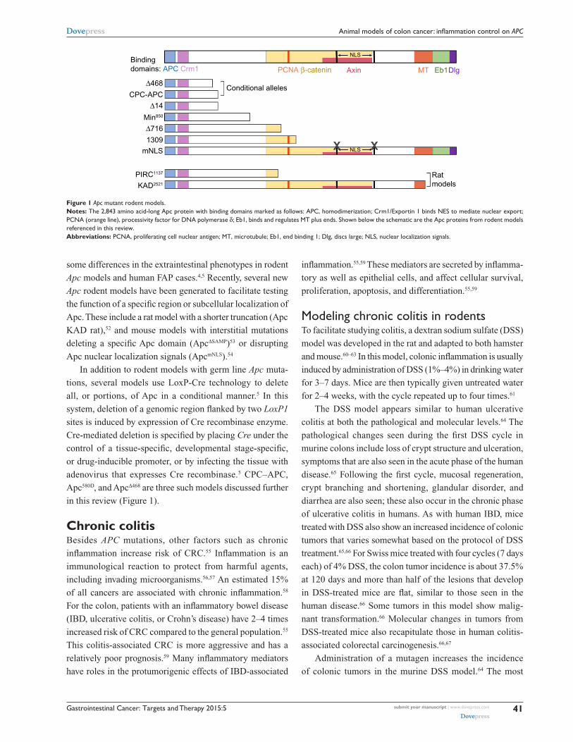
Modeling Apc in rodentsTo study APC biological functions in development and can-
cer, several mouse and rat models have been made. A more
comprehensive review of these models are provided in other
articles.4,5 Most of these models have mutations resulting in
truncated Apc, with lengths ranging from complete deletion
to deletion of only the C-terminal 300 amino acids. Figure
1 shows protein products resulting from Apc mutations in
rodent models that will be discussed in this review. These
models displayed some of the same phenotypes as patients
with germ line mutations of APC.5 Mice with Apc truncation
involving at least the C-terminal half of Apc develop intesti-
nal tumors, though the number of tumors does not correlate
with the extent of truncation.4 As in FAP patients, Apc trun-
cating mutations in these models are lethal in a homozygous
state, and tumor development requires mutation or loss of the
other (wild type) Apc allele.5 Tumors from these mouse mod-
els resemble those found in patients at both the histological
and molecular levels.49 However, the mouse tumors mainly
develop in small intestine, whereas FAP patients harbor
mostly colonic tumors.4 Rats with a mutation that truncates
Apc at amino acid 1137 develop tumors in both the small and
large intestine.50 In addition, unlike in humans, progression
to carcinoma is not typically seen in most Apc mutant mice,
presumably because of their limited lifespan.51 There are also
Gastrointestinal Cancer: Targets and Therapy 2015:5 submit your manuscript | www.dovepress.com
Dovepress
Dovepress
41
Animal models of colon cancer: inflammation control on APC
Bindingdomains: APC Crm1
CPC-APC
Min850
PIRC1137
KAD2521
∆468Conditional alleles
Ratmodels
PCNA β-catenin MTAxin
NLS
NLS
∆14
∆716
1309mNLS
Eb1Dlg
Figure 1 Apc mutant rodent models.Notes: The 2,843 amino acid-long Apc protein with binding domains marked as follows: APC, homodimerization; Crm1/exportin 1 binds NeS to mediate nuclear export; PCNA (orange line), processivity factor for DNA polymerase δ; eb1, binds and regulates MT plus ends. Shown below the schematic are the Apc proteins from rodent models referenced in this review.Abbreviations: PCNA, proliferating cell nuclear antigen; MT, microtubule; eb1, end binding 1; Dlg, discs large; NLS, nuclear localization signals.
some differences in the extraintestinal phenotypes in rodent
Apc models and human FAP cases.4,5 Recently, several new
Apc rodent models have been generated to facilitate testing
the function of a specific region or subcellular localization of
Apc. These include a rat model with a shorter truncation (Apc
KAD rat),52 and mouse models with interstitial mutations
deleting a specific Apc domain (Apc∆SAMP)53 or disrupting
Apc nuclear localization signals (ApcmNLS).54
In addition to rodent models with germ line Apc muta-
tions, several models use LoxP-Cre technology to delete
all, or portions, of Apc in a conditional manner.5 In this
system, deletion of a genomic region flanked by two LoxP1
sites is induced by expression of Cre recombinase enzyme.
Cre-mediated deletion is specified by placing Cre under the
control of a tissue-specific, developmental stage-specific,
or drug-inducible promoter, or by infecting the tissue with
adenovirus that expresses Cre recombinase.5 CPC–APC,
Apc580D, and Apc∆468 are three such models discussed further
in this review (Figure 1).
Chronic colitisBesides APC mutations, other factors such as chronic
inflammation increase risk of CRC.55 Inflammation is an
immunological reaction to protect from harmful agents,
including invading microorganisms.56,57 An estimated 15%
of all cancers are associated with chronic inflammation.58
For the colon, patients with an inflammatory bowel disease
(IBD, ulcerative colitis, or Crohn’s disease) have 2–4 times
increased risk of CRC compared to the general population.55
This colitis-associated CRC is more aggressive and has a
relatively poor prognosis.59 Many inflammatory mediators
have roles in the protumorigenic effects of IBD-associated
inflammation.55,59 These mediators are secreted by inflamma-
tory as well as epithelial cells, and affect cellular survival,
proliferation, apoptosis, and differentiation.55,59
Modeling chronic colitis in rodentsTo facilitate studying colitis, a dextran sodium sulfate (DSS)
model was developed in the rat and adapted to both hamster
and mouse.60–63 In this model, colonic inflammation is usually
induced by administration of DSS (1%–4%) in drinking water
for 3–7 days. Mice are then typically given untreated water
for 2–4 weeks, with the cycle repeated up to four times.61
The DSS model appears similar to human ulcerative
colitis at both the pathological and molecular levels.64 The
pathological changes seen during the first DSS cycle in
murine colons include loss of crypt structure and ulceration,
symptoms that are also seen in the acute phase of the human
disease.65 Following the first cycle, mucosal regeneration,
crypt branching and shortening, glandular disorder, and
diarrhea are also seen; these also occur in the chronic phase
of ulcerative colitis in humans. As with human IBD, mice
treated with DSS also show an increased incidence of colonic
tumors that varies somewhat based on the protocol of DSS
treatment.65,66 For Swiss mice treated with four cycles (7 days
each) of 4% DSS, the colon tumor incidence is about 37.5%
at 120 days and more than half of the lesions that develop
in DSS-treated mice are flat, similar to those seen in the
human disease.66 Some tumors in this model show malig-
nant transformation.66 Molecular changes in tumors from
DSS-treated mice also recapitulate those in human colitis-
associated colorectal carcinogenesis.66,67
Administration of a mutagen increases the incidence
of colonic tumors in the murine DSS model.64 The most

Gastrointestinal Cancer: Targets and Therapy 2015:5submit your manuscript | www.dovepress.com
Dovepress
Dovepress
42
Zeineldin and Neufeld
commonly used mutagen is azoxymethane (AOM), which
induces O6-methylguanine DNA adducts resulting in GA
transitions.64 A single intraperitoneal dose of AOM increases
the incidence of colonic cancer in DSS-treated mice to
100%.64 Another advantage of including a mutagen in the
protocol is that it allows reduction of the DSS dose in mice,
and decreases the mortality from DSS-associated acute
colitis. Again, different groups use different regimens of
AOM treatment: single or multiple doses of 7.5–20 mg/kg.
A single AOM dose of 10 mg/kg alone without DSS treatment
is not sufficient to induce tumors in wild-type mice.64
β-catenin mutations in exon 3 are detected in most
tumors from AOM–DSS-treated mice.68 These mutations
are expected to prevent phosphorylation and targeting of
β-catenin for destruction, resulting in cellular accumulation
and nuclear translocation of β-catenin, and promiscuous
activation of Wnt signaling.68 On the other hand, many AOM-
induced tumors in rats have Apc mutations.69 Both mice and
rats treated with AOM–DSS have activating mutations of the
proto-oncogene, Kras, in later stage tumors.68 Wnt and RAS
pathways are typically activated in human CRC.2
Intestinal epithelial barrier and gut microbiomeColon epithelial cells are exposed to a unique external
environment. The colon lumen contains hard fecal matter,
posing a potential threat of mechanical injury.70 In addition,
the colon is inhabited by over one hundred trillion bacterial
cells (almost ten times the number of cells in an adult human).
These gut microbes consume organic materials and secrete
various secondary metabolites.71,72 Intestinal epithelial cells
have several lines of defense that prevent bacterial invasion
or diffusion of harmful substances into the body while allow-
ing absorption of nutrients and beneficial substances.73 These
combined structural and physiological defenses are termed
the “intestinal epithelial barrier”.70,74
There are at least seven contributors to the intestinal epi-
thelial barrier (Figure 2). First is the actual physical barrier
created by mucus, which is continuously secreted by goblet
cells.75 This mucus is formed of two layers; an outer loose
layer and an inner adherent layer. The outer mucus lubricates
the solid contents of the colon to prevent mechanical injury
and also washes off microorganisms to prevent colonization.
1. Mucus secreted by Goblet cells is physical barrier
6. Gut associated lymphoid tissue (GALT)
M cell
Crypt
5. Paneth cells secrete anti-bacterial substances
3. Continuous epithelial cell replacement
7. TLR or NLR recognize PAMPs
4. Enteroendocrine secretions increase intestinal movement
Villus
2. Tight
junctions
Figure 2 intestinal epithelial barrier.Notes: Protecting the body from invasion by intestinal microbes requires many layers of defense. This illustration depicts the small intestine. The colon would have similar components but lack Paneth cells and the villus structure. Goblet cells (green); tight junctions (red); enteroendocrine cells (orange); Paneth cells (yellow).Abbreviations: DC, dendritic cell; T, T-cell; B, B-cell; TLR, Toll-like receptors; NLR, Nod-like receptors; PAMP, pathogen-associated molecular patterns.

Gastrointestinal Cancer: Targets and Therapy 2015:5 submit your manuscript | www.dovepress.com
Dovepress
Dovepress
43
Animal models of colon cancer: inflammation control on APC
The inner mucus layer prevents contact of microorganisms
and their products with the underlying epithelial cells.75
Second, epithelial cells lining the colon form a continuous
sheet with tight junctions that further prevent flora and harm-
ful molecules from penetration.76 Third, the continuous turn-
over of intestinal epithelial cells ensures rapid healing after
any damage or ulceration.70,77 Fourth, specialized epithelial
cells, enteroendocrine cells, respond to bacterial invasion
or toxic substances by secreting active amines to increase
intestinal movement and fluid secretion, thereby washing off
potential invaders.78 Fifth, in the small intestine, other special-
ized epithelial cells called Paneth cells secrete antibacterial
substances. Sixth, intestinal tissue also contains aggregations
of immune cells (gut-associated lymphoid tissues [GALT]
and other immune cells) that can detect foreign antigens and
defend the body against them. M-cells also contribute by
engulfing antigens and bacteria from the lumen and trans-
porting them to antigen presenting cells for immunological
processing.70 Seventh, intestinal epithelial cells themselves
detect different microbes and react to them by expressing
receptors that can recognize pathogen-associated molecular
patterns (PAMP) including Toll-like receptors (TLR) and
Nod-like receptors (NLR). These receptors do not recognize
specific antigens but specific molecular signatures associated
with pathogens eg, methylated DNA and peptidoglycans.79
Colitis and APC mutationsCRC is fundamentally a genetic disease, the result of accu-
mulated mutations in tumor suppressor genes and oncogenes.2
But the nature of the mutated genes and the order of their
mutation can vary with different precipitating factors.67,80,81
Activation of Wnt signaling is seen in the vast majority of
CRCs.2 Other signaling pathways that are commonly altered
during CRC progression include activation of K-ras, p53, and
TGF-β.82 Alterations in the same pathways are frequently seen
in cases with colitis-associated CRC. In addition, activation
of NF-κΒ and STAT3 pathways are also detected in colitis-
associated CRC. The sequence and role of these pathway
alterations in the development of CRC have been reviewed
previously.59 Here, we will focus on genetic mutations of the
tumor suppressor APC.
Mutation of APC is by far the most common genetic event
seen in CRC that leads to Wnt signal activation. Curiously,
APC mutations are not detected in other Wnt-dependent
tumors to nearly the same extent as seen in CRC. Rather, in
non-colonic tumors, mutations in other Wnt components, are
more commonly found,12 suggesting a colon-specific protec-
tive function of APC that is selected against during CRC
development. Furthermore, data from AOM–DSS models
suggest that Wnt signal activation alone is not sufficient for
effective initiation of colon tumorigenesis. Injection of mice
with a single dose of AOM, expected to induce oncogenic
β-catenin mutations which activate Wnt signaling, results
in no tumors or only a very low incidence of tumors.61,68
However, combing AOM with DSS-induced inflammation
results in robust tumor formation. Moreover, patients and
mice with germ line APC/Apc mutations develop intestinal
tumors with 100% penetrance.5,9
The data supporting an association between APC muta-
tions and inflammation are overwhelming. Inflammation can
greatly increase intestinal tumorigenesis in rodent models
with germ line Apc mutations. DSS treatment of ApcMin/+
mice increases their colon tumor multiplicity by 15–30-fold.83
Unlike AOM-induced tumors in wild-type mice treated with
DSS, which show β-catenin stabilizing mutations, colonic
tumors in DSS-treated ApcMin/+ mice typically show loss of
the wild-type Apc allele.83 The latter mechanism is similar
to that seen in tumors from ApcMin/+ mice not treated with
DSS.84 Of note, the multiplicity of tumors in DSS-treated
ApcMin/+ mice is higher than in wild-type mice treated with
the mutagen AOM followed by DSS.83 Collectively, these data
strongly support a colon-specific tumor suppressor function
for APC beyond that as a Wnt signal antagonist, potentially
to control colitis.
Experimental induction of inflammation in mouse intesti-
nal tumor models by methods other than DSS administration
also increases tumorigenesis. Germ line deletion of Il-10 (an
anti-inflammatory cytokines) or single immunoglobulin Il-1
receptor-related (SIGIRR) molecule increases intestinal
tumors in ApcMin/+ mice.85,86 Transgenic expression of Il-8
(a proinflammatory cytokine) enhances tumorigenesis in
both AOM–DSS and ApcMin/+ models.87 In addition, Nrf2
knockout mice display increased oxidative stress, increased
inflammatory markers, and colitis and accelerated intestinal
tumorigenesis.88,89 Conversely, reducing inflammation protects
from intestinal tumorigenesis. Nonsteroidal anti-inflammatory
drugs (NSAIDs) reduce polyp formation in FAP patients as
well as in ApcMin/+, Apc∆474/+, and Apc1309/+ mouse models.90–94
Experimental genetic deletion of proinflammatory mediators
CXCR2, CD24, TNF-α, and epimorphin significantly reduces
intestinal tumor numbers in ApcMin/+ mice.95–98
Inflammation might also contribute to some other known
risk and protective factors in CRC. For example, high fat
diets and obesity predispose humans to CRC, ApcMin/+ mice
to increased intestinal polyposis, and AOM-treated mice to
pre-cancerous colon lesions.99,100 Obesity has been associated

Gastrointestinal Cancer: Targets and Therapy 2015:5submit your manuscript | www.dovepress.com
Dovepress
Dovepress
44
Zeineldin and Neufeld
with adipose tissue macrophage malfunction and low-level
inflammation.101–103 A recent report showed increased inflam-
matory mediators in ApcMin/+ mice on high fat diet relative to
ApcMin/+ mice on regular lab diet.104 In addition, many natural
products including curcumin, grape antioxidant fibers, and
brown rice reduce colon tumors in various mouse models,
presumably by reducing inflammation.105–109
The mechanisms by which inflammation can enhance colon
tumorigenesis are not completely delineated. Inflammation
activates many pathways that synergize with Wnt signal
activation in CRC tumorigenesis including AKT, KRAS,
BRAF, HIF1-α, and TGF-β. DNA damage and epigenetic
changes that are associated with inflammation could also
contribute to tumor formation.59
Many inflammatory pathways converge to activate the
prosurvival NF-κB pathway,59 which is also activated in
colonic mucosa from IBD patients.110 NF-κB pathway activa-
tion increases proliferation and decreases apoptosis in CRC
cell lines and mouse colon mucosa,110,111 drugs that inhibit the
NF-κB pathway decrease intestinal tumorigenesis in ApcMin/+
mice.112 Aspirin, an NSAID that decreases intestinal poly-
posis in both mouse models and FAP patients and protects
from CRC, inhibits the NF-κB pathway and also increases
Apc/APC expression.113
Inflammation can increase DNA damage and acceler-
ate mutagenesis. The rate of reactive oxygen species (ROS)
production, including nitric oxide (NO), is augmented in
inflamed tissues. ROS are genotoxic and increase DNA muta-
tion rates.55,58,114,115 Inhibiting NO production reduces intestinal
polyp formation in ApcMin/+ mice as well as inflammatory mod-
els of colitis.116,117 Notably, activation of the NF-κB pathway
by constitutive activation of its upstream activator, IKKβ,
enhances intestinal polyposis and elevates DNA damage in
Apc580D/+ mice.118 NO synthase inhibitors reduce this DNA dam-
age and intestinal tumorigenesis, suggesting that accelerating
Apc LOH (loss of heterozygosity) due to the DNA damaging
effect of NO is the cause of enhanced tumorigenicity in these
mice.118 Inflammation may also induce DNA damage by
increasing the production of other mutagenic factors including
trans-4-hydroxy-2-nonenal from the activated inflammatory
cells, which can further induce chromosomal breakage in
nearby epithelial cells.119 Moreover, chronic inflammation can
also reduce DNA mismatch repair proteins.120,121
Chronic inflammation is also associated with epigenetic
changes including changes in miRNA, DNA hypermethyla-
tion, and aberrant methyl histone markings.122 Colitis leads
to upregulation of miRNA-155123; miRNA-155 targets APC
and thus, activates β-catenin.124 The protumorigenic effect of
chronic colitis has also been linked to prostaglandin (PG) for-
mation through induction of cyclooxygenase-2 (COX-2).125
COX-2 is the rate-limiting step in PGE2 formation from
arachidonic acid.122 Both Cox-2 and PGE2 promote Wnt
signaling, increase cellular proliferation, inhibit apopto-
sis, promote angiogenesis, and enhance metastasis.126–129
Conditional deletion of Cox-2 results in significant reduction
of intestinal tumors in ApcMin/+ and Apc∆716/+ mice,130,131 Cox-2
is also targeted by NSAIDs and selective Cox-2 inhibitors
such as Celebrex, both of which reduce intestinal tumori-
genesis in patients and mouse models with germ line APC/
Apc mutations.122
APC mutations and inflammationIn the previous section we presented evidence that inflam-
mation accelerates intestinal tumorigenesis in the presence
of Apc mutations. However, there is evidence that Apc
mutations can enhance colitis. Proinflammatory mediators
Cox-1, Cox-2, MIP-2, OPN, CXCR-2, and Gro-α mRNA
are upregulated in colonic polyps in ApcMin/+ mice relative
to epithelial cells from normal mice.132 Of these genes, only
Cox-2 is a defined Wnt target.133,134 The other mediators have
not been linked to activated Wnt signaling resulting from Apc
mutations. In addition, mRNA and serum protein levels of
proinflammatory cytokines MCP-1, IL-6, IL-1β, and TNF-α
increase with the progression of intestinal tumorigenesis and
correlate with tumor size.135 Moreover, a global expression
analysis showed differential expression of inflammatory
genes, Lcn2 and N4wbp4, in ApcMin/+ polyps.136 In another
mouse model (CPC–APC), conditional truncation of Apc
in the distal part of the small intestine and colon resulted in
inflammatory cell infiltration and upregulation of Il-17 and
Il-23 in the developing polyps.137
Recently, we described a mouse model with a germ line
Apc mutation that compromises the ability of Apc to locate
to the nucleus.54 These ApcmNLS/mNLS mice only rarely develop
tumors, and homozygous mutant mice are viable. However,
the ApcmNLS allele increases tumor formation when combined
with the ApcMin allele (ApcmNLS/Min mice).54 Notably, ApcmNLS/mNLS
mice have higher expression of inflammatory mediators
Cox-2 and MIP-2 and are more susceptible to DSS-induced
colitis and AOM–DSS-induced colon tumorigenesis.138 Rats
with germ line Apc mutation resulting in truncation of the
C-terminal 300 amino acids (KAD rats) do not develop tumors
but are also more susceptible to DSS-induced inflammation
and AOM–DSS-induced colon tumorigenesis.139
APC mutation can induce colitis by several mechanisms.
First, APC mutations can decrease mucus production and

Gastrointestinal Cancer: Targets and Therapy 2015:5 submit your manuscript | www.dovepress.com
Dovepress
Dovepress
45
Animal models of colon cancer: inflammation control on APC
therefore reduce the barrier between gut microbes and
intestinal tissues.137 Apc normally functions in promoting
cellular differentiation of intestinal lineages including mucus-
producing goblet cells.23,140 ApcmNLS/mNLS mice have reduced
expression of Hath-1 and fewer goblet cells in their small
intestines and less Muc-2 mRNA in their colons, relative
to their wild-type littermates.54,138 Hath-1 is a transcription
factor that participates in goblet cell differentiation and is
negatively regulated by Wnt signaling.17,141 Muc-2 is the
major protective mucin in the colon. Muc-2 knockout mice
develop colitis and have spontaneous colonic tumors.142,143
Muc-2 mutation also enhances intestinal tumorigenesis in
ApcMin/+ mice.143,144 Furthermore, induction of inflamma-
tion in ApcmNLS/mNLS mice using DSS results in significantly
fewer goblet cells and reduced Muc-2 mRNA, relative to
DSS-treated wild-type mice.138 Goblet cell differentiation
requires low Notch signal and treating ApcMin/+ mice with a
γ-secretase inhibitor, inhibited Notch signaling and increased
goblet cell differentiation in intestinal tumors.145 A potential
link between Notch signaling and APC is that APC is in a
double negative feedback loop with the transcription inhibitor
Msi-1.146 Msi-1 activates Notch signaling by inhibiting the
Notch repressor, Numb.147 In cases of Apc mutation, Msi-1
is upregulated; activating Notch signaling.23,148 However,
a direct role of Msi-1 in goblet cell differentiation has not
been examined. Finally, FAP patients and CPC–APC mice
with conditional truncation of APC/Apc showed reduced
mucus production of polyps, which displayed Apc LOH.137
Colonic mucosa in AOM-treated rats as well as FAP patients
shows foci with depleted mucin.149,150 These mucin-depleted
foci are correlated with tumor number and have high rates
of Apc mutations.151 Apc-mutant (PIRC) rats also show
mucin-depleted foci that increase in number as the rats age.152
Notably, the NSAID sulindac, reduces the number of polyps
as well as mucin-depleted foci in PIRC rats.152 Collectively,
these data suggest that Apc mutations predispose to the pre-
cancerous mucin-depleted foci.
Alteration of Apc can also affect other intestinal epithe-
lial barrier activities. APC loss effects localization of tight
junction protein ZO-1.153 Loss of APC and upregulated Wnt
signaling are also associated with increased expression of
tight junction protein claudin-1 in CRCs.154 Further, inducible
Apc truncation in CPC–APC mice leads to reduced junctional
claudin-3, -4, -5, and -7 and decreased levels of JAM-C
(junctional adhesion molecule-C) mRNA.137 The C-terminus
of Apc binds to the junctional protein DLG (Figure 1). In
KAD rats, Dlg5 fails to localize to the junction in endothelial
cells, resulting in delayed healing after DSS-induced inflam-
mation.155 Finally, APC interacts with cytoskeletal proteins
including those of microtubules and intermediate filaments,
which are important in formation and maintenance of tight
junctions.31,156,157 Apc mutations alter cytoskeletal organiza-
tion in intestinal epithelial cells and affect cell polarity.158
Whether these changes in epithelial organization enhance
colitis is not clear.
Apc mutations might also induce inflammation by acti-
vating Wnt signaling. Cox-2 and iNOS are Wnt targets.134,159
Cox-2 is the rate-limiting enzyme in PGE2 synthesis. PGE2
is involved in processes that lead to inflammation, including,
vasodilation, increasing vascular permeability, and chemo-
attraction of inflammatory cells.59
APC, colitis, and microbiome in CRCThe role of intestinal flora in health and disease is getting
increasing attention of late.160,161 The development of tools
such as deep sequencing has allowed rapid analysis of dif-
ferent intestinal bacteria. The gastrointestinal tract in general
and especially the distal portion is home to a large number of
microorganisms. The relationship between these florae and
the host is mostly symbiotic.160,161 The host provides a niche
and nutrients, while intestinal florae provide essential vita-
mins and are crucial for the development of the host immune
system. Particular intestinal florae also prevent overgrowth
of pathogenic microorganisms by competing with them for
limited resources. However, changes in the number, type, or
the relative abundance of different intestinal microorganisms
(dysbiosis) have been related to many pathological condi-
tions including IBD and CRC.160,162 The challenging task for
the intestinal epithelial barrier is to regulate the intestinal
microbiome by allowing the growth of beneficial species
and preventing the growth and invasion of pathogenic and
opportunistic organisms.
Disruption of the intestinal epithelial barrier is a hallmark
of IBD.163 However, the relationship between the epithelial
barrier, intestinal florae, and inflammation has multiple levels
of complexity. Mucus secretion is stimulated by bacterial
colonization.164 Germ-free mice have a thin mucus layer,
which can be restored to normal thickness by bacterial prod-
ucts including peptidoglycans and lipopolysaccharides.164,165
Bacterial products including butyrate and short chain fatty
acids also can induce Muc2 transcription via c-Fos/c-Jun and
by epigenetic histone alterations.166–169 On the other hand,
microbes or their metabolic products may induce inflam-
matory reactions in the colon. Some intestinal florae such as
Fusobacteria and Surpulina are enriched in the mucus layer
covering regions of enteric inflammation,170,171 consistent

Gastrointestinal Cancer: Targets and Therapy 2015:5submit your manuscript | www.dovepress.com
Dovepress
Dovepress
46
Zeineldin and Neufeld
with their ability to dissolve the mucus layer and thus provide
access to other microbes.172 Clostridia-like gram-positive seg-
mented filamentous bacteria induce intestinal inflammation
which predisposes to colitis but also protects mice from some
enteric infections.173 In contrast, some bacterial products
such as short chain fatty acids and butyrate inhibit colitis by
stimulating epithelial cells to secrete the anti-inflammatory
cytokines IL-10 and IL-18.174–176
Several mechanisms linking the colonic microbiome to
CRC have been proposed. In human patients, the florae of
colonic adenomas and adenocarcinomas are enriched with
fusobacterial species relative to normal colon tissue.177,178
Fusobacteria enhance intestinal tumorigenesis in ApcMin/+
mice resulting in a proinflammatory gene expression sig-
nature in the tumor cells.177 Reducing microbial-induced
inflammation by deleting the PAMP pathway adaptor
protein Myd88 decreases intestinal tumors in ApcMin/+ mice
and colon tumors in AOM-treated mice.179,180 Furthermore,
transplantation of bone marrow from mice with mutations
in genes encoding PAMP adaptor proteins Myd88, Tlr2, 4,
and 9 reduces inflammation and tumor load in CPC–APC
mice.137 Finally, deletion of anti-inflammatory cytokine
Il-10 alters the intestinal microbiota and increases the
intestinal tumor number in Apc∆468 mice; treating these
mice with broad-spectrum antibiotics decreased the overall
microbial diversity and also decreased the intestinal tumor
multiplicity.181
Microorganisms can also secrete carcinogenic metabo-
lites that can mutate DNA. In addition to ROS produced by
inflammatory cells as the result of bacterial-induced inflam-
mation, some colonic bacteria including the gram-positive
Enterococcus faecalis produce hydroxyl radicals.182–184
Still, other colon-inhabitant gram-negative, Escherichia
coli, produce a toxin that can cause DNA damage and
CRC.185 Bacteria may also secrete chemicals that directly
induce proliferation. For example, the exotoxin fragilysin
secreted by some Bacteroid species induces c-Myc which
stimulates cellular proliferation.186 Bacterial metabolites
such as H2S are produced by many Enterobacterial species
commonly found in the normal colon.187 H2S can activate
the RAS-MEK pathway and induce cellular proliferation
in mice.188
Goblet cell
Junctiondefects
Geneexpression
ROS/DNAdamage
Epigeneticchanges
Cytokines,protumorigenic
iNOS COX2
Barrier
MicrobesAPCmutation
Othermutations
CRC
Inflammation
differentiation
Figure 3 Potential roles for APC in inflammation.Notes: APC normally promotes differentiation of goblet cells which generate and secrete mucus. Protective mucus layers provide a physical barrier between luminal microbes and the epithelial cells lining the intestine. APC interacts with various junctional proteins, further contributing to a barrier between the luminal contents and the immune cells of the stroma. APC regulates expression of genes, some of which are involved in inflammation. Microbial breach of the intestinal barrier results in inflammation. Consequences of inflammation include DNA damage and epigenetic changes that can result in additional mutation of tumor suppressor genes and oncogenes that further promote colorectal carcinogenesis.Abbreviations: APC, adenomatous polyposis coli; iNOS, induced nitric oxide synthase; CRC, colorectal cancer; ROS, reactive oxygen species; COX-2, cyclooxygenase-2.

Gastrointestinal Cancer: Targets and Therapy 2015:5 submit your manuscript | www.dovepress.com
Dovepress
Dovepress
47
Animal models of colon cancer: inflammation control on APC
Several observations made in mouse models point to an
interaction between genetic lesions, intestinal florae and CRC.
Smad3-deficient mice develop colon tumors only in the pres-
ence of helicobacter infection.189 Tbx2 and Rag2-/- ulcerative
colitis (TRUC) mice develop colitis and colitis-associated
colon cancer, but not when raised in a germ-free environ-
ment.190 Similarly, Il10-/- mice develop colitis-associated
colon tumors only if they have intestinal bacteria.191 NLRP6
is a component of the innate immune response that senses
microbes, and NLRP6 deletion in intestinal epithelial cells
induces colitis and colitis-associated tumorigenesis.192 These
NLRP6-deficient mice also have changes in the bacterial flora
composition with more abundant Bacteroids in the colon.
Remarkably, cohousing these NLRP6-mutant mice with wild-
type mice results in development of colitis and colon tumors
in the wild-type mice, consistent with transmissible tumor
promoter.192,193 A similar transmissible, tumor-promoter has
been described in mice with mutations in other compo-
nents of the innate immune response, NOD2 and RIP2.194
Furthermore, expression of the secreted anti-inflammation
mediator/antimicrobial, Pla2g2a in intestinal epithelial cells
reduces the incidence of intestinal polyps in ApcMin/+ mice and
in orthotopic xenografts of human colon cancer cells.195,196
Notably, exogenous expression of the Pla2g2a gene pre-
vents colon tumorigenesis in Muc2-deficient mice.197
Although connections are starting to emerge, the precise
relationship between the tumor suppressor Apc and intestinal
flora is not well defined. ApcMin/+ mice raised in a germ-free
environment develop fewer polyps than ApcMin/+ mice housed
in standard conditions.198 However, this tumor reduction is
statistically significant only in the middle portion of the small
intestine, with no reduction in the number of tumors in the
colon.198 This region specificity may represent a varied role
for different microbial species in discrete regions of the gas-
trointestinal tract. On the other hand, Apc∆14/+ mice developed
more polyps when raised in germ-free conditions than in
standard housing conditions.199 Together, these data suggest
an allele-specific interaction of Apc with the microbial con-
tent of the gut. Notably, mutations in ApcMin/+ and Apc∆14/+ are
expected to result in truncated Apc proteins that differ by 403
amino acids.5 The contrasting effect of germ-free conditions
on polyp number in ApcMin/+ and Apc∆14/+could also represent
other contributing factors that vary between the two experi-
mental conditions including other genetic loci and diet.4
ConclusionThe results gathered from studies of rodent CRC models
reveal a complex interplay of genetics, inflammation, and
the microbiome that gives rise to a cancer phenotype. APC
is a major tumor suppressor in the colon. Although the
most universally appreciated APC role is that of Wnt signal
antagonist, APC is multifaceted. In this review, we describe
an emerging role for APC in colitis. We propose that this
APC role as regulator of the inflammatory response might
be particularly critical in the colon and thus contribute to the
high frequency of APC mutations seen in CRC compared to
cancers of other tissues (Figure 3). Unearthing the precise
role for APC in suppression of inflammation will expand
the repertoire of therapeutic strategies aimed at rescuing the
functions of this multifaceted and fascinating tumor sup-
pressor protein.
AcknowledgmentsThe authors recognize grants P30CA168524 and P20
RR016475 for financial support.
DisclosureThe authors report no conflicts of interest in this work.
References 1. American Cancer Society. Cancer Facts and Figures 2014. Atlanta,
GA: American Cancer Society; 2014. 2. Kinzler KW, Vogelstein B. Lessons from hereditary colorectal cancer.
Cell. 1996;87(2):159–170. 3. Singh S, Singh PP, Murad MH, Singh H, Samadder NJ. Prevalence, risk
factors, and outcomes of interval colorectal cancers: a systematic review and meta-analysis. Am J Gastroenterol. 2014;109(9):1375–1389.
4. Zeineldin M, Neufeld KL. Understanding phenotypic variation in rodent models with germline apc mutations. Cancer Res. 2013;73(8): 2389–2399.
5. Zeineldin M, Neufeld KL. More than two decades of Apc modeling in rodents. Biochim Biophys Acta. 2013;1836(1):80–89.
6. Powell SM, Zilz N, Beazer-Barclay Y, et al. APC mutations occur early during colorectal tumorigenesis. Nature. 1992;359(6392):235–237.
7. Kinzler KW, Nilbert MC, Su LK, et al. Identification of FAP locus genes from chromosome 5q21. Science. 1991;253(5020):661–665.
8. Groden J, Thliveris A, Samowitz W, et al. Identification and charac-terization of the familial adenomatous polyposis coli gene. Cell. 1991; 66(3):589–600.
9. Nieuwenhuis MH, Vasen HF. Correlations between mutation site in APC and phenotype of familial adenomatous polyposis (FAP): a review of the literature. Crit Rev Oncol Hematol. 2007;61(2):153–161.
10. Senda T, Iizuka-Kogo A, Onouchi T, Shimomura A. Adenomatous polyposis coli (APC) plays multiple roles in the intestinal and colorectal epithelia. Med Mol Morphol. 2007;40(2):68–81.
11. Phelps RA, Broadbent TJ, Stafforini DM, Jones DA. New perspectives on APC control of cell fate and proliferation in colorectal cancer. Cell Cycle. 2009;8(16):2549–2556.
12. Polakis P. Wnt signaling and cancer. Genes Dev. 2000;14(15): 1837–1851.
13. Klaus A, Birchmeier W. Wnt signalling and its impact on development and cancer. Nat Rev Cancer. 2008;8(5):387–398.
14. He TC, Sparks AB, Rago C, et al. Identification of c-MYC as a target of the APC pathway. Science. 1998;281(5382):1509–1512.
15. Tetsu O, McCormick F. Beta-catenin regulates expression of cyclin D1 in colon carcinoma cells. Nature. 1999;398(6726):422–426.

Gastrointestinal Cancer: Targets and Therapy 2015:5submit your manuscript | www.dovepress.com
Dovepress
Dovepress
48
Zeineldin and Neufeld
16. Yan D, Wiesmann M, Rohan M, et al. Elevated expression of axin2 and hnkd mRNA provides evidence that Wnt/beta-catenin signaling is activated in human colon tumors. Proc Natl Acad Sci U S A. 2001;98(26): 14973–14978.
17. Leow CC, Romero MS, Ross S, Polakis P, Gao WQ. Hath1, down-regulated in colon adenocarcinomas, inhibits proliferation and tumori-genesis of colon cancer cells. Cancer Res. 2004;64(17):6050–6057.
18. Sancho E, Batlle E, Clevers H. Signaling pathways in intestinal devel-opment and cancer. Annu Rev Cell Dev Biol. 2004;20:695–723.
19. Barker N. Adult intestinal stem cells: critical drivers of epithelial homeo-stasis and regeneration. Nat Rev Mol Cell Biol. 2014;15(1):19–33.
20. Cadigan KM, Nusse R. Wnt signaling: a common theme in animal development. Genes Dev. 1997;11(24):3286–3305.
21. Ricci-Vitiani L, Fabrizi E, Palio E, De Maria R. Colon cancer stem cells. J Mol Med. 2009;87(11):1097–1104.
22. Barker N, Clevers H. Tracking down the stem cells of the intestine: strategies to identify adult stem cells. Gastroenterology. 2007;133(6): 1755–1760.
23. Sansom OJ, Reed KR, Hayes AJ, et al. Loss of Apc in vivo immediately perturbs Wnt signaling, differentiation, and migration. Genes Dev. 2004;18(12):1385–1390.
24. Fodde R, Kuipers J, Rosenberg C, et al. Mutations in the APC tumour suppressor gene cause chromosomal instability. Nat Cell Biol. 2001;3(4):433–438.
25. Rubinfeld B, Souza B, Albert I, et al. Association of the APC gene product with beta-catenin. Science. 1993;262(5140):1731–1734.
26. Su LK, Vogelstein B, Kinzler KW. Association of the APC tumor sup-pressor protein with catenins. Science. 1993;262(5140):1734–1737.
27. Nathke IS, Adams CL, Polakis P, Sellin JH, Nelson J. The adenomatous polyposis coli tumor suppressor protein localizes to plasma membrane sites involved in active cell migration. J. Cell Biol. 1996;134:165–179.
28. Deka J, Kuhlmann J, Muller O. A domain within the tumor suppres-sor protein APC shows very similar biochemical properties as the microtubule-associated protein tau. Eur J Biochem. 1998;253(3): 591–597.
29. Su LK, Burrell M, Hill DE, et al. APC binds to the novel protein EB1. Cancer Res. 1995;55(14):2972–2977.
30. Askham JM, Moncur P, Markham AF, Morrison EE. Regulation and function of the interaction between the APC tumour suppressor protein and EB1. Oncogene. 2000;19(15):1950–1958.
31. Wang Y, Azuma Y, Friedman DB, Coffey RJ, Neufeld KL. Novel association of APC with intermediate filaments identified using a new versatile APC antibody. BMC Cell Biol. 2009;10:75.
32. Green RA, Kaplan KB. Chromosome instability in colorectal tumor cells is associated with defects in microtubule plus-end attachments caused by a dominant mutation in APC. J Cell Biol. 2003;163(5): 949–961.
33. Radulescu S, Ridgway RA, Appleton P, et al. Defining the role of APC in the mitotic spindle checkpoint in vivo: APC-deficient cells are resistant to Taxol. Oncogene. 2010;29(49):6418–6427.
34. Anderson CB, Neufeld KL, White RL. Subcellular distribution of Wnt pathway proteins in normal and neoplastic colon. Proc Natl Acad Sci U S A. 2002;99(13):8683–8688.
35. Henderson BR. Nuclear-cytoplasmic shuttling of APC regulates beta-catenin subcellular localization and turnover. Nat Cell Biol. 2000;2(9):653–660.
36. Neufeld KL. Nuclear APC. Adv Exp Med Biol. 2009;656:13–29. 37. Zhang F, White RL, Neufeld KL. Phosphorylation near nuclear local-
ization signal regulates nuclear import of adenomatous polyposis coli protein. Proc Natl Acad Sci U S A. 2000;97(23):12577–12582.
38. Neufeld KL, Zhang F, Cullen BR, White RL. APC-mediated down-regulation of beta-catenin activity involves nuclear sequestration and nuclear export. EMBO Rep. 2000;1(6):519–523.
39. Wang Y, Azuma Y, Moore D, Osheroff N, Neufeld KL. Interaction between tumor suppressor adenomatous polyposis coli and topoi-somerase II{alpha}: implication for the G2/M transition. Mol Biol Cell. 2008;19(10):4076–4085.
40. Wang Y, Coffey RJ, Osheroff N, Neufeld KL. Topoisomerase IIalpha binding domains of adenomatous polyposis coli influence cell cycle progression and aneuploidy. PLoS One. 2010;5(4):e9994.
41. Jaiswal AS, Balusu R, Armas ML, Kundu CN, Narayan S. Mechanism of adenomatous polyposis coli (APC)-mediated blockage of long-patch base excision repair. Biochemistry. 2006;45(51):15903–15914.
42. Jaiswal AS, Banerjee S, Aneja R, Sarkar FH, Ostrov DA, Narayan S. DNA polymerase beta as a novel target for chemotherapeutic interven-tion of colorectal cancer. PLoS One. 2011;6(2):e16691.
43. Jaiswal AS, Narayan S. A novel function of adenomatous polyposis coli (APC) in regulating DNA repair. Cancer Lett. 2008;271(2): 272–280.
44. Jaiswal AS, Narayan S. Assembly of the base excision repair complex on abasic DNA and role of adenomatous polyposis coli on its functional activity. Biochemistry. 2011;50(11):1901–1909.
45. Narayan S, Jaiswal AS, Balusu R. Tumor suppressor APC blocks DNA polymerase beta-dependent strand displacement synthesis dur-ing long patch but not short patch base excision repair and increases sensitivity to methylmethane sulfonate. J Biol Chem. 2005;280(8): 6942–6949.
46. Larabee JL, Shakir SM, Hightower L, Ballard JD. Adenomatous poly-posis coli protein associates with C/EBP beta and increases Bacillus anthracis edema toxin stimulated gene expression in macrophages. J Biol Chem. 2011;286(22):19364–19372.
47. Deka J, Herter P, Sprenger-Haussels M, et al. The APC protein binds to A/T rich DNA sequences. Oncogene. 1999;18(41):5654–5661.
48. Kohler EM, Derungs A, Daum G, Behrens J, Schneikert J. Functional definition of the mutation cluster region of adenomatous polyposis coli in colorectal tumours. Hum Mol Genet. 2008;17(13):1978–1987.
49. Gaspar C, Cardoso J, Franken P, et al. Cross-species comparison of human and mouse intestinal polyps reveals conserved mechanisms in adenomatous polyposis coli (APC)-driven tumorigenesis. Am J Pathol. 2008;172(5):1363–1380.
50. Amos-Landgraf JM, Kwong LN, Kendziorski CM, et al. A target- selected Apc-mutant rat kindred enhances the modeling of familial human colon cancer. Proc Natl Acad Sci U S A. 2007;104(10):4036–4041.
51. Moser AR, Pitot HC, Dove WF. A dominant mutation that predisposes to multiple intestinal neoplasia in the mouse. Science. 1990;247(4940): 322–324.
52. Yokoyama A, Nomura R, Kurosumi M, et al. The C-terminal domain of the adenomatous polyposis coli (Apc) protein is involved in thyroid mor-phogenesis and function. Med Mol Morphol. 2011;44(4):207–212.
53. Lewis A, Davis H, Deheragoda M, et al. The C-terminus of Apc does not influence intestinal adenoma development or progression. J Pathol. 2012;226(1):73–83.
54. Zeineldin M, Cunningham J, McGuinness W, et al. A knock-in mouse model reveals roles for nuclear Apc in cell proliferation, Wnt signal inhi-bition and tumor suppression. Oncogene. 2012;31(19):2423–2437.
55. Rizzo A, Pallone F, Monteleone G, Fantini MC. Intestinal inflammation and colorectal cancer: a double-edged sword? World J Gastroenterol. 2011;17(26):3092–3100.
56. Rowland KJ, Choi PM, Warner BW. The role of growth factors in intestinal regeneration and repair in necrotizing enterocolitis. Semin Pediatr Surg. 2013;22(2):101–111.
57. Borrello MG, Degl’Innocenti D, Pierotti MA. Inflammation and cancer: the oncogene-driven connection. Cancer Lett. 2008;267(2):262–270.
58. Rakoff-Nahoum S. Why cancer and inflammation? Yale J Biol Med. 2006;79(3–4):123–130.
59. Terzic J, Grivennikov S, Karin E, Karin M. Inflammation and colon cancer. Gastroenterology. 2010;138(6):2101–2114.
60. Okayasu I, Hatakeyama S, Yamada M, Ohkusa T, Inagaki Y, Nakaya R. A novel method in the induction of reliable experimental acute and chronic ulcerative colitis in mice. Gastroenterology. 1990;98(3):694–702.
61. Clapper ML, Cooper HS, Chang WC. Dextran sulfate sodium-induced colitis-associated neoplasia: a promising model for the development of chemopreventive interventions. Acta Pharmacol Sin. 2007;28(9):1450–1459.

Gastrointestinal Cancer: Targets and Therapy 2015:5 submit your manuscript | www.dovepress.com
Dovepress
Dovepress
49
Animal models of colon cancer: inflammation control on APC
62. Hirono I, Kuhara K, Hosaka S, Tomizawa S, Golberg L. Induction of intestinal tumors in rats by dextran sulfate sodium. J Natl Cancer Inst. 1981;66(3):579–583.
63. Ohkusa T. [Production of experimental ulcerative colitis in hamsters by dextran sulfate sodium and changes in intestinal microflora]. Nihon Shokakibyo Gakkai zasshi. 1985;82(5):1327–1336. Japanese.
64. Tanaka T, Kohno H, Suzuki R, Yamada Y, Sugie S, Mori H. A novel inflammation-related mouse colon carcinogenesis model induced by azoxymethane and dextran sodium sulfate. Cancer Sci. 2003;94(11): 965–973.
65. Cooper HS, Murthy SN, Shah RS, Sedergran DJ. Clinicopathologic study of dextran sulfate sodium experimental murine colitis. Lab Invest. 1993;69(2):238–249.
66. Cooper HS, Murthy S, Kido K, Yoshitake H, Flanigan A. Dysplasia and cancer in the dextran sulfate sodium mouse colitis model. Relevance to colitis-associated neoplasia in the human: a study of histopathol-ogy, B-catenin and p53 expression and the role of inflammation. Carcinogenesis. 2000;21(4):757–768.
67. Aust DE, Terdiman JP, Willenbucher RF, et al. The APC/beta-catenin pathway in ulcerative colitis-related colorectal carcinomas: a mutational analysis. Cancer. 2002;94(5):1421–1427.
68. Takahashi M, Wakabayashi K. Gene mutations and altered gene expres-sion in azoxymethane-induced colon carcinogenesis in rodents. Cancer Sci. 2004;95(6):475–480.
69. De Filippo C, Caderni G, Bazzicalupo M, et al. Mutations of the Apc gene in experimental colorectal carcinogenesis induced by azoxymethane in F344 rats. Br J Cancer. 1998;77(12):2148–2151.
70. Camilleri M, Madsen K, Spiller R, Van Meerveld BG, Verne GN. Intestinal barrier function in health and gastrointestinal disease. Neurogastroent Motil. 2012;24(10):976–976.
71. Voreades N, Kozil A, Weir TL. Diet and the development of the human intestinal microbiome. Front Microbiol. 2014;5:494.
72. Nuding S, Antoni L, Stange EF. The host and the flora. Dig Dis. 2013;31(3–4):286–292.
73. Serban DE. Gastrointestinal cancers: influence of gut microbiota, probiotics and prebiotics. Cancer Lett. 2014;345(2):258–270.
74. Dorofeyev AE, Vasilenko IV, Rassokhina OA, Kondratiuk RB. Mucosal barrier in ulcerative colitis and Crohn’s disease. Gastroenterol Res Pract. 2013;2013:431231.
75. Johansson ME, Hansson GC. Mucus and the goblet cell. Dig Dis. 2013;31(3–4):305–309.
76. Liang GH, Weber CR. Molecular aspects of tight junction barrier function. Curr Opin Pharmacol. 2014;19C:84–89.
77. Gunther C, Buchen B, Neurath MF, Becker C. Regulation and pathophysiological role of epithelial turnover in the gut. Semin Cell Dev Biol. 2014;35C:40–50.
78. Raybould HE. Gut microbiota, epithelial function and derangements in obesity. J Physiol. 2012;590(Pt 3):441–446.
79. Gkouskou KK, Deligianni C, Tsatsanis C, Eliopoulos AG. The gut microbiota in mouse models of inflammatory bowel disease. Front Cell Infect Microbiol. 2014;4:28.
80. Kern SE, Redston M, Seymour AB, et al. Molecular genetic profiles of colitis-associated neoplasms. Gastroenterology. 1994;107(2):420–428.
81. Fogt F, Vortmeyer AO, Goldman H, Giordano TJ, Merino MJ, Zhuang Z. Comparison of genetic alterations in colonic adenoma and ulcerative colitis-associated dysplasia and carcinoma. Hum Pathol. 1998;29(2):131–136.
82. Fearon ER, Vogelstein B. A genetic model for colorectal tumorigenesis. Cell. 1990;61(5):759–767.
83. Cooper HS, Everley L, Chang WC, et al. The role of mutant Apc in the development of dysplasia and cancer in the mouse model of dextran sulfate sodium-induced colitis. Gastroenterology. 2001;121(6): 1407–1416.
84. Luongo C, Moser AR, Gledhill S, Dove WF. Loss of Apc+ in intestinal adenomas from Min mice. Cancer Res. 1994;54(22):5947–5952.
85. Huang EH, Park JC, Appelman H, et al. Induction of inflammatory bowel disease accelerates adenoma formation in Min ± mice. Surgery. 2006;139(6):782–788.
86. Xiao H, Yin W, Khan MA, et al. Loss of single immunoglobulin interlukin-1 receptor-related molecule leads to enhanced colonic polyposis in Apc(min) mice. Gastroenterology. 2010;139(2): 574–585.
87. Asfaha S, Dubeykovskiy AN, Tomita H, et al. Mice that express human interleukin-8 have increased mobilization of immature myeloid cells, which exacerbates inflammation and accelerates colon carcinogenesis. Gastroenterology. 2013;144(1):155–166.
88. Cheung KL, Lee JH, Khor TO, et al. Nrf2 knockout enhances intestinal tumorigenesis in Apc(min/+) mice due to attenuation of anti-oxidative stress pathway while potentiates inflammation. Mol Carcinog. 2014; 53(1):77–84.
89. Khor TO, Huang MT, Prawan A, et al. Increased susceptibility of Nrf2 knockout mice to colitis-associated colorectal cancer. Cancer Prev Res (Phila). 2008;1(3):187–191.
90. Quesada CF, Kimata H, Mori M, Nishimura M, Tsuneyoshi T, Baba S. Piroxicam and acarbose as chemopreventive agents for spontaneous intestinal adenomas in APC gene 1309 knockout mice. Jpn J Cancer Res. 1998;89(4):392–396.
91. Sasai H, Masaki M, Wakitani K. Suppression of polypogenesis in a new mouse strain with a truncated Apc(Delta474) by a novel COX-2 inhibitor, JTE-522. Carcinogenesis. 2000;21(5):953–958.
92. Ishikawa H, Wakabayashi K, Suzuki S, et al. Preventive effects of low-dose aspirin on colorectal adenoma growth in patients with familial adenomatous polyposis: double-blind, randomized clinical trial. Cancer Med. 2013;2(1):50–56.
93. Barnes CJ, Lee M. Chemoprevention of spontaneous intestinal adenomas in the adenomatous polyposis coli Min mouse model with aspirin. Gastroenterology. 1998;114(5):873–877.
94. Guillen-Ahlers H, Buechler SA, Suckow MA, Castellino FJ, Ploplis VA. Sulindac treatment alters collagen and matrilysin expression in adenomas of ApcMin/+ mice. Carcinogenesis. 2008;29(7): 1421–1427.
95. Swietlicki EA, Bala S, Lu J, et al. Epimorphin deletion inhibits polyposis in the Apcmin/+ mouse model of colon carcinogenesis via decreased myofibroblast HGF secretion. Am J Physiol Gastrointest Liver Physiol. 2013;305(8):G564–G572.
96. Lee YS, Choi D, Kim NY, et al. CXCR2 inhibition enhances sulindac-mediated suppression of colon cancer development. Int J Cancer. 2014;135(1):232–237.
97. Naumov I, Zilberberg A, Shapira S, et al. CD24 knockout prevents colorectal cancer in chemically induced colon carcinogenesis and in APC(Min)/CD24 double knockout transgenic mice. Int J Cancer. 2014;135(5):1048–1059.
98. Sakai H, Yamada Y, Shimizu M, Saito K, Moriwaki H, Hara A. Genetic ablation of Tnfalpha demonstrates no detectable suppressive effect on inflammation-related mouse colon tumorigenesis. Chem Biol Interact. 2010;184(3):423–430.
99. Baltgalvis KA, Berger FG, Pena MM, Davis JM, Carson JA. The interaction of a high-fat diet and regular moderate intensity exercise on intestinal polyp development in Apc Min/+ mice. Cancer Prev Res (Phila). 2009;2(7):641–649.
100. Padidar S, Farquharson AJ, Williams LM, Kearney R, Arthur JR, Drew JE. High-fat diet alters gene expression in the liver and colon: links to increased development of aberrant crypt foci. Dig Dis Sci. 2012;57(7):1866–1874.
101. van Kruijsdijk RC, van der Wall E, Visseren FL. Obesity and cancer: the role of dysfunctional adipose tissue. Cancer Epidemiol Biomarkers Prev. 2009;18(10):2569–2578.
102. Prieto-Hontoria PL, Perez-Matute P, Fernandez-Galilea M, Bustos M, Martinez JA, Moreno-Aliaga MJ. Role of obesity-associated dysfunc-tional adipose tissue in cancer: a molecular nutrition approach. Biochim Biophys Acta. 2011;1807(6):664–678.
103. Newmark HL, Lipkin M, Maheshwari N. Colonic hyperplasia and hyperproliferation induced by a nutritional stress diet with four components of Western-style diet. J Natl Cancer Inst. 1990;82(6): 491–496.

Gastrointestinal Cancer: Targets and Therapy 2015:5submit your manuscript | www.dovepress.com
Dovepress
Dovepress
50
Zeineldin and Neufeld
104. Day SD, Enos RT, McClellan JL, Steiner JL, Velazquez KT, Murphy EA. Linking inflammation to tumorigenesis in a mouse model of high-fat-diet-enhanced colon cancer. Cytokine. 2013;64(1):454–462.
105. Villegas I, Sanchez-Fidalgo S, de la Lastra CA. Chemopreventive effect of dietary curcumin on inflammation-induced colorectal car-cinogenesis in mice. Mol Nutr Food Res. 2011;55(2):259–267.
106. Ng SC, Lam YT, Tsoi KK, Chan FK, Sung JJ, Wu JC. Systematic review: the efficacy of herbal therapy in inflammatory bowel disease. Aliment Pharmacol Ther. 2013;38(8):854–863.
107. Cheah KY, Bastian SE, Acott TM, Abimosleh SM, Lymn KA, Howarth GS. Grape seed extract reduces the severity of selected disease markers in the proximal colon of dextran sulphate sodium-induced colitis in rats. Dig Dis Sci. 2013;58(4):970–977.
108. Phutthaphadoong S, Yamada Y, Hirata A, et al. Chemopreventive effect of fermented brown rice and rice bran (FBRA) on the inflammation-related colorectal carcinogenesis in ApcMin/+ mice. Oncol Rep. 2010;23(1):53–59.
109. Sanchez-Tena S, Lizarraga D, Miranda A, et al. Grape antioxidant dietary fiber inhibits intestinal polyposis in ApcMin/+ mice: relation to cell cycle and immune response. Carcinogenesis. 2013;34(8): 1881–1888.
110. Kojima M, Morisaki T, Sasaki N, et al. Increased nuclear factor-kB activation in human colorectal carcinoma and its correlation with tumor progression. Anticancer Res. 2004;24(2B):675–681.
111. Umar S, Sarkar S, Wang Y, Singh P. Functional cross-talk between beta-catenin and NFkappaB signaling pathways in colonic crypts of mice in response to progastrin. J Biol Chem. 2009;284(33): 22274–22284.
112. Wang R, Wang Y, Gao Z, Qu X. The comparative study of acetyl-11-keto-beta-boswellic acid (AKBA) and aspirin in the prevention of intestinal adenomatous polyposis in APC(Min/+) mice. Drug Dis Ther. 2014;8(1):25–32.
113. Ashida N, Kishihata M, Tien DN, Kamei K, Kimura T, Yokode M. Aspirin augments the expression of Adenomatous Polyposis Coli protein by suppression of IKKbeta. Biochem Biophys Res Commun. 2014;446(2):460–464.
114. Meira LB, Bugni JM, Green SL, et al. DNA damage induced by chronic inflammation contributes to colon carcinogenesis in mice. J Clin Invest. 2008;118(7):2516–2525.
115. Westbrook AM, Wei B, Braun J, Schiestl RH. Intestinal mucosal inflammation leads to systemic genotoxicity in mice. Cancer Res. 2009;69(11):4827–4834.
116. Scott DJ, Hull MA, Cartwright EJ, et al. Lack of inducible nitric oxide synthase promotes intestinal tumorigenesis in the Apc(Min/+) mouse. Gastroenterology. 2001;121(4):889–899.
117. Kohno H, Takahashi M, Yasui Y, et al. A specific inducible nitric oxide synthase inhibitor, ONO-1714 attenuates inflammation-related large bowel carcinogenesis in male Apc(Min/+) mice. Int J Cancer. 2007;121(3):506–513.
118. Shaked H, Hofseth LJ, Chumanevich A, et al. Chronic epithelial NF-kappaB activation accelerates APC loss and intestinal tumor initiation through iNOS up-regulation. Proc Natl Acad Sci U S A. 2012;109(35):14007–14012.
119. Yang Y, Wang X, Huycke T, Moore DR, Lightfoot SA, Huycke MM. Colon macrophages polarized by commensal bacteria cause colitis and cancer through the bystander effect. Transl Oncol. 2013;6(5): 596–606.
120. Edwards RA, Witherspoon M, Wang K, et al. Epigenetic repression of DNA mismatch repair by inflammation and hypoxia in inflammatory bowel disease-associated colorectal cancer. Cancer Res. 2009;69(16): 6423–6429.
121. Chang CL, Marra G, Chauhan DP, et al. Oxidative stress inactivates the human DNA mismatch repair system. Am J Physiol Cell Physiol. 2002;283(1):C148–C154.
122. Colotta F, Allavena P, Sica A, Garlanda C, Mantovani A. Cancer-related inflammation, the seventh hallmark of cancer: links to genetic instabil-ity. Carcinogenesis. 2009;30(7):1073–1081.
123. Chen DF, Gong BD, Xie Q, Ben QW, Liu J, Yuan YZ. MicroRNA155 is induced in activated CD4(+) T cells of TNBS-induced colitis in mice. World J Gastroenterol. 2010;16(7):854–861.
124. Zhang X, Li M, Zuo K, et al. Upregulated miR-155 in papillary thyroid carcinoma promotes tumor growth by targeting APC and activating Wnt/beta-catenin signaling. J Clin Endocrinol Metab. 2013;98(8):E1305–E1313.
125. Sheehan KM, Sheahan K, O’Donoghue DP, et al. The relationship between cyclooxygenase-2 expression and colorectal cancer. JAMA. 1999;282(13):1254–1257.
126. Castellone MD, Teramoto H, Williams BO, Druey KM, Gutkind JS. Prostaglandin E2 promotes colon cancer cell growth through a Gs-axin-beta-catenin signaling axis. Science. 2005;310(5753): 1504–1510.
127. Half E, Arber N. Colon cancer: preventive agents and the present status of chemoprevention. Expert Opin Pharmacother. 2009;10(2): 211–219.
128. Jones MK, Wang H, Peskar BM, et al. Inhibition of angiogenesis by nonsteroidal anti-inflammatory drugs: insight into mechanisms and implications for cancer growth and ulcer healing. Nat Med. 1999;5(12):1418–1423.
129. Tessner TG, Muhale F, Riehl TE, Anant S, Stenson WF. Prostaglandin E2 reduces radiation-induced epithelial apoptosis through a mecha-nism involving AKT activation and bax translocation. J Clin Invest. 2004;114(11):1676–1685.
130. Oshima M, Dinchuk JE, Kargman SL, et al. Suppression of intestinal polyposis in Apc delta716 knockout mice by inhibition of cyclooxy-genase 2 (COX-2). Cell. 1996;87(5):803–809.
131. Chulada PC, Thompson MB, Mahler JF, et al. Genetic disruption of Ptgs-1, as well as Ptgs-2, reduces intestinal tumorigenesis in Min mice. Cancer Res. 2000;60(17):4705–4708.
132. Chen LC, Hao CY, Chiu YS, et al. Alteration of gene expression in normal-appearing colon mucosa of APC(min) mice and human cancer patients. Cancer Res. 2004;64(10):3694–3700.
133. Haertel-Wiesmann M, Liang Y, Fantl WJ, Williams LT. Regulation of cyclooxygenase-2 and periostin by Wnt-3 in mouse mammary epithelial cells. J Biol Chem. 2000;275(41):32046–32051.
134. Howe LR, Subbaramaiah K, Chung WJ, Dannenberg AJ, Brown AM. Transcriptional activation of cyclooxygenase-2 in Wnt-1-transformed mouse mammary epithelial cells. Cancer Res. 1999;59(7): 1572–1577.
135. McClellan JL, Davis JM, Steiner JL, et al. Intestinal inflammatory cytokine response in relation to tumorigenesis in the Apc(Min/+) mouse. Cytokine. 2012;57(1):113–119.
136. Reichling T, Goss KH, Carson DJ, et al. Transcriptional profiles of intestinal tumors in Apc(Min) mice are unique from those of embry-onic intestine and identify novel gene targets dysregulated in human colorectal tumors. Cancer Res. 2005;65(1):166–176.
137. Grivennikov SI, Wang K, Mucida D, et al. Adenoma-linked barrier defects and microbial products drive IL-23/IL-17-mediated tumour growth. Nature. 2012;491(7423):254–258.
138. Zeineldin M, Miller MA, Sullivan R, Neufeld KL. Nuclear adenoma-tous polyposis coli suppresses colitis-associated tumorigenesis in mice. Carcinogenesis. 2014;35(8):1881–1890.
139. Yoshimi K, Tanaka T, Takizawa A, et al. Enhanced colitis-associated colon carcinogenesis in a novel Apc mutant rat. Cancer Sci. 2009; 100(11):2022–2027.
140. Barker N, Ridgway RA, van Es JH, et al. Crypt stem cells as the cells-of-origin of intestinal cancer. Nature. 2009;457(7229):608–611.
141. Leow CC, Polakis P, Gao WQ. A role for Hath1, a bHLH transcrip-tion factor, in colon adenocarcinoma. Ann N Y Acad Sci. 2005;1059: 174–183.
142. Velcich A, Yang W, Heyer J, et al. Colorectal cancer in mice genetically deficient in the mucin Muc2. Science. 2002;295(5560):1726–1729.
143. Wenzel UA, Magnusson MK, Rydstrom A, et al. Spontaneous colitis in Muc2-deficient mice reflects clinical and cellular features of active ulcerative colitis. PLoS One. 2014;9(6):e100217.

Gastrointestinal Cancer: Targets and Therapy 2015:5 submit your manuscript | www.dovepress.com
Dovepress
Dovepress
51
Animal models of colon cancer: inflammation control on APC
144. Yang K, Popova NV, Yang WC, et al. Interaction of Muc2 and Apc on Wnt signaling and in intestinal tumorigenesis: potential role of chronic inflammation. Cancer Res. 2008;68(18):7313–7322.
145. van Es JH, van Gijn ME, Riccio O, et al. Notch/gamma-secretase inhibition turns proliferative cells in intestinal crypts and adenomas into goblet cells. Nature. 2005;435(7044):959–963.
146. Spears E, Neufeld KL. A novel double-negative feedback loop between adenomatous polyposis coli and musashi1 in colon epithelia. J Biol Chem. 2011;286(7):4946–4950.
147. Imai T, Tokunaga A, Yoshida T, et al. The neural RNA-binding protein Musashi1 translationally regulates mammalian numb gene expres-sion by interacting with its mRNA. Mol Cell Biol. 2001;21(12): 3888–3900.
148. Li D, Peng X, Yan D, et al. Msi-1 is a predictor of survival and a novel therapeutic target in colon cancer. Ann Surg Oncol. 2011;18(7): 2074–2083.
149. Femia AP, Giannini A, Fazi M, et al. Identification of mucin depleted foci in the human colon. Cancer Prev Res (Phila). 2008;1(7): 562–567.
150. Caderni G, Femia AP, Giannini A, et al. Identification of mucin-depleted foci in the unsectioned colon of azoxymethane-treated rats: correlation with carcinogenesis. Cancer Res. 2003;63(10):2388–2392.
151. Femia AP, Dolara P, Giannini A, Salvadori M, Biggeri A, Caderni G. Frequent mutation of Apc gene in rat colon tumors and mucin-depleted foci, preneoplastic lesions in experimental colon carcinogenesis. Cancer Res. 2007;67(2):445–449.
152. Femia AP, Luceri C, Soares PV, Lodovici M, Caderni G. Multiple mucin depleted foci, high proliferation and low apoptotic response in the onset of colon carcinogenesis of the PIRC rat, mutated in Apc. Int J Cancer. Epub September 25, 2014.
153. Breitman M, Zilberberg A, Caspi M, Rosin-Arbesfeld R. The arma-dillo repeat domain of the APC tumor suppressor protein interacts with Striatin family members. Biochim Biophys Acta. 2008;1783(10): 1792–1802.
154. Miwa N, Furuse M, Tsukita S, Niikawa N, Nakamura Y, Furukawa Y. Involvement of claudin-1 in the beta-catenin/Tcf signaling pathway and its frequent upregulation in human colorectal cancers. Oncol Res. 2001;12(11–12):469–476.
155. Yoshimi K, Tanaka T, Serikawa T, Kuramoto T. Tumor suppressor APC protein is essential in mucosal repair from colonic inflammation through angiogenesis. Am J Pathol. 2013;182(4):1263–1274.
156. Munemitsu S, Souza B, Muller O, Albert I, Rubinfeld B, Polakis P. The APC gene product associates with microtubules in vivo and promotes their assembly in vitro. Cancer Res. 1994;54(14):3676–3681.
157. Van Itallie CM, Anderson JM. Architecture of tight junctions and principles of molecular composition. Semin Cell Dev Biol. 2014;36C: 157–165.
158. Hinck L, Nathke I. Changes in cell and tissue organization in cancer of the breast and colon. Curr Opin Cell Biol. 2014;26:87–95.
159. Du Q, Park KS, Guo Z, et al. Regulation of human nitric oxide synthase 2 expression by Wnt beta-catenin signaling. Cancer Res. 2006;66(14):7024–7031.
160. Serban DE. The gut microbiota in the metagenomics era: sometimes a friend, sometimes a foe. Roum Arch Microbiol Immunol. 2011;70(3): 134–140.
161. Qin J, Li R, Raes J, et al. A human gut microbial gene cata-logue established by metagenomic sequencing. Nature. 2010; 464(7285):59–65.
162. Willing B, Halfvarson J, Dicksved J, et al. Twin studies reveal specific imbalances in the mucosa-associated microbiota of patients with ileal Crohn’s disease. Inflamm Bowel Dis. 2009;15(5):653–660.
163. Klag T, Stange EF, Wehkamp J. Defective antibacterial barrier in inflammatory bowel disease. Dig Dis. 2013;31(3–4):310–316.
164. Wrzosek L, Miquel S, Noordine ML, et al. Bacteroides thetaiotao-micron and Faecalibacterium prausnitzii influence the production of mucus glycans and the development of goblet cells in the colonic epithelium of a gnotobiotic model rodent. BMC Biol. 2013;11:61.
165. Petersson J, Schreiber O, Hansson GC, et al. Importance and regulation of the colonic mucus barrier in a mouse model of colitis. Am J Physiol Gastrointest Liver Physiol. 2011;300(2):G327–G333.
166. Gaudier E, Rival M, Buisine MP, Robineau I, Hoebler C. Butyrate enemas upregulate Muc genes expression but decrease adherent mucus thickness in mice colon. Physiol Res. 2009;58(1):111–119.
167. Zen Y, Harada K, Sasaki M, et al. Lipopolysaccharide induces over-expression of MUC2 and MUC5AC in cultured biliary epithelial cells: possible key phenomenon of hepatolithiasis. Am J Pathol. 2002; 161(4):1475–1484.
168. Burger-van Paassen N, Vincent A, Puiman PJ, et al. The regulation of intestinal mucin MUC2 expression by short-chain fatty acids: implica-tions for epithelial protection. Biochem J. 2009;420(2):211–219.
169. Hatayama H, Iwashita J, Kuwajima A, Abe T. The short chain fatty acid, butyrate, stimulates MUC2 mucin production in the human colon cancer cell line, LS174T. Biochem Biophys Res Commun. 2007;356(3): 599–603.
170. Jensen TK, Boye M, Moller K, Leser TD, Jorsal SE. Association of Serpulina hyodysenteriae with the colonic mucosa in experimental swine dysentery studied by fluorescent in situ hybridization. APMIS. 1998;106(11):1061–1068.
171. Strauss J, Kaplan GG, Beck PL, et al. Invasive potential of gut mucosa-derived Fusobacterium nucleatum positively correlates with IBD status of the host. Inflamm Bowel Dis. 2011;17(9):1971–1978.
172. Hansson GC, Johansson ME. The inner of the two Muc2 mucin-dependent mucus layers in colon is devoid of bacteria. Gut Microbes. 2010;1(1):51–54.
173. Ivanov II, Atarashi K, Manel N, et al. Induction of intestinal Th17 cells by segmented filamentous bacteria. Cell. 2009;139(3):485–498.
174. Round JL, Lee SM, Li J, et al. The Toll-like receptor 2 pathway estab-lishes colonization by a commensal of the human microbiota. Science. 2011;332(6032):974–977.
175. Arpaia N, Campbell C, Fan X, et al. Metabolites produced by com-mensal bacteria promote peripheral regulatory T-cell generation. Nature. 2013;504(7480):451–455.
176. Furusawa Y, Obata Y, Fukuda S, et al. Commensal microbe-derived butyrate induces the differentiation of colonic regulatory T cells. Nature. 2013;504(7480):446–450.
177. Kostic AD, Chun E, Robertson L, et al. Fusobacterium nucleatum potentiates intestinal tumorigenesis and modulates the tumor-immune microenvironment. Cell Host Microbe. 2013;14(2):207–215.
178. Rubinstein MR, Wang X, Liu W, Hao Y, Cai G, Han YW. Fusobacterium nucleatum promotes colorectal carcinogenesis by modulating E-cad-herin/beta-catenin signaling via its FadA adhesin. Cell Host Microbe. 2013;14(2):195–206.
179. Rakoff-Nahoum S, Medzhitov R. Regulation of spontaneous intes-tinal tumorigenesis through the adaptor protein MyD88. Science. 2007;317(5834):124–127.
180. Salcedo R, Worschech A, Cardone M, et al. MyD88-mediated signal-ing prevents development of adenocarcinomas of the colon: role of interleukin 18. J Exp Med. 2010;207(8):1625–1636.
181. Dennis KL, Wang Y, Blatner NR, et al. Adenomatous polyps are driven by microbe-instigated focal inflammation and are controlled by IL-10-producing T cells. Cancer Res. 2013;73(19):5905–5913.
182. Huycke MM, Moore D, Joyce W, et al. Extracellular superoxide pro-duction by Enterococcus faecalis requires demethylmenaquinone and is attenuated by functional terminal quinol oxidases. Mol Microbiol. 2001;42(3):729–740.
183. Huycke MM, Abrams V, Moore DR. Enterococcus faecalis produces extracellular superoxide and hydrogen peroxide that damages colonic epithelial cell DNA. Carcinogenesis. 2002;23(3):529–536.
184. Huycke MM, Moore DR. In vivo production of hydroxyl radical by Enterococcus faecalis colonizing the intestinal tract using aromatic hydroxylation. Free Radic Biol Med. 2002;33(6):818–826.
185. Lara-Tejero M, Galan JE. Cytolethal distending toxin: limited dam-age as a strategy to modulate cellular functions. Trends Microbiol. 2002;10(3):147–152.

Gastrointestinal Cancer: Targets and Therapy
Publish your work in this journal
Submit your manuscript here: http://www.dovepress.com/gastro-intestinal-cancer-targets-and-therapy-journal
Gastrointestinal Cancer: Targets and Therapy is an international, peer-reviewed, open access journal focusing on gastro-intestinal cancer research, identification of therapeutic targets and the optimal use of preventative and integrated treatment interventions to achieve improved outcomes, enhanced survival and quality of life for the
cancer patient. The manuscript management system is completely online and includes a very quick and fair peer-review system. Visit http://www.dovepress.com/testimonials.php to read real quotes from published authors.
Gastrointestinal Cancer: Targets and Therapy 2015:5submit your manuscript | www.dovepress.com
Dovepress
Dovepress
Dovepress
52
Zeineldin and Neufeld
186. Wu S, Morin PJ, Maouyo D, Sears CL. Bacteroides fragilis enterotoxin induces c-Myc expression and cellular proliferation. Gastroenterology. 2003;124(2):392–400.
187. Deplancke B, Finster K, Graham WV, Collier CT, Thurmond JE, Gaskins HR. Gastrointestinal and microbial responses to sulfate-supplemented drinking water in mice. Exp Biol Med. 2003;228(4): 424–433.
188. Zhang H, Moochhala SM, Bhatia M. Endogenous hydrogen sulfide regulates inflammatory response by activating the ERK pathway in polymicrobial sepsis. J Immunol. 2008;181(6):4320–4331.
189. Maggio-Price L, Treuting P, Zeng W, Tsang M, Bielefeldt-Ohmann H, Iritani BM. Helicobacter infection is required for inflammation and colon cancer in SMAD3-deficient mice. Cancer Res. 2006;66(2): 828–838.
190. Garrett WS, Punit S, Gallini CA, et al. Colitis-associated colorectal cancer driven by T-bet deficiency in dendritic cells. Cancer cell. 2009; 16(3):208–219.
191. Uronis JM, Muhlbauer M, Herfarth HH, Rubinas TC, Jones GS, Jobin C. Modulation of the intestinal microbiota alters colitis- associated colorectal cancer susceptibility. PLoS One. 2009;4(6):e6026.
192. Elinav E, Strowig T, Kau AL, et al. NLRP6 inflammasome regulates colonic microbial ecology and risk for colitis. Cell. 2011;145(5): 745–757.
193. Hu B, Elinav E, Huber S, et al. Microbiota-induced activation of epithelial IL-6 signaling links inflammasome-driven inflammation with transmissible cancer. Proc Natl Acad Sci U S A. 2013;110(24): 9862–9867.
194. Couturier-Maillard A, Secher T, Rehman A, et al. NOD2-mediated dysbiosis predisposes mice to transmissible colitis and colorectal cancer. J Clin Invest. 2013;123(2):700–711.
195. Zeineldin M, Jensen D, Paranjape SR, et al. Human cancer xenografts in outbred nude mice can be confounded by polymorphisms in a modi-fier of tumorigenesis. Genetics. 2014;197(4):1365–1376.
196. Cormier RT, Hong KH, Halberg RB, et al. Secretory phospholipase Pla2g2a confers resistance to intestinal tumorigenesis. Nat Genet. 1997;17(1):88–91.
197. Fijneman RJ, Peham JR, van de Wiel MA, et al. Expression of Pla2g2a prevents carcinogenesis in Muc2-deficient mice. Cancer Sci. 2008;99(11):2113–2119.
198. Dove WF, Clipson L, Gould KA, et al. Intestinal neoplasia in the ApcMin mouse: independence from the microbial and natural killer (beige locus) status. Cancer Res. 1997;57(5):812–814.
199. Colnot S, Niwa-Kawakita M, Hamard G, et al. Colorectal cancers in a new mouse model of familial adenomatous polyposis: influence of genetic and environmental modifiers. Lab Invest. 2004;84(12): 1619–1630.


![An Animal Model for Colon Cancer Metastasis: Establishment and Characterization of ... · [CANCER RESEARCH 47, 1398-1406, March 1, 1987] An Animal Model for Colon Cancer Metastasis:](https://static.fdocuments.us/doc/165x107/5ede0c63ad6a402d666950e1/an-animal-model-for-colon-cancer-metastasis-establishment-and-characterization.jpg)