New genotype of ASF in Ethiopia
12
ORIGINAL ARTICLE Identification of a New Genotype of African Swine Fever Virus in Domestic Pigs from Ethiopia J. E. Achenbach 1,* , C. Gallardo 2,* , E. Nieto-Pelegr ın 3,4 , B. Rivera-Arroyo 3,4 , T. Degefa-Negi 5 , M. Arias 2 , S. Jenberie 5 , D. D. Mulisa 6 , D. Gizaw 6 , E. Gelaye 1,5 , T. R. Chibssa 1,6 , A. Belaye 5 , A. Loitsch 7 , M. Forsa 6 , M. Yami 5 , A. Diallo 1 , A. Soler 2 , C. E. Lamien 1 and J. M. S anchez-Vizca ıno 3,4 1 Animal Production and Health Laboratory, Joint FAO/IAEA Division of Nuclear Techniques in Food and Agriculture, Department of Nuclear Sciences and Applications, International Atomic Energy Agency, Vienna, Austria 2 European Union Reference Laboratory for ASF: Centro de Investigaci on en Sanidad Animal, INIA, Madrid, Spain 3 OIE Reference Laboratory for ASF: VISAVET Health Surveillance Centre, Universidad Complutense Madrid, Madrid, Spain 4 Animal Health Department, Universidad Complutense Madrid, Madrid, Spain 5 National Veterinary Institute, Debre Ziet, Ethiopia 6 National Animal Health Diagnostic and Investigation Center (NAHDIC), Sebeta, Ethiopia 7 Institute for Veterinary Disease Control, Austrian Agency for Health and Food Safety, M€ odling, Austria Keywords: African swine fever; CVR; p72; genotype; Ethiopia Correspondence: J. E. Achenbach. Animal Production and Health Laboratory, Joint FAO/IAEA Division of Nuclear Techniques in Food and Agriculture, Department of Nuclear Sciences and Applications, International Atomic Energy Agency, Wagramer Strasse 5, P.O. Box 100, A-1400 Vienna, Austria. Tel.: +431260028267; Fax: +431260028222; E-mail: [email protected] *These authors contributed equally to this work. Received for publication January 11, 2016 doi:10.1111/tbed.12511 Summary African swine fever (ASF) is an important emerging transboundary animal disease (TAD), which currently has an impact on many countries in Africa, Eastern Eur- ope, the Caucasus and the Russian Federation. The current situation in Europe shows the ability of the virus to rapidly spread, which stands to threaten the global swine industry. At present, there is no viable vaccine to minimize spread of the disease and stamping out is the main source of control. In February 2011, Ethio- pia had reported its first suspected outbreaks of ASF. Genomic analyses of the col- lected ASF virus (ASFV) strains were undertaken using 23 tissue samples collected from domestic swine in Ethiopia from 2011 to 2014. The analysis of Ethiopian ASFVs partial p72 gene sequence showed the identification of a new genotype, genotype XXIII, that shares a common ancestor with genotypes IX and X, which comprise isolates circulating in Eastern African countries and the Republic of Congo. Analysis of the p54 gene also followed the p72 pattern and the deduced amino acid sequence of the central variable region (CVR) of the B602L gene showed novel tetramer repeats not previously characterized. Introduction African swine fever (ASF) is a devastating haemorrhagic disease of swine that ranges in severity from subacute to highly lethal acute disease depending on the doses and viral isolate involved (S anchez-Vizca ıno et al., 2015). It is caused by African swine fever virus (ASFV), the only DNA arbo- virus that consists of a large, linear, enveloped and double- stranded DNA between 170 and 190 kb in size and makes up the family Asfarviridae in the Asfivirus genus (Dixon et al., 2000). ASF is currently endemic in sub-Saharan Africa and in Sardinia (Italy). Since 2007, multiple countries of eastern Europe, the Caucasus and the Russian Federation have been affected by the introduction of ASF from East Africa (Rowlands et al., 2008; FAO 2013). Within the European Union (EU), Lithuania and Poland made the first notifica- tions of ASF cases in wild boar in early 2014 (Gallardo et al., 2014). ASFV has since spread in Estonia, Latvia, Lithuania and Poland with both domestic pigs and wild boar being equally affected (EFSA 2015). In sub-Saharan Africa, the number of countries reporting ASF outbreaks © 2016 The Authors. Transboundary and Emerging Diseases Published by Blackwell Verlag GmbH. 1 This is an open access article under the terms of the Creative Commons Attribution-NonCommercial License, which permits use, distribution and reproduction in any medium, provided the original work is properly cited and is not used for commercial purposes. Transboundary and Emerging Diseases
-
Upload
daniel-gizaw -
Category
Documents
-
view
46 -
download
0
Transcript of New genotype of ASF in Ethiopia
ORIGINAL ARTICLE
Identification of a New Genotype of African Swine FeverVirus in Domestic Pigs from EthiopiaJ. E. Achenbach1,*, C. Gallardo2,*, E. Nieto-Pelegr�ın3,4, B. Rivera-Arroyo3,4, T. Degefa-Negi5, M. Arias2,S. Jenberie5, D. D. Mulisa6, D. Gizaw6, E. Gelaye1,5, T. R. Chibssa1,6, A. Belaye5, A. Loitsch7, M. Forsa6,M. Yami5, A. Diallo1, A. Soler2, C. E. Lamien1 and J. M. S�anchez-Vizca�ıno3,4
1 Animal Production and Health Laboratory, Joint FAO/IAEA Division of Nuclear Techniques in Food and Agriculture, Department of Nuclear Sciences
and Applications, International Atomic Energy Agency, Vienna, Austria2 European Union Reference Laboratory for ASF: Centro de Investigaci�on en Sanidad Animal, INIA, Madrid, Spain3 OIE Reference Laboratory for ASF: VISAVET Health Surveillance Centre, Universidad Complutense Madrid, Madrid, Spain4 Animal Health Department, Universidad Complutense Madrid, Madrid, Spain5 National Veterinary Institute, Debre Ziet, Ethiopia6 National Animal Health Diagnostic and Investigation Center (NAHDIC), Sebeta, Ethiopia7 Institute for Veterinary Disease Control, Austrian Agency for Health and Food Safety, M€odling, Austria
Keywords:
African swine fever; CVR; p72; genotype;
Ethiopia
Correspondence:
J. E. Achenbach. Animal Production and
Health Laboratory, Joint FAO/IAEA Division of
Nuclear Techniques in Food and Agriculture,
Department of Nuclear Sciences and
Applications, International Atomic Energy
Agency, Wagramer Strasse 5, P.O. Box 100,
A-1400 Vienna, Austria.
Tel.: +431260028267;
Fax: +431260028222;
E-mail: [email protected]
*These authors contributed equally to this
work.
Received for publication January 11, 2016
doi:10.1111/tbed.12511
Summary
African swine fever (ASF) is an important emerging transboundary animal disease
(TAD), which currently has an impact on many countries in Africa, Eastern Eur-
ope, the Caucasus and the Russian Federation. The current situation in Europe
shows the ability of the virus to rapidly spread, which stands to threaten the global
swine industry. At present, there is no viable vaccine to minimize spread of the
disease and stamping out is the main source of control. In February 2011, Ethio-
pia had reported its first suspected outbreaks of ASF. Genomic analyses of the col-
lected ASF virus (ASFV) strains were undertaken using 23 tissue samples collected
from domestic swine in Ethiopia from 2011 to 2014. The analysis of Ethiopian
ASFVs partial p72 gene sequence showed the identification of a new genotype,
genotype XXIII, that shares a common ancestor with genotypes IX and X, which
comprise isolates circulating in Eastern African countries and the Republic of
Congo. Analysis of the p54 gene also followed the p72 pattern and the deduced
amino acid sequence of the central variable region (CVR) of the B602L gene
showed novel tetramer repeats not previously characterized.
Introduction
African swine fever (ASF) is a devastating haemorrhagic
disease of swine that ranges in severity from subacute to
highly lethal acute disease depending on the doses and viral
isolate involved (S�anchez-Vizca�ıno et al., 2015). It is caused
by African swine fever virus (ASFV), the only DNA arbo-
virus that consists of a large, linear, enveloped and double-
stranded DNA between 170 and 190 kb in size and makes
up the family Asfarviridae in the Asfivirus genus (Dixon
et al., 2000).
ASF is currently endemic in sub-Saharan Africa and in
Sardinia (Italy). Since 2007, multiple countries of eastern
Europe, the Caucasus and the Russian Federation have
been affected by the introduction of ASF from East Africa
(Rowlands et al., 2008; FAO 2013). Within the European
Union (EU), Lithuania and Poland made the first notifica-
tions of ASF cases in wild boar in early 2014 (Gallardo
et al., 2014). ASFV has since spread in Estonia, Latvia,
Lithuania and Poland with both domestic pigs and wild
boar being equally affected (EFSA 2015). In sub-Saharan
Africa, the number of countries reporting ASF outbreaks
© 2016 The Authors. Transboundary and Emerging Diseases Published by Blackwell Verlag GmbH. 1This is an open access article under the terms of the Creative Commons Attribution-NonCommercial License, which permits use,
distribution and reproduction in any medium, provided the original work is properly cited and is not used for commercial purposes.
Transboundary and Emerging Diseases

has significantly grown in the last decade with currently
more than 25 African countries infected with ASFV
(Penrith et al., 2013). This was clearly manifested with the
recent re-emergence of ASFV in 2014 and 2015 in the Ivory
Coast and on Cape Verde, respectively, after over 15 years
of silence (World Organisation for Animal Health (OIE)
2014; World Organisation for Animal Health (OIE) 2015a).
The risk of new introductions or reintroductions of the
virus in ASF-free areas is high, taking into account the
complex epidemiological situation of ASF in Africa. In east-
ern and southern African countries, ASFV has been main-
tained, for almost a century, in an ancient sylvatic cycle
involving soft ticks (Ornithodorus genus) and asymptomatic
wild African pigs, mainly warthogs (Phacochoerus spp),
which can act as potential long term carriers allowing the
virus to spill over into domestic species when the two inter-
act. Two additional cycles have been described in endemic
areas, namely a domestic pig/tick cycle, without warthog
involvement, and a domestic pig/pig cycle in which the
virus persists in domestic pigs in absence of vertebrate or
invertebrate hosts when proper disinfection measures are
not carried out (Haresnape et al., 1988; Wilkinson et al.,
1988; Oura et al., 1998; Kleiboeker & Scoles 2001; Bastos
et al., 2009; Penrith et al., 2004, 2013; Jori and Bastos,
2009; Jori et al.,2013; Costard et al., 2009; Gallardo et al.,
2011a; S�anchez-Vizca�ıno et al., 2015).
Over the last decade, the increase in pig production in
Africa as well as the rapid diagnosis of the disease has led to
the increased detection of ASFV in swine with a better epi-
demiological picture of the virus throughout infected
regions. The epidemiological complexity of ASF has been
clearly demonstrated in eastern and southern Africa, where
genetic characterization of ASFV based on the sequencing
of the C-terminal end of the major protein p72, revealed
the presence of the 22 genotypes as so far identified (Bosh-
off et al., 2007). This rich genetic diversity is promoted
through the sylvatic cycle and extended by the domestic
cycle with open borders and unrestricted movement of
swine in conflict areas. Several studies have assessed the role
of cross border pig movements and related them to the
occurrence of ASF outbreaks within the borders of several
countries such as Zambia, South Africa, Mozambique, Tan-
zania, Kenya and Uganda (Lubisi et al., 2005; Boshoff
et al., 2007; Gallardo et al., 2009, 2011a,b; Misinzo et al.,
2011; Atuhaire et al., 2013). Thus, genotypes that were
once thought to be specific to one country are now being
detected in neighbouring countries. In contrast, western
and Central Africa have traditionally shown the presence of
genotype I sequences with low genetic variability and an
absence of the sylvatic cycle, although transfer and dissemi-
nation of ASFV genotypes from eastern to western Africa
have been demonstrated (Bastos et al., 2003; Lubisi et al.,
2005; Boshoff et al., 2007; Gallardo et al., 2011a,b).
Prior to 2011, ASF had never been molecularly diag-
nosed in Ethiopia. On 2 January 2011, the first suspected
outbreak was reported in Welkite, located in Humera in
the northern Tigray region, on a small pig farm causing
100% mortality. The disease continued to spread
throughout 2011 with new reported outbreaks in Tigray,
Debre Zeit and Bahir Dar. This report describes the diag-
nosis and molecular characterization of the viruses col-
lected during the outbreaks in Ethiopia. The genotyping
strategy employed has involved standardized procedures
including the sequencing of the C-terminal end of the
gene encoding the p72 protein (Bastos et al., 2003) and
the full-length p54 gene to place isolates into major sub-
groups (Gallardo et al., 2009), followed by subtype analy-
sis of the tandem repeat sequences (TRS) located in the
central variable region (CVR) of the ASFV genome (Nix
et al., 2006). Collection of samples during outbreaks and
surveillance efforts, as well as analysing multiple genes, is
important to identify the origin of the virus during an
outbreak, which increases our knowledge of what viruses
are circulating and trace the spread of infection.
Materials and Methods
Study area and sampling
The rapid spread of outbreaks in early 2011 in Ethiopia
alerted officials and a laboratory team from the regional
veterinary laboratory and National Animal Health Diag-
nostic and Investigation Center (NAHDIC) investigated
the outbreaks. Routine post-mortem examination of dead
animals was performed, and tissue samples were collected
from domestic pigs at Tigray (n = 1), Debre Zeit
(n = 10) and Bahir Dar (n = 6). New outbreaks of sus-
pected ASF in November 2013 led to the collection of
20 additional tissue samples (lung, spleen, lymph node,
pancreas, kidney and liver) from a single farm located in
Debre Zeit. These 20 samples included ten tissues col-
lected between November and December 2013, six tissues
in December 2013 and four tissues in July 2014 during a
routine inspection. Finally, a total of 50 samples coming
from surveillance activities conducted in January 2014 at
several abattoirs located in Tatek (n = 12), Wollo
(n = 10), Gondar (n = 8), Mojo (n = 12) and Debre Zeit
(n = 8) were included in this study (Table 1). Samples
were initially tested for ASFV detection at NAHDIC or
the National Veterinary Institute (NVI) in Ethiopia. For
further confirmation and characterization of the viruses
responsible for the outbreaks, samples were sent to the
IAEA, Animal Production and Health Laboratory in
Seibersdorf, Austria, to the OIE reference laboratory for
ASF, ‘Health Surveillance Center – Animal Health
Department’ (VISAVET-UCM) Madrid, Spain, and to
the FAO Reference Laboratory for ASF, Centro de
© 2016 The Authors. Transboundary and Emerging Diseases Published by Blackwell Verlag GmbH.2
New ASFV Genotype in Ethiopia J. E. Achenbach et al.
Investigaci�on en Sanidad Animal (INIA-CISA), Valdeol-
mos, Madrid, Spain.
Detection of ASFV DNA by PCR
DNA was extracted from 10% suspensions of ground tis-
sues using the AllPrep DNA/RNA mini kit following the
manufacturer’s instructions (Qiagen, Hilden, Germany).
The OIE-prescribed real-time PCR assay using the ASF
diagnosis primers described by King et al., 2003 (World
Organisation for Animal Health (OIE), 2015b) was used to
confirm the presence of ASFV DNA.
ASF molecular characterization
The molecular characterization has been performed at the
IAEA APHL, Seibersdorf, Austria, the OIE reference labora-
tory for ASF, ‘Health Surveillance Center – Animal Health
Department” (VISAVET-UCM) Madrid, Spain, and in the
FAO Reference Laboratory for ASF, INIA-CISA, Valdeol-
mos, Madrid, Spain. For genetic characterization, PCR was
performed on nucleic acid extracted from ASFV selected
PCR-positive samples using specific primers, which amplify
three independent regions of the ASFV genome: (i) 478 bp
within the 30 end of p72 gene was amplified using primers
p72-U/D (Bastos et al., 2003); (ii) the full-length gene
encoding the vp54 protein was amplified using the primers
PPA89/722 (Gallardo et al., 2009); (iii) the primer pairs
CVR1/2 were used to amplify the central variable region
(CVR) within B602L gene (Gallardo et al., 2011b). PCR
products were purified using the Wizard SV Gel and PCR
clean-up system (Promega, Madison, WI, USA) per manu-
facturers’ recommendations. Nucleotide sequence of the
purified products was determined using an automated
3730 DNA analyser (Applied Biosystems, Foster City, CA,
USA) or by LGC genomics (Berlin, Germany). All nucleo-
tide sequences were deposited in GenBank (accession nos.
KT795353–79 and KU291450–KU291455).
Sequence analysis
Analysis of sequence data was performed with Vector NTI
11.5 software (Life Technologies, Carlsbad CA, USA),
Chromas (www.technelysium.com.au), BioEdit (www.
mbio.ncsu.edu/BioEdit/BioEdit.html) and Clustal Omega
(www.clustal.org). Two separate data sets were employed
for phylogenetic analyses conducted using the MEGAv.6
software (Tamura et al., 2013): (i) A p72-gene data set com-
prising 234 taxa representatives of the 22 p72 ASFV
described genotypes and (ii) A p54-gene data set comprising
122 taxa corresponding to East Africa ASF viruses. Neigh-
bour joining (NJ) and minimum evolution (ME) p72 and
p54 trees were constructed employing the p-distance
nucleotide substitution model as implemented in the MEGA
v.6 program. To determine the degree of statistical support
for each node in the resulting trees, data were resampled
1000 times using the bootstrap method. To analyse the tetra-
meric tandem amino acid repeat sequences within the CVR
of the B602L gene, deduced amino acid sequences were
manually aligned with the insertion of gaps to create a more
optimized alignment. Comparisons were made utilizing pre-
vious tandem repeat sequences (TRS) as described (Nix
et al., 2006; Boshoff et al., 2007; Misinzo et al., 2011, 2014).
Results
Epidemiological, clinical and laboratory investigation
The first case of ASF based on clinical–pathological signspresented on 2 January 2011 in Humera, in the Tigray
region, on a small pig farm affecting 100% of the popula-
tion. The second outbreak located also in the Tigray region
emerged a few weeks after the index case in another com-
mercial farm and resulted in losses of 47% (119/430) mor-
tality and 58% (252/430) morbidity. The disease affected
the 5- to 10-month-old domestic pig population and was
characterized by fever of 42°C, dyspnoea, nasal dischargeand anorexia that lasted for 2–3 weeks. Upon necropsy,
animals showed enlargement of lung, liver and lymph
nodes with haemorrhage of the liver, kidney and other
organs. In April 2011, subsequent ASF outbreaks were then
notified in Debre Zeit and Bahir Dar in farms with inten-
sive farming systems where pigs were confined and had no
potential interaction with wildlife. Affected animals pre-
sented an acute form of ASF with anorexia, cyanotic skin,
grunting and depression prior to death.
From November to December 2013, suspicions of a
haemorrhagic disease were notified in a farm located in
Table 1. Analysis of ASFV infection in domestic pigs on samples col-
lected in Ethiopia from ASF suspected outbreaks in 2011, 2013 and
2014 and in the surveillance programme in 2014
Origin Date sampled
No of tissue
samples
collected
ASF genomic
detection PCR
No positives
(%)
Outbreak Humera February 2011 1 1 (100)
Outbreak Debre Zeit April 2011 10 2 (20)
Outbreak Bahir Dar April 2011 6 4 (67)
Outbreak Debre Zeit November
2013–July
2014
20 14 (70)
Abattoir Tatek January 2014 12 0 (0)
Abattoir Wollo January 2014 10 0 (0)
Abattoir Gondar January 2014 8 2 (25)
Abattoir Mojo January 2014 12 0 (0)
Abattoir Debre Zeit January 2014 8 0 (0)
© 2016 The Authors. Transboundary and Emerging Diseases Published by Blackwell Verlag GmbH. • Transboundary and Emerging Diseases. 3
J. E. Achenbach et al. New ASFV Genotype in Ethiopia

Debre Zeit, 44 km south-east of the capital Addis Ababa, a
region with a large pig population as compared with other
livestock. The farm raised a total of 200 sows and 600 pig-
lets and is a farrow-to-finish farm that involves breeding
and farrowing sows as well as boars. Although gilts usually
come from internal replacement, this farm eventually
receives sows and boars from a farm located in Humera, in
northern Ethiopia, where the first official outbreak of the
disease occurred in 2011. The tissue samples were collected
from swine reared on extensive to intensive farming sys-
tems under free range to semiconfined conditions with no
suspected contact with wildlife.
Of the 37 samples obtained from domestic pigs
located in the affected farms, 21 were ASFV positive by
the OIE-prescribed real-time PCR including 7 from 2011
(1 from Humera, 2 from Debre Zeit, 4 from Bahir Dar)
and 14 from 2013 in Debre Zeit. Therefore, the presence
of the disease was confirmed within each of the loca-
tions affected. In addition, a positive PCR result was
obtained in two domestic pigs collected from the Gon-
dar abattoir in 2014 within the routine surveillance pro-
gramme. It is important to point out that the 50
samples collected in 2014 were taken from domestic pigs
with no reported illness (Table 1).
Molecular characterization of ASFV Ethiopia isolates
After the presence of ASFV was confirmed in Ethiopia, for
ASFV genotyping purposes, eleven ASFV positive samples
were selected from each of the outbreaks and one abattoir
(Table 2). Comparisons of 405 nucleotides of the p72
sequences among Ethiopia ASFV isolates revealed a level of
identity of 99.25%, with three nucleotide changes at posi-
tions 102, 105 and 336 which resulted in no amino acid
changes (data not shown). To place the ASF viruses
sequenced in one of the 22 p72 genotypes as so far
described, the sequences obtained were compared with 234
homologous sequences representatives of each p72
described genotypes. A minimum evolution tree mapped
the 11 ASFV Ethiopia isolates in a new p72 genotype,
named genotype XXIII, with a high bootstrap value (95%).
Genotype XXIII shares a common ancestor with genotypes
IX and X, which contain East African isolates from Kenya,
Uganda, Burundi and Tanzania and the Central African
ASFV isolate from the Republic of Congo (Table 3).
Within genotype XXIII, the Ethiopia ASF viruses were split
into two branches: branch A containing the ASF viruses
obtained from the 2011 outbreaks occurring in Humera
and Debre Zeit and named ETH/AA, ETH/1 and ETH/3,
and branch B with the viruses collected from the 2011 sam-
ples from Bahir Dar and Debre Zeit (ETH/1a, ETH/2a,
ETH/3a, ETH/5a), the 2013 samples (ET13/1504 and ET13/
1505) from Debre Zeit and the 2014 ASF viruses obtained
from healthy domestic pigs in the Gondar abattoir named
ETH/04 and ETH/017 (Fig. 1).
When comparing the 405 nt C-terminal region from the
Ethiopia ASFV isolates characterized in this study with rep-
resentative viruses of each of the XXII genotypes, it was
found that the percentage of nucleotide variation ranged
between 5.2% (21–22 changes) and 2.5% (9–10 changes).
The lower amino acid diversity was found between the
Ethiopia viruses and genotype IX viruses (0.7%) whereas
maximum divergence was reported with genotype XIII
(5.2%) (Table 4).
The value of p54 gene sequencing as additional, interme-
diate-resolution methods for typing of ASFV viruses has
been widely demonstrated (Rowlands et al., 2008; Gallardo
et al., 2009). The comparative sequence analysis of p54
PCR products obtained from the 11 ASFV Ethiopia isolates
produced similar results to those obtained using p72, in
that the Ethiopia viruses were split into two branches
within a separate p54 genotype closely related with the p54
genotype IX and X previously described by Gallardo et al.,
2009, 2011a,b; (Fig. 2).
Table 2. Ethiopia ASFV isolates selected for genotyping purposes obtained from domestic pigs whose nucleotide sequence was determined at three
loci directly from tissue samples
Isolate name
Sampling
localization
Date
sampled
p72 gene GenBank
accession no.
P54 gene GenBank
accession no.
CVR GenBank
accession no.
ETH/1 Debre Zeit farm 2011 KT795354 KT795366 KT795371
ETH/3 Debre Zeit farm 2011 KT795360 KT795367 KT795372
ETH/AA Humera farm 2011 KT795353 KT795362 KT795379
ETH/1a Bahir Dar farm 2011 KT795359 KT795363 KT795376
ETH/2a Bahir Dar farm 2011 KT795358 KT795364 KT795377
ETH/3a Bahir Dar farm 2011 KT795357 KT795365 KT795373
ETH/5a Bahir Dar farm 2011 KT795361 KT795370 KT795374
ET13/1504 Debre Zeit farm 2013 KU291454 KU291452 KU291450
ET13/1505 Debre Zeit farm 2013 KU291455 KU291453 KU291451
ETH/04 Gondar abattoir 2014 KT795356 KT795368 KT795378
ETH/17 Gondar abattoir 2014 KT795355 KT795369 KT795375
© 2016 The Authors. Transboundary and Emerging Diseases Published by Blackwell Verlag GmbH.4
New ASFV Genotype in Ethiopia J. E. Achenbach et al.
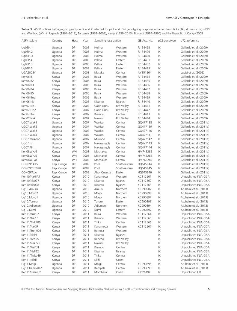
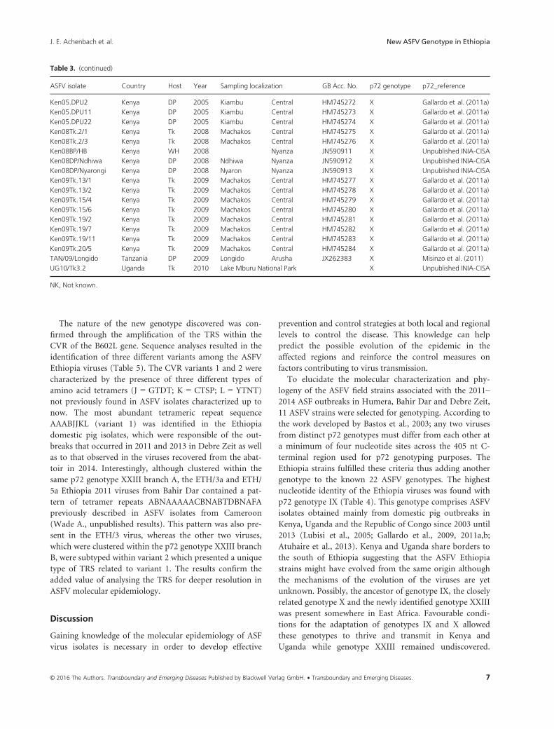
Table 3. ASFV isolates belonging to genotype IX and X selected for p72 and p54 genotyping purposes obtained from ticks (Tk), domestic pigs (DP)
and Warthog (WH) in Uganda (1964–2013), Tanzania (1968–2009), Kenya (1959–2013), Burundi (1984–1990) and the Republic of Congo 2009
ASFV isolate Country Host Year Sampling localization GB Acc. No. p72 genotype p72_reference
Ug03H.1 Uganda DP 2003 Hoima Western FJ154428 IX Gallardo et al. (2009)
Ug03H.2 Uganda DP 2003 Hoima Western FJ154429 IX Gallardo et al. (2009)
Ug03H.3 Uganda DP 2003 Hoima Western FJ154430 IX Gallardo et al. (2009)
Ug03P.4 Uganda DP 2003 Pallisa Eastern FJ154431 IX Gallardo et al. (2009)
Ug03P.5 Uganda DP 2003 Pallisa Eastern FJ154432 IX Gallardo et al. (2009)
Ug03P.6 Uganda DP 2003 Pallisa Eastern FJ154433 IX Gallardo et al. (2009)
UGA2003/1 Uganda DP 2003 Masaka Central AY351564 IX Lubisi et al. (2005)
Ken06.B1 Kenya DP 2006 Busia Western FJ154434 IX Gallardo et al. (2009)
Ken06.B2 Kenya DP 2006 Busia Western FJ154435 IX Gallardo et al. (2009)
Ken06.B3 Kenya DP 2006 Busia Western FJ154436 IX Gallardo et al. (2009)
Ken06.B4 Kenya DP 2006 Busia Western FJ154437 IX Gallardo et al. (2009)
Ken06.B5 Kenya DP 2006 Busia Western FJ154438 IX Gallardo et al. (2009)
Ken06.Bus Kenya DP 2006 Busia Western FJ154439 IX Gallardo et al. (2009)
Ken06.Kis Kenya DP 2006 Kisumu Nyanza FJ154440 IX Gallardo et al. (2009)
Ken07.Eld1 Kenya DP 2007 Uasin Gishu Rift Valley FJ154441 IX Gallardo et al. (2009)
Ken07.Eld2 Kenya DP 2007 Uasin Gishu Rift Valley FJ154442 IX Gallardo et al. (2009)
Ken07.Kia Kenya DP 2007 Kiambu Central FJ154443 IX Gallardo et al. (2009)
Ken07.Nak Kenya DP 2007 Nakuru Rift Valley FJ154444 IX Gallardo et al. (2009)
UG07.Wak1 Uganda DP 2007 Wakiso Central GQ477138 IX Gallardo et al. (2011a)
UG07.Wak2 Uganda DP 2007 Wakiso Central GQ477139 IX Gallardo et al. (2011a)
UG07.Wak3 Uganda DP 2007 Wakiso Central GQ477140 IX Gallardo et al. (2011a)
UG07.Wak4 Uganda DP 2007 Wakiso Central GQ477141 IX Gallardo et al. (2011a)
UG07.Mukono Uganda DP 2007 Mukono Central GQ477142 IX Gallardo et al. (2011a)
UG07.F7 Uganda DP 2007 Nakasangola Central GQ477143 IX Gallardo et al. (2011a)
UG07.F8 Uganda DP 2007 Nakasangola Central GQ477144 IX Gallardo et al. (2011a)
Ken08WH/4 Kenya WH 2008 Machakos Central HM745285 IX Gallardo et al. (2011a)
Ken08WH/5 Kenya WH 2008 Machakos Central HM745286 IX Gallardo et al. (2011a)
Ken08WH/8 Kenya WH 2008 Machakos Central HM745287 IX Gallardo et al. (2011a)
CON09/Pk45 Rep. Congo DP 2009 Pool Southeastern HQ645944 IX Gallardo et al. (2011a)
CON09/Bzz020 Rep. Congo DP 2009 Pool Southeastern HQ645945 IX Gallardo et al. (2011a)
CON09/Abo Rep. Congo DP 2009 Abo, Cuvette Eastern HQ645946 IX Gallardo et al. (2011a)
Ken10/KakFA1 Kenya DP 2010 Kakamega Western KC112561 IX Unpublished INIA-CISA
Ken10/Kis027 Kenya DP 2010 Kisumu Nyanza KC112562 IX Unpublished INIA-CISA
Ken10/Kis028 Kenya DP 2010 Kisumu Nyanza KC112563 IX Unpublished INIA-CISA
Ug10.Amuru Uganda DP 2010 Amuru Northern KC990902 IX Atuhaire et al. (2013)
Ug10.Moyo2 Uganda DP 2010 Moyo Northern KC990898 IX Atuhaire et al. (2013)
Ug10.Moyo1 Uganda DP 2010 Moyo Northern KC990897 IX Atuhaire et al. (2013)
Ug10.Tororo Uganda DP 2010 Tororo Eastern KC990896 IX Atuhaire et al. (2013)
Ug10.Adjumani Uganda DP 2010 Adjumani Northern KC990894 IX Atuhaire et al. (2013)
Ug10.Kumi Uganda DP 2010 Kumi Eastern KC990892 IX Atuhaire et al. (2013)
Ken11/Bus1.2 Kenya DP 2011 Busia Western KC112564 IX Unpublished INIA-CISA
Ken11/Kia2.1 Kenya DP 2011 Kiambu Western KC112565 IX Unpublished INIA-CISA
Ken11/ThikP06 Kenya DP 2011 Thika Central KC112566 IX Unpublished INIA-CISA
Ken11/KakSP Kenya DP 2011 Kakamega Western KC112567 IX Unpublished INIA-CISA
Ken11/Bum002 Kenya DP 2011 Bumula Western IX Unpublished INIA-CISA
Ken11/KisP1 Kenya DP 2011 Kisumu Nyanza IX Unpublished INIA-CISA
Ken11/KerP27 Kenya DP 2011 Kericho Rift Valley IX Unpublished INIA-CISA
Ken11/NakP29 Kenya DP 2011 Nakuru Rift Valley IX Unpublished INIA-CISA
Ken11/KiaP31 Kenya DP 2011 Kiambu Central IX Unpublished INIA-CISA
Ken11/KisP52 Kenya DP 2011 Kisumu Nyanza IX Unpublished INIA-CISA
Ken11/Thikp49 Kenya DP 2011 Thika Central IX Unpublished INIA-CISA
Ken11/Kilifili Kenya DP 2011 Kilifi Coast IX Unpublished INIA-CISA
Ug11.Mpigi Uganda DP 2011 Mpigi Central KC990895 IX Atuhaire et al. (2013)
Ug11.Kampala2 Uganda DP 2011 Kampala Central KC990893 IX Atuhaire et al. (2013)
Ken11/kisauni2 Kenya DP 2011 Mombasa Coast KJ626192 IX Unpublished ILRI
© 2016 The Authors. Transboundary and Emerging Diseases Published by Blackwell Verlag GmbH. • Transboundary and Emerging Diseases. 5
J. E. Achenbach et al. New ASFV Genotype in Ethiopia
Table 3. (continued)
ASFV isolate Country Host Year Sampling localization GB Acc. No. p72 genotype p72_reference
Ken11/kisauni1 Kenya DP 2011 Mombasa Coast KJ626191 IX Unpublished ILRI
Uga12.Kalungu3 Uganda DP 2012 Kalungu Central KF303313 IX Atuhaire et al. (2013)
Uga12.Kalungu2 Uganda DP 2012 Kalungu Central KF303312 IX Atuhaire et al. (2013)
Uga12.Kalungu1 Uganda DP 2012 Kalungu Central KF303311 IX Atuhaire et al. (2013)
Uga12.Nakasongola Uganda DP 2012 Nakasangola Central KF303310 IX Atuhaire et al. (2013)
Ug12.Kampala4 Uganda DP 2012 Kampala Central KC990904 IX Atuhaire et al. (2013)
Ug12.Kyenjojo Uganda DP 2012 Kyenjojo Western KC990903 IX Atuhaire et al. (2013)
Ug12.Wakiso Uganda DP 2012 Wakiso Central KC990901 IX Atuhaire et al. (2013)
Ug12.Kampala3 Uganda DP 2012 Kampala Central KC990900 IX Atuhaire et al. (2013)
Ug12.Lira Uganda DP 2012 Lira Northern KC990899 IX Atuhaire et al. (2013)
Ug12.Kabale1 Uganda DP 2012 Kabale Western KC990890 IX Atuhaire et al. (2013)
Uga12.Lango3 Uganda DP 2012 Lango Northern KF303321 IX Atuhaire et al. (2013)
Uga12.Lango2 Uganda DP 2012 Lango Northern KF303320 IX Atuhaire et al. (2013)
Uga12.Lango1 Uganda DP 2012 Lango Northern KF303319 IX Atuhaire et al. (2013)
Uga12.Busoga2 Uganda DP 2012 Busoga Eastern KF303318 IX Atuhaire et al. (2013)
Uga12.Busoga1 Uganda DP 2012 Busoga Eastern KF303317 IX Atuhaire et al. (2013)
Uga12.Nakaseke Uganda DP 2012 Nakaseke Central KF303316 IX Atuhaire et al. (2013)
Uga12.Kibaale Uganda DP 2012 Kibaale Western KF303315 IX Atuhaire et al. (2013)
Uga12.Sembabule Uganda DP 2012 Sembabule Central KF303314 IX Atuhaire et al. (2013)
Ug13.Busia2 Uganda DP 2013 Busia Eastern KC990906 IX Atuhaire et al. (2013)
Ug13.Busial Uganda DP 2013 Busia Eastern KC990905 IX Atuhaire et al. (2013)
Ug13.Kampala1 Uganda DP 2013 Kampala Central KC990891 IX Atuhaire et al. (2013)
ken13/nakuru.1 Kenya DP 2013 Nakuru Rift Valley KM000166 IX Unpublished ILRI
ken13/kakamega.1 Kenya DP 2013 Kakamega Western KM000164 IX Unpublished ILRI
ken13/nyadorera.1 Kenya DP 2013 Nyanza Western KM000163 IX Unpublished ILRI
ken13/busia.9 Kenya DP 2013 Busia Western KM000162 IX Unpublished ILRI
MWHOG/9 Kenya WH 1959 Rift Valley Rift Valley AY351565 X Lubisi et al. (2005)
Ug64 Uganda DP 1964 NK FJl74383 X Gallardo et al. (2009)
TAN/Kwh12 Tanzania WH 1968 Serengeti National Park AF301546 X Lubisi et al. (2005)
BUR/2/84 Burundi DP 1984 NK AF449464 X Bastos et al. (2003)
BUR/1/84 Burundi DP 1984 NK AF449463 X Bastos et al. (2003)
KIRW/891 Tanzania WH 1989 Serengeti National Park AY351514 X Lubisi et al. (2005)
KIRT/894 Tanzania Tk 1989 Serengeti National Park AY351513 X Lubisi et al. (2005)
KIRT/893 Tanzania Tk 1989 Serengeti National Park AY351512 X Lubisi et al. (2005)
KIRT/892 Tanzania Tk 1989 Serengeti National Park AY351511 X Lubisi et al. (2005)
BUR/90/3 Burundi DP 1990 Muyinga North AY351525 X Lubisi et al. (2005)
BUR/90/2 Burundi DP 1990 NK JX467634 X Unpublished OVI
BUR/90/1 Burundi DP 1990 NK AF449472 X Bastos et al. (2003)
Ken05/Tk1 Kenya Tk 2005 Machakos Central HM745253 X Gallardo et al. (2011a)
Ken05/Tk2 Kenya Tk 2005 Machakos Central HM745254 X Gallardo et al. (2011a)
Ken05/Tk3 Kenya Tk 2005 Machakos Central HM745255 X Gallardo et al. (2011a)
Ken05/Tk4 Kenya Tk 2005 Machakos Central HM745256 X Gallardo et al. (2011a)
Ken05/Tk5 Kenya Tk 2005 Machakos Central HM745257 X Gallardo et al. (2011a)
Ken05/Tk6 Kenya Tk 2005 Machakos Central HM745258 X Gallardo et al. (2011a)
Ken05/Tk7 Kenya Tk 2005 Machakos Central HM745259 X Gallardo et al. (2011a)
Ken05/Tk8 Kenya Tk 2005 Machakos Central HM745260 X Gallardo et al. (2011a)
Ken05/Tk9 Kenya Tk 2005 Machakos Central HM745261 X Gallardo et al. (2011a)
Ken05/Tk10 Kenya Tk 2005 Machakos Central HM745262 X Gallardo et al. (2011a)
Ken05.DPk2 Kenya DP 2005 Kiambu Central HM745263 X Gallardo et al. (2011a)
Ken05.DPk16 Kenya DP 2005 Kiambu Central HM745264 X Gallardo et al. (2011a)
Ken05.DPk18 Kenya DP 2005 Kiambu Central HM745265 X Gallardo et al. (2011a)
Ken05.DPk21 Kenya DP 2005 Kiambu Central HM745266 X Gallardo et al. (2011a)
Ken05.DPk27 Kenya DP 2005 Kiambu Central HM745267 X Gallardo et al. (2011a)
Ken05.DPN2 Kenya DP 2005 Nandi Central HM745268 X Gallardo et al. (2011a)
Ken05.DPN15 Kenya DP 2005 Nandi Central HM745269 X Gallardo et al. (2011a)
Ken05.DPU1 Kenya DP 2005 Kiambu Central HM745271 X Gallardo et al. (2011a)
© 2016 The Authors. Transboundary and Emerging Diseases Published by Blackwell Verlag GmbH.6
New ASFV Genotype in Ethiopia J. E. Achenbach et al.
The nature of the new genotype discovered was con-
firmed through the amplification of the TRS within the
CVR of the B602L gene. Sequence analyses resulted in the
identification of three different variants among the ASFV
Ethiopia viruses (Table 5). The CVR variants 1 and 2 were
characterized by the presence of three different types of
amino acid tetramers (J = GTDT; K = CTSP; L = YTNT)
not previously found in ASFV isolates characterized up to
now. The most abundant tetrameric repeat sequence
AAABJJKL (variant 1) was identified in the Ethiopia
domestic pig isolates, which were responsible of the out-
breaks that occurred in 2011 and 2013 in Debre Zeit as well
as to that observed in the viruses recovered from the abat-
toir in 2014. Interestingly, although clustered within the
same p72 genotype XXIII branch A, the ETH/3a and ETH/
5a Ethiopia 2011 viruses from Bahir Dar contained a pat-
tern of tetramer repeats ABNAAAAACBNABTDBNAFA
previously described in ASFV isolates from Cameroon
(Wade A., unpublished results). This pattern was also pre-
sent in the ETH/3 virus, whereas the other two viruses,
which were clustered within the p72 genotype XXIII branch
B, were subtyped within variant 2 which presented a unique
type of TRS related to variant 1. The results confirm the
added value of analysing the TRS for deeper resolution in
ASFV molecular epidemiology.
Discussion
Gaining knowledge of the molecular epidemiology of ASF
virus isolates is necessary in order to develop effective
prevention and control strategies at both local and regional
levels to control the disease. This knowledge can help
predict the possible evolution of the epidemic in the
affected regions and reinforce the control measures on
factors contributing to virus transmission.
To elucidate the molecular characterization and phy-
logeny of the ASFV field strains associated with the 2011–2014 ASF outbreaks in Humera, Bahir Dar and Debre Zeit,
11 ASFV strains were selected for genotyping. According to
the work developed by Bastos et al., 2003; any two viruses
from distinct p72 genotypes must differ from each other at
a minimum of four nucleotide sites across the 405 nt C-
terminal region used for p72 genotyping purposes. The
Ethiopia strains fulfilled these criteria thus adding another
genotype to the known 22 ASFV genotypes. The highest
nucleotide identity of the Ethiopia viruses was found with
p72 genotype IX (Table 4). This genotype comprises ASFV
isolates obtained mainly from domestic pig outbreaks in
Kenya, Uganda and the Republic of Congo since 2003 until
2013 (Lubisi et al., 2005; Gallardo et al., 2009, 2011a,b;
Atuhaire et al., 2013). Kenya and Uganda share borders to
the south of Ethiopia suggesting that the ASFV Ethiopia
strains might have evolved from the same origin although
the mechanisms of the evolution of the viruses are yet
unknown. Possibly, the ancestor of genotype IX, the closely
related genotype X and the newly identified genotype XXIII
was present somewhere in East Africa. Favourable condi-
tions for the adaptation of genotypes IX and X allowed
these genotypes to thrive and transmit in Kenya and
Uganda while genotype XXIII remained undiscovered.
Table 3. (continued)
ASFV isolate Country Host Year Sampling localization GB Acc. No. p72 genotype p72_reference
Ken05.DPU2 Kenya DP 2005 Kiambu Central HM745272 X Gallardo et al. (2011a)
Ken05.DPU11 Kenya DP 2005 Kiambu Central HM745273 X Gallardo et al. (2011a)
Ken05.DPU22 Kenya DP 2005 Kiambu Central HM745274 X Gallardo et al. (2011a)
Ken08Tk.2/1 Kenya Tk 2008 Machakos Central HM745275 X Gallardo et al. (2011a)
Ken08Tk.2/3 Kenya Tk 2008 Machakos Central HM745276 X Gallardo et al. (2011a)
Ken08BP/HB Kenya WH 2008 Nyanza JN590911 X Unpublished INIA-CISA
Ken08DP/Ndhiwa Kenya DP 2008 Ndhiwa Nyanza JN590912 X Unpublished INIA-CISA
Ken08DP/Nyarongi Kenya DP 2008 Nyaron Nyanza JN590913 X Unpublished INIA-CISA
Ken09Tk.13/1 Kenya Tk 2009 Machakos Central HM745277 X Gallardo et al. (2011a)
Ken09Tk.13/2 Kenya Tk 2009 Machakos Central HM745278 X Gallardo et al. (2011a)
Ken09Tk.15/4 Kenya Tk 2009 Machakos Central HM745279 X Gallardo et al. (2011a)
Ken09Tk.15/6 Kenya Tk 2009 Machakos Central HM745280 X Gallardo et al. (2011a)
Ken09Tk.19/2 Kenya Tk 2009 Machakos Central HM745281 X Gallardo et al. (2011a)
Ken09Tk.19/7 Kenya Tk 2009 Machakos Central HM745282 X Gallardo et al. (2011a)
Ken09Tk.19/11 Kenya Tk 2009 Machakos Central HM745283 X Gallardo et al. (2011a)
Ken09Tk.20/5 Kenya Tk 2009 Machakos Central HM745284 X Gallardo et al. (2011a)
TAN/09/Longido Tanzania DP 2009 Longido Arusha JX262383 X Misinzo et al. (2011)
UG10/Tk3.2 Uganda Tk 2010 Lake Mburu National Park X Unpublished INIA-CISA
NK, Not known.
© 2016 The Authors. Transboundary and Emerging Diseases Published by Blackwell Verlag GmbH. • Transboundary and Emerging Diseases. 7
J. E. Achenbach et al. New ASFV Genotype in Ethiopia

Genotype XXIII could have maintained a silent presence in
ticks and warthogs in Ethiopia until the increase in domes-
tic pig production in recent years allowed for the move-
ment into susceptible pig breeds within Ethiopia.
Further sequence analysis of ASFV isolates obtained dur-
ing the epidemic cases that occurred in 2013 at the Debre
Zeit farm, epidemiologically linked them with the index
case from 2011.
The results obtained throughout the p72 genotyping were
confirmed by the full sequencing of the p54 gene. In contrast,
the partial analysis of the hyper variable region located within
Fig. 1. Minimum evolution phylogenetic tree of the Ethiopia ASFV iso-
lates based on the analysis of the 405 nucleotides situated at the C-
terminal end of the p72 coding gene relative to the 22 p72 genotypes
(labelled I-XXII), including 234 nucleotide sequences. The tree was
inferred using the minimum evolution (ME) method following initial
application of a neighbour-joining algorithm. The evolutionary distances
were computed using the p-distance method and are in the units of the
number of base differences per site. The percentage of replicate trees
>50% in which the associated taxa clustered together by bootstrap
analysis (1000 replicates) is shown adjacent to the nodes. The robust-
ness of the ME tree was tested using the close-neighbour-interchange
(CNI) algorithm at a search level of 1. ASFV Ethiopia isolates genotyped
in this study are marked in red (●) within the genotype XXIII red
labelled.
Table 4. Nucleotide (NT) and amino acid (AA) variation across the 405
nt and 135 aa C-terminal region of p72 protein between the Ethiopia
ASFV isolates placed into the genotype 23 (branches A and B) and each
of the 22 p72 genotypes
ASFV
genotypes
P72 genotype 23
branch 1
P72 genotype 23
branch 2
NT
variation
AA
variation
NT
variation
AA
variation
No. % No. % No. % No. %
9 10 2.5 1 0.7 9 2.2 1 0.7
10 16 4.0 3 2.2 15 3.7 3 2.2
8 22 5.4 4 3.0 23 5.7 4 3.0
11 18 4.4 4 3.0 19 4.7 4 3.0
12 18 4.4 4 3.0 19 4.7 4 3.0
14 20 4.9 4 3.0 21 5.2 4 3.0
15 21 5.2 4 3.0 22 5.4 4 3.0
16 17 4.2 4 3.0 18 4.4 4 3.0
17 19 4.7 4 3.0 20 4.9 4 3.0
21 17 4.2 4 3.0 18 4.4 4 3.0
1 22 5.4 5 3.7 23 5.7 5 3.7
2 20 4.9 5 3.7 21 5.2 5 3.7
5 23 5.7 5 3.7 24 5.9 5 3.7
6 22 5.4 5 3.7 23 5.7 5 3.7
18 22 5.4 5 3.7 23 5.7 5 3.7
19 19 4.7 5 3.7 20 4.9 5 3.7
20 19 4.7 5 3.7 20 4.9 5 3.7
3 20 4.9 6 4.4 21 5.2 6 4.4
4 20 4.9 6 4.4 21 5.2 6 4.4
7 20 4.9 6 4.4 21 5.2 6 4.4
22 21 5.2 6 4.4 22 5.4 6 4.4
13 21 5.2 7 5.2 22 5.4 7 5.2
© 2016 The Authors. Transboundary and Emerging Diseases Published by Blackwell Verlag GmbH.8
New ASFV Genotype in Ethiopia J. E. Achenbach et al.

the B602L gene allowed us to identify three different CVR
variants, two of which are novel in sequence further support-
ing the separate genotype. The similarity of variant 3 between
Ethiopia and Cameroon is consistent with previous data,
which reported ASFV dissemination from East to West Africa
through persistently infected wild or domestic animals or by
the movement of infected pigs or pork products (Gallardo
et al., 2011b). It is therefore important to study ASF in
Central Africa with an emphasis on countries such as Central
African Republic and Chad to better understand and trace
the movement of virus between countries.
Interestingly, the virus recovered from apparently
healthy animals during the survey conducted in 2014 at the
Gondar abattoir presented 100% of identity in the three
ASFV genome regions analysed with the 2011 and 2013
ASFV strains, which induced an acute form of the disease
Fig. 2. Subtree depicting the p54 subtype X and IX of the full-length p54 gene (P54) generated using sequences from 122 East African ASFV isolates
including the Ethiopia viruses analysed in this study. The evolutionary history was inferred using the minimum evolution method (ME). The trees are
drawn to scale, with branch lengths in the same units as those of the evolutionary distances used to infer the phylogenetic tree. The ME trees were
further analysed using the close-neighbour-interchange (CNI) algorithm at a search level of 1. A neighbour-joining algorithm was used to generate
the initial trees using 1000 replicates. Bootstrap values > 50% are indicated next to the relevant node. (●) indicates ASF viruses characterized in this
study.
Table 5. Amino acid sequence of the tetrameric repeats that constitute the central variable region (CVR) of the B602L gene identified in the Ethiopia
viruses belonging to p72 genotype XXIII. Key: A (CAST); B (CADT); C (GAST); D (CASM); F (CANT, CAAT); J (GTDT); K (CTSP); L (YTNT); T (NVNT);
N (NVDT)
P72 genotype
XXIII Isolate Collection Date CVR VARIANT CVR amino acid sequence No repeats
A ETH/1a outbreak 2011 1 AAABJJKL 8
A ETH/2a outbreak 2011 1 AAABJJKL 8
A ET13/1504 outbreak 2013 1 AAABJJKL 8
A ET13/1505 outbreak 2013 1 AAABJJKL 8
A ETH/04 abattoir 2014 1 AAABJJKL 8
A ETH/17 abattoir 2014 1 AAABJJKL 8
B ETH/AA outbreak 2011 2 ABBBBFFBFBJJKL 14
B ETH/1 outbreak 2011 2 ABBBBFFBFBJJKL 14
B ETH/3 outbreak 2011 3 ABNAAAAACBNABTDBNAFA 20
A ETH/3a outbreak 2011 3 ABNAAAAACBNABTDBNAFA 20
A ETH/5a outbreak 2011 3 ABNAAAAACBNABTDBNAFA 20
© 2016 The Authors. Transboundary and Emerging Diseases Published by Blackwell Verlag GmbH. • Transboundary and Emerging Diseases. 9
J. E. Achenbach et al. New ASFV Genotype in Ethiopia
as it was reported in the affected farms. It should be consid-
ered that after the first introduction of the disease in a
region, increased numbers of subacute and subclinical
infections can occur over time and that mortality rates
decline. In such situations, the clinical manifestations are
more variable and recognition of the disease becomes diffi-
cult in the field, emphasizing the need of implementation
of appropriate surveillance programmes to control the dis-
ease (Mebus et al., 1978; Mebus and Dardiri, 1979, 1980;
Thomson et al., 1979; Hess, 1981; Wilkinson et al., 1981,
1983; Wilkinson, 1984; S�anchez-Botija, 1982; Nsalambi,
1993; Penrith et al., 2013; Gallardo et al., 2015; S�anchez-
Vizca�ıno et al., 2015).
There is currently no data regarding the presence of ASF
in other boarder countries such as Sudan, South Sudan,
Eritrea, Djibouti and Somalia. While many of these neigh-
bouring countries, due to religious preferences, have mini-
mal domestic swine, the sylvatic cycle between warthogs
and ticks remains a possibility and thus should be investi-
gated. As the domestic population of swine in Africa
continues to grow, so does the problematic economic con-
straints when there is no vaccine and the only form of con-
trol is stamping out of infected pigs. This makes the need
for a vaccine more imperative than ever to control this
TAD. So far, all attempts to produce safe vaccines against
ASFV infection have demonstrated only homologous pro-
tection (Manso-Ribeiro et al., 1963; Ruiz-Gonzalvo et al.,
1996). Additionally, swine that survive ASFV infection
against one genotype have shown solid immunity to chal-
lenge from a homologous strain but not usually against
heterologous strains even when they are in the same geno-
type (Manso-Ribeiro et al., 1963; Leit~ao et al., 2001; King
et al., 2011; Mulumba-Mfumu et al., 2015). It is therefore
likely that effective control of ASF will rely on the availabil-
ity of several vaccines based on the specific isolates circulat-
ing in each region. Increasing our knowledge of ASFV
isolates worldwide through whole genome sequencing will
aid in the development of promising research approaches
such as the creation of deletion mutants based on protec-
tion against the strains of the same genotypes.
While the detection of ASFV in Ethiopia is more recent
than other East African countries, the detection of a com-
pletely different genotype leaves a gap in understanding the
epidemiology of the virus. Questions remain whether these
strains have been circulating in the sylvatic cycle or if they
were transported from another unknown location where
ASF had not been detected. Continuation of surveillance is
important for us to better understand the epidemiology of
the virus so that a future vaccine can be prepared that
protects domestic swine from future outbreaks.
The discovery of a new ASFV genotype emphasises the
importance and high variability of ASFV isolates in Africa.
In addition and due to the increase of viruses sequenced
over the last decade, a need for reclassification of the
current ASFV isolates within the p72 genotypes should be
considered.
Acknowledgements
This study was partially funded by the International Atomic
Energy Agency (IAEA) project ‘Improvement of Veterinary
Laboratory Capacities in Sub-Saharan African Countries’,
the tripartite FAO/OIE/WHO IDENTIFY Project of the
USAID Emergent Pandemic Threats Program, the EU pro-
ject ‘Rapid Field Diagnostics and Screening in Veterinary
Medicine’ (Rapidia-Field, KBBE.2011.1.3-02) and by ‘Tar-
geted research effort on African swine fever’ (ASFORCE-
FP7 KBBE.2012.1.3-02).
Conflicts of interest
The authors declared no conflict of interest. The funders
had no role in study design, data collection and analysis,
decision to publish or preparation of the manuscript.
References
Atuhaire, D. K., M. Afayoa, S. Ochwo, S. Mwesigwa, J. B. Okuni,
W. Olaho-Mukani, and L. Ojok, 2013: Molecular characterization
and phylogenetic study of African swine fever virus isolates from
recent outbreaks in Uganda (2010–2013). Virol. J. 10, 247.Bastos, A. D., M. L. Penrith, C. Cruciere, J. L. Edrich, G.
Hutchings, F. Roger, E. Couacy-Hymann, and G. R. Thomson,
2003: Genotyping field strains of African swine fever virus by
partial p72 gene characterisation. Arch. Virol. 148, 693–706.Bastos, A. D., L. F. Arnot, M. D. Jacquier, and S. Maree, 2009: A
host species-informative internal control for molecular assess-
ment of African swine fever virus infection rates in the African
sylvatic cycle Ornithodoros vector. Med. Vet. Entomol. 23,
399–409.Boshoff, C. I., A. D. Bastos, L. J. Gerber, and W. Vosloo, 2007:
Genetic characterisation of African swine fever viruses from
outbreaks in southern Africa (1973-1999). Vet. Microbiol. 121,
45–55.Costard, S., B. Wieland, W. de Glanville, F. Jori, R. Rowlands,
W. Vosloo, F. Roger, D. U. Pfeiffer, and L. K. Dixon, 2009:
African swine fever: how can global spread be prevented? Phi-
los. Trans. R. Soc. Lond. B Biol. Sci. 364, 2683–2696.Dixon, L. K., J. V. Costa, J. M. Escribano, D. L. Rock, E. Vinuela,
and P. J. Wilkinson, 2000: Family Asfarviridae. In: Van
Regenmortel, M. H. V., C. M. Fauquel and D. H. L. Bishop
(eds), Virus Taxonomy, 7th Report of the ICTV, pp. 159–165.Academic Press, San Diego.
European Food Safety Authority (EFSA) and Scientific opinion
on African swine fever (ASF), 2015. Available at http://
www.efsa.europa.eu/sites/default/files/scientific_out-
put/files/main_documents/4163.pdf (accessed July 14, 2015).
© 2016 The Authors. Transboundary and Emerging Diseases Published by Blackwell Verlag GmbH.10
New ASFV Genotype in Ethiopia J. E. Achenbach et al.

Food and Agriculture Organization, United Nations, 2013:
African swine fever in the Russian Federation: risk factors for
Europe and beyond. EMPRES Watch. Vol. 28; 2013 May.
Available at http://www.fao.org/docrep/018/aq240e/
aq240e.pdf (accessed September 2, 2013).
Gallardo, C., D. M. Mwaengo, J. M. Macharia, M. Arias, E. A.
Taracha, A. Soler, E. Okoth, E. Martin, J. Kasiti, and R. P.
Bishop, 2009: Enhanced discrimination of African swine fever
virus isolates through nucleotide sequencing of the p54, p72,
and pB602L (CVR) genes. Virus Genes 38, 85–95.Gallardo, C., E. Okoth, V. Pelayo, R. Anchuelo, E. Mart�ın, A.
Sim�on, A. Llorente, R. Nieto, A. Soler, R. Mart�ın, M. Arias,
and R. P. Bishop, 2011a: African swine fever viruses with two
different genotypes, both of which occur in domestic pigs, are
associated with ticks and adult warthogs, respectively, at a
single geographical site. J. Gen. Virol. 92, 432–444.Gallardo, C., R. Anchuelo, V. Pelayo, F. Poudevigne, T. Leon, J.
Nzoussi, R. Bishop, C. P�erez, A. Soler, R. Nieto, H. Mart�ın,
and M. Arias, 2011b: African swine fever virus p72 genotype
IX in domestic pigs, Congo, 2009. Emerg. Infect. Dis. 17,
1556–1558.Gallardo, C., J. Fern�andez-Pinero, V. Pelayo, I. Gazaev, I. Mar-
kowska-Daniel, G. Pridotkas, R. Nieto, P. Fern�andez-Pacheco,
S. Bokhan, O. Nevolko, Z. Drozhzhe, C. P�erez, A. Soler, D.
Kolvasov, and M. Arias, 2014: Genetic variation among
African swine fever genotype II viruses, eastern and central
Europe. Emerg. Infect. Dis. 20, 1544–1547.Gallardo, C., A. Soler, R. Nieto, M. A. S�anchez, C. Martins, V.
Pelayo, A. Carrascosa, Y. Revilla, A. Sim�on, V. Briones, J. M.
S�anchez-Vizca�ıno, and M. Arias, 2015: Experimental Trans-
mission of African Swine Fever (ASF) Low Virulent Isolate
NH/P68 by Surviving Pigs. Transbound. Emerg. Dis. 62,
612–622.Haresnape, J. M., P. J. Wilkinson, and P. S. Mellor, 1988: Isola-
tion of African swine fever virus from ticks of the Ornitho-
doros moubata complex (Ixodoidea: Argasidae) collected
within the African swine fever enzootic area of Malawi.
Epidemiol. Infect. 101, 173–185.Hess, W. R., 1981: African swine fever: A reassessment. Adv. Vet.
Sci. Comp. Med. 25, 39–69.Jori, F., and A. D. Bastos, 2009: Role of wild suids in the epi-
demiology of African swine fever. EcoHealth 6, 296–310.Jori, F., L. Vial, M. L. Penrith, R. P�erez-S�anchez, E. Etter, E.
Albina, V. Michaud, and F. Roger, 2013: Review of the sylvatic
cycle of African swine fever in sub-Saharan Africa and the
Indian ocean. Virus Res. 173, 212–227.King, D. P., S. M. Reid, G. H. Hutchings, S. S. Grierson, P. J.
Wilkinson, L. K. Dixon, A. D. Bastos, and T. W. Drew, 2003:
Development of a TaqMan PCR assay with internal amplifica-
tion control for the detection of African swine fever virus.
J. Virol. Methods 107, 53–61.King, K., D. Chapman, J. M. Argilaguet, E. Fishbourne, E. Hutet,
R. Cariolet, G. Hutchings, C. A. Oura, C. L. Netherton, K.
Moffat, G. Taylor, M. F. Le Potier, L. K. Dixon, and H. H.
Takamatsu, 2011: Protection of European domestic pigs from
virulent African isolates of African swine fever virus by experi-
mental immunization. Vaccine 29, 4593–4600.Kleiboeker, S.B., and G.A. Scoles, 2001: Pathogenesis of African
swine fever virus in Ornithodoros ticks. Anim. Health Res.
Rev. 2, 121–128.Leit~ao, A., C. Cartaxeiro, R. Coelho, B. Cruz, R. M. Parkhouse,
F. Portugal, J. D. Vig�ario, and C. L. Martins, 2001: The non-
haemadsorbing African swine fever virus isolate ASFV/NH/
P68 provides a model for defining the protective antivirus
immune response. J. Gen. Virol. 82, 513–523.Lubisi, B. A., A. D. Bastos, R. M. Dwarka, and W. Vosloo, 2005:
Molecular epidemiology of African swine fever in East Africa.
Arch. Virol. 150, 2439–2452.Manso-Ribeiro, J., J. L. Nunes-Petisca, F. Lopez-Frazao, and M.
Sobral, 1963: Vaccination against ASF. Bull. Off. Int. Epizoot.
60, 921–937.Mebus, C. A., and A. H. Dardiri, 1979: Additional Characteris-
tics of disease caused by the African swine fever viruses iso-
lates from Brazil and the Dominican Republic. Proc. Annu.
Meet. U. S. Anim. Health Assoc. 83, 227–239.Mebus, C. A., and A. H. Dardiri, 1980: Western Hemisphere Iso-
lates of African Swine Fever Virus: Asymptomatic Carriers
and Resistance to Challenge Inoculation. Am. J. Vet. Res. 41,
1867–1869.Mebus, C. A., A. H. Dardiri, F. M. Hamdy, D. H. Ferris, W. R.
Hess, and J. J. Callis, 1978: Some Characteristics of disease
caused by the African swine fever viruses isolates from Brazil
and the Dominican Republic. Proc. Annu. Meet. U. S. Anim.
Health Assoc. 82, 232–236.Misinzo, G., J. Magambo, J. Masambu, M. G. Yongolo, D. J.
Van, and H. J. Nauwynck, 2011: Genetic characterization of
African swine fever viruses from a 2008 outbreak in Tanzania.
Transbound. Emerg. Dis. 58, 86–92.Misinzo, G., D. E. Kwavi, C. D. Sikombe, M. Makange, E. Peter,
A. P. Muhairwa, and M. J. Madege, 2014: Molecular charac-
terization of African swine fever virus from domestic pigs in
northern Tanzania during an outbreak in 2013. Trop. Anim.
Health Prod. 46, 1199–1207.Mulumba-Mfumu, L. K., L. C. Goatley, C. Saegerman, H. H.
Takamatsu, and L. K. Dixon, 2015: Immunization of African
Indigenous Pigs with Attenuated Genotype I African swine
fever Virus OURT88/3 Induces Protection Against Challenge
with Virulent Strains of Genotype I. Transbound. Emerg. Dis..
doi:10.1111/tbed.12303.
Nix, R. J., C. Gallardo, G. Hutchings, E. Blanco, and L. K. Dixon,
2006: Molecular epidemiology of African swine fever virus
studied by analysis of four variable genome regions. Arch.
Virol. 151, 2475–2494.Nsalambi, D., 1993: Differences cliniques et anatomo-pathologi-
ques de deux souches du virus de la peste porcine africaine
(PPA) en Angola. Revue d’Elevage et Medicine veterinaire du
Pays tropicaux. 46, 539–543.Oura, C. A., P. P. Powell, E. Anderson, and R. M. Parkhouse,
1998: The pathogenesis of African swine fever in the resistant
bushpig. J. Gen. Virol. 79, 1439–1443.
© 2016 The Authors. Transboundary and Emerging Diseases Published by Blackwell Verlag GmbH. • Transboundary and Emerging Diseases. 11
J. E. Achenbach et al. New ASFV Genotype in Ethiopia

Penrith, M. L., G. R. Thomson, A. D. Bastos, O. C. Phiri, B. A.
Lubisi, E. C. Du Plessis, F. Macome, F. Pinto, B. Botha, and J.
Esterhuysen, 2004: An investigation into natural resistance to
African swine fever in domestic pigs from an endemic area in
southern Africa. Rev. Sci. Tech. 23, 965–977.Penrith, M. L., W. Vosloo, F. Jori, and A. D. Bastos, 2013: African
swine fever virus eradication in Africa. Virus Res. 173, 228–246.Rowlands, R. J., V. Michaud, L. Heath, G. Hutchings, C. Oura,
W. Vosloo, R. Dwarka, T. Onashvili, E. Albina, and L. K.
Dixon, 2008: African swine fever virus isolate, Georgia, 2007.
Emerg. Infect. Dis. 14, 1870–1874.Ruiz-Gonzalvo, F., F. Rodriguez, and J. M. Escribano, 1996:
Functional and Immunological properties of the baculovirus-
expressed hemagglutinin of African swine fever virus. Virology
218, 285–289.S�anchez-Botija, C., 1982: African swine fever. New develop-
ments. R Rev. sci. tech. Off. int. Epiz., 1, 1065–1094.S�anchez-Vizca�ıno, J. M., L. Mur, J. C. Gomez-Villamandos, and
L. Carrasco, 2015: An update on the epidemiology and
pathology of African swine fever. J. Comp. Pathol. 152, 9–21.Tamura, K., G. Stecher, D. Peterson, A. Filipski, and S. Kumar,
2013: MEGA6: Molecular Evolutionary Genetics Analysis
version 6.0. Mol. Biol. Evol. 30, 2725–2729.Thomson, G. R., M. D. Gainaru, and A. F. Van Dellen, 1979:
African swine fever: Pathogenicity and immunogenicity of
two non-haemadsorbing viruses. Onderstepoort J. Vet. Res. 46,
149–154.Wilkinson, P. J., 1984: The persistence of African swine fever in
Africa and the Mediterranean. Prev. Vet. Med. 2, 71–82.
Wilkinson, P. J., R. C. Wardley, and S. M. Williams, 1981: Afri-
can swine fever virus (Malta/78) in pigs. J. Comp. Pathol. 91,
277–284.Wilkinson, P. J., R. C. Wardley, and S. M. Williams, 1983:
Studies in pigs infected with African swine fever virus
(Malta/78). In: P.J. Wilkinson, (ed.), CEC/FAO
Expert Consultation on African Swine Fever Research, pp.
74–84, September 1981. EEC Publication EUR 8466 EN,
Sardinia.
Wilkinson, P. J., R. G. Pegram, B. D. Perry, J. Lemche, and H. F.
Schels, 1988: The distribution of African swine fever virus iso-
lated from Ornithodoros moubata in Zambia. Epidemiol.
Infect. 101, 547–564.World Organisation for Animal Health (OIE), 2014: African
swine fever in Cote d’Ivoire. Immediate notification ref OIE
15914, Report Date: 27/08/2014, [cited 2014 August 27].
Available at http://www.oie.int/wahis_2/temp/reports/
en_imm_0000015914_20140828_131035.pdf
World Organisation for Animal Health (OIE), 2015a: African
swine fever in Cape Verde. Immediate notification ref OIE
17612, Report Date: 29/04/2015, [cited 2015 April 29]. Avail-
able at http://www.oie.int/wahis_2/temp/reports/en_imm_
0000017612_20150430_182816.pdf
World Organisation for Animal Health (OIE) 2015b: African
swine fever. In: Manual of Diagnostic Tests and Vaccines
for Terrestrial Animals 2013; Vol 2, Chapter 2.8.1. Available
at http://www.oie.int/international-standard-setting/
terrestrial-manual/access-online/ (accessed December 11,
2015).
© 2016 The Authors. Transboundary and Emerging Diseases Published by Blackwell Verlag GmbH.12
New ASFV Genotype in Ethiopia J. E. Achenbach et al.