
Neutrophil chemokine expression in healthy and diseased ...
79
i Neutrophil chemokine expression in healthy and diseased periodontal tissue Ara Rachelle Greer A dissertation submitted in partial fulfillment of the requirements for the degree of Doctorate of Philosophy University of Washington 2015 Reading Committee: Richard P. Darveau, Chair Susan W. Herring Frank A. Roberts Program Authorized to Offer Degree: Oral Biology
Transcript of Neutrophil chemokine expression in healthy and diseased ...

i
Neutrophil chemokine expression in healthy and diseased periodontal tissue
Ara Rachelle Greer
A dissertation submitted in partial fulfillment of the requirements
for the degree of
Doctorate of Philosophy
University of Washington 2015
Reading Committee: Richard P. Darveau, Chair
Susan W. Herring Frank A. Roberts
Program Authorized to Offer Degree:
Oral Biology

ii
©Copyright 2015
Ara Rachelle Greer

iii
University of Washington
Abstract
Neutrophil chemokine expression in healthy and diseased periodontal tissue
Ara Rachelle Greer
Chair of the Supervisory Committee:
Dr. Richard P. Darveau, PhD Professor and Chair, Department of Periodontics Professor, Department of Oral Health Sciences
The oral microbial community is the best-characterized bacterial community in the
human host. It has been shown that clinical health and disease states are strongly correlated with
the composition of the oral microbiota. As the role of dental plaque in the disease process has
been investigated, it has been revealed that periodontal disease is a microbial-shift disease based
on the well-characterized transition from mostly gram-positive bacteria to mostly gram-negative
species, as the transition from periodontal health to disease occurs. A key component of the
maintenance of healthy periodontal tissue is the recruitment of neutrophils and the associated
ligands that help to signal neutrophil migration to the periodontal tissue, in particular to the
junctional epithelium. In fact, the regulation of neutrophil numbers has been found to be a key
component in both the maintenance of periodontal health as well as the development of disease.
The subsequent pages are an examination and investigation of the role of commensal and
pathogenic bacteria on the process of neutrophil migration. They will provide evidence that the
presence of commensal bacteria influences the location of neutrophils and associated ligands
CXCL2 but not CXCL1 when examining tissue from the root associated mesial (anterior) of the
second molar to the root associated distal (posterior) of the second molar, with increased
expression in the tissue associated with the mesial and distal. In addition, it will be shown that

iv
individual species of bacteria can induce a migration pattern of neutrophils and CXCL2 similar
to the normal oral flora, and that this is different than the pattern expressed when no bacteria are
present. Next, the examination of the periodontal pathogen P. gingivalis and two
lipopolysaccharide (LPS) mutants 1587 and 1773, expanded on the findings associated with the
commensal bacteria and explored the interproximal region between the teeth. This showed
increased expression of neutrophils and ligands in the interproximal region, in addition to
revealing that both mutants caused a decrease in neutrophil migration and CXCL6 expression.
Additionally, 1587 had decreased CXCL2 expression. These results demonstrate the influence
that bacteria have on neutrophil migration and associated ligand expression in the junctional
epithelium and closely related tissue.

v
Table of Contents
Chapter One: Oral microbiota in periodontal health and disease ........................................ 1
1. History of Oral Microbiology .............................................................................................. 1
2. Current understanding of microbiology in the oral cavity................................................... 2
3. Dental plaque biofilms......................................................................................................... 3
4. Junctional epithelium........................................................................................................... 4
5. Role of neutrophil migration and associated ligands in periodontal tissue ......................... 5
a. Neutrophils ....................................................................................................................... 5
b. CXCR1 and CXCR2 ligands: CXCL1, CXCL2, CXCL6 ............................................... 7
6. Periodontal tissue: from Health to Disease.......................................................................... 9
Chapter Two: Influence of commensal bacteria on expression of neutrophil
migration and CXCL2 and CXCL1 expression in healthy periodontal tissue................... 12
1. Introduction........................................................................................................................ 12
2. Results................................................................................................................................ 14
a. Serial sectioning of the mouse molar to determine tissue specific neutrophil and
chemokine expression patterns .......................................................................................... 14
b. Oral commensal bacteria significantly impact both the number and location of
neutrophils in healthy periodontal tissue ........................................................................... 14

vi
c. Oral commensal bacteria significantly impact both the level and location of
CXCL2 but not CXCL1 expression in clinically healthy tissue ........................................ 15
3. Discussion .......................................................................................................................... 17
a. Location.......................................................................................................................... 17
b. Commensal bacteria ....................................................................................................... 18
c. Methodology .................................................................................................................. 21
4. Conclusion ......................................................................................................................... 22
Chapter Three: Effect of P. gingivalis on neutrophil migration and select CXCR1
and CXCR2 ligand expression in the junctional epithelium................................................ 28
1. Introduction........................................................................................................................ 28
2. Characterization of the P. gingivalis mutants used in this study ....................................... 31
3. Expanded serial sectioning of the mouse molar to determine tissue specific
neutrophil and chemokine expression patterns ...................................................................... 31
4. Results: The impact of oral pathogens, P. gingivalis and associated mutants, on the
quantity and location of neutrophils, CXCL1, CXCL2 and CXCL6 in periodontal
tissue ...................................................................................................................................... 32
a. Expression of neutrophils............................................................................................... 32
b. Expression of CXCL1, CXCL2 and CXCL6................................................................. 33
c. QPCR bacterial load....................................................................................................... 35
5. Discussion .......................................................................................................................... 36

vii
a. Differences in study methodology ................................................................................. 36
b. Pg wild type did not alter the community ...................................................................... 37
c. Microbial community levels........................................................................................... 38
d. The structure of the interdental region is potentally different than marginal regions
in a way that influences neutrophil migration.................................................................... 39
Chapter Four: Summary and future directions in health and disease ............................... 46
1. Summary ............................................................................................................................ 46
2. Future directions based on findings in Chapter Two......................................................... 46
3. Future directions based on findings in Chapter Three ....................................................... 48
4. Expansion of CXC receptors and associated ligands......................................................... 50
5. Oral bacteria single and multi species: health and disease ................................................ 51
6. Mechanisms of host response: The TLR’s......................................................................... 52
Chapter 5: Material and Methods.......................................................................................... 54
1. Animal (mouse) resource................................................................................................... 54
2. Oral Microbial challenge procedures................................................................................. 54
3. Histology and immunohistochemistry of mice .................................................................. 56
a. Chapter Two................................................................................................................... 56
b. Chapter Three................................................................................................................. 57
4. Data analysis ...................................................................................................................... 58
a. Chapter Two................................................................................................................... 58

viii
b. Chapter Three................................................................................................................. 59
5. Bone Loss Data Chapter Three .......................................................................................... 59
6. QPCR Chapter Three ......................................................................................................... 59
References ................................................................................................................................. 61

ix
Table of Figures
Figure 1. Diagram of the sectioning and sample selection in Chapter 2……………………. 24
Figure 2. Examination of neutrophil expression across the tooth in Chapter 2……………... 25
Figure 3. Examination of CXCL2 expression levels across the tooth in Chapter 2………… 26
Figure 4. Examination of CXCL1 expression levels across the tooth in Chapter 2………… 27
Figure 5. Diagram of the sectioning and sample selection in Chapter 3……………………. 41
Figure 6. Neutrophil expression levels across the tooth in Chapter 3………………………. 42
Figure 7. CXCL1 stain intensity levels across the tooth in Chapter 3………………………. 43
Figure 8. CXCL2 stain intensity levels across the tooth in Chapter 3………………………. 44
Figure 9. CXCL6 stain intensity levels across the tooth in Chapter 3………………………. 45

x
Acknowledgments/ Dedications
I am extremely grateful to Dr. Richard Darveau for his steadfast mentorship, support and
guidance through my studies. Also I want to thank my committee members and colleagues in
the Darveau lab, who were always there when I needed a sounding board or piece of advice.
I am thankful to my family who supported me in numerous ways through not one but two
graduate programs. Especially my husband Brent and daughter Alea who provided love and
support at home to ground me, and my unborn son who pushed his mother to finish before his
birth.

1
Chapter One: Oral microbiota in periodontal health and disease
1. History of Oral Microbiology
There are over 600 different species of bacteria that can be found in the oral cavity[1, 2].
These bacteria are part of highly specialized and distinctive communities from different
environmental niches in the human mouth. As a result, the human oral microbiome can be
viewed as a summation of discrete microbial communities drawn from a variety of surfaces in
the mouth such as the mucosal surfaces of the tongue, cheeks, palate, and tonsils and the
microbial biofilms that accumulate on the surfaces of the teeth. This complex microbial
community is constantly being bathed in saliva. It has been estimated that one milliliter of human
saliva from a healthy adult contains approximately 100 million bacterial cells[1]. If an individual
has an average normal salivary flow rate of 750 ml per day, approximately 8 X 10¹⁰ bacteria are
shed from the surfaces of the mouth every 24 hours, which is equivalent to 5–10 g wet weight
of bacterial cells [1]. The saliva, along with the bacteria and nutrients it carries, helps to maintain
and modify the microbial communities of the mouth.
The microbes of the oral cavity have been the subject of study for over three centuries, in
part owing to the ease of accessing and sampling the mouth, in addition to the known role of oral
bacteria with two of the most common diseases of humans: periodontal disease and caries. This
has resulted in oral bacteria being the best characterized microbiota in the human [1]. The study
of oral microbes started with Antonie van Leeuwenhoek in 1676 when he used his newly
manufactured microscopes to describe the “animacules” in the biofilm obtained from human
teeth. Throughout much of history, the study of bacterially induced disease has focused on
individual organisms based on Koch’s postulate, which provides criteria for the causative
relationship between microbes and disease [3]. As it became evident that the world was covered

2
by complex microbial biofilms, the focus has shifted to microbial communities rather than single
causative agents [2]. As techniques used to study bacteria have advanced to include non-culture
dependent molecular techniques, such as PCR, 16S RNA cloning and metagenomics as well as
advanced classic culture techniques, our understanding of the oral cavity and its complexity has
grown.
2. Current understanding of microbiology in the oral cavity
Of great importance to the microbial community structure are the niches available in the
oral cavity, determined by nutrient availability, for microbial communities to inhabit. The
transition from oral health to disease in the oral cavity is not caused by single pathogens; rather
this transition is a result of a shift in the composition of the microbial communities, which are
influenced by many factors in the oral environment such as nutrient availability and metabolic
relationships. The oral microbes in these communities are specialized to inhabit different niches
in the mouth.
As the scientific community endeavors to identify and characterize the oral microbiome,
advances in molecular analysis, such as data from 16S rRNA gene cloning and sequencing, have
revealed that a significant portion of the oral microbiota is uncultivable (estimates of 50%) with
our current techniques and has revealed an increase in the species present [3, 4]. The
uncultivable species present a challenge as the field explores how to evaluate their influence in
the oral microbiota. To examine the entire microbiome, novel techniques such as co-culturing of
previously uncultivable species, in addition to more traditional culture techniques, are being
utilized to examine the complex interactions of the oral microbiota [5]. This adds to the
complexity of studying the effects of the microbiome’s interactions with the host, as the in vitro
methods of studying the communities are only able to give a partial approximation to the inter-

3
workings of the communities, while in vivo models present challenges with trying to regulate an
assortment of variables.
3. Dental plaque biofilms
Dental plaque is a mixture of salivary glycoproteins, microorganisms, microbial polymers,
and other molecules supplied by the host. The formation of a dental plaque biofilm is achieved
by layering different elements. The initial formation of the plaque biofilm starts with adhesion
of bacteria to the salivary pellicle, which is a thin layer of saliva on the enamel surface. Initially
primary colonizers, which are often gram-positive bacteria, attach to the pellicle. Within four to
eight hours of the plaque formation, 60-80% of the primary colonizers are oral streptococci [6].
Primary colonizers help to recruit other bacteria to the biofilm. Fusobacterium nucleatum is one
of the most indiscriminate co-aggregation partners in the oral biofilm, and has the ability to co-
aggregate with almost any other oral bacteria; for this reason it is often referred to as a bridging
organism [6]. As the biofilm grows, the primary colonizers co-aggregate with other species of
bacteria, such as the bridging organisms and late colonizers. The late colonizers tend to be
considered the more pathogenic [3, 4, 6]. With increased biofilm growth there is an increase in
cell density, leading to the development of microenvironments within the biofilm. In the
microenvironments, cell signaling and metabolic products of different bacteria influence
neighboring bacteria and these interactions can be beneficial or detrimental to one or more of the
neighboring bacteria.
The bacteria in the oral microbiome have been classified in a variety of ways. They are
discussed based on the time they arrive in the layering of the biofilm, as described above. They
are also discussed based on their interaction with host/ability to cause disease. Socransky et al;
performed a study in which the microbial complexes in the subgingival plaque were examined.
Five major complexes of bacteria (red, orange, yellow, green and purple) that were consistently

4
identified in tightly related groups were found. Species within the complexes were closely
related, and the complexes themselves had specific relationships as well. For example, the red
complex consisted of bacteria including Bacteroides forsythus, Porphyromonas gingivalis and
Treponoma denticola. This complex was most highly associated with clinical parameters of
periodontal disease and was often associated with the orange complex. The orange complex
consisted of species including Fusobacterium nucleatum/periodonticum subspecies, Prevotella
intermedia, Prevotella nigrescens and Peptostreptococus micros. Red complex bacteria were
rarely found without the presence of orange complex bacteria, and the larger the number of
orange complex present, the higher the likelihood of seeing red complex bacteria [7]. These
associations help to characterize the community and relationship within the community. The
community also changes as the location of the environment changes.
Location of the tooth surface plays a role in composition of the microbial community.
Plaque is divided into supra-gingival plaque and sub-gingival plaque. Supra-gingival plaque
consists of more aerobic bacteria. Approaching the gingival margin, where the tooth and the gum
meet, there is a combination of gram-positive and gram-negative bacteria. As the community
goes sub-gingival, to form sub-gingival plaque, there is a shift to predominantly gram-negative
obligate anaerobic bacteria.
4. Junctional epithelium
The junctional epithelium is a small band of tissue in the gingival pocket that forms a
collar around the cervical portion of the tooth, forming a seal between the epithelium and the
tooth, and the free margin of the collar forms the bottom of the gingival sulcus [8]. The
junctional epithelium extends from the floor of the sulcus, where it is thickest, and extends
apically in apposition to the surface of the enamel, where it is only a few cells thick. It is
characterized by non-keratinized epithelium. Neutrophils tend to be retained near the bottom of
5
the sulcus. This is due to a gradient of IL-8 (CXCL 8), synthesized by the junctional epithelium
cells, which has increasing intensity as it approaches the bottom of the sulcus [9, 10]. The
neutrophils in the junctional epithelium are present to help phagocytize any bacteria that attempt
to invade the junctional epithelium [11]. This helps to establish the junctional epithelium as
critical to the defense of the periodontal tissue against microbial challenge.
5. Role of neutrophil migration and associated ligands in periodontal tissue
a. Neutrophils
Neutrophils are an indispensable component of the innate defense system that protects
periodontal tissue from disease. They are phagocytic cells crucial in protecting the host from
bacterial infection and invasion [12, 13]. There is an interesting dichotomy that occurs with
neutrophils; if too few neutrophils are present in the JE it can cause disease; but conversely, if
too many neutrophils are present for too long, it also can also cause disease [12]. Hence there is
a fine balance that must be struck for periodontal health. When the balance is disrupted in the
oral cavity, periodontitis can be one of the results. Disease associated with a lack of neutrophils
has been demonstrated in several different ways. For example studies have shown that
individuals who posses congenital deficiencies in either neutrophil numbers [14-17],
recruitment (LAD I-III) (most of the information about periodontitis and LAD are from LAD-1
patients) [12] or have chemically induced neutropenia by anti-mitotic agents such as
cyclophosamide [15, 18-20], often develop periodontitis. In animal studies, periodontitis has
also been observed in knockout (KO) mice that are defective in neutrophil transit [21-23]. The
observations of these studies are consistent with the notion that neutrophils help protect the host
from periodontal disease.
To maintain the correct number of neutrophils that results in periodontal health a highly
orchestrated expression of select innate host defense mediators is required. These mediators
6
facilitate the transit of neutrophils from the highly vascularized gingival connective tissue to the
gingival crevice [10, 24-27]. The result of this migration is neutrophils forming a “barrier”
between the host tissue and the dental plaque biofilm [10]. This “barrier” has benefits, but has
also been proven to be a detriment when there are too many neutrophils. Studies have indicated
that the presence of persistent normally functioning neutrophils is sufficient to cause damage.
Neutrophils which are able to detect bacterial presence but unable to engage the bacteria will
release their arsenal of killing mechanisms into the tissue [28]. The damage caused by
persistent neutrophils in gingivitis may cause tissue conditions that allow pathogens such as P.
gingivalis to grow and transition the disease state from gingivitis to periodontitis by impairing
neutrophil migration through the gingival epithelium [12, 29]. More supporting evidence of the
hypothesis that too many neutrophils are harmful is the failure to down regulate orchestrated
neutrophil transit as was observed in del-1 -/- mice, which were deficient in Del-1 (EDIL3) the
endogenous inhibitor of LFA-1 integrin-dependent neutrophil adhesion, which caused an
increase in neutrophil numbers in gingival tissue and resulted in a significant increase in
periodontal bone loss [30].
As stated above, neutrophil migration to the gingival crevice is highly regulated under
conditions of periodontal health, and is crucial to obtaining the right balance of neutrophils to
maintain control of dental plaque bacterial growth, yet not cause tissue damage. Recent studies
have examined neutrophil homing during several types of inflammatory responses and have
documented a highly orchestrated and select expression of different host neutrophil chemotactic
components [31, 32]. These recent studies have revealed numerous factors, both host and
bacterial, that can facilitate neutrophil recruitment. It was once thought that these regulatory
mechanisms were redundant, but that is being proven false [11]. On the contrary, through
7
distinct patterns of temporal and spatial expression, neutrophil chemo-attractants are skillfully
employed to direct neutrophils to the site of damage or infection [11]. Studies in arthritis
development have found that CXCR1 and CXCR2, two main chemokine receptors expressed on
neutrophils, which were once thought to have overlapping functions, are actually employed to
provide guidance at different times. The CXCR1 receptor provides early neutrophil recruitment,
whereas CXCR2 is involved in later and sustained infiltration [31]. However, little is known
concerning the mechanisms that facilitate the transit of neutrophils through periodontal tissue.
b. CXCR1 and CXCR2 ligands: CXCL1, CXCL2, CXCL6
Chemokines are a family of small proteins used in cell signaling. Throughout various
mammalian species, chemokine receptors and their associated ligands are well preserved and
have varying functions [33]. CXC chemokines, signaling proteins, promote the rapid
mobilization of neutrophils to sites of inflammation. CXCR1 and CXCR2 are receptors on
neutrophils that respond to CXC chemokines and promote the migration of neutrophils. In
humans it has been found that both CXCR1 and CXCR2 mRNA expression is increased in the
gingival tissue of patients with chronic periodontitis who had detectable levels of
Porphyromonas gingivalis and Tanerella forsythia [34]. In the following work presented in this
dissertation, a mouse model is utilized, raising the immediate issue of homology between
humans and mouse CXCR1 and CXCR2. This homology has been established, substantiating
the usefulness of studying mouse CXCR1 and CXCR2 activity to gain insight into the function
of human CXCR1 and CXCR2 [33]. The discovery of the mouse homolog of CXCR1 is
relatively recent compared to CXCL2, so there is less known about murine CXCL1 in the oral
periodontium. However, homology of the CXCR2 receptor has been identified for a longer
period of time, hence there is more literature on CXCR2. In the mouse, evidence has been
obtained that neutrophil chemokines that bind the CXCR2 receptor contribute to protective
8
neutrophil homing to the periodontium [21, 22]. One study examined the contribution of IL-17,
a cytokine known to contribute to neutrophil recruitment, and revealed that chemokine receptor
CXCR2 -/- mice demonstrated significant periodontal bone loss [21] in the absence of any
exogenously added periopathogen. This observation caused the authors to speculate that in the
absence of infection, CXCR2 chemokines contribute significantly to normal bone homeostasis.
Recently, the Darveau lab and collaborators have further characterized normal bone
homeostasis in the mouse periodontium [22]. They confirmed that CXCR2 -/- mice are severely
compromised in their ability to maintain normal alveolar bone levels and also demonstrated
significantly reduced alveolar bone levels in a second knockout model with mice missing
leukocyte function-associated antigen-1 (LFA-1), another neutrophil receptor required for tissue
homing [22]. They also showed both of these KO mice have a significantly higher oral
commensal bacterial load and that antibiotic treatment of LFA-1 -/- mice restored bone levels to
normal. These data support the hypothesis that CXCR2 contributes to periodontal homeostasis.
There are different chemokine ligands that bind CXCR1 and CXCR2, some of which
overlap. For example, CXCR1 binds CXCL6, CXCL7, CXCL8 and N-acetyl Pro-Gly-Pro
(acPGP), whereas CXCR2 binds CXCL1, CXCL2, CXCL3, CXCL5, CXCL6, CXCL7, CXCL8,
acPGP and migration inhibitory factor (MIF) [35]. In humans, the CXCR1 ligand CXCL6 has
been shown to correlate with the severity of periodontal disease. With increased expression
there is an increase in periodontal inflammation. This is also associated with the presence of
orange and red complex bacteria [36]. In different mouse models of bacterial infection,
inflammation or neutrophil homeostasis, studies have shown that chemokine ligands are
differentially expressed both temporally and spatially. For example, in one study CXCL6 (a
CXCR1 and CXCR2 ligand) was up regulated in mice with experimentally induced idiopathic
9
pulmonary fibrosis. When mice were treated with anti-mCXCL6 mAb, they were shown to have
inhibited neutrophil chemotaxis in vitro [37]. In another study, CXCL1 (KC) and CXCL2 (MIP-
2), two of the most studied CXCR2 chemokine ligands, have been shown to have differing
affinities for the CXCR2 receptor, resulting in a hierarchy of neutrophil chemotaxis and
activation [38, 39]. Furthermore, the kinetics of expression and tissue expression profiles in a
sepsis mouse model differ between these CXCR2 ligands, accounting for unique temporal and
spatial response patterns, and suggesting differing functional roles for these two ligands [40].
Finally, in an experimental Lyme arthritis mouse model, these two CXCR2 ligands displayed
different tissue expression profiles, and although increased expression of both CXCL1 and
CXCL2 was observed, depletion of CXCL2 alone was sufficient to cause mice to be resistant to
Lyme associated arthritis and carditis, indicating that these chemokines have different biological
functions [41]. It was proposed that CXCR2 chemokine ligands, such as CXCL1 or CXCL2,
may contribute to neutrophil regulation in other tissue sites [42]. These studies provided ample
evidence that multiple CXCR2 chemokine ligands display different functions when expressed in
different tissues.
6. Periodontal tissue: from Health to Disease
Health and disease in the oral cavity are two conditions that dental professionals
confront. When patients are in a state of health, dental professionals want to help them maintain
that state, but when patients are showing areas of disease, dental professionals want to stop and
reverse that process. Understanding the mechanisms that cause this transition from health to
disease and back is an important focus of research, not just in the oral cavity, but in the entire
body. This work begins to address the roles of neutrophils and associated ligands in both health
and disease to achieve a better understanding of how to maintain or transition patients into a
state of health. Understanding how the host regulates neutrophil transit through gingival tissue
10
is significant in that it will begin to define one mechanism of how periodontal tissue is regulated.
This is particularly relevant considering that it has been recently shown that normal mouse
periodontal homeostasis results in bone loss [22]. It was found that germ free (GF) mice had a
shorter distance between their cemento-enamel junction (CEJ) and the crest of the alveolar bone
than strain matched SPF mice. Co-caging GF and specific pathogen free (SPF) mice resulted in
GF mice experiencing an increase in the distance from the CEJ to the crest of the alveolar bone
similar to that of the SPF mice’s distance after 16 weeks. These observations demonstrated that
homeostasis in the periodontium normally results in some commensal bacteria induced bone
loss and requires properly honed neutrophils for protection against further pathologic bone loss.
So our state of health actually contains some bone loss, which classically, although incorrectly,
has been considered to be characteristic of disease.
Identifying the role of commensal and pathogenic bacteria in the delicate balance of
health and disease in the periodontium will help us better understand and develop treatments for
maintaining that balance. Studies have shown that commensal microbial colonization influences
innate host defense mediators in healthy periodontal tissue [43]. Investigation of the contribution
of oral commensal microbiome to periodontal tissue structure and function in health has revealed
that both GF and SPF mice recruited neutrophils to the junctional epithelium, although
significantly more neutrophils were recruited in the SPF mice [44]. Further analysis revealed that
the expression of CLXCL2, but not CXCL1, was modulated by commensal colonization [44].
The importance of neutrophils and CXCR1 and CXCR2 ligands in the development of
periodontitis has been demonstrated. For example, it has been demonstrated that loss of DEL-1
(EDIL3), an endogenous inhibitor of LFA-1 integrin-dependent neutrophil adhesion, regulation
increases IL-17 and triggers neutrophil recruitment and bone loss [30]. CXCR2 chemokine

11
ligands, CXCL1 and CXCL6, have also been shown to be involved in periodontitis. For
example, a four-fold increase in CXCL1 expression has been observed in mouse oral tissue with
the induction of periodontitis [45]. Another study showed increased bone loss in MMP-8 -/- mice
gavaged with P. gingivalis [46]. In this study it was observed that CXCL6 (Originally
published as CXCL5, renamed to CXCL6 at a Gordon Conference on “Chemotactic Cytokines”
in Italy, May 30th-June 4th 2010 based on the genetic and biological evidence as pointed out in
the review by Nomiyama et al. [47], and hereinafter referred to as CXCL6) levels were lower in
MMP-8 -/- mice gavaged with P. gingivalis compared to wild type mice gavaged with P.
gingivalis. This may have contributed to the observed increase in disease severity seen in the
MMP-8 -/- mice. Notably, as pointed out in the study, the activity of CXCL6 is significantly
increased after cleavage by MMP-8, and the lack of this augmentation mechanism in the MMP-
8 -/- mice also may have contributed to disease severity. Interestingly, MMP-8 did not cleave
nor modulate the activity of two other CXCR2 chemokines, CXCL1 and CXCL2 [48], and
therefore represents another potential mechanism by which CXCR2 chemokine ligands may
display differing activities in different tissues and models of inflammation.

12
Chapter Two: Influence of commensal bacteria on expression of neutrophil migration and CXCL2 and CXCL1 expression in healthy periodontal tissue
1. Introduction
Neutrophils represent a key component of the innate defense system that protects
clinically healthy periodontal tissue from disease. Individuals that have congenital deficiencies
in either neutrophil numbers [14-17] or transit (LAD 1 and 2) or have an induced neutropenia
by chemical induction with anti-mitotic agents such as cyclophosamide [15, 18-20] can develop
periodontitis. Likewise, studies have shown that KO mice, which are defective in neutrophil
transit, also develop periodontitis [21-23]. Consistent with the key contribution of neutrophils
in protection from periodontal disease, the periodontium contains a highly orchestrated
expression of select innate host defense mediators which facilitate the transit of neutrophils
from the highly vascularized gingival connective tissue to the gingival crevice [10, 24-27]
where they form a “wall” between the host tissue and the dental plaque biofilm [10].
Conversely, the prolonged presence of neutrophils in gingival tissue [12] or the failure to down
regulate orchestrated neutrophil transit as was observed in del-1 -/- mice results in an increase in
neutrophil numbers in gingival tissue and a significant increase in periodontal bone loss [30].
Therefore, neutrophil homing to the gingival crevice is highly regulated such that under
conditions of periodontal health the appropriate quantity of neutrophils are present to maintain
control of dental plaque bacterial growth and yet not elicit tissue damage. However, most
studies that have examined neutrophil homing into host tissue are concerned with inflammation
associated with disease states. Several of these recent studies have demonstrated a highly
orchestrated and select expression of different host neutrophil chemotaxis receptor ligands when
called in response to infection [31, 32]. These studies have revealed that the plethora of both
host and bacterial factors that can facilitate neutrophil recruitment do not represent host

13
redundancy as once thought [11]. Rather through distinct patterns of temporal and spatial
expression, neutrophil chemoattractants are effectively employed to direct neutrophils to the site
of damage or infection [11]. For example, the two commonly studied CXCR2 chemokine
ligands in the mouse, CXCL1 (Gro alpha, KC), and CXCL2 (Gro Beta, MIP-2) have been shown
to have differing affinities for the CXCR2 receptor resulting in a hierarchy of neutrophil
chemotaxis and activation [38, 39]. Furthermore, the kinetics of expression and tissue
expression profiles in several different models of infection have revealed that these CXCR2
ligands display both differences in temporal and spatial responses, suggesting differing
functional roles for these ligands in different disease models [40-42].
However, the contribution of different chemokine ligands to the neutrophil homing
process in clinically healthy tissue is not fully understood. It has been shown that CXCR2 is
required for neutrophil migration in clinically healthy mouse gingival tissue and that CXCL1
and CXCL2 are differentially regulated by commensal bacteria [44]. CXCL1 expression was
not altered by commensal bacteria nor modulated in MyD88 KO mice whereas CXCL2
demonstrated increased expression in SPF mice but was significantly ablated in MyD88 KO
mice [44]. These findings did not examine the importance of location of the tissue across the
tooth with respect to neutrophil and associated ligand expression. There is, however, reason to
believe that location is relevant and important. Early clinical signs of periodontal disease,
inflammation and increased periodontal probing depths, are often first observed in the
interproximal regions between teeth. This clinical observation supports the hypothesis that the
interproximal area has an increase in stimulation of the innate host response to bacterial
presence. In this work it is demonstrated that neutrophil migration and CXCL2, but not CXCL1,
expression was significantly shifted in gingival tissue sections taken across the tooth surface

14
consistent with the notion of select tissue expression patterns for neutrophil migration.
Furthermore, single oral commensal bacterial species were sufficient to induce both neutrophil
migration and CXCL2 expression.
2. Results
a. Serial sectioning of the mouse molar to determine tissue specific neutrophil and chemokine expression patterns
It has been previously shown that commensal colonization significantly increases the
number of neutrophils expressed in the junctional epithelium [44, 49]. This result clearly
demonstrated that oral commensal colonization of the oral cavity has a direct effect on the
periodontal innate defense status. However, the impact of oral commensal bacteria on the
location of neutrophils in healthy gingival tissue has not been investigated. Furthermore, it is not
known if individual oral commensal bacterial species can significantly modulate neutrophil
numbers or their migration pattern across the tooth surface. Therefore, gingival tissue was
obtained from serial sections of the second molar in groups of SPF, GF and GF mice gavaged
with either Streptococcus sp. or Lactobacillus sp. This method of sectioning allowed
visualization of the junctional epithelial tissue across the tooth from the root associated mesial
(anterior) to root associated distal (posterior) areas (See Fig. 1).
b. Oral commensal bacteria significantly impact both the number and location of neutrophils in healthy periodontal tissue
The examination of neutrophils across the tooth in SPF and GF mice revealed two
distinctly different patterns of neutrophil migration (Fig. 2). In SPF mice or mice gavaged with
either Streptococcus sp. or Lactobacillus sp. more neutrophils were in the root associated
anterior (mesial) and posterior (distal) junctional epithelium (JE) when compared to the straight
middle (buccal/lingual) portion of the tooth (Fig. 2A). In contrast, in GF mice nearly identical
numbers of neutrophils were found across the tooth surface. These data demonstrated that

15
commensal colonization, either by the indigenous oral microbiota or by gavage of select oral
commensal species, selectively modulated neutrophil migration across the tooth surface.
The significance of both the different migration pattern of neutrophils across the tooth
surface, from root associated mesial to root associated distal, as well as the total number of
neutrophils found in each experimental group was determined. A quadratic trend analysis was
employed to determine if the patterns of neutrophil migration among all of the mouse
experimental groups were significantly different. The analysis revealed that the neutrophil
expression pattern across the tooth surface found in SPF mice or mice that were gavaged with
either Streptococcus sp. or Lactobacillus sp. was significantly different (P=0.05) than that found
in GF mice (Fig. 2B). Therefore, the presence of oral commensal bacteria significantly altered
the location of neutrophils found next to the tooth surface. In contrast, no significant difference
was found in the location of neutrophils when SPF mice were compared to either of the mouse
groups that were selectively gavaged with individual oral commensal species (Fig. 2B).
However, the total quantity of neutrophils found in the junctional epithelium in each group was
significantly different (P=0.05) (Fig. 2C). As reported previously [44, 49], SPF mice contained
significantly more neutrophils than GF mice. This analysis also revealed, however, that the total
number of neutrophils found in either the Streptococcus sp. or Lactobacillus sp. gavaged groups
were significantly different from each other as well as the GF and SPF groups. Therefore,
although mice gavaged with select oral commensal bacteria displayed a similar pattern of
neutrophil migration across the tooth surface, the total number of neutrophils found in the
junctional epithelium in gavaged mice was significantly affected by the oral bacterial species
present.
c. Oral commensal bacteria significantly impact both the level and location of CXCL2 but not CXCL1 expression in clinically healthy tissue

16
It has been previously demonstrated that the expression level of CXCL2 but not CXCL1,
is modulated by oral commensal bacteria [44]. Therefore CXCL2 and CXCL1 expression levels
were determined in groups of SPF, GF, and GF mice gavaged with either Streptococcus sp and
Lactobacillus sp. in serial sections as described below (see Fig. 1).
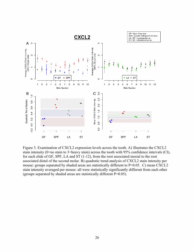
Examination of CXCL2 stain intensity across the second molar in SPF and GF mice
revealed two distinctly different patterns (Fig. 3A). In SPF mice, higher stain intensity was
found in the root associated mesial and distal JE as opposed to the straight buccal/lingual JE (Fig.
3A). In contrast, in GF mice the stain intensity of CXCL2 was uniform, exhibiting nearly
identical stain intensity in all locations (Fig. 3A). Examination of GF mice gavaged with either
Streptococcus sp. or Lactobacillus sp., revealed CXCL2 expression patterns similar to SPF mice,
indicating that the bacteria influenced the neutrophil expression pattern (Fig. 3A). This is in
contrast to the CXCL1 where the stain intensity was uniform across the tooth in all groups, GF,
SPF, GF-Lactobacillus sp, and GF-Streptococcus sp. mice (Fig. 4A).
The significance of the CXCL1 and CXCL2 expression patterns as well as their intensity
levels in the different experimental groups was determined. A quadratic trend analysis revealed
that the pattern of CXCL2 but not CXCL1 expression was significantly different (P=0.05) when
oral commensal bacteria were present in either SPF mice or mice gavaged with either
Streptococcus sp. or Lactobacillus sp. (Fig. 3B and 4B). An analysis of the mean values of the
total stain intensity of CXCL2 in the gingival tissue revealed that both SPF and mice selectively
gavaged with oral bacteria contained significantly higher expression levels of CXCL2 when
compared to GF mice (P=0.05). However, each group that contained oral commensal bacteria
was significantly different from each other such that the CXCL2 staining intensity in SPF mice
was greater in mice gavaged with Streptococcus sp., which was greater than mice gavaged with

17
Lactobacillus sp. (P=0.05) (Fig. 3C). In contrast, no significant differences were observed in
the mean intensity of CXCL1 among the different experimental groups (Fig. 4C). The
expression levels of CXCL1 and CXCL2 are consistent with our previous findings that CXCL2
expression was greater in SPF mice than GF mice, whereas CXCL1 expression was not
significantly different [44]. However, this analysis also revealed that the expression level of
CXCL2 but not CXCL1 varied across the tooth surface and that the CXCL2 expression levels
were modulated by individual species of oral commensal bacteria.
3. Discussion
Neutrophils are one of the most crucial components of the host immune system. Their
role and the mechanisms of recruitment have been studied in health, disease, and host
development [32, 44, 49, 50]. This study confirmed commensal bacterial influence on neutrophil
migration and revealed new information regarding neutrophil migration in clinically healthy
tissue. The study demonstrated: 1) the location of the gingival tissue from root associated mesial
of the 2nd molar to the root associated distal, was correlated with the migration pattern of
neutrophils and expression levels of CXCL2, but not CXCL1, 2) specific commensal bacteria
could induce neutrophil migration and CXCL2, but not CXCL1.
a. Location
Previous studies have reported increased neutrophil and CXCL2, but not CXCL1
presence in SPF versus GF mice [44, 49]. The data presented in this study are the first that
indicate the importance of tissue location with respect to neutrophil migration and ligand
expression. CXCR2 has been shown to be an important component in maintaining periodontal
homeostasis through CXCR2 ligands and resulting neutrophil migration [21, 44]. In this article,
it is confirmed that the role of CXCL2 and neutrophil migration in response to bacterial presence
and the importance of location of the tissue for expression of both CXCL2 and neutrophils is

18
shown. This novel finding expands our understanding of the tissue, demonstrating that not all
areas of the JE regulate neutrophil migration and CXCR2 expression in the same manner. This
provides further evidence that the host uses CXCR2 ligands to regulate its response to
commensal colonization in a highly specific manner. In addition, the findings raise questions
with regard to why the tissue exhibits differences in neutrophil numbers and CXCL2 but not
CXCL1 ligand expression levels. There are several potential reasons for the observed
differences, mainly relating to a variety of influences including the presence of bacteria. The
bacteria may be colonizing more heavily in the inter-dental areas than in the straight buccal and
lingual areas, causing the increase in expression as the interproximal region is approached. The
structure of the tissues in the inter-dental region, which has been found to differ from the buccal
and lingual regions of humans could be influencing the differential location of the neutrophils
and CXCL2 [51].
b. Commensal bacteria
The contribution of the commensal bacteria in the oral cavity on tissue structure and
function is subtle in the oral cavity compared to that of the intestinal tissue. In the gut, it is
relatively easy to demonstrate the influence of bacteria on the gut tissue in GF versus SPF mice:
the development of distinct structural components such as Peyer’s patches depends on the
presence of the bacteria [52, 53]. Conversely, demonstrating the influence of bacteria on the host
oral tissue in GF and SPF is less obvious because the presence of bacteria does not alter the
overall structural of the tissue, rather the bacteria influences the presence of specific cells such as
neutrophil numbers in the JE [52, 53].
The work of this study began an examination of the influence of specific species of
bacteria on the host immune system in the junctional epithelium. The study examined single
species of oral commensal bacteria and their effect on neutrophil migration and select ligand
19
expression. The results showed that two commensal bacteria, Streptococcus sp. and
Lactobacillus sp., could induce a similar, if not identical, pattern of neutrophil migration and
CXCL2 expression as the whole SPF community, yet not to the same level as the community as
a whole as demonstrated by the SPF mice. However, the data demonstrate that while bacterial
colonization is not necessary for neutrophils, CXCL2, or CXCL1 to be present, commensal
colonization is responsible for an increase in neutrophil and CXCL2, but not CXCL1, levels in
the oral cavity. This is the first time commensal bacteria have been added back to a GF mouse
and the role of neutrophil migration examined in the oral cavity. The results demonstrate at least
two individual species of bacteria can induce neutrophil migration and CXCL2 expression in a
similar pattern to wild type oral flora. This investigation of individual components lends insight
into the oral microbiota, which is composed of a community of bacteria, each having its own
unique role in the community and interaction with the host. The finding that while both the
Streptococcus sp and Lactobacillus sp induced a host response, but not at the same level,
supports the idea that not all bacteria are capable of eliciting the same host reaction, and that
their interaction with the host is unique rather than general. This idea is pervasive in the
literature, especially with respect to pathogenic bacteria of the red complex [1, 54]. These
findings in commensal bacteria suggest that different species of commensals induce different
levels of host inflammation.
The difference in the ability of the Streptococcus sp. and Lactobacillus sp. to induce
neutrophil migration and CXCL2 expression as previously stated gives insight into how the
bacteria interact to help maintain healthy homeostasis. The bacterial community in the oral
cavity has long been identified as a component in periodontal disease. Different hypotheses have
developed over the years such as the ‘nonspecific plaque’ hypothesis, where the quantity of

20
dental plaque was more important than the identity of the plaque. As the role of dental plaque in
the disease process has been investigated, it has been suggested that periodontal disease is a
microbial-shift disease based on the well-characterized shift from mostly gram-positive bacteria
to mostly gram-negative species, as the transition from periodontal health to disease occurs [55].
The mechanisms that cause the microbial shift are not well understood. The more that is
understood about how the individual bacteria in the plaque interact with the host and each other,
the more likely it will become possible to identify the particular mechanisms that cause the shift.
Both the Streptococcus sp. and Lactobacillus sp. studied in this experiment are associated with
healthy homeostasis in the periodontium, and both have shown the ability to influence the host to
modulate neutrophil and CXCL2 expression in similar ways, although as previously stated, not
to the same extent. This indicates that individual commensal bacteria can influence the host, but
that the influence is modulated when they are in the SPF community. Streptococcus sp. and
Lactobacillus sp. are just two of the hundreds of species present in periodontal health. The level
of neutrophil migration and CXCL2 expression in SPF mice is less than if one adds the
individual bacteria’s effect. These findings indicate that the community of bacteria in health
interacts to cause a host response that is less than the summation of the individual responses, but
more than that of its individual components alone. With this knowledge, future experiments
should include experimentation with more single species commensal bacteria, in addition to
adding different individual bacteria together, such as the Streptococcus sp. and Lactobacillus sp.
combination, to form limited communities. Further research should investigate how these
bacteria work in a community to induce the host response in homeostasis. The size of the
colonizing community and whether they are colonizing in greater numbers in the inter-dental
region should be investigated, to see how many bacteria are colonizing in these single species
21
gavages. Data from studies such as these could help reveal influencing factors on neutrophil
migration and associated ligand expression. Further investigation of these individual bacteria
and mini communities will improve the understanding of how commensal bacteria influence the
host response.
c. Methodology
The data generated for this study are based on the response of GF mice gavaged with
commensal bacteria, Streptococcus sp. and Lactobacillus sp., five days post-completion of the
gavage procedure. Thus these data represent a snapshot of neutrophil migration, CXCL2 and
CXCL1 ligand expression, which occurred immediately after the introduction of commensal
bacteria. If the tissues had been examined at a later time point, allowing the bacteria to have
been present for a longer amount of time, the results may have been different, as the host
response would have had more time to adapt to the bacterial presence. The significance of
examining this short time period is that it reveals that the body does not initially respond
identically to all types of bacteria. After five days, there appeared to be a stronger response to
CXCL2 with the presence of Streptococcus sp. over Lactobacillus sp. This gives strong
evidence that while both species are considered commensal, they do not activate the host
response to the same level.
To place the data in context of the natural oral community, SPF mice have been exposed
to a complex microbial community from the time they were born, and that community has been
changing and evolving over time. In contrast, the GF mice have never been exposed to any
bacteria. In this study the GF mice have only had a brief introduction to commensal bacteria
resulting in increased neutrophil migration and CXCL2, not CXCL1, ligand expression. To
continue this line of inquiry, further study of the host reaction to commensals could reveal more
information about SPF homeostasis. Studies have shown that GF mice have more bone than SPF
22
mice, and that the commensal microbiota of SPF mice can completely transfer to GF mice within
14 days of co-caging [22]. In addition, within 16 weeks of co-caging, SPF and GF mice
exhibited moderate bone loss comparable to age matched SPF mice, is observed in GF mice,
likely indicating that bone loss due to low grade infection occurred [22]. The results of this
study combined with the bone loss study raise additional questions, for example, whether
individual commensal bacteria or a limited number of bacteria induce the same inflammation and
bone loss as the entire SPF microbiota. Future experimentation could examine mice after
different exposure times to commensal bacteria, which could reveal the pattern of inflammation
that leads to natural bone loss observed in SPF mice. Such a finding could effectively challenge
the general consensus that commensal bacteria are not usually associated with acute and chronic
infections. It could provide additional evidence for, and a better understanding of how,
commensal bacteria induce low grade inflammation thought to cause the modest bone loss
observed [22].
4. Conclusion
The data presented provide the first evidence of the importance of tissue location when
examining the periodontium and the host mediators involved in health. In this Chapter it was
demonstrated that neutrophils and their associated CXCR2 ligand (CXCL2 but not CXCL1) were
more prevalent toward the interproximal regions of the tooth, revealing the importance of tissue
location. This indicates that not all areas of the junctional epithelium react the same to the
presence of bacteria. Bacteria have long been known to affect the host immune system triggering
different immune responses. The increase in neutrophils and CXCL2 ligands as the interproximal
region was approached is consistent with the fact that early clinical signs of periodontal disease,
include inflammation and increased periodontal probing depths, and are often first observed in
the interproximal regions. This clinical experience supports the hypothesis that the interproximal
23
area has an increase in stimulation of the innate host response to bacterial presence. The outcome
of this study, comparing the effects of a healthy biofilm versus single species from that
community, helps to further the understanding of commensal bacteria on periodontal health. We
show that not all bacteria cause the same level of host immune response. Understanding both
host and microbial factors that contribute to the clinical health of periodontal tissue will reveal
novel diagnostic and intervention practices. Examples could include specific chemokine ligand
inhibition or augmentation, probiotics or addition of bacterial based immune modulators.
24
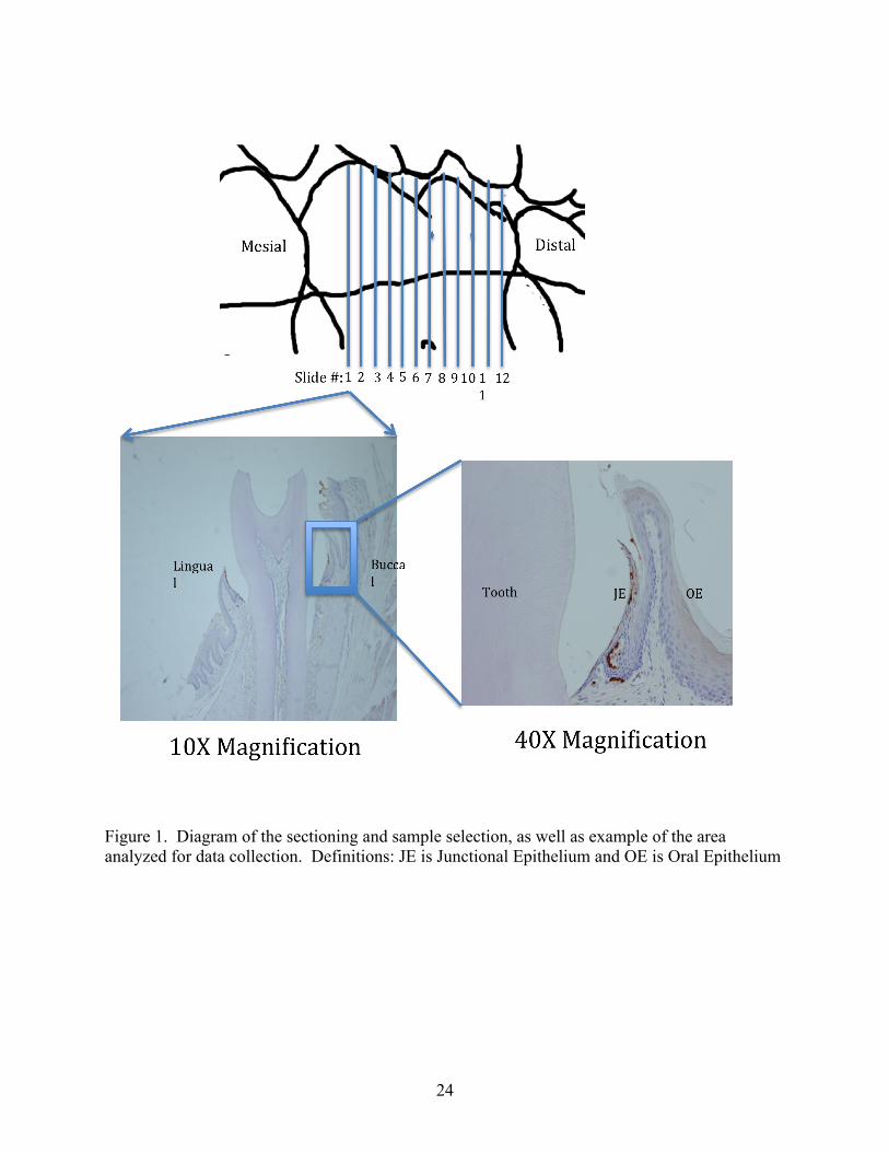
Figure 1. Diagram of the sectioning and sample selection, as well as example of the area analyzed for data collection. Definitions: JE is Junctional Epithelium and OE is Oral Epithelium

25
Figure 2. Examination of neutrophil expression across the tooth. A) illustrates the average number of neutrophils, with 95% confidence levels (CI), for each slide of GF, SPF, LA and ST (1-12), from the root associated mesial to the root associated distal of the second molar. B) Quadratic trend analysis of neutrophil per mouse: Groups separated by shaded areas are statistically different P=0.05. C) Mean neutrophil count per mouse: all groups were statistically significantly different from each other (groups separated by shaded areas are statistically different P=0.05).
26
Figure 3. Examination of CXCL2 expression levels across the tooth. A) illustrates the CXCL2 stain intensity (0=no stain to 3=heavy stain) across the tooth with 95% confidence intervals (CI), for each slide of GF, SPF, LA and ST (1-12), from the root associated mesial to the root associated distal of the second molar. B) quadratic trend analysis of CXCL2 stain intensity per mouse: groups separated by shaded areas are statistically different to P=0.05. C) mean CXCL2 stain intensity averaged per mouse: all were statistically significantly different from each other (groups separated by shaded areas are statistically different P=0.05).

27
Figure 4. Examination of CXCL1 expression levels across the tooth. A) illustrates the CXCL1 stain intensity (0=no stain to 3=heavy stain) across the tooth with 95% confidence intervals, for each slide of GF, SPF, LA and ST (1-12), from the root associated mesial to the root associated distal of the second molar, no pattern difference is observed. B) quadratic trend analysis of CXCL1 stain intensity per mouse shows no signficant difference between the groups of mice. C) mean CXCL1 stain intensity averaged per mouse shows no significant differnce between the groups of mice.

28
Chapter Three: Effect of P. gingivalis on neutrophil migration and select CXCR1 and CXCR2 ligand expression in the junctional epithelium
1. Introduction
P. gingivalis is one of the most studied periodontal pathogens and has been linked to the
induction and progression of chronic inflammatory diseases such as periodontal disease [54].
While P. gingivalis is linked with inflammatory disease, it is not able to induce as robust an
inflammatory response as some other bacteria [54]. P. gingivalis is an excellent immune
manipulator with the ability to selectively induce only a limited number of inflammatory
responses from leukocytes through receptor crosstalk mechanisms [54, 56]. To be more specific,
P. gingivalis can inhibit leukocyte-mediated bacteria-killing mechanisms [54]. P. gingivalis’s
involvement in the transition from health to disease has been proposed to be caused by disruption
of the homeostatic relationship between the commensal bacteria, thereby inducing a local
inflammatory response [55]. If the inflammation becomes chronic, it can result in oral bone
destruction, precipitating the diagnosis of periodontal disease. This affliction is not rare,
affecting approximately 100 million people in the United States [54, 57]. While P. gingivalis is
most commonly associated with disease in the oral cavity, it is now being linked with other
locations and conditions. P. gingivalis-mediated periodontal disease is being found in numerous
studies to be a risk factor for systemic diseases including diabetes, pre-term birth, stroke, and
cardiovascular diseases [57-64]. The following study focuses on the role of P. gingivalis in the
migration of neutrophils in the junctional epithelium and closely related tissue.
As discussed in Chapter One, neutrophils are a crucial, phagocytic component of the
innate defense system that protects periodontal tissue from disease [12, 13]. In periodontal
health, there is a fine balance in the quantity of neutrophils required; too few or too many

29
neutrophils present in the junctional epithelium can cause diseases such as periodontitis in the
oral cavity [12]. There are a number of diseases characterized by a lack of usual levels of
neutrophils and development of periodontitis [12, 14-20]. In animal studies it has been shown
that KO mice, which are defective in neutrophil transit, develop periodontitis [21-23]. The
observations of these studies are consistent with the hypothesis that neutrophils play a role in
protecting the host from periodontal disease. Neutrophil migration is regulated in the tissue by
different signaling modalities. For example, CXC chemokines, such as CXCL1, CXCL2 and
CXCL6, interact with receptors CXCR1 and CXCR2, to promote the rapid mobilization of
neutrophils to sites of inflammation. In humans, it has been found that both CXCR1 and
CXCR2 mRNA expression is increased in the gingival tissue of patients which chronic
periodontitis who had detectable levels of Porphyromonas gingivalis [34]. The homology of the
mouse and human CXCR1 and CXCR2 receptor has been established, substantiating the utility
of studying mouse CXCR1 and CXCR2 activity to gain insight into the function of human
CXCR1 and CXCR2 [33].
There are a number of pathogens that employ skillful mechanisms to evade host defenses,
such as inhibiting neutrophil activation, resulting in chronic infections. One way these
pathogens evade detection by the host is to elude the host pattern recognition receptors (PRRs),
which recognize conserved molecular patterns shared by many groups of microorganisms.
These PRRs include the Toll-like receptors (TLRs). TLRs play a critical role in the
identification of microbial components such as flagellin, lipopeptides, lipopolysaccharide and
CpGDNA [65, 66]. Activation of TLRs signals inflammation and is very important for the
detection and removal of pathogens. This has a short-term benefit to the host as it signals for
host clearance of the pathogen through recruitment and activation of cells and other factors
30
essential to that process. However excessive activation of such mediators or prolonged immune
activation can lead to chronic inflammation, which can be detrimental to the host [67, 68].
One important TLR, TLR4, an innate immune receptor, recognizes lipopolysaccharide
(LPS). The host uses TLR4 to identify gram-negative bacterial LPS expressed on the cell’s
membrane. Once the pathogen is recognized by the host, the host triggers an immune response,
inducing inflammation that attempts to remove the pathogen from the host [65]. A number of
gram-negative organisms have evolved mechanisms to evade immune detection so they can
establish infection. One way they evade detection is by modifying their LPS structure, in
particular their lipid A species [66]. Lipid A is actually the only area of LPS known to be
recognized by the innate immune system [66]. Unmodified lipid A produces a robust
inflammatory response in the host. This is well demonstrated by the structure typically expressed
by E. coli, which is highly stimulatory even in low concentration [66, 69]. Organisms which
modify the lipid A structure show alterations to the acyl chains or terminal phosphate groups,
and are less stimulatory to the host [69].
As mentioned above, P. gingivalis is one such low abundance oral anaerobic bacterium
that has evolved to evade host immune response through modification of its lipid A structure. P.
gingivalis is a red complex bacteria and is associated with clinical signs of periodontal disease
and related systemic conditions [64, 70-73]. It can induce chronic low-grade inflammation
associated with bone loss. P. gingivalis can also be considered a “keystone pathogen” which
supports and remodels a microbial community so as to promote disease pathogenesis [54]. This
study investigated the effects of P. gingivalis on the migration of neutrophils, CXCL1, 2, and 6,
and will increase understanding of P. gingivalis’s success in evading the host’s innate immune
responses, leading to a better understanding of the how to disrupt the disease state.

31
2. Characterization of the P. gingivalis mutants used in this study
The study utilized three types of P. gingivalis: wild type strain 381 and two lipid A
phosphatase mutants, PG1587381 and PG1773381. P. gingivalis wild-type strain 381 has a non-
phosphorylated and tetra-acylated lipid A structure that is predicted to have neither an agonist or
antagonist lipid A structure toward the TLR4 complex [69, 74]. The P. gingivalis mutant strains
lack lipid A 1- and 4’ phosphatase activities in the P. gingivalis 381 background. P. gingivalis
strain PG1773381, lacks the 1-phosphatase activity and has a TLR4 antagonist lipid A , while
PG1587381 lacks the 4’-phosphatase activity leading to a TLR4 agonist lipid A structure [74].
The modification of the lipid A structure not only modifies the TLR4 activity, but also alters the
bacteria’s susceptibility to being killed via cationic peptides [74, 75]. A deficiency in PG1587 in
three different P. gingivalis strains (33277, A7436, 381) has been shown to result in a
pronounced sensitivity to polymixin B, compared to the wild type and PG 1773 strains. This is
consistent with lipid A 4’ phosphotase having a critical role in bacterial susceptibility to this drug
[69, 74]. To confirm that the modifications of the strains only affect the TLR4 activity and
susceptibility to cationic killing, the strains have been verified to have phenotypes that are
consistent with bacterial lipid A modifications as opposed to other surface virulence factors such
as those that activate TLR2 via fimbriae and lipoproteins [74].
3. Expanded serial sectioning of the mouse molar to determine tissue specific neutrophil and chemokine expression patterns
The data in Chapter Two revealed that neutrophil migration and CXCL2 expression
levels were increased as the interproximal (area between two teeth) was approached. The study
in Chapter Two did not examine the non-root associated interproximal region between teeth. In
the following study it was decided to expand the area of tissue being examined to include the
interproximal region. To accomplish this, two anatomical points were selected: the distal most

32
root associated sections of first molar and the second molar. Then, 16 slides were evenly
distributed between the two points and stained, lining up the non-root associated interproximal
slides (Figure 5). The slides were then stained for neutrophils, CXCL1, CXCL2 and CXCL6
using standard histological procedures. The slides were then read, counting the number of
neutrophils or scoring the stain intensity (scale 0-3) as appropriate, in the same manner as
described in Chapter Two.
4. Results: The impact of oral pathogens, P. gingivalis and associated mutants, on the quantity and location of neutrophils, CXCL1, CXCL2 and CXCL6 in periodontal tissue
a. Expression of neutrophils
It has been previously shown (Chapter Two) that location of tissue is important when
examining the level of expression of neutrophils and CXCL2. It was shown that commensal
bacteria influence the pattern of expression of neutrophils and CXCL2, with an increase in both
neutrophil numbers and CXCL2 expression as the interproximal region is approached. This
study expands upon this finding and examines neutrophil migration through the entire
interproximal region between the first and second molar. The data show that neutrophil
migration to the JE and closely associated tissue is increased in the interproximal regions (slides
#3-8) in all groups examined: control group, P. gingivalis 381, PG1587381 and PG1773381 (Figure
6A). In addition, the overall pattern of expression across the entire area examined (the root
associated distal of the first molar to the root associated distal of the second molar) was similar.
This indicates that P. gingivalis 381, PG1587381 and PG1773381 do not affect the pattern of
neutrophil expression when compared to the wild type. This is consistent with the previous
finding in the SPF mice, GF-Streptococcus sp. and GF-Lactobacillus sp. groups examined in
Chapter Two, where the presence of bacteria induced a similar pattern of neutrophil migration.
The increase in neutrophils in the interproximal region is consistent with the fact that early

33
clinical signs of periodontal disease are recognized to include inflammation and increased
periodontal probing depths, and are often first observed in the interproximal regions. This
clinical experience lends credence to the hypothesis that this area has an increase in stimulation
of the innate host response to bacterial presence.
When examining the neutrophil counts as an average, per mouse type (Figure 6B), it was
shown that the average neutrophil counts for the control sham gavage group, using a 1-sided
permutation test, was higher than those of the two mutants, PG1773381 and PG1587381, (P=0.03)
and that the average neutrophil count of PG PG1587381 was even lower than PG PG1773381
(P=0.03). This indicates that the fixation of the LPS structure was affecting the neutrophil
migration levels in the JE and closely associated tissue. In P. gingivalis strain PG1773381, which
lacks the 1-phosphatase activity and has a TLR4 antagonist lipid A , this lipid A modification
altered neutrophil migration when compared to the wild type, but not to the same level as the
alteration of PG1587381, which lacks the 4’-phosphatase activity leading to a TLR4 agonist lipid
A structure. This indicates that the inability of the mutants to modify their structure is
influencing the level at which they can interact with the host. In addition, this finding suggests
that the wild type P. gingivalis 381 is altering its inflammatory structures in such a way as to
cause a greater variation in neutrophil migration as seen by the spread of the average levels of
migration, not all of the mice infected with wild type P. gingivalis 381 were expressing similar
levels of neutrophil migration. Overall these findings confirm the importance of LPS structure
on P. gingivalis’s influence on the host.
b. Expression of CXCL1, CXCL2 and CXCL6
It has been shown that CXCL2 was differentially regulated by commensal bacteria, while
there was no evidence for commensal bacterial regulation of CXCL1 expression (Chapter Two)
[44]. The present study examined CXCL1, CXCL2 and CXCL6 regulation in the presence of a

34
known periodontal pathogen, P. gingivalis, and two associated mutants, with either an agonist or
an antagonist LPS structure, P. gingivalis 381, PG1587381 and PG1773381 respectively. When
looking at the pattern of expression of CXCL1, CXCL2 and CXCL6 from the root associated
distal of the first molar to the root associated distal of the second molar, it is notable that all of
these ligands have a similar pattern of expression. All exhibited increased intensity in the
interproximal region when compared to the straight buccal and lingual regions (Figure 7A, 8A,
and 9A). This is a relevant distinction from the expression of CXCL1, which, as noted in
Chapter Two, did not show a variation based on location. The relatively static expression of
CXCL1 described in Chapter Two reflects the spatial limitation of the previous study to the
regions between the root associated mesial and the root associated distal. As it turns out, when
the area of investigation was widened (as described in this Chapter), the expression of CXCL1
was shown to increase in the interproximal region. When examining interproximal distribution
of CXCL1 in comparison to CXCL2 and CXCL6, the area of increased expression appeared over
a narrower range of slides than the CXCL2 and CXCL6 expression, indicating that the increase
in expression was more highly focused to the interproximal regions than that of CXCL2 and
CXCL6, which also began their increase in expression closer to the buccal and lingual tissue.
When examining the overall level of intensity of expression of the ligands, CXCL1 was
consistent with the results of Chapter Two, in that there was no change in overall average
expression of CXCL1 between the four groups: control group, P. gingivalis 381, PG1587381 and
PG1773381 (Figure 7). This indicates the overall expression level was not influenced by presence
of P. gingivalis or associated mutants, and that the variation seen based on location in the
interproximal region was independent of the addition of the periodontal pathogen. This contrasts
with the overall expression observed for CXCL2 and CXCL6 (Figure 8b and 9b). The average

35
expression level of CXCL2 was similar between control group, P. gingivalis 381, and PG1773381
(Figure 8b). There was, however, a decrease in average CXCL2 expression in PG1587381
(P=0.03) when compared to the control group and P. gingivalis 381. This CXCL2 decrease in
PG1587381 was similar to the decrease in average neutrophil counts when compared to the
control group (Figure 8b). However there was not a statistical difference when compared to
PG1773381 even though there was a lower level of neutrophil migration in PG1773381. Thus there
appears to be a threshold level of host stimulation, induced by the bacteria, required to cause a
noticeable change in CXCL2 ligand expression using the given methodology. This threshold
was reached by PG1587381 but not PG1773381. The average expression levels of CXCL6 are
slightly different than that of CXCL2 and neutrophils (Figure 6b and 9b). CXCL6 average
expression levels were similar between the control and P. gingivalis 381 groups. Yet the level of
expression was decreased for both PG1587381 and PG1773381 (P=0.03) when compared to the
control group. This shows that modification of the LPS structure modulated the CXCL6
expression in both cases.
c. QPCR bacterial load
The fluctuation in expression of neutrophils and associated CXCR1 and CXCR2 ligands
raises the question of how many bacteria are present in the oral cavity during the investigation. It
has been shown in both a rabbit model and a murine model that the presence of P. gingivalis and
associated mutants can cause changes in the microbiome, both in mass and in composition [22,
76]. This study attempted QPCR of snap frozen pieces of buccal tissue from the time of sacrifice.
Inconsistent amounts of DNA was recovered using a universal bacterial probe (approximately
101 to 102 bacteria per sample) and QPCR using a ISPG1 [22] gene to detect P. gingivalis was
unable to detect P. gingivalis reliably (data not shown).

36
5. Discussion
a. Differences in study methodology
A common approach to studying periodontitis is to inoculate SPF animals orally with P.
gingivalis and measure periodontal bone loss six weeks post-final inoculation [22, 76-78]. It has
been shown that significant bone loss in the murine model can take four or more weeks to
develop [22, 79-81]. A major difference in the study in this Chapter, in comparison to the study
in Chapter Two, is that the mice used for this study were the same animals used in Slocum et al.
2014, which examined them for vascular inflammation and periodontal bone loss. The Slocum
artherosclerosis model did not sacrifice the mice until 16 weeks post infection, although other
studies have involved earlier sacrifices, including at six weeks [74, 82, 83]. The vascular
inflammation portion of the Slocum study showed there was progressive luminal narrowing from
eight to 16 weeks with P. gingivalis 381 and PG1773381, when compared to strain PG1587381 and
the sham infected control, which showed minimal narrowing [74]. The PG1587381 gavaged group
showed more narrowing than the sham infected control group [74]. Interestingly, all of the mice
gavaged with P. gingivalis whether wild type or an LPS mutant, exhibited similar amounts of
bone loss around the molars. The observed bone loss in this study, is consistent with data from
the rabbit model where the presence of P. gingivalis A7436 and two similar lipid A phosphatase
mutants, PG1587A7436 and PG1773A7436, were all correlated with periodontal bone loss [76].
The bone loss and progressive luminal narrowing observed in Slocum et al. [74] is notable
because it correlates with the results obtained from the present study, showing that PG 1587381
had reduced average neutrophil migration and expression of CXCL2 and CXCL6 in comparison
to the other groups examined. This shows that alteration of the LPS structure on PG1587381 is
correlated with a significant reduction in the inflammatory response 16 weeks post gavage, not
only in the vessels but also in the junctional epithelium, when compared to P. gingivalis 381 and

37
PG1773381, yet still induced periodontal bone loss. This is an indication that lack of 4’
phosphatase is correlated with reduced inflammation after 16 weeks but was still able to induce
enough inflammation to cause periodontal bone loss. This may be correlated with the increased
susceptibility of PG 1587381 to cationic peptide killing [69, 74]. In addition, it was shown that
PG1773381 also had reduction in CXCL6 expression when compared to P. gingivalis 381,
indicating that the lack of 1-phosphatase activity also influences the CXCL6 expression levels,
yet did not affect the overall neutrophil or CXCL2 levels in the same way as P. gingivalis
1587381. These changes may be correlated with alterations in the oral microbial community. It
has been shown in the rabbit model that P. gingivalis wild type, but not the lipid A mutants, had
a significant impact on both the composition and microbial load of the oral microbiota [76]. This
reemphasizes the complex interactions of the bacterial host interaction.
b. Pg wild type did not alter the community
In this chapter it was found that SPF mice colonized with P. gingivalis did not experience
altered neutrophil migration, CXCL1, 2, or 6 expression when compared to sham gavage mice
16 weeks post gavage. This finding indicates that at the time of examination, P. gingivalis was
not exerting pressure to alter the response of these indicators. Interestingly, when examining the
average neutrophil counts of the four mice gavaged with the P. gingivalis 381wild type, the
average neutrophil count was spread over a wider range than the other three types of mice. This
observation could be due to the ability of P. gingivalis to modify its activity based on the
environment, indicating that the populations in the different mice were more divergent in their
activity without the fixed lipid A structure of the PG1773381 and 1587381. These findings with
regard to the ligand expression coincide with the findings of Hajishengallis et. al. [22] that
CXCL1 (a mouse homolog of human IL-8) was initially down regulated after P. gingivalis
inoculation when examined four days post gavage, but returned to baseline after six weeks [22].

38
In this study CXCL1 levels were examined 16 weeks post gavage. These two studies together
suggest that once CXCL1 levels return to baseline, they may become stable at that baseline for a
prolonged period of time. This is notable as P. gingivalis is known to inhibit IL8 induction by
gingival epithelial cells [22, 84, 85], yet the findings of this study and Hajishengallis et. al.[22]
indicate that this inhibition is most likely for only a short duration. These findings further support
Hajishengallis’s hypothesis that deregulation of leukocyte recruitment may contribute to P.
gingivalis-induced periodontitis in the murine model [22].
c. Microbial community levels
The finding that P. gingivalis 1587381 infected mice exhibited reduced inflammation, yet
still sustained bone loss, raises the issue of the bacterial load at 16 weeks post gavage. As stated
previously, the bacterial load in the oral cavity was examined using QPCR. There was only a
small amount of bacteria detected in general and specifically, with the P. gingivalis probe, we
were unable to detect any amount of the P. gingivalis pathogen. This indicates the bacterial load
was at minimal or sub-minimal detection levels. This may be due to sampling technique, as in
this study a small amount of tissue was utilized to try to determine the bacterial load. Other
studies have utilized either an oral swab or paper point collection of plaque to quantify the
bacterial load [1, 22, 76]. Hajishengallis et al. did perform QPCR on gingival tissue collected
from mice to quantify the amount of P. gingivalis, but the amount of tissue utilized was larger
than that available in this study. In addition, P. gingivalis has been detected in the oral cavity at
least 42 days (six weeks) after infection in immunocompetent and severe combined
immunodeficient (SCID) mice that were infected with P. gingivalis ATCC 53977 [82]. The fact
that levels of P. gingivalis 381, PG1773381, and 1587381 were undetectable in the present study,
when measured 112 days (16 weeks) post-infection, suggests that the P. gingivalis experienced a
reduction as time from intial infection passed.
39
The previous work of Slocum et al., which utilized the same animals as the present study,
revealed that all P. gingivalis types, wild and mutant, induced bone loss, and is consistent with
findings of other studies [22, 76]. The fact that bone loss was observed indicates that at some
point between infection and sacrifice, the mice experienced a host reaction inducing that bone
loss. This may have been due to a temporary microbial shift and the changes in the neutrophil
expression levels seen 16 weeks post gavage may be due to the residual result of the microbial
shift. It has been shown in the rabbit models that the presence of P. gingivalis causes a
significant increase in biomass of plaque in P. gingivalis wild type infected rabbits when
compared to rabbits infected with mutants 1587 and 1773, although the composition of the oral
community shifted in all three groups [76]. The introduction of P. gingivalis has been shown to
induce periodontal bone loss and increase the total cultivable load. In addition, it has been shown
to change the composition of the oral microbiota when compared to a non-infected SPF mouse
after six weeks [22]. The amount of P. gingivalis present was estimated to be less than 0.01% (4
to 5 log10 units lower) of the total bacterial count according to QPCR and Pg was not cultivable
from oral samples taken [22]. GF mice infected with P. gingivalis do not develop bone
resorption, which is a classic clinical sign of periodontitis. This indicates that P. gingivalis alone
does not induce periodontitis, but rather the presence of other bacteria is required to orchestrate
bone loss [22, 45]. This is consistent with the hypothesis that periodontal disease is caused by
dybiosis, a microbial imbalance. The community shift observed from the presence of P.
gingivalis causes a disturbance in the community that shifts the host from health to disease.
d. The structure of the interdental region is potentally different than marginal regions in a way that influences neutrophil migration
In humans it has been observed that the structure of the marginal gingiva when compared
to the interdental papilla exhibits distinct differences. For example, even in health, the

40
interdental region will display signs of inflammation that are not present in the marginal gingiva.
Also the components of the two different areas are different. For example, the interdental region
has less robust collagen fibers and a different mix of surrounding tissue [51]. These types of
differences, when translated to the mouse model, could explain some of the differences seen in
the neutrophil migration and CXCL1, 2, and 6 expression levels. Even if the structure of the
junctional epithelium is the same surrounding the tooth, differences in the surrounding
connective tissues could exert influene on them by modifying the levels of expression of
neutrophils and associated ligands. For instance, since the interdental region has two JE regions
in close proximity (the distal JE of the first molar and the mesial JE of the second molar),
abutting each other, and sharing connective tissue, it may be that the confluence of the signals
coming from the two JE areas is magnifying the response seen in this area, increasing the
number of neutrophils and ligand expression when compared to the buccal and lingual regions,
which only have the influence of mediators from one JE in the area.
In summary the following data support the idea that location of the tissue is important in
expression of inflammatory components and that modification of the LPS structure does
influence the bacterial host interaction. Further work in this area will increase the understanding
of this complex interaction. For example, examination of more time points post infection, and a
detailed examination of the the microbial community would further elucidate this complex
process.
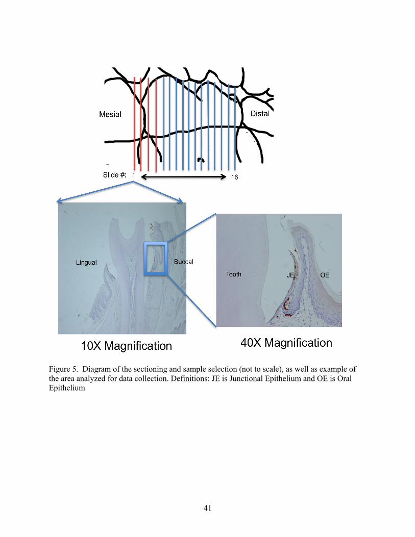
41
Figure 5. Diagram of the sectioning and sample selection (not to scale), as well as example of the area analyzed for data collection. Definitions: JE is Junctional Epithelium and OE is Oral Epithelium
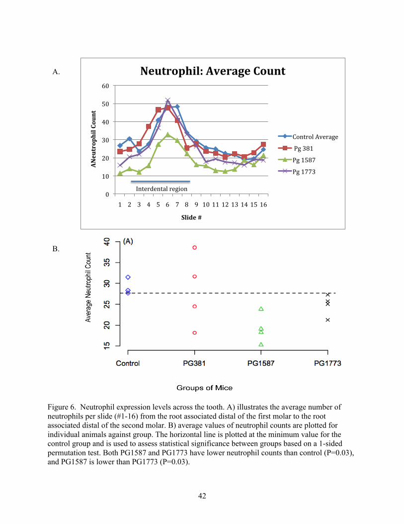
42
Figure 6. Neutrophil expression levels across the tooth. A) illustrates the average number of neutrophils per slide (#1-16) from the root associated distal of the first molar to the root associated distal of the second molar. B) average values of neutrophil counts are plotted for individual animals against group. The horizontal line is plotted at the minimum value for the control group and is used to assess statistical significance between groups based on a 1-sided permutation test. Both PG1587 and PG1773 have lower neutrophil counts than control (P=0.03), and PG1587 is lower than PG1773 (P=0.03).
0
10
20
30
40
50
60
1 2 3 4 5 6 7 8 9 10 11 12 13 14 15 16
ANeutrophil Count
Slide #
Neutrophil: Average Count
Control Average
Pg 381
Pg 1587
Pg 1773
Interdental region
A.
B.

43
Figure 7. CXCL1 stain intensity levels across the tooth. A) illustrates the average CXCL1 stain intensity (0=no stain to 3=heavy stain) per slide (#1-16) from the root associated distal of the first molar to the root associated distal of the second molar. B) average values of CXCL1 stain intensity plotted for individual animals against the group. The horizontal line is plotted at the minimum value for the control group and is used to assess statistical significance between groups based on a 1-sided permutation test. No significant differences were found for CXCL1.
0
0.5
1
1.5
2
2.5
3
1 2 3 4 5 6 7 8 9 10 11 12 13 14 15 16
Stain Intensity (Scale 0-‐3)
Slide #
CXCL1: Average Stain Intensity
Control Avererage
Pg381 Average
PG 1587 Average
PG 1773 Average Interdental region
A.
B.

44
Figure 8. CXCL2 stain intensity levels across the tooth. A) illustrates the average CXCL2 stain intensity (0=no stain to 3=heavy stain) per slide (#1-16) from the root associated distal of the first molar to the root associated distal of the second molar. B) average values CXCL2 stain intensity plotted for individual animals against group. The horizontal line is plotted at the minimum value for the control group and is used to assess statistical significance between groups based on a 1-sided permutation test. PG1587 has significant lower CXCL2 values than control and PG381 (P=0.03). No other significant differences between groups were found.
0
0.5
1
1.5
2
2.5
3
1 2 3 4 5 6 7 8 9 10 11 12 13 14 15 16
Stain Intensity (Scale 0-‐3)
Slide #
CXCL2: Average Stain Intensity
Control Avererage
Pg381 Average
1587 Average
PG 1773 Average
Interdental region
A.
B.
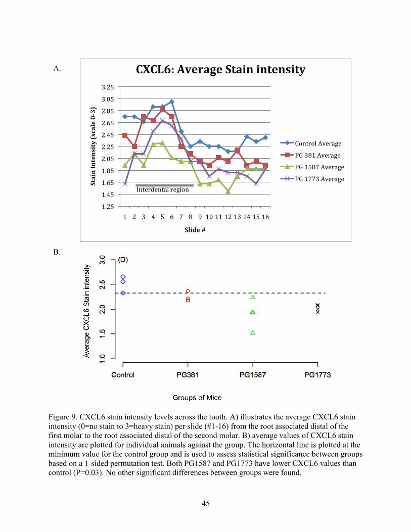
45
Figure 9. CXCL6 stain intensity levels across the tooth. A) illustrates the average CXCL6 stain intensity (0=no stain to 3=heavy stain) per slide (#1-16) from the root associated distal of the first molar to the root associated distal of the second molar. B) average values of CXCL6 stain intensity are plotted for individual animals against the group. The horizontal line is plotted at the minimum value for the control group and is used to assess statistical significance between groups based on a 1-sided permutation test. Both PG1587 and PG1773 have lower CXCL6 values than control (P=0.03). No other significant differences between groups were found.
1.25
1.45
1.65
1.85
2.05
2.25
2.45
2.65
2.85
3.05
3.25
1 2 3 4 5 6 7 8 9 10 11 12 13 14 15 16
Stain Intensity (scale 0-‐3)
Slide #
CXCL6: Average Stain intensity
Control Average
PG 381 Average
PG 1587 Average
PG 1773 Average Interdental region
A.
B.

46
Chapter Four: Summary and future directions in health and disease
1. Summary
In the previous chapters, it has been shown that commensal bacteria play a key role in
activating the innate immune response of the host in health. Chapter Two revealed that different
commensal bacteria induce different levels of location specific neutrophil migration and
expression of CXCL2, but not CXCL1, across the second molar from root associated mesial to
root associated distal of mouse molars. This study has expanded upon the findings by Zenobia et
al. [44] examining the importance of tissue location and presence of specific commensal bacteria
on the innate host response in disease. Chapter Three examined the effect of a well known
periodontal pathogen, P. gingivalis, and two associated LPS mutants, on neutrophil migration
and expression of associated ligands: CLXCL1, CXCL2 and CXLC6. This, in conjunction with
the atherosclerotic study by Slocum et al, gives insight into the relationship between LPS
modification and its effects on the host. The findings presented in this work illuminate the
importance of specific bacterial species and location of the tissue examined on neutrophil
presence and associated ligand expression. This Chapter explores potential future studies of the
bacterial host interaction, and is divided into three sections: 1) Findings specific to Chapter 2; 2)
Findings specific to Chapter 3; and 3) Areas of exploration that would be applicable to both.
These proposals for future study are designed to clarify the role of bacteria in the host innate
immune response in both health and disease.
2. Future directions based on findings in Chapter Two
It has been established that neutrophils are essential for oral health. This has been
demonstrated in studies that examine neutropenia and the resulting periodontitis from the lack of
neutrophils [12, 14-17]. In Chapter 2, examination of neutrophil migration in response to
47
commensal bacteria was performed. This work further expanded upon the findings that
commensal bacteria were responsible for the influx of neutrophils and associated ligands in
healthy tissue [12, 22, 44]. The work in Chapter Two examined the role of specific commensal
bacteria, Streptococcus sp. and Lactobacillus sp., in the host response. In addition, it examined
the importance of tissue location in the host, demonstrating that the level of host response is not
uniform, rather certain areas experience a higher level of neutrophil migration and associated
ligand expression than others. Due to the importance of neutrophils in health, it is essential to
understand the influence of regulatory components of commensals and pathogens in the
stimulation of neutrophils.
The findings of Chapter Two address select issues regarding neutrophil migration in
health, including the role of specific single bacteria in bacterial host interaction as well as
whether all areas of the JE experience the same stimulation level. This leads to a better
understanding of the bacterial host interaction, which will ultimately allow us to be better
prepared to manipulate the system. These findings broaden our understanding of the bacterial
host interaction but also raise more questions. The data show that not all commensal bacteria are
able to influence neutrophil migration and associated ligand expression to the same extent. As
only two bacteria were examined, the question of the influence of other kinds of commensals on
neutrophil and associated ligand expression remains open. Studies examining other commensal
bacteria’s effect on the host are needed to help illuminate the effect of other individual
commensal bacteria on of the host. Future experiments could include examination of the effect
of other individual oral commensal bacteria on neutrophil migration and CXCR2 ligand
expression. Potential bacteria could include other naturally occurring oral murine strains such as
Enterococcus faecalis, Escherichia coli, Staphylococcus xylosus, etc. As it has been shown that

48
single species are affecting the neutrophil migration, this raises the question how select
combinations of commensal bacteria would affect the host. How many species are required to
return the host response to that of the SPF mice? For example, what would be the effect of a
limited number of commensals on neutrophil migration and associated ligand expression? To
examine a limited community of commensal bacteria, one could combine Streptococcus sp. and
Lactobacillus sp. and examine the resulting migration of neutrophils and expression levels of
CXCL2. This would be a good starting combination, as we have data on the effect of the
individual species. The resulting data could contribute to the examination of questions such as
whether the effect of the combination would be additive. Finding the answers to these types of
questions will help us to better understand the role of commensal bacteria in oral health.
3. Future directions based on findings in Chapter Three
In Chapter Three, the focus was switched from looking at bacterial host interaction in
health to looking at the effects of a known periodontal pathogen and two associated LPS mutants
on the host response. In addition, the tissue area examined was expanded and a CXCR1/CXCR2
ligand, CXCL6 known to be expressed in the presence of P. gingivalis, was added to the analysis.
The findings in Chapter Three reinforced that location of the tissue is important in neutrophil and
associated ligand expression. In addition, the finding that CXCL1 expression level varied based
on location, while the variation did not appear to be associated with the presence of the P.
gingivalis mutants, raises the question of why there are differences in the level of expression of
CXCL1 when comparing the interproximal to the straight buccal and lingual. In humans,
differences in healthy tissue between the straight buccal and interproximal area have been
observed [51]. Could these differences also be observed in the murine model? A study
examining the histological structure of the two different areas could reveal whether structural
differences in the tissues could contribute to the variation of the neutrophil migration and
49
associated ligand expression observed in Chapter Three. Another potential contributing factor to
the differences in expression seen in the interproximal versus the straight buccal and lingual
could be the proximity of the two different JEs in the interproximal areas. Is the close proximity
of the JEs altering the expression levels of the neutrophils and associated ligands? It has been
shown that the IL8 gradient in the junctional epithelium is higher as it approaches the bottom of
the sulcus, helping to congregate neutrophils in this area [9, 10]. Could the signaling from JEs in
the interproximal area be magnifying this response when compared to the straight buccal and
lingual regions? An experiment that could potentially address this would be to examine the tissue
to the mesial of the first mouse molar or the distal of the third mouse molar as these areas are in
line with the interproximal areas but as they do not have an adjacent tooth; there is only one JE
present in the tissue. This line of investigation would pair well with different sectioning
methodology, which could reveal more information about the effect of tissue location. For
example if half the maxilla was sectioned coronally (this is the sectioning used in the studies
presented) and the other half sagittally, the resulting data would allow an examination of all areas
(interproximal as well as straight buccal and lingual) using the coronal sectioning and as well as
allow an examination of all interproximal areas (the mesial and distal tissue of all three molars)
at one time in the sagittal view. Another option would be to section from coronal to apical.
Sectioning in this orientation would potentially allow an examination of a single plane of the JE
as it surrounds the entire tooth in one section. However, one potential drawback is that it might
not be possible to capture a continuous JE, as the attachment of the JE to the tooth is not at a
uniform level on the straight buccal and lingual, when compared to the interproximal regions.
Pursuing these approaches could help us better localize the changes seen in the data presented.

50
Another explanation for the differences observed in the tissue could be related to
community dysbiosis. It was hypothesized that a dysbiosis resulting from P. gingivalis’s
presence could be partially responsible for the changes seen in neutrophil migration and
associated ligand expression. This is congruent with other studies, which have implicated P.
gingivalis in a dysbiosis of the commensal communities that ultimately caused disease. [22, 76]
When we tried to investigate this line of thought using qPCR, we were unable to enumerate the P.
gingivalis. There are a number of possible explanations for this. One is that that the sampling
was not adequate, as we had a smaller amount of tissue than in other studies to work with.[22]
Additionally we did not have oral swabs of the area, which other studies have used to
characterize the difference in the community [22, 76]. Another reason is that we were examining
the tissue 16 weeks post gavage rather than 6 weeks post gavage [22], and P. gingivalis not being
a natural murine pathogen, it may have been out-competed in the community and may either not
have been present or was present in such low numbers that it was not possible to detect. Further
optimizing oral sampling techniques for the study and investigation into the longevity of P.
gingivalis in the murine oral environment would help to further our understanding of the role of
P. gingivalis and associated mutants on the bacterial host interactions.
4. Expansion of CXC receptors and associated ligands
The studies presented in this work have examined some of the CXCR1 and CXCR2 ligands.
It was originally thought that the large number of chemokine ligands and receptors was
redundant in nature, but upon further investigation it is becoming clear that this is not the case
[35]. Inflammatory chemokines were the initial group of ligands identified; the ligand receptor
relationship was classified as promiscuous with one receptor binding multiple ligands and one
ligand interacting with multiple chemokine receptors [35]. The next group of chemokines
identified were found to be constitutive and restricted to specific organs or cells [35]. The more

51
that is understood about the chemokine family, the clearer it becomes that rather than
redundancy, the chemokines participate in multiple complex systems such as inflammatory
responses, innate and acquired immune responses, and development of the immune system [35].
As only a limited number of chemokine receptors and ligands have been investigated in these
studies and differences in expression were identified, it is logical to conclude that valuable
information would be obtained from examination of other receptors and ligands. Investigation of
a wider number of receptors and ligands would help give a more detailed picture of the complex
bacterial host interaction. For example there are other CXCR1 and CXCR2 ligands which have
not been investigated, CXCL3(GROg) which is similar to structure of CXCL1 and CXCL2 as
well as CXCL7 (NAP-2) which is a known neutrophil attractant [35]. In addition there are other
receptor ligands that have been implicated in periodontal disease. Studies have shown that
inflammation and bone resorption in periodontal disease have been associated with high stromal-
derived factor1/CXC chemokine ligand 12, a CXCR4 ligand [46]. Thus further investigation of
CXC receptors and associated ligands could reveal even more important information about the
host response.
5. Oral bacteria single and multi species: health and disease
As previously addressed in the “future directions” section of Chapter Two, further
investigation needs to be done on single and multispecies commensal bacterial influence in the
innate host. Another direction for this type of investigation, which could yield important
information, would be to examine what combination of commensal and pathogenic bacteria are
needed to induce a disease state. For example it has been shown that the introduction of P.
gingivalis in GF mice does not, alone, cause bone loss [22]. This finding leads to the question of
which bacteria P. gingivalis needs to be paired with to induce disease. Does P. gingivalis need
only one other species of bacteria to induce bone loss, the hallmark of periodontal disease, or
52
does it require a larger community? To address the questions raised about combining commensal
and pathogenic bacteria to induce disease, it would be prudent to combine this work with
proposed studies in “Future directions based on findings in Chapter Two.” The information
obtained from neutrophil migration and associated ligand expression in a greater number of
single commensal bacterial species and limited combination of those commensals could then be
used when combining those same single and multiple commensal species with pathogens such as
P. gingivalis to try to induce disease. The examination of the commensals alone, and then
combined with a pathogen, would allow one to compare the effects of the two different
classifications of bacteria.
6. Mechanisms of host response: The TLR’s
There are a variety of TLR’s that interact with different components of bacterial
pathogens to alert the host of their presence [65]. In humans, neutrophils have been shown to
express TLR1, 2, 4, 5, 6, 7, 8, 9, and 10 [86]. Investigation into TLR stimulation with respect to
the periodontal pathogen P. gingivalis has shown that TLR 4 interacts with the bacterial LPS.
Conversely, it has recently been shown that the four reported P. gingivalis TLR2 agonists,
lipopolysaccharide (LPS), fimbriae, the lipoprotein PG1828, and phosphoceramides, do not
account for the majority of TLR2 stimulation by P. gingivalis [87]. Rather it is a novel class of
agonists, most likely consisting of lipoproteins, that is responsible for TLR stimulation by P.
gingivalis [87]. This information indicates that P. gingivalis possesses multiple means to
stimulate the host response through TLR’s. The study in Chapter Three has begun to examine
the TLR4 host interaction, but further investigation into the response of TLR4 and TLR2 is
warranted. In addition, other studies have shown TL2 and TLR6 can function together to detect
53
gram-positive bacteria, and TLR2 can function with other TLR’s (other than TLR6) to identify
lipopeptides [65], indicating that they are functioning in complex pairings.
The scientific community is investigating TLR signaling via bacterial components, using
a variety of methods including immunohistochemistry and molecular methods [44, 88]. One
way to further the understanding of TLR activity, building on the knowledge gained from the
studies contained in this work, would be to utilize immunohistochemistry staining for further
investigation of neutrophils and associated ligands, focusing on location dependent expression of
neutrophils and associated ligands in tissues of knockout (KO) mice [44] such as MYD88KO,
TLR2KO, TLR4KO, or combined TLR2/4KO. Studies utilizing Chapter Three’s methodology,
substituting TLR knockout mice in place of SPF mice gavaged with P. gingivalis, could reveal
information about the contribution of these major bacterial recognition innate activation
pathways to neutrophil trafficking and chemokine ligand expression in the gingival tissue.
54
Chapter 5: Material and Methods
1. Animal (mouse) resource
All animal procedures were in compliance with established federal and state policies and
were approved by the institutional animal care and use committees.
Animals for the studies in Chapter Two consisted of Germ-free (GF) C3H/Orl mice
(Charles River Laboratories International) and were maintained in isolators at the Royal
Veterinary College, University of London. Sterility of the GF animals was verified by aerobic
and anaerobic culture of oral swabs and fecal pellets on non-selective media and by PCR
utilizing universal 16S Primers. Specific pathogen free (SPF) mice were created by taking half
of the original group of GF mice and placing them in conventional cages where they were raised
and propagated, thereby creating SFP mice. The SPF mice were maintained in individually
vented cages in the animal care facility of Queen Mary University of London.
Animals for the study in Chapter Three consisted of male atherosclerosis-prone ApoE-/-
mice, which were obtained from The Jackson Laboratory (Bar Harbor, Maine). The mice were
maintained under SPF conditions and cared for according to Boston University Institutional
Animal Care and Use Committee [74].
2. Oral Microbial challenge procedures
The oral challenge for Chapter Two utilized four groups of mice (n=3): Germ Free (GF),
Specific pathogen free (SPF), GF with Streptococcus sp. and GF with Lactobacillus murinus.
The GF and SPF mice were obtained and maintained as described above. The GF with
Streptococcus sp. and GF with Lactobacillus murinus groups of mice were created for use in this
study. To create these mice, first the strains Streptococcus sp. and Lactobacillus murinus were
55
collected from the mouse oral cavity as described by Hajishengallis, G., et al.[22]. In brief, the
mouse commensal bacteria were obtained from the SPF colony, used as the wild type control for
the study. The oral bacteria were harvested from the SPF murine oral cavity by swabbing the
oral cavity for 30 seconds using sterile fine tip cotton swabs (VWR, Randor, PA, USA), which
were then placed in a tube containing 100-µl transport medium. Serial dilutions of the extracts
were plated onto blood agar plates for aerobic and anaerobic growth (80% nitrogen, 10%
hydrogen and 10% carbon dioxide). Colony forming units (cfu) of cultivatable bacteria were
counted, purified by subculture and identified by 16S ribosomal DNA sequencing and MALDI
Biotyper (Bruker Daltonics). These bacteria were described in the recent publication from the
Darveau lab and collaborators [22] and are consistent with other reports that describe a limited
oral microbiota in the mouse [89, 90].
To create the GF with Streptococcus sp and GF with Lactobacillus murinus groups of
mice, groups of GF mice were inoculated with the bacteria of interest using the gavage method
described by Baker [77]. In brief, GF mice were gavaged on three separate occasions every
other day for a week, with each bacteria (109 cfu/ml), Streptococcus sp. or Lactobacillus murinus,
suspended in 2% carboxymethyl cellulose. After completion of the bacterial gavage method, oral
samples were taken for analysis, one and 21 days after the last gavage, and subjected to plate
count analysis. It was found that greater than 106 cfus of both Streptococcus and Lactobacillus
species were cultured from the oral cavity of mice at both time points.
The oral challenge for Chapter Three consisted of four groups of four six-week-old male
ApoE-/- mice, initially treated with a 10-day regimen of oral antibiotics to allow for P. gingivalis
colonization. After completion of the antibiotic regiment, the mice were then challenged by oral
application of the vehicle (2% carboxymethyl cellulose in PBS) or P. gingivalis strains (P.

56
gingivalis 381, PG1587381, or PG1773381) (1X10⁹ cfu in vehicle) at the buccal surface of the
maxilla five times per week for 3 weeks [74, 91, 92]. The P. gingivalis utilized in this challenge
was grown from frozen stocks anaerobically at 37°C on blood agar plates for 3-5 days. The
plate-grown bacteria were then used to inoculate Brain Heart Infusion broth (Becton-Dickinson
Bioscience, Sparks, MD, USA), supplemented with yeast extract (0.5%, Becton-Dickinson
Bioscience, Sparks, MD, USA), hemin (10µg/ml; Sigma Aldrich, St. Louis, MO, USA), and
menadione (1mg/ml; Sigma Aldrich, St. Louis, MO, USA), which was then grown anaerobically
for 16-18 hours. P. gingivalis lipid A mutants were grown with the additional presence of
erythromycin (5µg/ml, Sigma Aldrich, St. Louis, MO, USA) [74].
3. Histology and immunohistochemistry of mice
a. Chapter Two
All mice utilized in Chapter Two were sacrificed between 11-12 weeks in age. Three
mice per group were dissected and the mouse heads, which included the mandible and maxilla,
were prepared for immunohistochemistry. Tissues were fixed in 4% paraformaldehyde for two to
three days, and rinsed with 70% ethanol and demineralized in EDTA-cacodylate decalcification
solution. Tissues were processed according to standard histological procedures and embedded in
paraffin. Each tooth was sectioned using serial sectioning in a coronal orientation (5 mm) from
mesial to distal using a microtome and mounted as numbered serial sections on charged glass
slides (Fisher Scientific, Waltham, MA, USA), two sections per slide. This resulted in
approximately 100 sections per tooth, and 50 slides per tooth.
The Immunohistochemistry (IHC) for Chapter Two was performed on mouse mandible
and maxilla tissues. Every fourth section of serially sectioned tooth was stained, resulting in
approximately 24 stained sections/tooth for each primary antibody on 12 slides. Tissues were
stained using a modified procedure utilizing the Vectastain Elite ABC kit (Rat IgG: PK-6104 or

57
Rabbit IgG:PK-6101 Vector Laboratories Inc, Burlingam, California, USA). In brief, tissues
were deparaffinized in xylene, and rehydrated using decreased graded dilutions of ethanol.
Tissue specimens were blocked by incubation in 1.5% H2O2 in methanol solution for 30 minutes.
Primary antibodies [neutrophil elastase (Santa Cruz Biotechnology, Dallas, Texas, USA: sc-
71674), Gro Alpha (CXCL1) (Abcam, Cambridge, MA, USA: ab17882) and Gro Beta (CXCL2)
(Abcam, Cambridge, MA, USA: ab9950)] were used with biotinylated secondary antibodies
against rabbit or rat primary antibodies, as appropriate, and slides were developed using a
Peroxidase substrate kit DAB (Vector Laboratories Inc, Burlingam, California, USA). Positive
controls included staining in WT tissues, where immunolocalization of target proteins were well
characterized. Negative controls were performed without a primary antibody.
b. Chapter Three
All mice utilized in Chapter Three were sacrificed at 23-24 weeks in age. Four mice per
group were dissected and one-half of the maxilla was prepared for immunohistochemistry.
Tissues were fixed in Bouin’s solution for 24-48 hours, rinsed with 70% ethanol [44] and de-
mineralized in EDTA-Cacodylate decalcification solution (40g EDTA, 24.2g Sodium cacodylate,
15g Sodium hydroxide, 1000ml distilled water, adjusted Ph to 7.3) for 5-6 weeks on a rocking
platform. Tissues were processed according to standard histological procedures and embedded
in paraffin. Each tooth was sectioned using serial sectioning in a coronal orientation (5 mm)
across the first molar and into the root associated mesial of the third molar using a microtome
and mounted as numbered serial sections on charged glass slides, two sections per slide. This
resulted in approximately 500 sections per animal (approximately 250 slides).
IHC for Chapter Three was performed on maxillary tissues. Slides were selected for the
study by identifying the most distal root associated section of the first molar and the most distal
root associated section of the second molar and then evenly spacing 16 slides between the two

58
anatomical points (generally every 6-7th slide) making sure to align the non root associated
interproximal areas. Tissues were stained using a modified procedure utilizing the Vectastain
Elite ABC kit (Rat IgG: PK-6104 or Rabbit IgG:PK-6101 Vector Laboratories Inc, Burlingam,
California, USA). In brief, tissues were deparaffinized in Clearene (Leica Biosystems, Buffalo
Grove, IL, USA), a xylene substitute, and rehydrated using decreasing graded dilutions of
ethanol. Tissue specimens were blocked by incubation in 1.5% H2O2 in methanol solution for 30
minutes. Primary antibodies [neutrophil elastase (Santa Cruz Biotechnology, Dallas, Texas, USA,
sc-71674), Gro Alpha (CXCL1) (Abcam, Cambridge, MA, USA: ab17882) and Gro Beta
(CXCL2) (Abcam, Cambridge, MA, USA: ab9950), Lix (CXCL6) (PeproTech, Rocky Hill, NJ,
USA: 500-P146)] were used with biotinylated secondary antibodies against rabbit or rat primary
antibodies, as appropriate, and slides were developed using a Peroxidase substrate kit DAB
(Vector Laboratories Inc, Burlingam, California, USA). Positive controls included staining in
WT tissues, where immunolocalization of target proteins were well characterized. Negative
controls were performed without a primary antibody.
4. Data analysis
a. Chapter Two
The neutrophils were evaluated by direct count in the JE and closely associated tissue.
CXCL1 and CXCL2 were examined across the tooth, evaluating staining intensity, as the
staining for the ligand does not allow for the direct count of cells. All slides were blindly scored
for the number of neutrophils or expression levels of each chemokine on a scale of relative
staining intensity from 0 (no stain) to 3 (strongest staining) [44]. There was no observable
difference between the data for the maxilla versus the mandible.
59
All groups of mice were evaluated for similarities between the pattern of expression,
using quadratic analysis and mean analysis for each parameter using http://www.r-project.org,
software for statistical analysis and graphics. Three different mice were examined per group,
with a total of twelve teeth being the subject of this study for the Streptococcus sp. and
Lactobacillus sp., and nine teeth being the subject of the study of the SPF and GF controls.
b. Chapter Three
The evaluations of neutrophil number and stain intensity for CXCL1, CXCL2, and
CXCL6 were performed as described in Chapter Two methodology. As the pattern of expression
was similar between all groups, no quadratic analysis was performed. The average analysis for
each parameter was calculated using http://www.r-project.org, software for statistical analysis
and graphics. A maxillary molar of four different mice were examined per group gavaged with P.
gingivalis or associated mutant whereas a maxillary molar of three different mice were examined
for the control group.
5. Bone Loss Data Chapter Three
Bone loss was assessed by microcomputed tomography as described in Slocum et al [74,
93]. In brief, tissues were fixed in 4% buffered paraformaldehyde for 24– 48 hours. The tissues
were then stored in 70% ethanol at 4ºC until evaluation by microcomputed tomography. Using a
desktop micro-CT system (mCT 40; Scanco Medical AG, Bassersdorf, Switzerland) quantitative
three-dimensional analysis of the hemi-maxillae alveolar bone loss was performed.
6. QPCR Chapter Three
Gingival tissue was excised from the buccal of the maxillary molars, contralateral to the
side utilized for immunohistochemistry, and snap frozen in liquid nitrogen and stored at -80 C
until the time of analysis. For real time PCR of the bacterial genes, ISPg1 gene [22, 94] to
quantify P. gingivalis, and universal probe [95], which was designed for a broad range of
60
bacteria targeting a highly conserved region of gene sequences within the 16S rRNA gene, were
utilized. Real time PCR analysis consisted of isolation of the genomic DNA from the harvested
maxillary buccal tissue using the DNeasy kit (Qiagen, Venlo, Limburg, Netherlands). DNA was
quantified using PicoGreen® dsDNA quantitation assay (Life Technologies, Grand Island, NY,
USA) to verify extraction procedure. The samples were analyzed using TaqMan system on a
Roche light cycler (Basel, Switzerland). TaqMan probes, sense primers for detection and
quantification of the bacterial genes by real time PCR, were purchased from Sigma Aldrich, St.
Louis, MO, USA.
61
References 1. Curtis, M.A., C. Zenobia, and R.P. Darveau, The relationship of the oral microbiotia to
periodontal health and disease. Cell Host Microbe, 2011. 10(4): p. 302-6. 2. Dewhirst, F.E., et al., The human oral microbiome. J Bacteriol, 2010. 192(19): p. 5002-
17. 3. Kolenbrander, P.E., et al., Oral multispecies biofilm development and the key role of cell-
cell distance. Nat Rev Microbiol, 2010. 8(7): p. 471-80. 4. Jenkinson, H.F., Beyond the oral microbiome. Environ Microbiol, 2011. 13(12): p. 3077-
87. 5. He, X., et al., Cultivation of a human-associated TM7 phylotype reveals a reduced
genome and epibiotic parasitic lifestyle. Proc Natl Acad Sci U S A, 2015. 112(1): p. 244-9.
6. Jakubovics, N.S. and P.E. Kolenbrander, The road to ruin: the formation of disease-associated oral biofilms. Oral Dis, 2010. 16(8): p. 729-39.
7. Socransky, S.S., et al., Microbial complexes in subgingival plaque. J Clin Periodontol, 1998. 25(2): p. 134-44.
8. Nanci, A. and D.D. Bosshardt, Structure of periodontal tissues in health and disease. Periodontol 2000, 2006. 40: p. 11-28.
9. Schroeder, H.E. and M.A. Listgarten, The junctional epithelium: from strength to defense. J Dent Res, 2003. 82(3): p. 158-61.
10. Tonetti, M.S., et al., Localized expression of mRNA for phagocyte-specific hemotactic cytokines in human periodontal infections. Infect and Immun, 1994. 62: p. 4005-4014.
11. Sadik, C.D., N.D. Kim, and A.D. Luster, Neutrophils cascading their way to inflammation. Trends Immunol, 2011. 32(10): p. 452-60.
12. Nussbaum, G. and L. Shapira, How has neutrophil research improved our understanding of periodontal pathogenesis? J Clin Periodontol, 2011. 38 Suppl 11: p. 49-59.
13. Scott, D.A. and J. Krauss, Neutrophils in periodontal inflammation. Front Oral Biol, 2012. 15: p. 56-83.
14. Carrassi, A., et al., Periodontitis in a patient with chronic neutropenia. J Periodontol, 1989. 60(6): p. 352-7.
15. Attström, R. and H.E. Schroeder, Effect of experimental neutropenia on initial gingivitis in dogs. Scand J Dent Res, 1979. 87(1): p. 7-23.
16. Hart, T.C., L. Shapira, and T.E. Van Dyke, Neutrophil defects as risk factors for periodontal diseases. J Periodontol, 1994. 65(5 Suppl): p. 521-9.
17. Page, R.C., P. Beatty, and T.C. Waldrop, Molecular basis for the functional abnormality in neutrophils from patients with generalized prepubertal periodontitis. J Periodontal Res, 1987. 22(3): p. 182-3.
18. Sallay, K., et al., Bacterial invasion of oral tissues of immunosuppressed rats. Infect Immun, 1984. 43(3): p. 1091-3.
19. Hemmerle, J. and R.M. Frank, Bacterial invasion of periodontal tissues after experimental immunosuppression in rats. J Biol Buccale, 1991. 19(4): p. 271-82.
20. Yoshinari, N., et al., Effect of long-term methotrexate-induced neutropenia on experimental periodontal lesion in rats. J Periodontal Res, 1994. 29(6): p. 393-400.

62
21. Yu, J.J., et al., An essential role for IL-17 in preventing pathogen-initiated bone destruction: recruitment of neutrophils to inflamed bone requires IL-17 receptor-dependent signals. Blood, 2007. 109(9): p. 3794-802.
22. Hajishengallis, G., et al., Low-abundance biofilm species orchestrates inflammatory periodontal disease through the commensal microbiota and complement. Cell Host Microbe, 2011. 10(5): p. 497-506.
23. Niederman, R., et al., Infection-mediated early-onset periodontal disease in P/E-selectin-deficient mice. J Clin Periodontol, 2001. 28(6): p. 569-75.
24. Moughal, N.A., et al., Endothelial cell leukocyte adhesion molecule-1(ELAM-1) on endothelial cells in experimental gingivitis in humans. J Periodont, 1992. 64: p. 355-357.
25. Nylander, K., et al., Expression of the endothelial leukocyte adhesion molecule-1 (ELAM-1) on endothelial cells in experimental gingivitis in humans. J Periodontol, 1993. 64(5): p. 355-7.
26. Gemmell, E., et al., Adhesion molecule expression in chronic inflammatory periodontal disease tissue. J Periodontal Res, 1994. 29: p. 46-53.
27. Tonetti, M.S., Molecular factors associated with compartmentalization of gingival immune responses and transepithelial neutrophil migration. J Periodontal Res, 1997. 32: p. 104-9.
28. Nathan, C., Neutrophils and immunity: challenges and opportunities. Nat Rev Immunol, 2006. 6(3): p. 173-82.
29. Madianos, P.N., P.N. Papapanou, and J. Sandros, Porphyromonas gingivalis infection of oral epithelium inhibits neutrophil transepithelial migration. Infect Immun, 1997. 65(10): p. 3983-90.
30. Eskan, M.A., et al., The leukocyte integrin antagonist Del-1 inhibits IL-17-mediated inflammatory bone loss. Nat Immunol, 2012. 13(5): p. 465-73.
31. McDonald, B., et al., Intravascular danger signals guide neutrophils to sites of sterile inflammation. Science, 2010. 330(6002): p. 362-6.
32. Borregaard, N., Neutrophils, from marrow to microbes. Immunity, 2010. 33(5): p. 657-70. 33. Fan, X., et al., Murine CXCR1 is a functional receptor for GCP-2/CXCL6 and
interleukin-8/CXCL8. J Biol Chem, 2007. 282(16): p. 11658-66. 34. Noda, D., et al., Relationship between the presence of periodontopathic bacteria and the
expression of chemokine receptor mRNA in inflamed gingival tissues. J Periodontal Res, 2007. 42(6): p. 566-71.
35. Zlotnik, A. and O. Yoshie, The chemokine superfamily revisited. Immunity, 2012. 36(5): p. 705-16.
36. Kebschull, M., et al., Granulocyte chemotactic protein 2 (gcp-2/cxcl6) complements interleukin-8 in periodontal disease. J Periodontal Res, 2009. 44(4): p. 465-71.
37. Besnard, A.G., et al., CXCL6 antibody neutralization prevents lung inflammation and fibrosis in mice in the bleomycin model. J Leukoc Biol, 2013. 94(6): p. 1317-23.
38. Lee, J., et al., Chemokine binding and activities mediated by the mouse IL-8 receptor. J Immunol, 1995. 155(4): p. 2158-64.
39. Wuyts, A., et al., Identification of mouse granulocyte chemotactic protein-2 from fibroblasts and epithelial cells. Functional comparison with natural KC and macrophage inflammatory protein-2. J Immunol, 1996. 157(4): p. 1736-43.
40. Rovai, L.E., H.R. Herschman, and J.B. Smith, The murine neutrophil-chemoattractant chemokines LIX, KC, and MIP-2 have distinct induction kinetics, tissue distributions, and

63
tissue-specific sensitivities to glucocorticoid regulation in endotoxemia. J Leukoc Biol, 1998. 64(4): p. 494-502.
41. Ritzman, A.M., et al., The chemokine receptor CXCR2 ligand KC (CXCL1) mediates neutrophil recruitment and is critical for development of experimental Lyme arthritis and carditis. Infect Immun, 2010. 78(11): p. 4593-600.
42. Mei, J., et al., Cxcr2 and Cxcl5 regulate the IL-17/G-CSF axis and neutrophil homeostasis in mice. J Clin Invest, 2012. 122(3): p. 974-86.
43. Dixon, D.R., et al., Commensal bacteria influence innate status within gingival tissues: a pilot study. J Periodontol, 2004. 75(11): p. 1486-92.
44. Zenobia, C., et al., Commensal bacteria-dependent select expression of CXCL2 contributes to periodontal tissue homeostasis. Cell Microbiol, 2013. 15(8): p. 1419-26.
45. Jiao, Y., et al., Induction of bone loss by pathobiont-mediated Nod1 signaling in the oral cavity. Cell Host Microbe, 2013. 13(5): p. 595-601.
46. Hernandez, M., et al., Reduced expression of lipopolysaccharide-induced CXC chemokine in Porphyromonas gingivalis-induced experimental periodontitis in matrix metalloproteinase-8 null mice. J Periodontal Res, 2011. 46(1): p. 58-66.
47. Nomiyama, H., N. Osada, and O. Yoshie, The evolution of mammalian chemokine genes. Cytokine Growth Factor Rev, 2010. 21(4): p. 253-62.
48. Tester, A.M., et al., LPS responsiveness and neutrophil chemotaxis in vivo require PMN MMP-8 activity. PLoS One, 2007. 2(3): p. e312.
49. Tsukamoto, Y., et al., Role of the junctional epithelium in periodontal innate defense and homeostasis. J Periodontal Res, 2012. 47(6): p. 750-7.
50. Christopher, M.J. and D.C. Link, Regulation of neutrophil homeostasis. Curr Opin Hematol, 2007. 14(1): p. 3-8.
51. Csiszar, A., et al., Distinctive molecular composition of human gingival interdental papilla. J Periodontol, 2007. 78(2): p. 304-14.
52. Falk, P.G., et al., Creating and maintaining the gastrointestinal ecosystem: what we know and need to know from gnotobiology. Microbiol Mol Biol Rev, 1998. 62(4): p. 1157-70.
53. Duncan, H.E. and S.C. Edberg, Host-microbe interaction in the gastrointestinal tract. Crit Rev Microbiol, 1995. 21(2): p. 85-100.
54. Darveau, R.P., G. Hajishengallis, and M.A. Curtis, Porphyromonas gingivalis as a potential community activist for disease. J Dent Res, 2012. 91(9): p. 816-20.
55. Darveau, R.P., Periodontitis: a polymicrobial disruption of host homeostasis. Nat Rev Microbiol, 2010. 8(7): p. 481-90.
56. Hajishengallis, G., et al., Pathogen induction of CXCR4/TLR2 cross-talk impairs host defense function. Proc Natl Acad Sci U S A, 2008. 105(36): p. 13532-7.
57. Amar, S., et al., Periodontal disease is associated with brachial artery endothelial dysfunction and systemic inflammation. Arterioscler Thromb Vasc Biol, 2003. 23(7): p. 1245-9.
58. Gibson, F.C., 3rd, et al., Innate immune signaling and Porphyromonas gingivalis-accelerated atherosclerosis. J Dent Res, 2006. 85(2): p. 106-21.
59. Hayashi, C., et al., Review: Pathogen-induced inflammation at sites distant from oral infection: bacterial persistence and induction of cell-specific innate immune inflammatory pathways. Mol Oral Microbiol, 2010. 25(5): p. 305-16.

64
60. Gibson, F.C., 3rd, T. Ukai, and C.A. Genco, Engagement of specific innate immune signaling pathways during Porphyromonas gingivalis induced chronic inflammation and atherosclerosis. Front Biosci, 2008. 13: p. 2041-59.
61. Dasanayake, A.P., et al., Preterm low birth weight and periodontal disease among African Americans. Dent Clin North Am, 2003. 47(1): p. 115-25, x-xi.
62. Genco, R.J., Current view of risk factors for periodontal diseases. J Periodontol, 1996. 67(10 Suppl): p. 1041-9.
63. Pussinen, P.J., et al., Systemic exposure to Porphyromonas gingivalis predicts incident stroke. Atherosclerosis, 2007. 193(1): p. 222-8.
64. Tonetti, M.S., et al., Treatment of periodontitis and endothelial function. N Engl J Med, 2007. 356(9): p. 911-20.
65. Akira, S., K. Takeda, and T. Kaisho, Toll-like receptors: critical proteins linking innate and acquired immunity. Nat Immunol, 2001. 2(8): p. 675-80.
66. Miller, S.I., R.K. Ernst, and M.W. Bader, LPS, TLR4 and infectious disease diversity. Nat Rev Microbiol, 2005. 3(1): p. 36-46.
67. Dauphinee, S.M. and A. Karsan, Lipopolysaccharide signaling in endothelial cells. Lab Invest, 2006. 86(1): p. 9-22.
68. Zaric, S., et al., Impaired immune tolerance to Porphyromonas gingivalis lipopolysaccharide promotes neutrophil migration and decreased apoptosis. Infect Immun, 2010. 78(10): p. 4151-6.
69. Coats, S.R., et al., The lipid A phosphate position determines differential host Toll-like receptor 4 responses to phylogenetically related symbiotic and pathogenic bacteria. Infect Immun, 2011. 79(1): p. 203-10.
70. Desvarieux, M., et al., Periodontal microbiota and carotid intima-media thickness: the Oral Infections and Vascular Disease Epidemiology Study (INVEST). Circulation, 2005. 111(5): p. 576-82.
71. Genco, R.J. and T.E. Van Dyke, Prevention: Reducing the risk of CVD in patients with periodontitis. Nat Rev Cardiol, 2010. 7(9): p. 479-80.
72. Lundberg, K., et al., Periodontitis in RA-the citrullinated enolase connection. Nat Rev Rheumatol, 2010. 6(12): p. 727-30.
73. Pihlstrom, B.L., B.S. Michalowicz, and N.W. Johnson, Periodontal diseases. Lancet, 2005. 366(9499): p. 1809-20.
74. Slocum, C., et al., Distinct lipid a moieties contribute to pathogen-induced site-specific vascular inflammation. PLoS Pathog, 2014. 10(7): p. e1004215.
75. Coats, S.R., et al., Human Toll-like receptor 4 responses to P. gingivalis are regulated by lipid A 1- and 4'-phosphatase activities. Cell Microbiol, 2009. 11(11): p. 1587-99.
76. Zenobia, C., et al., Porphyromonas gingivalis lipid A phosphatase activity is critical for colonization and increasing the commensal load in the rabbit ligature model. Infect Immun, 2014. 82(2): p. 650-9.
77. Baker, P.J., M. Dixon, and D.C. Roopenian, Genetic control of susceptibility to Porphyromonas gingivalis-induced alveolar bone loss in mice. Infect Immun, 2000. 68(10): p. 5864-8.
78. Graves, D.T., et al., The use of rodent models to investigate host-bacteria interactions related to periodontal diseases. J Clin Periodontol, 2008. 35(2): p. 89-105.
79. Garlet, G.P., et al., Cytokine pattern determines the progression of experimental periodontal disease induced by Actinobacillus actinomycetemcomitans through the
65
modulation of MMPs, RANKL, and their physiological inhibitors. Oral Microbiol Immunol, 2006. 21(1): p. 12-20.
80. Hart, G.T., et al., Quantitative gene expression profiling implicates genes for susceptibility and resistance to alveolar bone loss. Infect Immun, 2004. 72(8): p. 4471-9.
81. Wilensky, A., et al., Three-dimensional quantification of alveolar bone loss in Porphyromonas gingivalis-infected mice using micro-computed tomography. J Periodontol, 2005. 76(8): p. 1282-6.
82. Baker, P.J., R.T. Evans, and D.C. Roopenian, Oral infection with Porphyromonas gingivalis and induced alveolar bone loss in immunocompetent and severe combined immunodeficient mice. Arch Oral Biol, 1994. 39(12): p. 1035-40.
83. Chou, H.H., et al., Porphyromonas gingivalis fimbria-dependent activation of inflammatory genes in human aortic endothelial cells. Infect Immun, 2005. 73(9): p. 5367-78.
84. Darveau, R.P., et al., Local chemokine paralysis, a novel pathogenic mechanism for Porphyromonas gingivalis. Infect Immun, 1998. 66(4): p. 1660-5.
85. Bainbridge, B., et al., Role of Porphyromonas gingivalis phosphoserine phosphatase enzyme SerB in inflammation, immune response, and induction of alveolar bone resorption in rats. Infect Immun, 2010. 78(11): p. 4560-9.
86. Hayashi, F., T.K. Means, and A.D. Luster, Toll-like receptors stimulate human neutrophil function. Blood, 2003. 102(7): p. 2660-9.
87. Jain, S., et al., A novel class of lipoprotein lipase-sensitive molecules mediates Toll-like receptor 2 activation by Porphyromonas gingivalis. Infect Immun, 2013. 81(4): p. 1277-86.
88. Maekawa, T., et al., Porphyromonas gingivalis manipulates complement and TLR signaling to uncouple bacterial clearance from inflammation and promote dysbiosis. Cell Host Microbe, 2014. 15(6): p. 768-78.
89. Trudel, L., et al., Bacteriology of the oral cavity of BALB/c mice. Can J Microbiol, 1986. 32(8): p. 673-8.
90. Wolff, L.F., M.J. Krupp, and W.F. Liljemark, Microbial changes associated with advancing periodontitis in STR/N mice. J Periodontal Res, 1985. 20(4): p. 378-85.
91. Hayashi, C., et al., Pathogen Mediated Inflammatory Atherosclerosis is Mediated in Part via TLR2 Induced Inflammatory Responses. J Innate Immun, 2010. 2: p. 334-343.
92. Gibson, F.C., 3rd, et al., Innate immune recognition of invasive bacteria accelerates atherosclerosis in apolipoprotein E-deficient mice. Circulation, 2004. 109(22): p. 2801-6.
93. Papadopoulos, G., et al., Macrophage-specific TLR2 signaling mediates pathogen-induced TNF-dependent inflammatory oral bone loss. J Immunol, 2013. 190(3): p. 1148-57.
94. Naito, M., et al., Determination of the genome sequence of Porphyromonas gingivalis strain ATCC 33277 and genomic comparison with strain W83 revealed extensive genome rearrangements in P. gingivalis. DNA Res, 2008. 15(4): p. 215-25.
95. Yoshida, A., et al., Development of a 5' fluorogenic nuclease-based real-time PCR assay for quantitative detection of Actinobacillus actinomycetemcomitans and Porphyromonas gingivalis. J Clin Microbiol, 2003. 41(2): p. 863-6.

66
CURRICULUM VITAE Name: Ara Rachelle Greer Birth date: November 18, 1978 Education Bowdoin College May 2001 BA University of Washington June 2010 DDS University or Washington 2015 PhD Practice Seattle, Washington 2010-present Part-time, DECOD Clinic UW Faculty Appointments Institution Department Rank Years % Time Univ. of Washington Oral Medicine Affiliate Instructor 2010-present 20% Hospital Appointments University of Washington Medical Center, Faculty Attending, 2010-present Memberships and Offices International Association for Dental Research Dental Licenses Washington Honors • University of Washington: DECOD (Dental Education in Care of Persons with Disabilities)
Fellowship Awarded (2010) • Special Care Dentistry Association and The Academy of Dentistry for Persons with
Disabilities: High Achievement in the Undergraduate Curriculum in Dental Care for Persons with Disabilities (2010)
• University of Washington: Most Distinguished Scholar Award, Department of Pediatric Dentistry (2008)
• Graduated with Honors from Bowdoin College (2001) • Sarah and James Bowdoin Scholar (1997-1998)

67
PhD Research, Advisor: Richard Darveau PhD, University of Washington (2010-present)
• Determine the abundance and specific location of neutrophils, CXCR1 and CXCR2 chemoattractants (CXCL1, CXCL2 and CXCL6) in conventionally raised (Specific Pathogen Free) mice, Germ Free mice and Germ Free mice with both commensal and pathogenic bacterial add back
• Tested the ability of different oral bacteria to form biofilms on a variety of dental cements used in the restoration of dental implants
• Tested the effect of different chocolate extracts on the formation of dental plaque biofilm
Other Research Projects
Summer Undergraduate Research Fellowship, Richard Darveau, University of Washington (2007)
• Directed project: Formation of Biofilms Containing Porphyromonas gingivalis Using Static Culture Method
• Awarded second place for Research Day Poster Competition • Awarded Research Advisory Committee Travel Award • Presented poster at the 37th AADR Meeting in Dallas, TX (2008)
Summer Undergraduate Research Fellow, Marilyn Roberts, University of Washington (2006) • Directed project aimed at chromosomal integration of the green fluorescent protein gene into
Streptococcus mutans and Streptococcus gordonii Honors Project, Bowdoin College (2000-2001) • Independent research on sperm competition in Caenorhabditis elegans (a type of nematode)
under the guidance of Michael F. Palopoli, PhD • Helped refine research protocol for sperm competition experiments • Used fluorescent probes to mark different genotypes of C. elegans that competed against
each other to determine which produced the most progeny Publications Greer A, Zenobia C, Darveau RP. Defensins and LL-37: a review of function in the gingival epithelium. Periodontology 2000. 2013 Oct; 63(1):67-79 Abstracts and Published Conference Proceedings Greer A, Irie K, Hashim A, Curtis MA, Darveau RP, University of Washington Seattle, WA,
Bacterial Influence on Neutrophils, CXCL1 and CXCL2 in Periodontal Tissue, Penn Periodontal Conference, 2013
Greer A, Ma H, Jain S, Bryers J, and Darveau RP, University of Washington, Seattle, WA,
Formation of Biofilms Containing P. gingivalis Using Static Culture Method, AADR 37th Annual Meeting and Exhibition, 2008

68
Teaching Responsibilities 2010-present Affiliate Faculty in DECOD Clinic at University of Washington: Supervise dental
students in the Clinic 2011-present Discussion Leader in ORALM 527 Introduction to Treatment Planning for 2nd year
dental students 2011-present Lecturer on Asepsis in ORALB 520 Mol. Micro and Oral Diseases for 1st year
dental students 2014-present Guest Lecturer in DPHS 201 Planning a career in dentistry for the future for
undergraduate students interested in dental careers 2014 Guest Lecturer in ORALM 528 Dental Education and Care of the Disabled for 2nd
year dental students
Undergraduate Dental Experience Fixed Prosthodontics Teaching Assistant, University of Washington (Fall 2009-2010) • Instructed second year dental students on crown and bridge fabrication, including
construction of working models, and casting of restorations Dental Education in Care of Persons with Disabilities (DECOD) Fellow, University of
Washington, (2009-2010) • Participated in fellowship to gain experience in treatment of various disabled populations • Experience in both traditional and mobile clinical settings
ADEA Educational Outreach Chair, University of Washington (2006-2010) • Organized events including: First Year Assisting Program, Pass/Match Lunch & Learns
Husky Smiles Student Dentist, University of Washington (2008) • Participated in Husky Smiles events providing exams and oral hygiene instruction for
underserved children Other Work Experience Environmental Scientist I, King County (2001-2005) • Performed various bacteriological tests on a number of different kinds of environmental
samples • Was integral part of the microbiology team and helped to develop different laboratory
procedures

69
Mentor, Baldwin Center for Learning and Teaching, Bowdoin College (Fall 1999-2001) • Recommended by faculty to provide information on time management and study skills to
students Business Intern, Lolly and Co. (Summer 2000) • Conducted study on products Lolly and Co. presented and sold to amazon.com • Provided customer service
Research Assistant, David Battaglia PhD, University of Washington (Summers 1998, 1999) • Carried out independent research to study the breakdown in ICSI (intracytoplasmic sperm
injection) egg fertilization • Developed a categorizing system to look for trends in the results • Performed medical chart reviews on patients in the study to assist categorization of the
results Tutor, Math Department at Bowdoin College (Fall 1998) • Recommended by faculty to provide assistance for four levels of calculus classes
Other Undergraduate College Activities Coordinator, AIDS Peer Education at Bowdoin (1997-2001) • Developed programs for the First-year Dorms and campus out reaches • Primary author of “Bowdoin Guide to Safe Sex” (Distributed by AIDS Peer Education) • Primary Bowdoin liaison to Merrymeeting AIDS (Local AIDS agency) for fundraising and
development of co-sponsored events promoting AIDS awareness Member, FORWARD (Students with Disabilities Committee) (1999-2001) • Provided support for students in the college community with various disabilities • Educated the campus community about students with special needs and their college
experience Member, Circle K (collegiate Kiwanis program) (1997-1999) • Vice President from 1997-1998 • Coordinated fund raising for support of Kiwanis programs


