
Neurotoxicology and Teratology - biobide.com et al. 2018.pdf · Abbreviations: FR, flame...
11
Contents lists available at ScienceDirect Neurotoxicology and Teratology journal homepage: www.elsevier.com/locate/neutera Toxicity profiling of flame retardants in zebrafish embryos using a battery of assays for developmental toxicity, neurotoxicity, cardiotoxicity and hepatotoxicity toward human relevance Ainhoa Alzualde a,1 , Mamta Behl b,1 , Nisha S. Sipes b , Jui-Hua Hsieh c , Aintzane Alday a , Raymond R. Tice b , Richard S. Paules b , Arantza Muriana a , Celia Quevedo a, ⁎ a BIOBIDE, Donostia - San Sebastián, Gipuzkoa, Spain b Division of the National Toxicology Program, National Institute of Environmental Health Sciences, Research Triangle Park, NC, United States of America c Kelly Government Solutions, Research Triangle Park, NC, United States of America ARTICLE INFO Keywords: Flame retardants Zebrafish Developmental toxicity Internal concentration Neurotoxicity Cardiotoxicity Hepatotoxicity ABSTRACT Following the voluntary phase-out of brominated flame retardants (BFRs) due to their environmental persistence and toxicity, the organophosphorus flame retardants (OPFRs) are emerging replacements. However, there is limited information on the potential human health effects of the OPFRs. Zebrafish embryos are a viable verte- brate model organism with many advantages for high throughput testing toward human hazard assessment. We utilized zebrafish embryos to assess developmental toxicity, neurotoxicity, cardiotoxicity and hepatotoxicity, of eight replacement OPFRs: (triphenyl phosphate [TPHP], isopropylated phenyl phosphate [IPP], 2-ethylhexyl diphenyl phosphate [EHDP], tert-butylated phenyl diphenyl phosphate [BPDP], trimethyl phenyl phosphate [TMPP], isodecyl diphenyl phosphate [IDDP], tris(1,3-dichloroisopropyl) phosphate [TDCIPP], and tris(2- chloroethyl) phosphate [TCEP]) and two BFRs (3,3′,5,5′- tetrabromobisphenol A [TBBPA] and 2,2′4,4′-bromi- nated diphenyl ether [BDE-47]). To determine potential effects on teratogenicity, embryos were exposed to flame retardants (FRs) at 4 h post fertilization (hpf) to 4 days post fertilization (dpf) and morphological al- terations and corresponding survival were evaluated at 2 and 4 dpf. Internal concentrations were measured in larvae used in this assay by liquid chromatography-mass spectrometry. Locomotor activity was assessed in larvae treated for 48 h (from 3 dpf to 5 dpf), followed by hepatotoxicity evaluation. Finally, alterations in heart rate and rhythmicity were assessed to determine cardiotoxicity in 48 hpf embryos exposed to compounds for 3 h. Results suggest that several OPFRs (BPDP, EHDP; IPP, TMPP; TPHP and TDCIPP) produced adverse effects in multiple target organs at concentrations comparable to the two BFRs. As these OPFRs have the capacity to disrupt an integrated vertebrate model, they potentially have the capacity to affect mammalian biology. Then, we com- pared the lowest effective levels (LEL) in zebrafish with estimated or measured human plasma concentrations using biomonitoring data (human plasma, breast milk, handwipe samples and house dust) and a high throughput toxicokinetic (HTTK) model. Results indicate that for some compounds, the nominal LELs were within the range of human exposures, while internal LELs in zebrafish are above internal exposures in humans. These findings demonstrate the value of the zebrafish model as a relevant screening tool and support the need for further hazard characterization of the OPFRs. 1. Introduction For several decades, flame retardants (FRs) have been added to polymers and resins used in commercial products, including electronics, furniture and textiles. Until 2005, the polybrominated diphenyl ethers (PBDEs) were the primary FRs used in household products such as https://doi.org/10.1016/j.ntt.2018.10.002 Received 27 July 2018; Received in revised form 3 October 2018; Accepted 8 October 2018 Abbreviations: FR, flame retardant; BFR, brominated flame retardants; PBDEs, polybrominated diphenyl ethers; TPHP, triphenyl phosphate; IPP, isopropylated phenyl phosphate; EHDP, 2-ethylhexyl diphenyl phosphate; BPDP, tert-butylated phenyl diphenyl phosphate; TMPP, trimethyl phenyl phosphate; IDDP, isodecyl diphenyl phosphate; TDCIPP, tris(1,3-dichloroisopropyl) phosphate; TCEP, tris(2-chloroethyl) phosphate; TBBPA, 3,3′,5,5′- tetrabromobisphenol A; BDE, 47–2,2′4,4′- brominated diphenyl ether; Hpf, hours post fertilization; Dpf, days post fertilization; DRF, dose range finding; LEL, lowest effective level ⁎ Corresponding author at: BBD-BioPhenix SL (Biobide), 56 Mikeletegi, 20009 San Sebastián, Guipuzkoa, Spain. 1 Contributed equally. E-mail address: [email protected] (C. Quevedo). Neurotoxicology and Teratology 70 (2018) 40–50 Available online 09 October 2018 0892-0362/ © 2018 The Authors. Published by Elsevier Inc. This is an open access article under the CC BY-NC-ND license (http://creativecommons.org/licenses/BY-NC-ND/4.0/). T
Transcript of Neurotoxicology and Teratology - biobide.com et al. 2018.pdf · Abbreviations: FR, flame...
Contents lists available at ScienceDirect
Neurotoxicology and Teratology
journal homepage: www.elsevier.com/locate/neutera
Toxicity profiling of flame retardants in zebrafish embryos using a battery ofassays for developmental toxicity, neurotoxicity, cardiotoxicity andhepatotoxicity toward human relevance
Ainhoa Alzualdea,1, Mamta Behlb,1, Nisha S. Sipesb, Jui-Hua Hsiehc, Aintzane Aldaya,Raymond R. Ticeb, Richard S. Paulesb, Arantza Murianaa, Celia Quevedoa,⁎
a BIOBIDE, Donostia - San Sebastián, Gipuzkoa, SpainbDivision of the National Toxicology Program, National Institute of Environmental Health Sciences, Research Triangle Park, NC, United States of Americac Kelly Government Solutions, Research Triangle Park, NC, United States of America
A R T I C L E I N F O
Keywords:Flame retardantsZebrafishDevelopmental toxicityInternal concentrationNeurotoxicityCardiotoxicityHepatotoxicity
A B S T R A C T
Following the voluntary phase-out of brominated flame retardants (BFRs) due to their environmental persistenceand toxicity, the organophosphorus flame retardants (OPFRs) are emerging replacements. However, there islimited information on the potential human health effects of the OPFRs. Zebrafish embryos are a viable verte-brate model organism with many advantages for high throughput testing toward human hazard assessment. Weutilized zebrafish embryos to assess developmental toxicity, neurotoxicity, cardiotoxicity and hepatotoxicity, ofeight replacement OPFRs: (triphenyl phosphate [TPHP], isopropylated phenyl phosphate [IPP], 2-ethylhexyldiphenyl phosphate [EHDP], tert-butylated phenyl diphenyl phosphate [BPDP], trimethyl phenyl phosphate[TMPP], isodecyl diphenyl phosphate [IDDP], tris(1,3-dichloroisopropyl) phosphate [TDCIPP], and tris(2-chloroethyl) phosphate [TCEP]) and two BFRs (3,3′,5,5′- tetrabromobisphenol A [TBBPA] and 2,2′4,4′-bromi-nated diphenyl ether [BDE-47]). To determine potential effects on teratogenicity, embryos were exposed toflame retardants (FRs) at 4 h post fertilization (hpf) to 4 days post fertilization (dpf) and morphological al-terations and corresponding survival were evaluated at 2 and 4 dpf. Internal concentrations were measured inlarvae used in this assay by liquid chromatography-mass spectrometry. Locomotor activity was assessed in larvaetreated for 48 h (from 3 dpf to 5 dpf), followed by hepatotoxicity evaluation. Finally, alterations in heart rate andrhythmicity were assessed to determine cardiotoxicity in 48 hpf embryos exposed to compounds for 3 h. Resultssuggest that several OPFRs (BPDP, EHDP; IPP, TMPP; TPHP and TDCIPP) produced adverse effects in multipletarget organs at concentrations comparable to the two BFRs. As these OPFRs have the capacity to disrupt anintegrated vertebrate model, they potentially have the capacity to affect mammalian biology. Then, we com-pared the lowest effective levels (LEL) in zebrafish with estimated or measured human plasma concentrationsusing biomonitoring data (human plasma, breast milk, handwipe samples and house dust) and a high throughputtoxicokinetic (HTTK) model. Results indicate that for some compounds, the nominal LELs were within the rangeof human exposures, while internal LELs in zebrafish are above internal exposures in humans. These findingsdemonstrate the value of the zebrafish model as a relevant screening tool and support the need for further hazardcharacterization of the OPFRs.
1. Introduction
For several decades, flame retardants (FRs) have been added to
polymers and resins used in commercial products, including electronics,furniture and textiles. Until 2005, the polybrominated diphenyl ethers(PBDEs) were the primary FRs used in household products such as
https://doi.org/10.1016/j.ntt.2018.10.002Received 27 July 2018; Received in revised form 3 October 2018; Accepted 8 October 2018
Abbreviations: FR, flame retardant; BFR, brominated flame retardants; PBDEs, polybrominated diphenyl ethers; TPHP, triphenyl phosphate; IPP, isopropylatedphenyl phosphate; EHDP, 2-ethylhexyl diphenyl phosphate; BPDP, tert-butylated phenyl diphenyl phosphate; TMPP, trimethyl phenyl phosphate; IDDP, isodecyldiphenyl phosphate; TDCIPP, tris(1,3-dichloroisopropyl) phosphate; TCEP, tris(2-chloroethyl) phosphate; TBBPA, 3,3′,5,5′- tetrabromobisphenol A; BDE, 47–2,2′4,4′-brominated diphenyl ether; Hpf, hours post fertilization; Dpf, days post fertilization; DRF, dose range finding; LEL, lowest effective level⁎ Corresponding author at: BBD-BioPhenix SL (Biobide), 56 Mikeletegi, 20009 San Sebastián, Guipuzkoa, Spain.
1 Contributed equally.E-mail address: [email protected] (C. Quevedo).
Neurotoxicology and Teratology 70 (2018) 40–50
Available online 09 October 20180892-0362/ © 2018 The Authors. Published by Elsevier Inc. This is an open access article under the CC BY-NC-ND license (http://creativecommons.org/licenses/BY-NC-ND/4.0/).
T
polyurethane foam and electronics. However, they were voluntarilyphased-out due to concerns with their environmental persistence,bioaccumulation, and association with adverse human health effectssuch as impaired neurodevelopment, altered circulating hormone le-vels, and decreased fertility (Frederiksen et al., 2009; Harley et al.,2010; Herbstman et al., 2010; Meeker and Stapleton, 2010; Vuonget al., 2017). Over the last decade, there has been growing evidence ofwidespread exposure to a number of alternative FRs, such as the or-ganophosphorus flame retardants (OPFRs), in house dust and furniturefoam, including in infant products (Stapleton et al., 2009; Meeker andStapleton, 2010; Stapleton et al., 2011), as well as in the urine of ele-mentary school children (Mizouchi et al., 2015) and in hand wipesamples from children (Stapleton et al., 2014; Hoffman et al., 2015;Cowell et al., 2017). A recent study showed a high correlation betweenneurodevelopmental impairments in children with increased exposureto some of the OPFRs during pregnancy and childhood (Castorina et al.,2017). However, there is relatively sparse information regarding theirsafety.
The zebrafish model is an integrative model system, which is beingused in a high content approach to predict adversity to biology in adeveloping vertebrate system (Sipes et al., 2011). There is increasingevidence linking the relevance of findings in zebrafish to mammalianmodels and humans (Sipes et al., 2011; Noyes et al., 2016; Bambino andChu, 2017). Some distinguishing features of the zebrafish (Danio rerio)as a promising integrative tool include: 1) production of hundreds ofoffspring at weekly intervals and the small size of the embryos allow thedevelop of high throughput screenings using microwell plates, 2) directcompound exposure into the embryo medium, 3) ability to observechemical effects due to transparency of the embryos and 4) importantly,many toxicity pathways are shared among fish and mammals due totheir generally well-conserved development, cellular networks andorgan systems (Kaufman et al., 2009; Noyes et al., 2016). Hence, thepurpose of this study was to use the zebrafish model as an integrativetool in the assessment of relative activity of some of the alternativeOPFRs to prioritize to further in vivo testing.
Recent findings suggest that the replacement OPFRs show compar-able activity to some of the phased-out PBDEs in vitro and in alternativeanimal models, including zebrafish (Bailey and Levin, 2015; Behl et al.,2015; Jarema et al., 2015; Noyes et al., 2015; Oliveri et al., 2015; Cano-Sancho et al., 2017; Yan et al., 2017). In this study, these compoundshave been evaluated for the first time using a system toxicity approachto include developmental toxicity, neurotoxicity, cardiotoxicity, andhepatotoxicity to better understand target organ toxicity of the re-placement OPFRs compared to phased-out BFRs (BDE-47 and TBBPA).In addition to nominal concentrations (concentrations in the water thatzebrafish were exposed to), internal concentration of compounds inlarvae following the developmental toxicity assay was measured, andfindings were contextualized with human biomonitoring data.
2. Materials and methods
2.1. Fish husbandry and egg production
Adult zebrafish were housed and maintained in accordance withstandard procedures. Briefly, fish were maintained under a photoperiodof 14:10 h light:dark at 28.5 °C in water continuously filtered atpH 7–7.8, conductivity 500–800 μS and O2 saturation at 60–90%.Adults were fed with ground dry pellets (Gemma 300, Skretting) andartemia (Catvis) twice a day each. Healthy mature zebrafish pairs wereused for egg production. Embryos were collected in E3 embryo mediacontaining 0.0001% methylene blue (Acros Organics, +96% purity)and 100 μg/mL ampicillin (Sigma-Aldrich) and kept in the incubator at28.5 °C until they reached the stage specified below for each assay.
Zebrafish were maintained in accordance with the EuropeanDirective 2010/63 for the protection of animals used for scientificpurposes and all experiments were approved by the ethical committee
for animal experimentation of IIS Biodonostia (San Sebastián,Gipuzkoa, Spain).
2.2. Chemicals
Dimethyl sulfoxide (DMSO) (CAS 67-68-5, purity 99.9%) (vehiclecontrol) was obtained from Scharlau, while Terfenadine (CAS 50679-08-8) (positive control for the cardiotoxicity assay) and 13-CIS-Retinoicacid (CAS 4759-48-2, purity> 98%) (positive control for the develop-mental toxicity assay based on Biobide internal validation) were ob-tained from Sigma-Aldrich. Tricaine (CAS 886-86-2) was obtained fromAcros Organics. Flame retardants used in this study were: 2,2′4,4′-Tetrabromodiphenyl ether (BDE-47), tert-Butylphenyl diphenyl phos-phate (BPDP), 2-Ethylhexyl diphenyl phosphate (EHDP), Isodecyl di-phenyl phosphate (IDDP), Phenol, isopropylated, phosphate (3:1) (IPP),Tricresyl phosphate (TMPP), Triphenyl phosphate (TPHP), 3,3′,5,5′-Tetrabromobisphenol A (TBBPA), Tris(2-chloroethyl) phosphate (TCEP)and Tris(1,3-dichloro-2-propyl) phosphate (TDCIPP) (SupplementalTable 1). Information about lot numbers, purity and suppliers is alsoprovided in supplemental table 2. Stock solutions of each chemical wereprepared (experiment 1) or received (experiment 2) in DMSO and thesewere further diluted to the desired concentration in E3 media con-taining 10mM HEPES (4-(2-Hydroxyethyl)piperazine-1-ethanesulfonicacid) (Sigma-Aldrich).
2.3. Experimental design
Details of the different assays performed are described below. Weconducted first an experiment (experiment 1) and to validate ourmethod and the robustness of the findings, we repeated the study in-dependently (experiment 2). Both experiments were conducted blinded.The only difference between the two studies was that in experiment 1,Biobide received the compounds from the NTP in powder form andwere then dissolved in DMSO; reference aliquots were prepared then atrelevant concentrations. In experiment 2, Biobide received the chemi-cals already dissolved at 20mM in DMSO and were also further dilutedin-house to obtain the relevant final concentrations.
2.3.1. Assessment of developmental toxicityTo determine concentrations for use in the main study, a dose-range
finding (DRF) study was first conducted at concentrations ranging from0.1 to 1000 μM in the first experiment (or from 0.2 to 100 μM in thesecond experiment). Fertilized embryos (from transgenic line expres-sing CopGFP under the myocardium specific promoter cmlc2,(Letamendia et al., 2012)) at 4 h post fertilization (hpf) were placed in24 well plates (5 embryos per well, 10 embryos per condition) with thecorresponding chemical concentration. The use of this transgenic lineallows a better visualization of the heart and, therefore, a more preciseanalysis of alterations as well as the presence or absence of heartbeat. Agroup of embryos treated with 0.5% DMSO (Hallare et al., 2006) wasused as a vehicle control. Plates were incubated at 28.5 °C for 4 daysand exposure solutions were renewed on the second day of treatment.Embryos were analyzed at 2 and 4 days post-fertilization (dpf) and theincidence of lethality and the presence of gross developmental defectswere recorded.
Following the DRF, the main experiments were carried out andembryos were treated with 8 concentrations of interest (SupplementalTable 3). Only 5 concentrations were tested for TCEP in the secondexperiment since no toxicity was detected in the DRF in this case.Embryos were treated in a similar manner as described in the DRFabove with the exception that a total of 15 embryos (instead of 10) weretested per experimental condition. In addition to vehicle control, agroup of embryos were treated with 100 nM retinoic acid (positivecontrol). Retinoic acid plays essential roles in early embryonic pat-terning and organogenesis in vertebrates and the alteration of its levelsduring development has shown to be teratogenic in mammals and
A. Alzualde et al. Neurotoxicology and Teratology 70 (2018) 40–50
41
zebrafish (Tembe et al., 1996; Malvasi et al., 2009; Selderslaghs et al.,2009; Selderslaghs et al., 2012). Detailed morphology analysis of em-bryo, including malformations in the head, heart and tail, deformedbody shape and the presence of edema (recorded as presence or ab-sence) and lethality was performed at 2 and 4 dpf. Expected mal-formations induced by retinoic acid were observed in the conductedassays (results not shown). Percentage of altered and dead embryos wasused for Effective Concentration 50% (EC50) and Lethal Concentration50% (LC50) calculations applying a nonlinear regression test (sigmoidaldose-response curve) using the GraphPad Prism (GraphPad Software). Ateratogenic Index (TI) was estimated as the ratio between LC50 andEC50. Two TIs were calculated, one per stage analyzed. Based on in-ternal validation (data not shown) a TI > 4, at least in one of the stagesanalyzed, was considered a clear indicator of teratogenic potential.
2.3.2. Internal concentration estimationAll larvae exposed to compounds at the LEL (lowest-effect-level) in
developmental toxicity assay (at 4 dpf) were pooled after analysis intoan Eppendorf tube, washed in E3 media and frozen at −80 °C untilbioanalysis. On the day of analysis, larvae were defrosted, resuspendedin Methanol (1 mL) and homogenized with vigorous agitation and ul-trasounds (10min each process). The extracts obtained after cen-trifugation at 15,000 rpm for 5min were then analyzed using a ThermoFisher Scientific -Dionex Ultimate 3000 ultra-performance liquidchromatography (UPLC) system (Dionex Softron GmbH, Part of ThermoFisher Scientific Inc., Germany) coupled to a mass spectrometer(Exactive™, Thermo Fisher Scientific, Germany). Both devices wereoperated using Trace Finder and Xcalibur software. The UPLC systemwas equipped with a 2.1× 100mm, 2.0 mm (ACE C18-PFP, HichromLtd., England) kept at 40 °C. A binary gradient mobile phase was used ata flow rate of 0.5mLmin−1 with solvent A (0.1% formic acid in waterType I) and solvent B (acetonitrile). The mass spectrometer was oper-ated in electrospray positive mode (ESI, Thermo Fisher Scientific,Germany), while data acquisition was performed using the ParallelReactions Monitoring mode. The source settings were as follows: sprayvoltage 3.500/5.500 V; capillary temperature 280 °C; sheath, auxiliaryand sweep gas 40, 20 and 1 ad respectively; probe heater temperature400 °C; S-Lens 60 V. The mass resolution was 35.000 and the errormass< 2 ppm. The results were quantified using Trace Finder software.Recoveries of all compounds were within 80–120%. For internal con-centration calculation the estimated volume of one larvae at 4 dpf was0.4 μL.
For all OPFRs tested, only the parent compound corresponding tothe CAS number was resolved in the chromatogram, except for IPP forwhich different peaks corresponding to TPHP, TPHP +1 propyl andTPHP +2 propyl groups were detected since it was known apriori someof the components of the isomeric mixture that IPP consisted of.Proportions of each component were unknown and therefore an ap-proximation to determine internal dosing in larvae was performed. Itwas assumed that all components had a similar response factor in LC/MS and therefore, the signal of each of the 3 more abundant compo-nents (area) was added together as if there was a single compound.
2.3.3. Assessment of behavior (locomotor activity)Wild-type AB embryos were obtained as described in Section 1 and
kept at 28.5 °C until they reached 3 dpf. At this stage, larvae weredispensed in a 96 squared-well plate (one embryo per well) and exposedto 5 concentrations per compound that were selected based on the re-sults obtained in the developmental toxicity assay (SupplementalTable 4). The LEL from the developmental toxicity assay was used asthe highest concentration evaluated in the behavioral assessment.Larvae were visually checked under the stereoscope after tracking tolook for the presence of morphological alterations. When malforma-tions appeared in a few embryos (< 20%), these embryos were re-moved from the analysis. But when malformations were common andlinked to the treatment, analysis was performed but concentration/s
causing malformations were not considered for conclusions. Higherconcentrations were not tested to ensure that locomotor effects oc-curred in the absence of overt developmental toxicity. 16 embryos weretreated per condition along with a group of vehicle controls (0.5%DMSO). After 48 h of incubation at 28.5 °C, plates were introduced inthe Daniovision automated tracking system powered by Ethovision(Noldus, The Netherlands). Temperature was set at 28.5 °C and after10min of habituation, tracking, which consisted in two rounds of10min light and 10min dark phases, started. Total duration of thetracking was 40min. Several parameters were analyzed such as velo-city, movement duration and frequency among others, but the totaldistance moved was selected as representative of locomotor activity.The mean of the total distance moved by embryos in each group wasmeasured in two-minute time bins and treated versus control groupswere compared using unpaired Student's t-test.
2.3.4. Assessment of hepatotoxicityFollowing evaluation of behavior, hepatotoxicity was assessed in the
same fish. Plates were recovered from Daniovision and larvae wereanesthetized with 0.12% tricaine and observed under the stereoscope.Liver in 5 dpf zebrafish larvae has a clearly recognizable peripheryagainst the neighboring tissues. Normally zebrafish liver is clear,whereas after the treatment with hepatotoxic drugs, it becomes darkerwith a brown or gray coloration, indicating degeneration and/or ne-crosis (He et al., 2013). When liver opacity was observed, embryos wereplaced in a plate previously filled with 3% methylcellulose, laterally ontheir right side, and images of the liver region were taken using a ste-reoscope (Lumar V12 Zeiss, Germany) equipped with a digital camera(ICc1, Zeiss, Germany). Then, the optical density of a central area insidethe liver was quantified on the pictures taken with ImageJ (NIH, Be-thesda, MD) software. Statistical analysis was applied (one-way ANOVAand Dunnett's post-test) to compare treated versus control groups.
2.3.5. Assessment of cardiotoxicityFor cardiotoxicity evaluation, embryos from the transgenic line
cmlc2:CopGFP were obtained as described in Section 1 and kept in anincubator at 28.5 °C until they reached 48–54 hpf. At this stage, em-bryos were dispensed in a 96 well plate (one embryo per well) andtreated with 5 concentrations per compound (1, 3, 10, 30 and 100 μM).20 embryos were treated per experimental condition. Embryos treatedwith 0.5% DMSO were used as the vehicle control; a group of larvaewere treated with cardiotoxic drug Terfenadine as a positive control(Sorkin and Heel, 1985; Letamendia et al., 2012) at 5 μM. Then, plateswere incubated at 28.5 °C for 3 h and heartbeat was analyzed as de-scribed in Letamendia et al. (2012). One video per embryo from aminimum of 7 embryos in each treated group were required for sta-tistical analysis. As heartbeat followed a non-Gaussian distribution(skewness value−0.9, p < 0,001), a Mann-Whitney U test was appliedto compare treated versus control groups.
2.4. Modeling biomonitoring data in humans
The literature was searched for measured internal plasma con-centrations for all compounds; however, limited data were available.Therefore, breastmilk, dust sample, and hand sample contaminationconcentrations were used for dose simulation in an HTTK model toestimate a child's internal plasma concentration from these exposures.Biomonitoring data were gathered from the public literature and con-verted into μM for plasma and serum, and a mg/kg dose for breast milkand dust samples. Multiple exposures are noted in the literature forsome chemicals and exposure scenarios; however, for this analysis thestudy with the highest measured concentration was used. SupplementalTable 5 contains all values from the respective publications, includingthe ones not used for comparisons, which are easily identified in thetable.
A. Alzualde et al. Neurotoxicology and Teratology 70 (2018) 40–50
42
2.4.1. Human adult and child plasma, and human cord blood serum valuesConcentrations of BDE47 (Wang et al., 2013) and TBBPA (Cariou
et al., 2008) were obtained from adult plasma, BDE47 (Stapleton et al.,2012) from child plasma, and TBBPA (Cariou et al., 2008) from cordblood serum. All values were in ng/g-lipid which were converted intoμM, assuming serum density is 1.06 kg/L. The minimum and maximumobserved were used in the evaluation, as well as the reported mean foradult plasma, geometric mean for child plasma, and median for cordblood serum.
2.4.2. Breast milk samplesBreast milk samples, maximum median (when available) and max-
imum observed, in ng/g lipid (Kim et al., 2014) were converted into anestimated ingestion dose (mg/kg-day) by using an infant intake rate of800 g lipid/day (Institute of Medicine (US) Committee on NutritionalStatus During Pregnancy and Lactation, 1991) for a 4 kg (i.e., 9 lb) in-fant.
2.4.3. Child handwipe samplesChild hand wipe samples (median, minimum, and maximum ob-
served) in ng (Sugeng et al., 2017) were converted into an estimatedingestion dose (mg/kg) per time point using the following: hand wipemeasurement× 0.5 transfer efficiency×0.1 fraction of the hand con-tacted (Stapleton et al., 2008). The child weight (kg) was estimatedusing: (3× age in years)+ 7 (Luscombe et al., 2011). The age used forcalculating biomonitoring dose was 15months, which is based on theparticipants' age ranges, which included six 9–12-month-old and fifteen13–18-month-old (Sugeng et al., 2017). Therefore, the estimated weightof a 15-month child is 10.75 kg. The contact is assumed to occur 18times per hour over a 12-hour exposure period and does not account forhandwashing (Stapleton et al., 2008).
2.4.4. House dust samplesHouse dust samples (median or geometric mean, minimum, and
maximum observed) in ng per g of house dust (Castorina et al., 2017;Sugeng et al., 2017) were converted into an estimated ingestion dose(mg/kg/day) based on a child ingestion of 100mg of dust/day(Stapleton et al., 2008). The age and weight were calculated as de-scribed above for the child hand wipe studies. When not stated, a 15-month-old child was used with a weight of 10.75 kg.
2.4.5. Estimation of internal plasma concentrations from breast milk anddust biomonitoring data
The High Throughput ToxicoKinetics (HTTK) R package (version1.7) (Wambaugh et al., 2015; Pearce et al., 2017) was used in R (ver-sion 3.4.1) (R. Development Core Team 2017) to estimate a child's in-ternal plasma concentrations from breast milk or dust exposure. Withinthis package, a generalized three-compartment toxicokinetic model,composed of the gut, liver, and rest of body with separate tissue andblood compartments was used. This model is governed by differentialequations describing changes in chemical concentration over time.Notable model assumptions include, rapid oral absorption (1/h), 100%bioavailability, the chemical exits the body through hepatic clearance(using CLint) and passive nonmetabolic renal clearance (glomerularfiltration rate× fup, being fup fraction of the chemical unbound inplasma), and perfusion-limited tissues (Wambaugh et al., 2015). Modelinputs include chemical-specific parameters such as acid dissociationconstant, octanol/water partition coefficient, fraction of the chemicalunbound in plasma (fup), and intrinsic metabolic clearance (CLint). Themodel assumed an average individual, not accounting for susceptiblepopulations.
Chemical specific model inputs, as used in the HTTK package, arelisted in Supplemental Table 5 (chem.physical_and_invitro.data tab).ADMET Predictor 7.2 (Simulations Plus Inc., Lancaster, CA, USA) wasused to calculate and predict all of the chemical-specific parameters formost chemicals and converted into applicable model units (Sipes et al.,
2017). Exceptions include TPHP and TDCIPP, which had measuredCLint and fup already listed in the HTTK package. In addition, threechemicals, TBBPA, BDE47, and TCEP had estimated CLint of zero fromADMET Predictor, using the method described in Sipes et al. (2017).Internal plasma concentrations were not calculated for TBBPA andBDE47, since measured plasma concentrations were available. The CLintvalue for TCEP, found to be 1.37 nmol/min-g of liver (Chapman et al.,1991), was converted to μL/min-106 cells using 1 g of liver/99 ∗ 106
cells.Peak plasma concentration (μM) over 365 days was estimated for
the various ingestion scenarios (breast milk, dust, hand wipe) usingspecific dosing protocols (Supplemental Table 5, exposure tab). For thebreast milk exposure simulation, the calculated mg/kg-day dose wasdivided over a day with ingestion once every 3 h. For the dust exposuresimulation, the calculated mg/kg-day dose was divided over a day withingestion 18 times per hour over a 12-hour awake period followed by a12-hour unexposed period. For the handwipe exposure simulation, thecalculated mg/kg-contact dose was ingested 18 times per hour over a12-hour awake period followed by a 12-hour unexposed period.
3. Results and discussion
Two distinct set of experiments (called experiment 1 & 2 as noted inthe methods above) were conducted to validate our method and toassess the robustness of our findings. Since there was high concordancebetween the experiments, for simplicity to the reader, we report find-ings for the experiment 1 in the main text, and all data for experiment 2is reported in supplemental material. While discussing our findings, wehighlight important differences between findings between both theexperiments when noticed.
3.1. Effects of flame retardants on developmental toxicity
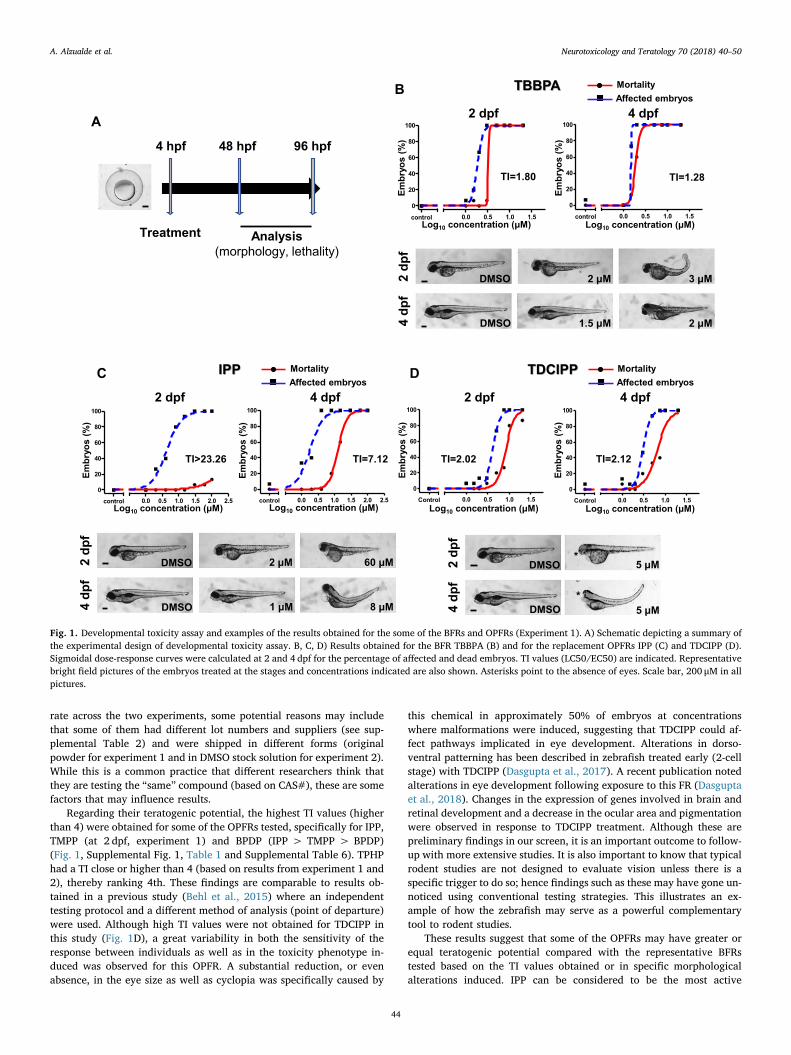
To evaluate developmental toxicity, embryo exposure started at 4hpf (Fig. 1A). Specific concentrations tested per chemical (Supple-mental Table 3) were selected based on previous range finding study(data not shown). Based on the analysis performed at 2 and 4 dpf,percentages for mortality and evidence of morphological alterationswere determined for each tested concentration. To determine the ter-atogenic potential of each compound a Teratogenic Index (TI) wascalculated as the ratio between LC50 (as a measure of general embry-otoxicity) and EC50 (reflecting teratogenic effects). A TI numericalthreshold to classify teratogenicity has been typically defined by atraining set of known in vivo positive and negative compounds (Brannenet al., 2010; Selderslaghs et al., 2012). While compounds with a TI (alsocalculated as LC50/EC50) higher than 2 can be consider teratogenic(Selderslaghs et al., 2012), based on our internal validation we decidedto increase our cutoff to 4 to ensure an accurate capture of ter-atogenicity.
In general, similar results were obtained in the two experimentsperformed (Table 1 and Supplemental Table 6). A LC50 was not ob-tained for BDE-47 and IDDP since these chemicals precipitated prior toinducing mortality (for BDE-47 at ≥25 μM in the first and ≥30 μM inthe second experiments, and for IDDP at ≥150 μM in the first and≥50 μM in the second experiments). Therefore, a TI value could not beestimated in these two cases. For the other tested compounds, TI couldbe accurately calculated at least at 4 dpf. No precipitation was noted forTMPP in the first experiment up to 100 μM (maximum concentrationtested) while it precipitated at concentrations ≥30 μM in the secondone. As a result, we were able to calculate a TI at 2 dpf only in the firstbut not in the second experiment. The nominal LEL at which toxicitywas induced was comparable between both experiments as well asbetween several of the OPFRs and the two BFRs (Table 1 and Supple-mental Table 6) even though precipitation was detected at differenttreatment concentrations in certain cases. Although we are not sureabout the exact reason for the differences in compound precipitation
A. Alzualde et al. Neurotoxicology and Teratology 70 (2018) 40–50
43
rate across the two experiments, some potential reasons may includethat some of them had different lot numbers and suppliers (see sup-plemental Table 2) and were shipped in different forms (originalpowder for experiment 1 and in DMSO stock solution for experiment 2).While this is a common practice that different researchers think thatthey are testing the “same” compound (based on CAS#), these are somefactors that may influence results.
Regarding their teratogenic potential, the highest TI values (higherthan 4) were obtained for some of the OPFRs tested, specifically for IPP,TMPP (at 2 dpf, experiment 1) and BPDP (IPP > TMPP > BPDP)(Fig. 1, Supplemental Fig. 1, Table 1 and Supplemental Table 6). TPHPhad a TI close or higher than 4 (based on results from experiment 1 and2), thereby ranking 4th. These findings are comparable to results ob-tained in a previous study (Behl et al., 2015) where an independenttesting protocol and a different method of analysis (point of departure)were used. Although high TI values were not obtained for TDCIPP inthis study (Fig. 1D), a great variability in both the sensitivity of theresponse between individuals as well as in the toxicity phenotype in-duced was observed for this OPFR. A substantial reduction, or evenabsence, in the eye size as well as cyclopia was specifically caused by
this chemical in approximately 50% of embryos at concentrationswhere malformations were induced, suggesting that TDCIPP could af-fect pathways implicated in eye development. Alterations in dorso-ventral patterning has been described in zebrafish treated early (2-cellstage) with TDCIPP (Dasgupta et al., 2017). A recent publication notedalterations in eye development following exposure to this FR (Dasguptaet al., 2018). Changes in the expression of genes involved in brain andretinal development and a decrease in the ocular area and pigmentationwere observed in response to TDCIPP treatment. Although these arepreliminary findings in our screen, it is an important outcome to follow-up with more extensive studies. It is also important to know that typicalrodent studies are not designed to evaluate vision unless there is aspecific trigger to do so; hence findings such as these may have gone un-noticed using conventional testing strategies. This illustrates an ex-ample of how the zebrafish may serve as a powerful complementarytool to rodent studies.
These results suggest that some of the OPFRs may have greater orequal teratogenic potential compared with the representative BFRstested based on the TI values obtained or in specific morphologicalalterations induced. IPP can be considered to be the most active
0
20
40
60
80
100
0.0 0.5 1.0 1.5 2.0 2.5control�
)%(
soyrbmE
Log10 concentration (μM)
A
Treatment
4 hpf 48 hpf 96 hpf
Analysis(morphology, lethality)
2 dpf
TI>23.26
0
20
40
60
80
100
0.0 0.5 1.0 1.5 2.0 2.5control�
Embr
yos
(%)
Log10 concentration (μM)
4 dpf
TI=7.12
MortalityAffected embryos
2 dp
f
DMSO 2 μM 60 μM
DMSO 1 μM 8 μM4 dp
f
0
20
40
60
80
100
0.0 0.5 1.0 1.5control�
Embr
yos
(%)
Log10 concentration (μM)
2 dpf
TI=1.80
0
20
40
60
80
100
0.0 0.5 1.0 1.5control�
4 dpf
TI=1.28
Embr
yos
(%)
Log10 concentration (μM)
2 dp
f
DMSO
DMSO4 dp
f
2 μM 3 μM
1.5 μM 2 μM
IPP
TBBPA MortalityAffected embryos
C
B
Embr
yos
(%)
2 dpf
TI=2.02
Embr
yos
(%)
4 dpf
TI=2.12
Log10 concentration (μM) Log10 concentration (μM)
D TDCIPP MortalityAffected embryos
2 dp
f
DMSO
DMSO4 dp
f
0
20
40
60
80
100
0.0 0.5 1.0 1.5Control�
0
20
40
60
80
100
0.0 0.5 1.0 1.5Control�
5 μM
5 μM*
*
Fig. 1. Developmental toxicity assay and examples of the results obtained for the some of the BFRs and OPFRs (Experiment 1). A) Schematic depicting a summary ofthe experimental design of developmental toxicity assay. B, C, D) Results obtained for the BFR TBBPA (B) and for the replacement OPFRs IPP (C) and TDCIPP (D).Sigmoidal dose-response curves were calculated at 2 and 4 dpf for the percentage of affected and dead embryos. TI values (LC50/EC50) are indicated. Representativebright field pictures of the embryos treated at the stages and concentrations indicated are also shown. Asterisks point to the absence of eyes. Scale bar, 200 μM in allpictures.
A. Alzualde et al. Neurotoxicology and Teratology 70 (2018) 40–50
44

compound for zebrafish in developmental toxicity assay since it showedthe highest TI (7.12 and 12.78 in the first and second experiment re-spectively at 4 dpf) and the lowest effective concentration (1 or 0.5 μMin the first and second experiment respectively at 4 dpf). Further studiesto evaluate the developmental toxicity effects of this FR, includingdevelopmental neurotoxicity in rodents, are underway at the NationalToxicology Program (Behl et al., 2015).
3.2. Internal concentration analysis
Although in most studies in the literature nominal concentrations(concentrations in the well at the beginning of exposure) are reported,they do not always reflect the internal exposure due to differences inchemical uptake based on physicochemical properties and kinetics(Berghmans et al., 2008; de Koning et al., 2015). Hence, we evaluatedinternal concentrations in 4 dpf larvae exposed to LEL following as-sessment of developmental toxicity. Except TCEP that showed a larvaeinternal concentration below the nominal concentration (around57%–87%), all other tested chemicals showed an accumulation/selec-tive absorption of various orders of magnitude (Table 1 and Supple-mental Table 6). Accumulation of highly lipophilic compounds (withhigh logP) in zebrafish embryos, including some OPFRs, have beenpreviously described (Dishaw et al., 2014; Wang et al., 2015). There-fore, while toxicity was noted at low nominal concentrations for most ofthe FRs tested, the equivalent internal exposures were generally up to200-fold higher compared to nominal concentrations (Table 1, Fig. 4).However, one of the OPFRs tested, IPP, was toxic at lower concentra-tions (4.21 μM in experiment 1). Although internal concentrations arean approximation due to the unknown proportion of each of derivativesthat conforms this mixture (see supplemental methods), these resultsconfirm that IPP is probably the most active compound for zebrafish indevelopmental toxicity assay. It is important to note that since this wasa screening study with a goal to prioritize compounds for furthertesting, in most cases we only measured levels of the major parentcompound inside the larvae; the presence of other isomers that could bepresent in the mixtures were not evaluated. Since compounds havedifferent physicochemical properties, they are expected to have dif-ferent rates of uptake. This has important implications for the
accumulation of these compounds for both, human exposure and eco-toxicity. Further studies are warranted to define the toxicokinetic pro-files of these compounds to better characterize hazard associated withexposure.
3.3. Effects of flame retardants in behavior alteration
Because of their structural similarities to organophosphate pesti-cides, which have been shown to be neurodevelopmentally toxic(Slotkin et al., 2006), neurotoxicity has been the main concern of al-ternative OPFRs. For the detection of neurotoxicity, potential altera-tions in locomotor activity caused by FRs were evaluated in 5 dpflarvae. Analysis was performed after 48 h of incubation with chemicalsonly in hatched and not malformed larvae. Although more specificneurotoxic effects are expected to be detected after shorter periods ofincubation, larvae used in this assay were also analyzed for the detec-tion of hepatotoxicity, for which longer exposures are necessary (Heet al., 2013). Therefore, acute neurotoxicity as well as effects morerelated with the inhibition of processes that occur late in nervoussystem development and maturation are expected to be detected withthis assay. Combining results of experiments 1 and 2, alterations inlocomotor activity were produced by most of the flame retardantsevaluated (Table 2, Supplemental Table 7, Fig. 2, Supplemental Fig. 2).Since the highest concentration evaluated in the behavioral assay wasthe LEL obtained in developmental toxicity, it was difficult to dis-criminate between general systemic toxicity versus specific neurotoxi-city when hypoactivity was the alteration caused only at the highestconcentration tested. This occurred for BPDP, IDDP, TMPP, TPHP,TCEP and TDCIPP and then, their effects were not considered specifi-cally neurotoxic but a consequence of general toxicity. However, forBDE-47, there was a clear concentration-dependent decrease in activityonly in the dark phase with no differences detected in the light phase,suggesting that this compound altered neurobehavior (Fig. 2B). ForEHDP, we noted a concentration-dependent hyperactivity under lightcondition and absence of responsiveness to light changes in both ourexperiments (Fig. 2C). Moreover, larvae treated with this chemical alsomanifested corkscrew movements and loss of equilibrium. Therefore,BDE-47 and EHDP were neuroactive/neurotoxic for zebrafish larvae.
Table 1Summary of the results obtained in the first experiment after the evaluation of developmental toxicity in zebrafish embryos. A comparison between nominalconcentration and estimated internal concentrations for LEL at 4 dpf is also shown. The numbers in the brackets indicate the 95% confidence intervals of the EC50and LC50 values given or if these intervals were very wide or interrupted. *Point out compounds that precipitated and concentrations at which precipitationoccurred. In light yellow values probably overestimated due to compound precipitation. High TI values, indicative of high teratogenic potential, are shown in red.
Test
item
NOAEL
(µM) EC50 (µM) LC50 (µM) TI
In
te
rn
al c
on
ce
ntra
tio
n
LEL (µM)
2 dpf 4 dpf 2 dpf 4 dpf 2 dpf 4 dpf 2 dpf 4 dpf Nominal Internal
BDE47* >25 2 – 12.01
(8.44 to 17.11)– >25 – >2.08 4 1040
BPDP 11.45
(10.56 to 12.42)
4.75
(0.086 to 263.1)
84.15
(80.72 to 87.72)
15.24
(12.33 to 18.84)7.35 3.21 8 1222
EHDP
8 4
>20 3 – 5.06
(4.89 to 5.24)–
9.78
(Very wide)– 1.93 5 2880
IDDP* – 77.23
(57.77 to 103.2)– >150 – >1.94 40 665.1
IPP 4.30
(3.66 to 5.05)
1.80
(1.31 to 2.47)>100
12.82
(11.97 to 13.73)>23.26 7.12 1 4.21
TMPP
>150 20
1 <1
8 2 11.48
(11.40 to 11.56)
3.00
(2.78 to 3.24)
143.8
(107.2 to 192.9)
9.52
(9.46 to 9.57)12.53 3.17 4 1078
TPHP 3.84
(3.41 to 4.33)
1.72
(1.61 to 1.84)
15.11
(very wide)
5.15
(Interrupted)3.93 2.99 1.5 335.2
TBBPA 1.81
(1.76 to 1.86)
1.48
(Very wide)
3.26
(Very wide)
1.90
(1.88 to 1.92)1.5 20.68
TCEP 521.2
(462.8 to 587.0)
415.2
(Very wide)>1000
977.6
(Very wide)600 342.7
TDCIPP
2 1
1.5 1
400 400
3 2
4.11
(3.68 to 4.58)
3.08
(2.79 to 3.40)
8.29
(7.15 to 9.61)
6.53
(5.07 to 8.40)
1.80 1.28
>1.92 2.35
2.02 2.12 3 76.68
A. Alzualde et al. Neurotoxicology and Teratology 70 (2018) 40–50
45
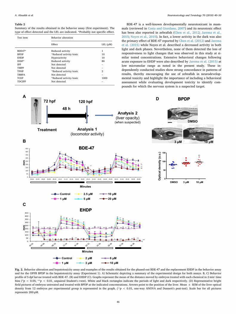
BDE-47 is a well-known developmentally neurotoxicant in mam-mals (reviewed in Costa and Giordano, 2007) and its neurotoxic effecthas been also reported in zebrafish (Chen et al., 2012; Jarema et al.,2015; Noyes et al., 2015). In fact, a lower activity in the dark was alsothe primary effect of BDE-47 reported by Chen et al. (2012) and Jaremaet al. (2015) while Noyes et al. described a decreased activity in bothlight and dark phases. Nevertheless, none of them detected the loss ofresponsiveness to light changes that was observed in this study at si-milar tested concentrations. Extensive behavioral changes followingacute exposure to EHDP were also described by Jarema et al. (2015) atlow micromolar range as noted in the present study. These in-dependently conducted studies show strong concordance in patterns ofresults, thereby encouraging the use of zebrafish in neurodevelop-mental toxicity and highlight the importance of including a behavioralassessment while evaluating development toxicity to identify com-pounds for which the nervous system is a suspected target.
Table 2Summary of the results obtained in the behavior assay (first experiment). Thetype of effect detected and the LEL are indicated. *Probably not specific effect.
Test item Behavior alteration
Effect LEL (μM)
BDE47* Reduced activity 1BPDP *Reduced activity/toxic 10EHDP Hyperactivity 10IDDP* Reduced activity 80IPP Not detected –TMPP Not detected –TPHP *Reduced activity/toxic 2TBBPA Not detected –TCEP *Reduced activity/toxic 1000TDCIPP Not detected –
A BPDP
0
50
100
150**
Opt
ical
den
sity
(a.u
.)
DMSO
3 μM 10 μM
*
**
**
*
*
*
**
** **
* *
** **
*
** **
****
**
* *
**
**
****
****
***
****
** ** **
****
** ** **** ** ** ** ** **
0
50
100
150
200
250
300
350
400
450
00-02 02-04 04-06 06-08 08-10 10-12 12-14 14-16 16-18 18-20 20-22 22-24 24-26 26-28 28-30 30-32 32-34 34-36 36-38 38-40
mm
Minutes
20 μMControl 10 μM
5 μM2.5 μM
1 μM
BDE-47
DMSO 3 μM 10 μM
72 hpf
48 h
Analysis 1(locomotor activity)
120 hpf
Treatment
Analysis 2(liver opacity)
(when suspected)
D
B
**
*
*
** *
*
** ** ** ** * * ** ** ** ** ** ** * *
* * ** ** ** ** ** * ** ** ** ****
0
50
100
150
200
250
300
350
400
450
Minutes
mm
EHDP
10 μMControl 6 μM
3 μM2 μM
1 μM
C
Fig. 2. Behavior alteration and hepatotoxicity assay and examples of the results obtained for the phased-out BDE-47 and the replacement EHDP in the behavior assayand for the OPFR BPDP in the hepatotoxicity assay (Experiment 1). A) Schematic depicting a summary of the experimental design for both assays. B, C) Behaviorprofile of 5 dpf larvae treated with BDE-47. (B) and EHDP (C). Graphs represent the mean of the distance moved by embryos treated with each chemical in 2min' timebins (*p < 0.05; **p < 0.01, unpaired Student's t-test). White and black rectangles indicate the periods of light and dark respectively. (D) Representative brightfield pictures of embryos untreated and treated with BPDP at the indicated concentrations. Arrows point to the position of the liver. Mean ± SEM of the liver opticaldensity from 12 embryos per experimental group is represented in the graph. (⁎⁎p < 0.01, one-way ANOVA and Dunnett's post-test). Scale bar for all picturesrepresents 200 μM.
A. Alzualde et al. Neurotoxicology and Teratology 70 (2018) 40–50
46
3.4. Effects of flame retardants on hepatotoxicity
After behavior analysis, embryos were visualized for the detectionof liver opacity that was suspected in larvae treated with BPDP andEHDP. This effect was confirmed after optical density quantification(Fig. 2D, Table 3, Supplemental Table 8) in both experiments
conducted. Other toxicity manifestations (mainly edemas) at con-centrations at which hepatotoxicity was detected were also present atthe time of analysis.
The potential adverse effects of OPFRs in liver have not been ade-quately tested using in vivo models. Although this is the first time thathepatotoxicity is being reported for BPDP and EHDP in zebrafish,changes in metabolism and liver gene expression for other OPFRs suchas TDCIPP and TPHP, have been previously described (Du et al., 2016;Liu et al., 2016). This suggests the need for more in-depth evaluation ofthe liver as a potential target of toxicity for this class of compounds.
3.5. Cardiotoxicity of tested flame retardants
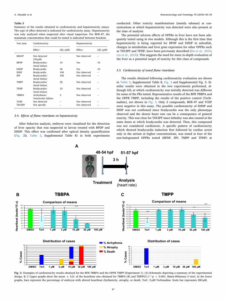
The results obtained following cardiotoxicity evaluation are shownin Table 3, Supplemental Table 8, Fig. 3 and Supplemental Fig. 3. Si-milar results were obtained in the two experiments performed, al-though LEL at which cardiotoxicity was initially detected was differentfor some of the FRs tested. Representative results of the BFR TBBPA andthe OPFR TMPP, including the results of the positive control (Terfe-nadine), are shown in Fig. 3. Only 2 compounds, BDE-47 and TCEPwere negative in this assay. The possible cardiotoxicity of EHDP andIDDP was not confirmed since bradycardia was the only phenotypeobserved and the slower heart rate can be a consequence of generaltoxicity. This was clear for TDCIPP since lethality was also caused at thesame doses at which bradycardia was detected. Then, this compoundwas not considered cardiotoxic. A specific pattern of cardiotoxicitywhich showed bradycardia induction first followed by cardiac arrestonly in the atrium at higher concentrations, was noted in four of thenon-halogenated OPFRs tested (BPDP, IPP, TMPP and TPHP) at
Table 3Summary of the results obtained in cardiotoxicity and hepatotoxicity assays.The type of effect detected is indicated for cardiotoxicity assay. Hepatotoxicitywas only analyzed when suspected after visual inspection. For BDE-47, themaximum concentration that could be tested is indicated between brackets.
Test item Cardiotoxicity Hepatotoxicity
Effect LEL (μM) Effect LEL (μM)
BDE47 Not detected(30 μM)
– Not observed –
BPDP Bradycardia/Atrial failure
10 Yes 10
EHDP Bradycardia 30 Yes 10IDDP Bradycardia 100 Not observed –IPP Bradycardia/
Atrial failure100 Not observed –
TMPP Bradycardia/Atrial failure
30 Not observed –
TPHP Bradycardia/Atrial failure
10 Not observed –
TBBPA Arrhythmia/Ventricular failure
3 Not observed –
TCEP Not detected – Not observed –TDCIPP Not specific – Not observed –
48-54 hpf
3 h
Analysis(heart rate)
51-57 hpf
Treatment
A
***
******
0,05,010,015,020,025,030,035,040,045,050,0
Hea
rtbe
at (b
eats
/15
s)
DMSO Terf 1 μM 3 μM 10 μM 30 μM 100 μM
Comparison of means
Distribution of cases
% C
ases
DMSO Terf. 1 μM 3 μM 10 μM 30 μM 100 μM
TMPPB
% Arrhythmia% Atrophy% Death
CTBBPA
* ***
0.05.0
10.015.020.0
25.030.035.0
40.045.0
DMSO 1 μM 3 μM 10 μM 30 μM 100 μM
)s51/staeb(taebtrae
H
Comparison of means
Terf.
0102030405060708090100
Distribution of cases
% C
ases
DMSO Terf. 1 μM 3 μM 10 μM 30 μM 100 μM
Fig. 3. Examples of cardiotoxicity results obtained for the BFR TBBPA and the OPFR TMPP (Experiment 1). (A) Schematic depicting a summary of the experimentaldesign. B, C Upper graphs show the mean ± S.D. of the heartbeat rate obtained for TBBPA (B) and TMPP(C) (⁎⁎⁎p < 0.001, Mann-Whiteney U test). In the lowergraphs, bars represent the percentage of embryos with altered heartbeat rhythmicity, atrophy, or death. Terf.: 5 μM Terfenadine. Scale bar represents 200 μM.
A. Alzualde et al. Neurotoxicology and Teratology 70 (2018) 40–50
47
concentrations between 10 and 100 μM (Supplemental Movies 1 and 2).Finally, TBBPA was the most active compound in this assay since car-diotoxicity was observed as low as 3 μM. Arrhythmia 2:1, a phenotypeindicative of ERG (ether-a-go-go-related gene) inhibition (Langheinrichet al., 2003) and related to long QT syndrome in humans, was caused bythe treatment with this FR (Supplemental Movie 3).
Alterations in heart development as well as defects in heartbeathave been previously described in zebrafish caused by some non-ha-logenated OPFRs as TPHP, isopropylated triaryl phosphate (ITP) andcresyl diphenyl phosphate (CDP) (McGee et al., 2013; Du et al., 2015).These findings are in agreement with previous published studies thathave shown the OPFRs to be bioactive in human stem cell derivedcardiomyocytes (Sirenko et al., 2017). These results indicate that car-diotoxicity may be a relevant target for the non-halogenated OPFRs.Further characterization of possible cardiotoxic effects of OPFRs inmammals is warranted.
3.6. Relevance of findings in zebrafish to biomonitoring data in humans
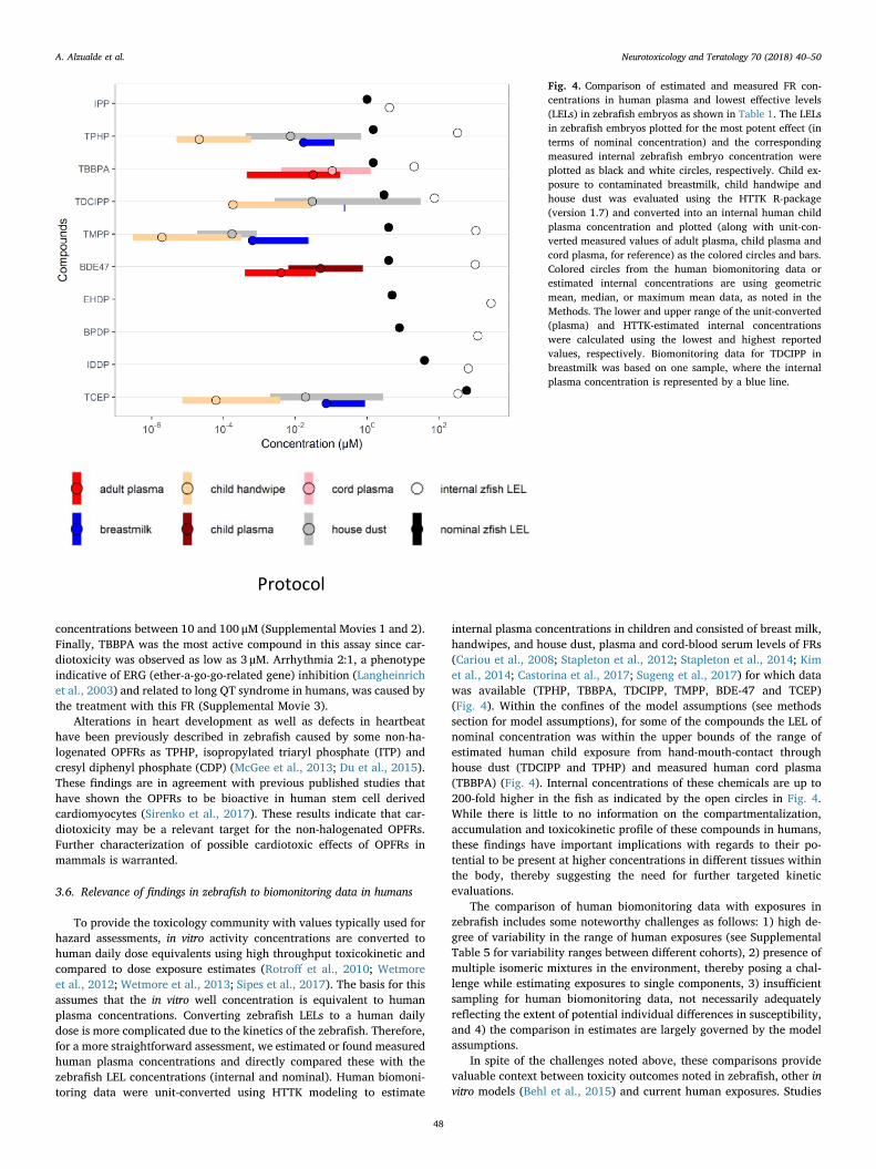
To provide the toxicology community with values typically used forhazard assessments, in vitro activity concentrations are converted tohuman daily dose equivalents using high throughput toxicokinetic andcompared to dose exposure estimates (Rotroff et al., 2010; Wetmoreet al., 2012; Wetmore et al., 2013; Sipes et al., 2017). The basis for thisassumes that the in vitro well concentration is equivalent to humanplasma concentrations. Converting zebrafish LELs to a human dailydose is more complicated due to the kinetics of the zebrafish. Therefore,for a more straightforward assessment, we estimated or found measuredhuman plasma concentrations and directly compared these with thezebrafish LEL concentrations (internal and nominal). Human biomoni-toring data were unit-converted using HTTK modeling to estimate
internal plasma concentrations in children and consisted of breast milk,handwipes, and house dust, plasma and cord-blood serum levels of FRs(Cariou et al., 2008; Stapleton et al., 2012; Stapleton et al., 2014; Kimet al., 2014; Castorina et al., 2017; Sugeng et al., 2017) for which datawas available (TPHP, TBBPA, TDCIPP, TMPP, BDE-47 and TCEP)(Fig. 4). Within the confines of the model assumptions (see methodssection for model assumptions), for some of the compounds the LEL ofnominal concentration was within the upper bounds of the range ofestimated human child exposure from hand-mouth-contact throughhouse dust (TDCIPP and TPHP) and measured human cord plasma(TBBPA) (Fig. 4). Internal concentrations of these chemicals are up to200-fold higher in the fish as indicated by the open circles in Fig. 4.While there is little to no information on the compartmentalization,accumulation and toxicokinetic profile of these compounds in humans,these findings have important implications with regards to their po-tential to be present at higher concentrations in different tissues withinthe body, thereby suggesting the need for further targeted kineticevaluations.
The comparison of human biomonitoring data with exposures inzebrafish includes some noteworthy challenges as follows: 1) high de-gree of variability in the range of human exposures (see SupplementalTable 5 for variability ranges between different cohorts), 2) presence ofmultiple isomeric mixtures in the environment, thereby posing a chal-lenge while estimating exposures to single components, 3) insufficientsampling for human biomonitoring data, not necessarily adequatelyreflecting the extent of potential individual differences in susceptibility,and 4) the comparison in estimates are largely governed by the modelassumptions.
In spite of the challenges noted above, these comparisons providevaluable context between toxicity outcomes noted in zebrafish, other invitro models (Behl et al., 2015) and current human exposures. Studies
Fig. 4. Comparison of estimated and measured FR con-centrations in human plasma and lowest effective levels(LELs) in zebrafish embryos as shown in Table 1. The LELsin zebrafish embryos plotted for the most potent effect (interms of nominal concentration) and the correspondingmeasured internal zebrafish embryo concentration wereplotted as black and white circles, respectively. Child ex-posure to contaminated breastmilk, child handwipe andhouse dust was evaluated using the HTTK R-package(version 1.7) and converted into an internal human childplasma concentration and plotted (along with unit-con-verted measured values of adult plasma, child plasma andcord plasma, for reference) as the colored circles and bars.Colored circles from the human biomonitoring data orestimated internal concentrations are using geometricmean, median, or maximum mean data, as noted in theMethods. The lower and upper range of the unit-converted(plasma) and HTTK-estimated internal concentrationswere calculated using the lowest and highest reportedvalues, respectively. Biomonitoring data for TDCIPP inbreastmilk was based on one sample, where the internalplasma concentration is represented by a blue line.
A. Alzualde et al. Neurotoxicology and Teratology 70 (2018) 40–50
48
are currently underway at the NTP to evaluate the effects of some ofthese compounds in rodent studies. Further toxicokinetic studies indeveloping and adult zebrafish are warranted to understand the dy-namics of these chemicals in relation to human exposure as well as toother aquatic organisms to determine the potential human and ecolo-gical hazards that these chemicals might pose.
4. Conclusion
In this study we demonstrated the utility of the zebrafish as an in-tegrative tool to screen for a class of compounds and showed how thesefindings may be related to human exposure. This is the first time that asystems toxicity approach has been used to compare OPFRs with re-presentative BFRs in zebrafish, and findings contextualized with humanbiomonitoring data. In general, some of the non-halogenated aromaticOPFRs (BPDP, IPP, TPP, EHDP, TMPP) showed comparable toxicity tothe phased-out BDE-47 and to TBBPA. A unique cyclopia was notedfollowing exposure to TDCIPP, distinct from all other FRs tested in thestudy. IPP manifested a high teratogenic potential; neurotoxicity wasclearly seen following exposure to BDE-47 and EHDP. Additionally,many of the non-halogenated OPFRs induced a characteristic cardio-toxic effect. Overall, it appears that the FRs affected multiple pathwaysduring zebrafish development thereby resulting in multiple target or-gans of toxicity. Generally, the results from both of our independentlyconducted blinded experiments largely corroborated with each other.Interestingly, these findings are also consistent with previous develop-mental toxicity data in the literature (Behl et al., 2015; Jarema et al.,2015) that used different protocols and zebrafish strains, thereby em-phasizing the robustness of this model as a reliable screening tool forpotential toxicants.
Finally, in comparing toxicity outcomes in the zebrafish with humanbiomonitoring data, our results suggest that in some cases, nominalconcentrations at which we noted toxicity in zebrafish were within theupper range of potential human exposure. Some of these compoundsaccumulate in the zebrafish up to ~200 fold higher, which has im-portant implications for other aquatic organisms and ecotoxicity.Further in-depth studies are warranted to better understand their tox-icokinetic profiles to characterize hazard in humans and wildlife.
Supplementary data to this article can be found online at https://doi.org/10.1016/j.ntt.2018.10.002.
Acknowledgements
The authors thank Drs. Troy Hubbard (NTP, NIEHS) and WindyBoyd (NTP, NIEHS) for their review of this manuscript. We also thankItziar Irijalba, Izaskun González and Olaia Holgado for their technicalsupport, and Jorge Hurtado (GRILab S.L.) for performing larvae bioa-nalysis and result interpretation. Finally, we thank Dr. JohnWambaugh, USEPA for his help with the dose conversions with theHTTK package.
Funding Information
This project was sponsored in part by the National ToxicologyProgram (NTP) under contract #s HHSN273201400015C andHHSN273201700005C.
Conflict of interest
Ainhoa Alzualde, Aintzane Alday, Arantza Muriana and CeliaQuevedo work for Biobide, a company that provide services usingzebrafish as experimental model.
References
Bailey, J.M., Levin, E.D., 2015. Neurotoxicity of FireMaster 550® in zebrafish (Danio
rerio): chronic developmental and acute adolescent exposures. Neurotoxicol. Teratol.52 (PtB), 210–219.
Bambino, K., Chu, J., 2017. Zebrafish in toxicology and environmental health. Curr. Top.Dev. Biol. 124, 331–367.
Behl, M., Hsieh, J.H., Shafer, T.J., Mundy, W.R., Rice, J.R., Boyd, W.A., et al., 2015. Useof alternative assays to identify and prioritize organophosphorus flame retardants forpotential developmental and neurotoxicity. Neurotoxicol. Teratol. 52, 181–193.
Berghmans, S., Butler, P., Goldsmith, P., Waldron, G., Gardner, I., Golder, Z., et al., 2008.Zebrafish based assays for the assessment of cardiac, visual and gut function—po-tential safety screens for early drug discovery. Toxicology Methods 58, 59–68.
Brannen, K.C., Panzica-Kelly, J.M., Danberry, T.L., Augustine-Rauch, K.A., 2010.Development of a zebrafish embryo teratogenicity assay and quantitative predictionmodel. Birth Defects Research. Part B, Developmental and Reproductive Toxicology89, 66–77.
Cano-Sancho, G., Smith, A., La Merrill, M.A., 2017. Triphenyl phosphate enhances adi-pogenic differentiation, glucose uptake and lipolysis via endocrine and noradrenergicmechanisms. Toxicol. in Vitro 40, 280–288.
Cariou, R., Antignac, J.P., Zalko, D., Berrebi, A., Cravedi, J.P., Maume, D., et al., 2008.Exposure assessment of French women and their newborns to tetrabromobisphenol-a:occurrence measurements in maternal adipose tissue, serum, breast milk and cordserum. Chemosphere 73, 1036–1041.
Castorina, R., Bradman, A., Stapleton, H.M., Butt, C., Avery, D., Harley, K.G., et al., 2017.Current-use flame retardants: maternal exposure and neurodevelopment in childrenof the CHAMACOS cohort. Chemosphere 189, 574–580.
Chapman, D.E., Michener, S.R., Powis, G., 1991. Metabolism of the flame retardantplasticizer tris(2-chloroethyl)phosphate by human and rat liver preparations.Fundam. Appl. Toxicol. 17, 215–224.
Chen, X., Huang, C., Wang, X., Chen, J., Bai, C., Chen, Y., et al., 2012. BDE-47 disruptsaxonal growth and motor behavior in developing zebrafish. Aquat. Toxicol. 120–121,35–44.
Costa, L.G., Giordano, G., 2007. Developmental neurotoxicity of polybrominated di-phenyl ether (PBDE) flame retardants. Neurotoxicology 28, 1047–1067.
Cowell, W.J., Stapleton, H.M., Holmes, D., Calero, L., Tobon, C., Perzanowski, M.,Herbstman, J.B., 2017. Prevalence of historical and replacement brominated flameretardant chemicals in New York City homes. Emerging Contaminants 3 (1), 32–39.
Dasgupta, S., Vliet, S.M., Kupsco, A., Leet, J.K., Altomare, D., Volz, D.C., 2017. Tris (1,3-dichloro-2-propyl) phosphate dorsoventral patterning in zebrafish embryos. Peer J 5,e4156. https://doi.org/10.7717/peerj.4156.
Dasgupta, S., Cheng, V., Vliet, S.M., Mitchell, C.A., Volz, D.C., 2018. Tris (1,3-dichloro-2-propyl) phosphate exposure during early-blastula alters the normal trajectory ofzebrafish embryogenesis. Environ. Sci. Technol. https://doi.org/10.1021/acs.est.8b03730.
Dishaw, L.V., Macaulay, L.J., Roberts, S.C., Stapleton, H.M., 2014. Exposures, mechan-isms, and impacts of endocrine-active flame retardants. Curr. Opin. Pharmacol. 19,125–133.
Du, Z., Wang, G., Gao, S., Wang, Z., 2015. Aryl organophosphate flame retardants inducedcardiotoxicity during zebrafish embryogenesis: by disturbing expression of thetranscriptional regulators. Aquat. Toxicol. 161, 25–32.
Du, Z., Zhang, Y., Wang, G., Peng, J., Wang, Z., Gao, S., 2016. TphP disturbs carbohydratemetabolism, and the DNA damage repair system in zebrafish liver. Sci. Rep. 6, 21827.https://doi.org/10.1038/srep21827.
Frederiksen, M., Thomsen, M., Vorkamp, K., Knudsen, L.E., 2009. Patterns and con-centration levels of polybrominated diphenyl ethers (PBDEs) in placental tissue ofwomen in Denmark. Chemosphere 76, 1464–1469.
Hallare, A., Nagel, K., Köhler, H.R., Triebskorn, R., 2006. Comparative embryotoxicityand proteotoxicity of three carrier solvents to zebrafish (Danio rerio) embryos.Ecotoxicol. Environ. Saf. 63 (3), 378–388.
Harley, K.G., Marks, A.R., Chevrier, J., Bradman, A., Sjodin, A., Eskenazi, B., 2010. PBDEconcentrations in women's serum and fecundability. Environ. Health Perspect. 118,699–704.
He, J.H., Guo, S.Y., Zhu, F., Zhu, J.J., Chen, Y.X., Huang, C.J., 2013. A zebrafish phe-notypic assay for assessing drug-induced hepatotoxicity. J. Pharmacol. Toxicol.Methods 67, 25–32.
Herbstman, J.B., Sjodi, A., Kurzo, M., Lederma, S.A., Jones, R.S., Rauh, V., et al., 2010.Prenatal exposure to PBDEs and neurodevelopment. Environ. Health Perspect. 118,712–719.
Hoffman, K., Garantziotis, S., Birnbaum, L.S., Stapleton, H.M., 2015. Monitoring indoorexposure to organophosphate flame retardants: hand wipes and house dust. Environ.Health Perspect. 123 (2), 160–165.
Institute of Medicine (US) Committee on Nutritional Status During Pregnancy andLactation, 1991. Nutrition during Lactation. National Academies Press (US),Washington (DC).
Jarema, K.A., Hunter, D.L., Shaffer, R.M., Behl, M., Padilla, S., 2015. Acute and devel-opmental behavioral effects of flame retardants and related chemicals in zebrafish.Neurotoxicol. Teratol. 52, 194–209.
Kaufman, C.K., White, R.M., Zon, L., 2009. Chemical genetic screening in the zebrafishembryo. Nat. Protoc. 4, 1422–1432.
Kim, J.W., Isobe, T., Muto, M., Tue, N.M., Katsura, K., Malarvannan, G., et al., 2014.Organophosphorus flame retardants (pfrs) in human breast milk from several Asiancountries. Chemosphere 116, 91–97.
de Koning, C., Beekhuijzen, M., Tobor-Kaplon, M., de Vries-Buitenweg, S., Schoutsen, D.,Leeijen, N., et al., 2015. Visualizing compound distribution during zebrafish embryosdevelopment: the effects of lipophilicity and DMSO. Birth Defects Research (Part B)104, 253–272.
Langheinrich, U., Vacun, G., Wagner, T., 2003. Zebrafish embryos express an orthologueof HERG and are sensitive toward a range of QT-prolonging drugs inducing severe
A. Alzualde et al. Neurotoxicology and Teratology 70 (2018) 40–50
49

arrhythmia. Toxicol. Appl. Pharmacol. 193, 370–382.Letamendia, A., Quevedo, C., Ibarbia, I., Virto, J.M., Holgado, O., Diez, M., et al., 2012.
Development and validation of an automated high-throughput system for zebrafish invivo screenings. PLoS One 5, e36690 (doi: 10.1371).
Liu, C., Su, G., Giesy, J.P., Letcher, R.J., Li, G., Agrawal, I., et al., 2016. Acute exposure toTris(1,3-dichloro-2-propyl) phosphate (TDCIPP) causes hepatic inflammation andleads to hepatotoxicity in zebrafish. Sci. Rep. 6, 19045. https://doi.org/10.1038/srep19045.
Luscombe, M.D., Owens, B.D., Burke, D., 2011. Weight estimation in paediatrics: acomparison of the APLS formula and the formula ‘weight= 3(age)+7’. Emerg. Med.J. 28, 590–593.
Malvasi, A., Tinelli, A., Buia, A., DeLuca, D.F., 2009. Possible long-term teratogenic effectof isotretinoin in pregnancy. European Review of Medical and PharmacologicalSciences 13 (5), 393–396.
McGee, S.P., Konstantinov, A., Stapleton, H.M., Volz, D.C., 2013. Aryl phosphate esterswithin a major PentaBDE replacement product induce cardiotoxicity in developingzebrafish embryos: potential role of the aryl hydrocarbon receptor. Toxicol. Sci. 133,144–156.
Meeker, J.D., Stapleton, H.M., 2010. House dust concentrations of organophosphateflame retardants in relation to hormone levels and semen quality parameters.Environ. Health Perspect. 118 (3), 318–323.
Mizouchi, S., Ichiba, M., Takigami, H., Kjjwara, M., Tkamuku, T., Miyajima, T., et al.,2015. Exposure assessment of organophosphorus and organobromine flame re-tardants via indoor dust from elementary schools and domestic houses. Chemosphere123, 17–25.
Noyes, P.D., Haggard, D.E., Gonnerman, G.D., Tanguay, R.L., 2015. Advances morpho-logical-behavioral test platform reveals neurodevelopmental defects in embryoniczebrafish exposed to comprehensive suite of halogenated and organophosphate flameretardants. Toxicol. Sci. 145 (1), 177–195.
Noyes, P.D., Garcia, G.R., Tanguay, R.L., 2016. Zebrafish as an in vivo model for sus-tainable chemical design. Green Chem. 18 (24), 6410–6430.
Oliveri, A.N., Bailey, J.M., Levin, E.D., 2015. Developmental exposure to organopho-sphate flame retardants causes behavioral effects in larval and adult zebrafish.Neurotoxicol. Teratol. 52 (PtB), 220–227.
Pearce, R.G., Setzer, R.W., Strope, C.L., Sipes, N.S., Wambaugh, J.F., 2017. Httk: Rpackage for high-throughput toxicokinetics. J. Stat. Softw. 79 (4).
Rotroff, D.M., Wetmore, B.A., Dix, D.J., Ferguson, S.S., Clewell, H.J., Houck, K.A., et al.,2010. Incorporating human dosimetry and exposure into high-throughput in vitrotoxicity screening. Toxicol. Sci. 117 (2), 348–358.
Selderslaghs, I.W., Van Rompay, A.R., De Coen, W., Witters, H.E., 2009. Development of ascreening assay to identity teratogenic and embryotoxic chemicals using the zebrafishembryo. Reprod. Toxicol. 28 (3), 308–320.
Selderslaghs, I.W.T., Blust, R., Witters, H.E., 2012. Feasibility study of the zebrafish assayas an alternative method to screen for developmental toxicity and embryotoxicityusing a training set of 27 compounds. Reprod. Toxicol. 33, 142–154.
Sipes, N.S., Padilla, S., Knudsen, T.B., 2011. Zebrafish: as an integrative model for twenty-first century toxicity testing. Bird Defects Research. Part C, Embryo Today 93 (3),256–267.
Sipes, N.S., Wambaugh, J.F., Pearce, R., Auerbach, S.S., Wetmore, B.A., Hsieh, J.H., et al.,2017. An intuitive approach for predicting potential human health risk with thetox21 10 k library. Environ. Sci. Technol. 51, 10786–10796.
Sirenko, O., Grimm, F.A., Ryan, K.R., Iwata, Y., Chiu, W.A., Parham, F., et al., 2017. Invitro cardiotoxicity assessment of environmental chemicals using an organotypic
human induced pluripotent stem cell-derived model. Toxicol. Appl. Pharmacol. 322,60–74.
Slotkin, T.A., Levin, E.D., Seidler, F.J., 2006. Comparative developmental neurotoxicity oforganophosphate insecticides: effects on brain development are separable from sys-temic toxicity. Environ. Health Perspect. 114, 746–751.
Sorkin, E., Heel, R.C., 1985. Terfenadine. A review of its pharmacodynamics propertiesand its therapeutic efficiency. Drugs 29 (1), 34–56.
Stapleton, H.M., Kelly, S.M., Allen, J.G., McClean, M.D., Webster, T.F., 2008.Measurement of polybrominated diphenyl ethers on hand wipes: estimating exposurefrom hand-to-mouth contact. Environ. Sci. Technol. 42, 3329–3334.
Stapleton, H.M., Klosterhaus, S., Eagle, S., Fuh, J., Meeker, J.D., Blum, A., Webster, T.F.,2009. Detection of organophosphate flame retardants in furniture foam and U.S.house dust. Environ. Sci. Technol. 43 (19), 7490–7495.
Stapleton, H.M., Eagle, S., Anthopolos, R., Wolkin, A., Miranda, M.L., 2011. Associationsbetween polybrominated diphenyl ether (PBDE) flame retardants, phenolic metabo-lites, and thyroid hormones during pregnancy. Environ. Health Perspect. 119 (10),1454–1459.
Stapleton, H.M., Eagle, S., Sjodin, A., Webster, T.F., 2012. Serum pbdes in a NorthCarolina toddler cohort: associations with handwipes, house dust, and socioeconomicvariables. Environ. Health Perspect. 120, 1049–1054.
Stapleton, H.M., Misenheimer, J., Hoffman, K., Webster, T.F., 2014. Flame retardant as-sociations between children's handwipes and house dust. Chemosphere 116, 54–60.
Sugeng, E.J., Leonards, P.E.G., van de Bor, M., 2017. Brominated and organophosphorusflame retardants in body wipes and house dust, and an estimation of house dust hand-loadings in Dutch toddlers. Environ. Res. 158, 789–797.
Tembe, E.A., Honeywell, R., Buss, N.E., Renwick, A.G., 1996. All-trans-retinoic acid inmaternal plasma and teratogenicity in rats and rabbits. Toxicol. Appl. Pharmacol. 141(2), 456–472.
Vuong, A.M., Yolton, K., Dietrich, K.N., Braun, J.M., Lanphear, B.P., Chen, A., 2017.Exposure to polybrominated diphenyl ethers (PBDEs) and child behavior: currentfindings and future directions. Horm. Behav. https://doi.org/10.1016/j.yhbeh.2017.11.008 Review.
Wambaugh, J.F., Wetmore, B.A., Pearce, R., Strope, C., Goldsmith, R., Sluka, J.P., et al.,2015. Toxicokinetic triage for environmental chemicals. Toxicol. Sci. 147 (1), 55–67.
Wang, H.S., Jiang, G.M., Chen, Z.J., Du, J., Man, Y.B., Giesy, J.P., et al., 2013.Concentrations and congener profiles of polybrominated diphenyl ethers (pbdes) inblood plasma from Hong Kong: implications for sources and exposure route. J.Hazard. Mater. 261, 253–259.
Wang, Q.W., Lam, J.C.W., Man, Y.C., Lai, N.L.S., Kwok, K.Y., Guo, Y.Y., et al., 2015.Bioconcentration, metabolism and neurotoxicity of the organophorous flame re-tardant 1,3-dichloro 2-propyl phosphate (TDCPP) to zebrafish. Aquat. Toxicol. 158,108–115.
Wetmore, B.A., Wambaugh, F.J., Ferguson, S.S., Sochaski, M.A., Rotroff, D.M., Freeman,K., et al., 2012. Integration of dosimetry, exposure, and high-throughput screeningdata in chemical toxicity assessment. Toxicol. Sci. 125 (1), 157–174.
Wetmore, B.A., Wambaugh, F.J., Ferguson, S.S., Li, L., Clewell 3rd, H.J., Judson, R.S.,et al., 2013. Relative impact of incorporating pharmacokinetics on predicting in vivohazard and mode of action from high-throughput in vitro toxicity assays. Toxicol. Sci.132 (2), 327–346.
Yan, S., Wu, H., Qin, J., Zha, J., Wang, Z., 2017. Halogen-free organophosphorus flameretardants caused oxidative stress and multixenobiotic resistance in Asian freshwaterclams (Corbicula fluminea). Environ. Pollut. 225, 559–568.
A. Alzualde et al. Neurotoxicology and Teratology 70 (2018) 40–50
50