NEUROSURGERY · Web viewpursuing an academic pathway will be expected to have demonstrated...
79
CURRICULUM / STATUTES & REGULATIONS FOR 5 YEARS DEGREE PROGRAMME IN NEUROSURGERY (MS Neurosurgery)
Transcript of NEUROSURGERY · Web viewpursuing an academic pathway will be expected to have demonstrated...

CURRICULUM / STATUTES & REGULATIONS
FOR
5 YEARS DEGREE PROGRAMMEIN
NEUROSURGERY(MS Neurosurgery)
RAWALPINDI MEDICAL UNIVERSITY

STATUTES
1. Nomenclature Of The Proposed Course
The name of degree program me shall be MS Neurosurgery. This name is well recognized and established for the last many decades worldwide.
2. Course Title:
MS Neurosurgery
3. Training Centres:
Departments of Neurosurgery Rawalpindi Medical University
4. Duration of Course
The duration of MS Neurosurgery course shall be five (5) years First two years in Part I and next three years in Part II with structured training in a recognized department under the guidance of an approved supervisor. The course is structured in three parts:
Part I is structured for the 1st and 2nd calendar years. The candidate will spend the first 06 months of induction period in the chosen specialty. Then the candidate shall undertake clinical training in fundamental concepts of Surgery. At the end of 2nd year the examination shall be held in fundamental concepts of Surgery. The clinical training in Neurosurgery shall start from 3rd year onwards in the recognized institutions.
Part II is structured for 3rd, 4th and 5th calendar years in MS Neurosurgery. It has two components; Clinical and Research. The candidate shall undergo clinical training to achieve educational objectives of MS Neurosurgery (knowledge & skills) along with rotation in relevant fields. Over the five years duration of the course, candidate will spend total time equivalent to one calendar year for research during the training. Research can be done as one block in 5th year of training or it can be done in the form of regular periodic rotations over five years as long as total research time is equivalent to one calendar year.
5. Admission Criteria
I. For admission in MS Neurosurgery course, the candidate shall be required to have:
MBBS Degree Completed one year house job Registration with PMDC

Passed Entry Test conducted by the University & aptitude interview by the Institute concerned
Having up to the mark credentials as per RMU rules (no. of attempts in each professional, any gold medals or distinctions, relevant work experience, Rural/ Army services, research experience in a recognized institution, any research article published in a National or International Journal) may also be considered on case to case basis.
II. Exemptions: A candidate holding FCPS/MRCS/Diplomate/equivalent qualification in General Surgery shall be exempted from Part-I Examination and shall be directly admitted to Part-II Examination, subject to fulfillment of requirements for the examination.
6. Registration And Enrollment: Total number of students enrolled for the course must not exceed 2 per
supervisor/year. The maximum number of trainees that can be attached with a supervisor at a given point
of time (inclusive of trainees in all years/phases of MS training), must not exceed 6. Beds to trainee ratio at the approved teaching site shall be at least 5 beds per
trainee. The University will approve supervisors for MS courses Candidates selected for the courses after their enrollment at the relevant institutions
shall be registered with RMU as per prescribed Registration Regulation.
7. Accreditation Related Issues Of The Institution:
A. Faculty Properly qualified teaching staff in accordance with the requirements of Pakistan Medical and Dental Council (PMDC)
B. Adequate Space Including class-rooms (with audiovisual aids), demonstration rooms, computer lab and clinical patholo gy lab etc.
C. Library Departmental library should have latest editions of recommended books, reference books and latest journals (National and International).
I. Accreditation of Neuro surgery training program can be suspended on temporary or permanent basis by the University, if the program does not comply with requirements for residents training as laid out in this curriculum.
II. Program should be presented to the University along with a plan for implementation of curriculum for training of residents.
III. Programs should have documentation of residents training activities and evaluation on monthly basis
IV. To ensure a uniform and standardized quality of training and availability of the training facilities, the University reserves the right to make surprise visits of the training program for monitoring purposes and may take appropriate action if deemed necessary.

AIMS AND OBJECTIVES OF THE COURSE
AIM
The aim of five years MS programme in Neurosurgery is to train residents to
acquire the competency of a specialist in the field so that they can become
good teachers, researchers and clinicians in their specialty after completion
of their training.
GENERAL OBJECTIVES
MS Neurosurgery training should enable a student to:
1. Access and apply relevant knowledge to clinical practice: Maintain currency of knowledge Apply scientific knowledge in practice Appropriate to patient need and context Critically evaluate new technology
2. Safely and effectively performs appropriate surgical procedures: Consistently demonstrate sound surgical skills Demonstrate procedural knowledge and technical skill at a level appropriate to the
level of training Demonstrate manual dexterity required to carry out procedures Adapt their skills in the context of each patient and procedure Maintain and acquire new skills Approach and carries out procedures with due attention to safety of patient, self and
others Critically analyze their own clinical performance for continuous improvement.
3. Design and implement effective management plans: Recognize the clinical features, accurately diagnose and manage neurological
problems Formulate a well-reasoned provisional diagnosis and management plan based on a
thorough history and examination Formulate a differential diagnosis based on investigative findings. Manage patients in ways that demonstrate sensitivity to their physical, social,
cultural and psychological needs Recognize disorders of the nervous system and differentiate those amenable to
surgical treatment Effectively manage the care of patients with neurotrauma including multiple system
trauma Effectively recognize and manage complications Accurately identify the benefits, risks and mechanisms of action of current and
evolving treatment modalities

Indicate alternatives in the process of interpreting investigations and in decision-making
Manage complexity and uncertainty Access and implement a risk management plan onside all issues relevant to the patient
Identify risk Assess and implement a risk management plan Critically evaluate and integrate new technologies and techniques.
4. Organize diagnostic testing, imaging and consultation as needed: Select medically appropriate investigative tools and monitoring techniques in a cost-
effective and useful manner Appraise and interpret appropriate diagnostic imaging and investigations according
to patients' needs Critically evaluates the advantages and disadvantages of different investigative
modalities 5. Communicate effectively
Communicate appropriate information to patients (and their family) about procedures, po tentialities and risks associated with surgery in ways that encourage their participation in informed decision making
Communicate with the patient (and their family) the treatment option as including benefits and risks of each
Communicate with and co-ordinate health management teams to achieve an optimal surgical environment
Initiate the resolution of misunderstandings or disputes Modify communication to accommodate cultural and linguistic sensitivities of the
patient 6. Recognize the value o f knowledge and research and its application to clinical practice
Assume responsibility for self-directed learning Critically appraise new trends in neurosurgery Facilitate the learning of others.
7. Appreciate ethical issues associated with Neurosurgery: Consistently apply ethical principles Identify ethical expectations that impact on medico-legal issues Recognize the current legal aspects of informed consent and confidentiality Be accountable for the management of their patients.
8. Prof essionalism by: Employing a critically reflective approach to Neurosurgery Adhering with current regulations concerning workplace harassment Regularly carrying out self and peer reviewed audit Acknowledging and have insight into their own limitations Acknowledging and learning from mistakes
9. Work in collaboration with m embers of an interdisciplinary team where appropriate Collaborate with other professionals in the selection and use of various types of
treatments assessing and weighing the indications and contraindications associated with each type
Develop a care plan for a patient in collaboration with members of an interdisciplinary team

Employ a consultative approach with colleagues and other prof essionals Recognize the need to refer patients to other professionals.
10. Management and Leadership Effective use of resources to balance patient care and system resources Identify and differentiate between system resources and patient needs Prioritize needs and demands dealing with limited system resources. Manage and lead clinical teams Recognize the importance of different types of expertise which contribute to the
effective functioning of clinical team. Maintain clinically relevant and accurate contemporaneous records
11. Health advocacy: Promote health maintenance of patients Advocate for appropriate health resource allocation Promote health maintenance of colleagues and self scholar and teacher
SPECIFIC LEARNING OUTCOMES On completion of the training programme, Neurosurgery trainees including those
pursuing an academic pathway will be expected to have demonstrated competence
in all aspects of the published syllabus. The specific training component would be
targeted for establishing clearly defined standards of knowledge and skills required
to practice Neurosurgery at secondary and tertiary care level with proficiency in
the Basic and applied clinical neurosciences, Basic neurosurgical care, Neurointensive care, Emergency (A&E) medicine and Complementary surgical disciplines.
1. Neuroanatomy:
To have a working knowledge of the structure and development of the central and peripheral nervous system together with the related parts of the head and spine and associated structures of neurosurgical importance.
2. Neurophysiology:
To be familiar with the normal and abnormal physiology and metabolism of the body and central nervous system.
To be familiar with the basic principles of neuropharmacology and Neurochemistry with special reference to the actions, interactions and to xic effects o f drugs currently used in neurosurgery.
To be familiar with the basic principles and interpretation of EEG, EMG and other techniques of applied neurophysiology, particularly those used intra-operatively and in neurointensive care.

3. Neuropathology:
To be familiar with the pathological changes and cellular organization of the central and peripheral nervous system during disease process.
To have a working knowledge of the gross and microscopic pathology of diseases affecting the nervous system
To recognize gross and microscopic preparations
To be familiar with the various pathogenic organisms responsible for infections of the nervous system.
4. Neuroradiology:
To be able to recognize and comment on abnormalities present on plain X-Rays of the skull, spine and other regions of neurosurgical interest and to interpret special investigations such as myelograms, angiograms, CT and MRI scans
To be familiar with the principles of radiobiology and radio therapy
To be familiar with the application of radionuclide studies to the diagnosis of neurological disorders.
5. Neurosurgery Related Clinical Competence
The ability to construct a diff erential diagnosis, interpret investigations and
construct a management plan for common conditions in practice of neurosurgery in the following specialties:
i. Clinical Neurology:
To be able to take a neurological history and to assess the value of different symptom patterns in indicating involvement of specific neurological systems and functio ns and/or particular disease processes
To be able to conduct and to demonstrate a reliable clinical examination relating to the nervous system and to elicit and interpret signs of dysfunction of different systems and their components
To be able to arrive at a well reasoned diagnosis and to recognize the common neurological disorders and differentiate those amenable to surgical treatment
To be conversant with all common neurosurgical disorders

To be able to describe in detail and to discuss the choice of the appropriate conventional neurosurgical procedures available
To be conversant with safety in the operating theatre, the use of instruments and infection control procedures
To demonstrate competence in all aspects of the care of the patient during diagnostic tests,
at operations, in the postoperative period and during rehabilitation
To be familiar with the principles of psychiatry, neuro-psychology, neuro- opthalmology, neuro-otology and neuro-anaesthesia
To be able to demonstrate tho se attitudes that ref lect awareness of, and respect for, individuality and autonomy of patients and careers at all stages of management, including counseling and providing explanations of the nature of disease and potential methods of treatment .
ii. Paediatric Neurosurgery:
The resident shall be proficient in the management of developmental disorders of the neuraxis including craniofacial anomalies and spinal dysraphism; all forms of hydrocephalus; intrinsic tumours of the brain and spine and a wide range of rarer pathologies. Paediatric neurosurgeons often contribute to the management o f related disorders such as hydrocephalus, spinal dysraphism and epilepsy presenting in young adults.
iii. Neuro-oncology:
The training is based on advances in basic oncological science and the so phisticated delivery of intra-lesional therapies for the management of malignant intrinsic tumours of the nervous system with refinement of surgical techniques using radiological and functional guidance; improvements in adjuvant chemotherapy and radiotherapy; greater understanding of the molecular biology of CNS tumours and better organization of oncology services.
iv. Functional Neurosurgery:
Functional neuro surgery involves the surgical management of a wide range of neurolo gical problems including intractable pain, epilepsy, spasticity and movement disorders. Traditional ablative surgery is being replaced by deep brain and spinal cord stimulation. Research into neuromodulation using gene therapy, biological vectors and pharmacological agents offers the prospect of effective treatment for neurodegenerative and disabling psychiatric diseases
v. Neurovascular Surgery:
Residents should be proficient in working closely with their interventional co lleagues dealing with complex aneurysms, vascular malfo rmations and occlusive cerebrovascular diseases.

vi. Skull-base surgery
Residents are expected to flourish in technical advances in microsurgery, surgical approaches and reconstructions in the routine practice of dealing with disorders of the skull-base including common tumours such as meningiomas, acoustic neuromas and pituitary adenomas. Skull-base surgery is often undertaken jointly with neuro-otological, plastic and maxillofacial surgeons. The resident should also be aware of the adjuvant treatments with so phisticated radiosurgery and fractio nated stereotactic radiotherapy for patients with skull-base tumours
vii. Spinal surgery
Spinal surgery is now the largest subspecialty in neurosurgery and accounts for more than 50% of the operative workload of some departments in European hospitals. The resident should demonstrate a comprehensive service delivery for primary and secondary spinal malignancy, spinal trauma, spinal pain and degenerative spinal disorders.
viii. Traumatology:
The resident must be able to provide a prompt neurosurgical intervention and neurointensive care and management in patients with head injury which remains a major cause of death and disability in children and young adults.
6. Research Experience:
All residents in the categorical pro gram are required to complete an academic outcomes-based research project during their training. This project can consist of original bench top laboratory research, clinical research or a combination of both. The research work shall be compiled in the form of a thesis which is to be submitted for evaluation by each resident before end of the training. The designated Faculty will organize and mentor the residents through the process, as well as journal clubs to teach critical appraisal of the literature.
REGULATIONS
1. Scheme of the course
A summary of five years course in MS Neurosurgery is presented as under:
Components ExaminationPart-I Fundamental concepts in Surgery
The candidate will spend the first 06 months of induction period in the chosen specialty. Training in basic clinical techniques of Surgery with compulsory rotation s for 18 months starting after completing 06 monthsRotations in Surgery & Allied specialities:3 elective rotations, of two months each in any of the following:
Urology Orthopaedic Surgery Plastic Surgery Thoracic Surgery Paediatric Surgery
Mandatory Workshops: Basic Surgical Skills Communication skills Computer skills and SPSS Biostatistics & Research Methodology
Part-1 examination at the end of 02nd year of MS Neurosurgery programme.
WrittenPaper 1 & 2 basic principle of Surgery
Oral & Practical/clinical examination OSCEClinical examination (long case, Short Case
Log Book
Part-II Clinical component of Part II
Professional Education in Neurosurgery :
Training in Neurosurgery during 3rd, 4th & 5thyear of MS Neurosurgery programme.
compulsory rotations in relevant fields,:-3 months rotation in Neurology3 weeks rotation in Oncology
3 weeks rotation in NeuroAnesthesiology3 weeks rotation in Neuroradiology
Research component of Part III
Part-II examination in specializedcompon ents of Neurosurgery at th een d of 5th year of MS Neurosurgeryprogramme.
Written.Papers 1 & 2: Problem-basedquestions in the su bject
Oral &Practical/Clinical ExaminationI. OSCEII. Clinical Examination (Long Case,
Short Cases Log Book
Part-II thesis examination with defense at the end of fifth year of MS Neurosurgery Programme.

Research and Thesis Writing:Research work/Thesis writing projectmust be completed and thesis besubmitted before the end of training
2. Examinations

Part-I Examination 1. All candidates admitted in MS Neurosurgery course shall appear in Part-I
examination at the end of second calendar year.
2. The examination shall be held on biannual basis.
3. The examination shall have the following components:
a. Written 200 Marks
b. OSCE 100 Marks
c. Clinical examination 100 Marks
4. There shall be one written paper of 100 marks and one paper of MCQs containing 100 MCQs of 100 marks.
Papers 1 & 2: Principles of General Surgery
5. The types of questions shall be of Short/Modified essay type and MCQs
(single best).
6. Oral & practical/clinical examination shall be held in clinical techniques
in General Surgery.
7. To be declared successful in Part-II examination the candidate must
secure 60% marks in each component.
8. Only those candidates, who pass in theory papers, will be eligible to
appear in the Oral & Practical/clinical Examination.
9. The candidates, who have passed written examination but failed in oral
& practical/ clinical examination, will re-appear only in oral &
practical/clinical examination.
10. The maximum number of attempts to re-appear in oral & practical
/clinical Examination alone shall be three, after which the candidate
shall have to appear in both written and oral & practical/clinical
examinations as a whole.
11. To be eligible to appear in Part-II examination the candidate must submit;

i. duly filled, prescribed Admission Form to the Controller of
Examinations duly recommended by the Principal/Head of the
Institution in which he/she is enrolled;
ii. a certificate by the Principal/Head of the Institution, that the
candidate has attended at least 75% of the lectures, seminars,
practical/clinical demonstrations;
iii. Examination fee as prescribed by the University.
Part-II Examination 1. All candidates admitted in MS Neurosurgery course shall appear in
Part-II (clinical) examination at the end of structured training
programme (end of 5th calendar year), and having passed the part I examinations. However, a candidate holding FCPS / MRCS / Diplomate / equivalent qualification in General Surgery shall be exempted from Part-I Examinations and shall be directly admitted to Part-II Examinations, subject to fulfillment of requirements for the examination.
2. The examination shall be held on biannual basis.
3. To be eligible to appear in Part-II examination the candidate must submit; =i. duly filled, prescribed Admission Form to the Controller of Examinations duly recommended by the Principal/Head of the Institution in which he/she is enrolled;
ii. a certificate by the Principal/Head of the Institution, that the candidate has attended at least 75% of the lectures, seminars, practical/clinical demonstrations;
iii. Original Log Book complete in all respect and duly signed by the Supervisor (for Oral & practical/clinical Examination);
iv. certificates of having passed the Part-I examinations;
v. Examination fee as prescribed by the University.
4. The Part-II clinical examination shall have the following components:
Written 200 marks Oral & practical/clinical examination 300 marks
5. There shall be one written paper of 100 marks and one MCQs paper containing 100 MCQs of 100 marks.
6. One paper shall have problem-based Short/Modified essay questions, one paper shall have MCQs.

7. Oral & practical/clinical examination shall have 300 marks for:
i. 1 Long Case 100
ii. 4 Short Cases 100(25 marks each)
iii. OSCE 100
8. To be declared successful in Part-III examination the candidate must secure 60% marks in each component.
9. Only those candidates who pass in theory papers, will be eligible to appear in the Oral & Practical/ Clinical Examination.
10. The candidates, who have passed written examination but failed in Oral & Practical/ Clinical Examination, will re-appear only in Oral & Practical / Clinical examination.
11. The maximum number of attempts to re-appear in oral & practical /clinical Examination alone shall be three, after which the candidate shall have to appear in both written and oral & practical/clinical examinations as a whole.
12. The candidate with 80% or above marks shall be deemed to have passed with distinction.
13. Log Book/Assignments: Through out the length of the course, the performance of the candidate shall be recorded on the Log Book.
14. The Supervisor shall certify every year that the Log Book is being maintained and signed regularly.
15. The Log Book will be developed & approved by the Advanced Studies & Research Board.
16. The evaluation will be maintained by the Supervisor (in consultation with the Co- Supervisor, if appointed).
17. The candidate shall be allowed to sit in the Part I examination only after submitting certificates of attendance for all four mandatory workshops and also the rotation certificates.
18. The candidate shall be allowed to sit in the Part II examination only after submitting the mandatory rotation certificates.
3. Submission / Evaluation of Synopsis:
1. The candidates shall prepare their synopsis as per guidelines provided by the Advanced Studies & Research Board, available on UHS website.
2. The research topic in clinical subject should have 30% component related to basic sciences and 70% component related to applied clinical sciences. The research topic must consist of a reasonable sample size and sufficient numbers of variables to give training to the candidate to conduct research, to collect & analyze the data.

3. Synopsis of research project shall be submitted by the end of the 3rd year of MS program. The synopsis after review by an Institutional Review Committee shall be submitted to the University for consideration by the Advanced Studies & Research Board, through the Principal / Dean /Head of the institution.
4. Submission of Thesis:
1. Thesis shall be submitted by the candidate duly recommended by the Supervisor.
2. The minimum duration between approval of synopsis and submission of thesis shall be one year, but the thesis cannot be submitted later than 8 years of enrolment.
3. The research thesis must be compiled and bound in accordance with the Thesis Format Guidelines approved by the University and available on website.
4. The research thesis will be submitted along with the fee prescribed by the University.
6. Award of MS Neurosurgery Degree After successful completion of the structured courses of MS Neurosurgery and qualifying Part-I and Part-II examinations, the degree with title MS Neurosurgery hall be awarded.
CONTENT OUTLINE:
Part I MS Neurosurgery Fundamental Principles of Surgery
1. History of surgery 2. Preparing a patient for surgery 3. Principles of operative surgery: asepsis, sterilization and antiseptics 4. Surgical infections and antibiotics 5. Basic principles of anaesthesia and pain management

6. Acute life support and critical care: 7. Pathophysiology and management of shock 8. Fluids and electrolyte balance/ acid base metabolism 9. Haemostasis, blood transfusion 10. Trauma: assessment of polytrauma, triage, basic and advanced trauma 11. Accident and emergency surgery 12. Wound healing and wound management 13. Nutrition and metabolism 14. Principles of burn management 15. Principles of surgical oncology 16. Principles of laparoscopy and endoscopy 17. Organ transplantation 18. Informed consent and medicolegal issues 19. Molecular biology and genetics 20. Operative procedures for common surgical manifestations e.g cysts, sinuses, fistula,
abscess, nodules, basic plastic and reconstructive surgery 21. Principles of basic diagnostic and interventional radiography 22. Principles and interpretation of conventional and advanced radiographic procedures
Common Surgical Skills
Incision of skin and subcutaneous tissue:
i. Langer’s lines ii. Healing mechanism
iii. Choice of instrument iv. Safe practice
Closure of skin and subcutaneous tissue:
i. Options for closure ii. Suture and needle choice
iii. Safe practice
Knot tying:
i. Choice of material ii. Single handed
iii. Double handed iv. Superficial v. Deep
Tissue retraction:
i. Choice of instruments

ii. Placement of wound retractors iii. Tissue forceps
Use of drains:
i. Indications ii. Types
iii. Insertion iv. Fixation v. Management/removal
Incision of skin and subcutaneous tissue:
Ability to use scalpel, diathermy and scissors
Closure of skin and subcutaneous tissue:
Accurate and tension free apposition of wound edges
Haemostasis:
i. Control of bleeding vessel (superficial) ii. Diathermy o Suture ligation
iii. Tie ligation iv. Clip application
Plan investigations
Clinical decision making
Case work up and evaluation; risk management
Pre-operative assessment and management:
i. Cardiorespiratory physiology ii. Diabetes mellitus
iii. Renal failure iv. Pathophysiology of blood loss v. Pathophysiology of sepsis
vi. Risk factors for surgery vii. Principles of day surgery
Management of comorbidity Intraoperative care:
i. Safety in theatre ii. Sharps safety
iii. Diathermy, laser use iv. Infection risks v. Radiation use and risks
vi. Tourniquets
vii. Principles of local, regional and general anaesthesia
Post-operative care:
i. Monitoring of postoperative patient ii. Postoperative analgesia
iii. Fluid and electrolyte management iv. Detection of impending organ failure v. Initial management of organ failure
vi. Complications specific to particular operation vii. Critical care
Blood products:
i. Components of blood ii. Alternatives to use of blood products
iii. Management of the complications of blood product transfusion including children
Antibiotics:
i. Common pathogens in surgical patients ii. Antibiotic sensitivities
iii. Antibiotic side-effects iv. Principles of prophylaxis and treatment
Safely assess the multiply injured patient:
i. History and examination ii. Investigation
iii. Resuscitation and early management iv. Referral to appropriate surgical subspecialties
Technical Skills
i. Central venous line insertion ii. Chest drain insertion
iii. Diagnostic peritoneal lavage iv. Bleeding diathesis & corrective measures, e.g. warming, packing v. Clotting mechanism; Effect of surgery and trauma on coagulation
vi. Tests for thrombophilia and other disorders of coagulation vii. Methods of investigation for suspected thromboembolic disease
viii. Anticoagulation, heparin and warfarin ix. Role of V/Q scanning, CT angiography and thrombolysis x. Place of pulmonary embolectomy
xi. Awareness of symptoms and signs associated with pulmonary embolism and DVT xii. Role of duplex scanning, venography and d-dimer measurement
xiii. Initiate and monitor treatment

Diagnosis and Management of Common Paediatric Surgical Conditions:
i. Child with abdominal pain ii. Vomiting child
iii. Trauma iv. Groin conditions
a. Hernia b. Hydrocoele c. Penile inflammatory conditions d. Undescended testis e. Acute scrotum
v. Abdominal wall pathologies vi. Urological conditions
vii. Constipation viii. Head / neck swellings
ix. Intussusception x. Abscess
xi. In growing toenail
In terms of general experience it is expected that trainees would have gained exposure to the following procedures and to be able to perform those marked (*) under direct supervision.
Elective Procedures
Inguinal hernia (not neo-natal)
o Orchidopexy o Circumcision* o Lymph node biopsy* o Abdominal wall herniae o Insertion of CV lines o Management of in growing toenails* o EUA rectum* o Manual evacuation* o Open rectal biopsy o Excision of skin lesions*
Emergency Procedures
Appendicectomy Incision and drainage of abscess* Pyloromyotomy Operation for testicular torsion* Insertion of pleural drain* Insertion of suprapubic catheter*

Reduction of intussusception
Part II- MS Neurosurgery Clinical Component: 1. Common Neurosurgical Disorders Congenital and Paediatric Neurosurgery
Neurological evaluation of the neonate and infant
Developmental malformations of the CNS and its coverings
Spina bifida and its variants; aetiology
Encephalocoele
Craniosynostosis; principles of craniofacial reconstruction
Paediatric head injury
Prevention and treatment of secondary insults relating to transfer and emergency surgery in head-injured children
Subdural effusions of infancy
Intracranial and spinal tumours in children
Phakomatoses (neurofibromatoses; tuberous sclerosis)
Craniovertebral anomalies
Vascular lesions in the paediatric age-group Epidemiology, natural history, pathophysiology and clinical features of subarachnoid haemorrhage, haemorrhagic stroke and ischaemia stroke in children secondary to intracranial aneurysms, arteriovenous malformations and fistulae, cavernomas, arterial dissection, moya-moya disease and venous sinus thrombosis
Surgical and endovascular strategies for the management of acute intracranial vascular disorders in children
Ethical considerations
Hydrocephalus and CSF disturbances
CSF physiology
Pathophysiology, investigation and classification of hydrocephalus and its complications
Benign intracranial hypertension

Medical and surgical methods of treatment of hydrocephalus and long term complications
Cerebrovascular Neurosurgery
Pathophysiology and clinical diagnosis of cerebral ischaemia
Extracranial carotid/vertebral disease; carotid endarterectomy; brain revascularisation
Medical prevention of occlusive cerebrovascular disease
Spontaneous intracranial/spinal haemorrhage especially SAH and intracerebral haemorrhage
Pathology, classification and natural history of cerebral aneurysms and AVM’s
Surgery of and perioperative management of aneurysms, AVM’s, cavernomas and haematomas
Miscellaneous cerebrovascular lesions e.g. Caroticocavernous fistulae, venous thrombosis.
Role of interventional radiology
Trauma - Head and Spine
(For neurointensive care and rehabilitation - see relevant sections)
i. Mechanisms and patterns of traumatic brain and spinal cord damage ii. Pathophysiology of CNS trauma
a. Cerebral perfusion and oxygenation b. Raised intracranial pressure c. Impaired intracranial compliance
iii. Intracranial herniation iv. Epidemiology and prevention of head and spinal injury v. Pathophysiology of spinal cord injury
vi. Classification of cervical spinal fracture dislocations vii. Biomechanics of spinal instability
viii. Indications for halo traction and external stabilization ix. Indications for and principles of open reduction and stabilization x. Transport, retrieval and pre-hospital care
xi. Initial resuscitation and triage xii. Clinical Assessment
xiii. Natural history of recovery from head injury including neurological, xiv. cognitive and behavioural disability and post- traumatic epilepsy xv. Management including operation for 'surgical' complications (eg. acute and chronic
haematoma, open injury, CSF fistula, traumatic vascular injuries, spinal instability, late hydrocephalus).
xvi. 'Medical' management of persisting unconsciousness xvii. Assessment of outcome, factors affecting prognosis and late sequelae
xviii. Perioperative and neuro-intensive care

xix. Respiratory functions and ventilation xx. Management of disorders of fluid balance; nutrition and feeding
xxi. Blood coagulation and transfusion xxii. DVT and pulmonary embolism
xxiii. Fever in neurosurgical patients xxiv. Confusion, restlessness and agitation in neurosurgery xxv. Informed consent and medicolegal aspects
xxvi. Postoperative seizures xxvii. Diagnosis of brainstem death
xxviii. Monitoring techniques in Neurointensive care and Theatre xxix. Principles of prophylactic drug treatment xxx. Other post-operative complications
xxxi. The neurogenic bladder
Infections
i. The pathophysiology of intracranial and spinal sepsis ii. Infective complications of neurosurgical procedures – treatment and prophylaxis
iii. Intracranial and spinal abscess/ empyema-clinical features, investigation and management
iv. The aetiology and pathophysiology of spinal sepsis v. Indications for drainage of spinal epidural abscess by laminectomy and multiple
laminotomies vi. Bacterial, viral, fungal and parasitic infections of the CNS and spine
vii. Opportunistic infections, HIV and AIDS viii. The aetiology and pathophysiology of vertebral osteomyelitis and discitis, including
pyogenic, tuberculous and atypical infections ix. Indications for percutaneous and open biopsy x. Principles of anti-microbial chemotherapy
xi. Indications for operative intervention xii. Principles of peri-operative care
xiii. Surgical complications and their management
Neuro-oncology
i. Presenting features and investigations of tumours involving the central nervous and peripheral nervous system
ii. Classification, natural history and pathology of benign and malignant intracranial neoplasia
iii. Pathophysiology of raised intracranial pressure associated with space occupying tumours
iv. Diagnostic imaging of intracranial tumours including the interpretation of CT and MRI scans and the role of MRS
v. Principles and techniques of tumour biopsy vi. Stereotaxy, robotics/ endoscopic techniques in CNS tumour management
vii. Operative management of intracranial and spinal tumours.

viii. Principles of fractionated radiotherapy, stereotactic radiotherapy and radiosurgery Role of adjuvant chemotherapy
ix. Principles of clinical trials and their application to neuro-oncology x. Specific management of tumours of the brain, skull base and orbit including glioma,
meningioma, pituitary and parasellar tumours, cerebellar pontine angle tumours, metastases, tumours of the ventricular system and pineal region, lymphoma, medulloblastoma, epidermoid, dermoid, haemangioblastoma and chordoma
xi. Specific management of primary and secondary tumours involving the spinal column, intramedullary, intra and extra dural tumours of the spinal canal and tumours of the nerve roots and peripheral nerves
xii. Prognosis of CNS and peripheral nerve tumours xiii. Principles of palliative care
Spinal disorders (for congenital, trauma, tumour and vascular disorders, see relevant sections)
i. Differential diagnosis of spinal cord compression and root dysfunction – investigation and management
ii. Biomechanics of the spine and principles of spinal stabilization/fusion; approaches to the spine
iii. Conservative management of spinal disorders iv. Degenerative and inflammatory spinal disease - e.g. rheumatoid arthritis, cervical
spondylotic myelopathy/radiculopathy, thoracic discs, lumbar disc disease, spinal stenosis and spondylolisthesis
v. Syringomyelia; arachnoiditis vi. Management of spasticity
Pain
Pathophysiology of pain; differential diagnosis
General and psychological factors in pain management
Analgesics and pain relief
Craniofacial pain syndromes
Trigeminal and glossopharyngeal neuralgia - history, drug treatment, percutaneous and posterior fossa approaches
Nerve blocks, electrical stimulation and RF lesions for pain relief; implants; cordotomy
DREZ lesions; Dorsal rhizotomy
Peripheral nerves
The diagnosis and treatment of common peripheral nerve problems

including entrapment neuropathies, thoracic outlet and brachial plexus, causalgia and sympathetic dystrophy
Theory and practice of nerve repair and cranial nerve reconstruction
Functional and Stereotactic Neurosurgery
i. Principles and techniques of stereotactic and computer-assisted imageguided surgery ii. Stereotactic radiosurgery
iii. Movement disorders and their surgical treatment iv. Investigation, medical and surgical management of epilepsy and other functional
disorders v. Classification, causes and presentations of dysphasias, speech dyspraxia and dyslexia
vi. Classification, causes and presentations of dysarthria vii. Role of speech and language therapists in assessment and treatment
viii. Neurological causes of dysphagia ix. Indications for laryngoscopy, videofluoroscopy, nasogastric and percutaneous gastric
feeding x. Aaetiology, differential diagnosis, investigation and initial management of patients
presenting with sphincteric disorders xi. Interpretation of urodynamic studies
xii. Aetiology, differential diagnosis, investigation and initial management of patients presenting with movement disorders
a. Parkinson's diseaseb. Iatrogenic movement disorders c. Dystonic syndromes d. Choreiform syndromes
xiii. Disorders of memory and cognition associated with head injury, subarachnoid haemorrhage, hydrocephalus, structural lesions of the frontal and temporal lobes and disorders of the limbic system
Neuro-ophthalmology / Neuro-otology
i. Visual acuity and visual fields; fundal examination ii. Patterns of visual loss in relation to common bulbar, retrobulbar, sellar, parasellar and
optic pathway disorders iii. Analysis of diplopia and nystagmus in relation to common cranial nerve and brainstem
disorders iv. Significance of abnormalities of the pupils, fundi, external ocular movements and the
visual fields v. Significance of abnormalities of hearing and of the vestibular system
vi. Common causes of conductive and sensorineural hearing loss vii. Principles of audiological assessment
Rehabilitation of the Neurosurgical Patient
i. Distinction between, and relevance of, concepts of limitation, disability and handicap
ii. Methods of assessment iii. Patterns of natural history of recovery after Neurosurgical treatment, outcome
and confounding factors iv. Use of components of rehabilitation provided by specific medical and
paramedical disciplines and interdisciplinary approaches, including community and family reintegration
Evidence based Neurosurgery; Audit and Trial design
i. To understand the provisional nature of knowledge ii. To be able to acknowledge and identify failure of current treatments
iii. To cope with uncertainty and biological variability iv. To be able to critically assess the neurosurgical literature v. To be aware of the relevant rational and quantitative methods to resolve
uncertainty
Relevant topics
i. Principles of audit and randomized controlled trials ii. Outcome assessment
iii. Design and appraisal of clinical studies - evaluation of published reportsiv. Clinical trials: design, randomization, patient numbers, end points and power;
statistical analysis, confidence intervals and clinical significance. v. Drug studies : phases 1 - 4
vi. Informed consent vii. Issues of organization and delivery of neurosurgical care
2. Common Neurosurgical Presentations 1. Impaired consciousness and non-traumatic coma due to:
- Meningitis - Encephalitis - Intracranial haemorrhage - Acutely raised ICP - Hydrocephalus - Hypoxaemia and ischaemia - Cardiogenic shock - Hypoglycaemia - Epilepsy - Metabolic encephalopathies - Drugs and toxins
2. Traumatic coma 3. Weakness and paralysis
- Ocular, cranial nerve, limb, trunk and respiratory muscle weakness
4. Headache - acute and chronic- associated with - Benign headache syndromes - Migraine, cluster headache and related syndromes - Space occupying lesions - Meningitic disorders - Intracranial haemorrhage - Trigeminal neuralgia - Atypical craniofacial pain syndrome
5. Dizziness, unsteadiness and falls - Cerebellar, vestibular, extrapyramidal and autonomic
dysfunction 6. Pain and sensory loss
- Musculoskeletal, neurogenic and neuropathic pain and sensory loss
7. Movement disorder associated with; - Parkinson's disease - Iatrogenic movement disorders - Dystonic syndromes - Choreiform syndromes
8. Hearing disorder - Conductive and sensorineural hearing loss
9. Visual disorder - Common bulbar, retrobulbar, sellar, parasellar and optic
pathway disorders - Nystagmus and diplopia
10. Language and speech disturbance presentations; - Dysphasias - Speech dyspraxia - Dyslexia - Dysarthria
11. Swallowing disorders with neurological causes of dysphagia 12. Disorders of the Sphincteric and sexual function
- Neurological enuresis - Constipation - Diarrhea - Urgency of micturition/dribbling
13. Memory and cognitive disorders associated with; - Head injury - Subarachnoid haemorrhage - Hydrocephalus - Structural lesions of the frontal and temporal lobes - Disorders of the limbic system
14. Acute and chronic presentations of organic and psychiatric behavioural disorders relating to;

- Alcohol and drug abuse - Encephalitis - Organic dementia - Psychosis
15. Ill child with hydrocephalus, impaired consciousness and sepsis
3. Common Neurosurgical Skills and Procedures On completion of the initial training in Part I, the trainees will be competent in all aspects of the basic, operative and non operative care of surgical patients During Part II training, they will understand the importance of neurosurgical care and management with particular reference to common neurosurgical presentations recognizing and preventing secondary insults to the central nervous system. They will be capable of resuscitating, assessing and initiating the surgical management of patients deteriorating as a result of intracranial and systemic complications. They will demonstrate sound judgment when seeking more senior support, prioritizing medical interventions and escalating the level of medical care. Neuro-Traumatology: General Management of the Head Injured Patient:
1. Medical management of acutely raised intracranial pressure 2. Indications for operation intervention including the use of pressure
monitoring 3. Principles, diagnosis and confirmation of brain death 4. Principles of intensive care of head injured patients 5. Principles of spinal stabilization and radiological assessment in head
injury patients 6. Role of neurological rehabilitation 7. Clinical assessment of the multiply-injured patient. 8. Neurological assessment of the head-injured patient including:
- Assessment and categorization of impaired consciousness - Recognition and interpretation of focal neurological deficits
9. Prioritization of clinical risk 10. Interpretation of CT scans and plain radiology 11. Accurate documentation 12. Indications for ICP monitoring 13. Insertion of ICP monitor 14. Insertion of frontal subdural and intraparenchymal ICP monitors
using a standard frontal burr hole and/or twist drill craniostomy 15. Calibration, zeroing and interpretation of ICP traces 16. Potential complications of the procedure

17. Burr hole evacuation of chronic subdural haematoma 18. Management of anti-platelet and anti-coagulant medication 19. Neurological assessment of patients with a CSDH 20. Interpretation of CT scans 21. Post-operative assessment and management 22. Performance of single and multiple frontal and parietal burr hole 23. Craniotomy for supratentorial traumatic haematoma, in particular:
I. Planning and siting of craniotomies for evacuation of extradural and subdural haematomas
II. Handling the "tight" brain III. Achieving haemostasis in the coagulopathic patientIV. Achieving haemostasis from the skull base and venous sinuses V. Elevation of compound depressed skull fracture with dural repair
24. Delayed cranioplasty of skull vault 25. Management of soft tissue trauma 26. Indications for primary and secondary closure of wounds 27. Indications for antibiotic prophylaxis 28. Assessment of tissue perfusion and viability 29. Wound exploration under local and general anaesthesia 30. Wound debridement 31. Arrest of scalp haemorrhage 32. Layered closure of the scalp without tension 33. Suturing technique 34. Wound drainage and head bandaging 35. Use of external mobilization including cervical collars and spinal
Boards 36. Application of halo traction 37. Application of a halo-body jacket 38. The role of posttraumatic neurological rehabilitation
General Management of Hydrocephalus:
1. The assessment and operative management of adult patients with communicating and non communicating hydrocephalus
2. The assessment of children with hydrocephalus; emergency external ventricular drainage in children with acute hydrocephalus
3. The insertion and revision of ventriculo-peritoneal, ventriculo-atrial and lumbo-peritoneal shunts; endoscopic third ventriculostomy
4. Image-guided placement of ventricular catheters 5. Management of neonatal post-haemorrhagic hydrocephalus
General Management of Subarachnoid Haemorrhage:
1. Principles of resuscitation and timing of interventions. 2. Indications for CT scanning, diagnostic lumbar punctuure, CT

angiography and digital subtraction angiography. 3. Principles of management of post-haemorrhagic hydrocephalus 4. Indications for endovascular and surgical intervention 5. Interpretation of CT scans including assessment of intracranial blood load,
haematomas and hydrocephalus 6. Basic interpretation of cerebral angiography 7. Diagnostic & therapeutic lumbar puncture 8. To undertake an atraumatic lumbar puncture 9. Interpretation of basic microscopy and biochemistry 10. Principles of spectrophotometry 11. Management of delayed secondary ischaemia 12. Principles governing the augmentation of cerebral blood flow 13. Assessment of a deteriorating patient 14. Recognition and management of secondary insults 15. Interpretation of CT scans 16. Management of hypervolaemic hypertension 17. Insertion of central venous catheter 18. Insertion of lumbar drain19. Insertion of external ventricular drain 20. Management of post-haemorrhagic hydrocephalus 21. Indications for external ventricular drainage and lumbar subarachnoid
drainage 22. Assessment of the unconscious and deteriorating SAH patient 23. Interpretation of CT scans 24. The management of hydrocephalus complicating intracranial
haemorrhage, head injury and intracranial space occupying lesions; 25. Insertion and taping of CSF reservoirs; insertion and maintenance of
lumbar and ventricular drains 26. External ventricular drainage, ventriculoperitoneal shunting, lumbar
CSF drainage and shunting, ventriculo-cisternostomy 27. Insertion of ventricular drain/access device
Neuro-Oncology:
All trainees will be competent to manage patients with high grade intrinsic tumours, metastases and convexity meningiomas. Trainees with a special interest in neuro-oncology will participate fully in the multidisciplinary management of neuro-oncology patients and will be familiar with current developments in molecular neuro-oncology, emerging surgical techniques and the ethical, regulatory and practical considerations governing clinical trials in neuro oncology.

Assessment and Peri-Operative Management of Patients with SpaceOccupying Intracranial Lesions:
1. Craniotomy for superficial, lobar supratentorial intrinsic tumour. In particular: a. Safe patient positioning b. Planning and siting of craniotomy with and without image-guidance c. Intra-operative management of raised ICP d. Appropriate exposure of the tumour, using operating microscope as necessary e. Safe use of fixed retractors f. Precise use of suction, electro-coagulation and ultrasonic aspiration g. Intracranial haemostasis
2. Advanced surgical techniques including awake craniotomy; stereotactic craniotomy, intraoperative neurophysiological monitoring
3. Advanced image guidance with integration of functional data; Intraoperative imaging techniques
4. Use of intraoperative chemotherapy wafers5. Third ventriculostomy 6. The management of low grade intrinsic tumours using advanced techniques 7. The surgical approaches to tumours of the ventricular system and pineal gland including
the transfrontal transventricular excision of intraventricular tumours and cysts 8. Transcallosal transventricular excision of lesions of the third ventricle and foramen of
Munro 9. Indications for biopsy of intracranial tumours 10. Risks of biopsy11. Principles of image-guided surgery 12. Principles of radiosurgery and stereotactic radiotherapy and the
indications for their use as adjunctive and/or primary treatment modalities.
13. Indications for neuroimaging 14. Image-guided frameless and/or frame-based stereotactic biopsy including Setting up a
computer workstation and importing and interrogating image data - Positioning the patient and applying a cranial fixator - Obtaining and confirming accurate patient registration - Positioning and performing a suitable burr hole - Passage of biopsy probe and biopsy - Preparation of smear histology (when available)
15. Management of raised intracranial pressure 16. Principles of operative management 17. Detection and management of post-operative complications e.g. cerebral swelling,
intracranial haematomas and intracranial sepsis; the management of post-operative seizures
18. Basic interpretation of CT and MRI scans 19. Interpretation of CT and MRI scans and selection of biopsy targets
Assessment and perioperative management of patients presenting with

Space occupying intra spinal lesions:
1. Assessment and perioperative management of patients presenting with acute spinal disorders e.g. cauda equina and spinal root compression
2. General and basic surgical management of patients with malignant spinal cord compression
3. The surgical management of degenerative spinal disorders e.g. lumbar compressive radiculopathies by lumbar microdiscectomy and associated microsurgical decompressions
4. The surgical management of compressive cervical myeloradiculopathies 5. Including the multi-disciplinary management of patients with intracranial
neoplasia 6. Extradural spinal biopsy and decompression by laminectomy in selected
patients without segmental instability 7. Instrumented posterior spinal stabilization 8. The management of spinal shock 9. The ward management of patients with spinal instability 10. The detection and initial management of postoperative complications
including compressing haematomas, CSF fistula and spinal sepsis 11. The operative management of supra-tentorial intrinsic tumours 12. The operative management of convexity meningiomas e.g. use of
duraplasty and cranioplasty
CNS Sepsis:
1. General management of CNS infections e.g. ventriculitis, cerebral abscess, subdural empyema and spinal epidural abscess
2. The operative management of cerebral abscess by burr hole aspiration
Paediatric Neurosurgery:
All trainees will undertake at least a six month placement in a paediatric neurosurgery service under the direct supervision of paediatric neurosurgeons with a full-time or major commitment to paediatric surgery. The service must provide a comprehensive range of paediatric neurosurgical care. On completion of general paediatric training trainees will be competent to assess and undertake the emergency neurosurgical management of the critically-ill child with raised intracranial pressure. On completion of a special interest fellowship in paediatric neurosurgery trainees will be competent in all aspects of the non-operative neurosurgical

management of children presenting with disorders of the nervous system. They will have detailed knowledge of the statutory framework governing the care of children, paediatric neurointensive care, the principles of paediatric neuro-rehabilitation and of the management of non-accidental injury. They will be competent to undertake all aspects of the emergency neurosurgical operative care of children and to undertake a range of elective procedures in the following fields with appropriate supervision:
Paediatric Neuro-oncology:
Stereotactic and image guided biopsy of paediatric tumours Endoscopic biopsy of third ventricular tumours Resection of supratentorial and infratentorial intrinsic tumours Approaches to suprasellar, third ventricular and pineal tumours Management of spinal cord tumours
Paediatric Head Injury:
Decompressive craniectomy Cranioplasty Management of growing fractures Craniofacial reconstruction including the management of simple
craniosynostosis, syndromic craniosynostosis, post-traumatic deformity Management of CSF fistulae
Paediatric Hydrocephalus:
Assessment of the ill child with hydrocephalus, impaired consciousness and sepsis
Differential diagnosis of shunt malfunction Interpretation of CT scans in shunted children Taping and draining from an Ommaya reservoir Taping a shunt External ventricular drainage
Spinal Dysraphism:
Management of neonatal spina bifida, meningoceles and encephaloceles Spinal cord tethering syndromes Management of congenital and acquired spinal deformity e.g. syndromic
spinal deformity and post-operative spinal deformity

Functional Neurosurgery: Trainees with a special interest in functional neurosurgery will develop additional expertise as follows:
Surgical Management of Pain: Implantation of spinal cord stimulators Insertion of intrathecal drug delivery systems Ablative surgical treatment for pain including DREZ lesioning, cordotomy
and myelotomy Neuromodulatory techniques including peripheral nerve, motor cortex
and deep brain stimulation. Neurovascular compression syndromes: including microvascular
decompression of the trigeminal nerve; microvascular decompression of the facial nerve; percutaneous trigeminal rhizotomy
Surgical Management of Spasticity: Medical and surgical treatments for spasticity Implantation of intrathecal drug delivery systems Other surgical treatments for spasticity including phenol blocks,
neurectomies and rhizotomy. Surgical Management of Epilepsy:
Multidisciplinary assessment and preparation of patients for epilepsy surgery
Stereotactic placement of depth electrodes and placement of subdural Electrode grids Temporal lobectomy Selective amygdalohippocampectomy Callosotomy Insertion of vagal nerve stimulators Hemispherectomy Multiple subpial transections
Surgical Management of Movement Disorders: Multidisciplinary assessment and management of patients with movement
disorders e.g. Parkinson’s disease and dystonia Selection, targeting and placement of deep brain stimulation electrodes Management of neuro-stimulators; radiofrequency lesioning
Neurovascular Surgery:
Special interest training will take place in units with extensive experience in the multi-disciplinary management of all common intracranial vascular disorders. Trainees with a special interest in neurovascular surgery will develop additional expertise in:

Intracranial Aneurysms: Surgical and endovascular strategies for the management of ruptured
and un-ruptured intracranial aneurysms Surgical treatment of ruptured aneurysms of the anterior circulation Principles of microvascular reconstruction and bypass for complex aneurysms.
Intracranial Vascular Malformations: Surgical, endovascular and radiosurgical strategies for the management
of arteriovenous malformations Surgical treatment of superficial cortical arteriovenous malformations
Other Vascular Disorders: Surgical and endovascular treatment of dural arteriovenous fistulae Image-guided resection of cavernomas Management of primary intracerebral haematomas The management of venous occlusive disorders Medical, surgical and endovascular management of extracranial arterial
occlusive disease
Skull-Base Surgery Special interest training in skull base surgery will take place in units with
extensive multi-disciplinary experience in the management of all
common skull-base disorders. Trainees with a special interest in skull
base surgery will develop additional expertise as follows:
Skull-Base and Craniofacial Surgical Access:
Standard variations of fronto-basal, fronto-orbital, transzygomatic infratemporal, transtemporal, far-lateral, transphenoidal and transmaxillary approaches
Cranial Base Meningiomas:
Resection of anterior fossa (olfactory groove and suprasellar) meningiomas; tentorial and petrous temporal meningiomas; petroclival meningiomas
Pituitary and Sellar Tumours:
Microsurgical and endoscopic transphenoidal resection of pituitary

tumours Pterional, subfrontal, interhemispheric and transventricular approaches
to suprasellar tumours
Acoustic Neuromas:
Retrosigmoid, translabyrinthine and middle fossa resection of acoustic neuromas
Other skull-base tumours:
Management of other cranial nerve schwannomas, glomus tumours and malignant primary and secondary tumours of the skull-base
Management of cranio-facial trauma:
Management of fronto-orbital disruption
Repair of CSF Fistulae:
Management of postoperative CSF fistulae Indications for endoscopic repair of basal CSF fistula Techniques for open repair and skull-base reconstruction
Spinal Surgery: On completion of a special interest fellowship in spinal surgery trainees
will be competent in all aspects of the emergency and urgent operativecare of patients with spinal disorders. They will develop additional expertise as follows: Spinal trauma:
Reduction and internal stabilization of atlanto-axial, sub-axial and thoraco-lumbar fractures and dislocations Metastatic Disease of the Spine:
Posterior decompression and stabilization using pedicle screw, hook and sub-laminar wire constructs
Corporectomy and instrumented reconstruction of the anterior column Primary tumours of the spine Techniques for local ablation of benign lesions and en bloc resections of
malignant tumours Transpedicular and open vertebral and disc biopsy in vertebral
osteomyelitis and discitis Intradural Tumours:
The radical resection of intradural, extra-medullary tumours; biopsy and optimal resection of intramedullary tumours Syringomyelia and Hind Brain Anomalies:

Foramen magnum decompression, syringostomy, syringopleural shunting, detethering and duroplasty Advanced Surgery of the Ageing and Degenerative Spine:
Management of osteoporotic collapse, vertebroplasty, kyphoplasty Stabilization of the osteoporotic spine Operative management degenerative spondylolisthesis and scoliosis The assessment, counseling and pre-operative preparation of patients
with lumbar radiculopathies Interpretation of plain radiographs, CT scan, MRI scans and CT
myelograms Primary lumbar microdiscectomy Primary posterior decompression (laminotomy, hemilaminectomy etc):
including - Identification of spinal level by pre and intra-operative fluoroscopy - Achieving safe access to the spinal canal by micro-surgical fenestration - Achieving full decompression of the spinal canal, lateral recess and foramen by appropriate bone and soft tissue resection - Protection and safe retraction of neural tissues
The assessment, counseling and pre-operative preparation of patients with cervical myeloradiculopathies
Interpretation of plain radiographs, CT scan, MRI scans and CT myelograms
Single level anterior cervical discectomy with and without fusion Standard anterolateral approach to the cervical spine Use of fluoroscopy or plain radiographs to confirm spinal level Radical and subtotal excision of cervical disc, PLL, and central and onco vertebral
osteophytes. Protection and full decompression of the spinal cord and spinal nerve
roots Interbody fusion using autologous bone with or without interbody cages
The Rheumatoid and Ankylosed Spine: Management of atlanto-axial subluxation Cranial settling and odontoid migration Sub-axial degeneration; cervico-dorsal kyphosis
Spinal Deformity: Multidisciplinary management of patients with spinal dysraphism,
diastematomyelia etc
METHODS OF INSTRUCTION/COURSE CONDUCTION:

As a policy, active participation of students at all levels will be encouraged. Following teaching modalities will be employed: 1. Lectures 2. Seminar Presentation and Journal Club Presentations 3. Group Discussions 4. Grand Rounds 5. Clinico-pathological Conferences 6. SEQ as assignments on the content areas 7. Skill teaching in ICU, Operation theatres, emergency and ward settings 8. Attend genetic clinics and rounds for at least one month. 9. Self study, assignments and use of internet 10. Bedside teaching rounds in ward 11. OPD & Follow up clinics 12. Long and short case presentations In addition to the conventional teaching methodologies interactive strategies like conferences will also be introduced to improve both communication and clinical skills in the upcoming consultants. Conferences must be conducted regularly as scheduled and attended by all available faculty and residents. Residents must actively request autopsies and participate in formal review of gross and microscopic pathological material from patients who have been under their care. It is essential that residents participate in planning and in conducting conferences. 1. Clinical Case Conference Each resident will be responsible for at least one clinical case conference each month. The cases discussed may be those seen on either the consultation or clinic service or during rotations in specialty areas. The resident, with the advice of the Attending Surgeon on the Consultation Service, will prepare and present the case(s) and review the relevant literature.
2. Monthly Student Meetings Each affiliated medical college approved to conduct training for MS Neurosurgery will provide a room for student meetings/discussions such as: a. Journal Club Meeting b. Core Curriculum Meetings c. Skill Development

a. Journal Club Meeting A resident will be assigned to present, in depth, a research article or topic of his/her choice of actual or potential broad interest and/or application. Two hours per month should be allocated to discussion of any current articles otopics introduced by any participant. Faculty or outside researchers will be invited to present outlines or results of current research activities. The article should be critically evaluated and its applicable results should be highlighted, which can be incorporated in clinical practice. Record of all such articles should be maintained in the relevant department. b. Core Curriculum Meetings All the core topics of Neurosurgery should be thoroughly discussed during these sessions. The duration of each session should be at least two hours once month. It should be chaired by the chief resident (elected by the residents of therelevant discipline). Each resident should be given an opportunity to brainstorm all topics included in the course and to generate new ideas regarding the improvement of the course structure c. Skill Development Two hours twice a month should be assigned for learning and practicing clinical skills. List of skills to be learnt during these sessions is as follows: 1. Residents must develop a comprehensive understanding of the indications, contraindications, limitations, complications, techniques, and interpretation of results of those technical procedures integral to the discipline 2. Residents must acquire knowledge of and skill in educating patients about the technique, rationale and ramifications of procedures and in obtaining procedure-specific informed consent. Faculty supervision of residents in their performance is required, and each resident's experience in such procedures must be documented by the program director.
3. Residents must have instruction in the evaluation of medical literature, clinical epidemiology, clinical study design, relative and absolute risks of disease medical statistics and medical decision making.
4. Training must include cultural, social, family, behavioral and economic issues, such as confidentiality of information, indications for life support systems, and allocation of limited resources.

5. Residents must be taught the social and economic impact of their decisions on patients, the primary care physician and society. This can be achieved by attending the bioethics lectures 6. Residents should have instruction and experience with patient counseling skills and community education. 7. This training should emphasize effective communication techniques for diverse populations, as well as organizational resources useful for patient and community education. 8. Residents should have experience in the performance of neurosurgery related clinical laboratory and radionuclide studies and basic laboratory techniques, including quality control, quality assurance and proficiency standards 9. Each resident will manage at least the following essential neurosurgical cases and observe and participate in each of the following procedures, preferably done on patients under supervision initially and then independently;
Essential Neurosurgical Conditions:
Cranial trauma Spontaneous intracranial haemorrhage Hydrocephalus Intracranial tumours CNS infections Spinal trauma Benign intradural tumours Malignant spinal cord compression Degenerative spinal disorders Emergency paediatric care
Essential Operative Competencies:
Initial Surgical Approaches Burr hole Craniotomy - convexity Craniotomy - pterional Craniotomy - midline supratentorial Craniotomy - midline posterior fossa Lateral posterior fossa Lumbar fenestration Laminectomy
General Procedures Insertion of lumbar drain Tapping/draining of CSF reservoir

Application of skull traction Image Guidance/Stereotaxy set up
Management of Cranial Trauma Insertion of Intracranial (ICP) monitor Burr hole evacuation of CSDH Elevation of depressed skull fracture Craniotomy for traumatic haematoma (ICH)
Management of Spontaneous Intracranial Haemorrhage Craniotomy for spontaneous intracerebral Haematoma (ICH supratentorial) Craniotomy for spontaneous intracerebellar Haematoma (ICH infratentorial) Management of Hydrocephalus Insertion of ventricular drain/access device Insertion of VP shunt Revision of VP shunt
Management of Intracranial Tumours Supratentorial tumour biopsy Craniotomy for supratentorial intrinsic tumour & metastasis Craniotomy for posterior fossa intrinsic tumour & metastasis Craniotomy for convexity meningioma
Management of Intradural Spinal Tumours Excision of intradural extramedullary tumour Management of degenerative spinal disorders Lumbar microdiscectomy Anterior cervical discectomy
Emergency Paediatric Care Insertion of EVD Evacuation of intracranial haematoma (ICH)
3. Annual Grand Meeting
Once a year all residents enrolled for MS Neurosurgery should be invited to the annual meeting at UHS Lahore.
One full day will be allocated to this event. All the chief residents from affiliated institutes will present their annual reports. Issues and concerns related to their
relevant courses will be discussed. Feedback should be collected and suggestions should be sought in order to involve residents in decision making. The research work done by residents and their literary work may be displayed. In the evening an informal gathering and dinner can be arranged. This will help in creating a sense of belonging and ownership among students and the faculty.
LOG BOOK
The residents must maintain a log book and get it signed regularly by the supervisor. A complete and duly certified log book should be part of the requirement to sit for MS examination. Log book should include adequate number of diagnostic and therapeutic procedures observed and performed, the indications for the procedure, any complications and the interpretation of the results, routine and emergency management of patients, case presentations in CPCs, journal club meetings and literature review.

NEUROSURGERY
LOGBOOK
Prof. Dr. Riaz Ahmed Prof. Dr. Nadeem AkhtarDean Orthopaedics, Neurosurgery & Trauma, Supervisor & Course DirectorHead of Orthopaedics Department, Head Of Neurosurgery Department

Rawalpindi Medical University Rawalpindi Medical University
Neurosurgery
LOGBOOK
Name of Candidate : _______________________________
Father’s Name : __________________________________________________________
CNIC/Passport No : _____________________ PMDC Registration No : ________________
Training Department : __________________________________________________________
Training Duration : From _____________ To _____________ (6 Months)
Training Supervisor : __________________________________________________________
TRAINING PROGRAM
(GENERAL INSTRUCTIONS)
A. PURPOSE OF LOGBOOK
This log book is the prerequisite for Neurosurgery training
It allows the trainee to record his experience in brief so that his experience can be recorded, deficiencies identified and corrected
To assist the Supervisor identify and address possible areas of deficiencies promptly.
To serve as a writer record for the Supervisor when assessing the Candidate’s overall training experience.
B. RESPONSIBILITIES OF THE CANDIDATE
• Trainee is required to maintain the log book throughout the course period and entries into the log book should be made from the beginning of the course
• Trainee is advised to carry the logbook with them at all times and to fill it on the same day of the activity
• All entries must be signed by the immediate supervisor
• The trainee should discuss the progress of the logbook with their supervisor at least every two months and a summary of experience must be signed at three months. This regular assessment allows deficiencies in either experience gained or experience available to be remedied early in the posting.
• The trainee should bring to the examination any completed log book in his/her possess ion as well as current log book.
• The trainee may consult with the supervisor/ course co-ordinator regarding logbook.
C. END OF ATTACHMENT
The original copy of logbook will be submitted by candidate at end of rotation. This is a pre-requisite for issuance of certificate of successful completion
The Candidate may request for a copy of the logbook at the end of his/her training.

TRAINING CONTENT– Date of discussion:
(Candidate must have this discussion with training supervisor before commencing attachment)
Training Objectives:
______________________________________________________________________________
______________________________________________________________________________
______________________________________________________________________________
Weekly Schedule:
Day A.M. P.M.
Monday
Tuesday
Wednesday
Thursday
Friday
Saturday
Sunday
Clinical Duties:
1. No. of outpatient sessions per week
2. No. of operating sessions per week
3. No. of emergency / duty per week
No. of formal departmental educational activities per week:
1. Lectures
2. Morbidity / Mortality rounds
3. Journal Club meetings
4. Audit meetings
5. Research meetings
6. Others
_______________________________ _________________________________
(Supervisor’s name and signature) (Candidate’s name and signature)

SUMMARY OF TEACHING EXPERIENCE
Undergraduates, interns, residents, nurses, allied Health professionals, consumer groups and Organizations.
Year of Training Summary (type of audience, topics, duration etc)

LIST OF SEMINAR / COURSES ATTENDED
Date / Venue Details
(State conference title and papers presented by trainee)

RESEARCH PROJECTS
Date of Commencement : _______________________________________________________________
Title/Aim of Research : __________________________________________________________________
_____________________________________________________________________________________
_____________________________________________________________________________________
Co-worker (if any) : _____________________________________________________________________
Date of Completion : ___________________________________________________________________
Conclusion & Remarks (List any resulting publications or presentations)
_____________________________________________________________________________________
_____________________________________________________________________________________
_____________________________________________________________________________________
_____________________________________________________________________________________
_____________________________________________________________________________________
_____________________________________________________________________________________
_____________________________________________________________________________________
_____________________________________________________________________________________
(Attach an abstract of published papers)
Papers Published
Author (s) Title Journal References

RECORD OF CASES
S. NO
CR no. Patient name Age / Sex
Diagnosis Procedure done
Level of participation
Outcome Supervisors Remarks

LEAVE RECORD: (Casual / Study / Sick)
S. No Type of leave From To

Summary of Periodical Discussion with Supervisor / Head of Department:
Candidate may use this section to record the details of discussions held with the supervisor / head of department for reference purposes.
Date Attended bySummary of Discussion
Follow-up required? If yes, please
provide details.
Acknowledgement by supervisor
/ HOD
qSupervisorq 1-lead of Department
qSupervisorq 1-lead of Department
qSupervisorq 1-lead of Department
qSupervisorq 1-lead of Department
qSupervisorq 1-lead of Department
qSupervisorq 1-lead of Department
Review of Logbook:
(Candidate must submit the logbook to Fellowship Coordinator following the schedule below.)
Schedule
Start Attachment:
1st submission (3rd Month)
Comments by Coordinator/Director:
Name & Signature:
Date:
2nd submission (6th Month)
Comments by Coordinator/Director:
Name & Signature:
Date:
3rd submission (9th Month)
Comments by Coordinator/Director:
Name & Signature:
Date:
4th submission (12th Month)
Comments by Coordinator/Director:
Name & Signature:
Date:

(Questionnaire for Candidates (at end attachment)
Please tick the boxes that represent your answer best and complete the open-ended questions when necessary.
Yes No Not Sure
Did the programme meet all the objectives
Did the programme meet all the objectives as you have indicated before commencing attachment?
if no, what do you feel were the possible reasons for not meeting the objectives?
❑ ❑ ❑
3.With this attachment be recognized as part of anyspecialist training / examination in your country?
❑ ❑ ❑
4.Will this kind of programme be beneficial to other colleagues in your country? ❑ ❑ ❑
5.Did the teaching faculty display sufficient knowledge in the field? ❑ ❑ ❑
6.Did the teaching faculty keep up-to date with the latest development in the field? ❑ ❑ ❑
7.Did the teaching faculty provide support and guidance for your training needs? ❑ ❑ ❑
8.Did you have enough discussion / evaluation sessions with you supervisor?
8-a. if yes, were the discussion / evaluation sessions helpful in improving your performance / competency?
❑
Please answer
Q8-a
❑ ❑
9.Did you perform any duties / responsibilities that you feel was irrelevant to your learning objectives?
9-a if yes, what were these irrelevant duties / responsibilities?
9-b what percentage of your time were spent on these duties / responsibilities?
❑
Please answer Q9-a
❑ ❑
10. Did you participate in any formal teaching sessions (e.g. lectures)?
10-aif yes, were the teaching sessions helpful in meeting your learning objectives?
10-bwhich sessions did you feel were not helpful, it any (if Q 10-a = No) ?
❑
Please answer Q-A
❑
Please answer Q-b
❑

Confidential Feedback Form We would be grateful if you could answer the following questions. Your feedback is really valuable for us in improving the standard of training you received.
We assure you that this document will remain strictly confidential.
Name __________________________________________ Training center_________________________________________
Email __________________________________________ Cell # _________________________________________________
Please answer the following questions
The option can be one of the following
• How do you evaluate yourself at preoperative assessment of the patient?
• Did you get ample opportunity to assist and perform cases of Neurosurgery trauma?
• After completion of the fellowship, how is your knowledge of basic Neurosurgery skills?
• After completion of training, are you able to perform trauma cases independently?
• Were there any arrangements of lectures for your better understanding of basic concepts of trauma?
• Are you familiar with postoperative rehabilitation protocols?
Poor Average Good Excellent
• How do you feel yourself at managements of preoperative / postoperative complications?
• How was the attitude of your supervisor regarding training?
• How many head trauma cases did you perform independently?
• Please write your valuable comments / suggestions to improve Neurosurgery training program
______________________________________________________________________________________________
_____________________
Poor Average Good Excellent
Poor Average Good Excellent
Poor Average Good Excellent
Poor Average Good Excellent
Poor Average Good Excellent
Poor Average Good Excellent
Poor Average Good Excellent
Poor Average Good Excellent

SUPERVISOR’S COMMENTS ON TRAINEE AND HIS TRAINING EXPERIENCE
(To be completed for every 3 months posting)
Period of Training: From: ____________________ To : ____________________
(Please 4)
Good Satisfactory Inadequate
Overall operating experience
Total number of surgeries performed
Technical & Management skills
Other Remarks:
(Communications/Attitude/Responsibility/Teamwork/Organizational skills,etc)
_____________________________________________________________________________________
_____________________________________________________________________________________
_____________________________________________________________________________________
_____________________________________________________________________________________
____________________
_____________________________________________________________________________________
_
Signature: _______________________________________ Date: ______________
Name & Designation of Supervisor: ________________________________________

EVALUATION & ASSESSMENT STRATEGIES
Assessment
It will consist of action and professional growth oriented student centered integrated assessment, with an additional component of informal internal assessment, formative assessment and measurement-based summative assessment.
Student centered integrated assessment
It views students as decision-makers in need of information about their own performance. Integrated Assessment is meant to give students responsibility for deciding what to evaluate, as well as how to evaluate it, encourages students to “own” the evaluation and to use it as a basis for self improvement.
In the proposed curriculum, it will be based on:
Self Assessment by the student Peer Assessment Informal Internal Assessment by the Faculty
Self Assessment by the Student
Each student will be provided with a pre-designed self-assessment form to evaluate his/her level of comfort and competency in dealing with different relevant clinical situations. It will be the responsibility of the student to correctly identify his/her areas of weakness and to take appropriate measures to address those weaknesses.
Peer Assessment:
The students will also be expected to evaluate their peers after the monthly small group meeting. This should be followed by a constructive feedback and

should be non-judgmental. This will enable students to become good mentors in future.
Informal Internal Assessment by the Faculty
There will be no formal allocation of marks for the component of Internal
Assessment so that students are willing to confront their weaknesses
rather than hiding them from their instructors.
a. Punctuality
b. Ward work
c. Monthly assessment (written tests to indicate particular areas
of weaknesses)
d. Participation in interactive sessions
Formative Assessment
Will help to improve the existing instructional methods and the curriculum
in use
Feedback to the faculty by the students:
After every three months students will be providing a written feedback regarding their course components and teaching methods. This will help to identify strengths and weaknesses of the relevant course, faculty members and to ascertain areas for further improvement.
Summative Assessment
It will be carried out at the end of the programme to empirically evaluate

cognitive, psychomotor and affective domains in order to award diplomas
for successful completion of courses.
MS Neurosurgery Examinations
Examinations
Part I MS Neurosurgery Total Marks: 400
All candidates admitted in MS Neurosurgery course shall appear in Part I examination at the end of second calendar year.
There shall be one written paper of 100 marks , one paper of MCQs containing 100 MCQs Oral & practical/ clinical examination of 200 marks.
Topics included in papers 1 & 2:
Basic Principles of Surgery
Components of Part I Examination
Theory:

Paper 1: 100 Marks 3 Hours
10 SEQs (No Choice; 10 marks each) 100 Marks
Paper 2: 100 Marks 3 Hours
100 MCQs 100 Marks
Only those candidates who pass in theory papers, will be eligible to appear in the Oral & Practical/Clinical Examination.
Oral & Practical/ Clinical Examination shall be held in clinical techniques relevant to General Surgery.
OSCE 100 Marks
10 stations each carrying 10 marks of 07 minutes duration; each evaluating performance based assessment with five of them interactive
Clinical 100 Marks
Four short cases (each 15 marks) 60 Marks
One long case: 40 Marks
Part II MS Neurosurgery
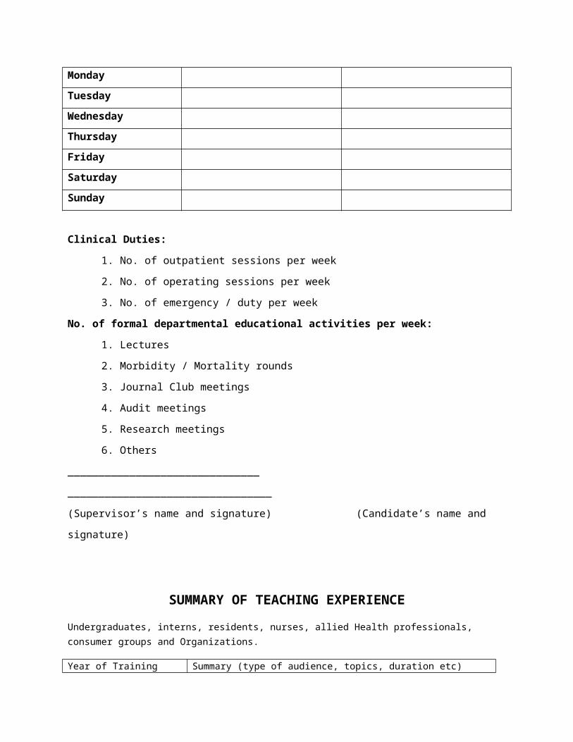
Total Marks: 500
All candidates shall appear in MS Examination part II, after completion of 5 years training program and after clearing Part I examination.
There shall be one written paper of 100 marks of SEQs pattern , one paper of MCQs containing 100 MCQs , Oral & practical/ clinical examination of 300 marks.
Clinical Examination
Total Marks: 300
Components of Part II Clinical Examination
Theory
Paper I 100 Marks 3 Hours
10 SEQs (No Choice) 100 Marks
Paper II 100 Marks 3 Hours
100 MCQs 100 Marks
Only those candidates, who pass in theory papers, will be eligible to appear
in the Oral & Practical/ Clinical Examination.
OSCE 100 Marks

10 stations each carrying 10 marks of 7minutes duration; each
evaluating performance based assessment with five of them interactive
Clinical 200 Marks
Four short cases (each 25 marks) 100 Marks
One long case: 100 Marks