Neuroscience and Mental Health - Imperial College Union | · Laminae- Layers of functional similar...
24
Collection of definitions Afferent neurone- Neurones which carry sensory information from periphery to the CNS Anterograde transport- Movement of molecules outwards from the soma towards the synapse or plasma membrane. Autonomic Nervous System: Controls visceral (organ) function Central Nervous System: Section of the nervous system containing the brain and the spinal cord Efferent neurones- Motor information from CNS to periphery. Fibre Tract- Groups of bundles of axons in the CNS that may or may not be myelinated. Examples included corpus callosum and internal capsule. Ganglion- Group of encapsulated cell bodies within the PNS. Examples include dorsal root ganglia and sympathetic ganglia. Laminae- Layers of functional similar cells such as the cerebral cortex grey matter and the cerebellar grey matter. Nerve- Discrete bundles of axons that are usually part of PNS Nucleus- Group of unencapsulated neuronal cell bodies within the CNS often of functionally similar cells. Peripheral Nervous System: Contains the nerves and ganglia outside of the CNS. Retrograde transport- Movement of molecules inwards from synapse to soma. It returns used synaptic vesicles and informs the soma of the conditions at the axon terminal. Somatic Nervous System: Controls motor and sensory function for body wall Accommodation Act of refocusing the visual image Action potential Brief reversal of the neuronal membrane potential, resulting from altered membrane permeability Affective disorder Psychiatric disorder of mood Afferent Conveying impulses towards a structure Agnosia Inability to recognise or attach meaning to objects Agraphia Inability to write Akinesia Loss or slowness of movement Amnesia Loss of memory Anaesthesia Loss of sensation Analgesia Loss of pain sensation Anterior Towards the front Aphasia Loss of ability to use language in the absence of motor and sensory loss Apraxia Inability to carry out a voluntary movement in the absence of paralysis, sensory loss and ataxia Areflexia Loss of reflexes Ataxia Loss of motor co-ordination Athetosis Involuntary movement: continuous, slow, writhing Atonia The absence of muscle tone Atrophy Reduction in size Axon Neuronal process carrying impulses away from the cell body Ballismus Involuntary, violent movements of the extremities Bradykinesia Slowness of movement Bulbar Relating to the brainstem Caudal Towards the tail Cerebral Relating to the cerebrum (=hemispheres + diencephalon) Chiasma (chiasm) Crossing over of fibres from optic nerve Column Bundle of axons within the CNS Coma State of unconsciousness from which the patient cannot be aroused by sensory stimulation Commissure Bundle of axons linking two sides of CNS Contralateral Situated in, or projecting to, the opposite side of the body Coronal plane Plane running vertically from side to side Cortex Outer layer of grey matter of the brain Decussation Crossing over of bundles of axons in the midline Dementia Loss of cognitive capabilities without objective loss of sensory or motor functions Dendrite Neuronal process that receives input from other neurones Denervation Lack of nervous control of part of the body Diplopia Double vision Dorsal Towards the back Dysarthria Difficulty with pronouncing words Dyskinesia Difficulty with movement Dysphagia Difficulty with swallowing Efferent Conveying impulses away from a structure EPSP Excitatory postsynaptic potential - local depolarisation of neuronal membrane brought about by synaptic activity Fasciculus Bundle of axons within the CNS Fibres Either bundles of axons in the CNS or whole nerves in the periphery Foramen Opening Funiculus Bundle of axons within the CNS Ganglion A collection of neurone cell bodies outside the CNS Grey matter Nervous tissue where neurone cell bodies are localised Gyrus A convolution of the cerebral cortex Hemiplegia Paralysis of one side of the body Hemianopia Loss of vision in half of the visual field Heteronymous Relating to different parts of the visual field in each eye Homonymous Relating to the same part of the visual field in each eye Hyperalgesia A reduced threshold or increased response to painful stimuli Hyperopia Condition in which only objects far from the eye are in focus Hypertonia Abnormal increase in muscle tone Idiopathic Without known cause Infarct(ion) Area of necrotic tissue resulting from vascular occlusion Guide for use of these notes First of all thank you for choosing to download these notes to study from, I hope you find them useful, please feel free to email me if you have any problems with the notes or if you notice any errors. I don't promise to respond to all emails but I'll do my best. For the Neuroscience notes I used "Vander's physiology" but mostly from the lecture notes and course guide. I organise my notes so that you should read the learning objectives on the left then proceed down the right hand side for a few learning objectives and then cross back over to the left and continue like that. Anything in this highlighted green is a definition or explains basically something's function. Text highlighted in yellow or with a star is what I would deem important and key to your information. Italics and bold just help to make certain terms stand out. The notes are a bit quirky but I hope you like them and find some of the memory aides strange enough so that they stick in your head. I provide them to you in OneNote format as that is how I created them, they can be saved as PDF but the formatting is not as nice. The one caveat with this is that these notes are freely copy able and editable. I would prefer if you didn't copy and paste my notes into your own but used them as a reference or preferably instead embellished these already existing notes by adding to them. Good luck with first year And I love Light Opera Soc Stuart Taylor Innervation Nerve supply Interneuron(e) Interconnects other neurones, usually locally Ipsilateral Situated in, or projecting to, the same side of the body IPSP Inhibitory postsynaptic potential – local hyperpolarisation of neuronal membrane brought about by synaptic activity Ischaemia Lack of sufficient blood supply, resulting in permanent damage if not restored rapidly Kinaesthesia Perception of movement Lateral Away from the midline Lemniscus Ribbon-like bundle of axons in CNS Lesion A circumscribed area of injury or disease Papilloedema Swelling of the head of the optic nerve due to raised intracranial pressure Pathway A chain of functionally connected neurones Plexus Interwoven nerves or blood vessels Posterior Towards the back Proprioception Conscious or unconscious reception by the brain of information from muscles, tendons and joints Psychosis Abnormal mental state including altered precepts (hallucinations) and false ideas (delusions) Ptosis Abnormal drooping of the upper eyelid Neuroscience and Mental Health 10 January 2012 13:58 Stuart's Neuroscience and Mental Health Page 1
Transcript of Neuroscience and Mental Health - Imperial College Union | · Laminae- Layers of functional similar...

Collection of definitions
Afferent neurone- Neurones which carry sensory information from periphery to the CNS
Anterograde transport- Movement of molecules outwards from the soma towards the synapse or plasma membrane
Autonomic Nervous System Controls visceral (organ) function
Central Nervous System Section of the nervous system containing the brain and the spinal cord
Efferent neurones- Motor information from CNS to periphery
Fibre Tract- Groups of bundles of axons in the CNS that may or may not be myelinated Examples included corpus callosum and internal capsule
Ganglion- Group of encapsulated cell bodies within the PNS Examples include dorsal root ganglia and sympathetic ganglia
Laminae- Layers of functional similar cells such as the cerebral cortex grey matter and the cerebellar grey matter
Nerve- Discrete bundles of axons that are usually part of PNS
Nucleus- Group of unencapsulated neuronal cell bodies within the CNS often of functionally similar cells
Peripheral Nervous System Contains the nerves and ganglia outside of the CNS
Retrograde transport- Movement of molecules inwards from synapse to soma It returns used synaptic vesicles and informs the soma of the conditions at the axon terminal
Somatic Nervous System Controls motor and sensory function for body wall
Accommodation Act of refocusing the visual imageAction potential Brief reversal of the neuronal membrane potential resulting from altered membrane permeabilityAffective disorder Psychiatric disorder of moodAfferent Conveying impulses towards a structureAgnosia Inability to recognise or attach meaning to objectsAgraphia Inability to writeAkinesia Loss or slowness of movementAmnesia Loss of memoryAnaesthesia Loss of sensationAnalgesia Loss of pain sensationAnterior Towards the frontAphasia Loss of ability to use language in the absence of motor and sensory loss Apraxia Inability to carry out a voluntary movement in the absence of paralysis sensory loss and ataxiaAreflexia Loss of reflexesAtaxia Loss of motor co-ordinationAthetosis Involuntary movement continuous slow writhingAtonia The absence of muscle tone Atrophy Reduction in sizeAxon Neuronal process carrying impulses away from the cell bodyBallismus Involuntary violent movements of the extremitiesBradykinesia Slowness of movementBulbar Relating to the brainstemCaudal Towards the tailCerebral Relating to the cerebrum (=hemispheres + diencephalon)Chiasma (chiasm) Crossing over of fibres from optic nerveColumn Bundle of axons within the CNSComa State of unconsciousness from which the patient cannot be aroused by sensory stimulationCommissure Bundle of axons linking two sides of CNSContralateral Situated in or projecting to the opposite side of the bodyCoronal plane Plane running vertically from side to sideCortex Outer layer of grey matter of the brainDecussation Crossing over of bundles of axons in the midlineDementia Loss of cognitive capabilities without objective loss of sensory or motor functionsDendrite Neuronal process that receives input from other neuronesDenervation Lack of nervous control of part of the bodyDiplopia Double visionDorsal Towards the backDysarthria Difficulty with pronouncing wordsDyskinesia Difficulty with movementDysphagia Difficulty with swallowingEfferent Conveying impulses away from a structureEPSP Excitatory postsynaptic potential - local depolarisation of neuronal membrane brought about by synaptic activityFasciculus Bundle of axons within the CNSFibres Either bundles of axons in the CNS or whole nerves in the peripheryForamen OpeningFuniculus Bundle of axons within the CNSGanglion A collection of neurone cell bodies outside the CNSGrey matter Nervous tissue where neurone cell bodies are localisedGyrus A convolution of the cerebral cortexHemiplegia Paralysis of one side of the bodyHemianopia Loss of vision in half of the visual fieldHeteronymous Relating to different parts of the visual field in each eyeHomonymous Relating to the same part of the visual field in each eyeHyperalgesia A reduced threshold or increased response to painful stimuliHyperopia Condition in which only objects far from the eye are in focusHypertonia Abnormal increase in muscle toneIdiopathic Without known causeInfarct(ion) Area of necrotic tissue resulting from vascular occlusion
Guide for use of these notes
First of all thank you for choosing to download these notes to study from I hope you find them useful please feel free to email me if you have any problems with the notes or if you notice any errors I dont promise to respond to all emails but Ill do my best
For the Neuroscience notes I used Vanders physiology but mostly from the lecture notes and course guide
I organise my notes so that you should read the learning objectives on the left then proceed down the right hand side for a few learning objectives and then cross back over to the left and continue like that
Anything in this highlighted green is a definition or explains basically somethings functionText highlighted in yellow or with a star is what I would deem important and key to your informationItalics and bold just help to make certain terms stand out
The notes are a bit quirky but I hope you like them and find some of the memory aides strange enough so that they stick in your head
I provide them to you in OneNote format as that is how I created them they can be saved as PDF but the formatting is not as nice The one caveat with this is that these notes are freely copy able and editable I would prefer if you didnt copy and paste my notes into your own but used them as a reference or preferably instead embellished these already existing notes by adding to them
Good luck with first yearAnd I love Light Opera Soc
Stuart Taylor
Innervation Nerve supply Interneuron(e) Interconnects other neurones usually locallyIpsilateral Situated in or projecting to the same side of the body IPSP Inhibitory postsynaptic potential ndash local hyperpolarisation of neuronal membrane brought about by synaptic activityIschaemia Lack of sufficient blood supply resulting in permanent damage if not restored rapidlyKinaesthesia Perception of movementLateral Away from the midline Lemniscus Ribbon-like bundle of axons in CNSLesion A circumscribed area of injury or disease
Papilloedema Swelling of the head of the optic nerve due to raised intracranial pressure Pathway A chain of functionally connected neuronesPlexus Interwoven nerves or blood vesselsPosterior Towards the backProprioception Conscious or unconscious reception by the brain of information from muscles tendons and jointsPsychosis Abnormal mental state including altered precepts (hallucinations) and false ideas (delusions)Ptosis Abnormal drooping of the upper eyelid
Neuroscience and Mental Health10 January 20121358
Stuarts Neuroscience and Mental Health Page 1
restored rapidlyKinaesthesia Perception of movementLateral Away from the midline Lemniscus Ribbon-like bundle of axons in CNSLesion A circumscribed area of injury or diseaseMania An abnormally elevated expansive or irritable moodMedial Towards the midlineMembrane potential Potential difference across the neuronal membrane maintained by differential permeabilityMemory The retention of learned informationMiosis Pupillary constrictionMydriasis Extreme dilation of the pupilMyopia Condition in which only objects close to the eye are in focusMotor Involved in movement or responseNeoplasia Cancerous overgrowth of tissueNeurite Process of a neurone dendrite or axonNeuroglia (glia) Cells which support the function of neurones in the CNSNeuron(e) Complete nerve cell comprising cell body dendrites and axonNeuropil Complex meshwork of dendrites axon terminals and neuroglial processesNeurotransmitter Specific chemical agent released by a presynaptic neurone on excitation which crosses the synaptic cleft to stimulate or inhibit the postsynaptic cellNociception Response to noxious stimulationNucleus A collection of neurone cell bodies within the CNSNystagmus Involuntary rapid movements of the eyeballsOedema Swelling of tissue due to accumulation of fluidOphthalmoplegia Paralysis of the extrinsic eye musclesPalsy Weakness or paralysis of musclesParaesthesia Distorted sensationParalysis Loss of voluntary movement following neural injuryParaplegia Paralysis of lower limbsParesis Weakness partial paralysis
Proprioception Conscious or unconscious reception by the brain of information from muscles tendons and jointsPsychosis Abnormal mental state including altered precepts (hallucinations) and false ideas (delusions)Ptosis Abnormal drooping of the upper eyelidRamus Branch (particularly of spinal nerve)Root Interconnects spinal cord and spinal nerveRostral Towards the headQuadriplegia Paralysis of all four limbs (tetraplegia)Saccade Small quick eye movements on changing point of fixationSagittal plane Plane running vertically from front to back Seizure Sudden disturbance of consciousness or sensorimotor functionSensory Involved in receiving information from the environment (internal or external)Sign In medicine an abnormality observed by the physician and independent of the observation of the patientSomatic Relating to the body framework as distinct from the internal organsSomatotopic The orderly representation of body parts in the CNSSpasticity Condition of increased muscle tone and exaggerated tendon reflexesStria Narrow band of axons in the CNSSulcus Groove between adjacent gyriSymptom In medicine an abnormality observed by the patient and reported to the physician - symptoms are necessarily subjectiveSynapse Site of functional contact between neuronsSyndrome Group of signs and symptoms which characterise a diseaseTetraplegia See quadriplegiaTract Bundle of axons within the CNSVentral Towards the frontVisceral Relating to the internal organsVisual field The total region of space that is viewed by both eyes when fixated on a pointWhite matter Nervous tissue made up mainly of axons
Stuarts Neuroscience and Mental Health Page 2
Learning Objectives
Define the following terms and explain how they interact with each other Central nervous system peripheral nervous system autonomic nervous system and somatic nervous system
Outline diagnostic methods and how to perform a neurological examination
Outline the major causes of neurological disorders
Refer to other important information
Define the following terms and explain how they interact with each other Central nervous system peripheral nervous system autonomic nervous system and somatic nervous system
Central Nervous System Section of the nervous system containing the brain and the spinal cord
Peripheral Nervous System Contains the nerves and ganglia outside of the CNS
Autonomic Nervous System Controls visceral (organ) function
Somatic Nervous System Controls motor and sensory function for body wall
Regulatory functions of ANS
Genitalia1Structures in the eye2Internal organs3Blood vessels4Glands5
Outline diagnostic methods and how to perform a neurological examination
Diagnostic methods
Taking a detailed history if possiblebullNeurological ExaminationbullImaging bullNeurophysiologybull
Neurological examination
Level of consciousnessbullSpeechbullMental state and cognitive abilitybullMotor functionbullSensory functionbullCranial nerve functionbull
ElectroencephalogrambullElectromyogrambullNerve conduction testbull
Neurophysiology
Imaging
CT scanbullMRIbull
Outline the major causes of neurological disorders
Memorisation tool MICE n TING
Metabolic disorders- Diabetic induced neuropathy Hypoglycaemia can cause coma likewise hyperglycaemia can result in ketoacidosis which likewise can result in coma
Immunological defects- Multiple sclerosis is a condition whereby there is an autoimmune response to the axon myelin sheath which causes demyelination Signal transduction is thus weaker which neurological effects can then occur from
Cerebrovascular incidents- Stroke or cerebrovascular infarct can occur reducing vital glucose and oxygen supply to brain and quickly causing necrosis Little or no storage of glucose in brain so a constant supply is vital If swelling occurs it can raise ICP which is similarly serious
Environmental factors- Examples being toxins and forms of recreational drugs Lead is one such example hence why there is such strict regulation on lead paint Alcohol with foetal alcohol syndrome being a case in point See mobile phones and recreational drugs for other areas Heavy metal encephalopathies
Trauma- Fracture of the skull or severing of the spinal cord can cause paralysis or the likelihood of developing potentially fatal haematomas Refer to PBL if interested
Infection- Meningitis which is inflammation of meninge membranes Two types viral and bacterial Viral causes serious illness whereas bacterial is often fatal Brain is relatively well protected against infection as a result of the blood brain barrier
Neoplasia- Abnormal proliferation which can cause cancers such as meningiomas Neurones are unlikely to be affected due to their cessation of mitosis after development Instead cancers can occur in supporting cells (glial- glioma) or the connective tissue cells Most cancers that are present are metastases from other locations in the body
Genetic defects- Extensive list of genetic conditions that can affect the brain One example is Huntingdons disorder whereby there is destruction of the caudate and putamen nuclei
Other important information
AXONS DO NOT I REPEAT DO NOT REGENERATE IN THE CNS They do however to a degree in the PNS Furthermore neurones as entire entities are also not replacedHigh energy requirement of brain means that there is little storage
Epilepsy- Neurological condition that can be caused by infection trauma or genetic factors Abnormal firing of neurones occurs within a epileptic focus which can spread to a large proportion of neurones in the brain causing the symptoms of the disorder such as fitting
Organisation and disorders10 January 20121408
Stuarts Neuroscience and Mental Health Page 3
Learning Objectives
Draw and label of a typical neuron identifying soma dendrites axon and terminals
Define the role of each cellular component in the specialised function of the neuron
Outline the organisation and functions of intracellular transport in the neuron
Define the functional subtypes of neurons and list the ways in which they are organised collectively in the nervous system
Describe the organisation of synapses
Name the main classes of neuroglia and explain their functions in the nervous system
Draw and label of a typical neuron identifying soma dendrites axon and terminals
Basic Information of the Neuron
Basic unit of the nervous system Each one can be thought of as a processing unit Responsible for the generation and transmission of electrical impulses Synapses are junctions between neurones Supported by glial cells of which there are many types Glial cells outnumber neurons 91
Nucleus
Schwann Cell
Axon TerminalsDendrites
Soma
Nodes of Ranvier
Myelin SheathDefine the role of each cellular component in the specialised function of the neuron
Soma is the metabolic centre of activity for the whole neuron
Large nucleusbullProminent NucleolusbullAbundant Rough ERbullWell developed Golgi- Required for the large amount of neurosecretionsbullAbundant mitochondriabullOrganised cytoskeletonbullMost metabolically active cell in bodybull
A highly branched structure originating from the cell body bullHas a large surface area which is improved further by the presence of protrusions called dendritic spines These structures receive the majority of synapses
bull
Large pyramidal neurons may have as many as 30-40000 spinesbull
Dendrites receives all the inputs into the cell
Axons the cellular component of the neuron concerned with the output mechanism
Impulses are generated at the axon hillock and progress away from the cell body Initiator of impulse- Axon hillock
bull
Usually only one per cell In picture above axon would be at the base of the pyramidal cell (cannot see due to focusing)
bull
Branching can occur after leaving the cell body or upon reaching the targetbullProminent microtubules and neurofilaments that contributes to cable like transporting system of the axon
bull
Can be myelinated or unmyelinated if so nodes of Ranvier are unmyelinated junctions where the action potential is propagated from
bull
As stated axons branch extensively close to a target- process known as terminal arbor
bull
Axon terminals form synapses with their targetsbullCan end with varicosities or boutons (think synaptic knob)bull
Axon terminals-
Highly organised cytoskeleton is required with microfilaments intermediate filaments and microtubules which are particularly abundant
bull
Neurofilaments play a critical role in determining axon calibrebull
Neuronal cytoskeleton-
Outline the organisation and functions of intracellular transport in the neuron
Transport of membrane associated materialsbullVesicles with associated motors are moved down the axon at 100-400mm per daybullDifferent membrane structures targeted to different componentsbullRetrograde moving organelles are morphologically and biochemically very different from anterograde vesicle
bull
Fast axonal transport
Anterograde transport- Movement of molecules outwards from the soma towards the synapse or plasma membrane
Fast- Transports synaptic vesicles transmitters and mitochondriabullSlow - Anterograde transport results in the delivery of cytoskeletal and cytoplasmic constituents
bull
Describe the organisation of synapses
Prevalent Golgi apparatus packages neurotransmitter into vesicles which is the transported by fast anterograde transport
bull
Mechanisms are in place for the successful completion of exocytosis and in particular the fusion of the vesicles to the plasma membrane
bull
Abundant mitochondria consume about 45 of cells energy expenditure This is required for the pumping of ions and synaptic transmission
bull
Neuronal integration occurs when multiple competing synaptic inputs are integrated in post synaptic neurone
bull
Three main types of synapse
Axo-dendritic- Often excitatory- Also Grays Type 1Axo- somatic- Often inhibitory- Also Grays Type 2Axo-axonic- Often modulatory-
Define the functional subtypes of neurons and list the ways in which they are organised collectively in the nervous system
Cells of the Nervous System20 January 20121315
Stuarts Neuroscience and Mental Health Page 4
Fast- Transports synaptic vesicles transmitters and mitochondriabullSlow - Anterograde transport results in the delivery of cytoskeletal and cytoplasmic constituents
bull
Transports substances from an extracellular spacebullUses microtubule-associated ATPase to drive particles along microtubulesbullTransports soluble cytoplasmic constituentsbullIs the process by which material returns from the terminals to the cell body either for degradation or recycling
bull
Retrograde transport- Movement of molecules inwards from synapse to soma It returns used synaptic vesicles and informs the soma of the conditions at the axon terminal
Define the functional subtypes of neurons and list the ways in which they are organised collectively in the nervous system
Pseudounipolar
Cell has two fused process which are axonal in structure This is a sensory neuronbullExample- Dorsal root ganglion cellsbull
Bipolar
Cells in retina in addition to white matter of cortexbullBIPOLAR THINK OPPOSITES BLACK RETINA ANDbullWHITE CORTEX
Golgi Type I multipolar
Highly branched dendritic treesbullPyramidal cells of cerebral cortexbullAnterior horn cells of the spinal cordbullPurkinje cells of cerebellum (15 million)bullExtremely long extending axonsbullRetinal ganglion cellsbullPAPERbull
Golgi Type II multipolar
Highly branched dendritic treesbullShort axons that terminate near to cell bodiesbullStellate cells of cerebral cortexbull
Define the functional subtypes of neurons and list the ways in which they are organised collectively in the nervous system
Types of neurons
Sensory1Motor2Interneurons (responsible for modification integration coordination facilitation and inhibition of sensory input)
3
Organisational structure of Neurons
Nucleus- Group of unencapsulated neuronal cell bodies within the CNS often of functionally similar cells
Examples include brain stem (Raphe) and deep cerebral cortex (Dentate)bullNNNucleus NNNoo capsule (unencapsulated)bull
Laminae rhymes with greybull
Laminae- Layers of functionally similar cells such as the cerebral cortex grey matter and the cerebellar grey matter
Ganglion- Group of encapsulated cell bodies within the PNS Examples include dorsal root ganglia and sympathetic ganglia
Fibre Tract- Groups of bundles of axons in the CNS that may or may not be myelinated Examples included corpus callosum and internal capsule
Bring information to the CNS from sensory receptors and bring axons to effector organs
bull
Often a combination of both motor and sensory neuronsbullExceptions to PNS are optic and olfactory nervesbull
Nerve- Discrete bundles of axons that are usually part of PNS
Name the main classes of neuroglia and explain their functions in the nervous system
These are the support cells of the nervous systembullEssential for correct functioning of neurones and have many and varied functionsbull
Astroglia
Oligodendroglia
Microglia
Immature progenitors
Ependymal cells- Epithelial lining of the ventricles
Schwann cells
Satellite glia
Examples includebull
Neuroglia
Star shaped cell (think astrology)bullMost abundant cell in CNSbullEach cell forms a specific territory that interfaces with microvasculaturebull
Fibrous astroglia (White Matter)i)Protoplasmic astroglia (Grey Matter)ii)Radial astrogliaiii)
Several different typesbull
Numerous IF bundles in cytoplasm of fibrous astrogliabullGap junctions suggest astroglia-astroglia signallingbullHas intimate associations with other cell typesbullInteracts with blood vessels in an ordered arrangement with little overlapbull
Astroglia
Scaffold for neuronal migration and axon growth during development1Formation of blood brain barrier2Transport of substances from blood to neurons3Segregation of neuronal processes (synapses)4Removal of neurotransmitters5Synthesis of neurotropic factors6Neuronal-glial and glial neuronal signalling7Potassium ion buffering8Glial scar formation9
SSS FTR NPGbull
Functions of astroglia
Myelin
Myelin is an insulating membrane found around nerve cells that is made up of lipid
bull
Up to 50 lamellae- thin plate like structurebullDark and light bands seen at EM levelbullHighly susceptible to damage and loss of either oligodendroglia or myelin is disastrous
bull
- AdrenoleucodystrophyMyelin disease states- Multiple Sclerosisbull
Stuarts Neuroscience and Mental Health Page 5
Potassium ion buffering8Glial scar formation9
SSS FTR NPGbull
Myelin producing cells of the CNSbullSmall spherical nucleibullFew thin processesbullProminent ER and GolgibullMetabolically highly activebull
Interfasicular oligodendroglia
Perineuronal oligodendroglia
Types includebull
Oligodendroglia
Functions of oligodendroglia
Production and maintenance of myelin sheath1Each cell can produce numerous sheaths (1-40)2
Derived from bone marrow during early developmentbullResident macrophage population of CNSbullInvolved in immune surveillancebullTypical macrophage functions- ie APCbullRole in tissue modellingbullSynaptic strippingbull
Microglia
These are the myelin producing cells of the PNSbullOnly produce one sheath per cellbullPromote axon regenerationbullSurround unmyelinated axonsbull
Peripheral Glial- Schwann Cells
Stuarts Neuroscience and Mental Health Page 6
Learning Objectives
Diffusion of an iono
Permeability of a cell membraneo
Electrochemical gradient of an iono
Define the following
Describe how a resting membrane potential can arise from a difference in concentration of an ion across a selectively permeable membrane (use diagrams)
Define electrochemical equilibrium for an ion
What is the equilibrium potential for an ion
The Nernst equation is Ex+ = (RTZF) ln (CoCi) You should know that Ex+ is the equilibrium potential of ion X+ R is the gas constant T is absolute temperature Z is the charge on the ion and F is Faradayrsquos number coulombs of charge per mol of ion
Substituting the values of the constants and T= 37oC gives 27
What are typical values for the concentration of K+ and for Na+ inside and outside a normal neuron
What is a typical value for the resting potential of a neuron
K+ concentration has a much stronger effect on the resting potential than Na+ concentration does Explain the basis of this difference
Diffusion of an ion- The passive movement of an ion down its concentration
gradient
Permeability of a cell membrane- The selectivity of a membrane to ions is its
permeability
Electrochemical gradient of an ion- The movement of ions from an area of
high concentration and relative charge to an area of low concentration and opposite charge
Define the following
Describe how a resting membrane potential can arise from a difference in concentration of an ion across a selectively permeable membrane (use diagrams)
Zero-reference is outside of the cellbullThe inside of the cell is negative compared to the outsidebullAll cells have a membrane potentialbullResting potential is -70mvbull
Concentration of at least one permanent ion is different on one side of the membranebull
Permeable pores that open and close that are selective for different types of ion eg (K+ Na+ Cl- Ca2+)
bull
Open by change in membrane potential
Voltage dependent-1)
Open all the time- these are the ones that produce the resting potential
Voltage independent2)
2 major typesbull
Permeability of membrane to potassium allows it to leave generating a negative charge within the cell- Resting potential Equilibrium of electrochemical gradient is produced resulting in small flux between compartments
bull
Permeability of membrane to sodium allows it to enter generating a negative charge outside and a positive charge inside- Action Potential
bull
Ion Channels
Define electrochemical equilibrium for an ion
Electrochemical equilibrium- Reached when the concentration gradient is balanced by the electrical gradient across the membrane
What is the equilibrium potential for an ion
Equilibrium potential- Potential that prevents diffusion down the ions concentration gradient
The Nernst equation is Ex+ = (RTZF) ln (CoCi) You should know that Ex+ is the equilibrium potential of ion X+ R is the gas constant T is absolute temperature Z is the charge on the ion and F is Faradayrsquos number coulombs of charge per mol of ion
Nernst equation
bull EX = (RTZF) ln (Co Ci)
ndash C is concentration of the ion [X+]
bull o = outside cell
bull i = inside cell
ndash Co = [X+] outside cell
ndash Ci = [X+] inside cell
bull R = gas constant
bull T = Temp o Kelvin
bull Z = charge on ion
bull -1 for Cl- +2 for Ca2+
bull F = Faradayrsquos number
bull charge per mol of ion
bull ln means log to base e
Ex- is the equilibrium potential of the ion X
Substituting the values of the constants and T= 37oC gives 27
EX = (27Z) ln (Co Ci)
Co
(mM)
Ci
(mM)
ENa
(mV)
Na+ 150 10 +73
Co
(mM)
Ci
(mM)
ENa
(mV)
K+ 5 150 -92
bull What sign for the equilibrium potential
bull The sign the inside needs to maintain the concentration
gradient
bull RTF 37 oC = 27
bull EK = (27 +1) ln(0005 M 015 M)
bull EK = +27 ln(003)
bull EK = +27 -34
bull EK = -92 mV
bull RTF 37 oC = 27
bull ENa = (27 +1) ln(015 M 001 M)
bull ENa = +27 ln(15)
bull ENa = +27 +27
bull ENa = +73 mV
What are typical values for the concentration of K+ and for Na+ inside and outside a normal neuron
Sodium Potassium
Inside (mM) 10 150
Outside (mM) 150 5
What is a typical value for the resting potential of a neuron
-70mVbull
K+ concentration has a much stronger effect on the resting potential than Na+
concentration does Explain the basis of this difference
Potassium sodium and chlorine concentrations all contribute to the real membrane potential
bull
The size of each ions contribution is proportional to how permeable the membrane is to the ion
bull
Goldman-Hodgkin-Katz voltage equation
This is an equation used to describe the resting membrane potentialbullTHINK- youd need a rest after saying that namebull
0 means the channel is completely close
05 means the channel is open half of the time
1 means the channel is fully open
Within the equation the letter P denotes permeability or channel open probabilitybull
GHK equation worked examples
bull 1) All channels are open all the time (PK PNa PCl = 1)
bull Vm = 27 ln(1[0005] + 1[015] + 1[0005] 1[015] + 1[001] + 1[011])
bull Vm = 27 ln(016 027)
bull Vm = 27 -05
bull Vm = -14 mV
bull 3) Now increase Na+ permeability by 5 (PK = 1 PNa = 005 PCl = 0)
bull Vm = 27 ln(1[0005] + 005[015] + 0[0005] 1[015] + 005[001] + 0[011])
bull Vm = 27 ln(00125 01505)
bull Vm = 27 -249
bull Vm = -67 mV
bull Increasing permeability for Na+ makes the cell more positive
RTF at 37 oC = 27
Concentration [ ] is in Moles
bull 2) Only K+ channels are open and Cl- and Na+ channels are closed (PK = 1 PNa = 0 PCl = 0)
bull Vm = 27 ln(1[0005] + 0[015] + 0[0005] 1[015] + 0[001] + 0[011])
bull Vm = 27 ln(0005 015)
bull Vm = 27 -34
bull Vm = -92 mV
bull Ion concentration has not changed BUT Vm moved to EK
Graded Potentials
Resting Potential14 May 20121248
Stuarts Neuroscience and Mental Health Page 7
EX = (27Z) ln (Co Ci)
Co
(mM)
Ci
(mM)
ENa
(mV)
Na+ 150 10 +73
Co
(mM)
Ci
(mM)
ENa
(mV)
K+ 5 150 -92
bull What sign for the equilibrium potential
bull The sign the inside needs to maintain the concentration
gradient
bull RTF 37 oC = 27
bull EK = (27 +1) ln(0005 M 015 M)
bull EK = +27 ln(003)
bull EK = +27 -34
bull EK = -92 mV
bull RTF 37 oC = 27
bull ENa = (27 +1) ln(015 M 001 M)
bull ENa = +27 ln(15)
bull ENa = +27 +27
bull ENa = +73 mV GHK equation worked examples
bull 1) All channels are open all the time (PK PNa PCl = 1)
bull Vm = 27 ln(1[0005] + 1[015] + 1[0005] 1[015] + 1[001] + 1[011])
bull Vm = 27 ln(016 027)
bull Vm = 27 -05
bull Vm = -14 mV
bull 3) Now increase Na+ permeability by 5 (PK = 1 PNa = 005 PCl = 0)
bull Vm = 27 ln(1[0005] + 005[015] + 0[0005] 1[015] + 005[001] + 0[011])
bull Vm = 27 ln(00125 01505)
bull Vm = 27 -249
bull Vm = -67 mV
bull Increasing permeability for Na+ makes the cell more positive
RTF at 37 oC = 27
Concentration [ ] is in Moles
bull 2) Only K+ channels are open and Cl- and Na+ channels are closed (PK = 1 PNa = 0 PCl = 0)
bull Vm = 27 ln(1[0005] + 0[015] + 0[0005] 1[015] + 0[001] + 0[011])
bull Vm = 27 ln(0005 015)
bull Vm = 27 -34
bull Vm = -92 mV
bull Ion concentration has not changed BUT Vm moved to EK
Through these series of equations we can clearly see that potassium contributes most to the resting potential as increasing the permeability of sodium by only 5 means that the resting potential is now slightly too positive
bull
Concentration is in moles not millimoles bull
Changes in membrane potential
Graded Potentials
Graded potentials occur at synapses and sensory receptor and their function is to contribute to initiating or preventing action potentials
bull
Change in membrane potential caused by alteration in permeability of ion channels Not of a fixed time and length like action potentials
bull
Graded potentials change in membrane potential in response to stimulation bull
Can be depolarizing or hyperpolarizing
A stronger stimulus produces a larger graded potential
Graded potentials get smaller the farther they travel (Decremental spread)
Properties of graded potentialsbull
Stuarts Neuroscience and Mental Health Page 8
Learning Objections
You should be able to explain in general terms what the function of the action potential is
Give some examples of other types of excitable cells (in addition to neurons) in which action potentials occur
ThresholdRefractory periodldquoAll or nothingrdquo behaviourDepolarizationRepolarizationHyperpolarizationSaltatory conduction
Define the following terms as they apply to action potentials
Voltage-gated channelChannel inactivationPositive feedback
Define the following terms as they apply to the membrane channels involved in producing the action potential
Outline the sequence of events during a typical action potential in the neuron Include changes in membrane potential changes in membrane permeability and fluxes of ions across the membrane (a diagram will help)
State the size and duration (including units) of a typical action potential in a neuron
Define the term ldquoregenerativerdquo as applied to action potentials and its significance for spread of the action potential along an axon
Explain how conduction of the action potential occurs (conduction here means spread along the axon alternatively this process may be called transmission or propagation)
List two structural features that affect the conduction velocity along normal axons Briefly explain why they affect velocity as they do
Be able to list at least one pathological condition that affects conduction velocity
You should be able to explain in general terms what the function of the action potential isGive some examples of other types of excitable cells (in addition to neurons) in which action potentials occur
The function of the action potential is to transport messages through the nervous system very rapidly
bull
Action potentials also exist in muscle cellsbull
Define the following terms as they apply to action potentials
Threshold- The level of depolarisation needed to cause the triggering of an
action potential
Refractory period- Unresponsive to threshold depolarization
Absolute refractory period- New action potential cannot be triggered even with very strong stimulus because inactivation gate is closed
Relative refractory period- Inactivation gate is open but sodium channel activation gate is closed meaning a stronger than normal stimulus is required to trigger an action potential
ldquoAll or nothingrdquo behaviour- Once triggered a full sized action potential
occurs
Depolarization- When the membrane becomes more positive towards
sodiums ion equilibrium potential
Repolarization- Efflux of potassium ions to restore membrane to negative
state
Hyperpolarization- Over efflux of potassium results in membrane potential
dropping below resting potential
Saltatory conduction- From the latin (hop or to leap) is the propagation of
action potentials along myelinated axons from one node of Ranvier to the next
Voltage-gated channel- Ion channels that only open when there is a
change in voltage
Channel inactivation- Closing of the inactivation gate in response often
to an extracellular positive charge
Positive feedback- A mechanism that means that action potentials
constantly regenerate
Define the following terms as they apply to the membrane channels involved in producing the action potential
Outline the sequence of events during a typical action potential in the neuron Include changes in membrane potential changes in membrane permeability and fluxes of ions across the membrane (a diagram will help)
At the resting potential voltage-gated ion channels (Na+ and K+) are closed
Note these are different to non-voltage gated channels that generate the resting potential
Resting potential PKgtgtPNa Where P is the permeability of the membrane1
Stimulus- depolarises the membrane potential and causes it to become more positive
2
PNa rapidly increases because the voltage gated Na+ channels open quicker than potassium channels
Potassium permeability increase as the voltage gated K+ channels start to slowly open and as the upstroke progresses more and more voltage gated K channels open
Potassium leaves the cell down the electrochemical gradient but is less than Na entering
Membrane potential moves toward the Na+ equilibrium potential
Depolarisation- Starts at the threshold potential3
Big decrease in PNa because the voltage-gated Na channels inactivation gate is closed and sodium entry stops generating the absolute refractory period
Moderate PK increase as there are more voltage gated K+ channels and they remain open for longer which means that K leaves by its electrochemical gradient
Membrane potential moves toward the K+ equilibrium potential
Repolarisation-4
State the size and duration (including units) of a typical action potential in a neuron
1-2ms for an action potentialbullSize= -70 to + 40 therefore 110mVbull
Define the term ldquoregenerativerdquo as applied to action potentials and its significance for spread of the action potential along an axon
If less than threshold then graded potentialbullOnce threshold is reached the cycle continues in a positive feedback mannerbullCycle continues until the voltage-gated sodium channels inactivate (closed and voltage insensitive)
bull
Membrane remains in a refractory state until the voltage gated sodium channels recover from inactivation
bull
Ions may move in and out but only a very small number of ions actually cross the membrane to change the potential ie the change in concentration is extremely small (less than 01)
bullIon Movements during action potential
Explain how conduction of the action potential occurs (conduction here means spread along the axon alternatively this process may be called
Action Potential14 May 20122245
Stuarts Neuroscience and Mental Health Page 9
periodModerate PK increase as there are more voltage gated K+ channels and they remain open for longer which means that K leaves by its electrochemical gradient
Membrane potential moves toward the K+ equilibrium potential
At rest voltage gated K channels are still open therefore K continues to leave down gradient
Membrane potential moves closer to the K+ equilibrium
Voltage gated K+ channels then close but can still possibly generate an AP if the stimulus is very strong- Relative Refractory period
Membrane potential returns to the resting potential
After hyperpolarization5
Time Course of changes in permeability
small (less than 01)
Explain how conduction of the action potential occurs (conduction here means spread along the axon alternatively this process may be called transmission or propagation)
Active Propagation
Passive propagation
Only resting K+ channels
openInternal (or axial) amp Membrane
Resistance alters propagation
distance and velocity
List two structural features that affect the conduction velocity along normal axons Briefly explain why they affect velocity as they do
Large diameter myelinated axons= 120msbullSmall diameter non myelinated = 1msbullSpeed of sound= 331msbull
Less resistance to current flow inside large diameter axons
Increases with axon diameter1
Action potentials only occur at nodes of Ranvier
Higher in myelinated than non-myelinated axons of the same diameter2
Reduced axon diameter (ie regrowth after injury)
Reduced myelination (MS and diphtheria)
Cold anoxia compression and drugs (some anaesthetics)
Conduction velocity is slowed by
Be able to list at least one pathological condition that affects conduction velocity
Multiple sclerosis and diphtheria causes demyelination of axonsbull
Stuarts Neuroscience and Mental Health Page 10
Learning Objectives
Define the essential components required for neurotransmitter release
Understand the differences between excitatory and inhibitory transmission
Define at least two mechanisms for the termination of neurotransmitter action at the synapse
Describe how modulation of the synaptic properties of GABA can be modulated pharmacologically to treat epilepsy
Features of neurotransmission
Transfer across a synapse requires the release of neurotransmitters and their interaction with postsynaptic receptors
bull
Transmitter released from 1st cell synaptic activation of 2nd cell signal integration and signal conduction by 2nd cell
bull
Rapid timescale1)Adaptability2)Plasticity 3)Diversity4)Learning and memory5)
Features
RAPDL- sounds like rapidbullDendrites receive inputs where it is integrated in the somabull
SynapseGap is about 20-100 nmbull5000 molecules of neurotransmitter per synaptic vesiclebullHigh energy dependency is satiated by high levels of mitochondriabullPost synaptic knob is electron densebull
Features of neurotransmitters
Provide enormous diversity in variety of transmitters and their receptorsbullAmino acids (eg glutamate gamma amino butyric acid [GABA] amines (eg noradrenaline [NA] and dopamine [DA] and neuropeptides (eg opioid peptides)
bull
May mediate rapid (micro to milli second responses) or slower effects (ms)bullVary in abundance from mM to nM CNS tissue concentrationbullNeurones receive multiple transmitter influence which are integrated to produce diverse functional responses
bull
Define the essential components required for neurotransmitter release
Essential componentsAn action potential is necessary to initiate neurotransmitter releasebullIt has to be very fast within ms (200 micro seconds)bullCalcium is essential- transmitter release requires an increase in intracellular Ca2+ to about 200microM
bull
A protein complex formation between vesicle membrane and cytoplasmic proteins to enable both vesicle docking and a rapid response to Ca entry leading to membrane fusion and exocytosis
bull
ATP and vesicle recyclingbull
Extra information on neurotransmittersSynaptic transmission is restricted to specialised structures ie the synapsebullSynaptic vesicles (SVs) provide the source of neurotransmitter (4000-10000 molecules per SV)
bull
Vary from mM to nM in CNS tissue concentrationsbull5-10 mM amino acids in brainbull
Activation of transmitter release is calcium dependent and requires rapid transduction
bull
Series of events= Membrane depolarisation-gt Calcium influx -gt Vesicle fusion -gt Vesicle exocytosis-gt Transmitter release
bull
From calcium influx to transmitter release has to be within this 200 microsecondsbullSuch rapid release rates can be achieved due to the vesicles being primed at the active zone close to Ca entry
bull
Localisation of vesicle at pre synaptic area Also alignment with calcium channel must occur so the response is rapid this is also helped by proteins associating between presynaptic membrane and vesicle to enable docking
bull
Vesicular proteins are targets for neurotoxinsbull
Types of neurotoxins
Botulinum toxin and tetanus destroys membrane association proteins and vesicles1Black widow spider- alpha latrotoxin that interferes with resealing of membrane so neurotransmitter is released to depletion
2
Vesicular proteins aretargets for neurotoxins
TETANUS toxinCtetani causes paralysis
BOTULINUM toxinCbotulinum causes flacid paralysis
alpha LATROTOXINblack widow spiderstimulates transmitterrelease to depletion
Zn2+-dependent endopeptidasesinhibit transmitter release
Steps of synaptic transmission
Biosynthesis packaging and release of neurotransmitter1
Membrane depolarisationaCalcium channels openbCalcium influxcVesicle fusiondVesicle exocytosis- fusion of vesicle to membrane and release of transmittereTransmitter releasef
Release
Receptor action2
Ion channel receptor- Extremely fast (msecs) mediate all fast excitatory and inhibitory transmission
bull
Glutamate can be synthesized from glucose via the TCA cycle
GABA is synthesised from Glutamate by removing a carboxyl group form N terminal Glutamate decarboxylase
CNS- Glutamate GABA
NMJ- Acetylcholine at nicotinic receptors
Examples bull
G- protein coupled receptor- Slow secs mins- Effectors may be enzymes (adenyl cyclase phospholipase C cGMP-PDE) or channels (eg Ca or K) Abundant in CNS
bull
CNS and PNS ACh at muscarinic receptors dopamine noradrenaline 5hydroxytryptamine (5ht) and neuropeptides eg enkaphalin
Examplesbull
Understand the differences between excitatory and inhibitory transmission
Glutamates effects propagated by sodiumbullimport in the post synaptic membraneExcitatory postsynaptic EPSPbull(membrane is becoming depolarised)
Excitatory-
Define at least two mechanisms for the termination of neurotransmitter action at the synapse
Excitatory CNS synapse mediated by Glutamate
G G
G G G
G
Excitatory amino acid
Transporter EAAT
GLIAL
CELL
Na+ Na+ and Ca2+
glucose
TCAcycleaKG
Glutamate
receptor
GLUR
GLUTAMINE
SYNTHETASE
Glutamine
Glutamate
Neurotransmission10 January 20121408
Stuarts Neuroscience and Mental Health Page 11
import in the post synaptic membraneExcitatory postsynaptic EPSPbull(membrane is becoming depolarised)
Excitatory postsynpatic potential (EPSP)
GABA- which results from Chlorine bullentering the post synaptic neuroneInhibitory postsynaptic EPSP (membrane bullis hyperpolarized which makes it veryhard to generate an action potential
Inhibitory-
Glutamate receptors
Alpha amino-3-hydroxy-5methyl-4-isoxazole propionic acid (AMPA)bullMajority of fast excitatory synapses with rapid onset offset and desensitisationbullInitiated by just Na entrybull
AMPA receptors
N-methyl-aspartatebullSlow component of excitatory transmissionbullServe as coincidence detectors which underlie learning mechanismsbullInitiate by Na and Ca entrybull
NMDA receptors
G G
G G G
G
Excitatory amino acid
Transporter EAAT
GLIAL
CELL
Na+ Na+ and Ca2+
glucose
TCAcycleaKG
Glutamate
receptor
GLUR
GLUTAMINE
SYNTHETASE
Glutamine
Glutamate
GLUR receptor takes up Na and Ca in PosSM (Post synaptic Membrane)bullGlutamate is the transported by Excitatory amino acid Transporter (EAAT) to glial cells
bull
Here it is converted to glutamine by glutamine synthasebullHence the glial cell has removed the neurotransmitter link to Cells of the Nervous System
bull
Inhibitory CNS synapse mediated by GABA
GABA GABA
GABA GABA GABA
GABA
GABA transporter GAT
GLIAL
CELL
Cl- Cl-
glucose
TCAcycle
aKG
GABA receptor
GABAAR
Cl-
GLUTAMATE
Glutamic acid
decarboxylase
GAD (B6)
GABA trans
-aminase
GABA-T
Succinate
semialdehyde
GABA
Glutamate is converted to GABA by Glutamic acid decarboxylase (GAD)bullGABA has effects on PSM receptor which causes chloride influxbullGABA is then relocated to Glial cell by GABA Transport (GAT)bullGABA trans aminase GABA-T converted GABA to Succinate semialdehydebull
Describe how modulation of the synaptic properties of GABA can be modulated pharmacologically to treat epilepsy
In epilepsy there is an excess of glutamate which causes abnormal neuronal firingbullThe concept of GABA drugs is that they will be used to inhibit the effects of glutamatebull
Frequency of opening of channel is increased when binding to benzodiazepinesbullBarbiturates- Open channel for longerbull
alpha
szlig
GABA
barbiturates
steroids
benzodiazepines
Zn
convulsantsethanol
Pentameric organisation of the GABA receptorand pharmacologically important binding domains
Drugs facilitating GABA transmission areantiepilepticanxiolyticsedativemuscle relaxant
Qu 1 Which of the following is NOT a normal route for neurotransmitter inactivation
Addition of a methyl group in the synaptic cleft
X Diffusion out of the cleft
Enzymatic destruction in the synaptic cleft-
Re-uptake by the pre-synaptic terminal- NE
Uptake by glial cells- GABA glutamate
X Mark = -2 (conf=2 )
Best Option Addition of a methyl group in the synaptic cleftSuch a method of inactivation does not existLecture Neurotransmitters
Pasted from lthttpswwwuclacuklaptlaptlitesysrunhtmicl08_neurof=cleari=icl1k=1u=_st1511i=Imperialgt
Stuarts Neuroscience and Mental Health Page 12
Session 4 Tutorial ndash Neurotransmitters and epilepsy
Tutorial notes
1 Epilepsy terminology
The term epilepsy refers to a disorder of brain function characterized by the periodic and unpredictable occurrence of seizures
bull
The term seizure refers to a transient alteration of behaviour due to the disordered synchronous and rhythmic firing of populations of brain neurones
bull
The pharmacological agents in current clinical use for inhibition of seizures are referred to as anticonvulsant or antiepileptic drugs
bull
Seizures are thought to arise from the cerebral cortex and they can be classified into bull
Partial seizures those beginning focally at a cortical site
Generalized seizures those that involve both hemispheres widely from the outset
The behavioural manifestations of a seizure are determined by the functions normally served by the cortical site at which the seizure arises
bull
LEFT HEMISPHERE WOULD CAUSE RIGHT SIDED JERKING
Thus for example a seizure involving the motor cortex is associated with clonic jerking of the body part controlled by this region of the cortex
bull
A simple partial seizure is associated with preservation of consciousness whilst acomplex partial seizure is associated with impairment of consciousness
bull
Examples of generalized seizures include absence myoclonic and tonic-clonic seizures You will be shown examples of the principal seizure types by video presentation during this teaching session
bull
2 Neurotransmitters in epilepsy
Epilepsy is a neurological disorder associated with abnormal neurotransmitter functionin the brain
bull
A decrease in GABA-mediated inhibition or an increase in glutamate-mediated excitation in the brain may result in seizure activity Indeed both glutamate and GABA are thought to play key roles in the brain mechanisms causing epilepsy in man
bull
Pharmacological evidence for a role of neurotransmitters in epilepsy
Dr Martin Croucher
Impairment of GABA-mediated inhibition causes seizures in animals eg impairment of synthesis release (tetanus toxin) or postsynaptic action (bicuculline picrotoxin)
bull
Enhancement of GABA-mediated inhibition leads to seizure suppression eg central (icv) administration of GABA or inhibition of the GABA metabolizing enzyme GABA-T (vigabatrin)
bull
Many clinically useful anticonvulsant drugs are known to act at least in part by potentiating central GABA-mediated inhibition eg benzodiazepines phenobarbital (see Section 3)
bull
Central (icv focal) administration of glutamate or glutamate receptor agonists causes seizure-like activity in animals
bull
Glutamate receptor antagonists are anticonvulsant in experimental models of epilepsy
bull
Some therapeutically effective anticonvulsant drugs act partly by blocking glutamate-mediated excitation in the brain eg phenobarbital
bull
Biochemical evidence for a role of neurotransmitters in epilepsy
Cobalt-induced seizures in rodents are associated with increased glutamate release
and with decreased GABA concentration GAD activity and GABA uptake (probably reflecting GABA neurone loss) at the seizure focus
bull
Audiogenic seizures in mice (DBA2 mice) are associated with glutamate receptor
binding in the brain and with GABA release from depolarized brain slices
bull
The baboon Papio papio which is highly sensitive to photically-induced seizures has a lower than normal CSF GABA concentration
bull
3 Some examples of antiepileptic drugs
Notes
Many neurological conditions are from malfunction in brainbullGABA malfunctions in epilepsy with propensity to repeated seizuresbull~1 in 100200bull
50 have well controlled epilepsy
25 have good control but side effects
25 have complex partial epilepsy which is poorly controlled
In epilepsy 502525 is an important ratiobull
Depression is associated with excessive noradrenalinebull
5 of people can have a seizure and 1 have epilepsybull
Epileptic focus- Excessively synchronous or sustained discharge of a group of neurones in the brain
Intracerebroventricular into lateral ventricles
ValiumLibrium are benzodiazepines
Whole list Important for exams possibly
Epilepsy can be causes by infection head trauma tumoursMeningitis-encephalitis Gliomas Stroke- ischaemic damage May occur in ~10 strokesGABA-interneurones small in CNS
Place in skull of rats and causes epileptic focus
Loud sounds causes seizures
Strobe of certain light frequency gives epilepsy Assumed that CSF is typical of brain
Generalized Seizures
Tonic- Clonic- Grand mal Tonic extension is continuous extensions which may cause the person to fall over Clonic rhythmical jerking 23 minutes
Absence- Petit mal Bit of eye fluttering 1015 secs phase out Occur very often
Myoclonic- Brief involuntary twitching of a muscle
Atonic- Lose muscle tone and posture fall forward onto face Epic face plant
Partial seizures-
Focal seizures can attribute to one direct point
Simple- Right motor cortex- left side activity Twitch tremor and face Can develop into secondary generalised seizure
Complex-Impairment of consciousness Lip smacking Temporal lobe epilepsy
Secondary generalised- Partial expands to general seizures
Parkinsons disease is caused by a lack of dopaminebullAkenisa- awkward gait Pill rolling tremorbullAlzheimers is caused by a lack of acetylcholinebull
4 Questions to be addressed
Review the process of neurotransmission occurring at central synapses utilising the inhibitory neurotransmitter GABA
bull
Present a convincing case for a role of neurotransmitter (glutamate or GABA) malfunction in the aetiology of epilepsy
bull
Draw a diagram to illustrate the principal steps in GABAergic neurotransmission (synthesis storage release etc) Indicate on your diagram i) established and ii) potential new target
bull
Epilepsy20 January 20120954
Stuarts Neuroscience and Mental Health Page 13
________________________________________________________________
Potential for drugs to solely target specific GABAA receptors to ensure only anti-convulsant and stop side effect of drowsiness
bull
Long acting- suicide anti convulsant
GABASynapse
Present a convincing case for a role of neurotransmitter (glutamate or GABA) malfunction in the aetiology of epilepsy
bull
Draw a diagram to illustrate the principal steps in GABAergic neurotransmission (synthesis storage release etc) Indicate on your diagram i) established and ii) potential new target sites for drugs acting to enhance GABA-mediated neurotransmission in the brain
bull
Stuarts Neuroscience and Mental Health Page 14
Learning Objectives
Draw a diagram to explain the relationship between the following major divisions of the CNS spinal cord brainstem cerebellum diencephalon cerebral hemispheres
Define the 3 components of the brainstem and state the main functions of the brainstem
Describe the functions of the 2 main structures in the diencephalon
State the functions of the basal ganglia and the cerebellum
Draw on a diagram of the cerebral hemisphere the cortical lobes and primary cortical areas
Recognise the main structures of the brain in a diagram or MRI
Describe the 3 layers of the meninges and explain their role in protecting the brain
Explain how the major divisions of the brain relate to the cranial fossae in the base of the skull
Explain the relationship between the spinal segments spinal nerves and vertebrae and state at what level a lumbar puncture can be performed safely
Define the functions of the dorsal and ventral horns of the spinal cord and explain how the dorsal and ventral roots and spinal nerves relate to them
Identify the components of the ventricular system and relate them to the divisions of the CNS
Explain the composition circulation and functions of CSF
State the average total volume and flow rate of CSF
Define hydrocephalus and outline how it may be treated
Distinguish between an epidural (extradural) and subdural haemorrhage
Draw a diagram to explain the relationship between the following major divisions of the CNS spinal cord brainstem cerebellum diencephalon cerebral hemispheres
pons
midbrain
medulla spinal cord
cerebellum
cerebral
hemisphere
diencephalon
brainstem
Define the 3 components of the brainstem and state the main functions of the brainstem
Midbrain1Pons-usually very tubular and has a bulge on the anterior surface2Medulla- With pons and cerebellum lies in hindbrain3
Midbrain pons and medulla share a lot of their functions These include controlling vital functions such as breathing consciousness and also innervating cranial nerve function
bull
Cranial Nerves
All are part of the PNS apart from the optic nervebullFunctional components less regularly organised than for spinal nervesbullSupply motor and sensory innervation to the headbullAutonomic (parasympathetic) innervation to head thoracic and abdominal organsbullSpecial senses eg vision hearing balancebull
Cerebral Hemispheres
Cortex of grey matter superficially that is responsible for most brain functions
1
Basal ganglia deep within brain that are involved with movement2
The cerebral hemisphere can be divided into two main partsbull
There is also the Corpus callosum which is a tract of fibres that connect the two hemispheres
bull
Central sulcus separates off the frontal lobe from the parietal lobebullSulcus are grooves and gyri are foldsbull
Cerebellum
Co-ordinates movement in terms of timing accuracy and precision (TAP) It may also have some involvement in cognitive function and in the fear and pleasure response
bull
TAP that cerebellumbullThe hindbrain contains the pons medulla and cerebellum
State the functions of the basal ganglia and the cerebellum
Draw on a diagram of the cerebral hemisphere the cortical lobes and primary cortical areas
Primary motor cortex is a strip Imagine that there is a man lying upside in the strip so damage to bottom of strip would affect his head or upper body
bull
Primary visual cortex- This area receives information from the retinas if there is a malfunction here it is likely you would become blind
bull
Auditory cortex receives information from inner earbullOther associated cortex areas seem to be related to higher functionbullWernickes area is involved with understanding language apparently according to recent literature the area should be moved and will then have more in common with experimental data and the area in primates
bull
Brocas area- Making speech case study evolved from a patient who had neurosyphillis destroying just that particular area and couldnt talk This area is more prominent in the left hemisphere
bullIdentify the components of the ventricular system and relate them to the divisions of the CNS
lateral ventricle
aqueduct
third ventricle
fourth ventricle
central canal
Describe the functions of the 2 main structures in the diencephalon
The diencephalon is a structure above the brain stembullIt is composed of the thalamus which relays information from lower structures to the cerebral cortex and the hypothalamus which is essential in coordinating homeostatic mechanisms and is the interface between CNS ANS and endocrine system
bull
Central sulcus
Lateral sulcus
CNS Lectures + Practicals31 January 20120900
Stuarts Neuroscience and Mental Health Page 15
lateral ventricle
aqueduct
third ventricle
fourth ventricle
central canal
Region of brain Region of ventricular system
Cerebral hemisphere Lateral ventricle
Diencephalon 3rd Ventricle
Midbrain Aqueduct
Pons and Medulla Fourth Ventricle
Lateral ventricle is C-shaped and there is one present in each hemispherebullFourth ventricle lies in front of the cerebellum and behind the pons and the medullabullHalfway down medulla it becomes the central canal and goes down into spinal cordbullEpendymal cells line the ventricular systembullChoroid plexus secretes CSFbull
Explain the composition circulation and functions of CSFState the average total volume and flow rate of CSF
The CSF is secreted by choroid plexus a structure within the lateral 3rd and 4th ventricles of the brain
bull
Most of the CSF leaves the 4th ventricle via holes and circulates via outside of the brain between the meninge membranes
bull
Lower number of cellsi)Decreased amount of proteinii)Lower Potassium and sodiumiii)Increased Magnesium and chlorine concentrationiv)
Differences between CSF and blood includebull
Circulates through ventricular system and subarachnoid space formed by the meninge membranes
bull
The CSF is reabsorbed into the venous sinuses via arachnoid villi which stops the accumulation of fluid possible oedema and a raised ICP
bull
Functions of CSF
Helps protective the brain against damage by suspending it in liquid 1Also has a few metabolic functions and waste products and helps distribute certain hormones
2
500ml per day is the flow rateTotal volume at any one time is 150ml
Describe the 3 layers of the meninges and explain their role in protecting the brain
3 layers of the meninges
Dura mater- Tough outer layer1Arachnoid mater - Delicate middle layer2Pia mater- Layer firmly attached to the surface of the brain3
Arachnoid villus drains CSF fluid into the venous sinusbull
Meningitis
Meningitis is most likely to infect Pia Mater and subarachnoid space but in the worst cases it can also spread to the upper layers of the cortex
bull
In order to determine between viral and bacterial and meningitis one can look at the CSFbull
BacterialThere should be a high white cell count in particular neutrophilsbullProtein concentration would be increased whereas there would be a decrease in glucose concentration- dangerous for brain metabolism
bull
Bacteria may actually be themselves identifiablebull
ViralLymphocytes account for the predominant change in leukocytes countbullProtein and glucose are normalbullViral identification is unlikelybull
Recognise the main structures of the brain in a diagram or MRI
Brain cut in mid-sagittal plane
Explain how the major divisions of the brain relate to the cranial fossae in the base of the skull
Inside of base of skull
Explain the relationship between the spinal segments spinal
Stuarts Neuroscience and Mental Health Page 16
Protein and glucose are normalbullViral identification is unlikelybull
Inferior aspect of brain
Cranial Fossae Part of brain
Anterior Cranial Frontal Lobe
Middle Cranial Temporal Lobe
Posterior Cranial Cerebellum
Lies above sphenoid bone Hypothalamus
Passes through the Foramen Magnum Medulla
Explain the relationship between the spinal segments spinal nerves and vertebrae and state at what level a lumbar puncture can be performed safely
Terminal end of spinal cord is called Conus MedullarisbullThere are 5 spinal segments cervical thoracic lumbar sacral and coccyx 7 12 5 5 4
bull
Spinal nerves are associated with each specific segment ie cervical etc but they do not necessarily have the same number of nerves to vertebrae and they terminate in different areas
bull
Type of vertebrae
Number of Vertebrae
Number of spinal nerves
Relationship of nerve to vertebrae
Cervical 7 8 Above
Thoracic 12 12 Below
Lumbar 5 5 Below
Sacrum 5 5 Below
Coccyx 4 (1 fused) 1 Below
The safe area to perform a lumber puncture is between L3 and L4- this is because the spinal cord has finished before this are and the nerves can be pushed aside by the needle without damage
bull
Spinal cord is much shorter than the vertebral column because the spinal cord develops mostly in the embryo whereas bones take much longer to develop
bull
Define the functions of the dorsal and ventral horns of the spinal cord and explain how the dorsal and ventral roots and spinal nerves relate to them
VRRMM VRRMM- sound like an engine VM= Ventral + Motor neurone cell bodies The more ridiculous the better right
Ventral Horn- Contains motor neurone cell bodiesbull
DI- Dorsal + interneurones Investigate what the bloody hell goes on in the spine (Detective Inspector- DI)
Dorsal Horn- Contains interneurones which receive sensory information from the body
bull
Afferent pathway is receives of sensory information via dorsal horn efferent pathway is motor information leaving the spinal cord Spinal nerve is collection on both and thus has bidirectional information transfer
bull
Grey matter contains the neuronal cell bodies and it is the white matter that contains the bundles of axons
bull
Define hydrocephalus and outline how it may be treated
Hydrocephalus- Water on the brain caused by CSF flow abnormalities
Communicating- All 4 ventricles affecteda)Non- communicating- Not all 4 enlargedb)
2 predominant types
Block in CSF absorption or CSF over brain surface caused bybullMeningitisbullHead injurybullCongenitalbull
MHCH
Haemorrhage (sub-arachnoid)bull
Communicating-
Block in ventricular system caused bybullAqueduct stenosisbullVentricular tumoursbull
AVPParaventricular tumoursbull
Non- Communicating-
Distinguish between an epidural (extradural) and subdural haemorrhage
EMA- Extradural= meningeal artery
Extradural haemorrhage is usually due to a damaged meningeal artery between the skull and the dura after head trauma
bull
Subdural haemorrhage- Usually due to a damaged vein between the dura and arachnoid membrane
bull
Both of these pathologies can cause a space-occupying lesion in the confined space bounded by the membranes and hence cause neurological pathologies
bull
Symptoms such as headache drowsiness vomiting or seizure are more likely to present quickly from an arterial (extradural) than a subdural haemorrhage
bull
Can also be confirmed by imagingbull
Subdural are more common than epiduralbullSubdural haemorrhages are more frequently seen in alcoholics the elderly patients on anticoagulants whereas epiduralextradural haemorrhages more commonly arise from blows to the side of the head
Pasted from lthttpswwwuclacuklaptlaptlitesysrunhtmicl08_neurof=cleari=icl1k=1u=_st1511i=Imperialgt
Stuarts Neuroscience and Mental Health Page 17
AVP
HeadachebullDrowsinessbullBlackoutsbullRaised intracranial pressurebullIncreased head circumference (in child)bull
Symptoms
Remove causes eg papillomabullPut in a shunt to divert CSFbullOpen alternate pathway- ventriculostomybull
Treatment
Stuarts Neuroscience and Mental Health Page 18
Brachial plexus forms at level C5-T1bull
Macrophage clears up debris along proximal end of axoniAxon sends at pseudopodia (projections of membrane) to make contact with Schwann cell which it grows through and coats it in myelin
ii
Axon reaches distal end of axon as a complete entityiiiNodes of Ranvier are reinserted however there are usually more than there was before which slows nerve conduction down slightly
iv
Axon sustains damage eg in example by forceps crushing it
Growth of axon is about 2-5mm per day
Soma undergoes chromatolysis
Failure to re-join proximal to distal end can result in Neuroma
Nerve degenerationbull
Qu 2 Which of the following statements about peripheral nerves is INCORRECT
Axons form bundles called fascicles
They may contain axons from difference spinal nerves
radic They contain only myelinated nerve fibres
They contain a mixture of fibres of different diameter
They contain a mixture of fibres of different conduction velocities
Pasted from lthttpswwwuclacuklaptlaptlitesysrunhtmicl08_neurof=cleari=icl1k=1u=_st1511i=Imperialgt
Definitions
Dorsal ramus- Posterior branches of the spinal nerves that are smaller than anteriorventral divisions Carry visceral motor somatic motor and sensory information to and from the skin and deep muscles of the backVentral ramus- Supply the antero-lateral parts of the trunk and limbs they are for the most part larger than the posterior divisionsDorsal root ganglion- Nodule on dorsal root that contains cell bodies of neurons in afferent spinal nervesDorsal root- Afferent sensory root of a spinal nerveVentral root- Efferent motor root of a spinal nerveGray ramus communicans- Contain unmyelinated postganglionic sympathetic fibres that are from ganglion of sympathetic trunkWhite ramus communicans- Preganglionic sympathetic outflow from the spinal cord
Additional info on white ramus communicans
The thoracic and the first and second lumbar nerves each contribute a branch white ramus communicans to the adjoining sympathetic ganglion Unlike the gray rami white rami communicants do not extend below L2 or above T1
bull
PNS18 May 20122302
Stuarts Neuroscience and Mental Health Page 19
Learning Objectives
Describe the basic anatomy of ANS innervation in terms of pre-ganglionic neurons ganglia and post-ganglionic fibres Contrast the anatomical location of sympathetic and parasympathetic ganglia
Describe the sympathetic and parasympathetic pathways and their centralspinal connections
Describe the thoracolumbar (sympathetic) and craniosacral (parasympathetic) central origins of the ANS
Describe the influence of the sympathetic and parasympathetic nervous system on the principal systemsorgans of the body (eg cardiovascular system lung gut exocrine glands) and understand the concept of dual innervation and autonomic tone (giving examples)
Understand the importance of the sacral parasympathetic outflow for innervation of structures within the pelvis eg the bladder
Classify the cholinoceptors found within the autonomic nervous system and identify the principal loci of (a) the nicotinic cholinoceptors and (b) the muscarinic cholinoceptors Note that the nicotinic receptors are ion-gated and the muscarinic receptors are G-protein coupled
Identify the neurotransmitter substances released at different levels within the autonomic nervous system and describe the principal steps involved in their biosynthesis and metabolism
CNS- ANS or SMS or NES
Principal ANS components
Exocrine glandsbullSmooth musclebullCardiac musclebullMetabolismbullHost defencebull
General information on ANS
Sympathetic- Fight and flightParasympathetic- Rest and digest
Often sympathetic and parasympathetic in many tissues are antagonists with often inhibitory functions
bull
An interesting point of note is that there are only parasympathetic nerves innervating the lungs which results in bronchoconstriction
bull
Furthermore although sympathetic is usually associated with dilation of blood vessels- maximum oxygen to muscles the sympathetic nervous system can cause vasoconstriction depending on the particular vessel receptor
bull
At rest there is usually a balance between parasympathetic and sympathetic nervous system
bull
SALIVARY GLANDS
Thick viscious secretion
Copious watery secretionSKIN
Piloerection
Increased sweating (C)HEART
rate and contractility
rate and contractility
GASTROINTESTINAL
motility and tone
Sphincter contraction
motility and tone
secretions
BLOOD VESSELS
Constriction
(skin mucous membranes and splanchnic area)
EYE
Dilatation (Pupil)
Constriction (Pupil)
Contraction (Ciliary Muscle)
TRACHEA AND BRONCHEOLES
Dilates (Ad)
Constriction
LIVER
Glycogenolysis
Gluconeogenesis
ADIPOSE
Lipolysis
KIDNEY
Increased renin secretion
URETERS AND BLADDER
Relaxes detrusor constriction of
trigone and sphincter
Contraction of detrusor relaxation
of trigone and sphincter
BLOOD VESSELS
(skeletal muscle) Dilatation
Yellow = sympathetic
Pink = parasympathetic
C = sympathetic cholinergic
Describe the thoracolumbar (sympathetic) and craniosacral (parasympathetic) central origins of the ANS
Parasympathetic efferent output
Known as craniosacral- although it may affect head there are no cervical nerves
bull
4 cranial nerves are in red Vagus is most interesting as it descends into thorax and has effect on the lungs and heart etc
bull
Sacral nerves are located inferiorly to cranial nerves and innervate lower internal viscera
bull
Cervical
Thoracic
Lumbar
Sacral
Lungs intestine
rectum
Reproductive organsSplachnic
nerve
Heart
Stomach
Pancreas
Small intestine
Lung
Eye
Lacrimal and salivary glands
Oculomotor nerve
Vagus
Glossopharyngeal nerve
Facial nerve
Describe the basic anatomy of ANS innervation in terms of pre-ganglionic neurons ganglia and post-ganglionic fibres Contrast the anatomical location of sympathetic and parasympathetic ganglia
Ganglia are a collection of neuronal cell bodies within the Peripheral Nervous System This is also where the neurons synapse
bull
The parasympathetic system has very long pre-ganglionic fibres cell bodies originate in CNS and move towards effector organs
bull
Ganglion are very close to effector organ- therefore short post-ganglionicbull
Parasympathetic- craniosacral
Sympathetic- thoracolumbar
Ganglion are grouped in structures just outside of spinal cord called the sympathetic trunk- this allows a very coordinated response
bull
Allows mass discharge of sympathetic nervesbullCan pass up and down sympathetic trunkbullOpposite set up very short pre-ganglionic fibres and very long post ganglionic fibres
bull
Innervation to adrenal medulla is atypical No official ganglion but adrenal medulla does almost act like one Short neurone from spinal cord and no post ganglionic neurone- neurotransmitters are released from medulla into blood stream instead
bull
Eye
Lacrimal and salivary glands
Lung
Heart
Foregut
Adrenal medulla
Midgut
Hindgut
Pelvic organs
All chain ganglia
supply the skin and
blood vessels
Describe the influence of the sympathetic and parasympathetic nervous system on the principal systemsorgans of the body (eg cardiovascular system lung gut exocrine glands) and understand the concept of dual innervation and autonomic tone (giving examples)
Cardiovascular
Cardiac output= SV x HR
Sympathetic activity increases the force of contraction of cardiac muscle (inotropic effect) therefore increases SV
bull
Increases HR (chronotropic effect)bullResult is a large increase in Cardiac Outputbull
Total peripheral resistance- Dual innervation
Sympathetic tone particularly affects arteriolesbullIncrease in sympathetic= vasoconstriction- increase TPRbullDecreased in sympathetic= vasodilation- decrease TPRbull
The combination of increased TPR and CO should result in increased arterial blood pressure
Vasodilation
Exceptions
Increased sympathetic activity to some blood vessels in skeletal muscle either cholinergic fibres or have adrenergic beta-receptors
bull
Local vasodilators (eg CO2 increased H+ nitric oxide and histamine etcbull
Sympathetic efferent output
Known as thoracolumbar refer to anatomy section for more detailbull
Autonomic Nervous System07 February 20120859
Stuarts Neuroscience and Mental Health Page 20
Cervical
Thoracic
Lumbar
Sacral
Lungs intestine
rectum
Reproductive organsSplachnic
nerve
Heart
Stomach
Pancreas
Small intestine
Lung
Eye
Lacrimal and salivary glands
Oculomotor nerve
Vagus
Glossopharyngeal nerve
Facial nerve
Eye
Lacrimal and salivary glands
Lung
Heart
Foregut
Adrenal medulla
Midgut
Hindgut
Pelvic organs
All chain ganglia
supply the skin and
blood vessels
Exceptions
Increased sympathetic activity to some blood vessels in skeletal muscle either cholinergic fibres or have adrenergic beta-receptors
bull
Local vasodilators (eg CO2 increased H+ nitric oxide and histamine etcbullAlthough parasympathetic doesnt usually have an effect it can have an effect in certain organs such as the penis clitoris GI and salivary glands
bull
Gastrointestinal tract
Sympathetic Activity Parasympathetic Activity
Generally decreases motility and tone Increases motility and tone
Usually stimulates contraction of sphincters
Usually causes relaxation of sphincters
Generally inhibits secretory activity Generally stimulates secretory activity
Think rest and digest Plus is digesting of food really an important focus if there was a lion in the lecture theatre
How can sympathetic affect lungs if there are no nerve fibres going there
Sympathetic effects via the adrenal glands and the secretion of adrenalineAdrenaline induces bronchodilation which increases oxygen delivery to the lungs
ANS innervation of the eye
Sympathetic activity Parasympathetic activity
Contracts radial muscle (pupil dilation)
Contracts pupillary sphincter (pupil contracts)
Contracts ciliary muscle lens bulges for near vision
Understand the importance of the sacral parasympathetic outflow for innervation of structures within the pelvis eg the bladder
Outline
Within the bladder there is both voluntary and ANS controlbullMain influences are parasympathetic coming from sacral region of the spinal cord
bull
Ganglion is located virtually within the tissuebullParasympathetic nerves fibres causes Detrussor muscle to contract and thus urination occurs
bull
Sympathetic nerves from the sympathetic trunk controls the internal sphincter whereby innervation closes it
bull
Voluntary control over sphincter carried by motor nervesbull
Detailed
Baroreceptors in the bladder sends information up the afferent pelvic nerveto sacral spinal cord this is known as the Micturition Reflex This reflex innervates the parasympathetic system and makes you more likely to urinateneed to urinate
bull
Afferent and efferent neurons are usually found together within the same larger nerve
bull
Decreased sympathetic outflow via hypogastric nervebullPudendal nerve is a somatic nerve that controls the ability to consciously urinate
bull
If pressure gets too great than voluntary control is overcomebull
Identify the neurotransmitter substances released at different levels within the autonomic nervous system and describe the principal steps involved in their biosynthesis and metabolism
The general synthesis of a neurotransmitter is as follows
Enzymatic conversion of precursor to active form1
Classify the cholinoceptors found within the autonomic nervous system and identify the principal loci of (a) the nicotinic cholinoceptors and (b) the muscarinic cholinoceptors Note that the nicotinic receptors are ion-gated and the muscarinic receptors are G-protein coupled
Acetylcholine
ACh binds to Nicotinic or Muscarinic receptors Nicotinic is an ion channel linked receptor that is present in the ganglion This ensures that pre-post ganglionic transmission is extremely quick In effector organs we have Muscarinic which is a G-Protein Coupled Receptor This is slower but still quick
bull
Adrenergic receptors are bound by noradrenaline which is a G-Protein coupled receptor
bull
Not actually activepresent until enzyme converts from dopaminebullDoes not get broken down in the synapse It has transportreuptake proteins that transports it back to the cell
bull
Degraded in neuronebullUptake 1- back to neuronebullUptake 2- extracellular tissuebull
Noradrenaline
PRESSURE
BUILDS UP
IN BLADDER
Internal sphincter
(relaxes with
decreased
sympathetic activity)
Sympathetic
control
Hypogastric nerve
parasympathetic
control Detrussor muscle
(contracts)
Parasympathetic
ganglion
Main Influence
Motor nerves
(voluntary control)
External sphincter
(relaxes following central inhibition)
Pudendal nerve
Sacral
segments of
spinal cord
Pelvic nerve
(micturition reflex)
Sympathetic efferent output
Known as thoracolumbar refer to anatomy section for more detailbull
Identify the neurotransmitter substances released at different levels within the autonomic nervous system and describe the principal steps involved in their biosynthesis and metabolism
Noradrenaline adrenaline and also dopamine belong to a group of chemicals called catecholamines because they have a catel ring
bull
All parasympathetic neurones release acetylcholine and it is ACh that also effects target organs
bull
Most sympathetic- Pre= Acetylcholine Post= Noradrenalinebull
Adrenal glands- ACh causes production of predominantly A (but also NA) by medulla
Some have ACh as pre and post ganglionic This is where sympathetic nervous are influencing secretion- sweat
Noradrenaline is main in sympathetic
Other variations exceptionsbull
Principal metabolites of noradrenaline and adrenaline
Noradrenaline Adrenaline
3-methoxy-4 hydroxy-
phenylethelene glycol
(MOPEG)Conjugation (glucuronide or sulphate)
Urine
Catechol -O-methyl transferase (COMT)
Monoamine oxidase A (MOA-A)
Stuarts Neuroscience and Mental Health Page 21
involved in their biosynthesis and metabolism
The general synthesis of a neurotransmitter is as follows
Enzymatic conversion of precursor to active form1Transmitter packaged into vesicles close to the surface of the neurone2Stimulation of nerve causes calcium entry into the neurone3Promotes exocytosis of neurotransmitter into synapse4Moves into synapse5Binds to receptor- random bouncing around by diffusion6Degradation of neurotransmitter by enzyme at some stage7Reuptake of certain molecules for resynthesis8
Acetylcholine
Same steps as above but the precursors are the chemicals acetyl Co-A and choline
bull
The enzyme that catalyses the formation of acetylcholine is Choline Acetyl Transferase
bull
Finally the enzyme that degrades ACh is Acetylcholin-esterasebull
Noradrenaline
The synthesis of Noradrenaline has a few more unique reactions that that of ACh
bull
Tyrosine enters the pre-synaptic neurone and is converted by Tyrosine Hydroxylase to form DOPA (dihydroxyphenylalanine) This reaction is the rate limiting step
bull
DOPA is converted by the enzyme DOPA carboxylase to dopaminebullDopamine is then packaged into a vesicle within the enzyme Dopamine βhydroxylase which converts it to Noradrenaline
bull
Unlike with ACh there is no further metabolism of the neurotransmitter after it has been bound to its receptor Instead a set of uptake proteins either take it back to the pre-synaptic neurone (Uptake Protein 1) or to the post-synaptic neurone (Uptake Protein 2)
bull
PreMOdona- Presynaptic= Mono-amine oxidase
In the PreSN an enzyme called Mono-amine oxidase A breaks down the noradrenaline back down to its metabolites inside the mitochondria
Way of remembering- Youll catch a cold if you are broken down
In the PostSN an enzyme called COMT (Catechol-O-Methyl Transferase) degrades the noradrenaline
Degredation of the noradrenaline is different depending upon its locationbull
Acetyl Co A
+
CholineCholine acetyl
transferaseACh + Co A
ACh
ACh
ACh
ACh
ReceptorEffector cell
Choline +
AcetateCa++
ACh = acetylcholine
6
3
7
8
1
2
4
5
NA
Ca++
Tyrosine
DOPA
DopamineDopamine
Dopamine hydroxylase
Tyrosine
hydroxylase
DOPA decarboxylase Monoamine oxidase
A (MAO-A)
Noradrenaline
NA
Metabolites
Tyrosine
NA
Adrenoceptor
Uptake 1
Uptake 2
Degradation
(COMT)
Tyrosine
hydroxylaseMetabolites
DOPA
Dopamine
Tyrosine
hydroxylaseDOPA
Dopamine
6
37
8
1
2
4
5
1
1
8
A
Ca++
Tyrosine
DOPA
DopamineDopamine
Dopamine
hydroxylase
Tyrosine
hydroxylase
DOPA decarboxylase
NoradrenalineNA
Metabolites
Tyrosine
A
Tyrosine
hydroxylaseDOPA
Dopamine
Tyrosine
hydroxylaseDOPA
Dopamine
6
3
1
2
4
5
1
1A
Phenylethanolamine
Methyl transferase
CapillaryA
Principal metabolites of noradrenaline and adrenaline
Noradrenaline Adrenaline
3-methoxy-4 hydroxy-
phenylethelene glycol
(MOPEG)Conjugation (glucuronide or sulphate)
Urine
Catechol -O-methyl transferase (COMT)
Monoamine oxidase A (MOA-A)
Sympathetic Parasympathetic
Is a diffuse system which allows it to stimulated multiple parts of the body at once (Mass discharge)
Relatively discrete system innervating individual target tissues via specific nerves
Can also have more discrete effects
The sympathetic nervous system
The acute stress response
BRAIN
(hypothalamus
and brain stem)
STRESS
ACTIONS
Spinal cord
ADRENAL
MEDULLA
sympathetic
ganglia
pre-ganglionic fibres post-ganglionic fibres
CATECHOLAMINES
Fight or flight response
First described by Cannon in 1915 as a mass sympathetic discharge in response to distress or alarm
bull
Results inIncreased arterial blood pressure1Increased blow flow to muscles and decreased levels to other areas such as splanchnic bed
2
Increased blood glucose concentration3Increased respiration4Increased awareness5
Adrenaline
Looks similar to noradrenaline in terms of synthesis but you must note that Adrenaline is NOT a neurotransmitter it is a hormone This is why there is no PostSN but a capillary instead
bull
Furthermore note that it is synthesised in Adrenal Chromaffin Cells which are specialised sympathetic post-ganglionic fibres
bull
Same initial steps as noradrenaline but Noradrenaline leaves vesicle and is changed by enzyme Phenylethanolamine Methyl Transferase (PNMT) to give Adrenaline
bull
Stuarts Neuroscience and Mental Health Page 22
Learning Objectives
Describe the basic anatomy of ANS innervation in terms of pre-ganglionic neurons ganglia and post-ganglionic fibres Contrast the anatomical location of sympathetic and parasympathetic ganglia
Describe the thoracolumbar (sympathetic) and craniosacral (parasympathetic) central origins of the ANS
Understand the terms sympathetic trunk plexus and subsidiary ganglia
Identify the rich sympathetic plexuses that surround the major organs and blood vessels
Understand the pre-ganglionic nature of the thoracic and lumbar splanchnic nerves and their synapses in subsidiary ganglia eg coeliac ganglion
Understand the importance of the sacral parasympathetic outflow for innervation of structures within the pelvis eg the bladder
Identify which of the cranial nerves contain parasympathetic pre-ganglionic fibres
Describe the basic anatomy of ANS innervation in terms of pre-ganglionic neurons ganglia and post-ganglionic fibres Contrast the anatomical location of sympathetic and parasympathetic gangliaDescribe the thoracolumbar (sympathetic) and craniosacral (parasympathetic) central origins of the ANS
Sympathetic
Sympathetic trunks (x2)aNerve plexusesbSubsidiary gangliac
Largest division of the ANS which consists ofbull
To cause vasoconstriction in blood vessels
Secretomotor function in sweat glands and motor function in the erector pilli muscles of hair
To accompany motor nerves to voluntary muscles but only distributed to blood vessels supplying the muscles
Viscera- dilation of pupils dilation of arterioles movement of alimentary tract and urinary bladder (ie relaxes wall constricts sphincters)
Functionsbull
Thoracic spinal nerves
Upper lumbar spinal nerves (L1-3)
Preganglionic efferent fibres arise frombull
Pre ganglionic neurons are very short and are situated in the lateral column grey matter of spinal cord T1 to L3
bull
They emerge from the spinal cord via the ventral root of the spinal nervebullThey pass through ventral ramus to white rami communicans to ganglionbullSynapse within ganglion or send fibres up and down to other ganglionbull
Postganglionic fibres are distributed to effector organ via grey rami communicansbullTravel in dorsal or ventral ramibull
Understand the importance of the sacral parasympathetic outflow for innervation of structures within the pelvis eg the bladder
Parasympathetic
Cranial nerves
Sacral (spinal) nerves
Preganglionic efferent fibres arise frombull
Anterior rami of S2-4
Visceral branches passing directly to pelvic viscera ie pelvic splanchnic nerves
Minute ganglia in wall of viscera giving rise to post ganglionic fibres
Sacral outflowbull
Motor fibres to rectum and bladder wall
Inhibitory fibres to bladder sphincter
Erection of penisclitoris via vasodilator fibres
Fibres also pass superiorly to supply large part of the gut with visceromotor innervation
Pelvic splanchnic nerves
Understand the terms sympathetic trunk plexus and subsidiary ganglia
The sympathetic trunks are located on either side of the spinal cord and go from the base of the skull to the coccyx and are divided as so
bull
Cervical- 3 ganglia- Superior cervical ganglion follows internal carotid arteryThoracic- 11 or 12 gangliaLumbar- 4 or 5 gangliaSacral- 4 or 5 ganglia
Sympathetic Trunks
Identify which of the cranial nerves contain parasympathetic pre-ganglionic fibres
Anatomy of the ANS19 May 20122057
Stuarts Neuroscience and Mental Health Page 23
Horners syndrome- Loss of sympathetic supply around the eye Ptosis- drooping upper eye lid Pinpoint pupil- unopposed parasympathetic activity Anhydrosis- loss of sweating around the eye
bull
Caused by tumour in the thorax- apex of lungbull
All parasympathetic ganglions are subsidiary ganglionsbullTherefore I would attempt to define a subsidiary ganglion as sobullSubsidiary ganglion- Any ganglions that are not part of the sympathetic system and are located next to the organ they innervate
bull
Identify the rich sympathetic plexuses that surround the major organs and blood vessels
Plexus around pharynx
Cardiac plexus
Thyroid plexus
PCTP
Pulmonary plexus
Cervicalbull
Plexus around thoracic aorta
Thoracic aorta supply
Pierces diaphragm and enters abdominal plexus through the great vessels
Greater
Pierces diaphragm and enter abdominal plexus through the aorta
Lesser
Pierces diaphragm and enters abdominal plexus through gut
Least
Splanchnic nerves
Thoracicbull
4 lumbar ganglia
Lumbar splanchnic nerves take part in all plexi of sympathetic nerves in abdominal and pelvic regions
Lumbarbull
Sympathetic Plexuses
Identify which of the cranial nerves contain parasympathetic pre-ganglionic fibres
Ciliary ganglionbullPost ganglionic fibres to sphincter pupillae and ciliary muscle inside the eyebull
Oculomotor Nerve (CN III)
Submandibular ganglionbullPost ganglionic fibres to submandibular and sublingual salivary glandsbullPterygopalatine ganglionbullPost ganglionic fibres to paranasal sinuses and lacrimal glandsbull
Facial nerve (CN VII)
Otic ganglionbullPostganglionic fibres to parotid glandbull
Glossopharyngeal nerve (CN IX)
Enters neck and thorax via carotid sheathbullBranches to lungs heart esophagus stomach and intestinesbull
3 7 9 10bull
Vagus nerve (CN X)
Enteric System
Myenteric (Auerbachs) plexus
Submucous (Meissners) plexus
In walls of alimentary tractbull
Sensory- monitoring mechanical chemical and hormonal activity of gutbullMotor- gut motility secretion vessel tonebullCan be overridden by sympathetic and parasympathetic systemsbull
Qu 5 Provides cannular access to the sacral epidural space for the administration of anaesthetics
Best Option Sacral hiatusThis is a naturally occurring gap at the lower end of the sacrum Because there is a failure of the laminae of the last sacral segments to coalesce this space is available for cannular use
Pasted from lthttpswwwuclacuklaptlaptlitesysrunhtmicl08_neurof=cleari=icl1k=1u=_st1511i=Imperialgt
Stuarts Neuroscience and Mental Health Page 24
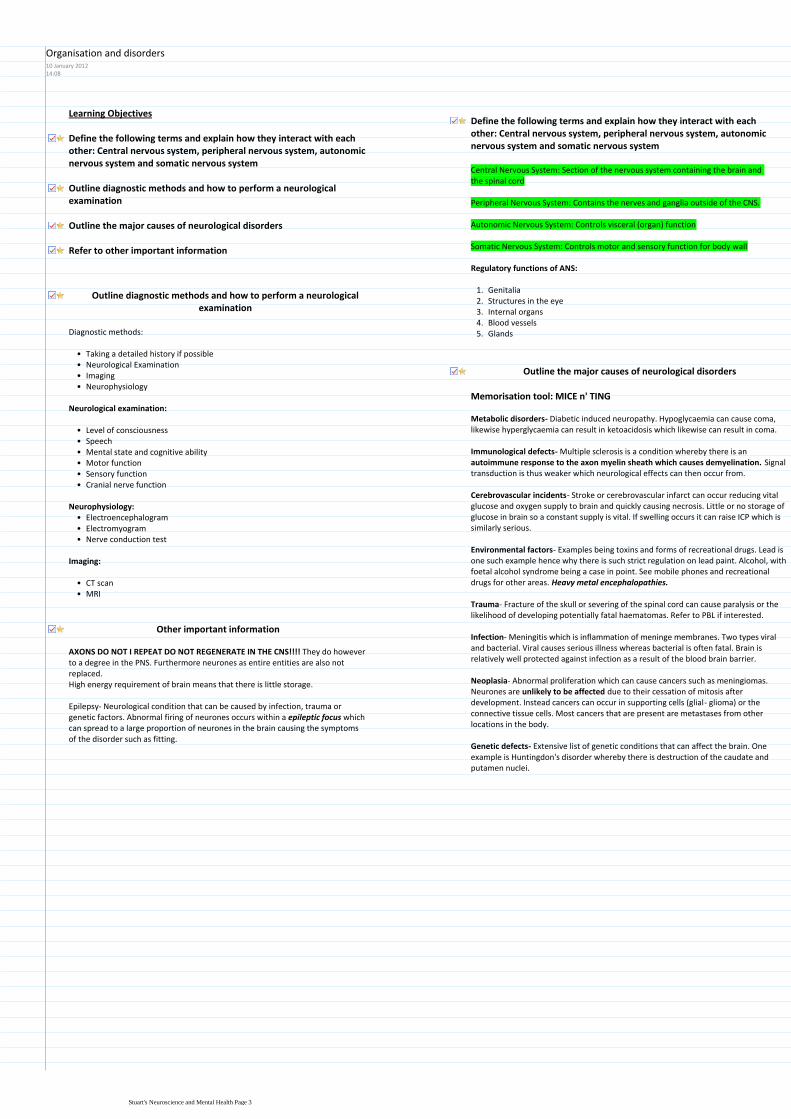
Learning Objectives
Define the following terms and explain how they interact with each other Central nervous system peripheral nervous system autonomic nervous system and somatic nervous system
Outline diagnostic methods and how to perform a neurological examination
Outline the major causes of neurological disorders
Refer to other important information
Define the following terms and explain how they interact with each other Central nervous system peripheral nervous system autonomic nervous system and somatic nervous system
Central Nervous System Section of the nervous system containing the brain and the spinal cord
Peripheral Nervous System Contains the nerves and ganglia outside of the CNS
Autonomic Nervous System Controls visceral (organ) function
Somatic Nervous System Controls motor and sensory function for body wall
Regulatory functions of ANS
Genitalia1Structures in the eye2Internal organs3Blood vessels4Glands5
Outline diagnostic methods and how to perform a neurological examination
Diagnostic methods
Taking a detailed history if possiblebullNeurological ExaminationbullImaging bullNeurophysiologybull
Neurological examination
Level of consciousnessbullSpeechbullMental state and cognitive abilitybullMotor functionbullSensory functionbullCranial nerve functionbull
ElectroencephalogrambullElectromyogrambullNerve conduction testbull
Neurophysiology
Imaging
CT scanbullMRIbull
Outline the major causes of neurological disorders
Memorisation tool MICE n TING
Metabolic disorders- Diabetic induced neuropathy Hypoglycaemia can cause coma likewise hyperglycaemia can result in ketoacidosis which likewise can result in coma
Immunological defects- Multiple sclerosis is a condition whereby there is an autoimmune response to the axon myelin sheath which causes demyelination Signal transduction is thus weaker which neurological effects can then occur from
Cerebrovascular incidents- Stroke or cerebrovascular infarct can occur reducing vital glucose and oxygen supply to brain and quickly causing necrosis Little or no storage of glucose in brain so a constant supply is vital If swelling occurs it can raise ICP which is similarly serious
Environmental factors- Examples being toxins and forms of recreational drugs Lead is one such example hence why there is such strict regulation on lead paint Alcohol with foetal alcohol syndrome being a case in point See mobile phones and recreational drugs for other areas Heavy metal encephalopathies
Trauma- Fracture of the skull or severing of the spinal cord can cause paralysis or the likelihood of developing potentially fatal haematomas Refer to PBL if interested
Infection- Meningitis which is inflammation of meninge membranes Two types viral and bacterial Viral causes serious illness whereas bacterial is often fatal Brain is relatively well protected against infection as a result of the blood brain barrier
Neoplasia- Abnormal proliferation which can cause cancers such as meningiomas Neurones are unlikely to be affected due to their cessation of mitosis after development Instead cancers can occur in supporting cells (glial- glioma) or the connective tissue cells Most cancers that are present are metastases from other locations in the body
Genetic defects- Extensive list of genetic conditions that can affect the brain One example is Huntingdons disorder whereby there is destruction of the caudate and putamen nuclei
Other important information
AXONS DO NOT I REPEAT DO NOT REGENERATE IN THE CNS They do however to a degree in the PNS Furthermore neurones as entire entities are also not replacedHigh energy requirement of brain means that there is little storage
Epilepsy- Neurological condition that can be caused by infection trauma or genetic factors Abnormal firing of neurones occurs within a epileptic focus which can spread to a large proportion of neurones in the brain causing the symptoms of the disorder such as fitting
Organisation and disorders10 January 20121408
Stuarts Neuroscience and Mental Health Page 3
Learning Objectives
Draw and label of a typical neuron identifying soma dendrites axon and terminals
Define the role of each cellular component in the specialised function of the neuron
Outline the organisation and functions of intracellular transport in the neuron
Define the functional subtypes of neurons and list the ways in which they are organised collectively in the nervous system
Describe the organisation of synapses
Name the main classes of neuroglia and explain their functions in the nervous system
Draw and label of a typical neuron identifying soma dendrites axon and terminals
Basic Information of the Neuron
Basic unit of the nervous system Each one can be thought of as a processing unit Responsible for the generation and transmission of electrical impulses Synapses are junctions between neurones Supported by glial cells of which there are many types Glial cells outnumber neurons 91
Nucleus
Schwann Cell
Axon TerminalsDendrites
Soma
Nodes of Ranvier
Myelin SheathDefine the role of each cellular component in the specialised function of the neuron
Soma is the metabolic centre of activity for the whole neuron
Large nucleusbullProminent NucleolusbullAbundant Rough ERbullWell developed Golgi- Required for the large amount of neurosecretionsbullAbundant mitochondriabullOrganised cytoskeletonbullMost metabolically active cell in bodybull
A highly branched structure originating from the cell body bullHas a large surface area which is improved further by the presence of protrusions called dendritic spines These structures receive the majority of synapses
bull
Large pyramidal neurons may have as many as 30-40000 spinesbull
Dendrites receives all the inputs into the cell
Axons the cellular component of the neuron concerned with the output mechanism
Impulses are generated at the axon hillock and progress away from the cell body Initiator of impulse- Axon hillock
bull
Usually only one per cell In picture above axon would be at the base of the pyramidal cell (cannot see due to focusing)
bull
Branching can occur after leaving the cell body or upon reaching the targetbullProminent microtubules and neurofilaments that contributes to cable like transporting system of the axon
bull
Can be myelinated or unmyelinated if so nodes of Ranvier are unmyelinated junctions where the action potential is propagated from
bull
As stated axons branch extensively close to a target- process known as terminal arbor
bull
Axon terminals form synapses with their targetsbullCan end with varicosities or boutons (think synaptic knob)bull
Axon terminals-
Highly organised cytoskeleton is required with microfilaments intermediate filaments and microtubules which are particularly abundant
bull
Neurofilaments play a critical role in determining axon calibrebull
Neuronal cytoskeleton-
Outline the organisation and functions of intracellular transport in the neuron
Transport of membrane associated materialsbullVesicles with associated motors are moved down the axon at 100-400mm per daybullDifferent membrane structures targeted to different componentsbullRetrograde moving organelles are morphologically and biochemically very different from anterograde vesicle
bull
Fast axonal transport
Anterograde transport- Movement of molecules outwards from the soma towards the synapse or plasma membrane
Fast- Transports synaptic vesicles transmitters and mitochondriabullSlow - Anterograde transport results in the delivery of cytoskeletal and cytoplasmic constituents
bull
Describe the organisation of synapses
Prevalent Golgi apparatus packages neurotransmitter into vesicles which is the transported by fast anterograde transport
bull
Mechanisms are in place for the successful completion of exocytosis and in particular the fusion of the vesicles to the plasma membrane
bull
Abundant mitochondria consume about 45 of cells energy expenditure This is required for the pumping of ions and synaptic transmission
bull
Neuronal integration occurs when multiple competing synaptic inputs are integrated in post synaptic neurone
bull
Three main types of synapse
Axo-dendritic- Often excitatory- Also Grays Type 1Axo- somatic- Often inhibitory- Also Grays Type 2Axo-axonic- Often modulatory-
Define the functional subtypes of neurons and list the ways in which they are organised collectively in the nervous system
Cells of the Nervous System20 January 20121315
Stuarts Neuroscience and Mental Health Page 4
Fast- Transports synaptic vesicles transmitters and mitochondriabullSlow - Anterograde transport results in the delivery of cytoskeletal and cytoplasmic constituents
bull
Transports substances from an extracellular spacebullUses microtubule-associated ATPase to drive particles along microtubulesbullTransports soluble cytoplasmic constituentsbullIs the process by which material returns from the terminals to the cell body either for degradation or recycling
bull
Retrograde transport- Movement of molecules inwards from synapse to soma It returns used synaptic vesicles and informs the soma of the conditions at the axon terminal
Define the functional subtypes of neurons and list the ways in which they are organised collectively in the nervous system
Pseudounipolar
Cell has two fused process which are axonal in structure This is a sensory neuronbullExample- Dorsal root ganglion cellsbull
Bipolar
Cells in retina in addition to white matter of cortexbullBIPOLAR THINK OPPOSITES BLACK RETINA ANDbullWHITE CORTEX
Golgi Type I multipolar
Highly branched dendritic treesbullPyramidal cells of cerebral cortexbullAnterior horn cells of the spinal cordbullPurkinje cells of cerebellum (15 million)bullExtremely long extending axonsbullRetinal ganglion cellsbullPAPERbull
Golgi Type II multipolar
Highly branched dendritic treesbullShort axons that terminate near to cell bodiesbullStellate cells of cerebral cortexbull
Define the functional subtypes of neurons and list the ways in which they are organised collectively in the nervous system
Types of neurons
Sensory1Motor2Interneurons (responsible for modification integration coordination facilitation and inhibition of sensory input)
3
Organisational structure of Neurons
Nucleus- Group of unencapsulated neuronal cell bodies within the CNS often of functionally similar cells
Examples include brain stem (Raphe) and deep cerebral cortex (Dentate)bullNNNucleus NNNoo capsule (unencapsulated)bull
Laminae rhymes with greybull
Laminae- Layers of functionally similar cells such as the cerebral cortex grey matter and the cerebellar grey matter
Ganglion- Group of encapsulated cell bodies within the PNS Examples include dorsal root ganglia and sympathetic ganglia
Fibre Tract- Groups of bundles of axons in the CNS that may or may not be myelinated Examples included corpus callosum and internal capsule
Bring information to the CNS from sensory receptors and bring axons to effector organs
bull
Often a combination of both motor and sensory neuronsbullExceptions to PNS are optic and olfactory nervesbull
Nerve- Discrete bundles of axons that are usually part of PNS
Name the main classes of neuroglia and explain their functions in the nervous system
These are the support cells of the nervous systembullEssential for correct functioning of neurones and have many and varied functionsbull
Astroglia
Oligodendroglia
Microglia
Immature progenitors
Ependymal cells- Epithelial lining of the ventricles
Schwann cells
Satellite glia
Examples includebull
Neuroglia
Star shaped cell (think astrology)bullMost abundant cell in CNSbullEach cell forms a specific territory that interfaces with microvasculaturebull
Fibrous astroglia (White Matter)i)Protoplasmic astroglia (Grey Matter)ii)Radial astrogliaiii)
Several different typesbull
Numerous IF bundles in cytoplasm of fibrous astrogliabullGap junctions suggest astroglia-astroglia signallingbullHas intimate associations with other cell typesbullInteracts with blood vessels in an ordered arrangement with little overlapbull
Astroglia
Scaffold for neuronal migration and axon growth during development1Formation of blood brain barrier2Transport of substances from blood to neurons3Segregation of neuronal processes (synapses)4Removal of neurotransmitters5Synthesis of neurotropic factors6Neuronal-glial and glial neuronal signalling7Potassium ion buffering8Glial scar formation9
SSS FTR NPGbull
Functions of astroglia
Myelin
Myelin is an insulating membrane found around nerve cells that is made up of lipid
bull
Up to 50 lamellae- thin plate like structurebullDark and light bands seen at EM levelbullHighly susceptible to damage and loss of either oligodendroglia or myelin is disastrous
bull
- AdrenoleucodystrophyMyelin disease states- Multiple Sclerosisbull
Stuarts Neuroscience and Mental Health Page 5
Potassium ion buffering8Glial scar formation9
SSS FTR NPGbull
Myelin producing cells of the CNSbullSmall spherical nucleibullFew thin processesbullProminent ER and GolgibullMetabolically highly activebull
Interfasicular oligodendroglia
Perineuronal oligodendroglia
Types includebull
Oligodendroglia
Functions of oligodendroglia
Production and maintenance of myelin sheath1Each cell can produce numerous sheaths (1-40)2
Derived from bone marrow during early developmentbullResident macrophage population of CNSbullInvolved in immune surveillancebullTypical macrophage functions- ie APCbullRole in tissue modellingbullSynaptic strippingbull
Microglia
These are the myelin producing cells of the PNSbullOnly produce one sheath per cellbullPromote axon regenerationbullSurround unmyelinated axonsbull
Peripheral Glial- Schwann Cells
Stuarts Neuroscience and Mental Health Page 6
Learning Objectives
Diffusion of an iono
Permeability of a cell membraneo
Electrochemical gradient of an iono
Define the following
Describe how a resting membrane potential can arise from a difference in concentration of an ion across a selectively permeable membrane (use diagrams)
Define electrochemical equilibrium for an ion
What is the equilibrium potential for an ion
The Nernst equation is Ex+ = (RTZF) ln (CoCi) You should know that Ex+ is the equilibrium potential of ion X+ R is the gas constant T is absolute temperature Z is the charge on the ion and F is Faradayrsquos number coulombs of charge per mol of ion
Substituting the values of the constants and T= 37oC gives 27
What are typical values for the concentration of K+ and for Na+ inside and outside a normal neuron
What is a typical value for the resting potential of a neuron
K+ concentration has a much stronger effect on the resting potential than Na+ concentration does Explain the basis of this difference
Diffusion of an ion- The passive movement of an ion down its concentration
gradient
Permeability of a cell membrane- The selectivity of a membrane to ions is its
permeability
Electrochemical gradient of an ion- The movement of ions from an area of
high concentration and relative charge to an area of low concentration and opposite charge
Define the following
Describe how a resting membrane potential can arise from a difference in concentration of an ion across a selectively permeable membrane (use diagrams)
Zero-reference is outside of the cellbullThe inside of the cell is negative compared to the outsidebullAll cells have a membrane potentialbullResting potential is -70mvbull
Concentration of at least one permanent ion is different on one side of the membranebull
Permeable pores that open and close that are selective for different types of ion eg (K+ Na+ Cl- Ca2+)
bull
Open by change in membrane potential
Voltage dependent-1)
Open all the time- these are the ones that produce the resting potential
Voltage independent2)
2 major typesbull
Permeability of membrane to potassium allows it to leave generating a negative charge within the cell- Resting potential Equilibrium of electrochemical gradient is produced resulting in small flux between compartments
bull
Permeability of membrane to sodium allows it to enter generating a negative charge outside and a positive charge inside- Action Potential
bull
Ion Channels
Define electrochemical equilibrium for an ion
Electrochemical equilibrium- Reached when the concentration gradient is balanced by the electrical gradient across the membrane
What is the equilibrium potential for an ion
Equilibrium potential- Potential that prevents diffusion down the ions concentration gradient
The Nernst equation is Ex+ = (RTZF) ln (CoCi) You should know that Ex+ is the equilibrium potential of ion X+ R is the gas constant T is absolute temperature Z is the charge on the ion and F is Faradayrsquos number coulombs of charge per mol of ion
Nernst equation
bull EX = (RTZF) ln (Co Ci)
ndash C is concentration of the ion [X+]
bull o = outside cell
bull i = inside cell
ndash Co = [X+] outside cell
ndash Ci = [X+] inside cell
bull R = gas constant
bull T = Temp o Kelvin
bull Z = charge on ion
bull -1 for Cl- +2 for Ca2+
bull F = Faradayrsquos number
bull charge per mol of ion
bull ln means log to base e
Ex- is the equilibrium potential of the ion X
Substituting the values of the constants and T= 37oC gives 27
EX = (27Z) ln (Co Ci)
Co
(mM)
Ci
(mM)
ENa
(mV)
Na+ 150 10 +73
Co
(mM)
Ci
(mM)
ENa
(mV)
K+ 5 150 -92
bull What sign for the equilibrium potential
bull The sign the inside needs to maintain the concentration
gradient
bull RTF 37 oC = 27
bull EK = (27 +1) ln(0005 M 015 M)
bull EK = +27 ln(003)
bull EK = +27 -34
bull EK = -92 mV
bull RTF 37 oC = 27
bull ENa = (27 +1) ln(015 M 001 M)
bull ENa = +27 ln(15)
bull ENa = +27 +27
bull ENa = +73 mV
What are typical values for the concentration of K+ and for Na+ inside and outside a normal neuron
Sodium Potassium
Inside (mM) 10 150
Outside (mM) 150 5
What is a typical value for the resting potential of a neuron
-70mVbull
K+ concentration has a much stronger effect on the resting potential than Na+
concentration does Explain the basis of this difference
Potassium sodium and chlorine concentrations all contribute to the real membrane potential
bull
The size of each ions contribution is proportional to how permeable the membrane is to the ion
bull
Goldman-Hodgkin-Katz voltage equation
This is an equation used to describe the resting membrane potentialbullTHINK- youd need a rest after saying that namebull
0 means the channel is completely close
05 means the channel is open half of the time
1 means the channel is fully open
Within the equation the letter P denotes permeability or channel open probabilitybull
GHK equation worked examples
bull 1) All channels are open all the time (PK PNa PCl = 1)
bull Vm = 27 ln(1[0005] + 1[015] + 1[0005] 1[015] + 1[001] + 1[011])
bull Vm = 27 ln(016 027)
bull Vm = 27 -05
bull Vm = -14 mV
bull 3) Now increase Na+ permeability by 5 (PK = 1 PNa = 005 PCl = 0)
bull Vm = 27 ln(1[0005] + 005[015] + 0[0005] 1[015] + 005[001] + 0[011])
bull Vm = 27 ln(00125 01505)
bull Vm = 27 -249
bull Vm = -67 mV
bull Increasing permeability for Na+ makes the cell more positive
RTF at 37 oC = 27
Concentration [ ] is in Moles
bull 2) Only K+ channels are open and Cl- and Na+ channels are closed (PK = 1 PNa = 0 PCl = 0)
bull Vm = 27 ln(1[0005] + 0[015] + 0[0005] 1[015] + 0[001] + 0[011])
bull Vm = 27 ln(0005 015)
bull Vm = 27 -34
bull Vm = -92 mV
bull Ion concentration has not changed BUT Vm moved to EK
Graded Potentials
Resting Potential14 May 20121248
Stuarts Neuroscience and Mental Health Page 7
EX = (27Z) ln (Co Ci)
Co
(mM)
Ci
(mM)
ENa
(mV)
Na+ 150 10 +73
Co
(mM)
Ci
(mM)
ENa
(mV)
K+ 5 150 -92
bull What sign for the equilibrium potential
bull The sign the inside needs to maintain the concentration
gradient
bull RTF 37 oC = 27
bull EK = (27 +1) ln(0005 M 015 M)
bull EK = +27 ln(003)
bull EK = +27 -34
bull EK = -92 mV
bull RTF 37 oC = 27
bull ENa = (27 +1) ln(015 M 001 M)
bull ENa = +27 ln(15)
bull ENa = +27 +27
bull ENa = +73 mV GHK equation worked examples
bull 1) All channels are open all the time (PK PNa PCl = 1)
bull Vm = 27 ln(1[0005] + 1[015] + 1[0005] 1[015] + 1[001] + 1[011])
bull Vm = 27 ln(016 027)
bull Vm = 27 -05
bull Vm = -14 mV
bull 3) Now increase Na+ permeability by 5 (PK = 1 PNa = 005 PCl = 0)
bull Vm = 27 ln(1[0005] + 005[015] + 0[0005] 1[015] + 005[001] + 0[011])
bull Vm = 27 ln(00125 01505)
bull Vm = 27 -249
bull Vm = -67 mV
bull Increasing permeability for Na+ makes the cell more positive
RTF at 37 oC = 27
Concentration [ ] is in Moles
bull 2) Only K+ channels are open and Cl- and Na+ channels are closed (PK = 1 PNa = 0 PCl = 0)
bull Vm = 27 ln(1[0005] + 0[015] + 0[0005] 1[015] + 0[001] + 0[011])
bull Vm = 27 ln(0005 015)
bull Vm = 27 -34
bull Vm = -92 mV
bull Ion concentration has not changed BUT Vm moved to EK
Through these series of equations we can clearly see that potassium contributes most to the resting potential as increasing the permeability of sodium by only 5 means that the resting potential is now slightly too positive
bull
Concentration is in moles not millimoles bull
Changes in membrane potential
Graded Potentials
Graded potentials occur at synapses and sensory receptor and their function is to contribute to initiating or preventing action potentials
bull
Change in membrane potential caused by alteration in permeability of ion channels Not of a fixed time and length like action potentials
bull
Graded potentials change in membrane potential in response to stimulation bull
Can be depolarizing or hyperpolarizing
A stronger stimulus produces a larger graded potential
Graded potentials get smaller the farther they travel (Decremental spread)
Properties of graded potentialsbull
Stuarts Neuroscience and Mental Health Page 8
Learning Objections
You should be able to explain in general terms what the function of the action potential is
Give some examples of other types of excitable cells (in addition to neurons) in which action potentials occur
ThresholdRefractory periodldquoAll or nothingrdquo behaviourDepolarizationRepolarizationHyperpolarizationSaltatory conduction
Define the following terms as they apply to action potentials
Voltage-gated channelChannel inactivationPositive feedback
Define the following terms as they apply to the membrane channels involved in producing the action potential
Outline the sequence of events during a typical action potential in the neuron Include changes in membrane potential changes in membrane permeability and fluxes of ions across the membrane (a diagram will help)
State the size and duration (including units) of a typical action potential in a neuron
Define the term ldquoregenerativerdquo as applied to action potentials and its significance for spread of the action potential along an axon
Explain how conduction of the action potential occurs (conduction here means spread along the axon alternatively this process may be called transmission or propagation)
List two structural features that affect the conduction velocity along normal axons Briefly explain why they affect velocity as they do
Be able to list at least one pathological condition that affects conduction velocity
You should be able to explain in general terms what the function of the action potential isGive some examples of other types of excitable cells (in addition to neurons) in which action potentials occur
The function of the action potential is to transport messages through the nervous system very rapidly
bull
Action potentials also exist in muscle cellsbull
Define the following terms as they apply to action potentials
Threshold- The level of depolarisation needed to cause the triggering of an
action potential
Refractory period- Unresponsive to threshold depolarization
Absolute refractory period- New action potential cannot be triggered even with very strong stimulus because inactivation gate is closed
Relative refractory period- Inactivation gate is open but sodium channel activation gate is closed meaning a stronger than normal stimulus is required to trigger an action potential
ldquoAll or nothingrdquo behaviour- Once triggered a full sized action potential
occurs
Depolarization- When the membrane becomes more positive towards
sodiums ion equilibrium potential
Repolarization- Efflux of potassium ions to restore membrane to negative
state
Hyperpolarization- Over efflux of potassium results in membrane potential
dropping below resting potential
Saltatory conduction- From the latin (hop or to leap) is the propagation of
action potentials along myelinated axons from one node of Ranvier to the next
Voltage-gated channel- Ion channels that only open when there is a
change in voltage
Channel inactivation- Closing of the inactivation gate in response often
to an extracellular positive charge
Positive feedback- A mechanism that means that action potentials
constantly regenerate
Define the following terms as they apply to the membrane channels involved in producing the action potential
Outline the sequence of events during a typical action potential in the neuron Include changes in membrane potential changes in membrane permeability and fluxes of ions across the membrane (a diagram will help)
At the resting potential voltage-gated ion channels (Na+ and K+) are closed
Note these are different to non-voltage gated channels that generate the resting potential
Resting potential PKgtgtPNa Where P is the permeability of the membrane1
Stimulus- depolarises the membrane potential and causes it to become more positive
2
PNa rapidly increases because the voltage gated Na+ channels open quicker than potassium channels
Potassium permeability increase as the voltage gated K+ channels start to slowly open and as the upstroke progresses more and more voltage gated K channels open
Potassium leaves the cell down the electrochemical gradient but is less than Na entering
Membrane potential moves toward the Na+ equilibrium potential
Depolarisation- Starts at the threshold potential3
Big decrease in PNa because the voltage-gated Na channels inactivation gate is closed and sodium entry stops generating the absolute refractory period
Moderate PK increase as there are more voltage gated K+ channels and they remain open for longer which means that K leaves by its electrochemical gradient
Membrane potential moves toward the K+ equilibrium potential
Repolarisation-4
State the size and duration (including units) of a typical action potential in a neuron
1-2ms for an action potentialbullSize= -70 to + 40 therefore 110mVbull
Define the term ldquoregenerativerdquo as applied to action potentials and its significance for spread of the action potential along an axon
If less than threshold then graded potentialbullOnce threshold is reached the cycle continues in a positive feedback mannerbullCycle continues until the voltage-gated sodium channels inactivate (closed and voltage insensitive)
bull
Membrane remains in a refractory state until the voltage gated sodium channels recover from inactivation
bull
Ions may move in and out but only a very small number of ions actually cross the membrane to change the potential ie the change in concentration is extremely small (less than 01)
bullIon Movements during action potential
Explain how conduction of the action potential occurs (conduction here means spread along the axon alternatively this process may be called
Action Potential14 May 20122245
Stuarts Neuroscience and Mental Health Page 9
periodModerate PK increase as there are more voltage gated K+ channels and they remain open for longer which means that K leaves by its electrochemical gradient
Membrane potential moves toward the K+ equilibrium potential
At rest voltage gated K channels are still open therefore K continues to leave down gradient
Membrane potential moves closer to the K+ equilibrium
Voltage gated K+ channels then close but can still possibly generate an AP if the stimulus is very strong- Relative Refractory period
Membrane potential returns to the resting potential
After hyperpolarization5
Time Course of changes in permeability
small (less than 01)
Explain how conduction of the action potential occurs (conduction here means spread along the axon alternatively this process may be called transmission or propagation)
Active Propagation
Passive propagation
Only resting K+ channels
openInternal (or axial) amp Membrane
Resistance alters propagation
distance and velocity
List two structural features that affect the conduction velocity along normal axons Briefly explain why they affect velocity as they do
Large diameter myelinated axons= 120msbullSmall diameter non myelinated = 1msbullSpeed of sound= 331msbull
Less resistance to current flow inside large diameter axons
Increases with axon diameter1
Action potentials only occur at nodes of Ranvier
Higher in myelinated than non-myelinated axons of the same diameter2
Reduced axon diameter (ie regrowth after injury)
Reduced myelination (MS and diphtheria)
Cold anoxia compression and drugs (some anaesthetics)
Conduction velocity is slowed by
Be able to list at least one pathological condition that affects conduction velocity
Multiple sclerosis and diphtheria causes demyelination of axonsbull
Stuarts Neuroscience and Mental Health Page 10
Learning Objectives
Define the essential components required for neurotransmitter release
Understand the differences between excitatory and inhibitory transmission
Define at least two mechanisms for the termination of neurotransmitter action at the synapse
Describe how modulation of the synaptic properties of GABA can be modulated pharmacologically to treat epilepsy
Features of neurotransmission
Transfer across a synapse requires the release of neurotransmitters and their interaction with postsynaptic receptors
bull
Transmitter released from 1st cell synaptic activation of 2nd cell signal integration and signal conduction by 2nd cell
bull
Rapid timescale1)Adaptability2)Plasticity 3)Diversity4)Learning and memory5)
Features
RAPDL- sounds like rapidbullDendrites receive inputs where it is integrated in the somabull
SynapseGap is about 20-100 nmbull5000 molecules of neurotransmitter per synaptic vesiclebullHigh energy dependency is satiated by high levels of mitochondriabullPost synaptic knob is electron densebull
Features of neurotransmitters
Provide enormous diversity in variety of transmitters and their receptorsbullAmino acids (eg glutamate gamma amino butyric acid [GABA] amines (eg noradrenaline [NA] and dopamine [DA] and neuropeptides (eg opioid peptides)
bull
May mediate rapid (micro to milli second responses) or slower effects (ms)bullVary in abundance from mM to nM CNS tissue concentrationbullNeurones receive multiple transmitter influence which are integrated to produce diverse functional responses
bull
Define the essential components required for neurotransmitter release
Essential componentsAn action potential is necessary to initiate neurotransmitter releasebullIt has to be very fast within ms (200 micro seconds)bullCalcium is essential- transmitter release requires an increase in intracellular Ca2+ to about 200microM
bull
A protein complex formation between vesicle membrane and cytoplasmic proteins to enable both vesicle docking and a rapid response to Ca entry leading to membrane fusion and exocytosis
bull
ATP and vesicle recyclingbull
Extra information on neurotransmittersSynaptic transmission is restricted to specialised structures ie the synapsebullSynaptic vesicles (SVs) provide the source of neurotransmitter (4000-10000 molecules per SV)
bull
Vary from mM to nM in CNS tissue concentrationsbull5-10 mM amino acids in brainbull
Activation of transmitter release is calcium dependent and requires rapid transduction
bull
Series of events= Membrane depolarisation-gt Calcium influx -gt Vesicle fusion -gt Vesicle exocytosis-gt Transmitter release
bull
From calcium influx to transmitter release has to be within this 200 microsecondsbullSuch rapid release rates can be achieved due to the vesicles being primed at the active zone close to Ca entry
bull
Localisation of vesicle at pre synaptic area Also alignment with calcium channel must occur so the response is rapid this is also helped by proteins associating between presynaptic membrane and vesicle to enable docking
bull
Vesicular proteins are targets for neurotoxinsbull
Types of neurotoxins
Botulinum toxin and tetanus destroys membrane association proteins and vesicles1Black widow spider- alpha latrotoxin that interferes with resealing of membrane so neurotransmitter is released to depletion
2
Vesicular proteins aretargets for neurotoxins
TETANUS toxinCtetani causes paralysis
BOTULINUM toxinCbotulinum causes flacid paralysis
alpha LATROTOXINblack widow spiderstimulates transmitterrelease to depletion
Zn2+-dependent endopeptidasesinhibit transmitter release
Steps of synaptic transmission
Biosynthesis packaging and release of neurotransmitter1
Membrane depolarisationaCalcium channels openbCalcium influxcVesicle fusiondVesicle exocytosis- fusion of vesicle to membrane and release of transmittereTransmitter releasef
Release
Receptor action2
Ion channel receptor- Extremely fast (msecs) mediate all fast excitatory and inhibitory transmission
bull
Glutamate can be synthesized from glucose via the TCA cycle
GABA is synthesised from Glutamate by removing a carboxyl group form N terminal Glutamate decarboxylase
CNS- Glutamate GABA
NMJ- Acetylcholine at nicotinic receptors
Examples bull
G- protein coupled receptor- Slow secs mins- Effectors may be enzymes (adenyl cyclase phospholipase C cGMP-PDE) or channels (eg Ca or K) Abundant in CNS
bull
CNS and PNS ACh at muscarinic receptors dopamine noradrenaline 5hydroxytryptamine (5ht) and neuropeptides eg enkaphalin
Examplesbull
Understand the differences between excitatory and inhibitory transmission
Glutamates effects propagated by sodiumbullimport in the post synaptic membraneExcitatory postsynaptic EPSPbull(membrane is becoming depolarised)
Excitatory-
Define at least two mechanisms for the termination of neurotransmitter action at the synapse
Excitatory CNS synapse mediated by Glutamate
G G
G G G
G
Excitatory amino acid
Transporter EAAT
GLIAL
CELL
Na+ Na+ and Ca2+
glucose
TCAcycleaKG
Glutamate
receptor
GLUR
GLUTAMINE
SYNTHETASE
Glutamine
Glutamate
Neurotransmission10 January 20121408
Stuarts Neuroscience and Mental Health Page 11
import in the post synaptic membraneExcitatory postsynaptic EPSPbull(membrane is becoming depolarised)
Excitatory postsynpatic potential (EPSP)
GABA- which results from Chlorine bullentering the post synaptic neuroneInhibitory postsynaptic EPSP (membrane bullis hyperpolarized which makes it veryhard to generate an action potential
Inhibitory-
Glutamate receptors
Alpha amino-3-hydroxy-5methyl-4-isoxazole propionic acid (AMPA)bullMajority of fast excitatory synapses with rapid onset offset and desensitisationbullInitiated by just Na entrybull
AMPA receptors
N-methyl-aspartatebullSlow component of excitatory transmissionbullServe as coincidence detectors which underlie learning mechanismsbullInitiate by Na and Ca entrybull
NMDA receptors
G G
G G G
G
Excitatory amino acid
Transporter EAAT
GLIAL
CELL
Na+ Na+ and Ca2+
glucose
TCAcycleaKG
Glutamate
receptor
GLUR
GLUTAMINE
SYNTHETASE
Glutamine
Glutamate
GLUR receptor takes up Na and Ca in PosSM (Post synaptic Membrane)bullGlutamate is the transported by Excitatory amino acid Transporter (EAAT) to glial cells
bull
Here it is converted to glutamine by glutamine synthasebullHence the glial cell has removed the neurotransmitter link to Cells of the Nervous System
bull
Inhibitory CNS synapse mediated by GABA
GABA GABA
GABA GABA GABA
GABA
GABA transporter GAT
GLIAL
CELL
Cl- Cl-
glucose
TCAcycle
aKG
GABA receptor
GABAAR
Cl-
GLUTAMATE
Glutamic acid
decarboxylase
GAD (B6)
GABA trans
-aminase
GABA-T
Succinate
semialdehyde
GABA
Glutamate is converted to GABA by Glutamic acid decarboxylase (GAD)bullGABA has effects on PSM receptor which causes chloride influxbullGABA is then relocated to Glial cell by GABA Transport (GAT)bullGABA trans aminase GABA-T converted GABA to Succinate semialdehydebull
Describe how modulation of the synaptic properties of GABA can be modulated pharmacologically to treat epilepsy
In epilepsy there is an excess of glutamate which causes abnormal neuronal firingbullThe concept of GABA drugs is that they will be used to inhibit the effects of glutamatebull
Frequency of opening of channel is increased when binding to benzodiazepinesbullBarbiturates- Open channel for longerbull
alpha
szlig
GABA
barbiturates
steroids
benzodiazepines
Zn
convulsantsethanol
Pentameric organisation of the GABA receptorand pharmacologically important binding domains
Drugs facilitating GABA transmission areantiepilepticanxiolyticsedativemuscle relaxant
Qu 1 Which of the following is NOT a normal route for neurotransmitter inactivation
Addition of a methyl group in the synaptic cleft
X Diffusion out of the cleft
Enzymatic destruction in the synaptic cleft-
Re-uptake by the pre-synaptic terminal- NE
Uptake by glial cells- GABA glutamate
X Mark = -2 (conf=2 )
Best Option Addition of a methyl group in the synaptic cleftSuch a method of inactivation does not existLecture Neurotransmitters
Pasted from lthttpswwwuclacuklaptlaptlitesysrunhtmicl08_neurof=cleari=icl1k=1u=_st1511i=Imperialgt
Stuarts Neuroscience and Mental Health Page 12
Session 4 Tutorial ndash Neurotransmitters and epilepsy
Tutorial notes
1 Epilepsy terminology
The term epilepsy refers to a disorder of brain function characterized by the periodic and unpredictable occurrence of seizures
bull
The term seizure refers to a transient alteration of behaviour due to the disordered synchronous and rhythmic firing of populations of brain neurones
bull
The pharmacological agents in current clinical use for inhibition of seizures are referred to as anticonvulsant or antiepileptic drugs
bull
Seizures are thought to arise from the cerebral cortex and they can be classified into bull
Partial seizures those beginning focally at a cortical site
Generalized seizures those that involve both hemispheres widely from the outset
The behavioural manifestations of a seizure are determined by the functions normally served by the cortical site at which the seizure arises
bull
LEFT HEMISPHERE WOULD CAUSE RIGHT SIDED JERKING
Thus for example a seizure involving the motor cortex is associated with clonic jerking of the body part controlled by this region of the cortex
bull
A simple partial seizure is associated with preservation of consciousness whilst acomplex partial seizure is associated with impairment of consciousness
bull
Examples of generalized seizures include absence myoclonic and tonic-clonic seizures You will be shown examples of the principal seizure types by video presentation during this teaching session
bull
2 Neurotransmitters in epilepsy
Epilepsy is a neurological disorder associated with abnormal neurotransmitter functionin the brain
bull
A decrease in GABA-mediated inhibition or an increase in glutamate-mediated excitation in the brain may result in seizure activity Indeed both glutamate and GABA are thought to play key roles in the brain mechanisms causing epilepsy in man
bull
Pharmacological evidence for a role of neurotransmitters in epilepsy
Dr Martin Croucher
Impairment of GABA-mediated inhibition causes seizures in animals eg impairment of synthesis release (tetanus toxin) or postsynaptic action (bicuculline picrotoxin)
bull
Enhancement of GABA-mediated inhibition leads to seizure suppression eg central (icv) administration of GABA or inhibition of the GABA metabolizing enzyme GABA-T (vigabatrin)
bull
Many clinically useful anticonvulsant drugs are known to act at least in part by potentiating central GABA-mediated inhibition eg benzodiazepines phenobarbital (see Section 3)
bull
Central (icv focal) administration of glutamate or glutamate receptor agonists causes seizure-like activity in animals
bull
Glutamate receptor antagonists are anticonvulsant in experimental models of epilepsy
bull
Some therapeutically effective anticonvulsant drugs act partly by blocking glutamate-mediated excitation in the brain eg phenobarbital
bull
Biochemical evidence for a role of neurotransmitters in epilepsy
Cobalt-induced seizures in rodents are associated with increased glutamate release
and with decreased GABA concentration GAD activity and GABA uptake (probably reflecting GABA neurone loss) at the seizure focus
bull
Audiogenic seizures in mice (DBA2 mice) are associated with glutamate receptor
binding in the brain and with GABA release from depolarized brain slices
bull
The baboon Papio papio which is highly sensitive to photically-induced seizures has a lower than normal CSF GABA concentration
bull
3 Some examples of antiepileptic drugs
Notes
Many neurological conditions are from malfunction in brainbullGABA malfunctions in epilepsy with propensity to repeated seizuresbull~1 in 100200bull
50 have well controlled epilepsy
25 have good control but side effects
25 have complex partial epilepsy which is poorly controlled
In epilepsy 502525 is an important ratiobull
Depression is associated with excessive noradrenalinebull
5 of people can have a seizure and 1 have epilepsybull
Epileptic focus- Excessively synchronous or sustained discharge of a group of neurones in the brain
Intracerebroventricular into lateral ventricles
ValiumLibrium are benzodiazepines
Whole list Important for exams possibly
Epilepsy can be causes by infection head trauma tumoursMeningitis-encephalitis Gliomas Stroke- ischaemic damage May occur in ~10 strokesGABA-interneurones small in CNS
Place in skull of rats and causes epileptic focus
Loud sounds causes seizures
Strobe of certain light frequency gives epilepsy Assumed that CSF is typical of brain
Generalized Seizures
Tonic- Clonic- Grand mal Tonic extension is continuous extensions which may cause the person to fall over Clonic rhythmical jerking 23 minutes
Absence- Petit mal Bit of eye fluttering 1015 secs phase out Occur very often
Myoclonic- Brief involuntary twitching of a muscle
Atonic- Lose muscle tone and posture fall forward onto face Epic face plant
Partial seizures-
Focal seizures can attribute to one direct point
Simple- Right motor cortex- left side activity Twitch tremor and face Can develop into secondary generalised seizure
Complex-Impairment of consciousness Lip smacking Temporal lobe epilepsy
Secondary generalised- Partial expands to general seizures
Parkinsons disease is caused by a lack of dopaminebullAkenisa- awkward gait Pill rolling tremorbullAlzheimers is caused by a lack of acetylcholinebull
4 Questions to be addressed
Review the process of neurotransmission occurring at central synapses utilising the inhibitory neurotransmitter GABA
bull
Present a convincing case for a role of neurotransmitter (glutamate or GABA) malfunction in the aetiology of epilepsy
bull
Draw a diagram to illustrate the principal steps in GABAergic neurotransmission (synthesis storage release etc) Indicate on your diagram i) established and ii) potential new target
bull
Epilepsy20 January 20120954
Stuarts Neuroscience and Mental Health Page 13
________________________________________________________________
Potential for drugs to solely target specific GABAA receptors to ensure only anti-convulsant and stop side effect of drowsiness
bull
Long acting- suicide anti convulsant
GABASynapse
Present a convincing case for a role of neurotransmitter (glutamate or GABA) malfunction in the aetiology of epilepsy
bull
Draw a diagram to illustrate the principal steps in GABAergic neurotransmission (synthesis storage release etc) Indicate on your diagram i) established and ii) potential new target sites for drugs acting to enhance GABA-mediated neurotransmission in the brain
bull
Stuarts Neuroscience and Mental Health Page 14
Learning Objectives
Draw a diagram to explain the relationship between the following major divisions of the CNS spinal cord brainstem cerebellum diencephalon cerebral hemispheres
Define the 3 components of the brainstem and state the main functions of the brainstem
Describe the functions of the 2 main structures in the diencephalon
State the functions of the basal ganglia and the cerebellum
Draw on a diagram of the cerebral hemisphere the cortical lobes and primary cortical areas
Recognise the main structures of the brain in a diagram or MRI
Describe the 3 layers of the meninges and explain their role in protecting the brain
Explain how the major divisions of the brain relate to the cranial fossae in the base of the skull
Explain the relationship between the spinal segments spinal nerves and vertebrae and state at what level a lumbar puncture can be performed safely
Define the functions of the dorsal and ventral horns of the spinal cord and explain how the dorsal and ventral roots and spinal nerves relate to them
Identify the components of the ventricular system and relate them to the divisions of the CNS
Explain the composition circulation and functions of CSF
State the average total volume and flow rate of CSF
Define hydrocephalus and outline how it may be treated
Distinguish between an epidural (extradural) and subdural haemorrhage
Draw a diagram to explain the relationship between the following major divisions of the CNS spinal cord brainstem cerebellum diencephalon cerebral hemispheres
pons
midbrain
medulla spinal cord
cerebellum
cerebral
hemisphere
diencephalon
brainstem
Define the 3 components of the brainstem and state the main functions of the brainstem
Midbrain1Pons-usually very tubular and has a bulge on the anterior surface2Medulla- With pons and cerebellum lies in hindbrain3
Midbrain pons and medulla share a lot of their functions These include controlling vital functions such as breathing consciousness and also innervating cranial nerve function
bull
Cranial Nerves
All are part of the PNS apart from the optic nervebullFunctional components less regularly organised than for spinal nervesbullSupply motor and sensory innervation to the headbullAutonomic (parasympathetic) innervation to head thoracic and abdominal organsbullSpecial senses eg vision hearing balancebull
Cerebral Hemispheres
Cortex of grey matter superficially that is responsible for most brain functions
1
Basal ganglia deep within brain that are involved with movement2
The cerebral hemisphere can be divided into two main partsbull
There is also the Corpus callosum which is a tract of fibres that connect the two hemispheres
bull
Central sulcus separates off the frontal lobe from the parietal lobebullSulcus are grooves and gyri are foldsbull
Cerebellum
Co-ordinates movement in terms of timing accuracy and precision (TAP) It may also have some involvement in cognitive function and in the fear and pleasure response
bull
TAP that cerebellumbullThe hindbrain contains the pons medulla and cerebellum
State the functions of the basal ganglia and the cerebellum
Draw on a diagram of the cerebral hemisphere the cortical lobes and primary cortical areas
Primary motor cortex is a strip Imagine that there is a man lying upside in the strip so damage to bottom of strip would affect his head or upper body
bull
Primary visual cortex- This area receives information from the retinas if there is a malfunction here it is likely you would become blind
bull
Auditory cortex receives information from inner earbullOther associated cortex areas seem to be related to higher functionbullWernickes area is involved with understanding language apparently according to recent literature the area should be moved and will then have more in common with experimental data and the area in primates
bull
Brocas area- Making speech case study evolved from a patient who had neurosyphillis destroying just that particular area and couldnt talk This area is more prominent in the left hemisphere
bullIdentify the components of the ventricular system and relate them to the divisions of the CNS
lateral ventricle
aqueduct
third ventricle
fourth ventricle
central canal
Describe the functions of the 2 main structures in the diencephalon
The diencephalon is a structure above the brain stembullIt is composed of the thalamus which relays information from lower structures to the cerebral cortex and the hypothalamus which is essential in coordinating homeostatic mechanisms and is the interface between CNS ANS and endocrine system
bull
Central sulcus
Lateral sulcus
CNS Lectures + Practicals31 January 20120900
Stuarts Neuroscience and Mental Health Page 15
lateral ventricle
aqueduct
third ventricle
fourth ventricle
central canal
Region of brain Region of ventricular system
Cerebral hemisphere Lateral ventricle
Diencephalon 3rd Ventricle
Midbrain Aqueduct
Pons and Medulla Fourth Ventricle
Lateral ventricle is C-shaped and there is one present in each hemispherebullFourth ventricle lies in front of the cerebellum and behind the pons and the medullabullHalfway down medulla it becomes the central canal and goes down into spinal cordbullEpendymal cells line the ventricular systembullChoroid plexus secretes CSFbull
Explain the composition circulation and functions of CSFState the average total volume and flow rate of CSF
The CSF is secreted by choroid plexus a structure within the lateral 3rd and 4th ventricles of the brain
bull
Most of the CSF leaves the 4th ventricle via holes and circulates via outside of the brain between the meninge membranes
bull
Lower number of cellsi)Decreased amount of proteinii)Lower Potassium and sodiumiii)Increased Magnesium and chlorine concentrationiv)
Differences between CSF and blood includebull
Circulates through ventricular system and subarachnoid space formed by the meninge membranes
bull
The CSF is reabsorbed into the venous sinuses via arachnoid villi which stops the accumulation of fluid possible oedema and a raised ICP
bull
Functions of CSF
Helps protective the brain against damage by suspending it in liquid 1Also has a few metabolic functions and waste products and helps distribute certain hormones
2
500ml per day is the flow rateTotal volume at any one time is 150ml
Describe the 3 layers of the meninges and explain their role in protecting the brain
3 layers of the meninges
Dura mater- Tough outer layer1Arachnoid mater - Delicate middle layer2Pia mater- Layer firmly attached to the surface of the brain3
Arachnoid villus drains CSF fluid into the venous sinusbull
Meningitis
Meningitis is most likely to infect Pia Mater and subarachnoid space but in the worst cases it can also spread to the upper layers of the cortex
bull
In order to determine between viral and bacterial and meningitis one can look at the CSFbull
BacterialThere should be a high white cell count in particular neutrophilsbullProtein concentration would be increased whereas there would be a decrease in glucose concentration- dangerous for brain metabolism
bull
Bacteria may actually be themselves identifiablebull
ViralLymphocytes account for the predominant change in leukocytes countbullProtein and glucose are normalbullViral identification is unlikelybull
Recognise the main structures of the brain in a diagram or MRI
Brain cut in mid-sagittal plane
Explain how the major divisions of the brain relate to the cranial fossae in the base of the skull
Inside of base of skull
Explain the relationship between the spinal segments spinal
Stuarts Neuroscience and Mental Health Page 16
Protein and glucose are normalbullViral identification is unlikelybull
Inferior aspect of brain
Cranial Fossae Part of brain
Anterior Cranial Frontal Lobe
Middle Cranial Temporal Lobe
Posterior Cranial Cerebellum
Lies above sphenoid bone Hypothalamus
Passes through the Foramen Magnum Medulla
Explain the relationship between the spinal segments spinal nerves and vertebrae and state at what level a lumbar puncture can be performed safely
Terminal end of spinal cord is called Conus MedullarisbullThere are 5 spinal segments cervical thoracic lumbar sacral and coccyx 7 12 5 5 4
bull
Spinal nerves are associated with each specific segment ie cervical etc but they do not necessarily have the same number of nerves to vertebrae and they terminate in different areas
bull
Type of vertebrae
Number of Vertebrae
Number of spinal nerves
Relationship of nerve to vertebrae
Cervical 7 8 Above
Thoracic 12 12 Below
Lumbar 5 5 Below
Sacrum 5 5 Below
Coccyx 4 (1 fused) 1 Below
The safe area to perform a lumber puncture is between L3 and L4- this is because the spinal cord has finished before this are and the nerves can be pushed aside by the needle without damage
bull
Spinal cord is much shorter than the vertebral column because the spinal cord develops mostly in the embryo whereas bones take much longer to develop
bull
Define the functions of the dorsal and ventral horns of the spinal cord and explain how the dorsal and ventral roots and spinal nerves relate to them
VRRMM VRRMM- sound like an engine VM= Ventral + Motor neurone cell bodies The more ridiculous the better right
Ventral Horn- Contains motor neurone cell bodiesbull
DI- Dorsal + interneurones Investigate what the bloody hell goes on in the spine (Detective Inspector- DI)
Dorsal Horn- Contains interneurones which receive sensory information from the body
bull
Afferent pathway is receives of sensory information via dorsal horn efferent pathway is motor information leaving the spinal cord Spinal nerve is collection on both and thus has bidirectional information transfer
bull
Grey matter contains the neuronal cell bodies and it is the white matter that contains the bundles of axons
bull
Define hydrocephalus and outline how it may be treated
Hydrocephalus- Water on the brain caused by CSF flow abnormalities
Communicating- All 4 ventricles affecteda)Non- communicating- Not all 4 enlargedb)
2 predominant types
Block in CSF absorption or CSF over brain surface caused bybullMeningitisbullHead injurybullCongenitalbull
MHCH
Haemorrhage (sub-arachnoid)bull
Communicating-
Block in ventricular system caused bybullAqueduct stenosisbullVentricular tumoursbull
AVPParaventricular tumoursbull
Non- Communicating-
Distinguish between an epidural (extradural) and subdural haemorrhage
EMA- Extradural= meningeal artery
Extradural haemorrhage is usually due to a damaged meningeal artery between the skull and the dura after head trauma
bull
Subdural haemorrhage- Usually due to a damaged vein between the dura and arachnoid membrane
bull
Both of these pathologies can cause a space-occupying lesion in the confined space bounded by the membranes and hence cause neurological pathologies
bull
Symptoms such as headache drowsiness vomiting or seizure are more likely to present quickly from an arterial (extradural) than a subdural haemorrhage
bull
Can also be confirmed by imagingbull
Subdural are more common than epiduralbullSubdural haemorrhages are more frequently seen in alcoholics the elderly patients on anticoagulants whereas epiduralextradural haemorrhages more commonly arise from blows to the side of the head
Pasted from lthttpswwwuclacuklaptlaptlitesysrunhtmicl08_neurof=cleari=icl1k=1u=_st1511i=Imperialgt
Stuarts Neuroscience and Mental Health Page 17
AVP
HeadachebullDrowsinessbullBlackoutsbullRaised intracranial pressurebullIncreased head circumference (in child)bull
Symptoms
Remove causes eg papillomabullPut in a shunt to divert CSFbullOpen alternate pathway- ventriculostomybull
Treatment
Stuarts Neuroscience and Mental Health Page 18
Brachial plexus forms at level C5-T1bull
Macrophage clears up debris along proximal end of axoniAxon sends at pseudopodia (projections of membrane) to make contact with Schwann cell which it grows through and coats it in myelin
ii
Axon reaches distal end of axon as a complete entityiiiNodes of Ranvier are reinserted however there are usually more than there was before which slows nerve conduction down slightly
iv
Axon sustains damage eg in example by forceps crushing it
Growth of axon is about 2-5mm per day
Soma undergoes chromatolysis
Failure to re-join proximal to distal end can result in Neuroma
Nerve degenerationbull
Qu 2 Which of the following statements about peripheral nerves is INCORRECT
Axons form bundles called fascicles
They may contain axons from difference spinal nerves
radic They contain only myelinated nerve fibres
They contain a mixture of fibres of different diameter
They contain a mixture of fibres of different conduction velocities
Pasted from lthttpswwwuclacuklaptlaptlitesysrunhtmicl08_neurof=cleari=icl1k=1u=_st1511i=Imperialgt
Definitions
Dorsal ramus- Posterior branches of the spinal nerves that are smaller than anteriorventral divisions Carry visceral motor somatic motor and sensory information to and from the skin and deep muscles of the backVentral ramus- Supply the antero-lateral parts of the trunk and limbs they are for the most part larger than the posterior divisionsDorsal root ganglion- Nodule on dorsal root that contains cell bodies of neurons in afferent spinal nervesDorsal root- Afferent sensory root of a spinal nerveVentral root- Efferent motor root of a spinal nerveGray ramus communicans- Contain unmyelinated postganglionic sympathetic fibres that are from ganglion of sympathetic trunkWhite ramus communicans- Preganglionic sympathetic outflow from the spinal cord
Additional info on white ramus communicans
The thoracic and the first and second lumbar nerves each contribute a branch white ramus communicans to the adjoining sympathetic ganglion Unlike the gray rami white rami communicants do not extend below L2 or above T1
bull
PNS18 May 20122302
Stuarts Neuroscience and Mental Health Page 19
Learning Objectives
Describe the basic anatomy of ANS innervation in terms of pre-ganglionic neurons ganglia and post-ganglionic fibres Contrast the anatomical location of sympathetic and parasympathetic ganglia
Describe the sympathetic and parasympathetic pathways and their centralspinal connections
Describe the thoracolumbar (sympathetic) and craniosacral (parasympathetic) central origins of the ANS
Describe the influence of the sympathetic and parasympathetic nervous system on the principal systemsorgans of the body (eg cardiovascular system lung gut exocrine glands) and understand the concept of dual innervation and autonomic tone (giving examples)
Understand the importance of the sacral parasympathetic outflow for innervation of structures within the pelvis eg the bladder
Classify the cholinoceptors found within the autonomic nervous system and identify the principal loci of (a) the nicotinic cholinoceptors and (b) the muscarinic cholinoceptors Note that the nicotinic receptors are ion-gated and the muscarinic receptors are G-protein coupled
Identify the neurotransmitter substances released at different levels within the autonomic nervous system and describe the principal steps involved in their biosynthesis and metabolism
CNS- ANS or SMS or NES
Principal ANS components
Exocrine glandsbullSmooth musclebullCardiac musclebullMetabolismbullHost defencebull
General information on ANS
Sympathetic- Fight and flightParasympathetic- Rest and digest
Often sympathetic and parasympathetic in many tissues are antagonists with often inhibitory functions
bull
An interesting point of note is that there are only parasympathetic nerves innervating the lungs which results in bronchoconstriction
bull
Furthermore although sympathetic is usually associated with dilation of blood vessels- maximum oxygen to muscles the sympathetic nervous system can cause vasoconstriction depending on the particular vessel receptor
bull
At rest there is usually a balance between parasympathetic and sympathetic nervous system
bull
SALIVARY GLANDS
Thick viscious secretion
Copious watery secretionSKIN
Piloerection
Increased sweating (C)HEART
rate and contractility
rate and contractility
GASTROINTESTINAL
motility and tone
Sphincter contraction
motility and tone
secretions
BLOOD VESSELS
Constriction
(skin mucous membranes and splanchnic area)
EYE
Dilatation (Pupil)
Constriction (Pupil)
Contraction (Ciliary Muscle)
TRACHEA AND BRONCHEOLES
Dilates (Ad)
Constriction
LIVER
Glycogenolysis
Gluconeogenesis
ADIPOSE
Lipolysis
KIDNEY
Increased renin secretion
URETERS AND BLADDER
Relaxes detrusor constriction of
trigone and sphincter
Contraction of detrusor relaxation
of trigone and sphincter
BLOOD VESSELS
(skeletal muscle) Dilatation
Yellow = sympathetic
Pink = parasympathetic
C = sympathetic cholinergic
Describe the thoracolumbar (sympathetic) and craniosacral (parasympathetic) central origins of the ANS
Parasympathetic efferent output
Known as craniosacral- although it may affect head there are no cervical nerves
bull
4 cranial nerves are in red Vagus is most interesting as it descends into thorax and has effect on the lungs and heart etc
bull
Sacral nerves are located inferiorly to cranial nerves and innervate lower internal viscera
bull
Cervical
Thoracic
Lumbar
Sacral
Lungs intestine
rectum
Reproductive organsSplachnic
nerve
Heart
Stomach
Pancreas
Small intestine
Lung
Eye
Lacrimal and salivary glands
Oculomotor nerve
Vagus
Glossopharyngeal nerve
Facial nerve
Describe the basic anatomy of ANS innervation in terms of pre-ganglionic neurons ganglia and post-ganglionic fibres Contrast the anatomical location of sympathetic and parasympathetic ganglia
Ganglia are a collection of neuronal cell bodies within the Peripheral Nervous System This is also where the neurons synapse
bull
The parasympathetic system has very long pre-ganglionic fibres cell bodies originate in CNS and move towards effector organs
bull
Ganglion are very close to effector organ- therefore short post-ganglionicbull
Parasympathetic- craniosacral
Sympathetic- thoracolumbar
Ganglion are grouped in structures just outside of spinal cord called the sympathetic trunk- this allows a very coordinated response
bull
Allows mass discharge of sympathetic nervesbullCan pass up and down sympathetic trunkbullOpposite set up very short pre-ganglionic fibres and very long post ganglionic fibres
bull
Innervation to adrenal medulla is atypical No official ganglion but adrenal medulla does almost act like one Short neurone from spinal cord and no post ganglionic neurone- neurotransmitters are released from medulla into blood stream instead
bull
Eye
Lacrimal and salivary glands
Lung
Heart
Foregut
Adrenal medulla
Midgut
Hindgut
Pelvic organs
All chain ganglia
supply the skin and
blood vessels
Describe the influence of the sympathetic and parasympathetic nervous system on the principal systemsorgans of the body (eg cardiovascular system lung gut exocrine glands) and understand the concept of dual innervation and autonomic tone (giving examples)
Cardiovascular
Cardiac output= SV x HR
Sympathetic activity increases the force of contraction of cardiac muscle (inotropic effect) therefore increases SV
bull
Increases HR (chronotropic effect)bullResult is a large increase in Cardiac Outputbull
Total peripheral resistance- Dual innervation
Sympathetic tone particularly affects arteriolesbullIncrease in sympathetic= vasoconstriction- increase TPRbullDecreased in sympathetic= vasodilation- decrease TPRbull
The combination of increased TPR and CO should result in increased arterial blood pressure
Vasodilation
Exceptions
Increased sympathetic activity to some blood vessels in skeletal muscle either cholinergic fibres or have adrenergic beta-receptors
bull
Local vasodilators (eg CO2 increased H+ nitric oxide and histamine etcbull
Sympathetic efferent output
Known as thoracolumbar refer to anatomy section for more detailbull
Autonomic Nervous System07 February 20120859
Stuarts Neuroscience and Mental Health Page 20
Cervical
Thoracic
Lumbar
Sacral
Lungs intestine
rectum
Reproductive organsSplachnic
nerve
Heart
Stomach
Pancreas
Small intestine
Lung
Eye
Lacrimal and salivary glands
Oculomotor nerve
Vagus
Glossopharyngeal nerve
Facial nerve
Eye
Lacrimal and salivary glands
Lung
Heart
Foregut
Adrenal medulla
Midgut
Hindgut
Pelvic organs
All chain ganglia
supply the skin and
blood vessels
Exceptions
Increased sympathetic activity to some blood vessels in skeletal muscle either cholinergic fibres or have adrenergic beta-receptors
bull
Local vasodilators (eg CO2 increased H+ nitric oxide and histamine etcbullAlthough parasympathetic doesnt usually have an effect it can have an effect in certain organs such as the penis clitoris GI and salivary glands
bull
Gastrointestinal tract
Sympathetic Activity Parasympathetic Activity
Generally decreases motility and tone Increases motility and tone
Usually stimulates contraction of sphincters
Usually causes relaxation of sphincters
Generally inhibits secretory activity Generally stimulates secretory activity
Think rest and digest Plus is digesting of food really an important focus if there was a lion in the lecture theatre
How can sympathetic affect lungs if there are no nerve fibres going there
Sympathetic effects via the adrenal glands and the secretion of adrenalineAdrenaline induces bronchodilation which increases oxygen delivery to the lungs
ANS innervation of the eye
Sympathetic activity Parasympathetic activity
Contracts radial muscle (pupil dilation)
Contracts pupillary sphincter (pupil contracts)
Contracts ciliary muscle lens bulges for near vision
Understand the importance of the sacral parasympathetic outflow for innervation of structures within the pelvis eg the bladder
Outline
Within the bladder there is both voluntary and ANS controlbullMain influences are parasympathetic coming from sacral region of the spinal cord
bull
Ganglion is located virtually within the tissuebullParasympathetic nerves fibres causes Detrussor muscle to contract and thus urination occurs
bull
Sympathetic nerves from the sympathetic trunk controls the internal sphincter whereby innervation closes it
bull
Voluntary control over sphincter carried by motor nervesbull
Detailed
Baroreceptors in the bladder sends information up the afferent pelvic nerveto sacral spinal cord this is known as the Micturition Reflex This reflex innervates the parasympathetic system and makes you more likely to urinateneed to urinate
bull
Afferent and efferent neurons are usually found together within the same larger nerve
bull
Decreased sympathetic outflow via hypogastric nervebullPudendal nerve is a somatic nerve that controls the ability to consciously urinate
bull
If pressure gets too great than voluntary control is overcomebull
Identify the neurotransmitter substances released at different levels within the autonomic nervous system and describe the principal steps involved in their biosynthesis and metabolism
The general synthesis of a neurotransmitter is as follows
Enzymatic conversion of precursor to active form1
Classify the cholinoceptors found within the autonomic nervous system and identify the principal loci of (a) the nicotinic cholinoceptors and (b) the muscarinic cholinoceptors Note that the nicotinic receptors are ion-gated and the muscarinic receptors are G-protein coupled
Acetylcholine
ACh binds to Nicotinic or Muscarinic receptors Nicotinic is an ion channel linked receptor that is present in the ganglion This ensures that pre-post ganglionic transmission is extremely quick In effector organs we have Muscarinic which is a G-Protein Coupled Receptor This is slower but still quick
bull
Adrenergic receptors are bound by noradrenaline which is a G-Protein coupled receptor
bull
Not actually activepresent until enzyme converts from dopaminebullDoes not get broken down in the synapse It has transportreuptake proteins that transports it back to the cell
bull
Degraded in neuronebullUptake 1- back to neuronebullUptake 2- extracellular tissuebull
Noradrenaline
PRESSURE
BUILDS UP
IN BLADDER
Internal sphincter
(relaxes with
decreased
sympathetic activity)
Sympathetic
control
Hypogastric nerve
parasympathetic
control Detrussor muscle
(contracts)
Parasympathetic
ganglion
Main Influence
Motor nerves
(voluntary control)
External sphincter
(relaxes following central inhibition)
Pudendal nerve
Sacral
segments of
spinal cord
Pelvic nerve
(micturition reflex)
Sympathetic efferent output
Known as thoracolumbar refer to anatomy section for more detailbull
Identify the neurotransmitter substances released at different levels within the autonomic nervous system and describe the principal steps involved in their biosynthesis and metabolism
Noradrenaline adrenaline and also dopamine belong to a group of chemicals called catecholamines because they have a catel ring
bull
All parasympathetic neurones release acetylcholine and it is ACh that also effects target organs
bull
Most sympathetic- Pre= Acetylcholine Post= Noradrenalinebull
Adrenal glands- ACh causes production of predominantly A (but also NA) by medulla
Some have ACh as pre and post ganglionic This is where sympathetic nervous are influencing secretion- sweat
Noradrenaline is main in sympathetic
Other variations exceptionsbull
Principal metabolites of noradrenaline and adrenaline
Noradrenaline Adrenaline
3-methoxy-4 hydroxy-
phenylethelene glycol
(MOPEG)Conjugation (glucuronide or sulphate)
Urine
Catechol -O-methyl transferase (COMT)
Monoamine oxidase A (MOA-A)
Stuarts Neuroscience and Mental Health Page 21
involved in their biosynthesis and metabolism
The general synthesis of a neurotransmitter is as follows
Enzymatic conversion of precursor to active form1Transmitter packaged into vesicles close to the surface of the neurone2Stimulation of nerve causes calcium entry into the neurone3Promotes exocytosis of neurotransmitter into synapse4Moves into synapse5Binds to receptor- random bouncing around by diffusion6Degradation of neurotransmitter by enzyme at some stage7Reuptake of certain molecules for resynthesis8
Acetylcholine
Same steps as above but the precursors are the chemicals acetyl Co-A and choline
bull
The enzyme that catalyses the formation of acetylcholine is Choline Acetyl Transferase
bull
Finally the enzyme that degrades ACh is Acetylcholin-esterasebull
Noradrenaline
The synthesis of Noradrenaline has a few more unique reactions that that of ACh
bull
Tyrosine enters the pre-synaptic neurone and is converted by Tyrosine Hydroxylase to form DOPA (dihydroxyphenylalanine) This reaction is the rate limiting step
bull
DOPA is converted by the enzyme DOPA carboxylase to dopaminebullDopamine is then packaged into a vesicle within the enzyme Dopamine βhydroxylase which converts it to Noradrenaline
bull
Unlike with ACh there is no further metabolism of the neurotransmitter after it has been bound to its receptor Instead a set of uptake proteins either take it back to the pre-synaptic neurone (Uptake Protein 1) or to the post-synaptic neurone (Uptake Protein 2)
bull
PreMOdona- Presynaptic= Mono-amine oxidase
In the PreSN an enzyme called Mono-amine oxidase A breaks down the noradrenaline back down to its metabolites inside the mitochondria
Way of remembering- Youll catch a cold if you are broken down
In the PostSN an enzyme called COMT (Catechol-O-Methyl Transferase) degrades the noradrenaline
Degredation of the noradrenaline is different depending upon its locationbull
Acetyl Co A
+
CholineCholine acetyl
transferaseACh + Co A
ACh
ACh
ACh
ACh
ReceptorEffector cell
Choline +
AcetateCa++
ACh = acetylcholine
6
3
7
8
1
2
4
5
NA
Ca++
Tyrosine
DOPA
DopamineDopamine
Dopamine hydroxylase
Tyrosine
hydroxylase
DOPA decarboxylase Monoamine oxidase
A (MAO-A)
Noradrenaline
NA
Metabolites
Tyrosine
NA
Adrenoceptor
Uptake 1
Uptake 2
Degradation
(COMT)
Tyrosine
hydroxylaseMetabolites
DOPA
Dopamine
Tyrosine
hydroxylaseDOPA
Dopamine
6
37
8
1
2
4
5
1
1
8
A
Ca++
Tyrosine
DOPA
DopamineDopamine
Dopamine
hydroxylase
Tyrosine
hydroxylase
DOPA decarboxylase
NoradrenalineNA
Metabolites
Tyrosine
A
Tyrosine
hydroxylaseDOPA
Dopamine
Tyrosine
hydroxylaseDOPA
Dopamine
6
3
1
2
4
5
1
1A
Phenylethanolamine
Methyl transferase
CapillaryA
Principal metabolites of noradrenaline and adrenaline
Noradrenaline Adrenaline
3-methoxy-4 hydroxy-
phenylethelene glycol
(MOPEG)Conjugation (glucuronide or sulphate)
Urine
Catechol -O-methyl transferase (COMT)
Monoamine oxidase A (MOA-A)
Sympathetic Parasympathetic
Is a diffuse system which allows it to stimulated multiple parts of the body at once (Mass discharge)
Relatively discrete system innervating individual target tissues via specific nerves
Can also have more discrete effects
The sympathetic nervous system
The acute stress response
BRAIN
(hypothalamus
and brain stem)
STRESS
ACTIONS
Spinal cord
ADRENAL
MEDULLA
sympathetic
ganglia
pre-ganglionic fibres post-ganglionic fibres
CATECHOLAMINES
Fight or flight response
First described by Cannon in 1915 as a mass sympathetic discharge in response to distress or alarm
bull
Results inIncreased arterial blood pressure1Increased blow flow to muscles and decreased levels to other areas such as splanchnic bed
2
Increased blood glucose concentration3Increased respiration4Increased awareness5
Adrenaline
Looks similar to noradrenaline in terms of synthesis but you must note that Adrenaline is NOT a neurotransmitter it is a hormone This is why there is no PostSN but a capillary instead
bull
Furthermore note that it is synthesised in Adrenal Chromaffin Cells which are specialised sympathetic post-ganglionic fibres
bull
Same initial steps as noradrenaline but Noradrenaline leaves vesicle and is changed by enzyme Phenylethanolamine Methyl Transferase (PNMT) to give Adrenaline
bull
Stuarts Neuroscience and Mental Health Page 22
Learning Objectives
Describe the basic anatomy of ANS innervation in terms of pre-ganglionic neurons ganglia and post-ganglionic fibres Contrast the anatomical location of sympathetic and parasympathetic ganglia
Describe the thoracolumbar (sympathetic) and craniosacral (parasympathetic) central origins of the ANS
Understand the terms sympathetic trunk plexus and subsidiary ganglia
Identify the rich sympathetic plexuses that surround the major organs and blood vessels
Understand the pre-ganglionic nature of the thoracic and lumbar splanchnic nerves and their synapses in subsidiary ganglia eg coeliac ganglion
Understand the importance of the sacral parasympathetic outflow for innervation of structures within the pelvis eg the bladder
Identify which of the cranial nerves contain parasympathetic pre-ganglionic fibres
Describe the basic anatomy of ANS innervation in terms of pre-ganglionic neurons ganglia and post-ganglionic fibres Contrast the anatomical location of sympathetic and parasympathetic gangliaDescribe the thoracolumbar (sympathetic) and craniosacral (parasympathetic) central origins of the ANS
Sympathetic
Sympathetic trunks (x2)aNerve plexusesbSubsidiary gangliac
Largest division of the ANS which consists ofbull
To cause vasoconstriction in blood vessels
Secretomotor function in sweat glands and motor function in the erector pilli muscles of hair
To accompany motor nerves to voluntary muscles but only distributed to blood vessels supplying the muscles
Viscera- dilation of pupils dilation of arterioles movement of alimentary tract and urinary bladder (ie relaxes wall constricts sphincters)
Functionsbull
Thoracic spinal nerves
Upper lumbar spinal nerves (L1-3)
Preganglionic efferent fibres arise frombull
Pre ganglionic neurons are very short and are situated in the lateral column grey matter of spinal cord T1 to L3
bull
They emerge from the spinal cord via the ventral root of the spinal nervebullThey pass through ventral ramus to white rami communicans to ganglionbullSynapse within ganglion or send fibres up and down to other ganglionbull
Postganglionic fibres are distributed to effector organ via grey rami communicansbullTravel in dorsal or ventral ramibull
Understand the importance of the sacral parasympathetic outflow for innervation of structures within the pelvis eg the bladder
Parasympathetic
Cranial nerves
Sacral (spinal) nerves
Preganglionic efferent fibres arise frombull
Anterior rami of S2-4
Visceral branches passing directly to pelvic viscera ie pelvic splanchnic nerves
Minute ganglia in wall of viscera giving rise to post ganglionic fibres
Sacral outflowbull
Motor fibres to rectum and bladder wall
Inhibitory fibres to bladder sphincter
Erection of penisclitoris via vasodilator fibres
Fibres also pass superiorly to supply large part of the gut with visceromotor innervation
Pelvic splanchnic nerves
Understand the terms sympathetic trunk plexus and subsidiary ganglia
The sympathetic trunks are located on either side of the spinal cord and go from the base of the skull to the coccyx and are divided as so
bull
Cervical- 3 ganglia- Superior cervical ganglion follows internal carotid arteryThoracic- 11 or 12 gangliaLumbar- 4 or 5 gangliaSacral- 4 or 5 ganglia
Sympathetic Trunks
Identify which of the cranial nerves contain parasympathetic pre-ganglionic fibres
Anatomy of the ANS19 May 20122057
Stuarts Neuroscience and Mental Health Page 23
Horners syndrome- Loss of sympathetic supply around the eye Ptosis- drooping upper eye lid Pinpoint pupil- unopposed parasympathetic activity Anhydrosis- loss of sweating around the eye
bull
Caused by tumour in the thorax- apex of lungbull
All parasympathetic ganglions are subsidiary ganglionsbullTherefore I would attempt to define a subsidiary ganglion as sobullSubsidiary ganglion- Any ganglions that are not part of the sympathetic system and are located next to the organ they innervate
bull
Identify the rich sympathetic plexuses that surround the major organs and blood vessels
Plexus around pharynx
Cardiac plexus
Thyroid plexus
PCTP
Pulmonary plexus
Cervicalbull
Plexus around thoracic aorta
Thoracic aorta supply
Pierces diaphragm and enters abdominal plexus through the great vessels
Greater
Pierces diaphragm and enter abdominal plexus through the aorta
Lesser
Pierces diaphragm and enters abdominal plexus through gut
Least
Splanchnic nerves
Thoracicbull
4 lumbar ganglia
Lumbar splanchnic nerves take part in all plexi of sympathetic nerves in abdominal and pelvic regions
Lumbarbull
Sympathetic Plexuses
Identify which of the cranial nerves contain parasympathetic pre-ganglionic fibres
Ciliary ganglionbullPost ganglionic fibres to sphincter pupillae and ciliary muscle inside the eyebull
Oculomotor Nerve (CN III)
Submandibular ganglionbullPost ganglionic fibres to submandibular and sublingual salivary glandsbullPterygopalatine ganglionbullPost ganglionic fibres to paranasal sinuses and lacrimal glandsbull
Facial nerve (CN VII)
Otic ganglionbullPostganglionic fibres to parotid glandbull
Glossopharyngeal nerve (CN IX)
Enters neck and thorax via carotid sheathbullBranches to lungs heart esophagus stomach and intestinesbull
3 7 9 10bull
Vagus nerve (CN X)
Enteric System
Myenteric (Auerbachs) plexus
Submucous (Meissners) plexus
In walls of alimentary tractbull
Sensory- monitoring mechanical chemical and hormonal activity of gutbullMotor- gut motility secretion vessel tonebullCan be overridden by sympathetic and parasympathetic systemsbull
Qu 5 Provides cannular access to the sacral epidural space for the administration of anaesthetics
Best Option Sacral hiatusThis is a naturally occurring gap at the lower end of the sacrum Because there is a failure of the laminae of the last sacral segments to coalesce this space is available for cannular use
Pasted from lthttpswwwuclacuklaptlaptlitesysrunhtmicl08_neurof=cleari=icl1k=1u=_st1511i=Imperialgt
Stuarts Neuroscience and Mental Health Page 24
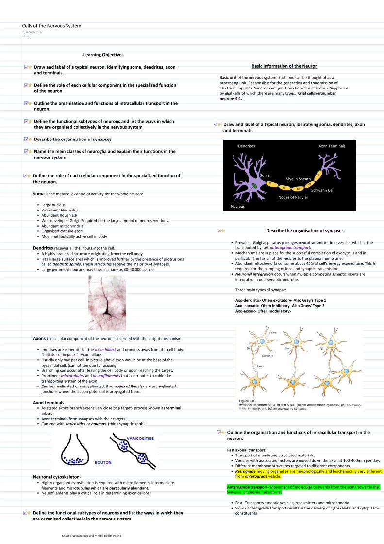
Learning Objectives
Draw and label of a typical neuron identifying soma dendrites axon and terminals
Define the role of each cellular component in the specialised function of the neuron
Outline the organisation and functions of intracellular transport in the neuron
Define the functional subtypes of neurons and list the ways in which they are organised collectively in the nervous system
Describe the organisation of synapses
Name the main classes of neuroglia and explain their functions in the nervous system
Draw and label of a typical neuron identifying soma dendrites axon and terminals
Basic Information of the Neuron
Basic unit of the nervous system Each one can be thought of as a processing unit Responsible for the generation and transmission of electrical impulses Synapses are junctions between neurones Supported by glial cells of which there are many types Glial cells outnumber neurons 91
Nucleus
Schwann Cell
Axon TerminalsDendrites
Soma
Nodes of Ranvier
Myelin SheathDefine the role of each cellular component in the specialised function of the neuron
Soma is the metabolic centre of activity for the whole neuron
Large nucleusbullProminent NucleolusbullAbundant Rough ERbullWell developed Golgi- Required for the large amount of neurosecretionsbullAbundant mitochondriabullOrganised cytoskeletonbullMost metabolically active cell in bodybull
A highly branched structure originating from the cell body bullHas a large surface area which is improved further by the presence of protrusions called dendritic spines These structures receive the majority of synapses
bull
Large pyramidal neurons may have as many as 30-40000 spinesbull
Dendrites receives all the inputs into the cell
Axons the cellular component of the neuron concerned with the output mechanism
Impulses are generated at the axon hillock and progress away from the cell body Initiator of impulse- Axon hillock
bull
Usually only one per cell In picture above axon would be at the base of the pyramidal cell (cannot see due to focusing)
bull
Branching can occur after leaving the cell body or upon reaching the targetbullProminent microtubules and neurofilaments that contributes to cable like transporting system of the axon
bull
Can be myelinated or unmyelinated if so nodes of Ranvier are unmyelinated junctions where the action potential is propagated from
bull
As stated axons branch extensively close to a target- process known as terminal arbor
bull
Axon terminals form synapses with their targetsbullCan end with varicosities or boutons (think synaptic knob)bull
Axon terminals-
Highly organised cytoskeleton is required with microfilaments intermediate filaments and microtubules which are particularly abundant
bull
Neurofilaments play a critical role in determining axon calibrebull
Neuronal cytoskeleton-
Outline the organisation and functions of intracellular transport in the neuron
Transport of membrane associated materialsbullVesicles with associated motors are moved down the axon at 100-400mm per daybullDifferent membrane structures targeted to different componentsbullRetrograde moving organelles are morphologically and biochemically very different from anterograde vesicle
bull
Fast axonal transport
Anterograde transport- Movement of molecules outwards from the soma towards the synapse or plasma membrane
Fast- Transports synaptic vesicles transmitters and mitochondriabullSlow - Anterograde transport results in the delivery of cytoskeletal and cytoplasmic constituents
bull
Describe the organisation of synapses
Prevalent Golgi apparatus packages neurotransmitter into vesicles which is the transported by fast anterograde transport
bull
Mechanisms are in place for the successful completion of exocytosis and in particular the fusion of the vesicles to the plasma membrane
bull
Abundant mitochondria consume about 45 of cells energy expenditure This is required for the pumping of ions and synaptic transmission
bull
Neuronal integration occurs when multiple competing synaptic inputs are integrated in post synaptic neurone
bull
Three main types of synapse
Axo-dendritic- Often excitatory- Also Grays Type 1Axo- somatic- Often inhibitory- Also Grays Type 2Axo-axonic- Often modulatory-
Define the functional subtypes of neurons and list the ways in which they are organised collectively in the nervous system
Cells of the Nervous System20 January 20121315
Stuarts Neuroscience and Mental Health Page 4
Fast- Transports synaptic vesicles transmitters and mitochondriabullSlow - Anterograde transport results in the delivery of cytoskeletal and cytoplasmic constituents
bull
Transports substances from an extracellular spacebullUses microtubule-associated ATPase to drive particles along microtubulesbullTransports soluble cytoplasmic constituentsbullIs the process by which material returns from the terminals to the cell body either for degradation or recycling
bull
Retrograde transport- Movement of molecules inwards from synapse to soma It returns used synaptic vesicles and informs the soma of the conditions at the axon terminal
Define the functional subtypes of neurons and list the ways in which they are organised collectively in the nervous system
Pseudounipolar
Cell has two fused process which are axonal in structure This is a sensory neuronbullExample- Dorsal root ganglion cellsbull
Bipolar
Cells in retina in addition to white matter of cortexbullBIPOLAR THINK OPPOSITES BLACK RETINA ANDbullWHITE CORTEX
Golgi Type I multipolar
Highly branched dendritic treesbullPyramidal cells of cerebral cortexbullAnterior horn cells of the spinal cordbullPurkinje cells of cerebellum (15 million)bullExtremely long extending axonsbullRetinal ganglion cellsbullPAPERbull
Golgi Type II multipolar
Highly branched dendritic treesbullShort axons that terminate near to cell bodiesbullStellate cells of cerebral cortexbull
Define the functional subtypes of neurons and list the ways in which they are organised collectively in the nervous system
Types of neurons
Sensory1Motor2Interneurons (responsible for modification integration coordination facilitation and inhibition of sensory input)
3
Organisational structure of Neurons
Nucleus- Group of unencapsulated neuronal cell bodies within the CNS often of functionally similar cells
Examples include brain stem (Raphe) and deep cerebral cortex (Dentate)bullNNNucleus NNNoo capsule (unencapsulated)bull
Laminae rhymes with greybull
Laminae- Layers of functionally similar cells such as the cerebral cortex grey matter and the cerebellar grey matter
Ganglion- Group of encapsulated cell bodies within the PNS Examples include dorsal root ganglia and sympathetic ganglia
Fibre Tract- Groups of bundles of axons in the CNS that may or may not be myelinated Examples included corpus callosum and internal capsule
Bring information to the CNS from sensory receptors and bring axons to effector organs
bull
Often a combination of both motor and sensory neuronsbullExceptions to PNS are optic and olfactory nervesbull
Nerve- Discrete bundles of axons that are usually part of PNS
Name the main classes of neuroglia and explain their functions in the nervous system
These are the support cells of the nervous systembullEssential for correct functioning of neurones and have many and varied functionsbull
Astroglia
Oligodendroglia
Microglia
Immature progenitors
Ependymal cells- Epithelial lining of the ventricles
Schwann cells
Satellite glia
Examples includebull
Neuroglia
Star shaped cell (think astrology)bullMost abundant cell in CNSbullEach cell forms a specific territory that interfaces with microvasculaturebull
Fibrous astroglia (White Matter)i)Protoplasmic astroglia (Grey Matter)ii)Radial astrogliaiii)
Several different typesbull
Numerous IF bundles in cytoplasm of fibrous astrogliabullGap junctions suggest astroglia-astroglia signallingbullHas intimate associations with other cell typesbullInteracts with blood vessels in an ordered arrangement with little overlapbull
Astroglia
Scaffold for neuronal migration and axon growth during development1Formation of blood brain barrier2Transport of substances from blood to neurons3Segregation of neuronal processes (synapses)4Removal of neurotransmitters5Synthesis of neurotropic factors6Neuronal-glial and glial neuronal signalling7Potassium ion buffering8Glial scar formation9
SSS FTR NPGbull
Functions of astroglia
Myelin
Myelin is an insulating membrane found around nerve cells that is made up of lipid
bull
Up to 50 lamellae- thin plate like structurebullDark and light bands seen at EM levelbullHighly susceptible to damage and loss of either oligodendroglia or myelin is disastrous
bull
- AdrenoleucodystrophyMyelin disease states- Multiple Sclerosisbull
Stuarts Neuroscience and Mental Health Page 5
Potassium ion buffering8Glial scar formation9
SSS FTR NPGbull
Myelin producing cells of the CNSbullSmall spherical nucleibullFew thin processesbullProminent ER and GolgibullMetabolically highly activebull
Interfasicular oligodendroglia
Perineuronal oligodendroglia
Types includebull
Oligodendroglia
Functions of oligodendroglia
Production and maintenance of myelin sheath1Each cell can produce numerous sheaths (1-40)2
Derived from bone marrow during early developmentbullResident macrophage population of CNSbullInvolved in immune surveillancebullTypical macrophage functions- ie APCbullRole in tissue modellingbullSynaptic strippingbull
Microglia
These are the myelin producing cells of the PNSbullOnly produce one sheath per cellbullPromote axon regenerationbullSurround unmyelinated axonsbull
Peripheral Glial- Schwann Cells
Stuarts Neuroscience and Mental Health Page 6
Learning Objectives
Diffusion of an iono
Permeability of a cell membraneo
Electrochemical gradient of an iono
Define the following
Describe how a resting membrane potential can arise from a difference in concentration of an ion across a selectively permeable membrane (use diagrams)
Define electrochemical equilibrium for an ion
What is the equilibrium potential for an ion
The Nernst equation is Ex+ = (RTZF) ln (CoCi) You should know that Ex+ is the equilibrium potential of ion X+ R is the gas constant T is absolute temperature Z is the charge on the ion and F is Faradayrsquos number coulombs of charge per mol of ion
Substituting the values of the constants and T= 37oC gives 27
What are typical values for the concentration of K+ and for Na+ inside and outside a normal neuron
What is a typical value for the resting potential of a neuron
K+ concentration has a much stronger effect on the resting potential than Na+ concentration does Explain the basis of this difference
Diffusion of an ion- The passive movement of an ion down its concentration
gradient
Permeability of a cell membrane- The selectivity of a membrane to ions is its
permeability
Electrochemical gradient of an ion- The movement of ions from an area of
high concentration and relative charge to an area of low concentration and opposite charge
Define the following
Describe how a resting membrane potential can arise from a difference in concentration of an ion across a selectively permeable membrane (use diagrams)
Zero-reference is outside of the cellbullThe inside of the cell is negative compared to the outsidebullAll cells have a membrane potentialbullResting potential is -70mvbull
Concentration of at least one permanent ion is different on one side of the membranebull
Permeable pores that open and close that are selective for different types of ion eg (K+ Na+ Cl- Ca2+)
bull
Open by change in membrane potential
Voltage dependent-1)
Open all the time- these are the ones that produce the resting potential
Voltage independent2)
2 major typesbull
Permeability of membrane to potassium allows it to leave generating a negative charge within the cell- Resting potential Equilibrium of electrochemical gradient is produced resulting in small flux between compartments
bull
Permeability of membrane to sodium allows it to enter generating a negative charge outside and a positive charge inside- Action Potential
bull
Ion Channels
Define electrochemical equilibrium for an ion
Electrochemical equilibrium- Reached when the concentration gradient is balanced by the electrical gradient across the membrane
What is the equilibrium potential for an ion
Equilibrium potential- Potential that prevents diffusion down the ions concentration gradient
The Nernst equation is Ex+ = (RTZF) ln (CoCi) You should know that Ex+ is the equilibrium potential of ion X+ R is the gas constant T is absolute temperature Z is the charge on the ion and F is Faradayrsquos number coulombs of charge per mol of ion
Nernst equation
bull EX = (RTZF) ln (Co Ci)
ndash C is concentration of the ion [X+]
bull o = outside cell
bull i = inside cell
ndash Co = [X+] outside cell
ndash Ci = [X+] inside cell
bull R = gas constant
bull T = Temp o Kelvin
bull Z = charge on ion
bull -1 for Cl- +2 for Ca2+
bull F = Faradayrsquos number
bull charge per mol of ion
bull ln means log to base e
Ex- is the equilibrium potential of the ion X
Substituting the values of the constants and T= 37oC gives 27
EX = (27Z) ln (Co Ci)
Co
(mM)
Ci
(mM)
ENa
(mV)
Na+ 150 10 +73
Co
(mM)
Ci
(mM)
ENa
(mV)
K+ 5 150 -92
bull What sign for the equilibrium potential
bull The sign the inside needs to maintain the concentration
gradient
bull RTF 37 oC = 27
bull EK = (27 +1) ln(0005 M 015 M)
bull EK = +27 ln(003)
bull EK = +27 -34
bull EK = -92 mV
bull RTF 37 oC = 27
bull ENa = (27 +1) ln(015 M 001 M)
bull ENa = +27 ln(15)
bull ENa = +27 +27
bull ENa = +73 mV
What are typical values for the concentration of K+ and for Na+ inside and outside a normal neuron
Sodium Potassium
Inside (mM) 10 150
Outside (mM) 150 5
What is a typical value for the resting potential of a neuron
-70mVbull
K+ concentration has a much stronger effect on the resting potential than Na+
concentration does Explain the basis of this difference
Potassium sodium and chlorine concentrations all contribute to the real membrane potential
bull
The size of each ions contribution is proportional to how permeable the membrane is to the ion
bull
Goldman-Hodgkin-Katz voltage equation
This is an equation used to describe the resting membrane potentialbullTHINK- youd need a rest after saying that namebull
0 means the channel is completely close
05 means the channel is open half of the time
1 means the channel is fully open
Within the equation the letter P denotes permeability or channel open probabilitybull
GHK equation worked examples
bull 1) All channels are open all the time (PK PNa PCl = 1)
bull Vm = 27 ln(1[0005] + 1[015] + 1[0005] 1[015] + 1[001] + 1[011])
bull Vm = 27 ln(016 027)
bull Vm = 27 -05
bull Vm = -14 mV
bull 3) Now increase Na+ permeability by 5 (PK = 1 PNa = 005 PCl = 0)
bull Vm = 27 ln(1[0005] + 005[015] + 0[0005] 1[015] + 005[001] + 0[011])
bull Vm = 27 ln(00125 01505)
bull Vm = 27 -249
bull Vm = -67 mV
bull Increasing permeability for Na+ makes the cell more positive
RTF at 37 oC = 27
Concentration [ ] is in Moles
bull 2) Only K+ channels are open and Cl- and Na+ channels are closed (PK = 1 PNa = 0 PCl = 0)
bull Vm = 27 ln(1[0005] + 0[015] + 0[0005] 1[015] + 0[001] + 0[011])
bull Vm = 27 ln(0005 015)
bull Vm = 27 -34
bull Vm = -92 mV
bull Ion concentration has not changed BUT Vm moved to EK
Graded Potentials
Resting Potential14 May 20121248
Stuarts Neuroscience and Mental Health Page 7
EX = (27Z) ln (Co Ci)
Co
(mM)
Ci
(mM)
ENa
(mV)
Na+ 150 10 +73
Co
(mM)
Ci
(mM)
ENa
(mV)
K+ 5 150 -92
bull What sign for the equilibrium potential
bull The sign the inside needs to maintain the concentration
gradient
bull RTF 37 oC = 27
bull EK = (27 +1) ln(0005 M 015 M)
bull EK = +27 ln(003)
bull EK = +27 -34
bull EK = -92 mV
bull RTF 37 oC = 27
bull ENa = (27 +1) ln(015 M 001 M)
bull ENa = +27 ln(15)
bull ENa = +27 +27
bull ENa = +73 mV GHK equation worked examples
bull 1) All channels are open all the time (PK PNa PCl = 1)
bull Vm = 27 ln(1[0005] + 1[015] + 1[0005] 1[015] + 1[001] + 1[011])
bull Vm = 27 ln(016 027)
bull Vm = 27 -05
bull Vm = -14 mV
bull 3) Now increase Na+ permeability by 5 (PK = 1 PNa = 005 PCl = 0)
bull Vm = 27 ln(1[0005] + 005[015] + 0[0005] 1[015] + 005[001] + 0[011])
bull Vm = 27 ln(00125 01505)
bull Vm = 27 -249
bull Vm = -67 mV
bull Increasing permeability for Na+ makes the cell more positive
RTF at 37 oC = 27
Concentration [ ] is in Moles
bull 2) Only K+ channels are open and Cl- and Na+ channels are closed (PK = 1 PNa = 0 PCl = 0)
bull Vm = 27 ln(1[0005] + 0[015] + 0[0005] 1[015] + 0[001] + 0[011])
bull Vm = 27 ln(0005 015)
bull Vm = 27 -34
bull Vm = -92 mV
bull Ion concentration has not changed BUT Vm moved to EK
Through these series of equations we can clearly see that potassium contributes most to the resting potential as increasing the permeability of sodium by only 5 means that the resting potential is now slightly too positive
bull
Concentration is in moles not millimoles bull
Changes in membrane potential
Graded Potentials
Graded potentials occur at synapses and sensory receptor and their function is to contribute to initiating or preventing action potentials
bull
Change in membrane potential caused by alteration in permeability of ion channels Not of a fixed time and length like action potentials
bull
Graded potentials change in membrane potential in response to stimulation bull
Can be depolarizing or hyperpolarizing
A stronger stimulus produces a larger graded potential
Graded potentials get smaller the farther they travel (Decremental spread)
Properties of graded potentialsbull
Stuarts Neuroscience and Mental Health Page 8
Learning Objections
You should be able to explain in general terms what the function of the action potential is
Give some examples of other types of excitable cells (in addition to neurons) in which action potentials occur
ThresholdRefractory periodldquoAll or nothingrdquo behaviourDepolarizationRepolarizationHyperpolarizationSaltatory conduction
Define the following terms as they apply to action potentials
Voltage-gated channelChannel inactivationPositive feedback
Define the following terms as they apply to the membrane channels involved in producing the action potential
Outline the sequence of events during a typical action potential in the neuron Include changes in membrane potential changes in membrane permeability and fluxes of ions across the membrane (a diagram will help)
State the size and duration (including units) of a typical action potential in a neuron
Define the term ldquoregenerativerdquo as applied to action potentials and its significance for spread of the action potential along an axon
Explain how conduction of the action potential occurs (conduction here means spread along the axon alternatively this process may be called transmission or propagation)
List two structural features that affect the conduction velocity along normal axons Briefly explain why they affect velocity as they do
Be able to list at least one pathological condition that affects conduction velocity
You should be able to explain in general terms what the function of the action potential isGive some examples of other types of excitable cells (in addition to neurons) in which action potentials occur
The function of the action potential is to transport messages through the nervous system very rapidly
bull
Action potentials also exist in muscle cellsbull
Define the following terms as they apply to action potentials
Threshold- The level of depolarisation needed to cause the triggering of an
action potential
Refractory period- Unresponsive to threshold depolarization
Absolute refractory period- New action potential cannot be triggered even with very strong stimulus because inactivation gate is closed
Relative refractory period- Inactivation gate is open but sodium channel activation gate is closed meaning a stronger than normal stimulus is required to trigger an action potential
ldquoAll or nothingrdquo behaviour- Once triggered a full sized action potential
occurs
Depolarization- When the membrane becomes more positive towards
sodiums ion equilibrium potential
Repolarization- Efflux of potassium ions to restore membrane to negative
state
Hyperpolarization- Over efflux of potassium results in membrane potential
dropping below resting potential
Saltatory conduction- From the latin (hop or to leap) is the propagation of
action potentials along myelinated axons from one node of Ranvier to the next
Voltage-gated channel- Ion channels that only open when there is a
change in voltage
Channel inactivation- Closing of the inactivation gate in response often
to an extracellular positive charge
Positive feedback- A mechanism that means that action potentials
constantly regenerate
Define the following terms as they apply to the membrane channels involved in producing the action potential
Outline the sequence of events during a typical action potential in the neuron Include changes in membrane potential changes in membrane permeability and fluxes of ions across the membrane (a diagram will help)
At the resting potential voltage-gated ion channels (Na+ and K+) are closed
Note these are different to non-voltage gated channels that generate the resting potential
Resting potential PKgtgtPNa Where P is the permeability of the membrane1
Stimulus- depolarises the membrane potential and causes it to become more positive
2
PNa rapidly increases because the voltage gated Na+ channels open quicker than potassium channels
Potassium permeability increase as the voltage gated K+ channels start to slowly open and as the upstroke progresses more and more voltage gated K channels open
Potassium leaves the cell down the electrochemical gradient but is less than Na entering
Membrane potential moves toward the Na+ equilibrium potential
Depolarisation- Starts at the threshold potential3
Big decrease in PNa because the voltage-gated Na channels inactivation gate is closed and sodium entry stops generating the absolute refractory period
Moderate PK increase as there are more voltage gated K+ channels and they remain open for longer which means that K leaves by its electrochemical gradient
Membrane potential moves toward the K+ equilibrium potential
Repolarisation-4
State the size and duration (including units) of a typical action potential in a neuron
1-2ms for an action potentialbullSize= -70 to + 40 therefore 110mVbull
Define the term ldquoregenerativerdquo as applied to action potentials and its significance for spread of the action potential along an axon
If less than threshold then graded potentialbullOnce threshold is reached the cycle continues in a positive feedback mannerbullCycle continues until the voltage-gated sodium channels inactivate (closed and voltage insensitive)
bull
Membrane remains in a refractory state until the voltage gated sodium channels recover from inactivation
bull
Ions may move in and out but only a very small number of ions actually cross the membrane to change the potential ie the change in concentration is extremely small (less than 01)
bullIon Movements during action potential
Explain how conduction of the action potential occurs (conduction here means spread along the axon alternatively this process may be called
Action Potential14 May 20122245
Stuarts Neuroscience and Mental Health Page 9
periodModerate PK increase as there are more voltage gated K+ channels and they remain open for longer which means that K leaves by its electrochemical gradient
Membrane potential moves toward the K+ equilibrium potential
At rest voltage gated K channels are still open therefore K continues to leave down gradient
Membrane potential moves closer to the K+ equilibrium
Voltage gated K+ channels then close but can still possibly generate an AP if the stimulus is very strong- Relative Refractory period
Membrane potential returns to the resting potential
After hyperpolarization5
Time Course of changes in permeability
small (less than 01)
Explain how conduction of the action potential occurs (conduction here means spread along the axon alternatively this process may be called transmission or propagation)
Active Propagation
Passive propagation
Only resting K+ channels
openInternal (or axial) amp Membrane
Resistance alters propagation
distance and velocity
List two structural features that affect the conduction velocity along normal axons Briefly explain why they affect velocity as they do
Large diameter myelinated axons= 120msbullSmall diameter non myelinated = 1msbullSpeed of sound= 331msbull
Less resistance to current flow inside large diameter axons
Increases with axon diameter1
Action potentials only occur at nodes of Ranvier
Higher in myelinated than non-myelinated axons of the same diameter2
Reduced axon diameter (ie regrowth after injury)
Reduced myelination (MS and diphtheria)
Cold anoxia compression and drugs (some anaesthetics)
Conduction velocity is slowed by
Be able to list at least one pathological condition that affects conduction velocity
Multiple sclerosis and diphtheria causes demyelination of axonsbull
Stuarts Neuroscience and Mental Health Page 10
Learning Objectives
Define the essential components required for neurotransmitter release
Understand the differences between excitatory and inhibitory transmission
Define at least two mechanisms for the termination of neurotransmitter action at the synapse
Describe how modulation of the synaptic properties of GABA can be modulated pharmacologically to treat epilepsy
Features of neurotransmission
Transfer across a synapse requires the release of neurotransmitters and their interaction with postsynaptic receptors
bull
Transmitter released from 1st cell synaptic activation of 2nd cell signal integration and signal conduction by 2nd cell
bull
Rapid timescale1)Adaptability2)Plasticity 3)Diversity4)Learning and memory5)
Features
RAPDL- sounds like rapidbullDendrites receive inputs where it is integrated in the somabull
SynapseGap is about 20-100 nmbull5000 molecules of neurotransmitter per synaptic vesiclebullHigh energy dependency is satiated by high levels of mitochondriabullPost synaptic knob is electron densebull
Features of neurotransmitters
Provide enormous diversity in variety of transmitters and their receptorsbullAmino acids (eg glutamate gamma amino butyric acid [GABA] amines (eg noradrenaline [NA] and dopamine [DA] and neuropeptides (eg opioid peptides)
bull
May mediate rapid (micro to milli second responses) or slower effects (ms)bullVary in abundance from mM to nM CNS tissue concentrationbullNeurones receive multiple transmitter influence which are integrated to produce diverse functional responses
bull
Define the essential components required for neurotransmitter release
Essential componentsAn action potential is necessary to initiate neurotransmitter releasebullIt has to be very fast within ms (200 micro seconds)bullCalcium is essential- transmitter release requires an increase in intracellular Ca2+ to about 200microM
bull
A protein complex formation between vesicle membrane and cytoplasmic proteins to enable both vesicle docking and a rapid response to Ca entry leading to membrane fusion and exocytosis
bull
ATP and vesicle recyclingbull
Extra information on neurotransmittersSynaptic transmission is restricted to specialised structures ie the synapsebullSynaptic vesicles (SVs) provide the source of neurotransmitter (4000-10000 molecules per SV)
bull
Vary from mM to nM in CNS tissue concentrationsbull5-10 mM amino acids in brainbull
Activation of transmitter release is calcium dependent and requires rapid transduction
bull
Series of events= Membrane depolarisation-gt Calcium influx -gt Vesicle fusion -gt Vesicle exocytosis-gt Transmitter release
bull
From calcium influx to transmitter release has to be within this 200 microsecondsbullSuch rapid release rates can be achieved due to the vesicles being primed at the active zone close to Ca entry
bull
Localisation of vesicle at pre synaptic area Also alignment with calcium channel must occur so the response is rapid this is also helped by proteins associating between presynaptic membrane and vesicle to enable docking
bull
Vesicular proteins are targets for neurotoxinsbull
Types of neurotoxins
Botulinum toxin and tetanus destroys membrane association proteins and vesicles1Black widow spider- alpha latrotoxin that interferes with resealing of membrane so neurotransmitter is released to depletion
2
Vesicular proteins aretargets for neurotoxins
TETANUS toxinCtetani causes paralysis
BOTULINUM toxinCbotulinum causes flacid paralysis
alpha LATROTOXINblack widow spiderstimulates transmitterrelease to depletion
Zn2+-dependent endopeptidasesinhibit transmitter release
Steps of synaptic transmission
Biosynthesis packaging and release of neurotransmitter1
Membrane depolarisationaCalcium channels openbCalcium influxcVesicle fusiondVesicle exocytosis- fusion of vesicle to membrane and release of transmittereTransmitter releasef
Release
Receptor action2
Ion channel receptor- Extremely fast (msecs) mediate all fast excitatory and inhibitory transmission
bull
Glutamate can be synthesized from glucose via the TCA cycle
GABA is synthesised from Glutamate by removing a carboxyl group form N terminal Glutamate decarboxylase
CNS- Glutamate GABA
NMJ- Acetylcholine at nicotinic receptors
Examples bull
G- protein coupled receptor- Slow secs mins- Effectors may be enzymes (adenyl cyclase phospholipase C cGMP-PDE) or channels (eg Ca or K) Abundant in CNS
bull
CNS and PNS ACh at muscarinic receptors dopamine noradrenaline 5hydroxytryptamine (5ht) and neuropeptides eg enkaphalin
Examplesbull
Understand the differences between excitatory and inhibitory transmission
Glutamates effects propagated by sodiumbullimport in the post synaptic membraneExcitatory postsynaptic EPSPbull(membrane is becoming depolarised)
Excitatory-
Define at least two mechanisms for the termination of neurotransmitter action at the synapse
Excitatory CNS synapse mediated by Glutamate
G G
G G G
G
Excitatory amino acid
Transporter EAAT
GLIAL
CELL
Na+ Na+ and Ca2+
glucose
TCAcycleaKG
Glutamate
receptor
GLUR
GLUTAMINE
SYNTHETASE
Glutamine
Glutamate
Neurotransmission10 January 20121408
Stuarts Neuroscience and Mental Health Page 11
import in the post synaptic membraneExcitatory postsynaptic EPSPbull(membrane is becoming depolarised)
Excitatory postsynpatic potential (EPSP)
GABA- which results from Chlorine bullentering the post synaptic neuroneInhibitory postsynaptic EPSP (membrane bullis hyperpolarized which makes it veryhard to generate an action potential
Inhibitory-
Glutamate receptors
Alpha amino-3-hydroxy-5methyl-4-isoxazole propionic acid (AMPA)bullMajority of fast excitatory synapses with rapid onset offset and desensitisationbullInitiated by just Na entrybull
AMPA receptors
N-methyl-aspartatebullSlow component of excitatory transmissionbullServe as coincidence detectors which underlie learning mechanismsbullInitiate by Na and Ca entrybull
NMDA receptors
G G
G G G
G
Excitatory amino acid
Transporter EAAT
GLIAL
CELL
Na+ Na+ and Ca2+
glucose
TCAcycleaKG
Glutamate
receptor
GLUR
GLUTAMINE
SYNTHETASE
Glutamine
Glutamate
GLUR receptor takes up Na and Ca in PosSM (Post synaptic Membrane)bullGlutamate is the transported by Excitatory amino acid Transporter (EAAT) to glial cells
bull
Here it is converted to glutamine by glutamine synthasebullHence the glial cell has removed the neurotransmitter link to Cells of the Nervous System
bull
Inhibitory CNS synapse mediated by GABA
GABA GABA
GABA GABA GABA
GABA
GABA transporter GAT
GLIAL
CELL
Cl- Cl-
glucose
TCAcycle
aKG
GABA receptor
GABAAR
Cl-
GLUTAMATE
Glutamic acid
decarboxylase
GAD (B6)
GABA trans
-aminase
GABA-T
Succinate
semialdehyde
GABA
Glutamate is converted to GABA by Glutamic acid decarboxylase (GAD)bullGABA has effects on PSM receptor which causes chloride influxbullGABA is then relocated to Glial cell by GABA Transport (GAT)bullGABA trans aminase GABA-T converted GABA to Succinate semialdehydebull
Describe how modulation of the synaptic properties of GABA can be modulated pharmacologically to treat epilepsy
In epilepsy there is an excess of glutamate which causes abnormal neuronal firingbullThe concept of GABA drugs is that they will be used to inhibit the effects of glutamatebull
Frequency of opening of channel is increased when binding to benzodiazepinesbullBarbiturates- Open channel for longerbull
alpha
szlig
GABA
barbiturates
steroids
benzodiazepines
Zn
convulsantsethanol
Pentameric organisation of the GABA receptorand pharmacologically important binding domains
Drugs facilitating GABA transmission areantiepilepticanxiolyticsedativemuscle relaxant
Qu 1 Which of the following is NOT a normal route for neurotransmitter inactivation
Addition of a methyl group in the synaptic cleft
X Diffusion out of the cleft
Enzymatic destruction in the synaptic cleft-
Re-uptake by the pre-synaptic terminal- NE
Uptake by glial cells- GABA glutamate
X Mark = -2 (conf=2 )
Best Option Addition of a methyl group in the synaptic cleftSuch a method of inactivation does not existLecture Neurotransmitters
Pasted from lthttpswwwuclacuklaptlaptlitesysrunhtmicl08_neurof=cleari=icl1k=1u=_st1511i=Imperialgt
Stuarts Neuroscience and Mental Health Page 12
Session 4 Tutorial ndash Neurotransmitters and epilepsy
Tutorial notes
1 Epilepsy terminology
The term epilepsy refers to a disorder of brain function characterized by the periodic and unpredictable occurrence of seizures
bull
The term seizure refers to a transient alteration of behaviour due to the disordered synchronous and rhythmic firing of populations of brain neurones
bull
The pharmacological agents in current clinical use for inhibition of seizures are referred to as anticonvulsant or antiepileptic drugs
bull
Seizures are thought to arise from the cerebral cortex and they can be classified into bull
Partial seizures those beginning focally at a cortical site
Generalized seizures those that involve both hemispheres widely from the outset
The behavioural manifestations of a seizure are determined by the functions normally served by the cortical site at which the seizure arises
bull
LEFT HEMISPHERE WOULD CAUSE RIGHT SIDED JERKING
Thus for example a seizure involving the motor cortex is associated with clonic jerking of the body part controlled by this region of the cortex
bull
A simple partial seizure is associated with preservation of consciousness whilst acomplex partial seizure is associated with impairment of consciousness
bull
Examples of generalized seizures include absence myoclonic and tonic-clonic seizures You will be shown examples of the principal seizure types by video presentation during this teaching session
bull
2 Neurotransmitters in epilepsy
Epilepsy is a neurological disorder associated with abnormal neurotransmitter functionin the brain
bull
A decrease in GABA-mediated inhibition or an increase in glutamate-mediated excitation in the brain may result in seizure activity Indeed both glutamate and GABA are thought to play key roles in the brain mechanisms causing epilepsy in man
bull
Pharmacological evidence for a role of neurotransmitters in epilepsy
Dr Martin Croucher
Impairment of GABA-mediated inhibition causes seizures in animals eg impairment of synthesis release (tetanus toxin) or postsynaptic action (bicuculline picrotoxin)
bull
Enhancement of GABA-mediated inhibition leads to seizure suppression eg central (icv) administration of GABA or inhibition of the GABA metabolizing enzyme GABA-T (vigabatrin)
bull
Many clinically useful anticonvulsant drugs are known to act at least in part by potentiating central GABA-mediated inhibition eg benzodiazepines phenobarbital (see Section 3)
bull
Central (icv focal) administration of glutamate or glutamate receptor agonists causes seizure-like activity in animals
bull
Glutamate receptor antagonists are anticonvulsant in experimental models of epilepsy
bull
Some therapeutically effective anticonvulsant drugs act partly by blocking glutamate-mediated excitation in the brain eg phenobarbital
bull
Biochemical evidence for a role of neurotransmitters in epilepsy
Cobalt-induced seizures in rodents are associated with increased glutamate release
and with decreased GABA concentration GAD activity and GABA uptake (probably reflecting GABA neurone loss) at the seizure focus
bull
Audiogenic seizures in mice (DBA2 mice) are associated with glutamate receptor
binding in the brain and with GABA release from depolarized brain slices
bull
The baboon Papio papio which is highly sensitive to photically-induced seizures has a lower than normal CSF GABA concentration
bull
3 Some examples of antiepileptic drugs
Notes
Many neurological conditions are from malfunction in brainbullGABA malfunctions in epilepsy with propensity to repeated seizuresbull~1 in 100200bull
50 have well controlled epilepsy
25 have good control but side effects
25 have complex partial epilepsy which is poorly controlled
In epilepsy 502525 is an important ratiobull
Depression is associated with excessive noradrenalinebull
5 of people can have a seizure and 1 have epilepsybull
Epileptic focus- Excessively synchronous or sustained discharge of a group of neurones in the brain
Intracerebroventricular into lateral ventricles
ValiumLibrium are benzodiazepines
Whole list Important for exams possibly
Epilepsy can be causes by infection head trauma tumoursMeningitis-encephalitis Gliomas Stroke- ischaemic damage May occur in ~10 strokesGABA-interneurones small in CNS
Place in skull of rats and causes epileptic focus
Loud sounds causes seizures
Strobe of certain light frequency gives epilepsy Assumed that CSF is typical of brain
Generalized Seizures
Tonic- Clonic- Grand mal Tonic extension is continuous extensions which may cause the person to fall over Clonic rhythmical jerking 23 minutes
Absence- Petit mal Bit of eye fluttering 1015 secs phase out Occur very often
Myoclonic- Brief involuntary twitching of a muscle
Atonic- Lose muscle tone and posture fall forward onto face Epic face plant
Partial seizures-
Focal seizures can attribute to one direct point
Simple- Right motor cortex- left side activity Twitch tremor and face Can develop into secondary generalised seizure
Complex-Impairment of consciousness Lip smacking Temporal lobe epilepsy
Secondary generalised- Partial expands to general seizures
Parkinsons disease is caused by a lack of dopaminebullAkenisa- awkward gait Pill rolling tremorbullAlzheimers is caused by a lack of acetylcholinebull
4 Questions to be addressed
Review the process of neurotransmission occurring at central synapses utilising the inhibitory neurotransmitter GABA
bull
Present a convincing case for a role of neurotransmitter (glutamate or GABA) malfunction in the aetiology of epilepsy
bull
Draw a diagram to illustrate the principal steps in GABAergic neurotransmission (synthesis storage release etc) Indicate on your diagram i) established and ii) potential new target
bull
Epilepsy20 January 20120954
Stuarts Neuroscience and Mental Health Page 13
________________________________________________________________
Potential for drugs to solely target specific GABAA receptors to ensure only anti-convulsant and stop side effect of drowsiness
bull
Long acting- suicide anti convulsant
GABASynapse
Present a convincing case for a role of neurotransmitter (glutamate or GABA) malfunction in the aetiology of epilepsy
bull
Draw a diagram to illustrate the principal steps in GABAergic neurotransmission (synthesis storage release etc) Indicate on your diagram i) established and ii) potential new target sites for drugs acting to enhance GABA-mediated neurotransmission in the brain
bull
Stuarts Neuroscience and Mental Health Page 14
Learning Objectives
Draw a diagram to explain the relationship between the following major divisions of the CNS spinal cord brainstem cerebellum diencephalon cerebral hemispheres
Define the 3 components of the brainstem and state the main functions of the brainstem
Describe the functions of the 2 main structures in the diencephalon
State the functions of the basal ganglia and the cerebellum
Draw on a diagram of the cerebral hemisphere the cortical lobes and primary cortical areas
Recognise the main structures of the brain in a diagram or MRI
Describe the 3 layers of the meninges and explain their role in protecting the brain
Explain how the major divisions of the brain relate to the cranial fossae in the base of the skull
Explain the relationship between the spinal segments spinal nerves and vertebrae and state at what level a lumbar puncture can be performed safely
Define the functions of the dorsal and ventral horns of the spinal cord and explain how the dorsal and ventral roots and spinal nerves relate to them
Identify the components of the ventricular system and relate them to the divisions of the CNS
Explain the composition circulation and functions of CSF
State the average total volume and flow rate of CSF
Define hydrocephalus and outline how it may be treated
Distinguish between an epidural (extradural) and subdural haemorrhage
Draw a diagram to explain the relationship between the following major divisions of the CNS spinal cord brainstem cerebellum diencephalon cerebral hemispheres
pons
midbrain
medulla spinal cord
cerebellum
cerebral
hemisphere
diencephalon
brainstem
Define the 3 components of the brainstem and state the main functions of the brainstem
Midbrain1Pons-usually very tubular and has a bulge on the anterior surface2Medulla- With pons and cerebellum lies in hindbrain3
Midbrain pons and medulla share a lot of their functions These include controlling vital functions such as breathing consciousness and also innervating cranial nerve function
bull
Cranial Nerves
All are part of the PNS apart from the optic nervebullFunctional components less regularly organised than for spinal nervesbullSupply motor and sensory innervation to the headbullAutonomic (parasympathetic) innervation to head thoracic and abdominal organsbullSpecial senses eg vision hearing balancebull
Cerebral Hemispheres
Cortex of grey matter superficially that is responsible for most brain functions
1
Basal ganglia deep within brain that are involved with movement2
The cerebral hemisphere can be divided into two main partsbull
There is also the Corpus callosum which is a tract of fibres that connect the two hemispheres
bull
Central sulcus separates off the frontal lobe from the parietal lobebullSulcus are grooves and gyri are foldsbull
Cerebellum
Co-ordinates movement in terms of timing accuracy and precision (TAP) It may also have some involvement in cognitive function and in the fear and pleasure response
bull
TAP that cerebellumbullThe hindbrain contains the pons medulla and cerebellum
State the functions of the basal ganglia and the cerebellum
Draw on a diagram of the cerebral hemisphere the cortical lobes and primary cortical areas
Primary motor cortex is a strip Imagine that there is a man lying upside in the strip so damage to bottom of strip would affect his head or upper body
bull
Primary visual cortex- This area receives information from the retinas if there is a malfunction here it is likely you would become blind
bull
Auditory cortex receives information from inner earbullOther associated cortex areas seem to be related to higher functionbullWernickes area is involved with understanding language apparently according to recent literature the area should be moved and will then have more in common with experimental data and the area in primates
bull
Brocas area- Making speech case study evolved from a patient who had neurosyphillis destroying just that particular area and couldnt talk This area is more prominent in the left hemisphere
bullIdentify the components of the ventricular system and relate them to the divisions of the CNS
lateral ventricle
aqueduct
third ventricle
fourth ventricle
central canal
Describe the functions of the 2 main structures in the diencephalon
The diencephalon is a structure above the brain stembullIt is composed of the thalamus which relays information from lower structures to the cerebral cortex and the hypothalamus which is essential in coordinating homeostatic mechanisms and is the interface between CNS ANS and endocrine system
bull
Central sulcus
Lateral sulcus
CNS Lectures + Practicals31 January 20120900
Stuarts Neuroscience and Mental Health Page 15
lateral ventricle
aqueduct
third ventricle
fourth ventricle
central canal
Region of brain Region of ventricular system
Cerebral hemisphere Lateral ventricle
Diencephalon 3rd Ventricle
Midbrain Aqueduct
Pons and Medulla Fourth Ventricle
Lateral ventricle is C-shaped and there is one present in each hemispherebullFourth ventricle lies in front of the cerebellum and behind the pons and the medullabullHalfway down medulla it becomes the central canal and goes down into spinal cordbullEpendymal cells line the ventricular systembullChoroid plexus secretes CSFbull
Explain the composition circulation and functions of CSFState the average total volume and flow rate of CSF
The CSF is secreted by choroid plexus a structure within the lateral 3rd and 4th ventricles of the brain
bull
Most of the CSF leaves the 4th ventricle via holes and circulates via outside of the brain between the meninge membranes
bull
Lower number of cellsi)Decreased amount of proteinii)Lower Potassium and sodiumiii)Increased Magnesium and chlorine concentrationiv)
Differences between CSF and blood includebull
Circulates through ventricular system and subarachnoid space formed by the meninge membranes
bull
The CSF is reabsorbed into the venous sinuses via arachnoid villi which stops the accumulation of fluid possible oedema and a raised ICP
bull
Functions of CSF
Helps protective the brain against damage by suspending it in liquid 1Also has a few metabolic functions and waste products and helps distribute certain hormones
2
500ml per day is the flow rateTotal volume at any one time is 150ml
Describe the 3 layers of the meninges and explain their role in protecting the brain
3 layers of the meninges
Dura mater- Tough outer layer1Arachnoid mater - Delicate middle layer2Pia mater- Layer firmly attached to the surface of the brain3
Arachnoid villus drains CSF fluid into the venous sinusbull
Meningitis
Meningitis is most likely to infect Pia Mater and subarachnoid space but in the worst cases it can also spread to the upper layers of the cortex
bull
In order to determine between viral and bacterial and meningitis one can look at the CSFbull
BacterialThere should be a high white cell count in particular neutrophilsbullProtein concentration would be increased whereas there would be a decrease in glucose concentration- dangerous for brain metabolism
bull
Bacteria may actually be themselves identifiablebull
ViralLymphocytes account for the predominant change in leukocytes countbullProtein and glucose are normalbullViral identification is unlikelybull
Recognise the main structures of the brain in a diagram or MRI
Brain cut in mid-sagittal plane
Explain how the major divisions of the brain relate to the cranial fossae in the base of the skull
Inside of base of skull
Explain the relationship between the spinal segments spinal
Stuarts Neuroscience and Mental Health Page 16
Protein and glucose are normalbullViral identification is unlikelybull
Inferior aspect of brain
Cranial Fossae Part of brain
Anterior Cranial Frontal Lobe
Middle Cranial Temporal Lobe
Posterior Cranial Cerebellum
Lies above sphenoid bone Hypothalamus
Passes through the Foramen Magnum Medulla
Explain the relationship between the spinal segments spinal nerves and vertebrae and state at what level a lumbar puncture can be performed safely
Terminal end of spinal cord is called Conus MedullarisbullThere are 5 spinal segments cervical thoracic lumbar sacral and coccyx 7 12 5 5 4
bull
Spinal nerves are associated with each specific segment ie cervical etc but they do not necessarily have the same number of nerves to vertebrae and they terminate in different areas
bull
Type of vertebrae
Number of Vertebrae
Number of spinal nerves
Relationship of nerve to vertebrae
Cervical 7 8 Above
Thoracic 12 12 Below
Lumbar 5 5 Below
Sacrum 5 5 Below
Coccyx 4 (1 fused) 1 Below
The safe area to perform a lumber puncture is between L3 and L4- this is because the spinal cord has finished before this are and the nerves can be pushed aside by the needle without damage
bull
Spinal cord is much shorter than the vertebral column because the spinal cord develops mostly in the embryo whereas bones take much longer to develop
bull
Define the functions of the dorsal and ventral horns of the spinal cord and explain how the dorsal and ventral roots and spinal nerves relate to them
VRRMM VRRMM- sound like an engine VM= Ventral + Motor neurone cell bodies The more ridiculous the better right
Ventral Horn- Contains motor neurone cell bodiesbull
DI- Dorsal + interneurones Investigate what the bloody hell goes on in the spine (Detective Inspector- DI)
Dorsal Horn- Contains interneurones which receive sensory information from the body
bull
Afferent pathway is receives of sensory information via dorsal horn efferent pathway is motor information leaving the spinal cord Spinal nerve is collection on both and thus has bidirectional information transfer
bull
Grey matter contains the neuronal cell bodies and it is the white matter that contains the bundles of axons
bull
Define hydrocephalus and outline how it may be treated
Hydrocephalus- Water on the brain caused by CSF flow abnormalities
Communicating- All 4 ventricles affecteda)Non- communicating- Not all 4 enlargedb)
2 predominant types
Block in CSF absorption or CSF over brain surface caused bybullMeningitisbullHead injurybullCongenitalbull
MHCH
Haemorrhage (sub-arachnoid)bull
Communicating-
Block in ventricular system caused bybullAqueduct stenosisbullVentricular tumoursbull
AVPParaventricular tumoursbull
Non- Communicating-
Distinguish between an epidural (extradural) and subdural haemorrhage
EMA- Extradural= meningeal artery
Extradural haemorrhage is usually due to a damaged meningeal artery between the skull and the dura after head trauma
bull
Subdural haemorrhage- Usually due to a damaged vein between the dura and arachnoid membrane
bull
Both of these pathologies can cause a space-occupying lesion in the confined space bounded by the membranes and hence cause neurological pathologies
bull
Symptoms such as headache drowsiness vomiting or seizure are more likely to present quickly from an arterial (extradural) than a subdural haemorrhage
bull
Can also be confirmed by imagingbull
Subdural are more common than epiduralbullSubdural haemorrhages are more frequently seen in alcoholics the elderly patients on anticoagulants whereas epiduralextradural haemorrhages more commonly arise from blows to the side of the head
Pasted from lthttpswwwuclacuklaptlaptlitesysrunhtmicl08_neurof=cleari=icl1k=1u=_st1511i=Imperialgt
Stuarts Neuroscience and Mental Health Page 17
AVP
HeadachebullDrowsinessbullBlackoutsbullRaised intracranial pressurebullIncreased head circumference (in child)bull
Symptoms
Remove causes eg papillomabullPut in a shunt to divert CSFbullOpen alternate pathway- ventriculostomybull
Treatment
Stuarts Neuroscience and Mental Health Page 18
Brachial plexus forms at level C5-T1bull
Macrophage clears up debris along proximal end of axoniAxon sends at pseudopodia (projections of membrane) to make contact with Schwann cell which it grows through and coats it in myelin
ii
Axon reaches distal end of axon as a complete entityiiiNodes of Ranvier are reinserted however there are usually more than there was before which slows nerve conduction down slightly
iv
Axon sustains damage eg in example by forceps crushing it
Growth of axon is about 2-5mm per day
Soma undergoes chromatolysis
Failure to re-join proximal to distal end can result in Neuroma
Nerve degenerationbull
Qu 2 Which of the following statements about peripheral nerves is INCORRECT
Axons form bundles called fascicles
They may contain axons from difference spinal nerves
radic They contain only myelinated nerve fibres
They contain a mixture of fibres of different diameter
They contain a mixture of fibres of different conduction velocities
Pasted from lthttpswwwuclacuklaptlaptlitesysrunhtmicl08_neurof=cleari=icl1k=1u=_st1511i=Imperialgt
Definitions
Dorsal ramus- Posterior branches of the spinal nerves that are smaller than anteriorventral divisions Carry visceral motor somatic motor and sensory information to and from the skin and deep muscles of the backVentral ramus- Supply the antero-lateral parts of the trunk and limbs they are for the most part larger than the posterior divisionsDorsal root ganglion- Nodule on dorsal root that contains cell bodies of neurons in afferent spinal nervesDorsal root- Afferent sensory root of a spinal nerveVentral root- Efferent motor root of a spinal nerveGray ramus communicans- Contain unmyelinated postganglionic sympathetic fibres that are from ganglion of sympathetic trunkWhite ramus communicans- Preganglionic sympathetic outflow from the spinal cord
Additional info on white ramus communicans
The thoracic and the first and second lumbar nerves each contribute a branch white ramus communicans to the adjoining sympathetic ganglion Unlike the gray rami white rami communicants do not extend below L2 or above T1
bull
PNS18 May 20122302
Stuarts Neuroscience and Mental Health Page 19
Learning Objectives
Describe the basic anatomy of ANS innervation in terms of pre-ganglionic neurons ganglia and post-ganglionic fibres Contrast the anatomical location of sympathetic and parasympathetic ganglia
Describe the sympathetic and parasympathetic pathways and their centralspinal connections
Describe the thoracolumbar (sympathetic) and craniosacral (parasympathetic) central origins of the ANS
Describe the influence of the sympathetic and parasympathetic nervous system on the principal systemsorgans of the body (eg cardiovascular system lung gut exocrine glands) and understand the concept of dual innervation and autonomic tone (giving examples)
Understand the importance of the sacral parasympathetic outflow for innervation of structures within the pelvis eg the bladder
Classify the cholinoceptors found within the autonomic nervous system and identify the principal loci of (a) the nicotinic cholinoceptors and (b) the muscarinic cholinoceptors Note that the nicotinic receptors are ion-gated and the muscarinic receptors are G-protein coupled
Identify the neurotransmitter substances released at different levels within the autonomic nervous system and describe the principal steps involved in their biosynthesis and metabolism
CNS- ANS or SMS or NES
Principal ANS components
Exocrine glandsbullSmooth musclebullCardiac musclebullMetabolismbullHost defencebull
General information on ANS
Sympathetic- Fight and flightParasympathetic- Rest and digest
Often sympathetic and parasympathetic in many tissues are antagonists with often inhibitory functions
bull
An interesting point of note is that there are only parasympathetic nerves innervating the lungs which results in bronchoconstriction
bull
Furthermore although sympathetic is usually associated with dilation of blood vessels- maximum oxygen to muscles the sympathetic nervous system can cause vasoconstriction depending on the particular vessel receptor
bull
At rest there is usually a balance between parasympathetic and sympathetic nervous system
bull
SALIVARY GLANDS
Thick viscious secretion
Copious watery secretionSKIN
Piloerection
Increased sweating (C)HEART
rate and contractility
rate and contractility
GASTROINTESTINAL
motility and tone
Sphincter contraction
motility and tone
secretions
BLOOD VESSELS
Constriction
(skin mucous membranes and splanchnic area)
EYE
Dilatation (Pupil)
Constriction (Pupil)
Contraction (Ciliary Muscle)
TRACHEA AND BRONCHEOLES
Dilates (Ad)
Constriction
LIVER
Glycogenolysis
Gluconeogenesis
ADIPOSE
Lipolysis
KIDNEY
Increased renin secretion
URETERS AND BLADDER
Relaxes detrusor constriction of
trigone and sphincter
Contraction of detrusor relaxation
of trigone and sphincter
BLOOD VESSELS
(skeletal muscle) Dilatation
Yellow = sympathetic
Pink = parasympathetic
C = sympathetic cholinergic
Describe the thoracolumbar (sympathetic) and craniosacral (parasympathetic) central origins of the ANS
Parasympathetic efferent output
Known as craniosacral- although it may affect head there are no cervical nerves
bull
4 cranial nerves are in red Vagus is most interesting as it descends into thorax and has effect on the lungs and heart etc
bull
Sacral nerves are located inferiorly to cranial nerves and innervate lower internal viscera
bull
Cervical
Thoracic
Lumbar
Sacral
Lungs intestine
rectum
Reproductive organsSplachnic
nerve
Heart
Stomach
Pancreas
Small intestine
Lung
Eye
Lacrimal and salivary glands
Oculomotor nerve
Vagus
Glossopharyngeal nerve
Facial nerve
Describe the basic anatomy of ANS innervation in terms of pre-ganglionic neurons ganglia and post-ganglionic fibres Contrast the anatomical location of sympathetic and parasympathetic ganglia
Ganglia are a collection of neuronal cell bodies within the Peripheral Nervous System This is also where the neurons synapse
bull
The parasympathetic system has very long pre-ganglionic fibres cell bodies originate in CNS and move towards effector organs
bull
Ganglion are very close to effector organ- therefore short post-ganglionicbull
Parasympathetic- craniosacral
Sympathetic- thoracolumbar
Ganglion are grouped in structures just outside of spinal cord called the sympathetic trunk- this allows a very coordinated response
bull
Allows mass discharge of sympathetic nervesbullCan pass up and down sympathetic trunkbullOpposite set up very short pre-ganglionic fibres and very long post ganglionic fibres
bull
Innervation to adrenal medulla is atypical No official ganglion but adrenal medulla does almost act like one Short neurone from spinal cord and no post ganglionic neurone- neurotransmitters are released from medulla into blood stream instead
bull
Eye
Lacrimal and salivary glands
Lung
Heart
Foregut
Adrenal medulla
Midgut
Hindgut
Pelvic organs
All chain ganglia
supply the skin and
blood vessels
Describe the influence of the sympathetic and parasympathetic nervous system on the principal systemsorgans of the body (eg cardiovascular system lung gut exocrine glands) and understand the concept of dual innervation and autonomic tone (giving examples)
Cardiovascular
Cardiac output= SV x HR
Sympathetic activity increases the force of contraction of cardiac muscle (inotropic effect) therefore increases SV
bull
Increases HR (chronotropic effect)bullResult is a large increase in Cardiac Outputbull
Total peripheral resistance- Dual innervation
Sympathetic tone particularly affects arteriolesbullIncrease in sympathetic= vasoconstriction- increase TPRbullDecreased in sympathetic= vasodilation- decrease TPRbull
The combination of increased TPR and CO should result in increased arterial blood pressure
Vasodilation
Exceptions
Increased sympathetic activity to some blood vessels in skeletal muscle either cholinergic fibres or have adrenergic beta-receptors
bull
Local vasodilators (eg CO2 increased H+ nitric oxide and histamine etcbull
Sympathetic efferent output
Known as thoracolumbar refer to anatomy section for more detailbull
Autonomic Nervous System07 February 20120859
Stuarts Neuroscience and Mental Health Page 20
Cervical
Thoracic
Lumbar
Sacral
Lungs intestine
rectum
Reproductive organsSplachnic
nerve
Heart
Stomach
Pancreas
Small intestine
Lung
Eye
Lacrimal and salivary glands
Oculomotor nerve
Vagus
Glossopharyngeal nerve
Facial nerve
Eye
Lacrimal and salivary glands
Lung
Heart
Foregut
Adrenal medulla
Midgut
Hindgut
Pelvic organs
All chain ganglia
supply the skin and
blood vessels
Exceptions
Increased sympathetic activity to some blood vessels in skeletal muscle either cholinergic fibres or have adrenergic beta-receptors
bull
Local vasodilators (eg CO2 increased H+ nitric oxide and histamine etcbullAlthough parasympathetic doesnt usually have an effect it can have an effect in certain organs such as the penis clitoris GI and salivary glands
bull
Gastrointestinal tract
Sympathetic Activity Parasympathetic Activity
Generally decreases motility and tone Increases motility and tone
Usually stimulates contraction of sphincters
Usually causes relaxation of sphincters
Generally inhibits secretory activity Generally stimulates secretory activity
Think rest and digest Plus is digesting of food really an important focus if there was a lion in the lecture theatre
How can sympathetic affect lungs if there are no nerve fibres going there
Sympathetic effects via the adrenal glands and the secretion of adrenalineAdrenaline induces bronchodilation which increases oxygen delivery to the lungs
ANS innervation of the eye
Sympathetic activity Parasympathetic activity
Contracts radial muscle (pupil dilation)
Contracts pupillary sphincter (pupil contracts)
Contracts ciliary muscle lens bulges for near vision
Understand the importance of the sacral parasympathetic outflow for innervation of structures within the pelvis eg the bladder
Outline
Within the bladder there is both voluntary and ANS controlbullMain influences are parasympathetic coming from sacral region of the spinal cord
bull
Ganglion is located virtually within the tissuebullParasympathetic nerves fibres causes Detrussor muscle to contract and thus urination occurs
bull
Sympathetic nerves from the sympathetic trunk controls the internal sphincter whereby innervation closes it
bull
Voluntary control over sphincter carried by motor nervesbull
Detailed
Baroreceptors in the bladder sends information up the afferent pelvic nerveto sacral spinal cord this is known as the Micturition Reflex This reflex innervates the parasympathetic system and makes you more likely to urinateneed to urinate
bull
Afferent and efferent neurons are usually found together within the same larger nerve
bull
Decreased sympathetic outflow via hypogastric nervebullPudendal nerve is a somatic nerve that controls the ability to consciously urinate
bull
If pressure gets too great than voluntary control is overcomebull
Identify the neurotransmitter substances released at different levels within the autonomic nervous system and describe the principal steps involved in their biosynthesis and metabolism
The general synthesis of a neurotransmitter is as follows
Enzymatic conversion of precursor to active form1
Classify the cholinoceptors found within the autonomic nervous system and identify the principal loci of (a) the nicotinic cholinoceptors and (b) the muscarinic cholinoceptors Note that the nicotinic receptors are ion-gated and the muscarinic receptors are G-protein coupled
Acetylcholine
ACh binds to Nicotinic or Muscarinic receptors Nicotinic is an ion channel linked receptor that is present in the ganglion This ensures that pre-post ganglionic transmission is extremely quick In effector organs we have Muscarinic which is a G-Protein Coupled Receptor This is slower but still quick
bull
Adrenergic receptors are bound by noradrenaline which is a G-Protein coupled receptor
bull
Not actually activepresent until enzyme converts from dopaminebullDoes not get broken down in the synapse It has transportreuptake proteins that transports it back to the cell
bull
Degraded in neuronebullUptake 1- back to neuronebullUptake 2- extracellular tissuebull
Noradrenaline
PRESSURE
BUILDS UP
IN BLADDER
Internal sphincter
(relaxes with
decreased
sympathetic activity)
Sympathetic
control
Hypogastric nerve
parasympathetic
control Detrussor muscle
(contracts)
Parasympathetic
ganglion
Main Influence
Motor nerves
(voluntary control)
External sphincter
(relaxes following central inhibition)
Pudendal nerve
Sacral
segments of
spinal cord
Pelvic nerve
(micturition reflex)
Sympathetic efferent output
Known as thoracolumbar refer to anatomy section for more detailbull
Identify the neurotransmitter substances released at different levels within the autonomic nervous system and describe the principal steps involved in their biosynthesis and metabolism
Noradrenaline adrenaline and also dopamine belong to a group of chemicals called catecholamines because they have a catel ring
bull
All parasympathetic neurones release acetylcholine and it is ACh that also effects target organs
bull
Most sympathetic- Pre= Acetylcholine Post= Noradrenalinebull
Adrenal glands- ACh causes production of predominantly A (but also NA) by medulla
Some have ACh as pre and post ganglionic This is where sympathetic nervous are influencing secretion- sweat
Noradrenaline is main in sympathetic
Other variations exceptionsbull
Principal metabolites of noradrenaline and adrenaline
Noradrenaline Adrenaline
3-methoxy-4 hydroxy-
phenylethelene glycol
(MOPEG)Conjugation (glucuronide or sulphate)
Urine
Catechol -O-methyl transferase (COMT)
Monoamine oxidase A (MOA-A)
Stuarts Neuroscience and Mental Health Page 21
involved in their biosynthesis and metabolism
The general synthesis of a neurotransmitter is as follows
Enzymatic conversion of precursor to active form1Transmitter packaged into vesicles close to the surface of the neurone2Stimulation of nerve causes calcium entry into the neurone3Promotes exocytosis of neurotransmitter into synapse4Moves into synapse5Binds to receptor- random bouncing around by diffusion6Degradation of neurotransmitter by enzyme at some stage7Reuptake of certain molecules for resynthesis8
Acetylcholine
Same steps as above but the precursors are the chemicals acetyl Co-A and choline
bull
The enzyme that catalyses the formation of acetylcholine is Choline Acetyl Transferase
bull
Finally the enzyme that degrades ACh is Acetylcholin-esterasebull
Noradrenaline
The synthesis of Noradrenaline has a few more unique reactions that that of ACh
bull
Tyrosine enters the pre-synaptic neurone and is converted by Tyrosine Hydroxylase to form DOPA (dihydroxyphenylalanine) This reaction is the rate limiting step
bull
DOPA is converted by the enzyme DOPA carboxylase to dopaminebullDopamine is then packaged into a vesicle within the enzyme Dopamine βhydroxylase which converts it to Noradrenaline
bull
Unlike with ACh there is no further metabolism of the neurotransmitter after it has been bound to its receptor Instead a set of uptake proteins either take it back to the pre-synaptic neurone (Uptake Protein 1) or to the post-synaptic neurone (Uptake Protein 2)
bull
PreMOdona- Presynaptic= Mono-amine oxidase
In the PreSN an enzyme called Mono-amine oxidase A breaks down the noradrenaline back down to its metabolites inside the mitochondria
Way of remembering- Youll catch a cold if you are broken down
In the PostSN an enzyme called COMT (Catechol-O-Methyl Transferase) degrades the noradrenaline
Degredation of the noradrenaline is different depending upon its locationbull
Acetyl Co A
+
CholineCholine acetyl
transferaseACh + Co A
ACh
ACh
ACh
ACh
ReceptorEffector cell
Choline +
AcetateCa++
ACh = acetylcholine
6
3
7
8
1
2
4
5
NA
Ca++
Tyrosine
DOPA
DopamineDopamine
Dopamine hydroxylase
Tyrosine
hydroxylase
DOPA decarboxylase Monoamine oxidase
A (MAO-A)
Noradrenaline
NA
Metabolites
Tyrosine
NA
Adrenoceptor
Uptake 1
Uptake 2
Degradation
(COMT)
Tyrosine
hydroxylaseMetabolites
DOPA
Dopamine
Tyrosine
hydroxylaseDOPA
Dopamine
6
37
8
1
2
4
5
1
1
8
A
Ca++
Tyrosine
DOPA
DopamineDopamine
Dopamine
hydroxylase
Tyrosine
hydroxylase
DOPA decarboxylase
NoradrenalineNA
Metabolites
Tyrosine
A
Tyrosine
hydroxylaseDOPA
Dopamine
Tyrosine
hydroxylaseDOPA
Dopamine
6
3
1
2
4
5
1
1A
Phenylethanolamine
Methyl transferase
CapillaryA
Principal metabolites of noradrenaline and adrenaline
Noradrenaline Adrenaline
3-methoxy-4 hydroxy-
phenylethelene glycol
(MOPEG)Conjugation (glucuronide or sulphate)
Urine
Catechol -O-methyl transferase (COMT)
Monoamine oxidase A (MOA-A)
Sympathetic Parasympathetic
Is a diffuse system which allows it to stimulated multiple parts of the body at once (Mass discharge)
Relatively discrete system innervating individual target tissues via specific nerves
Can also have more discrete effects
The sympathetic nervous system
The acute stress response
BRAIN
(hypothalamus
and brain stem)
STRESS
ACTIONS
Spinal cord
ADRENAL
MEDULLA
sympathetic
ganglia
pre-ganglionic fibres post-ganglionic fibres
CATECHOLAMINES
Fight or flight response
First described by Cannon in 1915 as a mass sympathetic discharge in response to distress or alarm
bull
Results inIncreased arterial blood pressure1Increased blow flow to muscles and decreased levels to other areas such as splanchnic bed
2
Increased blood glucose concentration3Increased respiration4Increased awareness5
Adrenaline
Looks similar to noradrenaline in terms of synthesis but you must note that Adrenaline is NOT a neurotransmitter it is a hormone This is why there is no PostSN but a capillary instead
bull
Furthermore note that it is synthesised in Adrenal Chromaffin Cells which are specialised sympathetic post-ganglionic fibres
bull
Same initial steps as noradrenaline but Noradrenaline leaves vesicle and is changed by enzyme Phenylethanolamine Methyl Transferase (PNMT) to give Adrenaline
bull
Stuarts Neuroscience and Mental Health Page 22
Learning Objectives
Describe the basic anatomy of ANS innervation in terms of pre-ganglionic neurons ganglia and post-ganglionic fibres Contrast the anatomical location of sympathetic and parasympathetic ganglia
Describe the thoracolumbar (sympathetic) and craniosacral (parasympathetic) central origins of the ANS
Understand the terms sympathetic trunk plexus and subsidiary ganglia
Identify the rich sympathetic plexuses that surround the major organs and blood vessels
Understand the pre-ganglionic nature of the thoracic and lumbar splanchnic nerves and their synapses in subsidiary ganglia eg coeliac ganglion
Understand the importance of the sacral parasympathetic outflow for innervation of structures within the pelvis eg the bladder
Identify which of the cranial nerves contain parasympathetic pre-ganglionic fibres
Describe the basic anatomy of ANS innervation in terms of pre-ganglionic neurons ganglia and post-ganglionic fibres Contrast the anatomical location of sympathetic and parasympathetic gangliaDescribe the thoracolumbar (sympathetic) and craniosacral (parasympathetic) central origins of the ANS
Sympathetic
Sympathetic trunks (x2)aNerve plexusesbSubsidiary gangliac
Largest division of the ANS which consists ofbull
To cause vasoconstriction in blood vessels
Secretomotor function in sweat glands and motor function in the erector pilli muscles of hair
To accompany motor nerves to voluntary muscles but only distributed to blood vessels supplying the muscles
Viscera- dilation of pupils dilation of arterioles movement of alimentary tract and urinary bladder (ie relaxes wall constricts sphincters)
Functionsbull
Thoracic spinal nerves
Upper lumbar spinal nerves (L1-3)
Preganglionic efferent fibres arise frombull
Pre ganglionic neurons are very short and are situated in the lateral column grey matter of spinal cord T1 to L3
bull
They emerge from the spinal cord via the ventral root of the spinal nervebullThey pass through ventral ramus to white rami communicans to ganglionbullSynapse within ganglion or send fibres up and down to other ganglionbull
Postganglionic fibres are distributed to effector organ via grey rami communicansbullTravel in dorsal or ventral ramibull
Understand the importance of the sacral parasympathetic outflow for innervation of structures within the pelvis eg the bladder
Parasympathetic
Cranial nerves
Sacral (spinal) nerves
Preganglionic efferent fibres arise frombull
Anterior rami of S2-4
Visceral branches passing directly to pelvic viscera ie pelvic splanchnic nerves
Minute ganglia in wall of viscera giving rise to post ganglionic fibres
Sacral outflowbull
Motor fibres to rectum and bladder wall
Inhibitory fibres to bladder sphincter
Erection of penisclitoris via vasodilator fibres
Fibres also pass superiorly to supply large part of the gut with visceromotor innervation
Pelvic splanchnic nerves
Understand the terms sympathetic trunk plexus and subsidiary ganglia
The sympathetic trunks are located on either side of the spinal cord and go from the base of the skull to the coccyx and are divided as so
bull
Cervical- 3 ganglia- Superior cervical ganglion follows internal carotid arteryThoracic- 11 or 12 gangliaLumbar- 4 or 5 gangliaSacral- 4 or 5 ganglia
Sympathetic Trunks
Identify which of the cranial nerves contain parasympathetic pre-ganglionic fibres
Anatomy of the ANS19 May 20122057
Stuarts Neuroscience and Mental Health Page 23
Horners syndrome- Loss of sympathetic supply around the eye Ptosis- drooping upper eye lid Pinpoint pupil- unopposed parasympathetic activity Anhydrosis- loss of sweating around the eye
bull
Caused by tumour in the thorax- apex of lungbull
All parasympathetic ganglions are subsidiary ganglionsbullTherefore I would attempt to define a subsidiary ganglion as sobullSubsidiary ganglion- Any ganglions that are not part of the sympathetic system and are located next to the organ they innervate
bull
Identify the rich sympathetic plexuses that surround the major organs and blood vessels
Plexus around pharynx
Cardiac plexus
Thyroid plexus
PCTP
Pulmonary plexus
Cervicalbull
Plexus around thoracic aorta
Thoracic aorta supply
Pierces diaphragm and enters abdominal plexus through the great vessels
Greater
Pierces diaphragm and enter abdominal plexus through the aorta
Lesser
Pierces diaphragm and enters abdominal plexus through gut
Least
Splanchnic nerves
Thoracicbull
4 lumbar ganglia
Lumbar splanchnic nerves take part in all plexi of sympathetic nerves in abdominal and pelvic regions
Lumbarbull
Sympathetic Plexuses
Identify which of the cranial nerves contain parasympathetic pre-ganglionic fibres
Ciliary ganglionbullPost ganglionic fibres to sphincter pupillae and ciliary muscle inside the eyebull
Oculomotor Nerve (CN III)
Submandibular ganglionbullPost ganglionic fibres to submandibular and sublingual salivary glandsbullPterygopalatine ganglionbullPost ganglionic fibres to paranasal sinuses and lacrimal glandsbull
Facial nerve (CN VII)
Otic ganglionbullPostganglionic fibres to parotid glandbull
Glossopharyngeal nerve (CN IX)
Enters neck and thorax via carotid sheathbullBranches to lungs heart esophagus stomach and intestinesbull
3 7 9 10bull
Vagus nerve (CN X)
Enteric System
Myenteric (Auerbachs) plexus
Submucous (Meissners) plexus
In walls of alimentary tractbull
Sensory- monitoring mechanical chemical and hormonal activity of gutbullMotor- gut motility secretion vessel tonebullCan be overridden by sympathetic and parasympathetic systemsbull
Qu 5 Provides cannular access to the sacral epidural space for the administration of anaesthetics
Best Option Sacral hiatusThis is a naturally occurring gap at the lower end of the sacrum Because there is a failure of the laminae of the last sacral segments to coalesce this space is available for cannular use
Pasted from lthttpswwwuclacuklaptlaptlitesysrunhtmicl08_neurof=cleari=icl1k=1u=_st1511i=Imperialgt
Stuarts Neuroscience and Mental Health Page 24

Learning Objectives
Diffusion of an iono
Permeability of a cell membraneo
Electrochemical gradient of an iono
Define the following
Describe how a resting membrane potential can arise from a difference in concentration of an ion across a selectively permeable membrane (use diagrams)
Define electrochemical equilibrium for an ion
What is the equilibrium potential for an ion
The Nernst equation is Ex+ = (RTZF) ln (CoCi) You should know that Ex+ is the equilibrium potential of ion X+ R is the gas constant T is absolute temperature Z is the charge on the ion and F is Faradayrsquos number coulombs of charge per mol of ion
Substituting the values of the constants and T= 37oC gives 27
What are typical values for the concentration of K+ and for Na+ inside and outside a normal neuron
What is a typical value for the resting potential of a neuron
K+ concentration has a much stronger effect on the resting potential than Na+ concentration does Explain the basis of this difference
Diffusion of an ion- The passive movement of an ion down its concentration
gradient
Permeability of a cell membrane- The selectivity of a membrane to ions is its
permeability
Electrochemical gradient of an ion- The movement of ions from an area of
high concentration and relative charge to an area of low concentration and opposite charge
Define the following
Describe how a resting membrane potential can arise from a difference in concentration of an ion across a selectively permeable membrane (use diagrams)
Zero-reference is outside of the cellbullThe inside of the cell is negative compared to the outsidebullAll cells have a membrane potentialbullResting potential is -70mvbull
Concentration of at least one permanent ion is different on one side of the membranebull
Permeable pores that open and close that are selective for different types of ion eg (K+ Na+ Cl- Ca2+)
bull
Open by change in membrane potential
Voltage dependent-1)
Open all the time- these are the ones that produce the resting potential
Voltage independent2)
2 major typesbull
Permeability of membrane to potassium allows it to leave generating a negative charge within the cell- Resting potential Equilibrium of electrochemical gradient is produced resulting in small flux between compartments
bull
Permeability of membrane to sodium allows it to enter generating a negative charge outside and a positive charge inside- Action Potential
bull
Ion Channels
Define electrochemical equilibrium for an ion
Electrochemical equilibrium- Reached when the concentration gradient is balanced by the electrical gradient across the membrane
What is the equilibrium potential for an ion
Equilibrium potential- Potential that prevents diffusion down the ions concentration gradient
The Nernst equation is Ex+ = (RTZF) ln (CoCi) You should know that Ex+ is the equilibrium potential of ion X+ R is the gas constant T is absolute temperature Z is the charge on the ion and F is Faradayrsquos number coulombs of charge per mol of ion
Nernst equation
bull EX = (RTZF) ln (Co Ci)
ndash C is concentration of the ion [X+]
bull o = outside cell
bull i = inside cell
ndash Co = [X+] outside cell
ndash Ci = [X+] inside cell
bull R = gas constant
bull T = Temp o Kelvin
bull Z = charge on ion
bull -1 for Cl- +2 for Ca2+
bull F = Faradayrsquos number
bull charge per mol of ion
bull ln means log to base e
Ex- is the equilibrium potential of the ion X
Substituting the values of the constants and T= 37oC gives 27
EX = (27Z) ln (Co Ci)
Co
(mM)
Ci
(mM)
ENa
(mV)
Na+ 150 10 +73
Co
(mM)
Ci
(mM)
ENa
(mV)
K+ 5 150 -92
bull What sign for the equilibrium potential
bull The sign the inside needs to maintain the concentration
gradient
bull RTF 37 oC = 27
bull EK = (27 +1) ln(0005 M 015 M)
bull EK = +27 ln(003)
bull EK = +27 -34
bull EK = -92 mV
bull RTF 37 oC = 27
bull ENa = (27 +1) ln(015 M 001 M)
bull ENa = +27 ln(15)
bull ENa = +27 +27
bull ENa = +73 mV
What are typical values for the concentration of K+ and for Na+ inside and outside a normal neuron
Sodium Potassium
Inside (mM) 10 150
Outside (mM) 150 5
What is a typical value for the resting potential of a neuron
-70mVbull
K+ concentration has a much stronger effect on the resting potential than Na+
concentration does Explain the basis of this difference
Potassium sodium and chlorine concentrations all contribute to the real membrane potential
bull
The size of each ions contribution is proportional to how permeable the membrane is to the ion
bull
Goldman-Hodgkin-Katz voltage equation
This is an equation used to describe the resting membrane potentialbullTHINK- youd need a rest after saying that namebull
0 means the channel is completely close
05 means the channel is open half of the time
1 means the channel is fully open
Within the equation the letter P denotes permeability or channel open probabilitybull
GHK equation worked examples
bull 1) All channels are open all the time (PK PNa PCl = 1)
bull Vm = 27 ln(1[0005] + 1[015] + 1[0005] 1[015] + 1[001] + 1[011])
bull Vm = 27 ln(016 027)
bull Vm = 27 -05
bull Vm = -14 mV
bull 3) Now increase Na+ permeability by 5 (PK = 1 PNa = 005 PCl = 0)
bull Vm = 27 ln(1[0005] + 005[015] + 0[0005] 1[015] + 005[001] + 0[011])
bull Vm = 27 ln(00125 01505)
bull Vm = 27 -249
bull Vm = -67 mV
bull Increasing permeability for Na+ makes the cell more positive
RTF at 37 oC = 27
Concentration [ ] is in Moles
bull 2) Only K+ channels are open and Cl- and Na+ channels are closed (PK = 1 PNa = 0 PCl = 0)
bull Vm = 27 ln(1[0005] + 0[015] + 0[0005] 1[015] + 0[001] + 0[011])
bull Vm = 27 ln(0005 015)
bull Vm = 27 -34
bull Vm = -92 mV
bull Ion concentration has not changed BUT Vm moved to EK
Graded Potentials
Resting Potential14 May 20121248
Stuarts Neuroscience and Mental Health Page 7
EX = (27Z) ln (Co Ci)
Co
(mM)
Ci
(mM)
ENa
(mV)
Na+ 150 10 +73
Co
(mM)
Ci
(mM)
ENa
(mV)
K+ 5 150 -92
bull What sign for the equilibrium potential
bull The sign the inside needs to maintain the concentration
gradient
bull RTF 37 oC = 27
bull EK = (27 +1) ln(0005 M 015 M)
bull EK = +27 ln(003)
bull EK = +27 -34
bull EK = -92 mV
bull RTF 37 oC = 27
bull ENa = (27 +1) ln(015 M 001 M)
bull ENa = +27 ln(15)
bull ENa = +27 +27
bull ENa = +73 mV GHK equation worked examples
bull 1) All channels are open all the time (PK PNa PCl = 1)
bull Vm = 27 ln(1[0005] + 1[015] + 1[0005] 1[015] + 1[001] + 1[011])
bull Vm = 27 ln(016 027)
bull Vm = 27 -05
bull Vm = -14 mV
bull 3) Now increase Na+ permeability by 5 (PK = 1 PNa = 005 PCl = 0)
bull Vm = 27 ln(1[0005] + 005[015] + 0[0005] 1[015] + 005[001] + 0[011])
bull Vm = 27 ln(00125 01505)
bull Vm = 27 -249
bull Vm = -67 mV
bull Increasing permeability for Na+ makes the cell more positive
RTF at 37 oC = 27
Concentration [ ] is in Moles
bull 2) Only K+ channels are open and Cl- and Na+ channels are closed (PK = 1 PNa = 0 PCl = 0)
bull Vm = 27 ln(1[0005] + 0[015] + 0[0005] 1[015] + 0[001] + 0[011])
bull Vm = 27 ln(0005 015)
bull Vm = 27 -34
bull Vm = -92 mV
bull Ion concentration has not changed BUT Vm moved to EK
Through these series of equations we can clearly see that potassium contributes most to the resting potential as increasing the permeability of sodium by only 5 means that the resting potential is now slightly too positive
bull
Concentration is in moles not millimoles bull
Changes in membrane potential
Graded Potentials
Graded potentials occur at synapses and sensory receptor and their function is to contribute to initiating or preventing action potentials
bull
Change in membrane potential caused by alteration in permeability of ion channels Not of a fixed time and length like action potentials
bull
Graded potentials change in membrane potential in response to stimulation bull
Can be depolarizing or hyperpolarizing
A stronger stimulus produces a larger graded potential
Graded potentials get smaller the farther they travel (Decremental spread)
Properties of graded potentialsbull
Stuarts Neuroscience and Mental Health Page 8
Learning Objections
You should be able to explain in general terms what the function of the action potential is
Give some examples of other types of excitable cells (in addition to neurons) in which action potentials occur
ThresholdRefractory periodldquoAll or nothingrdquo behaviourDepolarizationRepolarizationHyperpolarizationSaltatory conduction
Define the following terms as they apply to action potentials
Voltage-gated channelChannel inactivationPositive feedback
Define the following terms as they apply to the membrane channels involved in producing the action potential
Outline the sequence of events during a typical action potential in the neuron Include changes in membrane potential changes in membrane permeability and fluxes of ions across the membrane (a diagram will help)
State the size and duration (including units) of a typical action potential in a neuron
Define the term ldquoregenerativerdquo as applied to action potentials and its significance for spread of the action potential along an axon
Explain how conduction of the action potential occurs (conduction here means spread along the axon alternatively this process may be called transmission or propagation)
List two structural features that affect the conduction velocity along normal axons Briefly explain why they affect velocity as they do
Be able to list at least one pathological condition that affects conduction velocity
You should be able to explain in general terms what the function of the action potential isGive some examples of other types of excitable cells (in addition to neurons) in which action potentials occur
The function of the action potential is to transport messages through the nervous system very rapidly
bull
Action potentials also exist in muscle cellsbull
Define the following terms as they apply to action potentials
Threshold- The level of depolarisation needed to cause the triggering of an
action potential
Refractory period- Unresponsive to threshold depolarization
Absolute refractory period- New action potential cannot be triggered even with very strong stimulus because inactivation gate is closed
Relative refractory period- Inactivation gate is open but sodium channel activation gate is closed meaning a stronger than normal stimulus is required to trigger an action potential
ldquoAll or nothingrdquo behaviour- Once triggered a full sized action potential
occurs
Depolarization- When the membrane becomes more positive towards
sodiums ion equilibrium potential
Repolarization- Efflux of potassium ions to restore membrane to negative
state
Hyperpolarization- Over efflux of potassium results in membrane potential
dropping below resting potential
Saltatory conduction- From the latin (hop or to leap) is the propagation of
action potentials along myelinated axons from one node of Ranvier to the next
Voltage-gated channel- Ion channels that only open when there is a
change in voltage
Channel inactivation- Closing of the inactivation gate in response often
to an extracellular positive charge
Positive feedback- A mechanism that means that action potentials
constantly regenerate
Define the following terms as they apply to the membrane channels involved in producing the action potential
Outline the sequence of events during a typical action potential in the neuron Include changes in membrane potential changes in membrane permeability and fluxes of ions across the membrane (a diagram will help)
At the resting potential voltage-gated ion channels (Na+ and K+) are closed
Note these are different to non-voltage gated channels that generate the resting potential
Resting potential PKgtgtPNa Where P is the permeability of the membrane1
Stimulus- depolarises the membrane potential and causes it to become more positive
2
PNa rapidly increases because the voltage gated Na+ channels open quicker than potassium channels
Potassium permeability increase as the voltage gated K+ channels start to slowly open and as the upstroke progresses more and more voltage gated K channels open
Potassium leaves the cell down the electrochemical gradient but is less than Na entering
Membrane potential moves toward the Na+ equilibrium potential
Depolarisation- Starts at the threshold potential3
Big decrease in PNa because the voltage-gated Na channels inactivation gate is closed and sodium entry stops generating the absolute refractory period
Moderate PK increase as there are more voltage gated K+ channels and they remain open for longer which means that K leaves by its electrochemical gradient
Membrane potential moves toward the K+ equilibrium potential
Repolarisation-4
State the size and duration (including units) of a typical action potential in a neuron
1-2ms for an action potentialbullSize= -70 to + 40 therefore 110mVbull
Define the term ldquoregenerativerdquo as applied to action potentials and its significance for spread of the action potential along an axon
If less than threshold then graded potentialbullOnce threshold is reached the cycle continues in a positive feedback mannerbullCycle continues until the voltage-gated sodium channels inactivate (closed and voltage insensitive)
bull
Membrane remains in a refractory state until the voltage gated sodium channels recover from inactivation
bull
Ions may move in and out but only a very small number of ions actually cross the membrane to change the potential ie the change in concentration is extremely small (less than 01)
bullIon Movements during action potential
Explain how conduction of the action potential occurs (conduction here means spread along the axon alternatively this process may be called
Action Potential14 May 20122245
Stuarts Neuroscience and Mental Health Page 9
periodModerate PK increase as there are more voltage gated K+ channels and they remain open for longer which means that K leaves by its electrochemical gradient
Membrane potential moves toward the K+ equilibrium potential
At rest voltage gated K channels are still open therefore K continues to leave down gradient
Membrane potential moves closer to the K+ equilibrium
Voltage gated K+ channels then close but can still possibly generate an AP if the stimulus is very strong- Relative Refractory period
Membrane potential returns to the resting potential
After hyperpolarization5
Time Course of changes in permeability
small (less than 01)
Explain how conduction of the action potential occurs (conduction here means spread along the axon alternatively this process may be called transmission or propagation)
Active Propagation
Passive propagation
Only resting K+ channels
openInternal (or axial) amp Membrane
Resistance alters propagation
distance and velocity
List two structural features that affect the conduction velocity along normal axons Briefly explain why they affect velocity as they do
Large diameter myelinated axons= 120msbullSmall diameter non myelinated = 1msbullSpeed of sound= 331msbull
Less resistance to current flow inside large diameter axons
Increases with axon diameter1
Action potentials only occur at nodes of Ranvier
Higher in myelinated than non-myelinated axons of the same diameter2
Reduced axon diameter (ie regrowth after injury)
Reduced myelination (MS and diphtheria)
Cold anoxia compression and drugs (some anaesthetics)
Conduction velocity is slowed by
Be able to list at least one pathological condition that affects conduction velocity
Multiple sclerosis and diphtheria causes demyelination of axonsbull
Stuarts Neuroscience and Mental Health Page 10
Learning Objectives
Define the essential components required for neurotransmitter release
Understand the differences between excitatory and inhibitory transmission
Define at least two mechanisms for the termination of neurotransmitter action at the synapse
Describe how modulation of the synaptic properties of GABA can be modulated pharmacologically to treat epilepsy
Features of neurotransmission
Transfer across a synapse requires the release of neurotransmitters and their interaction with postsynaptic receptors
bull
Transmitter released from 1st cell synaptic activation of 2nd cell signal integration and signal conduction by 2nd cell
bull
Rapid timescale1)Adaptability2)Plasticity 3)Diversity4)Learning and memory5)
Features
RAPDL- sounds like rapidbullDendrites receive inputs where it is integrated in the somabull
SynapseGap is about 20-100 nmbull5000 molecules of neurotransmitter per synaptic vesiclebullHigh energy dependency is satiated by high levels of mitochondriabullPost synaptic knob is electron densebull
Features of neurotransmitters
Provide enormous diversity in variety of transmitters and their receptorsbullAmino acids (eg glutamate gamma amino butyric acid [GABA] amines (eg noradrenaline [NA] and dopamine [DA] and neuropeptides (eg opioid peptides)
bull
May mediate rapid (micro to milli second responses) or slower effects (ms)bullVary in abundance from mM to nM CNS tissue concentrationbullNeurones receive multiple transmitter influence which are integrated to produce diverse functional responses
bull
Define the essential components required for neurotransmitter release
Essential componentsAn action potential is necessary to initiate neurotransmitter releasebullIt has to be very fast within ms (200 micro seconds)bullCalcium is essential- transmitter release requires an increase in intracellular Ca2+ to about 200microM
bull
A protein complex formation between vesicle membrane and cytoplasmic proteins to enable both vesicle docking and a rapid response to Ca entry leading to membrane fusion and exocytosis
bull
ATP and vesicle recyclingbull
Extra information on neurotransmittersSynaptic transmission is restricted to specialised structures ie the synapsebullSynaptic vesicles (SVs) provide the source of neurotransmitter (4000-10000 molecules per SV)
bull
Vary from mM to nM in CNS tissue concentrationsbull5-10 mM amino acids in brainbull
Activation of transmitter release is calcium dependent and requires rapid transduction
bull
Series of events= Membrane depolarisation-gt Calcium influx -gt Vesicle fusion -gt Vesicle exocytosis-gt Transmitter release
bull
From calcium influx to transmitter release has to be within this 200 microsecondsbullSuch rapid release rates can be achieved due to the vesicles being primed at the active zone close to Ca entry
bull
Localisation of vesicle at pre synaptic area Also alignment with calcium channel must occur so the response is rapid this is also helped by proteins associating between presynaptic membrane and vesicle to enable docking
bull
Vesicular proteins are targets for neurotoxinsbull
Types of neurotoxins
Botulinum toxin and tetanus destroys membrane association proteins and vesicles1Black widow spider- alpha latrotoxin that interferes with resealing of membrane so neurotransmitter is released to depletion
2
Vesicular proteins aretargets for neurotoxins
TETANUS toxinCtetani causes paralysis
BOTULINUM toxinCbotulinum causes flacid paralysis
alpha LATROTOXINblack widow spiderstimulates transmitterrelease to depletion
Zn2+-dependent endopeptidasesinhibit transmitter release
Steps of synaptic transmission
Biosynthesis packaging and release of neurotransmitter1
Membrane depolarisationaCalcium channels openbCalcium influxcVesicle fusiondVesicle exocytosis- fusion of vesicle to membrane and release of transmittereTransmitter releasef
Release
Receptor action2
Ion channel receptor- Extremely fast (msecs) mediate all fast excitatory and inhibitory transmission
bull
Glutamate can be synthesized from glucose via the TCA cycle
GABA is synthesised from Glutamate by removing a carboxyl group form N terminal Glutamate decarboxylase
CNS- Glutamate GABA
NMJ- Acetylcholine at nicotinic receptors
Examples bull
G- protein coupled receptor- Slow secs mins- Effectors may be enzymes (adenyl cyclase phospholipase C cGMP-PDE) or channels (eg Ca or K) Abundant in CNS
bull
CNS and PNS ACh at muscarinic receptors dopamine noradrenaline 5hydroxytryptamine (5ht) and neuropeptides eg enkaphalin
Examplesbull
Understand the differences between excitatory and inhibitory transmission
Glutamates effects propagated by sodiumbullimport in the post synaptic membraneExcitatory postsynaptic EPSPbull(membrane is becoming depolarised)
Excitatory-
Define at least two mechanisms for the termination of neurotransmitter action at the synapse
Excitatory CNS synapse mediated by Glutamate
G G
G G G
G
Excitatory amino acid
Transporter EAAT
GLIAL
CELL
Na+ Na+ and Ca2+
glucose
TCAcycleaKG
Glutamate
receptor
GLUR
GLUTAMINE
SYNTHETASE
Glutamine
Glutamate
Neurotransmission10 January 20121408
Stuarts Neuroscience and Mental Health Page 11
import in the post synaptic membraneExcitatory postsynaptic EPSPbull(membrane is becoming depolarised)
Excitatory postsynpatic potential (EPSP)
GABA- which results from Chlorine bullentering the post synaptic neuroneInhibitory postsynaptic EPSP (membrane bullis hyperpolarized which makes it veryhard to generate an action potential
Inhibitory-
Glutamate receptors
Alpha amino-3-hydroxy-5methyl-4-isoxazole propionic acid (AMPA)bullMajority of fast excitatory synapses with rapid onset offset and desensitisationbullInitiated by just Na entrybull
AMPA receptors
N-methyl-aspartatebullSlow component of excitatory transmissionbullServe as coincidence detectors which underlie learning mechanismsbullInitiate by Na and Ca entrybull
NMDA receptors
G G
G G G
G
Excitatory amino acid
Transporter EAAT
GLIAL
CELL
Na+ Na+ and Ca2+
glucose
TCAcycleaKG
Glutamate
receptor
GLUR
GLUTAMINE
SYNTHETASE
Glutamine
Glutamate
GLUR receptor takes up Na and Ca in PosSM (Post synaptic Membrane)bullGlutamate is the transported by Excitatory amino acid Transporter (EAAT) to glial cells
bull
Here it is converted to glutamine by glutamine synthasebullHence the glial cell has removed the neurotransmitter link to Cells of the Nervous System
bull
Inhibitory CNS synapse mediated by GABA
GABA GABA
GABA GABA GABA
GABA
GABA transporter GAT
GLIAL
CELL
Cl- Cl-
glucose
TCAcycle
aKG
GABA receptor
GABAAR
Cl-
GLUTAMATE
Glutamic acid
decarboxylase
GAD (B6)
GABA trans
-aminase
GABA-T
Succinate
semialdehyde
GABA
Glutamate is converted to GABA by Glutamic acid decarboxylase (GAD)bullGABA has effects on PSM receptor which causes chloride influxbullGABA is then relocated to Glial cell by GABA Transport (GAT)bullGABA trans aminase GABA-T converted GABA to Succinate semialdehydebull
Describe how modulation of the synaptic properties of GABA can be modulated pharmacologically to treat epilepsy
In epilepsy there is an excess of glutamate which causes abnormal neuronal firingbullThe concept of GABA drugs is that they will be used to inhibit the effects of glutamatebull
Frequency of opening of channel is increased when binding to benzodiazepinesbullBarbiturates- Open channel for longerbull
alpha
szlig
GABA
barbiturates
steroids
benzodiazepines
Zn
convulsantsethanol
Pentameric organisation of the GABA receptorand pharmacologically important binding domains
Drugs facilitating GABA transmission areantiepilepticanxiolyticsedativemuscle relaxant
Qu 1 Which of the following is NOT a normal route for neurotransmitter inactivation
Addition of a methyl group in the synaptic cleft
X Diffusion out of the cleft
Enzymatic destruction in the synaptic cleft-
Re-uptake by the pre-synaptic terminal- NE
Uptake by glial cells- GABA glutamate
X Mark = -2 (conf=2 )
Best Option Addition of a methyl group in the synaptic cleftSuch a method of inactivation does not existLecture Neurotransmitters
Pasted from lthttpswwwuclacuklaptlaptlitesysrunhtmicl08_neurof=cleari=icl1k=1u=_st1511i=Imperialgt
Stuarts Neuroscience and Mental Health Page 12
Session 4 Tutorial ndash Neurotransmitters and epilepsy
Tutorial notes
1 Epilepsy terminology
The term epilepsy refers to a disorder of brain function characterized by the periodic and unpredictable occurrence of seizures
bull
The term seizure refers to a transient alteration of behaviour due to the disordered synchronous and rhythmic firing of populations of brain neurones
bull
The pharmacological agents in current clinical use for inhibition of seizures are referred to as anticonvulsant or antiepileptic drugs
bull
Seizures are thought to arise from the cerebral cortex and they can be classified into bull
Partial seizures those beginning focally at a cortical site
Generalized seizures those that involve both hemispheres widely from the outset
The behavioural manifestations of a seizure are determined by the functions normally served by the cortical site at which the seizure arises
bull
LEFT HEMISPHERE WOULD CAUSE RIGHT SIDED JERKING
Thus for example a seizure involving the motor cortex is associated with clonic jerking of the body part controlled by this region of the cortex
bull
A simple partial seizure is associated with preservation of consciousness whilst acomplex partial seizure is associated with impairment of consciousness
bull
Examples of generalized seizures include absence myoclonic and tonic-clonic seizures You will be shown examples of the principal seizure types by video presentation during this teaching session
bull
2 Neurotransmitters in epilepsy
Epilepsy is a neurological disorder associated with abnormal neurotransmitter functionin the brain
bull
A decrease in GABA-mediated inhibition or an increase in glutamate-mediated excitation in the brain may result in seizure activity Indeed both glutamate and GABA are thought to play key roles in the brain mechanisms causing epilepsy in man
bull
Pharmacological evidence for a role of neurotransmitters in epilepsy
Dr Martin Croucher
Impairment of GABA-mediated inhibition causes seizures in animals eg impairment of synthesis release (tetanus toxin) or postsynaptic action (bicuculline picrotoxin)
bull
Enhancement of GABA-mediated inhibition leads to seizure suppression eg central (icv) administration of GABA or inhibition of the GABA metabolizing enzyme GABA-T (vigabatrin)
bull
Many clinically useful anticonvulsant drugs are known to act at least in part by potentiating central GABA-mediated inhibition eg benzodiazepines phenobarbital (see Section 3)
bull
Central (icv focal) administration of glutamate or glutamate receptor agonists causes seizure-like activity in animals
bull
Glutamate receptor antagonists are anticonvulsant in experimental models of epilepsy
bull
Some therapeutically effective anticonvulsant drugs act partly by blocking glutamate-mediated excitation in the brain eg phenobarbital
bull
Biochemical evidence for a role of neurotransmitters in epilepsy
Cobalt-induced seizures in rodents are associated with increased glutamate release
and with decreased GABA concentration GAD activity and GABA uptake (probably reflecting GABA neurone loss) at the seizure focus
bull
Audiogenic seizures in mice (DBA2 mice) are associated with glutamate receptor
binding in the brain and with GABA release from depolarized brain slices
bull
The baboon Papio papio which is highly sensitive to photically-induced seizures has a lower than normal CSF GABA concentration
bull
3 Some examples of antiepileptic drugs
Notes
Many neurological conditions are from malfunction in brainbullGABA malfunctions in epilepsy with propensity to repeated seizuresbull~1 in 100200bull
50 have well controlled epilepsy
25 have good control but side effects
25 have complex partial epilepsy which is poorly controlled
In epilepsy 502525 is an important ratiobull
Depression is associated with excessive noradrenalinebull
5 of people can have a seizure and 1 have epilepsybull
Epileptic focus- Excessively synchronous or sustained discharge of a group of neurones in the brain
Intracerebroventricular into lateral ventricles
ValiumLibrium are benzodiazepines
Whole list Important for exams possibly
Epilepsy can be causes by infection head trauma tumoursMeningitis-encephalitis Gliomas Stroke- ischaemic damage May occur in ~10 strokesGABA-interneurones small in CNS
Place in skull of rats and causes epileptic focus
Loud sounds causes seizures
Strobe of certain light frequency gives epilepsy Assumed that CSF is typical of brain
Generalized Seizures
Tonic- Clonic- Grand mal Tonic extension is continuous extensions which may cause the person to fall over Clonic rhythmical jerking 23 minutes
Absence- Petit mal Bit of eye fluttering 1015 secs phase out Occur very often
Myoclonic- Brief involuntary twitching of a muscle
Atonic- Lose muscle tone and posture fall forward onto face Epic face plant
Partial seizures-
Focal seizures can attribute to one direct point
Simple- Right motor cortex- left side activity Twitch tremor and face Can develop into secondary generalised seizure
Complex-Impairment of consciousness Lip smacking Temporal lobe epilepsy
Secondary generalised- Partial expands to general seizures
Parkinsons disease is caused by a lack of dopaminebullAkenisa- awkward gait Pill rolling tremorbullAlzheimers is caused by a lack of acetylcholinebull
4 Questions to be addressed
Review the process of neurotransmission occurring at central synapses utilising the inhibitory neurotransmitter GABA
bull
Present a convincing case for a role of neurotransmitter (glutamate or GABA) malfunction in the aetiology of epilepsy
bull
Draw a diagram to illustrate the principal steps in GABAergic neurotransmission (synthesis storage release etc) Indicate on your diagram i) established and ii) potential new target
bull
Epilepsy20 January 20120954
Stuarts Neuroscience and Mental Health Page 13
________________________________________________________________
Potential for drugs to solely target specific GABAA receptors to ensure only anti-convulsant and stop side effect of drowsiness
bull
Long acting- suicide anti convulsant
GABASynapse
Present a convincing case for a role of neurotransmitter (glutamate or GABA) malfunction in the aetiology of epilepsy
bull
Draw a diagram to illustrate the principal steps in GABAergic neurotransmission (synthesis storage release etc) Indicate on your diagram i) established and ii) potential new target sites for drugs acting to enhance GABA-mediated neurotransmission in the brain
bull
Stuarts Neuroscience and Mental Health Page 14
Learning Objectives
Draw a diagram to explain the relationship between the following major divisions of the CNS spinal cord brainstem cerebellum diencephalon cerebral hemispheres
Define the 3 components of the brainstem and state the main functions of the brainstem
Describe the functions of the 2 main structures in the diencephalon
State the functions of the basal ganglia and the cerebellum
Draw on a diagram of the cerebral hemisphere the cortical lobes and primary cortical areas
Recognise the main structures of the brain in a diagram or MRI
Describe the 3 layers of the meninges and explain their role in protecting the brain
Explain how the major divisions of the brain relate to the cranial fossae in the base of the skull
Explain the relationship between the spinal segments spinal nerves and vertebrae and state at what level a lumbar puncture can be performed safely
Define the functions of the dorsal and ventral horns of the spinal cord and explain how the dorsal and ventral roots and spinal nerves relate to them
Identify the components of the ventricular system and relate them to the divisions of the CNS
Explain the composition circulation and functions of CSF
State the average total volume and flow rate of CSF
Define hydrocephalus and outline how it may be treated
Distinguish between an epidural (extradural) and subdural haemorrhage
Draw a diagram to explain the relationship between the following major divisions of the CNS spinal cord brainstem cerebellum diencephalon cerebral hemispheres
pons
midbrain
medulla spinal cord
cerebellum
cerebral
hemisphere
diencephalon
brainstem
Define the 3 components of the brainstem and state the main functions of the brainstem
Midbrain1Pons-usually very tubular and has a bulge on the anterior surface2Medulla- With pons and cerebellum lies in hindbrain3
Midbrain pons and medulla share a lot of their functions These include controlling vital functions such as breathing consciousness and also innervating cranial nerve function
bull
Cranial Nerves
All are part of the PNS apart from the optic nervebullFunctional components less regularly organised than for spinal nervesbullSupply motor and sensory innervation to the headbullAutonomic (parasympathetic) innervation to head thoracic and abdominal organsbullSpecial senses eg vision hearing balancebull
Cerebral Hemispheres
Cortex of grey matter superficially that is responsible for most brain functions
1
Basal ganglia deep within brain that are involved with movement2
The cerebral hemisphere can be divided into two main partsbull
There is also the Corpus callosum which is a tract of fibres that connect the two hemispheres
bull
Central sulcus separates off the frontal lobe from the parietal lobebullSulcus are grooves and gyri are foldsbull
Cerebellum
Co-ordinates movement in terms of timing accuracy and precision (TAP) It may also have some involvement in cognitive function and in the fear and pleasure response
bull
TAP that cerebellumbullThe hindbrain contains the pons medulla and cerebellum
State the functions of the basal ganglia and the cerebellum
Draw on a diagram of the cerebral hemisphere the cortical lobes and primary cortical areas
Primary motor cortex is a strip Imagine that there is a man lying upside in the strip so damage to bottom of strip would affect his head or upper body
bull
Primary visual cortex- This area receives information from the retinas if there is a malfunction here it is likely you would become blind
bull
Auditory cortex receives information from inner earbullOther associated cortex areas seem to be related to higher functionbullWernickes area is involved with understanding language apparently according to recent literature the area should be moved and will then have more in common with experimental data and the area in primates
bull
Brocas area- Making speech case study evolved from a patient who had neurosyphillis destroying just that particular area and couldnt talk This area is more prominent in the left hemisphere
bullIdentify the components of the ventricular system and relate them to the divisions of the CNS
lateral ventricle
aqueduct
third ventricle
fourth ventricle
central canal
Describe the functions of the 2 main structures in the diencephalon
The diencephalon is a structure above the brain stembullIt is composed of the thalamus which relays information from lower structures to the cerebral cortex and the hypothalamus which is essential in coordinating homeostatic mechanisms and is the interface between CNS ANS and endocrine system
bull
Central sulcus
Lateral sulcus
CNS Lectures + Practicals31 January 20120900
Stuarts Neuroscience and Mental Health Page 15
lateral ventricle
aqueduct
third ventricle
fourth ventricle
central canal
Region of brain Region of ventricular system
Cerebral hemisphere Lateral ventricle
Diencephalon 3rd Ventricle
Midbrain Aqueduct
Pons and Medulla Fourth Ventricle
Lateral ventricle is C-shaped and there is one present in each hemispherebullFourth ventricle lies in front of the cerebellum and behind the pons and the medullabullHalfway down medulla it becomes the central canal and goes down into spinal cordbullEpendymal cells line the ventricular systembullChoroid plexus secretes CSFbull
Explain the composition circulation and functions of CSFState the average total volume and flow rate of CSF
The CSF is secreted by choroid plexus a structure within the lateral 3rd and 4th ventricles of the brain
bull
Most of the CSF leaves the 4th ventricle via holes and circulates via outside of the brain between the meninge membranes
bull
Lower number of cellsi)Decreased amount of proteinii)Lower Potassium and sodiumiii)Increased Magnesium and chlorine concentrationiv)
Differences between CSF and blood includebull
Circulates through ventricular system and subarachnoid space formed by the meninge membranes
bull
The CSF is reabsorbed into the venous sinuses via arachnoid villi which stops the accumulation of fluid possible oedema and a raised ICP
bull
Functions of CSF
Helps protective the brain against damage by suspending it in liquid 1Also has a few metabolic functions and waste products and helps distribute certain hormones
2
500ml per day is the flow rateTotal volume at any one time is 150ml
Describe the 3 layers of the meninges and explain their role in protecting the brain
3 layers of the meninges
Dura mater- Tough outer layer1Arachnoid mater - Delicate middle layer2Pia mater- Layer firmly attached to the surface of the brain3
Arachnoid villus drains CSF fluid into the venous sinusbull
Meningitis
Meningitis is most likely to infect Pia Mater and subarachnoid space but in the worst cases it can also spread to the upper layers of the cortex
bull
In order to determine between viral and bacterial and meningitis one can look at the CSFbull
BacterialThere should be a high white cell count in particular neutrophilsbullProtein concentration would be increased whereas there would be a decrease in glucose concentration- dangerous for brain metabolism
bull
Bacteria may actually be themselves identifiablebull
ViralLymphocytes account for the predominant change in leukocytes countbullProtein and glucose are normalbullViral identification is unlikelybull
Recognise the main structures of the brain in a diagram or MRI
Brain cut in mid-sagittal plane
Explain how the major divisions of the brain relate to the cranial fossae in the base of the skull
Inside of base of skull
Explain the relationship between the spinal segments spinal
Stuarts Neuroscience and Mental Health Page 16
Protein and glucose are normalbullViral identification is unlikelybull
Inferior aspect of brain
Cranial Fossae Part of brain
Anterior Cranial Frontal Lobe
Middle Cranial Temporal Lobe
Posterior Cranial Cerebellum
Lies above sphenoid bone Hypothalamus
Passes through the Foramen Magnum Medulla
Explain the relationship between the spinal segments spinal nerves and vertebrae and state at what level a lumbar puncture can be performed safely
Terminal end of spinal cord is called Conus MedullarisbullThere are 5 spinal segments cervical thoracic lumbar sacral and coccyx 7 12 5 5 4
bull
Spinal nerves are associated with each specific segment ie cervical etc but they do not necessarily have the same number of nerves to vertebrae and they terminate in different areas
bull
Type of vertebrae
Number of Vertebrae
Number of spinal nerves
Relationship of nerve to vertebrae
Cervical 7 8 Above
Thoracic 12 12 Below
Lumbar 5 5 Below
Sacrum 5 5 Below
Coccyx 4 (1 fused) 1 Below
The safe area to perform a lumber puncture is between L3 and L4- this is because the spinal cord has finished before this are and the nerves can be pushed aside by the needle without damage
bull
Spinal cord is much shorter than the vertebral column because the spinal cord develops mostly in the embryo whereas bones take much longer to develop
bull
Define the functions of the dorsal and ventral horns of the spinal cord and explain how the dorsal and ventral roots and spinal nerves relate to them
VRRMM VRRMM- sound like an engine VM= Ventral + Motor neurone cell bodies The more ridiculous the better right
Ventral Horn- Contains motor neurone cell bodiesbull
DI- Dorsal + interneurones Investigate what the bloody hell goes on in the spine (Detective Inspector- DI)
Dorsal Horn- Contains interneurones which receive sensory information from the body
bull
Afferent pathway is receives of sensory information via dorsal horn efferent pathway is motor information leaving the spinal cord Spinal nerve is collection on both and thus has bidirectional information transfer
bull
Grey matter contains the neuronal cell bodies and it is the white matter that contains the bundles of axons
bull
Define hydrocephalus and outline how it may be treated
Hydrocephalus- Water on the brain caused by CSF flow abnormalities
Communicating- All 4 ventricles affecteda)Non- communicating- Not all 4 enlargedb)
2 predominant types
Block in CSF absorption or CSF over brain surface caused bybullMeningitisbullHead injurybullCongenitalbull
MHCH
Haemorrhage (sub-arachnoid)bull
Communicating-
Block in ventricular system caused bybullAqueduct stenosisbullVentricular tumoursbull
AVPParaventricular tumoursbull
Non- Communicating-
Distinguish between an epidural (extradural) and subdural haemorrhage
EMA- Extradural= meningeal artery
Extradural haemorrhage is usually due to a damaged meningeal artery between the skull and the dura after head trauma
bull
Subdural haemorrhage- Usually due to a damaged vein between the dura and arachnoid membrane
bull
Both of these pathologies can cause a space-occupying lesion in the confined space bounded by the membranes and hence cause neurological pathologies
bull
Symptoms such as headache drowsiness vomiting or seizure are more likely to present quickly from an arterial (extradural) than a subdural haemorrhage
bull
Can also be confirmed by imagingbull
Subdural are more common than epiduralbullSubdural haemorrhages are more frequently seen in alcoholics the elderly patients on anticoagulants whereas epiduralextradural haemorrhages more commonly arise from blows to the side of the head
Pasted from lthttpswwwuclacuklaptlaptlitesysrunhtmicl08_neurof=cleari=icl1k=1u=_st1511i=Imperialgt
Stuarts Neuroscience and Mental Health Page 17
AVP
HeadachebullDrowsinessbullBlackoutsbullRaised intracranial pressurebullIncreased head circumference (in child)bull
Symptoms
Remove causes eg papillomabullPut in a shunt to divert CSFbullOpen alternate pathway- ventriculostomybull
Treatment
Stuarts Neuroscience and Mental Health Page 18
Brachial plexus forms at level C5-T1bull
Macrophage clears up debris along proximal end of axoniAxon sends at pseudopodia (projections of membrane) to make contact with Schwann cell which it grows through and coats it in myelin
ii
Axon reaches distal end of axon as a complete entityiiiNodes of Ranvier are reinserted however there are usually more than there was before which slows nerve conduction down slightly
iv
Axon sustains damage eg in example by forceps crushing it
Growth of axon is about 2-5mm per day
Soma undergoes chromatolysis
Failure to re-join proximal to distal end can result in Neuroma
Nerve degenerationbull
Qu 2 Which of the following statements about peripheral nerves is INCORRECT
Axons form bundles called fascicles
They may contain axons from difference spinal nerves
radic They contain only myelinated nerve fibres
They contain a mixture of fibres of different diameter
They contain a mixture of fibres of different conduction velocities
Pasted from lthttpswwwuclacuklaptlaptlitesysrunhtmicl08_neurof=cleari=icl1k=1u=_st1511i=Imperialgt
Definitions
Dorsal ramus- Posterior branches of the spinal nerves that are smaller than anteriorventral divisions Carry visceral motor somatic motor and sensory information to and from the skin and deep muscles of the backVentral ramus- Supply the antero-lateral parts of the trunk and limbs they are for the most part larger than the posterior divisionsDorsal root ganglion- Nodule on dorsal root that contains cell bodies of neurons in afferent spinal nervesDorsal root- Afferent sensory root of a spinal nerveVentral root- Efferent motor root of a spinal nerveGray ramus communicans- Contain unmyelinated postganglionic sympathetic fibres that are from ganglion of sympathetic trunkWhite ramus communicans- Preganglionic sympathetic outflow from the spinal cord
Additional info on white ramus communicans
The thoracic and the first and second lumbar nerves each contribute a branch white ramus communicans to the adjoining sympathetic ganglion Unlike the gray rami white rami communicants do not extend below L2 or above T1
bull
PNS18 May 20122302
Stuarts Neuroscience and Mental Health Page 19
Learning Objectives
Describe the basic anatomy of ANS innervation in terms of pre-ganglionic neurons ganglia and post-ganglionic fibres Contrast the anatomical location of sympathetic and parasympathetic ganglia
Describe the sympathetic and parasympathetic pathways and their centralspinal connections
Describe the thoracolumbar (sympathetic) and craniosacral (parasympathetic) central origins of the ANS
Describe the influence of the sympathetic and parasympathetic nervous system on the principal systemsorgans of the body (eg cardiovascular system lung gut exocrine glands) and understand the concept of dual innervation and autonomic tone (giving examples)
Understand the importance of the sacral parasympathetic outflow for innervation of structures within the pelvis eg the bladder
Classify the cholinoceptors found within the autonomic nervous system and identify the principal loci of (a) the nicotinic cholinoceptors and (b) the muscarinic cholinoceptors Note that the nicotinic receptors are ion-gated and the muscarinic receptors are G-protein coupled
Identify the neurotransmitter substances released at different levels within the autonomic nervous system and describe the principal steps involved in their biosynthesis and metabolism
CNS- ANS or SMS or NES
Principal ANS components
Exocrine glandsbullSmooth musclebullCardiac musclebullMetabolismbullHost defencebull
General information on ANS
Sympathetic- Fight and flightParasympathetic- Rest and digest
Often sympathetic and parasympathetic in many tissues are antagonists with often inhibitory functions
bull
An interesting point of note is that there are only parasympathetic nerves innervating the lungs which results in bronchoconstriction
bull
Furthermore although sympathetic is usually associated with dilation of blood vessels- maximum oxygen to muscles the sympathetic nervous system can cause vasoconstriction depending on the particular vessel receptor
bull
At rest there is usually a balance between parasympathetic and sympathetic nervous system
bull
SALIVARY GLANDS
Thick viscious secretion
Copious watery secretionSKIN
Piloerection
Increased sweating (C)HEART
rate and contractility
rate and contractility
GASTROINTESTINAL
motility and tone
Sphincter contraction
motility and tone
secretions
BLOOD VESSELS
Constriction
(skin mucous membranes and splanchnic area)
EYE
Dilatation (Pupil)
Constriction (Pupil)
Contraction (Ciliary Muscle)
TRACHEA AND BRONCHEOLES
Dilates (Ad)
Constriction
LIVER
Glycogenolysis
Gluconeogenesis
ADIPOSE
Lipolysis
KIDNEY
Increased renin secretion
URETERS AND BLADDER
Relaxes detrusor constriction of
trigone and sphincter
Contraction of detrusor relaxation
of trigone and sphincter
BLOOD VESSELS
(skeletal muscle) Dilatation
Yellow = sympathetic
Pink = parasympathetic
C = sympathetic cholinergic
Describe the thoracolumbar (sympathetic) and craniosacral (parasympathetic) central origins of the ANS
Parasympathetic efferent output
Known as craniosacral- although it may affect head there are no cervical nerves
bull
4 cranial nerves are in red Vagus is most interesting as it descends into thorax and has effect on the lungs and heart etc
bull
Sacral nerves are located inferiorly to cranial nerves and innervate lower internal viscera
bull
Cervical
Thoracic
Lumbar
Sacral
Lungs intestine
rectum
Reproductive organsSplachnic
nerve
Heart
Stomach
Pancreas
Small intestine
Lung
Eye
Lacrimal and salivary glands
Oculomotor nerve
Vagus
Glossopharyngeal nerve
Facial nerve
Describe the basic anatomy of ANS innervation in terms of pre-ganglionic neurons ganglia and post-ganglionic fibres Contrast the anatomical location of sympathetic and parasympathetic ganglia
Ganglia are a collection of neuronal cell bodies within the Peripheral Nervous System This is also where the neurons synapse
bull
The parasympathetic system has very long pre-ganglionic fibres cell bodies originate in CNS and move towards effector organs
bull
Ganglion are very close to effector organ- therefore short post-ganglionicbull
Parasympathetic- craniosacral
Sympathetic- thoracolumbar
Ganglion are grouped in structures just outside of spinal cord called the sympathetic trunk- this allows a very coordinated response
bull
Allows mass discharge of sympathetic nervesbullCan pass up and down sympathetic trunkbullOpposite set up very short pre-ganglionic fibres and very long post ganglionic fibres
bull
Innervation to adrenal medulla is atypical No official ganglion but adrenal medulla does almost act like one Short neurone from spinal cord and no post ganglionic neurone- neurotransmitters are released from medulla into blood stream instead
bull
Eye
Lacrimal and salivary glands
Lung
Heart
Foregut
Adrenal medulla
Midgut
Hindgut
Pelvic organs
All chain ganglia
supply the skin and
blood vessels
Describe the influence of the sympathetic and parasympathetic nervous system on the principal systemsorgans of the body (eg cardiovascular system lung gut exocrine glands) and understand the concept of dual innervation and autonomic tone (giving examples)
Cardiovascular
Cardiac output= SV x HR
Sympathetic activity increases the force of contraction of cardiac muscle (inotropic effect) therefore increases SV
bull
Increases HR (chronotropic effect)bullResult is a large increase in Cardiac Outputbull
Total peripheral resistance- Dual innervation
Sympathetic tone particularly affects arteriolesbullIncrease in sympathetic= vasoconstriction- increase TPRbullDecreased in sympathetic= vasodilation- decrease TPRbull
The combination of increased TPR and CO should result in increased arterial blood pressure
Vasodilation
Exceptions
Increased sympathetic activity to some blood vessels in skeletal muscle either cholinergic fibres or have adrenergic beta-receptors
bull
Local vasodilators (eg CO2 increased H+ nitric oxide and histamine etcbull
Sympathetic efferent output
Known as thoracolumbar refer to anatomy section for more detailbull
Autonomic Nervous System07 February 20120859
Stuarts Neuroscience and Mental Health Page 20
Cervical
Thoracic
Lumbar
Sacral
Lungs intestine
rectum
Reproductive organsSplachnic
nerve
Heart
Stomach
Pancreas
Small intestine
Lung
Eye
Lacrimal and salivary glands
Oculomotor nerve
Vagus
Glossopharyngeal nerve
Facial nerve
Eye
Lacrimal and salivary glands
Lung
Heart
Foregut
Adrenal medulla
Midgut
Hindgut
Pelvic organs
All chain ganglia
supply the skin and
blood vessels
Exceptions
Increased sympathetic activity to some blood vessels in skeletal muscle either cholinergic fibres or have adrenergic beta-receptors
bull
Local vasodilators (eg CO2 increased H+ nitric oxide and histamine etcbullAlthough parasympathetic doesnt usually have an effect it can have an effect in certain organs such as the penis clitoris GI and salivary glands
bull
Gastrointestinal tract
Sympathetic Activity Parasympathetic Activity
Generally decreases motility and tone Increases motility and tone
Usually stimulates contraction of sphincters
Usually causes relaxation of sphincters
Generally inhibits secretory activity Generally stimulates secretory activity
Think rest and digest Plus is digesting of food really an important focus if there was a lion in the lecture theatre
How can sympathetic affect lungs if there are no nerve fibres going there
Sympathetic effects via the adrenal glands and the secretion of adrenalineAdrenaline induces bronchodilation which increases oxygen delivery to the lungs
ANS innervation of the eye
Sympathetic activity Parasympathetic activity
Contracts radial muscle (pupil dilation)
Contracts pupillary sphincter (pupil contracts)
Contracts ciliary muscle lens bulges for near vision
Understand the importance of the sacral parasympathetic outflow for innervation of structures within the pelvis eg the bladder
Outline
Within the bladder there is both voluntary and ANS controlbullMain influences are parasympathetic coming from sacral region of the spinal cord
bull
Ganglion is located virtually within the tissuebullParasympathetic nerves fibres causes Detrussor muscle to contract and thus urination occurs
bull
Sympathetic nerves from the sympathetic trunk controls the internal sphincter whereby innervation closes it
bull
Voluntary control over sphincter carried by motor nervesbull
Detailed
Baroreceptors in the bladder sends information up the afferent pelvic nerveto sacral spinal cord this is known as the Micturition Reflex This reflex innervates the parasympathetic system and makes you more likely to urinateneed to urinate
bull
Afferent and efferent neurons are usually found together within the same larger nerve
bull
Decreased sympathetic outflow via hypogastric nervebullPudendal nerve is a somatic nerve that controls the ability to consciously urinate
bull
If pressure gets too great than voluntary control is overcomebull
Identify the neurotransmitter substances released at different levels within the autonomic nervous system and describe the principal steps involved in their biosynthesis and metabolism
The general synthesis of a neurotransmitter is as follows
Enzymatic conversion of precursor to active form1
Classify the cholinoceptors found within the autonomic nervous system and identify the principal loci of (a) the nicotinic cholinoceptors and (b) the muscarinic cholinoceptors Note that the nicotinic receptors are ion-gated and the muscarinic receptors are G-protein coupled
Acetylcholine
ACh binds to Nicotinic or Muscarinic receptors Nicotinic is an ion channel linked receptor that is present in the ganglion This ensures that pre-post ganglionic transmission is extremely quick In effector organs we have Muscarinic which is a G-Protein Coupled Receptor This is slower but still quick
bull
Adrenergic receptors are bound by noradrenaline which is a G-Protein coupled receptor
bull
Not actually activepresent until enzyme converts from dopaminebullDoes not get broken down in the synapse It has transportreuptake proteins that transports it back to the cell
bull
Degraded in neuronebullUptake 1- back to neuronebullUptake 2- extracellular tissuebull
Noradrenaline
PRESSURE
BUILDS UP
IN BLADDER
Internal sphincter
(relaxes with
decreased
sympathetic activity)
Sympathetic
control
Hypogastric nerve
parasympathetic
control Detrussor muscle
(contracts)
Parasympathetic
ganglion
Main Influence
Motor nerves
(voluntary control)
External sphincter
(relaxes following central inhibition)
Pudendal nerve
Sacral
segments of
spinal cord
Pelvic nerve
(micturition reflex)
Sympathetic efferent output
Known as thoracolumbar refer to anatomy section for more detailbull
Identify the neurotransmitter substances released at different levels within the autonomic nervous system and describe the principal steps involved in their biosynthesis and metabolism
Noradrenaline adrenaline and also dopamine belong to a group of chemicals called catecholamines because they have a catel ring
bull
All parasympathetic neurones release acetylcholine and it is ACh that also effects target organs
bull
Most sympathetic- Pre= Acetylcholine Post= Noradrenalinebull
Adrenal glands- ACh causes production of predominantly A (but also NA) by medulla
Some have ACh as pre and post ganglionic This is where sympathetic nervous are influencing secretion- sweat
Noradrenaline is main in sympathetic
Other variations exceptionsbull
Principal metabolites of noradrenaline and adrenaline
Noradrenaline Adrenaline
3-methoxy-4 hydroxy-
phenylethelene glycol
(MOPEG)Conjugation (glucuronide or sulphate)
Urine
Catechol -O-methyl transferase (COMT)
Monoamine oxidase A (MOA-A)
Stuarts Neuroscience and Mental Health Page 21
involved in their biosynthesis and metabolism
The general synthesis of a neurotransmitter is as follows
Enzymatic conversion of precursor to active form1Transmitter packaged into vesicles close to the surface of the neurone2Stimulation of nerve causes calcium entry into the neurone3Promotes exocytosis of neurotransmitter into synapse4Moves into synapse5Binds to receptor- random bouncing around by diffusion6Degradation of neurotransmitter by enzyme at some stage7Reuptake of certain molecules for resynthesis8
Acetylcholine
Same steps as above but the precursors are the chemicals acetyl Co-A and choline
bull
The enzyme that catalyses the formation of acetylcholine is Choline Acetyl Transferase
bull
Finally the enzyme that degrades ACh is Acetylcholin-esterasebull
Noradrenaline
The synthesis of Noradrenaline has a few more unique reactions that that of ACh
bull
Tyrosine enters the pre-synaptic neurone and is converted by Tyrosine Hydroxylase to form DOPA (dihydroxyphenylalanine) This reaction is the rate limiting step
bull
DOPA is converted by the enzyme DOPA carboxylase to dopaminebullDopamine is then packaged into a vesicle within the enzyme Dopamine βhydroxylase which converts it to Noradrenaline
bull
Unlike with ACh there is no further metabolism of the neurotransmitter after it has been bound to its receptor Instead a set of uptake proteins either take it back to the pre-synaptic neurone (Uptake Protein 1) or to the post-synaptic neurone (Uptake Protein 2)
bull
PreMOdona- Presynaptic= Mono-amine oxidase
In the PreSN an enzyme called Mono-amine oxidase A breaks down the noradrenaline back down to its metabolites inside the mitochondria
Way of remembering- Youll catch a cold if you are broken down
In the PostSN an enzyme called COMT (Catechol-O-Methyl Transferase) degrades the noradrenaline
Degredation of the noradrenaline is different depending upon its locationbull
Acetyl Co A
+
CholineCholine acetyl
transferaseACh + Co A
ACh
ACh
ACh
ACh
ReceptorEffector cell
Choline +
AcetateCa++
ACh = acetylcholine
6
3
7
8
1
2
4
5
NA
Ca++
Tyrosine
DOPA
DopamineDopamine
Dopamine hydroxylase
Tyrosine
hydroxylase
DOPA decarboxylase Monoamine oxidase
A (MAO-A)
Noradrenaline
NA
Metabolites
Tyrosine
NA
Adrenoceptor
Uptake 1
Uptake 2
Degradation
(COMT)
Tyrosine
hydroxylaseMetabolites
DOPA
Dopamine
Tyrosine
hydroxylaseDOPA
Dopamine
6
37
8
1
2
4
5
1
1
8
A
Ca++
Tyrosine
DOPA
DopamineDopamine
Dopamine
hydroxylase
Tyrosine
hydroxylase
DOPA decarboxylase
NoradrenalineNA
Metabolites
Tyrosine
A
Tyrosine
hydroxylaseDOPA
Dopamine
Tyrosine
hydroxylaseDOPA
Dopamine
6
3
1
2
4
5
1
1A
Phenylethanolamine
Methyl transferase
CapillaryA
Principal metabolites of noradrenaline and adrenaline
Noradrenaline Adrenaline
3-methoxy-4 hydroxy-
phenylethelene glycol
(MOPEG)Conjugation (glucuronide or sulphate)
Urine
Catechol -O-methyl transferase (COMT)
Monoamine oxidase A (MOA-A)
Sympathetic Parasympathetic
Is a diffuse system which allows it to stimulated multiple parts of the body at once (Mass discharge)
Relatively discrete system innervating individual target tissues via specific nerves
Can also have more discrete effects
The sympathetic nervous system
The acute stress response
BRAIN
(hypothalamus
and brain stem)
STRESS
ACTIONS
Spinal cord
ADRENAL
MEDULLA
sympathetic
ganglia
pre-ganglionic fibres post-ganglionic fibres
CATECHOLAMINES
Fight or flight response
First described by Cannon in 1915 as a mass sympathetic discharge in response to distress or alarm
bull
Results inIncreased arterial blood pressure1Increased blow flow to muscles and decreased levels to other areas such as splanchnic bed
2
Increased blood glucose concentration3Increased respiration4Increased awareness5
Adrenaline
Looks similar to noradrenaline in terms of synthesis but you must note that Adrenaline is NOT a neurotransmitter it is a hormone This is why there is no PostSN but a capillary instead
bull
Furthermore note that it is synthesised in Adrenal Chromaffin Cells which are specialised sympathetic post-ganglionic fibres
bull
Same initial steps as noradrenaline but Noradrenaline leaves vesicle and is changed by enzyme Phenylethanolamine Methyl Transferase (PNMT) to give Adrenaline
bull
Stuarts Neuroscience and Mental Health Page 22
Learning Objectives
Describe the basic anatomy of ANS innervation in terms of pre-ganglionic neurons ganglia and post-ganglionic fibres Contrast the anatomical location of sympathetic and parasympathetic ganglia
Describe the thoracolumbar (sympathetic) and craniosacral (parasympathetic) central origins of the ANS
Understand the terms sympathetic trunk plexus and subsidiary ganglia
Identify the rich sympathetic plexuses that surround the major organs and blood vessels
Understand the pre-ganglionic nature of the thoracic and lumbar splanchnic nerves and their synapses in subsidiary ganglia eg coeliac ganglion
Understand the importance of the sacral parasympathetic outflow for innervation of structures within the pelvis eg the bladder
Identify which of the cranial nerves contain parasympathetic pre-ganglionic fibres
Describe the basic anatomy of ANS innervation in terms of pre-ganglionic neurons ganglia and post-ganglionic fibres Contrast the anatomical location of sympathetic and parasympathetic gangliaDescribe the thoracolumbar (sympathetic) and craniosacral (parasympathetic) central origins of the ANS
Sympathetic
Sympathetic trunks (x2)aNerve plexusesbSubsidiary gangliac
Largest division of the ANS which consists ofbull
To cause vasoconstriction in blood vessels
Secretomotor function in sweat glands and motor function in the erector pilli muscles of hair
To accompany motor nerves to voluntary muscles but only distributed to blood vessels supplying the muscles
Viscera- dilation of pupils dilation of arterioles movement of alimentary tract and urinary bladder (ie relaxes wall constricts sphincters)
Functionsbull
Thoracic spinal nerves
Upper lumbar spinal nerves (L1-3)
Preganglionic efferent fibres arise frombull
Pre ganglionic neurons are very short and are situated in the lateral column grey matter of spinal cord T1 to L3
bull
They emerge from the spinal cord via the ventral root of the spinal nervebullThey pass through ventral ramus to white rami communicans to ganglionbullSynapse within ganglion or send fibres up and down to other ganglionbull
Postganglionic fibres are distributed to effector organ via grey rami communicansbullTravel in dorsal or ventral ramibull
Understand the importance of the sacral parasympathetic outflow for innervation of structures within the pelvis eg the bladder
Parasympathetic
Cranial nerves
Sacral (spinal) nerves
Preganglionic efferent fibres arise frombull
Anterior rami of S2-4
Visceral branches passing directly to pelvic viscera ie pelvic splanchnic nerves
Minute ganglia in wall of viscera giving rise to post ganglionic fibres
Sacral outflowbull
Motor fibres to rectum and bladder wall
Inhibitory fibres to bladder sphincter
Erection of penisclitoris via vasodilator fibres
Fibres also pass superiorly to supply large part of the gut with visceromotor innervation
Pelvic splanchnic nerves
Understand the terms sympathetic trunk plexus and subsidiary ganglia
The sympathetic trunks are located on either side of the spinal cord and go from the base of the skull to the coccyx and are divided as so
bull
Cervical- 3 ganglia- Superior cervical ganglion follows internal carotid arteryThoracic- 11 or 12 gangliaLumbar- 4 or 5 gangliaSacral- 4 or 5 ganglia
Sympathetic Trunks
Identify which of the cranial nerves contain parasympathetic pre-ganglionic fibres
Anatomy of the ANS19 May 20122057
Stuarts Neuroscience and Mental Health Page 23
Horners syndrome- Loss of sympathetic supply around the eye Ptosis- drooping upper eye lid Pinpoint pupil- unopposed parasympathetic activity Anhydrosis- loss of sweating around the eye
bull
Caused by tumour in the thorax- apex of lungbull
All parasympathetic ganglions are subsidiary ganglionsbullTherefore I would attempt to define a subsidiary ganglion as sobullSubsidiary ganglion- Any ganglions that are not part of the sympathetic system and are located next to the organ they innervate
bull
Identify the rich sympathetic plexuses that surround the major organs and blood vessels
Plexus around pharynx
Cardiac plexus
Thyroid plexus
PCTP
Pulmonary plexus
Cervicalbull
Plexus around thoracic aorta
Thoracic aorta supply
Pierces diaphragm and enters abdominal plexus through the great vessels
Greater
Pierces diaphragm and enter abdominal plexus through the aorta
Lesser
Pierces diaphragm and enters abdominal plexus through gut
Least
Splanchnic nerves
Thoracicbull
4 lumbar ganglia
Lumbar splanchnic nerves take part in all plexi of sympathetic nerves in abdominal and pelvic regions
Lumbarbull
Sympathetic Plexuses
Identify which of the cranial nerves contain parasympathetic pre-ganglionic fibres
Ciliary ganglionbullPost ganglionic fibres to sphincter pupillae and ciliary muscle inside the eyebull
Oculomotor Nerve (CN III)
Submandibular ganglionbullPost ganglionic fibres to submandibular and sublingual salivary glandsbullPterygopalatine ganglionbullPost ganglionic fibres to paranasal sinuses and lacrimal glandsbull
Facial nerve (CN VII)
Otic ganglionbullPostganglionic fibres to parotid glandbull
Glossopharyngeal nerve (CN IX)
Enters neck and thorax via carotid sheathbullBranches to lungs heart esophagus stomach and intestinesbull
3 7 9 10bull
Vagus nerve (CN X)
Enteric System
Myenteric (Auerbachs) plexus
Submucous (Meissners) plexus
In walls of alimentary tractbull
Sensory- monitoring mechanical chemical and hormonal activity of gutbullMotor- gut motility secretion vessel tonebullCan be overridden by sympathetic and parasympathetic systemsbull
Qu 5 Provides cannular access to the sacral epidural space for the administration of anaesthetics
Best Option Sacral hiatusThis is a naturally occurring gap at the lower end of the sacrum Because there is a failure of the laminae of the last sacral segments to coalesce this space is available for cannular use
Pasted from lthttpswwwuclacuklaptlaptlitesysrunhtmicl08_neurof=cleari=icl1k=1u=_st1511i=Imperialgt
Stuarts Neuroscience and Mental Health Page 24
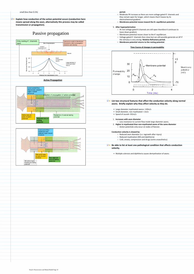

Learning Objectives
Define the essential components required for neurotransmitter release
Understand the differences between excitatory and inhibitory transmission
Define at least two mechanisms for the termination of neurotransmitter action at the synapse
Describe how modulation of the synaptic properties of GABA can be modulated pharmacologically to treat epilepsy
Features of neurotransmission
Transfer across a synapse requires the release of neurotransmitters and their interaction with postsynaptic receptors
bull
Transmitter released from 1st cell synaptic activation of 2nd cell signal integration and signal conduction by 2nd cell
bull
Rapid timescale1)Adaptability2)Plasticity 3)Diversity4)Learning and memory5)
Features
RAPDL- sounds like rapidbullDendrites receive inputs where it is integrated in the somabull
SynapseGap is about 20-100 nmbull5000 molecules of neurotransmitter per synaptic vesiclebullHigh energy dependency is satiated by high levels of mitochondriabullPost synaptic knob is electron densebull
Features of neurotransmitters
Provide enormous diversity in variety of transmitters and their receptorsbullAmino acids (eg glutamate gamma amino butyric acid [GABA] amines (eg noradrenaline [NA] and dopamine [DA] and neuropeptides (eg opioid peptides)
bull
May mediate rapid (micro to milli second responses) or slower effects (ms)bullVary in abundance from mM to nM CNS tissue concentrationbullNeurones receive multiple transmitter influence which are integrated to produce diverse functional responses
bull
Define the essential components required for neurotransmitter release
Essential componentsAn action potential is necessary to initiate neurotransmitter releasebullIt has to be very fast within ms (200 micro seconds)bullCalcium is essential- transmitter release requires an increase in intracellular Ca2+ to about 200microM
bull
A protein complex formation between vesicle membrane and cytoplasmic proteins to enable both vesicle docking and a rapid response to Ca entry leading to membrane fusion and exocytosis
bull
ATP and vesicle recyclingbull
Extra information on neurotransmittersSynaptic transmission is restricted to specialised structures ie the synapsebullSynaptic vesicles (SVs) provide the source of neurotransmitter (4000-10000 molecules per SV)
bull
Vary from mM to nM in CNS tissue concentrationsbull5-10 mM amino acids in brainbull
Activation of transmitter release is calcium dependent and requires rapid transduction
bull
Series of events= Membrane depolarisation-gt Calcium influx -gt Vesicle fusion -gt Vesicle exocytosis-gt Transmitter release
bull
From calcium influx to transmitter release has to be within this 200 microsecondsbullSuch rapid release rates can be achieved due to the vesicles being primed at the active zone close to Ca entry
bull
Localisation of vesicle at pre synaptic area Also alignment with calcium channel must occur so the response is rapid this is also helped by proteins associating between presynaptic membrane and vesicle to enable docking
bull
Vesicular proteins are targets for neurotoxinsbull
Types of neurotoxins
Botulinum toxin and tetanus destroys membrane association proteins and vesicles1Black widow spider- alpha latrotoxin that interferes with resealing of membrane so neurotransmitter is released to depletion
2
Vesicular proteins aretargets for neurotoxins
TETANUS toxinCtetani causes paralysis
BOTULINUM toxinCbotulinum causes flacid paralysis
alpha LATROTOXINblack widow spiderstimulates transmitterrelease to depletion
Zn2+-dependent endopeptidasesinhibit transmitter release
Steps of synaptic transmission
Biosynthesis packaging and release of neurotransmitter1
Membrane depolarisationaCalcium channels openbCalcium influxcVesicle fusiondVesicle exocytosis- fusion of vesicle to membrane and release of transmittereTransmitter releasef
Release
Receptor action2
Ion channel receptor- Extremely fast (msecs) mediate all fast excitatory and inhibitory transmission
bull
Glutamate can be synthesized from glucose via the TCA cycle
GABA is synthesised from Glutamate by removing a carboxyl group form N terminal Glutamate decarboxylase
CNS- Glutamate GABA
NMJ- Acetylcholine at nicotinic receptors
Examples bull
G- protein coupled receptor- Slow secs mins- Effectors may be enzymes (adenyl cyclase phospholipase C cGMP-PDE) or channels (eg Ca or K) Abundant in CNS
bull
CNS and PNS ACh at muscarinic receptors dopamine noradrenaline 5hydroxytryptamine (5ht) and neuropeptides eg enkaphalin
Examplesbull
Understand the differences between excitatory and inhibitory transmission
Glutamates effects propagated by sodiumbullimport in the post synaptic membraneExcitatory postsynaptic EPSPbull(membrane is becoming depolarised)
Excitatory-
Define at least two mechanisms for the termination of neurotransmitter action at the synapse
Excitatory CNS synapse mediated by Glutamate
G G
G G G
G
Excitatory amino acid
Transporter EAAT
GLIAL
CELL
Na+ Na+ and Ca2+
glucose
TCAcycleaKG
Glutamate
receptor
GLUR
GLUTAMINE
SYNTHETASE
Glutamine
Glutamate
Neurotransmission10 January 20121408
Stuarts Neuroscience and Mental Health Page 11
import in the post synaptic membraneExcitatory postsynaptic EPSPbull(membrane is becoming depolarised)
Excitatory postsynpatic potential (EPSP)
GABA- which results from Chlorine bullentering the post synaptic neuroneInhibitory postsynaptic EPSP (membrane bullis hyperpolarized which makes it veryhard to generate an action potential
Inhibitory-
Glutamate receptors
Alpha amino-3-hydroxy-5methyl-4-isoxazole propionic acid (AMPA)bullMajority of fast excitatory synapses with rapid onset offset and desensitisationbullInitiated by just Na entrybull
AMPA receptors
N-methyl-aspartatebullSlow component of excitatory transmissionbullServe as coincidence detectors which underlie learning mechanismsbullInitiate by Na and Ca entrybull
NMDA receptors
G G
G G G
G
Excitatory amino acid
Transporter EAAT
GLIAL
CELL
Na+ Na+ and Ca2+
glucose
TCAcycleaKG
Glutamate
receptor
GLUR
GLUTAMINE
SYNTHETASE
Glutamine
Glutamate
GLUR receptor takes up Na and Ca in PosSM (Post synaptic Membrane)bullGlutamate is the transported by Excitatory amino acid Transporter (EAAT) to glial cells
bull
Here it is converted to glutamine by glutamine synthasebullHence the glial cell has removed the neurotransmitter link to Cells of the Nervous System
bull
Inhibitory CNS synapse mediated by GABA
GABA GABA
GABA GABA GABA
GABA
GABA transporter GAT
GLIAL
CELL
Cl- Cl-
glucose
TCAcycle
aKG
GABA receptor
GABAAR
Cl-
GLUTAMATE
Glutamic acid
decarboxylase
GAD (B6)
GABA trans
-aminase
GABA-T
Succinate
semialdehyde
GABA
Glutamate is converted to GABA by Glutamic acid decarboxylase (GAD)bullGABA has effects on PSM receptor which causes chloride influxbullGABA is then relocated to Glial cell by GABA Transport (GAT)bullGABA trans aminase GABA-T converted GABA to Succinate semialdehydebull
Describe how modulation of the synaptic properties of GABA can be modulated pharmacologically to treat epilepsy
In epilepsy there is an excess of glutamate which causes abnormal neuronal firingbullThe concept of GABA drugs is that they will be used to inhibit the effects of glutamatebull
Frequency of opening of channel is increased when binding to benzodiazepinesbullBarbiturates- Open channel for longerbull
alpha
szlig
GABA
barbiturates
steroids
benzodiazepines
Zn
convulsantsethanol
Pentameric organisation of the GABA receptorand pharmacologically important binding domains
Drugs facilitating GABA transmission areantiepilepticanxiolyticsedativemuscle relaxant
Qu 1 Which of the following is NOT a normal route for neurotransmitter inactivation
Addition of a methyl group in the synaptic cleft
X Diffusion out of the cleft
Enzymatic destruction in the synaptic cleft-
Re-uptake by the pre-synaptic terminal- NE
Uptake by glial cells- GABA glutamate
X Mark = -2 (conf=2 )
Best Option Addition of a methyl group in the synaptic cleftSuch a method of inactivation does not existLecture Neurotransmitters
Pasted from lthttpswwwuclacuklaptlaptlitesysrunhtmicl08_neurof=cleari=icl1k=1u=_st1511i=Imperialgt
Stuarts Neuroscience and Mental Health Page 12
Session 4 Tutorial ndash Neurotransmitters and epilepsy
Tutorial notes
1 Epilepsy terminology
The term epilepsy refers to a disorder of brain function characterized by the periodic and unpredictable occurrence of seizures
bull
The term seizure refers to a transient alteration of behaviour due to the disordered synchronous and rhythmic firing of populations of brain neurones
bull
The pharmacological agents in current clinical use for inhibition of seizures are referred to as anticonvulsant or antiepileptic drugs
bull
Seizures are thought to arise from the cerebral cortex and they can be classified into bull
Partial seizures those beginning focally at a cortical site
Generalized seizures those that involve both hemispheres widely from the outset
The behavioural manifestations of a seizure are determined by the functions normally served by the cortical site at which the seizure arises
bull
LEFT HEMISPHERE WOULD CAUSE RIGHT SIDED JERKING
Thus for example a seizure involving the motor cortex is associated with clonic jerking of the body part controlled by this region of the cortex
bull
A simple partial seizure is associated with preservation of consciousness whilst acomplex partial seizure is associated with impairment of consciousness
bull
Examples of generalized seizures include absence myoclonic and tonic-clonic seizures You will be shown examples of the principal seizure types by video presentation during this teaching session
bull
2 Neurotransmitters in epilepsy
Epilepsy is a neurological disorder associated with abnormal neurotransmitter functionin the brain
bull
A decrease in GABA-mediated inhibition or an increase in glutamate-mediated excitation in the brain may result in seizure activity Indeed both glutamate and GABA are thought to play key roles in the brain mechanisms causing epilepsy in man
bull
Pharmacological evidence for a role of neurotransmitters in epilepsy
Dr Martin Croucher
Impairment of GABA-mediated inhibition causes seizures in animals eg impairment of synthesis release (tetanus toxin) or postsynaptic action (bicuculline picrotoxin)
bull
Enhancement of GABA-mediated inhibition leads to seizure suppression eg central (icv) administration of GABA or inhibition of the GABA metabolizing enzyme GABA-T (vigabatrin)
bull
Many clinically useful anticonvulsant drugs are known to act at least in part by potentiating central GABA-mediated inhibition eg benzodiazepines phenobarbital (see Section 3)
bull
Central (icv focal) administration of glutamate or glutamate receptor agonists causes seizure-like activity in animals
bull
Glutamate receptor antagonists are anticonvulsant in experimental models of epilepsy
bull
Some therapeutically effective anticonvulsant drugs act partly by blocking glutamate-mediated excitation in the brain eg phenobarbital
bull
Biochemical evidence for a role of neurotransmitters in epilepsy
Cobalt-induced seizures in rodents are associated with increased glutamate release
and with decreased GABA concentration GAD activity and GABA uptake (probably reflecting GABA neurone loss) at the seizure focus
bull
Audiogenic seizures in mice (DBA2 mice) are associated with glutamate receptor
binding in the brain and with GABA release from depolarized brain slices
bull
The baboon Papio papio which is highly sensitive to photically-induced seizures has a lower than normal CSF GABA concentration
bull
3 Some examples of antiepileptic drugs
Notes
Many neurological conditions are from malfunction in brainbullGABA malfunctions in epilepsy with propensity to repeated seizuresbull~1 in 100200bull
50 have well controlled epilepsy
25 have good control but side effects
25 have complex partial epilepsy which is poorly controlled
In epilepsy 502525 is an important ratiobull
Depression is associated with excessive noradrenalinebull
5 of people can have a seizure and 1 have epilepsybull
Epileptic focus- Excessively synchronous or sustained discharge of a group of neurones in the brain
Intracerebroventricular into lateral ventricles
ValiumLibrium are benzodiazepines
Whole list Important for exams possibly
Epilepsy can be causes by infection head trauma tumoursMeningitis-encephalitis Gliomas Stroke- ischaemic damage May occur in ~10 strokesGABA-interneurones small in CNS
Place in skull of rats and causes epileptic focus
Loud sounds causes seizures
Strobe of certain light frequency gives epilepsy Assumed that CSF is typical of brain
Generalized Seizures
Tonic- Clonic- Grand mal Tonic extension is continuous extensions which may cause the person to fall over Clonic rhythmical jerking 23 minutes
Absence- Petit mal Bit of eye fluttering 1015 secs phase out Occur very often
Myoclonic- Brief involuntary twitching of a muscle
Atonic- Lose muscle tone and posture fall forward onto face Epic face plant
Partial seizures-
Focal seizures can attribute to one direct point
Simple- Right motor cortex- left side activity Twitch tremor and face Can develop into secondary generalised seizure
Complex-Impairment of consciousness Lip smacking Temporal lobe epilepsy
Secondary generalised- Partial expands to general seizures
Parkinsons disease is caused by a lack of dopaminebullAkenisa- awkward gait Pill rolling tremorbullAlzheimers is caused by a lack of acetylcholinebull
4 Questions to be addressed
Review the process of neurotransmission occurring at central synapses utilising the inhibitory neurotransmitter GABA
bull
Present a convincing case for a role of neurotransmitter (glutamate or GABA) malfunction in the aetiology of epilepsy
bull
Draw a diagram to illustrate the principal steps in GABAergic neurotransmission (synthesis storage release etc) Indicate on your diagram i) established and ii) potential new target
bull
Epilepsy20 January 20120954
Stuarts Neuroscience and Mental Health Page 13
________________________________________________________________
Potential for drugs to solely target specific GABAA receptors to ensure only anti-convulsant and stop side effect of drowsiness
bull
Long acting- suicide anti convulsant
GABASynapse
Present a convincing case for a role of neurotransmitter (glutamate or GABA) malfunction in the aetiology of epilepsy
bull
Draw a diagram to illustrate the principal steps in GABAergic neurotransmission (synthesis storage release etc) Indicate on your diagram i) established and ii) potential new target sites for drugs acting to enhance GABA-mediated neurotransmission in the brain
bull
Stuarts Neuroscience and Mental Health Page 14
Learning Objectives
Draw a diagram to explain the relationship between the following major divisions of the CNS spinal cord brainstem cerebellum diencephalon cerebral hemispheres
Define the 3 components of the brainstem and state the main functions of the brainstem
Describe the functions of the 2 main structures in the diencephalon
State the functions of the basal ganglia and the cerebellum
Draw on a diagram of the cerebral hemisphere the cortical lobes and primary cortical areas
Recognise the main structures of the brain in a diagram or MRI
Describe the 3 layers of the meninges and explain their role in protecting the brain
Explain how the major divisions of the brain relate to the cranial fossae in the base of the skull
Explain the relationship between the spinal segments spinal nerves and vertebrae and state at what level a lumbar puncture can be performed safely
Define the functions of the dorsal and ventral horns of the spinal cord and explain how the dorsal and ventral roots and spinal nerves relate to them
Identify the components of the ventricular system and relate them to the divisions of the CNS
Explain the composition circulation and functions of CSF
State the average total volume and flow rate of CSF
Define hydrocephalus and outline how it may be treated
Distinguish between an epidural (extradural) and subdural haemorrhage
Draw a diagram to explain the relationship between the following major divisions of the CNS spinal cord brainstem cerebellum diencephalon cerebral hemispheres
pons
midbrain
medulla spinal cord
cerebellum
cerebral
hemisphere
diencephalon
brainstem
Define the 3 components of the brainstem and state the main functions of the brainstem
Midbrain1Pons-usually very tubular and has a bulge on the anterior surface2Medulla- With pons and cerebellum lies in hindbrain3
Midbrain pons and medulla share a lot of their functions These include controlling vital functions such as breathing consciousness and also innervating cranial nerve function
bull
Cranial Nerves
All are part of the PNS apart from the optic nervebullFunctional components less regularly organised than for spinal nervesbullSupply motor and sensory innervation to the headbullAutonomic (parasympathetic) innervation to head thoracic and abdominal organsbullSpecial senses eg vision hearing balancebull
Cerebral Hemispheres
Cortex of grey matter superficially that is responsible for most brain functions
1
Basal ganglia deep within brain that are involved with movement2
The cerebral hemisphere can be divided into two main partsbull
There is also the Corpus callosum which is a tract of fibres that connect the two hemispheres
bull
Central sulcus separates off the frontal lobe from the parietal lobebullSulcus are grooves and gyri are foldsbull
Cerebellum
Co-ordinates movement in terms of timing accuracy and precision (TAP) It may also have some involvement in cognitive function and in the fear and pleasure response
bull
TAP that cerebellumbullThe hindbrain contains the pons medulla and cerebellum
State the functions of the basal ganglia and the cerebellum
Draw on a diagram of the cerebral hemisphere the cortical lobes and primary cortical areas
Primary motor cortex is a strip Imagine that there is a man lying upside in the strip so damage to bottom of strip would affect his head or upper body
bull
Primary visual cortex- This area receives information from the retinas if there is a malfunction here it is likely you would become blind
bull
Auditory cortex receives information from inner earbullOther associated cortex areas seem to be related to higher functionbullWernickes area is involved with understanding language apparently according to recent literature the area should be moved and will then have more in common with experimental data and the area in primates
bull
Brocas area- Making speech case study evolved from a patient who had neurosyphillis destroying just that particular area and couldnt talk This area is more prominent in the left hemisphere
bullIdentify the components of the ventricular system and relate them to the divisions of the CNS
lateral ventricle
aqueduct
third ventricle
fourth ventricle
central canal
Describe the functions of the 2 main structures in the diencephalon
The diencephalon is a structure above the brain stembullIt is composed of the thalamus which relays information from lower structures to the cerebral cortex and the hypothalamus which is essential in coordinating homeostatic mechanisms and is the interface between CNS ANS and endocrine system
bull
Central sulcus
Lateral sulcus
CNS Lectures + Practicals31 January 20120900
Stuarts Neuroscience and Mental Health Page 15
lateral ventricle
aqueduct
third ventricle
fourth ventricle
central canal
Region of brain Region of ventricular system
Cerebral hemisphere Lateral ventricle
Diencephalon 3rd Ventricle
Midbrain Aqueduct
Pons and Medulla Fourth Ventricle
Lateral ventricle is C-shaped and there is one present in each hemispherebullFourth ventricle lies in front of the cerebellum and behind the pons and the medullabullHalfway down medulla it becomes the central canal and goes down into spinal cordbullEpendymal cells line the ventricular systembullChoroid plexus secretes CSFbull
Explain the composition circulation and functions of CSFState the average total volume and flow rate of CSF
The CSF is secreted by choroid plexus a structure within the lateral 3rd and 4th ventricles of the brain
bull
Most of the CSF leaves the 4th ventricle via holes and circulates via outside of the brain between the meninge membranes
bull
Lower number of cellsi)Decreased amount of proteinii)Lower Potassium and sodiumiii)Increased Magnesium and chlorine concentrationiv)
Differences between CSF and blood includebull
Circulates through ventricular system and subarachnoid space formed by the meninge membranes
bull
The CSF is reabsorbed into the venous sinuses via arachnoid villi which stops the accumulation of fluid possible oedema and a raised ICP
bull
Functions of CSF
Helps protective the brain against damage by suspending it in liquid 1Also has a few metabolic functions and waste products and helps distribute certain hormones
2
500ml per day is the flow rateTotal volume at any one time is 150ml
Describe the 3 layers of the meninges and explain their role in protecting the brain
3 layers of the meninges
Dura mater- Tough outer layer1Arachnoid mater - Delicate middle layer2Pia mater- Layer firmly attached to the surface of the brain3
Arachnoid villus drains CSF fluid into the venous sinusbull
Meningitis
Meningitis is most likely to infect Pia Mater and subarachnoid space but in the worst cases it can also spread to the upper layers of the cortex
bull
In order to determine between viral and bacterial and meningitis one can look at the CSFbull
BacterialThere should be a high white cell count in particular neutrophilsbullProtein concentration would be increased whereas there would be a decrease in glucose concentration- dangerous for brain metabolism
bull
Bacteria may actually be themselves identifiablebull
ViralLymphocytes account for the predominant change in leukocytes countbullProtein and glucose are normalbullViral identification is unlikelybull
Recognise the main structures of the brain in a diagram or MRI
Brain cut in mid-sagittal plane
Explain how the major divisions of the brain relate to the cranial fossae in the base of the skull
Inside of base of skull
Explain the relationship between the spinal segments spinal
Stuarts Neuroscience and Mental Health Page 16
Protein and glucose are normalbullViral identification is unlikelybull
Inferior aspect of brain
Cranial Fossae Part of brain
Anterior Cranial Frontal Lobe
Middle Cranial Temporal Lobe
Posterior Cranial Cerebellum
Lies above sphenoid bone Hypothalamus
Passes through the Foramen Magnum Medulla
Explain the relationship between the spinal segments spinal nerves and vertebrae and state at what level a lumbar puncture can be performed safely
Terminal end of spinal cord is called Conus MedullarisbullThere are 5 spinal segments cervical thoracic lumbar sacral and coccyx 7 12 5 5 4
bull
Spinal nerves are associated with each specific segment ie cervical etc but they do not necessarily have the same number of nerves to vertebrae and they terminate in different areas
bull
Type of vertebrae
Number of Vertebrae
Number of spinal nerves
Relationship of nerve to vertebrae
Cervical 7 8 Above
Thoracic 12 12 Below
Lumbar 5 5 Below
Sacrum 5 5 Below
Coccyx 4 (1 fused) 1 Below
The safe area to perform a lumber puncture is between L3 and L4- this is because the spinal cord has finished before this are and the nerves can be pushed aside by the needle without damage
bull
Spinal cord is much shorter than the vertebral column because the spinal cord develops mostly in the embryo whereas bones take much longer to develop
bull
Define the functions of the dorsal and ventral horns of the spinal cord and explain how the dorsal and ventral roots and spinal nerves relate to them
VRRMM VRRMM- sound like an engine VM= Ventral + Motor neurone cell bodies The more ridiculous the better right
Ventral Horn- Contains motor neurone cell bodiesbull
DI- Dorsal + interneurones Investigate what the bloody hell goes on in the spine (Detective Inspector- DI)
Dorsal Horn- Contains interneurones which receive sensory information from the body
bull
Afferent pathway is receives of sensory information via dorsal horn efferent pathway is motor information leaving the spinal cord Spinal nerve is collection on both and thus has bidirectional information transfer
bull
Grey matter contains the neuronal cell bodies and it is the white matter that contains the bundles of axons
bull
Define hydrocephalus and outline how it may be treated
Hydrocephalus- Water on the brain caused by CSF flow abnormalities
Communicating- All 4 ventricles affecteda)Non- communicating- Not all 4 enlargedb)
2 predominant types
Block in CSF absorption or CSF over brain surface caused bybullMeningitisbullHead injurybullCongenitalbull
MHCH
Haemorrhage (sub-arachnoid)bull
Communicating-
Block in ventricular system caused bybullAqueduct stenosisbullVentricular tumoursbull
AVPParaventricular tumoursbull
Non- Communicating-
Distinguish between an epidural (extradural) and subdural haemorrhage
EMA- Extradural= meningeal artery
Extradural haemorrhage is usually due to a damaged meningeal artery between the skull and the dura after head trauma
bull
Subdural haemorrhage- Usually due to a damaged vein between the dura and arachnoid membrane
bull
Both of these pathologies can cause a space-occupying lesion in the confined space bounded by the membranes and hence cause neurological pathologies
bull
Symptoms such as headache drowsiness vomiting or seizure are more likely to present quickly from an arterial (extradural) than a subdural haemorrhage
bull
Can also be confirmed by imagingbull
Subdural are more common than epiduralbullSubdural haemorrhages are more frequently seen in alcoholics the elderly patients on anticoagulants whereas epiduralextradural haemorrhages more commonly arise from blows to the side of the head
Pasted from lthttpswwwuclacuklaptlaptlitesysrunhtmicl08_neurof=cleari=icl1k=1u=_st1511i=Imperialgt
Stuarts Neuroscience and Mental Health Page 17
AVP
HeadachebullDrowsinessbullBlackoutsbullRaised intracranial pressurebullIncreased head circumference (in child)bull
Symptoms
Remove causes eg papillomabullPut in a shunt to divert CSFbullOpen alternate pathway- ventriculostomybull
Treatment
Stuarts Neuroscience and Mental Health Page 18
Brachial plexus forms at level C5-T1bull
Macrophage clears up debris along proximal end of axoniAxon sends at pseudopodia (projections of membrane) to make contact with Schwann cell which it grows through and coats it in myelin
ii
Axon reaches distal end of axon as a complete entityiiiNodes of Ranvier are reinserted however there are usually more than there was before which slows nerve conduction down slightly
iv
Axon sustains damage eg in example by forceps crushing it
Growth of axon is about 2-5mm per day
Soma undergoes chromatolysis
Failure to re-join proximal to distal end can result in Neuroma
Nerve degenerationbull
Qu 2 Which of the following statements about peripheral nerves is INCORRECT
Axons form bundles called fascicles
They may contain axons from difference spinal nerves
radic They contain only myelinated nerve fibres
They contain a mixture of fibres of different diameter
They contain a mixture of fibres of different conduction velocities
Pasted from lthttpswwwuclacuklaptlaptlitesysrunhtmicl08_neurof=cleari=icl1k=1u=_st1511i=Imperialgt
Definitions
Dorsal ramus- Posterior branches of the spinal nerves that are smaller than anteriorventral divisions Carry visceral motor somatic motor and sensory information to and from the skin and deep muscles of the backVentral ramus- Supply the antero-lateral parts of the trunk and limbs they are for the most part larger than the posterior divisionsDorsal root ganglion- Nodule on dorsal root that contains cell bodies of neurons in afferent spinal nervesDorsal root- Afferent sensory root of a spinal nerveVentral root- Efferent motor root of a spinal nerveGray ramus communicans- Contain unmyelinated postganglionic sympathetic fibres that are from ganglion of sympathetic trunkWhite ramus communicans- Preganglionic sympathetic outflow from the spinal cord
Additional info on white ramus communicans
The thoracic and the first and second lumbar nerves each contribute a branch white ramus communicans to the adjoining sympathetic ganglion Unlike the gray rami white rami communicants do not extend below L2 or above T1
bull
PNS18 May 20122302
Stuarts Neuroscience and Mental Health Page 19
Learning Objectives
Describe the basic anatomy of ANS innervation in terms of pre-ganglionic neurons ganglia and post-ganglionic fibres Contrast the anatomical location of sympathetic and parasympathetic ganglia
Describe the sympathetic and parasympathetic pathways and their centralspinal connections
Describe the thoracolumbar (sympathetic) and craniosacral (parasympathetic) central origins of the ANS
Describe the influence of the sympathetic and parasympathetic nervous system on the principal systemsorgans of the body (eg cardiovascular system lung gut exocrine glands) and understand the concept of dual innervation and autonomic tone (giving examples)
Understand the importance of the sacral parasympathetic outflow for innervation of structures within the pelvis eg the bladder
Classify the cholinoceptors found within the autonomic nervous system and identify the principal loci of (a) the nicotinic cholinoceptors and (b) the muscarinic cholinoceptors Note that the nicotinic receptors are ion-gated and the muscarinic receptors are G-protein coupled
Identify the neurotransmitter substances released at different levels within the autonomic nervous system and describe the principal steps involved in their biosynthesis and metabolism
CNS- ANS or SMS or NES
Principal ANS components
Exocrine glandsbullSmooth musclebullCardiac musclebullMetabolismbullHost defencebull
General information on ANS
Sympathetic- Fight and flightParasympathetic- Rest and digest
Often sympathetic and parasympathetic in many tissues are antagonists with often inhibitory functions
bull
An interesting point of note is that there are only parasympathetic nerves innervating the lungs which results in bronchoconstriction
bull
Furthermore although sympathetic is usually associated with dilation of blood vessels- maximum oxygen to muscles the sympathetic nervous system can cause vasoconstriction depending on the particular vessel receptor
bull
At rest there is usually a balance between parasympathetic and sympathetic nervous system
bull
SALIVARY GLANDS
Thick viscious secretion
Copious watery secretionSKIN
Piloerection
Increased sweating (C)HEART
rate and contractility
rate and contractility
GASTROINTESTINAL
motility and tone
Sphincter contraction
motility and tone
secretions
BLOOD VESSELS
Constriction
(skin mucous membranes and splanchnic area)
EYE
Dilatation (Pupil)
Constriction (Pupil)
Contraction (Ciliary Muscle)
TRACHEA AND BRONCHEOLES
Dilates (Ad)
Constriction
LIVER
Glycogenolysis
Gluconeogenesis
ADIPOSE
Lipolysis
KIDNEY
Increased renin secretion
URETERS AND BLADDER
Relaxes detrusor constriction of
trigone and sphincter
Contraction of detrusor relaxation
of trigone and sphincter
BLOOD VESSELS
(skeletal muscle) Dilatation
Yellow = sympathetic
Pink = parasympathetic
C = sympathetic cholinergic
Describe the thoracolumbar (sympathetic) and craniosacral (parasympathetic) central origins of the ANS
Parasympathetic efferent output
Known as craniosacral- although it may affect head there are no cervical nerves
bull
4 cranial nerves are in red Vagus is most interesting as it descends into thorax and has effect on the lungs and heart etc
bull
Sacral nerves are located inferiorly to cranial nerves and innervate lower internal viscera
bull
Cervical
Thoracic
Lumbar
Sacral
Lungs intestine
rectum
Reproductive organsSplachnic
nerve
Heart
Stomach
Pancreas
Small intestine
Lung
Eye
Lacrimal and salivary glands
Oculomotor nerve
Vagus
Glossopharyngeal nerve
Facial nerve
Describe the basic anatomy of ANS innervation in terms of pre-ganglionic neurons ganglia and post-ganglionic fibres Contrast the anatomical location of sympathetic and parasympathetic ganglia
Ganglia are a collection of neuronal cell bodies within the Peripheral Nervous System This is also where the neurons synapse
bull
The parasympathetic system has very long pre-ganglionic fibres cell bodies originate in CNS and move towards effector organs
bull
Ganglion are very close to effector organ- therefore short post-ganglionicbull
Parasympathetic- craniosacral
Sympathetic- thoracolumbar
Ganglion are grouped in structures just outside of spinal cord called the sympathetic trunk- this allows a very coordinated response
bull
Allows mass discharge of sympathetic nervesbullCan pass up and down sympathetic trunkbullOpposite set up very short pre-ganglionic fibres and very long post ganglionic fibres
bull
Innervation to adrenal medulla is atypical No official ganglion but adrenal medulla does almost act like one Short neurone from spinal cord and no post ganglionic neurone- neurotransmitters are released from medulla into blood stream instead
bull
Eye
Lacrimal and salivary glands
Lung
Heart
Foregut
Adrenal medulla
Midgut
Hindgut
Pelvic organs
All chain ganglia
supply the skin and
blood vessels
Describe the influence of the sympathetic and parasympathetic nervous system on the principal systemsorgans of the body (eg cardiovascular system lung gut exocrine glands) and understand the concept of dual innervation and autonomic tone (giving examples)
Cardiovascular
Cardiac output= SV x HR
Sympathetic activity increases the force of contraction of cardiac muscle (inotropic effect) therefore increases SV
bull
Increases HR (chronotropic effect)bullResult is a large increase in Cardiac Outputbull
Total peripheral resistance- Dual innervation
Sympathetic tone particularly affects arteriolesbullIncrease in sympathetic= vasoconstriction- increase TPRbullDecreased in sympathetic= vasodilation- decrease TPRbull
The combination of increased TPR and CO should result in increased arterial blood pressure
Vasodilation
Exceptions
Increased sympathetic activity to some blood vessels in skeletal muscle either cholinergic fibres or have adrenergic beta-receptors
bull
Local vasodilators (eg CO2 increased H+ nitric oxide and histamine etcbull
Sympathetic efferent output
Known as thoracolumbar refer to anatomy section for more detailbull
Autonomic Nervous System07 February 20120859
Stuarts Neuroscience and Mental Health Page 20
Cervical
Thoracic
Lumbar
Sacral
Lungs intestine
rectum
Reproductive organsSplachnic
nerve
Heart
Stomach
Pancreas
Small intestine
Lung
Eye
Lacrimal and salivary glands
Oculomotor nerve
Vagus
Glossopharyngeal nerve
Facial nerve
Eye
Lacrimal and salivary glands
Lung
Heart
Foregut
Adrenal medulla
Midgut
Hindgut
Pelvic organs
All chain ganglia
supply the skin and
blood vessels
Exceptions
Increased sympathetic activity to some blood vessels in skeletal muscle either cholinergic fibres or have adrenergic beta-receptors
bull
Local vasodilators (eg CO2 increased H+ nitric oxide and histamine etcbullAlthough parasympathetic doesnt usually have an effect it can have an effect in certain organs such as the penis clitoris GI and salivary glands
bull
Gastrointestinal tract
Sympathetic Activity Parasympathetic Activity
Generally decreases motility and tone Increases motility and tone
Usually stimulates contraction of sphincters
Usually causes relaxation of sphincters
Generally inhibits secretory activity Generally stimulates secretory activity
Think rest and digest Plus is digesting of food really an important focus if there was a lion in the lecture theatre
How can sympathetic affect lungs if there are no nerve fibres going there
Sympathetic effects via the adrenal glands and the secretion of adrenalineAdrenaline induces bronchodilation which increases oxygen delivery to the lungs
ANS innervation of the eye
Sympathetic activity Parasympathetic activity
Contracts radial muscle (pupil dilation)
Contracts pupillary sphincter (pupil contracts)
Contracts ciliary muscle lens bulges for near vision
Understand the importance of the sacral parasympathetic outflow for innervation of structures within the pelvis eg the bladder
Outline
Within the bladder there is both voluntary and ANS controlbullMain influences are parasympathetic coming from sacral region of the spinal cord
bull
Ganglion is located virtually within the tissuebullParasympathetic nerves fibres causes Detrussor muscle to contract and thus urination occurs
bull
Sympathetic nerves from the sympathetic trunk controls the internal sphincter whereby innervation closes it
bull
Voluntary control over sphincter carried by motor nervesbull
Detailed
Baroreceptors in the bladder sends information up the afferent pelvic nerveto sacral spinal cord this is known as the Micturition Reflex This reflex innervates the parasympathetic system and makes you more likely to urinateneed to urinate
bull
Afferent and efferent neurons are usually found together within the same larger nerve
bull
Decreased sympathetic outflow via hypogastric nervebullPudendal nerve is a somatic nerve that controls the ability to consciously urinate
bull
If pressure gets too great than voluntary control is overcomebull
Identify the neurotransmitter substances released at different levels within the autonomic nervous system and describe the principal steps involved in their biosynthesis and metabolism
The general synthesis of a neurotransmitter is as follows
Enzymatic conversion of precursor to active form1
Classify the cholinoceptors found within the autonomic nervous system and identify the principal loci of (a) the nicotinic cholinoceptors and (b) the muscarinic cholinoceptors Note that the nicotinic receptors are ion-gated and the muscarinic receptors are G-protein coupled
Acetylcholine
ACh binds to Nicotinic or Muscarinic receptors Nicotinic is an ion channel linked receptor that is present in the ganglion This ensures that pre-post ganglionic transmission is extremely quick In effector organs we have Muscarinic which is a G-Protein Coupled Receptor This is slower but still quick
bull
Adrenergic receptors are bound by noradrenaline which is a G-Protein coupled receptor
bull
Not actually activepresent until enzyme converts from dopaminebullDoes not get broken down in the synapse It has transportreuptake proteins that transports it back to the cell
bull
Degraded in neuronebullUptake 1- back to neuronebullUptake 2- extracellular tissuebull
Noradrenaline
PRESSURE
BUILDS UP
IN BLADDER
Internal sphincter
(relaxes with
decreased
sympathetic activity)
Sympathetic
control
Hypogastric nerve
parasympathetic
control Detrussor muscle
(contracts)
Parasympathetic
ganglion
Main Influence
Motor nerves
(voluntary control)
External sphincter
(relaxes following central inhibition)
Pudendal nerve
Sacral
segments of
spinal cord
Pelvic nerve
(micturition reflex)
Sympathetic efferent output
Known as thoracolumbar refer to anatomy section for more detailbull
Identify the neurotransmitter substances released at different levels within the autonomic nervous system and describe the principal steps involved in their biosynthesis and metabolism
Noradrenaline adrenaline and also dopamine belong to a group of chemicals called catecholamines because they have a catel ring
bull
All parasympathetic neurones release acetylcholine and it is ACh that also effects target organs
bull
Most sympathetic- Pre= Acetylcholine Post= Noradrenalinebull
Adrenal glands- ACh causes production of predominantly A (but also NA) by medulla
Some have ACh as pre and post ganglionic This is where sympathetic nervous are influencing secretion- sweat
Noradrenaline is main in sympathetic
Other variations exceptionsbull
Principal metabolites of noradrenaline and adrenaline
Noradrenaline Adrenaline
3-methoxy-4 hydroxy-
phenylethelene glycol
(MOPEG)Conjugation (glucuronide or sulphate)
Urine
Catechol -O-methyl transferase (COMT)
Monoamine oxidase A (MOA-A)
Stuarts Neuroscience and Mental Health Page 21
involved in their biosynthesis and metabolism
The general synthesis of a neurotransmitter is as follows
Enzymatic conversion of precursor to active form1Transmitter packaged into vesicles close to the surface of the neurone2Stimulation of nerve causes calcium entry into the neurone3Promotes exocytosis of neurotransmitter into synapse4Moves into synapse5Binds to receptor- random bouncing around by diffusion6Degradation of neurotransmitter by enzyme at some stage7Reuptake of certain molecules for resynthesis8
Acetylcholine
Same steps as above but the precursors are the chemicals acetyl Co-A and choline
bull
The enzyme that catalyses the formation of acetylcholine is Choline Acetyl Transferase
bull
Finally the enzyme that degrades ACh is Acetylcholin-esterasebull
Noradrenaline
The synthesis of Noradrenaline has a few more unique reactions that that of ACh
bull
Tyrosine enters the pre-synaptic neurone and is converted by Tyrosine Hydroxylase to form DOPA (dihydroxyphenylalanine) This reaction is the rate limiting step
bull
DOPA is converted by the enzyme DOPA carboxylase to dopaminebullDopamine is then packaged into a vesicle within the enzyme Dopamine βhydroxylase which converts it to Noradrenaline
bull
Unlike with ACh there is no further metabolism of the neurotransmitter after it has been bound to its receptor Instead a set of uptake proteins either take it back to the pre-synaptic neurone (Uptake Protein 1) or to the post-synaptic neurone (Uptake Protein 2)
bull
PreMOdona- Presynaptic= Mono-amine oxidase
In the PreSN an enzyme called Mono-amine oxidase A breaks down the noradrenaline back down to its metabolites inside the mitochondria
Way of remembering- Youll catch a cold if you are broken down
In the PostSN an enzyme called COMT (Catechol-O-Methyl Transferase) degrades the noradrenaline
Degredation of the noradrenaline is different depending upon its locationbull
Acetyl Co A
+
CholineCholine acetyl
transferaseACh + Co A
ACh
ACh
ACh
ACh
ReceptorEffector cell
Choline +
AcetateCa++
ACh = acetylcholine
6
3
7
8
1
2
4
5
NA
Ca++
Tyrosine
DOPA
DopamineDopamine
Dopamine hydroxylase
Tyrosine
hydroxylase
DOPA decarboxylase Monoamine oxidase
A (MAO-A)
Noradrenaline
NA
Metabolites
Tyrosine
NA
Adrenoceptor
Uptake 1
Uptake 2
Degradation
(COMT)
Tyrosine
hydroxylaseMetabolites
DOPA
Dopamine
Tyrosine
hydroxylaseDOPA
Dopamine
6
37
8
1
2
4
5
1
1
8
A
Ca++
Tyrosine
DOPA
DopamineDopamine
Dopamine
hydroxylase
Tyrosine
hydroxylase
DOPA decarboxylase
NoradrenalineNA
Metabolites
Tyrosine
A
Tyrosine
hydroxylaseDOPA
Dopamine
Tyrosine
hydroxylaseDOPA
Dopamine
6
3
1
2
4
5
1
1A
Phenylethanolamine
Methyl transferase
CapillaryA
Principal metabolites of noradrenaline and adrenaline
Noradrenaline Adrenaline
3-methoxy-4 hydroxy-
phenylethelene glycol
(MOPEG)Conjugation (glucuronide or sulphate)
Urine
Catechol -O-methyl transferase (COMT)
Monoamine oxidase A (MOA-A)
Sympathetic Parasympathetic
Is a diffuse system which allows it to stimulated multiple parts of the body at once (Mass discharge)
Relatively discrete system innervating individual target tissues via specific nerves
Can also have more discrete effects
The sympathetic nervous system
The acute stress response
BRAIN
(hypothalamus
and brain stem)
STRESS
ACTIONS
Spinal cord
ADRENAL
MEDULLA
sympathetic
ganglia
pre-ganglionic fibres post-ganglionic fibres
CATECHOLAMINES
Fight or flight response
First described by Cannon in 1915 as a mass sympathetic discharge in response to distress or alarm
bull
Results inIncreased arterial blood pressure1Increased blow flow to muscles and decreased levels to other areas such as splanchnic bed
2
Increased blood glucose concentration3Increased respiration4Increased awareness5
Adrenaline
Looks similar to noradrenaline in terms of synthesis but you must note that Adrenaline is NOT a neurotransmitter it is a hormone This is why there is no PostSN but a capillary instead
bull
Furthermore note that it is synthesised in Adrenal Chromaffin Cells which are specialised sympathetic post-ganglionic fibres
bull
Same initial steps as noradrenaline but Noradrenaline leaves vesicle and is changed by enzyme Phenylethanolamine Methyl Transferase (PNMT) to give Adrenaline
bull
Stuarts Neuroscience and Mental Health Page 22
Learning Objectives
Describe the basic anatomy of ANS innervation in terms of pre-ganglionic neurons ganglia and post-ganglionic fibres Contrast the anatomical location of sympathetic and parasympathetic ganglia
Describe the thoracolumbar (sympathetic) and craniosacral (parasympathetic) central origins of the ANS
Understand the terms sympathetic trunk plexus and subsidiary ganglia
Identify the rich sympathetic plexuses that surround the major organs and blood vessels
Understand the pre-ganglionic nature of the thoracic and lumbar splanchnic nerves and their synapses in subsidiary ganglia eg coeliac ganglion
Understand the importance of the sacral parasympathetic outflow for innervation of structures within the pelvis eg the bladder
Identify which of the cranial nerves contain parasympathetic pre-ganglionic fibres
Describe the basic anatomy of ANS innervation in terms of pre-ganglionic neurons ganglia and post-ganglionic fibres Contrast the anatomical location of sympathetic and parasympathetic gangliaDescribe the thoracolumbar (sympathetic) and craniosacral (parasympathetic) central origins of the ANS
Sympathetic
Sympathetic trunks (x2)aNerve plexusesbSubsidiary gangliac
Largest division of the ANS which consists ofbull
To cause vasoconstriction in blood vessels
Secretomotor function in sweat glands and motor function in the erector pilli muscles of hair
To accompany motor nerves to voluntary muscles but only distributed to blood vessels supplying the muscles
Viscera- dilation of pupils dilation of arterioles movement of alimentary tract and urinary bladder (ie relaxes wall constricts sphincters)
Functionsbull
Thoracic spinal nerves
Upper lumbar spinal nerves (L1-3)
Preganglionic efferent fibres arise frombull
Pre ganglionic neurons are very short and are situated in the lateral column grey matter of spinal cord T1 to L3
bull
They emerge from the spinal cord via the ventral root of the spinal nervebullThey pass through ventral ramus to white rami communicans to ganglionbullSynapse within ganglion or send fibres up and down to other ganglionbull
Postganglionic fibres are distributed to effector organ via grey rami communicansbullTravel in dorsal or ventral ramibull
Understand the importance of the sacral parasympathetic outflow for innervation of structures within the pelvis eg the bladder
Parasympathetic
Cranial nerves
Sacral (spinal) nerves
Preganglionic efferent fibres arise frombull
Anterior rami of S2-4
Visceral branches passing directly to pelvic viscera ie pelvic splanchnic nerves
Minute ganglia in wall of viscera giving rise to post ganglionic fibres
Sacral outflowbull
Motor fibres to rectum and bladder wall
Inhibitory fibres to bladder sphincter
Erection of penisclitoris via vasodilator fibres
Fibres also pass superiorly to supply large part of the gut with visceromotor innervation
Pelvic splanchnic nerves
Understand the terms sympathetic trunk plexus and subsidiary ganglia
The sympathetic trunks are located on either side of the spinal cord and go from the base of the skull to the coccyx and are divided as so
bull
Cervical- 3 ganglia- Superior cervical ganglion follows internal carotid arteryThoracic- 11 or 12 gangliaLumbar- 4 or 5 gangliaSacral- 4 or 5 ganglia
Sympathetic Trunks
Identify which of the cranial nerves contain parasympathetic pre-ganglionic fibres
Anatomy of the ANS19 May 20122057
Stuarts Neuroscience and Mental Health Page 23
Horners syndrome- Loss of sympathetic supply around the eye Ptosis- drooping upper eye lid Pinpoint pupil- unopposed parasympathetic activity Anhydrosis- loss of sweating around the eye
bull
Caused by tumour in the thorax- apex of lungbull
All parasympathetic ganglions are subsidiary ganglionsbullTherefore I would attempt to define a subsidiary ganglion as sobullSubsidiary ganglion- Any ganglions that are not part of the sympathetic system and are located next to the organ they innervate
bull
Identify the rich sympathetic plexuses that surround the major organs and blood vessels
Plexus around pharynx
Cardiac plexus
Thyroid plexus
PCTP
Pulmonary plexus
Cervicalbull
Plexus around thoracic aorta
Thoracic aorta supply
Pierces diaphragm and enters abdominal plexus through the great vessels
Greater
Pierces diaphragm and enter abdominal plexus through the aorta
Lesser
Pierces diaphragm and enters abdominal plexus through gut
Least
Splanchnic nerves
Thoracicbull
4 lumbar ganglia
Lumbar splanchnic nerves take part in all plexi of sympathetic nerves in abdominal and pelvic regions
Lumbarbull
Sympathetic Plexuses
Identify which of the cranial nerves contain parasympathetic pre-ganglionic fibres
Ciliary ganglionbullPost ganglionic fibres to sphincter pupillae and ciliary muscle inside the eyebull
Oculomotor Nerve (CN III)
Submandibular ganglionbullPost ganglionic fibres to submandibular and sublingual salivary glandsbullPterygopalatine ganglionbullPost ganglionic fibres to paranasal sinuses and lacrimal glandsbull
Facial nerve (CN VII)
Otic ganglionbullPostganglionic fibres to parotid glandbull
Glossopharyngeal nerve (CN IX)
Enters neck and thorax via carotid sheathbullBranches to lungs heart esophagus stomach and intestinesbull
3 7 9 10bull
Vagus nerve (CN X)
Enteric System
Myenteric (Auerbachs) plexus
Submucous (Meissners) plexus
In walls of alimentary tractbull
Sensory- monitoring mechanical chemical and hormonal activity of gutbullMotor- gut motility secretion vessel tonebullCan be overridden by sympathetic and parasympathetic systemsbull
Qu 5 Provides cannular access to the sacral epidural space for the administration of anaesthetics
Best Option Sacral hiatusThis is a naturally occurring gap at the lower end of the sacrum Because there is a failure of the laminae of the last sacral segments to coalesce this space is available for cannular use
Pasted from lthttpswwwuclacuklaptlaptlitesysrunhtmicl08_neurof=cleari=icl1k=1u=_st1511i=Imperialgt
Stuarts Neuroscience and Mental Health Page 24
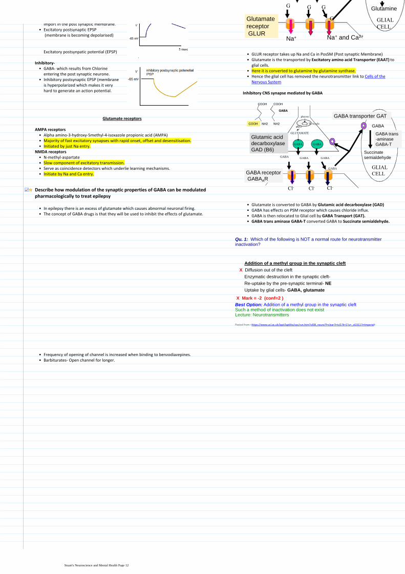
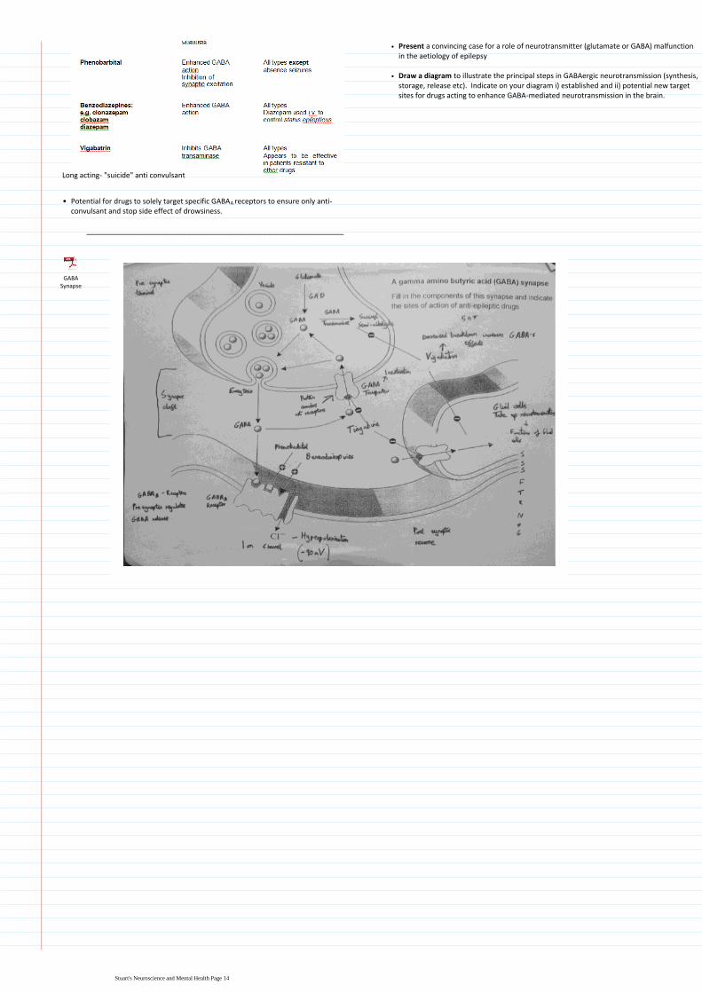

Learning Objectives
Draw a diagram to explain the relationship between the following major divisions of the CNS spinal cord brainstem cerebellum diencephalon cerebral hemispheres
Define the 3 components of the brainstem and state the main functions of the brainstem
Describe the functions of the 2 main structures in the diencephalon
State the functions of the basal ganglia and the cerebellum
Draw on a diagram of the cerebral hemisphere the cortical lobes and primary cortical areas
Recognise the main structures of the brain in a diagram or MRI
Describe the 3 layers of the meninges and explain their role in protecting the brain
Explain how the major divisions of the brain relate to the cranial fossae in the base of the skull
Explain the relationship between the spinal segments spinal nerves and vertebrae and state at what level a lumbar puncture can be performed safely
Define the functions of the dorsal and ventral horns of the spinal cord and explain how the dorsal and ventral roots and spinal nerves relate to them
Identify the components of the ventricular system and relate them to the divisions of the CNS
Explain the composition circulation and functions of CSF
State the average total volume and flow rate of CSF
Define hydrocephalus and outline how it may be treated
Distinguish between an epidural (extradural) and subdural haemorrhage
Draw a diagram to explain the relationship between the following major divisions of the CNS spinal cord brainstem cerebellum diencephalon cerebral hemispheres
pons
midbrain
medulla spinal cord
cerebellum
cerebral
hemisphere
diencephalon
brainstem
Define the 3 components of the brainstem and state the main functions of the brainstem
Midbrain1Pons-usually very tubular and has a bulge on the anterior surface2Medulla- With pons and cerebellum lies in hindbrain3
Midbrain pons and medulla share a lot of their functions These include controlling vital functions such as breathing consciousness and also innervating cranial nerve function
bull
Cranial Nerves
All are part of the PNS apart from the optic nervebullFunctional components less regularly organised than for spinal nervesbullSupply motor and sensory innervation to the headbullAutonomic (parasympathetic) innervation to head thoracic and abdominal organsbullSpecial senses eg vision hearing balancebull
Cerebral Hemispheres
Cortex of grey matter superficially that is responsible for most brain functions
1
Basal ganglia deep within brain that are involved with movement2
The cerebral hemisphere can be divided into two main partsbull
There is also the Corpus callosum which is a tract of fibres that connect the two hemispheres
bull
Central sulcus separates off the frontal lobe from the parietal lobebullSulcus are grooves and gyri are foldsbull
Cerebellum
Co-ordinates movement in terms of timing accuracy and precision (TAP) It may also have some involvement in cognitive function and in the fear and pleasure response
bull
TAP that cerebellumbullThe hindbrain contains the pons medulla and cerebellum
State the functions of the basal ganglia and the cerebellum
Draw on a diagram of the cerebral hemisphere the cortical lobes and primary cortical areas
Primary motor cortex is a strip Imagine that there is a man lying upside in the strip so damage to bottom of strip would affect his head or upper body
bull
Primary visual cortex- This area receives information from the retinas if there is a malfunction here it is likely you would become blind
bull
Auditory cortex receives information from inner earbullOther associated cortex areas seem to be related to higher functionbullWernickes area is involved with understanding language apparently according to recent literature the area should be moved and will then have more in common with experimental data and the area in primates
bull
Brocas area- Making speech case study evolved from a patient who had neurosyphillis destroying just that particular area and couldnt talk This area is more prominent in the left hemisphere
bullIdentify the components of the ventricular system and relate them to the divisions of the CNS
lateral ventricle
aqueduct
third ventricle
fourth ventricle
central canal
Describe the functions of the 2 main structures in the diencephalon
The diencephalon is a structure above the brain stembullIt is composed of the thalamus which relays information from lower structures to the cerebral cortex and the hypothalamus which is essential in coordinating homeostatic mechanisms and is the interface between CNS ANS and endocrine system
bull
Central sulcus
Lateral sulcus
CNS Lectures + Practicals31 January 20120900
Stuarts Neuroscience and Mental Health Page 15
lateral ventricle
aqueduct
third ventricle
fourth ventricle
central canal
Region of brain Region of ventricular system
Cerebral hemisphere Lateral ventricle
Diencephalon 3rd Ventricle
Midbrain Aqueduct
Pons and Medulla Fourth Ventricle
Lateral ventricle is C-shaped and there is one present in each hemispherebullFourth ventricle lies in front of the cerebellum and behind the pons and the medullabullHalfway down medulla it becomes the central canal and goes down into spinal cordbullEpendymal cells line the ventricular systembullChoroid plexus secretes CSFbull
Explain the composition circulation and functions of CSFState the average total volume and flow rate of CSF
The CSF is secreted by choroid plexus a structure within the lateral 3rd and 4th ventricles of the brain
bull
Most of the CSF leaves the 4th ventricle via holes and circulates via outside of the brain between the meninge membranes
bull
Lower number of cellsi)Decreased amount of proteinii)Lower Potassium and sodiumiii)Increased Magnesium and chlorine concentrationiv)
Differences between CSF and blood includebull
Circulates through ventricular system and subarachnoid space formed by the meninge membranes
bull
The CSF is reabsorbed into the venous sinuses via arachnoid villi which stops the accumulation of fluid possible oedema and a raised ICP
bull
Functions of CSF
Helps protective the brain against damage by suspending it in liquid 1Also has a few metabolic functions and waste products and helps distribute certain hormones
2
500ml per day is the flow rateTotal volume at any one time is 150ml
Describe the 3 layers of the meninges and explain their role in protecting the brain
3 layers of the meninges
Dura mater- Tough outer layer1Arachnoid mater - Delicate middle layer2Pia mater- Layer firmly attached to the surface of the brain3
Arachnoid villus drains CSF fluid into the venous sinusbull
Meningitis
Meningitis is most likely to infect Pia Mater and subarachnoid space but in the worst cases it can also spread to the upper layers of the cortex
bull
In order to determine between viral and bacterial and meningitis one can look at the CSFbull
BacterialThere should be a high white cell count in particular neutrophilsbullProtein concentration would be increased whereas there would be a decrease in glucose concentration- dangerous for brain metabolism
bull
Bacteria may actually be themselves identifiablebull
ViralLymphocytes account for the predominant change in leukocytes countbullProtein and glucose are normalbullViral identification is unlikelybull
Recognise the main structures of the brain in a diagram or MRI
Brain cut in mid-sagittal plane
Explain how the major divisions of the brain relate to the cranial fossae in the base of the skull
Inside of base of skull
Explain the relationship between the spinal segments spinal
Stuarts Neuroscience and Mental Health Page 16
Protein and glucose are normalbullViral identification is unlikelybull
Inferior aspect of brain
Cranial Fossae Part of brain
Anterior Cranial Frontal Lobe
Middle Cranial Temporal Lobe
Posterior Cranial Cerebellum
Lies above sphenoid bone Hypothalamus
Passes through the Foramen Magnum Medulla
Explain the relationship between the spinal segments spinal nerves and vertebrae and state at what level a lumbar puncture can be performed safely
Terminal end of spinal cord is called Conus MedullarisbullThere are 5 spinal segments cervical thoracic lumbar sacral and coccyx 7 12 5 5 4
bull
Spinal nerves are associated with each specific segment ie cervical etc but they do not necessarily have the same number of nerves to vertebrae and they terminate in different areas
bull
Type of vertebrae
Number of Vertebrae
Number of spinal nerves
Relationship of nerve to vertebrae
Cervical 7 8 Above
Thoracic 12 12 Below
Lumbar 5 5 Below
Sacrum 5 5 Below
Coccyx 4 (1 fused) 1 Below
The safe area to perform a lumber puncture is between L3 and L4- this is because the spinal cord has finished before this are and the nerves can be pushed aside by the needle without damage
bull
Spinal cord is much shorter than the vertebral column because the spinal cord develops mostly in the embryo whereas bones take much longer to develop
bull
Define the functions of the dorsal and ventral horns of the spinal cord and explain how the dorsal and ventral roots and spinal nerves relate to them
VRRMM VRRMM- sound like an engine VM= Ventral + Motor neurone cell bodies The more ridiculous the better right
Ventral Horn- Contains motor neurone cell bodiesbull
DI- Dorsal + interneurones Investigate what the bloody hell goes on in the spine (Detective Inspector- DI)
Dorsal Horn- Contains interneurones which receive sensory information from the body
bull
Afferent pathway is receives of sensory information via dorsal horn efferent pathway is motor information leaving the spinal cord Spinal nerve is collection on both and thus has bidirectional information transfer
bull
Grey matter contains the neuronal cell bodies and it is the white matter that contains the bundles of axons
bull
Define hydrocephalus and outline how it may be treated
Hydrocephalus- Water on the brain caused by CSF flow abnormalities
Communicating- All 4 ventricles affecteda)Non- communicating- Not all 4 enlargedb)
2 predominant types
Block in CSF absorption or CSF over brain surface caused bybullMeningitisbullHead injurybullCongenitalbull
MHCH
Haemorrhage (sub-arachnoid)bull
Communicating-
Block in ventricular system caused bybullAqueduct stenosisbullVentricular tumoursbull
AVPParaventricular tumoursbull
Non- Communicating-
Distinguish between an epidural (extradural) and subdural haemorrhage
EMA- Extradural= meningeal artery
Extradural haemorrhage is usually due to a damaged meningeal artery between the skull and the dura after head trauma
bull
Subdural haemorrhage- Usually due to a damaged vein between the dura and arachnoid membrane
bull
Both of these pathologies can cause a space-occupying lesion in the confined space bounded by the membranes and hence cause neurological pathologies
bull
Symptoms such as headache drowsiness vomiting or seizure are more likely to present quickly from an arterial (extradural) than a subdural haemorrhage
bull
Can also be confirmed by imagingbull
Subdural are more common than epiduralbullSubdural haemorrhages are more frequently seen in alcoholics the elderly patients on anticoagulants whereas epiduralextradural haemorrhages more commonly arise from blows to the side of the head
Pasted from lthttpswwwuclacuklaptlaptlitesysrunhtmicl08_neurof=cleari=icl1k=1u=_st1511i=Imperialgt
Stuarts Neuroscience and Mental Health Page 17
AVP
HeadachebullDrowsinessbullBlackoutsbullRaised intracranial pressurebullIncreased head circumference (in child)bull
Symptoms
Remove causes eg papillomabullPut in a shunt to divert CSFbullOpen alternate pathway- ventriculostomybull
Treatment
Stuarts Neuroscience and Mental Health Page 18
Brachial plexus forms at level C5-T1bull
Macrophage clears up debris along proximal end of axoniAxon sends at pseudopodia (projections of membrane) to make contact with Schwann cell which it grows through and coats it in myelin
ii
Axon reaches distal end of axon as a complete entityiiiNodes of Ranvier are reinserted however there are usually more than there was before which slows nerve conduction down slightly
iv
Axon sustains damage eg in example by forceps crushing it
Growth of axon is about 2-5mm per day
Soma undergoes chromatolysis
Failure to re-join proximal to distal end can result in Neuroma
Nerve degenerationbull
Qu 2 Which of the following statements about peripheral nerves is INCORRECT
Axons form bundles called fascicles
They may contain axons from difference spinal nerves
radic They contain only myelinated nerve fibres
They contain a mixture of fibres of different diameter
They contain a mixture of fibres of different conduction velocities
Pasted from lthttpswwwuclacuklaptlaptlitesysrunhtmicl08_neurof=cleari=icl1k=1u=_st1511i=Imperialgt
Definitions
Dorsal ramus- Posterior branches of the spinal nerves that are smaller than anteriorventral divisions Carry visceral motor somatic motor and sensory information to and from the skin and deep muscles of the backVentral ramus- Supply the antero-lateral parts of the trunk and limbs they are for the most part larger than the posterior divisionsDorsal root ganglion- Nodule on dorsal root that contains cell bodies of neurons in afferent spinal nervesDorsal root- Afferent sensory root of a spinal nerveVentral root- Efferent motor root of a spinal nerveGray ramus communicans- Contain unmyelinated postganglionic sympathetic fibres that are from ganglion of sympathetic trunkWhite ramus communicans- Preganglionic sympathetic outflow from the spinal cord
Additional info on white ramus communicans
The thoracic and the first and second lumbar nerves each contribute a branch white ramus communicans to the adjoining sympathetic ganglion Unlike the gray rami white rami communicants do not extend below L2 or above T1
bull
PNS18 May 20122302
Stuarts Neuroscience and Mental Health Page 19
Learning Objectives
Describe the basic anatomy of ANS innervation in terms of pre-ganglionic neurons ganglia and post-ganglionic fibres Contrast the anatomical location of sympathetic and parasympathetic ganglia
Describe the sympathetic and parasympathetic pathways and their centralspinal connections
Describe the thoracolumbar (sympathetic) and craniosacral (parasympathetic) central origins of the ANS
Describe the influence of the sympathetic and parasympathetic nervous system on the principal systemsorgans of the body (eg cardiovascular system lung gut exocrine glands) and understand the concept of dual innervation and autonomic tone (giving examples)
Understand the importance of the sacral parasympathetic outflow for innervation of structures within the pelvis eg the bladder
Classify the cholinoceptors found within the autonomic nervous system and identify the principal loci of (a) the nicotinic cholinoceptors and (b) the muscarinic cholinoceptors Note that the nicotinic receptors are ion-gated and the muscarinic receptors are G-protein coupled
Identify the neurotransmitter substances released at different levels within the autonomic nervous system and describe the principal steps involved in their biosynthesis and metabolism
CNS- ANS or SMS or NES
Principal ANS components
Exocrine glandsbullSmooth musclebullCardiac musclebullMetabolismbullHost defencebull
General information on ANS
Sympathetic- Fight and flightParasympathetic- Rest and digest
Often sympathetic and parasympathetic in many tissues are antagonists with often inhibitory functions
bull
An interesting point of note is that there are only parasympathetic nerves innervating the lungs which results in bronchoconstriction
bull
Furthermore although sympathetic is usually associated with dilation of blood vessels- maximum oxygen to muscles the sympathetic nervous system can cause vasoconstriction depending on the particular vessel receptor
bull
At rest there is usually a balance between parasympathetic and sympathetic nervous system
bull
SALIVARY GLANDS
Thick viscious secretion
Copious watery secretionSKIN
Piloerection
Increased sweating (C)HEART
rate and contractility
rate and contractility
GASTROINTESTINAL
motility and tone
Sphincter contraction
motility and tone
secretions
BLOOD VESSELS
Constriction
(skin mucous membranes and splanchnic area)
EYE
Dilatation (Pupil)
Constriction (Pupil)
Contraction (Ciliary Muscle)
TRACHEA AND BRONCHEOLES
Dilates (Ad)
Constriction
LIVER
Glycogenolysis
Gluconeogenesis
ADIPOSE
Lipolysis
KIDNEY
Increased renin secretion
URETERS AND BLADDER
Relaxes detrusor constriction of
trigone and sphincter
Contraction of detrusor relaxation
of trigone and sphincter
BLOOD VESSELS
(skeletal muscle) Dilatation
Yellow = sympathetic
Pink = parasympathetic
C = sympathetic cholinergic
Describe the thoracolumbar (sympathetic) and craniosacral (parasympathetic) central origins of the ANS
Parasympathetic efferent output
Known as craniosacral- although it may affect head there are no cervical nerves
bull
4 cranial nerves are in red Vagus is most interesting as it descends into thorax and has effect on the lungs and heart etc
bull
Sacral nerves are located inferiorly to cranial nerves and innervate lower internal viscera
bull
Cervical
Thoracic
Lumbar
Sacral
Lungs intestine
rectum
Reproductive organsSplachnic
nerve
Heart
Stomach
Pancreas
Small intestine
Lung
Eye
Lacrimal and salivary glands
Oculomotor nerve
Vagus
Glossopharyngeal nerve
Facial nerve
Describe the basic anatomy of ANS innervation in terms of pre-ganglionic neurons ganglia and post-ganglionic fibres Contrast the anatomical location of sympathetic and parasympathetic ganglia
Ganglia are a collection of neuronal cell bodies within the Peripheral Nervous System This is also where the neurons synapse
bull
The parasympathetic system has very long pre-ganglionic fibres cell bodies originate in CNS and move towards effector organs
bull
Ganglion are very close to effector organ- therefore short post-ganglionicbull
Parasympathetic- craniosacral
Sympathetic- thoracolumbar
Ganglion are grouped in structures just outside of spinal cord called the sympathetic trunk- this allows a very coordinated response
bull
Allows mass discharge of sympathetic nervesbullCan pass up and down sympathetic trunkbullOpposite set up very short pre-ganglionic fibres and very long post ganglionic fibres
bull
Innervation to adrenal medulla is atypical No official ganglion but adrenal medulla does almost act like one Short neurone from spinal cord and no post ganglionic neurone- neurotransmitters are released from medulla into blood stream instead
bull
Eye
Lacrimal and salivary glands
Lung
Heart
Foregut
Adrenal medulla
Midgut
Hindgut
Pelvic organs
All chain ganglia
supply the skin and
blood vessels
Describe the influence of the sympathetic and parasympathetic nervous system on the principal systemsorgans of the body (eg cardiovascular system lung gut exocrine glands) and understand the concept of dual innervation and autonomic tone (giving examples)
Cardiovascular
Cardiac output= SV x HR
Sympathetic activity increases the force of contraction of cardiac muscle (inotropic effect) therefore increases SV
bull
Increases HR (chronotropic effect)bullResult is a large increase in Cardiac Outputbull
Total peripheral resistance- Dual innervation
Sympathetic tone particularly affects arteriolesbullIncrease in sympathetic= vasoconstriction- increase TPRbullDecreased in sympathetic= vasodilation- decrease TPRbull
The combination of increased TPR and CO should result in increased arterial blood pressure
Vasodilation
Exceptions
Increased sympathetic activity to some blood vessels in skeletal muscle either cholinergic fibres or have adrenergic beta-receptors
bull
Local vasodilators (eg CO2 increased H+ nitric oxide and histamine etcbull
Sympathetic efferent output
Known as thoracolumbar refer to anatomy section for more detailbull
Autonomic Nervous System07 February 20120859
Stuarts Neuroscience and Mental Health Page 20
Cervical
Thoracic
Lumbar
Sacral
Lungs intestine
rectum
Reproductive organsSplachnic
nerve
Heart
Stomach
Pancreas
Small intestine
Lung
Eye
Lacrimal and salivary glands
Oculomotor nerve
Vagus
Glossopharyngeal nerve
Facial nerve
Eye
Lacrimal and salivary glands
Lung
Heart
Foregut
Adrenal medulla
Midgut
Hindgut
Pelvic organs
All chain ganglia
supply the skin and
blood vessels
Exceptions
Increased sympathetic activity to some blood vessels in skeletal muscle either cholinergic fibres or have adrenergic beta-receptors
bull
Local vasodilators (eg CO2 increased H+ nitric oxide and histamine etcbullAlthough parasympathetic doesnt usually have an effect it can have an effect in certain organs such as the penis clitoris GI and salivary glands
bull
Gastrointestinal tract
Sympathetic Activity Parasympathetic Activity
Generally decreases motility and tone Increases motility and tone
Usually stimulates contraction of sphincters
Usually causes relaxation of sphincters
Generally inhibits secretory activity Generally stimulates secretory activity
Think rest and digest Plus is digesting of food really an important focus if there was a lion in the lecture theatre
How can sympathetic affect lungs if there are no nerve fibres going there
Sympathetic effects via the adrenal glands and the secretion of adrenalineAdrenaline induces bronchodilation which increases oxygen delivery to the lungs
ANS innervation of the eye
Sympathetic activity Parasympathetic activity
Contracts radial muscle (pupil dilation)
Contracts pupillary sphincter (pupil contracts)
Contracts ciliary muscle lens bulges for near vision
Understand the importance of the sacral parasympathetic outflow for innervation of structures within the pelvis eg the bladder
Outline
Within the bladder there is both voluntary and ANS controlbullMain influences are parasympathetic coming from sacral region of the spinal cord
bull
Ganglion is located virtually within the tissuebullParasympathetic nerves fibres causes Detrussor muscle to contract and thus urination occurs
bull
Sympathetic nerves from the sympathetic trunk controls the internal sphincter whereby innervation closes it
bull
Voluntary control over sphincter carried by motor nervesbull
Detailed
Baroreceptors in the bladder sends information up the afferent pelvic nerveto sacral spinal cord this is known as the Micturition Reflex This reflex innervates the parasympathetic system and makes you more likely to urinateneed to urinate
bull
Afferent and efferent neurons are usually found together within the same larger nerve
bull
Decreased sympathetic outflow via hypogastric nervebullPudendal nerve is a somatic nerve that controls the ability to consciously urinate
bull
If pressure gets too great than voluntary control is overcomebull
Identify the neurotransmitter substances released at different levels within the autonomic nervous system and describe the principal steps involved in their biosynthesis and metabolism
The general synthesis of a neurotransmitter is as follows
Enzymatic conversion of precursor to active form1
Classify the cholinoceptors found within the autonomic nervous system and identify the principal loci of (a) the nicotinic cholinoceptors and (b) the muscarinic cholinoceptors Note that the nicotinic receptors are ion-gated and the muscarinic receptors are G-protein coupled
Acetylcholine
ACh binds to Nicotinic or Muscarinic receptors Nicotinic is an ion channel linked receptor that is present in the ganglion This ensures that pre-post ganglionic transmission is extremely quick In effector organs we have Muscarinic which is a G-Protein Coupled Receptor This is slower but still quick
bull
Adrenergic receptors are bound by noradrenaline which is a G-Protein coupled receptor
bull
Not actually activepresent until enzyme converts from dopaminebullDoes not get broken down in the synapse It has transportreuptake proteins that transports it back to the cell
bull
Degraded in neuronebullUptake 1- back to neuronebullUptake 2- extracellular tissuebull
Noradrenaline
PRESSURE
BUILDS UP
IN BLADDER
Internal sphincter
(relaxes with
decreased
sympathetic activity)
Sympathetic
control
Hypogastric nerve
parasympathetic
control Detrussor muscle
(contracts)
Parasympathetic
ganglion
Main Influence
Motor nerves
(voluntary control)
External sphincter
(relaxes following central inhibition)
Pudendal nerve
Sacral
segments of
spinal cord
Pelvic nerve
(micturition reflex)
Sympathetic efferent output
Known as thoracolumbar refer to anatomy section for more detailbull
Identify the neurotransmitter substances released at different levels within the autonomic nervous system and describe the principal steps involved in their biosynthesis and metabolism
Noradrenaline adrenaline and also dopamine belong to a group of chemicals called catecholamines because they have a catel ring
bull
All parasympathetic neurones release acetylcholine and it is ACh that also effects target organs
bull
Most sympathetic- Pre= Acetylcholine Post= Noradrenalinebull
Adrenal glands- ACh causes production of predominantly A (but also NA) by medulla
Some have ACh as pre and post ganglionic This is where sympathetic nervous are influencing secretion- sweat
Noradrenaline is main in sympathetic
Other variations exceptionsbull
Principal metabolites of noradrenaline and adrenaline
Noradrenaline Adrenaline
3-methoxy-4 hydroxy-
phenylethelene glycol
(MOPEG)Conjugation (glucuronide or sulphate)
Urine
Catechol -O-methyl transferase (COMT)
Monoamine oxidase A (MOA-A)
Stuarts Neuroscience and Mental Health Page 21
involved in their biosynthesis and metabolism
The general synthesis of a neurotransmitter is as follows
Enzymatic conversion of precursor to active form1Transmitter packaged into vesicles close to the surface of the neurone2Stimulation of nerve causes calcium entry into the neurone3Promotes exocytosis of neurotransmitter into synapse4Moves into synapse5Binds to receptor- random bouncing around by diffusion6Degradation of neurotransmitter by enzyme at some stage7Reuptake of certain molecules for resynthesis8
Acetylcholine
Same steps as above but the precursors are the chemicals acetyl Co-A and choline
bull
The enzyme that catalyses the formation of acetylcholine is Choline Acetyl Transferase
bull
Finally the enzyme that degrades ACh is Acetylcholin-esterasebull
Noradrenaline
The synthesis of Noradrenaline has a few more unique reactions that that of ACh
bull
Tyrosine enters the pre-synaptic neurone and is converted by Tyrosine Hydroxylase to form DOPA (dihydroxyphenylalanine) This reaction is the rate limiting step
bull
DOPA is converted by the enzyme DOPA carboxylase to dopaminebullDopamine is then packaged into a vesicle within the enzyme Dopamine βhydroxylase which converts it to Noradrenaline
bull
Unlike with ACh there is no further metabolism of the neurotransmitter after it has been bound to its receptor Instead a set of uptake proteins either take it back to the pre-synaptic neurone (Uptake Protein 1) or to the post-synaptic neurone (Uptake Protein 2)
bull
PreMOdona- Presynaptic= Mono-amine oxidase
In the PreSN an enzyme called Mono-amine oxidase A breaks down the noradrenaline back down to its metabolites inside the mitochondria
Way of remembering- Youll catch a cold if you are broken down
In the PostSN an enzyme called COMT (Catechol-O-Methyl Transferase) degrades the noradrenaline
Degredation of the noradrenaline is different depending upon its locationbull
Acetyl Co A
+
CholineCholine acetyl
transferaseACh + Co A
ACh
ACh
ACh
ACh
ReceptorEffector cell
Choline +
AcetateCa++
ACh = acetylcholine
6
3
7
8
1
2
4
5
NA
Ca++
Tyrosine
DOPA
DopamineDopamine
Dopamine hydroxylase
Tyrosine
hydroxylase
DOPA decarboxylase Monoamine oxidase
A (MAO-A)
Noradrenaline
NA
Metabolites
Tyrosine
NA
Adrenoceptor
Uptake 1
Uptake 2
Degradation
(COMT)
Tyrosine
hydroxylaseMetabolites
DOPA
Dopamine
Tyrosine
hydroxylaseDOPA
Dopamine
6
37
8
1
2
4
5
1
1
8
A
Ca++
Tyrosine
DOPA
DopamineDopamine
Dopamine
hydroxylase
Tyrosine
hydroxylase
DOPA decarboxylase
NoradrenalineNA
Metabolites
Tyrosine
A
Tyrosine
hydroxylaseDOPA
Dopamine
Tyrosine
hydroxylaseDOPA
Dopamine
6
3
1
2
4
5
1
1A
Phenylethanolamine
Methyl transferase
CapillaryA
Principal metabolites of noradrenaline and adrenaline
Noradrenaline Adrenaline
3-methoxy-4 hydroxy-
phenylethelene glycol
(MOPEG)Conjugation (glucuronide or sulphate)
Urine
Catechol -O-methyl transferase (COMT)
Monoamine oxidase A (MOA-A)
Sympathetic Parasympathetic
Is a diffuse system which allows it to stimulated multiple parts of the body at once (Mass discharge)
Relatively discrete system innervating individual target tissues via specific nerves
Can also have more discrete effects
The sympathetic nervous system
The acute stress response
BRAIN
(hypothalamus
and brain stem)
STRESS
ACTIONS
Spinal cord
ADRENAL
MEDULLA
sympathetic
ganglia
pre-ganglionic fibres post-ganglionic fibres
CATECHOLAMINES
Fight or flight response
First described by Cannon in 1915 as a mass sympathetic discharge in response to distress or alarm
bull
Results inIncreased arterial blood pressure1Increased blow flow to muscles and decreased levels to other areas such as splanchnic bed
2
Increased blood glucose concentration3Increased respiration4Increased awareness5
Adrenaline
Looks similar to noradrenaline in terms of synthesis but you must note that Adrenaline is NOT a neurotransmitter it is a hormone This is why there is no PostSN but a capillary instead
bull
Furthermore note that it is synthesised in Adrenal Chromaffin Cells which are specialised sympathetic post-ganglionic fibres
bull
Same initial steps as noradrenaline but Noradrenaline leaves vesicle and is changed by enzyme Phenylethanolamine Methyl Transferase (PNMT) to give Adrenaline
bull
Stuarts Neuroscience and Mental Health Page 22
Learning Objectives
Describe the basic anatomy of ANS innervation in terms of pre-ganglionic neurons ganglia and post-ganglionic fibres Contrast the anatomical location of sympathetic and parasympathetic ganglia
Describe the thoracolumbar (sympathetic) and craniosacral (parasympathetic) central origins of the ANS
Understand the terms sympathetic trunk plexus and subsidiary ganglia
Identify the rich sympathetic plexuses that surround the major organs and blood vessels
Understand the pre-ganglionic nature of the thoracic and lumbar splanchnic nerves and their synapses in subsidiary ganglia eg coeliac ganglion
Understand the importance of the sacral parasympathetic outflow for innervation of structures within the pelvis eg the bladder
Identify which of the cranial nerves contain parasympathetic pre-ganglionic fibres
Describe the basic anatomy of ANS innervation in terms of pre-ganglionic neurons ganglia and post-ganglionic fibres Contrast the anatomical location of sympathetic and parasympathetic gangliaDescribe the thoracolumbar (sympathetic) and craniosacral (parasympathetic) central origins of the ANS
Sympathetic
Sympathetic trunks (x2)aNerve plexusesbSubsidiary gangliac
Largest division of the ANS which consists ofbull
To cause vasoconstriction in blood vessels
Secretomotor function in sweat glands and motor function in the erector pilli muscles of hair
To accompany motor nerves to voluntary muscles but only distributed to blood vessels supplying the muscles
Viscera- dilation of pupils dilation of arterioles movement of alimentary tract and urinary bladder (ie relaxes wall constricts sphincters)
Functionsbull
Thoracic spinal nerves
Upper lumbar spinal nerves (L1-3)
Preganglionic efferent fibres arise frombull
Pre ganglionic neurons are very short and are situated in the lateral column grey matter of spinal cord T1 to L3
bull
They emerge from the spinal cord via the ventral root of the spinal nervebullThey pass through ventral ramus to white rami communicans to ganglionbullSynapse within ganglion or send fibres up and down to other ganglionbull
Postganglionic fibres are distributed to effector organ via grey rami communicansbullTravel in dorsal or ventral ramibull
Understand the importance of the sacral parasympathetic outflow for innervation of structures within the pelvis eg the bladder
Parasympathetic
Cranial nerves
Sacral (spinal) nerves
Preganglionic efferent fibres arise frombull
Anterior rami of S2-4
Visceral branches passing directly to pelvic viscera ie pelvic splanchnic nerves
Minute ganglia in wall of viscera giving rise to post ganglionic fibres
Sacral outflowbull
Motor fibres to rectum and bladder wall
Inhibitory fibres to bladder sphincter
Erection of penisclitoris via vasodilator fibres
Fibres also pass superiorly to supply large part of the gut with visceromotor innervation
Pelvic splanchnic nerves
Understand the terms sympathetic trunk plexus and subsidiary ganglia
The sympathetic trunks are located on either side of the spinal cord and go from the base of the skull to the coccyx and are divided as so
bull
Cervical- 3 ganglia- Superior cervical ganglion follows internal carotid arteryThoracic- 11 or 12 gangliaLumbar- 4 or 5 gangliaSacral- 4 or 5 ganglia
Sympathetic Trunks
Identify which of the cranial nerves contain parasympathetic pre-ganglionic fibres
Anatomy of the ANS19 May 20122057
Stuarts Neuroscience and Mental Health Page 23
Horners syndrome- Loss of sympathetic supply around the eye Ptosis- drooping upper eye lid Pinpoint pupil- unopposed parasympathetic activity Anhydrosis- loss of sweating around the eye
bull
Caused by tumour in the thorax- apex of lungbull
All parasympathetic ganglions are subsidiary ganglionsbullTherefore I would attempt to define a subsidiary ganglion as sobullSubsidiary ganglion- Any ganglions that are not part of the sympathetic system and are located next to the organ they innervate
bull
Identify the rich sympathetic plexuses that surround the major organs and blood vessels
Plexus around pharynx
Cardiac plexus
Thyroid plexus
PCTP
Pulmonary plexus
Cervicalbull
Plexus around thoracic aorta
Thoracic aorta supply
Pierces diaphragm and enters abdominal plexus through the great vessels
Greater
Pierces diaphragm and enter abdominal plexus through the aorta
Lesser
Pierces diaphragm and enters abdominal plexus through gut
Least
Splanchnic nerves
Thoracicbull
4 lumbar ganglia
Lumbar splanchnic nerves take part in all plexi of sympathetic nerves in abdominal and pelvic regions
Lumbarbull
Sympathetic Plexuses
Identify which of the cranial nerves contain parasympathetic pre-ganglionic fibres
Ciliary ganglionbullPost ganglionic fibres to sphincter pupillae and ciliary muscle inside the eyebull
Oculomotor Nerve (CN III)
Submandibular ganglionbullPost ganglionic fibres to submandibular and sublingual salivary glandsbullPterygopalatine ganglionbullPost ganglionic fibres to paranasal sinuses and lacrimal glandsbull
Facial nerve (CN VII)
Otic ganglionbullPostganglionic fibres to parotid glandbull
Glossopharyngeal nerve (CN IX)
Enters neck and thorax via carotid sheathbullBranches to lungs heart esophagus stomach and intestinesbull
3 7 9 10bull
Vagus nerve (CN X)
Enteric System
Myenteric (Auerbachs) plexus
Submucous (Meissners) plexus
In walls of alimentary tractbull
Sensory- monitoring mechanical chemical and hormonal activity of gutbullMotor- gut motility secretion vessel tonebullCan be overridden by sympathetic and parasympathetic systemsbull
Qu 5 Provides cannular access to the sacral epidural space for the administration of anaesthetics
Best Option Sacral hiatusThis is a naturally occurring gap at the lower end of the sacrum Because there is a failure of the laminae of the last sacral segments to coalesce this space is available for cannular use
Pasted from lthttpswwwuclacuklaptlaptlitesysrunhtmicl08_neurof=cleari=icl1k=1u=_st1511i=Imperialgt
Stuarts Neuroscience and Mental Health Page 24
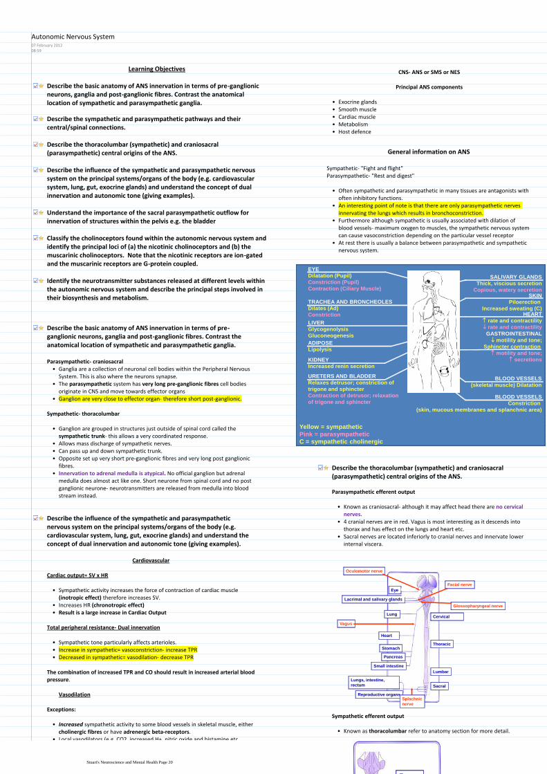
Learning Objectives
Describe the basic anatomy of ANS innervation in terms of pre-ganglionic neurons ganglia and post-ganglionic fibres Contrast the anatomical location of sympathetic and parasympathetic ganglia
Describe the sympathetic and parasympathetic pathways and their centralspinal connections
Describe the thoracolumbar (sympathetic) and craniosacral (parasympathetic) central origins of the ANS
Describe the influence of the sympathetic and parasympathetic nervous system on the principal systemsorgans of the body (eg cardiovascular system lung gut exocrine glands) and understand the concept of dual innervation and autonomic tone (giving examples)
Understand the importance of the sacral parasympathetic outflow for innervation of structures within the pelvis eg the bladder
Classify the cholinoceptors found within the autonomic nervous system and identify the principal loci of (a) the nicotinic cholinoceptors and (b) the muscarinic cholinoceptors Note that the nicotinic receptors are ion-gated and the muscarinic receptors are G-protein coupled
Identify the neurotransmitter substances released at different levels within the autonomic nervous system and describe the principal steps involved in their biosynthesis and metabolism
CNS- ANS or SMS or NES
Principal ANS components
Exocrine glandsbullSmooth musclebullCardiac musclebullMetabolismbullHost defencebull
General information on ANS
Sympathetic- Fight and flightParasympathetic- Rest and digest
Often sympathetic and parasympathetic in many tissues are antagonists with often inhibitory functions
bull
An interesting point of note is that there are only parasympathetic nerves innervating the lungs which results in bronchoconstriction
bull
Furthermore although sympathetic is usually associated with dilation of blood vessels- maximum oxygen to muscles the sympathetic nervous system can cause vasoconstriction depending on the particular vessel receptor
bull
At rest there is usually a balance between parasympathetic and sympathetic nervous system
bull
SALIVARY GLANDS
Thick viscious secretion
Copious watery secretionSKIN
Piloerection
Increased sweating (C)HEART
rate and contractility
rate and contractility
GASTROINTESTINAL
motility and tone
Sphincter contraction
motility and tone
secretions
BLOOD VESSELS
Constriction
(skin mucous membranes and splanchnic area)
EYE
Dilatation (Pupil)
Constriction (Pupil)
Contraction (Ciliary Muscle)
TRACHEA AND BRONCHEOLES
Dilates (Ad)
Constriction
LIVER
Glycogenolysis
Gluconeogenesis
ADIPOSE
Lipolysis
KIDNEY
Increased renin secretion
URETERS AND BLADDER
Relaxes detrusor constriction of
trigone and sphincter
Contraction of detrusor relaxation
of trigone and sphincter
BLOOD VESSELS
(skeletal muscle) Dilatation
Yellow = sympathetic
Pink = parasympathetic
C = sympathetic cholinergic
Describe the thoracolumbar (sympathetic) and craniosacral (parasympathetic) central origins of the ANS
Parasympathetic efferent output
Known as craniosacral- although it may affect head there are no cervical nerves
bull
4 cranial nerves are in red Vagus is most interesting as it descends into thorax and has effect on the lungs and heart etc
bull
Sacral nerves are located inferiorly to cranial nerves and innervate lower internal viscera
bull
Cervical
Thoracic
Lumbar
Sacral
Lungs intestine
rectum
Reproductive organsSplachnic
nerve
Heart
Stomach
Pancreas
Small intestine
Lung
Eye
Lacrimal and salivary glands
Oculomotor nerve
Vagus
Glossopharyngeal nerve
Facial nerve
Describe the basic anatomy of ANS innervation in terms of pre-ganglionic neurons ganglia and post-ganglionic fibres Contrast the anatomical location of sympathetic and parasympathetic ganglia
Ganglia are a collection of neuronal cell bodies within the Peripheral Nervous System This is also where the neurons synapse
bull
The parasympathetic system has very long pre-ganglionic fibres cell bodies originate in CNS and move towards effector organs
bull
Ganglion are very close to effector organ- therefore short post-ganglionicbull
Parasympathetic- craniosacral
Sympathetic- thoracolumbar
Ganglion are grouped in structures just outside of spinal cord called the sympathetic trunk- this allows a very coordinated response
bull
Allows mass discharge of sympathetic nervesbullCan pass up and down sympathetic trunkbullOpposite set up very short pre-ganglionic fibres and very long post ganglionic fibres
bull
Innervation to adrenal medulla is atypical No official ganglion but adrenal medulla does almost act like one Short neurone from spinal cord and no post ganglionic neurone- neurotransmitters are released from medulla into blood stream instead
bull
Eye
Lacrimal and salivary glands
Lung
Heart
Foregut
Adrenal medulla
Midgut
Hindgut
Pelvic organs
All chain ganglia
supply the skin and
blood vessels
Describe the influence of the sympathetic and parasympathetic nervous system on the principal systemsorgans of the body (eg cardiovascular system lung gut exocrine glands) and understand the concept of dual innervation and autonomic tone (giving examples)
Cardiovascular
Cardiac output= SV x HR
Sympathetic activity increases the force of contraction of cardiac muscle (inotropic effect) therefore increases SV
bull
Increases HR (chronotropic effect)bullResult is a large increase in Cardiac Outputbull
Total peripheral resistance- Dual innervation
Sympathetic tone particularly affects arteriolesbullIncrease in sympathetic= vasoconstriction- increase TPRbullDecreased in sympathetic= vasodilation- decrease TPRbull
The combination of increased TPR and CO should result in increased arterial blood pressure
Vasodilation
Exceptions
Increased sympathetic activity to some blood vessels in skeletal muscle either cholinergic fibres or have adrenergic beta-receptors
bull
Local vasodilators (eg CO2 increased H+ nitric oxide and histamine etcbull
Sympathetic efferent output
Known as thoracolumbar refer to anatomy section for more detailbull
Autonomic Nervous System07 February 20120859
Stuarts Neuroscience and Mental Health Page 20
Cervical
Thoracic
Lumbar
Sacral
Lungs intestine
rectum
Reproductive organsSplachnic
nerve
Heart
Stomach
Pancreas
Small intestine
Lung
Eye
Lacrimal and salivary glands
Oculomotor nerve
Vagus
Glossopharyngeal nerve
Facial nerve
Eye
Lacrimal and salivary glands
Lung
Heart
Foregut
Adrenal medulla
Midgut
Hindgut
Pelvic organs
All chain ganglia
supply the skin and
blood vessels
Exceptions
Increased sympathetic activity to some blood vessels in skeletal muscle either cholinergic fibres or have adrenergic beta-receptors
bull
Local vasodilators (eg CO2 increased H+ nitric oxide and histamine etcbullAlthough parasympathetic doesnt usually have an effect it can have an effect in certain organs such as the penis clitoris GI and salivary glands
bull
Gastrointestinal tract
Sympathetic Activity Parasympathetic Activity
Generally decreases motility and tone Increases motility and tone
Usually stimulates contraction of sphincters
Usually causes relaxation of sphincters
Generally inhibits secretory activity Generally stimulates secretory activity
Think rest and digest Plus is digesting of food really an important focus if there was a lion in the lecture theatre
How can sympathetic affect lungs if there are no nerve fibres going there
Sympathetic effects via the adrenal glands and the secretion of adrenalineAdrenaline induces bronchodilation which increases oxygen delivery to the lungs
ANS innervation of the eye
Sympathetic activity Parasympathetic activity
Contracts radial muscle (pupil dilation)
Contracts pupillary sphincter (pupil contracts)
Contracts ciliary muscle lens bulges for near vision
Understand the importance of the sacral parasympathetic outflow for innervation of structures within the pelvis eg the bladder
Outline
Within the bladder there is both voluntary and ANS controlbullMain influences are parasympathetic coming from sacral region of the spinal cord
bull
Ganglion is located virtually within the tissuebullParasympathetic nerves fibres causes Detrussor muscle to contract and thus urination occurs
bull
Sympathetic nerves from the sympathetic trunk controls the internal sphincter whereby innervation closes it
bull
Voluntary control over sphincter carried by motor nervesbull
Detailed
Baroreceptors in the bladder sends information up the afferent pelvic nerveto sacral spinal cord this is known as the Micturition Reflex This reflex innervates the parasympathetic system and makes you more likely to urinateneed to urinate
bull
Afferent and efferent neurons are usually found together within the same larger nerve
bull
Decreased sympathetic outflow via hypogastric nervebullPudendal nerve is a somatic nerve that controls the ability to consciously urinate
bull
If pressure gets too great than voluntary control is overcomebull
Identify the neurotransmitter substances released at different levels within the autonomic nervous system and describe the principal steps involved in their biosynthesis and metabolism
The general synthesis of a neurotransmitter is as follows
Enzymatic conversion of precursor to active form1
Classify the cholinoceptors found within the autonomic nervous system and identify the principal loci of (a) the nicotinic cholinoceptors and (b) the muscarinic cholinoceptors Note that the nicotinic receptors are ion-gated and the muscarinic receptors are G-protein coupled
Acetylcholine
ACh binds to Nicotinic or Muscarinic receptors Nicotinic is an ion channel linked receptor that is present in the ganglion This ensures that pre-post ganglionic transmission is extremely quick In effector organs we have Muscarinic which is a G-Protein Coupled Receptor This is slower but still quick
bull
Adrenergic receptors are bound by noradrenaline which is a G-Protein coupled receptor
bull
Not actually activepresent until enzyme converts from dopaminebullDoes not get broken down in the synapse It has transportreuptake proteins that transports it back to the cell
bull
Degraded in neuronebullUptake 1- back to neuronebullUptake 2- extracellular tissuebull
Noradrenaline
PRESSURE
BUILDS UP
IN BLADDER
Internal sphincter
(relaxes with
decreased
sympathetic activity)
Sympathetic
control
Hypogastric nerve
parasympathetic
control Detrussor muscle
(contracts)
Parasympathetic
ganglion
Main Influence
Motor nerves
(voluntary control)
External sphincter
(relaxes following central inhibition)
Pudendal nerve
Sacral
segments of
spinal cord
Pelvic nerve
(micturition reflex)
Sympathetic efferent output
Known as thoracolumbar refer to anatomy section for more detailbull
Identify the neurotransmitter substances released at different levels within the autonomic nervous system and describe the principal steps involved in their biosynthesis and metabolism
Noradrenaline adrenaline and also dopamine belong to a group of chemicals called catecholamines because they have a catel ring
bull
All parasympathetic neurones release acetylcholine and it is ACh that also effects target organs
bull
Most sympathetic- Pre= Acetylcholine Post= Noradrenalinebull
Adrenal glands- ACh causes production of predominantly A (but also NA) by medulla
Some have ACh as pre and post ganglionic This is where sympathetic nervous are influencing secretion- sweat
Noradrenaline is main in sympathetic
Other variations exceptionsbull
Principal metabolites of noradrenaline and adrenaline
Noradrenaline Adrenaline
3-methoxy-4 hydroxy-
phenylethelene glycol
(MOPEG)Conjugation (glucuronide or sulphate)
Urine
Catechol -O-methyl transferase (COMT)
Monoamine oxidase A (MOA-A)
Stuarts Neuroscience and Mental Health Page 21
involved in their biosynthesis and metabolism
The general synthesis of a neurotransmitter is as follows
Enzymatic conversion of precursor to active form1Transmitter packaged into vesicles close to the surface of the neurone2Stimulation of nerve causes calcium entry into the neurone3Promotes exocytosis of neurotransmitter into synapse4Moves into synapse5Binds to receptor- random bouncing around by diffusion6Degradation of neurotransmitter by enzyme at some stage7Reuptake of certain molecules for resynthesis8
Acetylcholine
Same steps as above but the precursors are the chemicals acetyl Co-A and choline
bull
The enzyme that catalyses the formation of acetylcholine is Choline Acetyl Transferase
bull
Finally the enzyme that degrades ACh is Acetylcholin-esterasebull
Noradrenaline
The synthesis of Noradrenaline has a few more unique reactions that that of ACh
bull
Tyrosine enters the pre-synaptic neurone and is converted by Tyrosine Hydroxylase to form DOPA (dihydroxyphenylalanine) This reaction is the rate limiting step
bull
DOPA is converted by the enzyme DOPA carboxylase to dopaminebullDopamine is then packaged into a vesicle within the enzyme Dopamine βhydroxylase which converts it to Noradrenaline
bull
Unlike with ACh there is no further metabolism of the neurotransmitter after it has been bound to its receptor Instead a set of uptake proteins either take it back to the pre-synaptic neurone (Uptake Protein 1) or to the post-synaptic neurone (Uptake Protein 2)
bull
PreMOdona- Presynaptic= Mono-amine oxidase
In the PreSN an enzyme called Mono-amine oxidase A breaks down the noradrenaline back down to its metabolites inside the mitochondria
Way of remembering- Youll catch a cold if you are broken down
In the PostSN an enzyme called COMT (Catechol-O-Methyl Transferase) degrades the noradrenaline
Degredation of the noradrenaline is different depending upon its locationbull
Acetyl Co A
+
CholineCholine acetyl
transferaseACh + Co A
ACh
ACh
ACh
ACh
ReceptorEffector cell
Choline +
AcetateCa++
ACh = acetylcholine
6
3
7
8
1
2
4
5
NA
Ca++
Tyrosine
DOPA
DopamineDopamine
Dopamine hydroxylase
Tyrosine
hydroxylase
DOPA decarboxylase Monoamine oxidase
A (MAO-A)
Noradrenaline
NA
Metabolites
Tyrosine
NA
Adrenoceptor
Uptake 1
Uptake 2
Degradation
(COMT)
Tyrosine
hydroxylaseMetabolites
DOPA
Dopamine
Tyrosine
hydroxylaseDOPA
Dopamine
6
37
8
1
2
4
5
1
1
8
A
Ca++
Tyrosine
DOPA
DopamineDopamine
Dopamine
hydroxylase
Tyrosine
hydroxylase
DOPA decarboxylase
NoradrenalineNA
Metabolites
Tyrosine
A
Tyrosine
hydroxylaseDOPA
Dopamine
Tyrosine
hydroxylaseDOPA
Dopamine
6
3
1
2
4
5
1
1A
Phenylethanolamine
Methyl transferase
CapillaryA
Principal metabolites of noradrenaline and adrenaline
Noradrenaline Adrenaline
3-methoxy-4 hydroxy-
phenylethelene glycol
(MOPEG)Conjugation (glucuronide or sulphate)
Urine
Catechol -O-methyl transferase (COMT)
Monoamine oxidase A (MOA-A)
Sympathetic Parasympathetic
Is a diffuse system which allows it to stimulated multiple parts of the body at once (Mass discharge)
Relatively discrete system innervating individual target tissues via specific nerves
Can also have more discrete effects
The sympathetic nervous system
The acute stress response
BRAIN
(hypothalamus
and brain stem)
STRESS
ACTIONS
Spinal cord
ADRENAL
MEDULLA
sympathetic
ganglia
pre-ganglionic fibres post-ganglionic fibres
CATECHOLAMINES
Fight or flight response
First described by Cannon in 1915 as a mass sympathetic discharge in response to distress or alarm
bull
Results inIncreased arterial blood pressure1Increased blow flow to muscles and decreased levels to other areas such as splanchnic bed
2
Increased blood glucose concentration3Increased respiration4Increased awareness5
Adrenaline
Looks similar to noradrenaline in terms of synthesis but you must note that Adrenaline is NOT a neurotransmitter it is a hormone This is why there is no PostSN but a capillary instead
bull
Furthermore note that it is synthesised in Adrenal Chromaffin Cells which are specialised sympathetic post-ganglionic fibres
bull
Same initial steps as noradrenaline but Noradrenaline leaves vesicle and is changed by enzyme Phenylethanolamine Methyl Transferase (PNMT) to give Adrenaline
bull
Stuarts Neuroscience and Mental Health Page 22
Learning Objectives
Describe the basic anatomy of ANS innervation in terms of pre-ganglionic neurons ganglia and post-ganglionic fibres Contrast the anatomical location of sympathetic and parasympathetic ganglia
Describe the thoracolumbar (sympathetic) and craniosacral (parasympathetic) central origins of the ANS
Understand the terms sympathetic trunk plexus and subsidiary ganglia
Identify the rich sympathetic plexuses that surround the major organs and blood vessels
Understand the pre-ganglionic nature of the thoracic and lumbar splanchnic nerves and their synapses in subsidiary ganglia eg coeliac ganglion
Understand the importance of the sacral parasympathetic outflow for innervation of structures within the pelvis eg the bladder
Identify which of the cranial nerves contain parasympathetic pre-ganglionic fibres
Describe the basic anatomy of ANS innervation in terms of pre-ganglionic neurons ganglia and post-ganglionic fibres Contrast the anatomical location of sympathetic and parasympathetic gangliaDescribe the thoracolumbar (sympathetic) and craniosacral (parasympathetic) central origins of the ANS
Sympathetic
Sympathetic trunks (x2)aNerve plexusesbSubsidiary gangliac
Largest division of the ANS which consists ofbull
To cause vasoconstriction in blood vessels
Secretomotor function in sweat glands and motor function in the erector pilli muscles of hair
To accompany motor nerves to voluntary muscles but only distributed to blood vessels supplying the muscles
Viscera- dilation of pupils dilation of arterioles movement of alimentary tract and urinary bladder (ie relaxes wall constricts sphincters)
Functionsbull
Thoracic spinal nerves
Upper lumbar spinal nerves (L1-3)
Preganglionic efferent fibres arise frombull
Pre ganglionic neurons are very short and are situated in the lateral column grey matter of spinal cord T1 to L3
bull
They emerge from the spinal cord via the ventral root of the spinal nervebullThey pass through ventral ramus to white rami communicans to ganglionbullSynapse within ganglion or send fibres up and down to other ganglionbull
Postganglionic fibres are distributed to effector organ via grey rami communicansbullTravel in dorsal or ventral ramibull
Understand the importance of the sacral parasympathetic outflow for innervation of structures within the pelvis eg the bladder
Parasympathetic
Cranial nerves
Sacral (spinal) nerves
Preganglionic efferent fibres arise frombull
Anterior rami of S2-4
Visceral branches passing directly to pelvic viscera ie pelvic splanchnic nerves
Minute ganglia in wall of viscera giving rise to post ganglionic fibres
Sacral outflowbull
Motor fibres to rectum and bladder wall
Inhibitory fibres to bladder sphincter
Erection of penisclitoris via vasodilator fibres
Fibres also pass superiorly to supply large part of the gut with visceromotor innervation
Pelvic splanchnic nerves
Understand the terms sympathetic trunk plexus and subsidiary ganglia
The sympathetic trunks are located on either side of the spinal cord and go from the base of the skull to the coccyx and are divided as so
bull
Cervical- 3 ganglia- Superior cervical ganglion follows internal carotid arteryThoracic- 11 or 12 gangliaLumbar- 4 or 5 gangliaSacral- 4 or 5 ganglia
Sympathetic Trunks
Identify which of the cranial nerves contain parasympathetic pre-ganglionic fibres
Anatomy of the ANS19 May 20122057
Stuarts Neuroscience and Mental Health Page 23
Horners syndrome- Loss of sympathetic supply around the eye Ptosis- drooping upper eye lid Pinpoint pupil- unopposed parasympathetic activity Anhydrosis- loss of sweating around the eye
bull
Caused by tumour in the thorax- apex of lungbull
All parasympathetic ganglions are subsidiary ganglionsbullTherefore I would attempt to define a subsidiary ganglion as sobullSubsidiary ganglion- Any ganglions that are not part of the sympathetic system and are located next to the organ they innervate
bull
Identify the rich sympathetic plexuses that surround the major organs and blood vessels
Plexus around pharynx
Cardiac plexus
Thyroid plexus
PCTP
Pulmonary plexus
Cervicalbull
Plexus around thoracic aorta
Thoracic aorta supply
Pierces diaphragm and enters abdominal plexus through the great vessels
Greater
Pierces diaphragm and enter abdominal plexus through the aorta
Lesser
Pierces diaphragm and enters abdominal plexus through gut
Least
Splanchnic nerves
Thoracicbull
4 lumbar ganglia
Lumbar splanchnic nerves take part in all plexi of sympathetic nerves in abdominal and pelvic regions
Lumbarbull
Sympathetic Plexuses
Identify which of the cranial nerves contain parasympathetic pre-ganglionic fibres
Ciliary ganglionbullPost ganglionic fibres to sphincter pupillae and ciliary muscle inside the eyebull
Oculomotor Nerve (CN III)
Submandibular ganglionbullPost ganglionic fibres to submandibular and sublingual salivary glandsbullPterygopalatine ganglionbullPost ganglionic fibres to paranasal sinuses and lacrimal glandsbull
Facial nerve (CN VII)
Otic ganglionbullPostganglionic fibres to parotid glandbull
Glossopharyngeal nerve (CN IX)
Enters neck and thorax via carotid sheathbullBranches to lungs heart esophagus stomach and intestinesbull
3 7 9 10bull
Vagus nerve (CN X)
Enteric System
Myenteric (Auerbachs) plexus
Submucous (Meissners) plexus
In walls of alimentary tractbull
Sensory- monitoring mechanical chemical and hormonal activity of gutbullMotor- gut motility secretion vessel tonebullCan be overridden by sympathetic and parasympathetic systemsbull
Qu 5 Provides cannular access to the sacral epidural space for the administration of anaesthetics
Best Option Sacral hiatusThis is a naturally occurring gap at the lower end of the sacrum Because there is a failure of the laminae of the last sacral segments to coalesce this space is available for cannular use
Pasted from lthttpswwwuclacuklaptlaptlitesysrunhtmicl08_neurof=cleari=icl1k=1u=_st1511i=Imperialgt
Stuarts Neuroscience and Mental Health Page 24