Neurophysiology of Vestibular Rehabilitation 9 4... · vestibular (Scarpa’s) ganglion, which is...
32
Neurophysiology of Vestibular Rehabilitation Timothy C. Hain, M.D. Professor of Otolaryngology and Physical Therapy Northwestern University 645 N. Michigan, Suite 410 Chicago Il 60611 ABSTRACT The vestibular system is a sophisticated human control system. Accurate processing of sensory input about rapid head and postural motion is critical. Not surprisingly, the body uses multiple, partially redundant sensory inputs and motor outputs, combined with a very competent central repair capability. The system as a whole can adapt to substantial peripheral vestibular dysfunction. The Achilles’ heel of the vestibular system is a relative inability to repair central vestibular dysfunction. Keywords: Semicircular canals, otoliths, push-pull, common mode rejection, sensorimotor integration, vestibuloocular reflex, vestibulospinal reflex, vestibulocollic reflex, velocity storage, internal models, Kalman filter, Ewald's second law, adaptive plasticity, context dependency. This is a Post-print version of the original work that was published in NeuroRehabilitation 29 (2011) 127—141. The final publication is available at IOS Press through http://dx.doi.org/10.3233/NRE-2011-0687. Hain, TC. NeuroRehabilitation 29 (2011) 127—141. 1
-
Upload
vuongxuyen -
Category
Documents
-
view
224 -
download
0
Transcript of Neurophysiology of Vestibular Rehabilitation 9 4... · vestibular (Scarpa’s) ganglion, which is...
Neurophysiology of Vestibular
Rehabilitation
Timothy C. Hain, M.D.
Professor of Otolaryngology and Physical Therapy
Northwestern University
645 N. Michigan, Suite 410
Chicago Il 60611
ABSTRACT
The vestibular system is a sophisticated human control system. Accurate processing of
sensory input about rapid head and postural motion is critical. Not surprisingly, the body
uses multiple, partially redundant sensory inputs and motor outputs, combined with a very
competent central repair capability. The system as a whole can adapt to substantial
peripheral vestibular dysfunction. The Achilles’ heel of the vestibular system is a relative
inability to repair central vestibular dysfunction.
Keywords:
Semicircular canals, otoliths, push-pull, common mode rejection, sensorimotor
integration, vestibuloocular reflex, vestibulospinal reflex, vestibulocollic reflex, velocity
storage, internal models, Kalman filter, Ewald's second law, adaptive plasticity, context
dependency.
This is a Post-print version of the original work that was published in
NeuroRehabilitation 29 (2011) 127—141. The final publication is available at IOS Press
through
http://dx.doi.org/10.3233/NRE-2011-0687.
Hain, TC. NeuroRehabilitation 29 (2011) 127—141.
1
INTRODUCTION:
The peripheral vestibular system senses rotational and linear motion of the head. This
information is used to stabilize the eyes so as to maintain gaze on targets of interest in
spite of any head movement, as well as to maintain one’s desired orientation to one’s
environment, often an upright posture.
Figure 1 about here
The peripheral vestibular system does not act in isolation. It is an important but not
completely essential part of an orientation system mainly localized in the brainstem that
integrates together information from that of other senses (e.g. vision, somatosensation),
motor efference, and expected head and body orientation. The composite signal is used to
estimate body motion and stabilize eye and body movement.
After an acute loss of peripheral vestibular function, postural and oculomotor deficits
appear. With the head still, spontaneous jumping of the eyes (nystagmus) and tilting of
the body away from upright appears. When the head is moved, vision and balance is
further impaired. Associated with these deficits are behavioral changes aimed at
minimizing the risk of disorientation by avoiding visual input, minimizing the risk of
falling by adopting a more cautious and stable stance, and a reduced propensity to move
the head.
Recovery from vestibular lesions has been studied on for over 100 years (Von Bechterew,
1883). Orientation in space and being able to walk upright are critical functions. It is
understandable then that the vestibular system is supported by multiple vestibular repair
mechanisms. The capability for repair and adaptation is remarkable ! Plasticity consists of
neural adjustments that restore original function. This supplemented by substitution of
other sensory input or internal estimates. Finally, one may change one’s behavior to
“work around” problems presented by a vestibular lesion.
Given sufficient time, persons with up to approximately 50% loss of vestibular function
adapt so well that a casual observer may find them indistinguishable from someone
without a vestibular lesion. Nevertheless, such persons can rarely attain the same degree
of performance as normal, and a sophisticated clinician can nearly always detect this
situation.
Our purpose in the following text is to describe the neurophysiology of the vestibular
system, paying particular attention to aspects relevant to rehabilitation. We will proceed
from the peripheral to central structures, and conclude with a discussion of “higher-level”
problems in vestibular neurophysiology, which are relevant to rehabilitation.
Hain, TC. NeuroRehabilitation 29 (2011) 127—141.
2

THE PERIPHERAL SENSORY APPARATUS
Figure 2 illustrates the peripheral vestibular system, which lies within the inner ear. The
periphery includes the membranous and bony labyrinths, and hair cells, which are the
motion receptors of the vestibular system. It is bordered laterally by the air-filled middle
ear, medially by temporal bone, and it is posterior to the cochlea.
Bony Labyrinth
The bony labyrinth consists of three semicircular canals (SCC), the cochlea, and a central
chamber, called the vestibule (see Fig. 3). The bony labyrinth is filled with perilymphatic
fluid, which has chemistry similar to cerebrospinal fluid (high Na:K ratio). Perilymphatic
fluid communicates via the cochlear aqueduct (not shown) with cerebrospinal fluid.
Because of this communication, disorders that affect spinal fluid pressure (such as lumbar
puncture), can also affect inner ear function.
Membranous Labyrinth
The membranous labyrinth is suspended within the bony labyrinth by perilymphatic fluid
and supportive connective tissue. It contains five sensory organs, namely the membranous
portions of the three semicircular canals and the two otolith organs, the utricle and
saccule. Note that one end of each semicircular canal is widened in diameter to form an
ampulla. This widening will be relevant when we later discuss a common vestibular
condition, benign paroxysmal positional vertigo.
The membranous labyrinth is filled with endolymphatic fluid (Fig. 3). In contrast to
perilymph, the electrolyte composition of endolymph resembles intracellular fluid (high
K:Na ratio). Under normal circumstances, no direct communication exists between the
endolymph and perilymph compartments.
Hair Cells
Hair cells contained in each ampulla and otolith organ convert displacement due to head
motion into neural firing (Fig. 4). The hair cells of the ampullae rest on a tuft of blood
vessels, nerve fibers, and supporting tissue, called the crista ampullaris. The hair cells of
the saccule and utricle, the maculae, are located on the medial wall of the saccule and the
floor of the utricle. Each hair cell is innervated by an afferent neuron located in the
vestibular (Scarpa’s) ganglion, which is located close to the ampullae. When hairs are
bent toward or away from the longest process of the hair cell, firing rate increases or
decreases in the vestibular nerve (see Fig. 4A). A flexible, diaphragmatic membrane
called the cupula overlies each crista and completely seals the ampulla from the adjacent
vestibule. Associated with angular head motion, endolymphatic pressure differentials
across the cupula and causes the cupula to bend back and forth which stimulates the hair
cells (Fig. 4B).
Hain, TC. NeuroRehabilitation 29 (2011) 127—141.
3
The otolithic membranes are structures similar to the cupulae, but are weighted
membrane. They contain calcium carbonate (limestone) crystals called otoconia and have
substantially more mass than the cupulae (Fig. 5). The mass of the otolithic membrane
causes the maculae to be sensitive to gravity and linear acceleration. In contrast, the
cupulae normally have the same density as the surrounding endolymphatic fluid and are
insensitive to gravity.
PHYSIOLOGY OF THE PERIPHERY
The hair cells of the canals and otoliths convert the mechanical energy generated by head
motion into neural discharges directed to specific areas of the brainstem and the
cerebellum. By virtue of their orientation, the canals and otolith organs are able to
respond selectively to head motion in particular directions. Due to differences in their
fluid mechanics, the canals respond to angular velocity, and the otoliths to linear -
acceleration.
Semicircular Canals
The semicircular canals provide sensory input about head velocity, which enables the
VOR to generate an eye movement that matches the velocity of the head movement. The
desired result is that the eye remains still in space during head motion, enabling clear
vision. Neural firing in the vestibular nerve is proportional to head velocity over the range
of frequencies in which the head commonly moves (0.5 to 7 Hz).
This fact poses a significant problem: How do the hair cells of the semicircular canals,
which are activated by displacement, produce sensory input proportional to velocity? The
labyrinth must have a method of converting head velocity into displacement. Biophysical
properties of the semicircular canals loops accomplish the conversion. (Wilson and Jones
1979) The membranous canal loops have very thin walls and a small lumen diameter
relative to the radius of the loop curvature. These characteristics make viscous drag on the
endolymph very powerful. Viscosity, or in other words fluidic friction, slows down
endolymph flow in a way similar to how honey slowly runs down the side of a jar. In a
frictionless system with a freely moving cupula, for a step of constant rotational velocity,
endolymph displacement would be proportional to velocity times time, or rotational
position. The viscosity creates resistance to endolymph movement so that transcupular
pressure and displacement become more closely proportional to head velocity. Because of
these considerations, over the usual frequencies of head movement, endolymph
displacement is proportional to angular head velocity, and the semicircular canals
transmit a velocity signal to the brain.
Hain, TC. NeuroRehabilitation 29 (2011) 127—141.
4

A second important dynamic characteristic of the canals has to do with their response to
prolonged rotation at constant velocity. Instead of producing a signal proportional to
velocity, the canals respond reasonably well only in the first second or so, because output
decays exponentially with a time constant of about 7 seconds. This behavior is due to a
spring like action of the cupula that tends to restore it to its resting position. (Wilson and
Jones 1979)
Three important spatial arrangements characterize the alignment of the semicircular
canals loops. First, each canal plane within each labyrinth is perpendicular to the other
canal planes, analogous to the spatial relationship between two walls and the floor of a
rectangular room (Fig. 6). Second, paired planes of the semicircular canals between the
labyrinths conform very closely to each other. The six individual semicircular canals
become three coplanar pairs: (1) right and left lateral, (2) left anterior and right
posterior, and (3) left posterior and right anterior. Third, the planes of the canals are close
to the planes of the extraocular muscles, thus allowing relatively simple connections
between sensory neurons related to individual canals, and motor output neurons, related
to individual ocular muscles.
The coplanar pairing of canals is associated with a push-pull change in the quantity of
semicircular canals output. When angular head motion occurs within their shared plane,
the endolymph of the coplanar pair is displaced in opposite directions with respect to their
ampullae, and neural firing increases in one vestibular nerve and decreases on the
opposite side. For the lateral canals, displacement of the cupula towards the ampulla
(ampullopetal flow) is excitatory, whereas for the vertical canals, displacement of the
cupula away from the ampulla (ampullofugal flow) is excitatory.
There are three advantages to the push-pull arrangement of coplanar pairing. First, pairing
provides sensory redundancy. If disease affects the semicircular canals input from one
member of a pair (e.g., as in vestibular neuritis), the central nervous system will still
receive vestibular information about head velocity within that plane from the contralateral
member of the coplanar pair.
Second, such a pairing allows the brain to ignore changes in neural firing that occur on
both sides simultaneously, such as might occur due to changes in body temperature or
chemistry. These changes are not related to head motion and are common-mode noise.
The engineering term for this desirable characteristic is common-mode rejection. Third,
as will be discussed in a later section, a push-pull configuration assists in compensation
for overload.
Hain, TC. NeuroRehabilitation 29 (2011) 127—141.
5

Otoliths
The otoliths register forces related to linear acceleration (Fig. 7). They respond to both
linear head motion and static tilt with respect to the gravitational axis. The function of the
otoliths is illustrated by the situation of a passenger in a commercial jet. During flight at a
constant velocity, we have no sense that we are traveling at 300 miles per hour. However,
in the process of taking off and ascending to cruising altitude, we sense the change in
velocity (acceleration) as well as the tilt of the plane on ascent. The otoliths therefore
differ from the semicircular canals in two basic ways: they respond to linear motion
instead of angular motion and they respond to acceleration rather than to velocity.
(Wilson and Jones 1979)
The otoliths have a simpler task to perform than the canals. Unlike the canals, that
convert head velocity into cupular displacement through hydrodynamic viscosity, the
otoliths need no special hydrodynamic system to do their job. Exquisite sensitivity to
gravity and linear acceleration is obtained by incorporating the mass of the otoconia into
the otolithic membrane (see Fig. 5). As force is equal to mass times acceleration, by
incorporating a large mass, a given acceleration produces enough shearing force to make
the otoliths extremely sensitive (shearing force refers to force that is directed
perpendicularly to the processes of the hair cells).
Like the canals, the otoliths are arranged to enable them to respond to motion in all three
dimensions (Fig. 8). However, unlike the canals, which have one sensory organ per axis
of angular motion, there are only two sensory organs for three axes of linear motion. In an
upright individual, the saccule is vertical (parasagittal), while the utricle is horizontally
oriented (near the plane of the lateral semicircular canals). In this posture, the saccule can
sense linear acceleration in its plane, which includes the acceleration oriented along the
occipitocaudal axis and also linear motion along the anterior-posterior axis. The utricle
senses acceleration in its plane, which includes lateral accelerations along the interaural
axis, as well as again, anterior-posterior motion.
Because earth’s gravitational field is a linear acceleration field, on earth, the otoliths
register tilt. For example, as the head is tilted laterally (which is also called roll; see Fig.
7), shear force is exerted upon the utricle, causing excitation, while shear force is lessened
upon the saccule. Similar changes occur when the head is tilted forwards or backwards
(called pitch). Because linear acceleration can come from two sources—earths
gravitational field as well as from linear motion—there is an ambiguity problem. We will
discuss strategies that the central nervous system might use to solve this problem in our
section on higher-level vestibular processing.
Hain, TC. NeuroRehabilitation 29 (2011) 127—141.
6

For the otoliths, as in the canals, there is redundancy as there are organs similar in
orientation and function located on both sides of the head. Push-pull processing for the
otoliths is also incorporated into the geometry of each of the otolithic membranes. Within
each otolithic macula, a curving zone, the striola, separates the direction of hair cell
polarization on each side. Consequently, head tilt results in increased afferent discharge
from one part of a macula, while reducing the afferent discharge from another portion of
the same macula. This extra redundancy compared to the semicircular canals, probably
makes the otoliths less vulnerable to unilateral vestibular lesions.
The Vestibular Nerve
Vestibular nerve fibers are the afferent projections from the bipolar neurons of Scarpa’s
(vestibular) ganglion. The vestibular nerve transmits afferent signals from the labyrinths
along its course through the internal auditory canal (IAC).
There are two patterns of firing in vestibular afferent neurons. Regular afferents usually
have a tonic rate and little variability in interspike intervals. Irregular afferents often show
no firing at rest, and when stimulated by head motion develop highly variable interspike
intervals. (Goldberg and Fernandez 1971) Regular afferents appear to be the most
important type for the VOR, because irregular afferents can be turned off with electrical
stimulation without much change in the VOR of monkeys (Minor and Goldberg 1991).
However, irregular afferents may be important for the VSR and in coordinating responses
between the otoliths and canals.
Regular afferents of the monkey have tonic firing rates of about 90 spikes per second, and
sensitivity to head velocity of about 0.5 spikes per degree per second. (Fernandez and
Goldberg 1971; Miles and Braitman 1980) We can speculate about what happens
immediately after a sudden change in head velocity. Humans can easily move their heads
at velocities exceeding 300° per second. As noted previously, the semicircular canals are
connected in push-pull, so that one side is always being inhibited while the other is being
excited. Given the sensitivity and tonic rate noted above, the vestibular nerve, which is
being inhibited, should be driven to a firing rate of 0 spikes per second, for head
velocities of only 180° per second! In other words, head velocities greater than 180° per
second may be unquantifiable by half of the vestibular system. This cutoff behavior has
been advanced as the explanation for Ewald’s second law, which says that responses to
rotations that excite a canal are greater than for rotation that inhibits a canal. (Ewald
1892; Baloh, Honrubia et al. 1977) Cutoff behavior explains why patients with unilateral
vestibular loss avoid head motion towards the side of their lesion. More will be said about
this when we discuss how the central nervous system may compensate for overload.
Hain, TC. NeuroRehabilitation 29 (2011) 127—141.
7

CENTRAL PROCESSING OF VESTIBULAR INPUT
There are two main targets for vestibular input from primary afferents: the vestibular
nuclear complex and the cerebellum (see Fig. 1). The vestibular nuclear complex is the
primary processor of vestibular input, and implements direct, fast connections between
incoming afferent information and motor output neurons. The cerebellum is the main
adaptive processor—it monitors vestibular performance and readjusts central vestibular
processing if necessary. At both locations, vestibular sensory input is processed in
association with somatosensory and visual sensory input.
Vestibular Nucleus
The vestibular nuclear complex consists of four major nuclei (superior, medial, lateral,
and descending) and at least seven minor nuclei (Fig. 9). This large structure, located
primarily within the pons, also extends caudally into the medulla. The superior and
medial vestibular nuclei are relays for the VOR. The medial vestibular nucleus is also
involved in vestibulospinal reflexes, and coordinates head and eye movements that occur
together. The lateral vestibular nucleus is the principal nucleus for the vestibulospinal
reflex. The descending nucleus is connected to all of the other nuclei and the cerebellum,
but has no primary outflow of its own. The vestibular nuclei between the two sides of the
brainstem are laced together via a system of commissures, which are mutually inhibitory.
The commissures allow information to be shared between the two sides of the brainstem
and implement the push-pull pairing of canals discussed earlier.
In the vestibular nuclear complex, processing of the vestibular sensory input occurs
concurrently with the processing of extravestibular sensory information (proprioceptive,
visual, tactile, and auditory). This process, sometimes called sensorimotor integration,
must also communicate with the internal estimation process discussed later under the
heading of higher level vestibular processing . Extensive connections between the
vestibular nuclear complex, cerebellum, ocular motor nuclei, and brainstem reticular
activating systems are required to formulate appropriate efferent signals to the VOR and
VSR effector organs, the extraocular and skeletal muscles.
Cerebellum
The cerebellum is a major recipient of outflow from the vestibular nucleus complex, and
is also a major source of input itself. Although not required for vestibular reflexes,
vestibular reflexes become uncalibrated and ineffective when the cerebellum is removed.
The cerebellar flocculus adjusts and maintains the gain of the VOR. Lesions of the
flocculus reduce the ability of experimental animals to adapt to disorders, which reduce
or increase the gain of the VOR. Patients with cerebellar degenerations or the Arnold-
Chiari malformation typically have flocculus disorders.
Hain, TC. NeuroRehabilitation 29 (2011) 127—141.
8

The cerebellar nodulus adjusts the duration of VOR responses, and is also involved with
processing of otolith input. Patients with lesions of the cerebellar nodulus, such as
patients with medulloblastoma, show gait ataxia and often have nystagmus, which is
strongly affected by the position of the head with respect to the gravitational axis. The
nodulus is required for motion sickness (Bard, Woolsey et al. 1947).
Lesions of the anterior-superior vermis of the cerebellum affect the VSR, and cause a
profound gait ataxia with truncal instability. These patients are unable to use sensory
input from their lower extremities to stabilize their posture. These lesions are commonly
related to excessive alcohol intake and thiamine deficiency.
Neural Integrator
Thus far, we have discussed processing of velocity signals from the canals or acceleration
signals from the otoliths. These signals are not suitable for driving the ocular motor
neurons, which need a neural signal encoding eye position. The transformation of velocity
to position is accomplished by a brainstem structure called the neural integrator. The
nucleus prepositus hypoglossi, located just below the medial vestibular nucleus, appears
to provide this function for the horizontal oculomotor system (Cannon and Robinson
1987). Although a similar structure must exist for the connections with the
vestibulospinal system (Ezure, Sasaki et al. 1978), the location of the VSR neural
integrator is presently unknown.
MOTOR OUTPUT OF THE VESTIBULAR SYSTEM NEURONS
Output for the Vestibulo-Ocular Reflex
Figure 9 about here
The output neurons of the VOR are the motor neurons of the ocular motor nuclei, which
drive the extraocular muscles. The extraocular muscles are arranged in pairs, which are
oriented in planes very close to those of the canals. This geometrical arrangement enables
a single pair of canals to be connected predominantly to a single pair of extraocular
muscles. The result is conjugate movements of the eyes in same plane as head motion.
There are two white matter tracts that carry output from the vestibular nuclear complex to
the ocular motor nuclei. The ascending tract of Deiters carries output from the vestibular
nucleus to the ipsilateral abducens nucleus (lateral rectus) during the horizontal VOR. All
other VOR-related output to the ocular motor nuclei is transmitted by the medial
longitudinal fasciculus (MLF) (Fig. 9). As the MLF is often injured in multiple sclerosis,
this connection may account for central vestibular symptoms in these patients.
Hain, TC. NeuroRehabilitation 29 (2011) 127—141.
9
Output for the Vestibulospinal Reflex
The output neurons of the VSR are the anterior horn cells of the spinal cord gray matter,
which drive skeletal muscle. However, the connection between the vestibular nuclear
complex and the motor neurons is more complicated than for the VOR. The VSR has a
much more difficult task than the VOR, because there are multiple strategies that can be
used to prevent falls, which involve entirely different motor synergies. For example,
when shoved from behind, one’s center of gravity might become displaced anteriorly. In
order to restore “balance,” one might (1) plantar-flex at the ankles; (2) take a step; (3)
grab for support; or (4) use some combination of all three activities. The VSR also has to
adjust limb motion appropriately for the position of the head on the body (see the frame
of reference problem discussed in the section on higher-level problems in vestibular
processing). The VSR must also use otolith input, reflecting linear motion, to a greater
extent than the VOR. While the eyes can only rotate and thus can do little to compensate
for linear motion, the body can both rotate and translate.
There are three major white matter pathways that connect the vestibular nucleus to the
anterior horn cells of the spinal cord. The lateral vestibulospinal tract originates from the
ipsilateral lateral vestibular nucleus, which receives the majority of its input from the
otoliths and the cerebellum (see Fig. 9). This pathway generates antigravity postural
motor activity or protective extension, primarily in the lower extremities, in response to
the head position changes, which occur with respect to gravity. The medial
vestibulospinal tract originates from the contralateral medial, superior, and descending
vestibular nuclei (see Fig. 9) and mediates ongoing postural changes or head righting in
response to semicircular canal sensory input (angular head motion). The medial
vestibulospinal tract descends only through the cervical spinal cord in the medial
longitudinal fasciculus and activates cervical axial musculature.
The reticulospinal tract receives sensory input from all of the vestibular nuclei, as well as
all of the other sensory and motor systems involved with maintaining balance. This
projection has both crossed and uncrossed components, and is very highly collateralized.
As a result, the reticulospinal tract through the entire extent of the spinal cord is poorly
defined, but is probably involved in most balance reflex motor actions, including postural
adjustments made to extravestibular sensory input (auditory, visual, and tactile stimuli).
VESTIBULAR REFLEXES
The sensory, central, and motor output components of the vestibular system have been
described. We will now discuss their integration into reflexes called the VOR, VSR, and
VCR. Additionally, we include brief descriptions of cervical, visual, and somatosensory
reflexes. Although not directly mediated by the vestibular apparatus, these reflexes have a
close interaction with vestibular reflexes.
Hain, TC. NeuroRehabilitation 29 (2011) 127—141.
10
The Vestibulo-Ocular Reflex
The VOR normally acts to maintain stable vision during head motion. The VOR has two
components. The angular VOR, mediated by the semicircular canals, compensates for
rotation. The linear VOR, mediated by the otoliths, compensates for translation. The
angular VOR is primarily responsible for gaze stabilization. The linear VOR is most
important in situations where near targets are being viewed and the head is being moved
at relatively high frequencies. An example of how the horizontal canal VOR is
orchestrated is given below:
1. When the head turns to the right, endolymphatic flow deflects the cupulae to the left
(see Fig. 4B).
2. The discharge rate from hair cells in the right crista increases in proportion to the
velocity of the head motion, while the discharge rate from hair cells in the left lateral
crista decreases (see Fig. 4A).
3. These changes in firing rate are transmitted along the vestibular nerve and influence
the discharge of the neurons of the medial and superior vestibular nuclei and
cerebellum.
4. Excitatory impulses are transmitted via white matter tracts in the brainstem to the
oculomotor nuclei which activate the right (ipsilateral) medial rectus and the left
(contralateral) lateral rectus. Inhibitory impulses are also transmitted to their
antagonists.
5. Simultaneous contraction of the left lateral rectus and right medial rectus muscles, and
relaxation of the left medial rectus and right lateral rectus occurs, resulting in lateral
compensatory eye movements toward the left.
6. If the eye velocity is not adequate for the given head velocity and retina image motion
is >2per second, the cerebellar projection to the vestibular nuclei will modify the
firing rate of the neurons within the vestibular nuclei to reduce the error.
The Vestibulospinal Reflex
The purpose of the VSR is to stabilize the body. The VSR actually consists of an
assemblage of several reflexes named according to the timing (dynamic vs. static or tonic)
and sensory input (canal vs. otolith). As an example of a vestibulospinal reflex, let us
examine the sequence of events involved in generating a labyrinthine reflex.
1. When the head is tilted to one side, both the canals and otoliths are stimulated.
Endolymphatic flow deflects the cupula and shear force deflects hair cells within the
otoliths.
2. The vestibular nerve and vestibular nucleus are activated.
3. Impulses are transmitted via the lateral and medial vestibulospinal tracts to the spinal
cord.
4. Extensor activity is induced on the side to which the head is inclined, and flexor
activity is induced on the opposite side. The head movement opposes the movement
registered by the vestibular system.
Hain, TC. NeuroRehabilitation 29 (2011) 127—141.
11

The Vestibulocollic Reflex
The vestibulocollic reflex (VCR) acts on the neck musculature to stabilize the head. The
reflex head movement produced counters the movement sensed by the otolithic or
semicircular canal organs. The precise pathways mediating this reflex have yet to be
detailed.
CERVICAL REFLEXES
The Cervico-Ocular Reflex
The cervico-ocular reflex (COR) interacts with the VOR. The COR consists of eye
movements driven by neck proprioceptors that can supplement the VOR under certain
circumstances. Normally, the gain of the COR is very low. (Peterson 1988) The COR is
facilitated when the vestibular apparatus is injured (Kasai and Zee 1978; Botros 1979). It
is rare, however, that the COR has any clinical significance.
The Cervicospinal Reflex
The cervicospinal reflex (CSR) is defined as changes in limb position driven by neck
afferent activity. Analogous to the COR, which supplements the VOR under certain
circumstances, the CSR can supplement the VSR by altering motor tone in the body. Like
the VSR, the CSR consists of an assemblage of several reflexes. Two pathways are
thought to mediate these reflex signals—an excitatory pathway from the lateral vestibular
nucleus and an inhibitory pathway from the medial part of the medullary reticular
formation. When the body is rotated with head stable, neurons of the excitatory
vestibulospinal system increase their rate of firing on the side to which the chin is
pointed. At the same time, neurons thought to be in the inhibitory reticulospinal system
show a reduced rate of firing. This activity leads to extension of the limb on the side to
which the chain is pointed and flexion of the limb on the contralateral side. Vestibular
receptors influence both of these systems by modulating the firing of medullary neurons
in a pattern opposite to that elicited by neck receptors. The interaction between the effects
on the body of vestibular and neck inputs tend to cancel one another when the head
moves freely on the body so that posture remains stable. (Pompeiano 1988)
The Cervicocollic Reflex
The cervicocollic reflex (CCR) is a cervical reflex that stabilizes the head on the body.
The afferent sensory changes caused by changes in neck position create opposition to that
stretch by way of reflexive contractions of appropriate neck muscles. (Peterson 1988) The
degree to which the CCR contributes to head stabilization in normal humans is presently
uncertain, but it seems likely that it is useful primarily to stabilize head movement in the
vertical plane, and it may also be facilitated after labyrinthine loss.
Hain, TC. NeuroRehabilitation 29 (2011) 127—141.
12
VISUAL REFLEXES
The visual system is a capable and sophisticated sensory system that influences vestibular
central circuitry and drive visual following responses (i.e., smooth pursuit) and postural
reactions. Because of intrinsic delays in multisynaptic visual mechanisms, the earliest
visual responses occur at a substantially longer latency (about 80 msec) compared to
vestibular responses (about 14 msec) and are much less suited to tracking at frequencies
above about 0.5 Hz than vestibular responses (Lisberger 1990). Visual tracking responses
may be facilitated after vestibular loss.
SOMATOSENSORY REFLEXES
Somatosensory mechanisms are involved in postural stability as well. Bles and associates
documented somatosensory induced nystagmus (“stepping around nystagmus”). (Bles, de
Jong et al. 1984) Interestingly, the subjects with bilateral vestibular loss developed a more
pronounced nystagmus than did normal subjects. This implies that subjects with bilateral
vestibular loss use somatosensory information to a greater extent than normal subjects.
HIGHER-LEVEL VESTIBULAR PROCESSING
In this section we will discuss some of the more sophisticated aspects of central vestibular
processing, which are not reflexes but rather require much more processing, are generally
much more accurate, and often are at least partially under conscious control. Because
these mechanisms are more modifiable than vestibular reflexes, they are especially
relevant to rehabilitation. Most of these mechanisms process multiple sensory inputs.
Velocity Storage
How good does the VOR have to be? In order to keep the eye still in space while the head
is moving, the velocity of eyes should be exactly opposite to head movement. When this
happens, the ratio of eye movement to head movement velocity, called the gain, equals -
1.0. In order to maintain normal vision, retinal image motion must be less than 2° per
second. In other words, for a head velocity of 100° per second, which is easily produced
by an ordinary head movement, the gain of the VOR must be 98 percent accurate, as any
greater error would cause vision to be obscured.
Hain, TC. NeuroRehabilitation 29 (2011) 127—141.
13

The normal VOR can deliver this high standard of performance only for brief head
movements. In other words, the VOR is compensatory for high-frequency head motion,
but is not compensatory for low-frequency head motion. This fact can be most easily seen
by considering the response of the semicircular canals to a sustained head movement,
which has a constant velocity. The canals respond by producing an exponentially
decaying change in neural firing in the vestibular nerve. The time constant of the
exponential is about 7 seconds, or in other words, the firing rate decays to 32 percent of
the initial amount in 7 seconds. Ideally, the time constant should be infinite, which would
be associated with no response decline. Apparently, a time constant of 7 seconds is not
long enough, because the central nervous system goes to the trouble to perseverate the
response, and replace the peripheral time constant of 7 seconds with a central time
constant of about 20 seconds. The perseveration is provided via a brainstem structure
called the velocity storage mechanism. (Raphan, Matsuo et al. 1979)
The velocity storage mechanism is used as a repository for information about head
velocity derived from several kinds of motion receptors. During rotation in the light, the
vestibular nucleus is supplied with retinal slip information. Retinal slip is the difference
between eye velocity and head velocity. Retinal slip can drive the velocity storage
mechanism, and keep vestibular-related responses going even after vestibular afferent
information decays. The vestibular system also uses somatosensory and otolithic
information to drive the velocity storage mechanism. (Hain 1986) This example shows
how the vestibular system resolves multiple, partially redundant sensory inputs.
Internal Estimation – going beyond reflexes
Reflexes are simple sensory processors that rapidly convert sensory input into motor
outflow. What happens when sensory input is not available (such as when the eyes are
closed) or inaccurate (such as when a person with positional vertigo tilts their head), or
noisy ? A mechanism that combines sensory inputs, weights them according to their
relevance, and provides a reasonable estimate of orientation in space, even without any
recent sensory input, is needed.
In engineering terms, we are discussing an “estimator”. Navigating the space shuttle
involves similar problems. There are 100’s of transducers and many actuators. Some
transducers respond quickly, and some slowly. They may differ in accuracy, scaling,
coordinate frame, timing, and noise characteristics. A mechanism is needed to integrate
transducer output and develop an internal estimate of the state of the system (i.e. position,
velocity, acceleration), to keep the shuttle from crashing.
FIGURE 10 about here (Kalman filter -- optimal estimator).
Hain, TC. NeuroRehabilitation 29 (2011) 127—141.
14

The engineering solution to this problem developed out of work done by Kalman at MIT,
and is often called a “Kalman filter”. It is also called an “optimal estimator”, or an
“internal model”. We will use the term “optimal estimator”. The essentials of an optimal
estimator are shown in figure10. There is very strong evidence that mechanisms similar to
optimal estimators are used for human sensorimotor processing (Wolpert and Miall 1996)
and vestibular processing (Oman 1990).
Estimators are far more powerful than simple reflexes. There are several key concepts
that need to be introduced to understand how it is different. First, internal models of
transducers and motor output are used to develop an estimate of the current state. These
internal models are adjusted according to experience, and track changes in bodily
function. It seems highly likely that vestibular rehabilitation improves internal models.
Second, body state is not computed from sensory input directly, but rather the difference
between sensory input and predicted sensory input is used to update an estimate of body
state. This design makes it possible for the estimator to continue to work even in the
absence of sensory input, which is clearly a desirable feature.
Third, a “Kalman Gain” weights the degree to which a sensory input affects the ongoing
state estimate. This provides a method of adjusting for the salience and reliability of
sensory streams. Again, it seems highly likely that vestibular rehabilitation adjusts
something similar to the Kalman gain to teach persons with vestibular deficits to
compensate for unreliable sensory streams.
Overall, estimators are fundamentally superior to vestibular reflexes –they are
intrinsically far more accurate. Estimators function even in the absence of sensory input.
Estimators are modifiable to a greater extent than reflexes by experience. Estimators are
just not as fast as reflexes as they are not “hard wired”.
HIGHER LEVEL PROBLEMS OF THE VESTIBULAR SYSTEM
Compensation for Overload
Humans can easily move their heads at velocities exceeding 300° per second. For
example, while driving in the car, when one hears a horn to the side, the head may rapidly
rotate to visualize the problem and potentially to avoid an impending collision. Similarly,
during certain sports (e.g., racquetball), head velocity and acceleration reach high levels.
One must be able to see during these sorts of activities, but the vestibular nerve is not
well suited to transmission of high velocity signals. The reason is the cutoff behavior
discussed in the section regarding the motor output of the vestibular system. High-
velocity head movement may cause the nerve on the inhibited side to be driven to a firing
rate of 0.
Hain, TC. NeuroRehabilitation 29 (2011) 127—141.
15

In this instance, the vestibular system must depend on the excited side, which is wired in
“push-pull” with the inhibited side. Whereas the inhibited side can only be driven to 0
spikes per second, the side being excited can be driven to much higher levels. Thus, the
push-pull arrangement takes care of part of the overload problem. Note, however, that
patients with unilateral vestibular loss do not have this mechanism available to deal with
the overload problem, and are commonly disturbed by rapid head motion toward the side
of their lesion.
Vestibular Ambiguity
Sensory input from the otoliths is intrinsically ambiguous, as the same pattern of otolith
activation can be produced by either a linear acceleration or a tilt. In other words, in the
absence of other information, we have no method of deciding whether we are being
whisked off along an axis, or if the whole room just tilted. Canal information may not be
that useful in resolving the ambiguity, because one might be rotating and tilting at the
same time. These sorts of problems are graphically demonstrated in subway cars and
airplanes, which can both tilt and/or translate briskly.
Outside of moving vehicles, vision and tactile sensation can be used to decide what is
happening, perhaps using an optimal estimator as discussed above. As long as one does
not have to make a quick decision, these senses may be perfectly adequate. However,
remember that visual input takes 80 ms to get to the vestibular nucleus, and that tactile
input must be considered in the context of joint position, and intrinsic neural transmission
delays between the point of contact and the vestibular nuclear complex.
Another strategy that the brain can use to separate tilt from linear acceleration is filtering.
In most instances, tilts are prolonged, while linear accelerations are brief. Neural filters
that pass low and high frequencies can be used to tell one from the other. Nevertheless, in
humans, evolution apparently has decided that the ambiguity problem is not worth
solving. Otolith-ocular reflexes appropriate to compensate for linear acceleration or tilt do
exist in darkness, but are extremely weak in normal humans. (Israel and Berthoz 1989)
Stronger otolith-ocular reflexes are generally only seen in the light when vision is
available to solve the ambiguity problem. Sensory ambiguity becomes most problematic
for patients who have multiple sensory deficits because they cannot use other senses to
formulate appropriate vestibulospinal responses.
Hain, TC. NeuroRehabilitation 29 (2011) 127—141.
16
Motion Sickness
An instructive illustration of how the brain routinely processes multiple channels of
sensory information simultaneously is found in the motion sickness syndrome. The
motion sickness syndrome consists of dizziness, nausea or emesis, and malaise following
motion. It is thought to be caused by a conflict between movement information in related
sensory channels, such as visual-vestibular conflicts or conflict between an actual and an
anticipated sensory input. For example, motion sickness is often triggered by reading a
book while riding in a car. In this instance, the vestibular and proprioceptive systems
signal movement but the visual system signals relative stability.
The vestibular apparatus provides partially redundant information, and this allows for the
possibility of intralabyrinthine conflict. Space motion sickness is thought to be caused by
intralabyrinthine conflict. About 50 percent of space shuttle astronauts experience motion
sickness during the initial 24 to 72 hours of orbital flight. It is currently thought that space
motion sickness is due to a disturbance in “otolith-tilt translation.” (Parker, Reschke et al.
1985) The otoliths normally function in the context of a gravitational field, so that, at any
moment, the total force acting on the otoliths is the vector sum of that due to gravity and
that due to linear acceleration of the head. The central nervous system expects linear
acceleration to be mainly related to tilt because linear acceleration due to gravity is
usually much greater than that due to acceleration of the head. When outside of earth’s
gravitational field, such as is the situation for astronauts in outer space, the only source of
otolith stimulation is linear acceleration of the head. In susceptible individuals, the central
nervous system continues to interpret linear acceleration as being primarily related to tilt,
which is now untrue, causing the motion sickness syndrome. (Parker, Reschke et al. 1985;
Oman, Lichtenberg et al. 1986)
Structures that are generally required for motion sickness include: (1) intact labyrinth and
central vestibular connections, (2) cerebellar nodulus and uvula that coordinate
labyrinthine stimuli(Bard, Woolsey et al. 1947), (3) the chemoreceptive trigger zone
located in the area postrema, and (4) the medullary vomiting center. (Harm 1990) Why
certain subjects are more prone to motion sickness than others is not completely
understood.
Repair
Thus far, we have described some of the problems posed by the limitations of the
vestibular apparatus and the constraints of physics. In normal individuals, these problems
can be satisfactorily resolved by relying on redundancy of sensory input and central signal
processing. In addition to these intrinsic problems, there are also extrinsic problems that
are related to ongoing changes in sensory apparatus, central processing capabilities, and
motor output channels. Because being able to see while one’s head is moving and
avoiding falls is so important to survival, the repair facility of the vestibular system must
be considered as an integral part of its physiology, and for this reason, it is our final topic.
Hain, TC. NeuroRehabilitation 29 (2011) 127—141.
17

Adaptive plasticity for peripheral vestibular lesions is amazingly competent, even
enabling the vestibular system to adapt to peculiar sensory situations requiring a reversal
of the VOR. (Gonshor and Melvill-Jones 1976) Adjustments of internal models and
weighting of sensory inputs (e.g. Kalman gain, see above) is likely at least as important as
readjustment of reflexes, as internal models provide many important features that reflexes
cannot provide (such as functioning in the absence of sensory input).
Although most people are capable of abstract thought and can generalize from one
context to another, there is a high degree of context dependency to the repair of peripheral
vestibular lesions. In other words, adaptations learned within one sensory context may not
work within another. For example, a patient who can stabilize gaze on a target with the
head upright may not be able to do so when making the same head movements from a
supine posture. Experimentally, in the cat, VOR gain adaptations can be produced that
depend on the orientation of the head. (Baker, Wickland et al. 1987) Similarly, when the
VOR of cats is trained using head movements of low frequency, no training effect is seen
at high frequencies. (Godaux, Halleux et al. 1983)
Another type of context dependency relates to the vestibulospinal reflexes and has to do
with the difference in reference frames between the head and body. Because the head can
move on the body, information about how the head is moving may be rotated with respect
to the body. For example, consider the situation when the head is turned 90° to the right.
In this situation, the coronal plane of the head is aligned with the sagittal plane of the
body and motor synergies intended to prevent a fall for a given vestibular input must also
be rotated by 90°. For example, patients with vestibular impairment who undergo gait
training in which all procedures are performed only in a particular head posture (such as
upright) may show little improvement in natural situations where the head assumes other
postures, such as looking down at one’s feet. Little is understood about the physiology of
context dependency.
Repair of central lesions is much more limited than that available for peripheral lesions;
this is the “Achilles’ heel” of the vestibular apparatus. Symptoms due to central lesions
last much longer than symptoms due to peripheral vestibular problems. The reason for
this vulnerability is not difficult to understand. To use a commonplace analogy, if your
television breaks down you can take it to the repair shop and get it fixed. If, however,
both your television and the repair shop are broken, you have a much bigger problem. The
cerebellum fulfills the role of the repair shop for the vestibular system. When there are
cerebellar lesions, or lesions in the pathways to and from the cerebellum, symptoms of
vestibular dysfunction can be profound and permanent. Clinicians use this reasoning
when they attempt to separate peripheral from central vestibular lesions. A spontaneous
nystagmus, which persists over several weeks, is generally due to a central lesion; a
peripheral nystagmus can be repaired by an intact brainstem and cerebellum.
Hain, TC. NeuroRehabilitation 29 (2011) 127—141.
18

SUMMARY
The vestibular system is an old and sophisticated human control system. Accurate
processing of sensory input about rapid head and postural motion is difficult, as well as
critical to survival. Not surprisingly, the body uses multiple, partially redundant sensory
inputs and motor outputs, combined with a competent central repair capability. The
system as a whole can withstand and adapt to major amounts of peripheral vestibular
dysfunction. The Achilles’ heel of the vestibular system is a relative inability to repair
central vestibular dysfunction.
Hain, TC. NeuroRehabilitation 29 (2011) 127—141.
19

FIGURE 1. Block diagram illustrating the organization of the vestibular system.
(Copyright Timothy C. Hain, M.D)
FIGURE 2. Anatomy of the peripheral vestibular system. (Copyright Northwestern
University)
FIGURE 3. The membranous and bony labyrinths. Adapted from (Pender 1992).
FIGURE 4. Effects of head rotation on the canals. (A) The direction from which hair cells
are deflected determines whether or not hair-cell discharge frequency increases or
decreases. (B) Cross-section of the membranous labyrinth illustrating endolymph flow
and cupular deflection in response to head motion. Adapted from (Bach-Y-Rita and al
1971)
FIGURE 5. The otolithic macula and its overlying membrane. Reprinted from Baloh RW,
Honrubia V, 1990, with permission of the publisher, F. A. Davis.
FIGURE 6. The spatial arrangement of the semicircular canals. The canals on each side
are mutually perpendicular, are paired with conjugate canals on the opposite side of the
head, and also are closely aligned with the optimal pulling directions of the extraocular
muscles.
FIGURE 7. The otoliths register linear acceleration and static tilt.
FIGURE 8. Geometry of the otoliths. Reprinted from Baloh RW, Honrubia V, 1990, with
permission of the publisher, F. A. Davis. (Baloh and Honrubia 1990)
FIGURE 9. The VOR and VSR reflex arcs. S, L, M, and D indicate the superior, lateral,
medial, and descending vestibular nuclei, respectively. The lateral vestibulospinal and
medial vestibulospinal tracts are shown as heavy and light lines, beginning in the lateral
and medial vestibular nucleus, respectively. From (Brodal 1981), with permission of the
publisher, Oxford University Press.
FIGURE: 10. An internal model like this may be used for sensorimotor integration.
Sensory inflow and motor outflow are used to estimate the current state. From (Wolpert,
1997) with permission of the publisher, CellPress.
Hain, TC. NeuroRehabilitation 29 (2011) 127—141.
20

REFERENCES
Bach-Y-Rita, P. and e. al (1971). The control of eye movements. New York, Academic Press.
Baker, J., C. Wickland, et al. (1987). "Dependence of cat vestibulo-ocular reflex direction adaptation on animal orientation during adaptation and rotation in darkness." Brain Res 408(1-2): 339-343.
Baloh, R. and V. Honrubia (1990). Clinical Neurophysiology of the Vestibular System. Philadelphia, F.A.Davis.
Baloh, R. W., V. Honrubia, et al. (1977). "Ewald's second law re-evaluated." Acta Oto-Laryngologica 83(5-6): 475-479.
Bard, P., C. N. Woolsey, et al. (1947). "Delimitation of central nervous mechanisms involved in motion sickness." Fed Proc 6(1 Pt 2): 72.
Bles, W., J. M. de Jong, et al. (1984). "Somatosensory compensation for loss of labyrinthine function." Acta Otolaryngol 97(3-4): 213-221.
Botros, G. (1979). "The tonic oculomotor function of the cervical joint and muscle receptors." Adv Otorhinolaryngol 25: 214-220.
Brodal, A. (1981). Neurological Anatomy in Relation to Clinical Medicine. New York, Oxford Press.
Cannon, S. C. and D. A. Robinson (1987). "Loss of the neural integrator of the oculomotor system from brain stem lesions in monkey." J Neurophysiol 57(5): 1383-1409.
Ewald, R. (1892). Physiologische Untersuchungen ueber das Endorgan des Nervus Octavus. Wiesbaden, Bergman.
Ezure, K., S. Sasaki, et al. (1978). "Frequency-response analysis of vestibular-induced neck reflex in cat. II. Functional significance of cervical afferents and polysynaptic descending pathways." J Neurophysiol 41(2): 459-471.
Fernandez, C. and J. M. Goldberg (1971). "Physiology of peripheral neurons innervating semicircular canals of the squirrel monkey. II. Response to sinusoidal stimulation and dynamics of peripheral vestibular system." J Neurophysiol 34(4): 661-675.
Godaux, E., J. Halleux, et al. (1983). "Adaptive change of the vestibulo-ocular reflex in the cat: the effects of a long-term frequency-selective procedure." Exp Brain Res 49(1): 28-34.
Goldberg, J. M. and C. Fernandez (1971). "Physiology of peripheral neurons innervating semicircular canals of the squirrel monkey. 3. Variantions among units in their discharge properties." J Neurophysiol 34(4): 676-684.
Gonshor, A. and G. Melvill-Jones (1976). "Extreme vestibulo-ocular adaptation induced by prolonged optical reversal of vision." J. Physiol (Lond) 256: 381.
Hain, T. C. (1986). "A model of the nystagmus induced by off vertical axis rotation." Biological Cybernetics 54(4-5): 337-350.
Harm, D. (1990). Physiology of motion sickness symptoms. Motion and Space Sickness. G. Crampton. Boca Ratan, CRC Press.
Israel, I. and A. Berthoz (1989). "Contribution of the otoliths to the calculation of linear displacement." J Neurophysiol 62(1): 247-263.
Kasai, T. and D. S. Zee (1978). "Eye-head coordination in labyrinthine-defective human beings." Brain Research 144(1): 123-141.
Lisberger, S. G. (1990). "Visual tracking in monkeys: evidence for short-latency suppression of the vestibuloocular reflex." J Neurophysiol 63(4): 676-688.
Miles, F. A. and D. J. Braitman (1980). "Long-term adaptive changes in primate vestibuloocular reflex. II. Electrophysiological observations on semicircular canal primary afferents." J Neurophysiol 43(5): 1426-1436.
Hain, TC. NeuroRehabilitation 29 (2011) 127—141.
21
Minor, L. B. and J. M. Goldberg (1991). "Vestibular-nerve inputs to the vestibulo-ocular reflex: a functional-ablation study in the squirrel monkey." J Neurosci 11(6): 1636-1648.
Oman, C. M. (1990). "Motion sickness: a synthesis and evaluation of the sensory conflict theory." Can J Physiol Pharmacol 68(2): 294-303.
Oman, C. M., B. K. Lichtenberg, et al. (1986). "M.I.T./Canadian vestibular experiments on the Spacelab-1 mission: 4. Space motion sickness: symptoms, stimuli, and predictability." Exp Brain Res 64(2): 316-334.
Parker, D. E., M. F. Reschke, et al. (1985). "Otolith tilt-translation reinterpretation following prolonged weightlessness: implications for preflight training." Aviat Space Environ Med 56(6): 601-606.
Pender, D. (1992). Practical Otology. Philadelphia, JB Lippincott. Peterson, B. (1988). Cervicocollic and Cervicoocular reflexes. Control of Head
Movement. B. Peterson and F. Richmond. New York, Oxford: 90-99. Pompeiano, O. (1988). The tonic neck reflex: Supraspinal Control. Control of Head
Movement. B. Peterson and F. Richmond. New York, Oxford: 108-119. Raphan, T., V. Matsuo, et al. (1979). "Velocity storage in the vestibulo-ocular reflex arc
(VOR)." Exp Brain Res 35(2): 229-248. Wilson, V. and M. Jones (1979). Mammalian Vestibular Physiology. New York, Plenum. Wolpert, D. M. and R. C. Miall (1996). "Forward Models for Physiological Motor
Control." Neural Netw 9(8): 1265-1279.
Hain, TC. NeuroRehabilitation 29 (2011) 127—141.
22

Figure 1: Block diagram illustrating the organization of the vestibular
system. (copyright Timothy C. Hain, M.D)
Hain, TC. NeuroRehabilitation 29 (2011) 127—141.
23

Figure 2: Anatomy of the peripheral vestibular system. (copyright
Northwestern University)
Hain, TC. NeuroRehabilitation 29 (2011) 127—141.
24

Figure 3Figure 3: The membranous and bony labyrinths. Adapted from
(Pender 1992).
Hain, TC. NeuroRehabilitation 29 (2011) 127—141.
25

AB
Figure 4: Effects of head rotation on the canals. B. Adapted from (Bach-Y-
Rita and al 1971)
Hain, TC. NeuroRehabilitation 29 (2011) 127—141.
26

Figure 5: The otolithic macula and its overlying membrane. Reprinted from Baloh
RW, Honrubia V, 1990, with permission of the publisher, F. A. Davis.
Hain, TC. NeuroRehabilitation 29 (2011) 127—141.
27

Figure 6: The spatial arrangement
of the semicircular canals
Hain, TC. NeuroRehabilitation 29 (2011) 127—141.
28

FIGURE 7. The otoliths register linear acceleration and static tilt.
(Copyright Timothy C. Hain, M.)
Hain, TC. NeuroRehabilitation 29 (2011) 127—141.
29
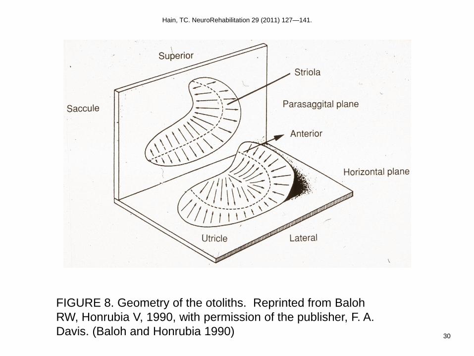
FIGURE 8. Geometry of the otoliths. Reprinted from Baloh
RW, Honrubia V, 1990, with permission of the publisher, F. A.
Davis. (Baloh and Honrubia 1990)
Hain, TC. NeuroRehabilitation 29 (2011) 127—141.
30

FIGURE 9. The VOR and VSR reflex arcs. From (Brodal
1981), with permission of the publisher, Oxford University
Press.
Hain, TC. NeuroRehabilitation 29 (2011) 127—141.
31

Figure 10. An internal model like this may be used for sensorimotor integration. Sensory inflow and
motor outflow are used to estimate the current state. From (Wolpert, 1997) with permission of the
publisher, CellPress.
Hain, TC. NeuroRehabilitation 29 (2011) 127—141.
32