Neuropathologic Diagnosis of Central Nervous System Viral Diseases
22
2 Neuropathologic Diagnosis of Central Nervous System Viral Diseases Kymberly A. Gyure West Virginia University School of Medicine U.S.A. 1. Introduction The principal aim of this chapter is to provide an illustrated review of the neuropathologic features of common and recently emerging central nervous system (CNS) viral diseases. An overview of the histopathologic features common to most viral infections of the central nervous system will be provided. This will be followed by a description of the gross and microscopic findings characteristic of specific viral infections of the CNS. Conditions which can be accurately diagnosed via brain biopsy and their differential diagnosis will be emphasized. Where appropriate, ancillary studies including molecular diagnostic techniques will be covered. 2. Overview of neuropathologic findings in CNS viral diseases The accurate diagnosis of viral meningoencephalitis is important in order to institute appropriate specific therapy to improve survival and reduce the extent of permanent brain injury. In many cases, a diagnosis can be made based on medical history and examination followed by a combination of cerebrospinal fluid (CSF) analysis, serology, and neuroimaging studies (Johnson & Power, 2008; Steiner et al., 2010). However, in clinically unusual and diagnostically difficult cases, brain biopsy may be performed. Although the pathologic features of viral encephalitis vary somewhat depending on the specific infectious agent and the immunologic status of the patient, most viral infections of the CNS are characterized by a triad of findings including perivascular chronic inflammation, microglial nodules, and neuronophagia (Figure 1). The distribution of these findings as well as the presence of characteristic intranuclear or intracytoplasmic viral inclusions can lead to a specific diagnosis in an appropriate clinical setting. Ancillary techniques, including immunohistochemistry (IHC), in-situ hybridization (ISH), or polymerase chain reaction (PCR) amplification, are useful in some settings. 3. DNA Viruses 3.1 Herpesviruses The herpesviruses are double-stranded DNA viruses which have the capacity to cause latent infections in the nervous system. www.intechopen.com
Transcript of Neuropathologic Diagnosis of Central Nervous System Viral Diseases
2
Neuropathologic Diagnosis of Central Nervous System
Viral Diseases
Kymberly A. Gyure West Virginia University School of Medicine
U.S.A.
1. Introduction
The principal aim of this chapter is to provide an illustrated review of the neuropathologic features of common and recently emerging central nervous system (CNS) viral diseases. An overview of the histopathologic features common to most viral infections of the central nervous system will be provided. This will be followed by a description of the gross and microscopic findings characteristic of specific viral infections of the CNS. Conditions which can be accurately diagnosed via brain biopsy and their differential diagnosis will be emphasized. Where appropriate, ancillary studies including molecular diagnostic techniques will be covered.
2. Overview of neuropathologic findings in CNS viral diseases
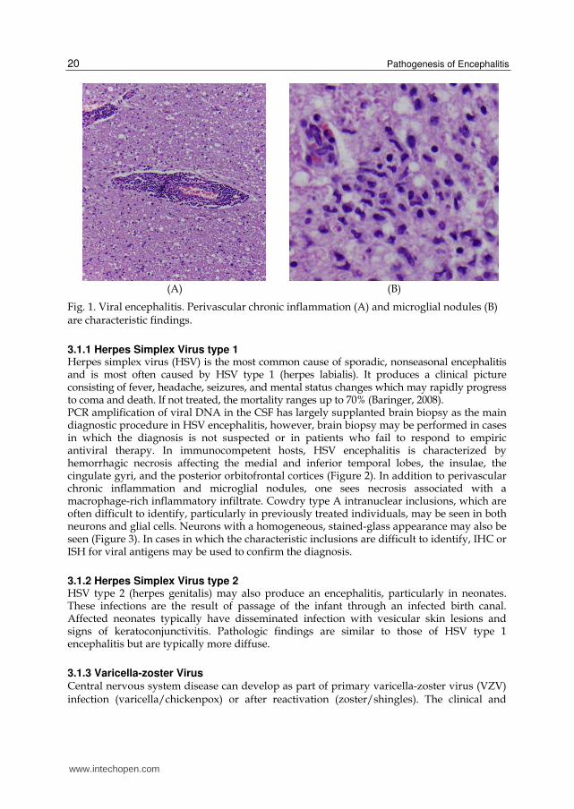
The accurate diagnosis of viral meningoencephalitis is important in order to institute appropriate specific therapy to improve survival and reduce the extent of permanent brain injury. In many cases, a diagnosis can be made based on medical history and examination followed by a combination of cerebrospinal fluid (CSF) analysis, serology, and neuroimaging studies (Johnson & Power, 2008; Steiner et al., 2010). However, in clinically unusual and diagnostically difficult cases, brain biopsy may be performed. Although the pathologic features of viral encephalitis vary somewhat depending on the specific infectious agent and the immunologic status of the patient, most viral infections of the CNS are characterized by a triad of findings including perivascular chronic inflammation, microglial nodules, and neuronophagia (Figure 1). The distribution of these findings as well as the presence of characteristic intranuclear or intracytoplasmic viral inclusions can lead to a specific diagnosis in an appropriate clinical setting. Ancillary techniques, including immunohistochemistry (IHC), in-situ hybridization (ISH), or polymerase chain reaction (PCR) amplification, are useful in some settings.
3. DNA Viruses
3.1 Herpesviruses The herpesviruses are double-stranded DNA viruses which have the capacity to cause latent infections in the nervous system.
www.intechopen.com
Pathogenesis of Encephalitis 20
(A) (B)
Fig. 1. Viral encephalitis. Perivascular chronic inflammation (A) and microglial nodules (B) are characteristic findings.
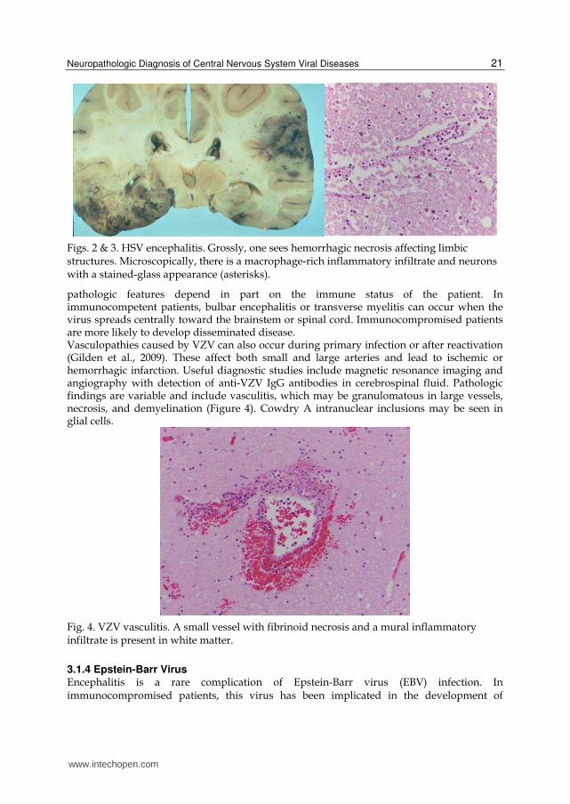
3.1.1 Herpes Simplex Virus type 1 Herpes simplex virus (HSV) is the most common cause of sporadic, nonseasonal encephalitis and is most often caused by HSV type 1 (herpes labialis). It produces a clinical picture consisting of fever, headache, seizures, and mental status changes which may rapidly progress to coma and death. If not treated, the mortality ranges up to 70% (Baringer, 2008). PCR amplification of viral DNA in the CSF has largely supplanted brain biopsy as the main diagnostic procedure in HSV encephalitis, however, brain biopsy may be performed in cases in which the diagnosis is not suspected or in patients who fail to respond to empiric antiviral therapy. In immunocompetent hosts, HSV encephalitis is characterized by hemorrhagic necrosis affecting the medial and inferior temporal lobes, the insulae, the cingulate gyri, and the posterior orbitofrontal cortices (Figure 2). In addition to perivascular chronic inflammation and microglial nodules, one sees necrosis associated with a macrophage-rich inflammatory infiltrate. Cowdry type A intranuclear inclusions, which are often difficult to identify, particularly in previously treated individuals, may be seen in both neurons and glial cells. Neurons with a homogeneous, stained-glass appearance may also be seen (Figure 3). In cases in which the characteristic inclusions are difficult to identify, IHC or ISH for viral antigens may be used to confirm the diagnosis.
3.1.2 Herpes Simplex Virus type 2 HSV type 2 (herpes genitalis) may also produce an encephalitis, particularly in neonates. These infections are the result of passage of the infant through an infected birth canal. Affected neonates typically have disseminated infection with vesicular skin lesions and signs of keratoconjunctivitis. Pathologic findings are similar to those of HSV type 1 encephalitis but are typically more diffuse.
3.1.3 Varicella-zoster Virus Central nervous system disease can develop as part of primary varicella-zoster virus (VZV) infection (varicella/chickenpox) or after reactivation (zoster/shingles). The clinical and
www.intechopen.com
Neuropathologic Diagnosis of Central Nervous System Viral Diseases 21
Figs. 2 & 3. HSV encephalitis. Grossly, one sees hemorrhagic necrosis affecting limbic structures. Microscopically, there is a macrophage-rich inflammatory infiltrate and neurons with a stained-glass appearance (asterisks).
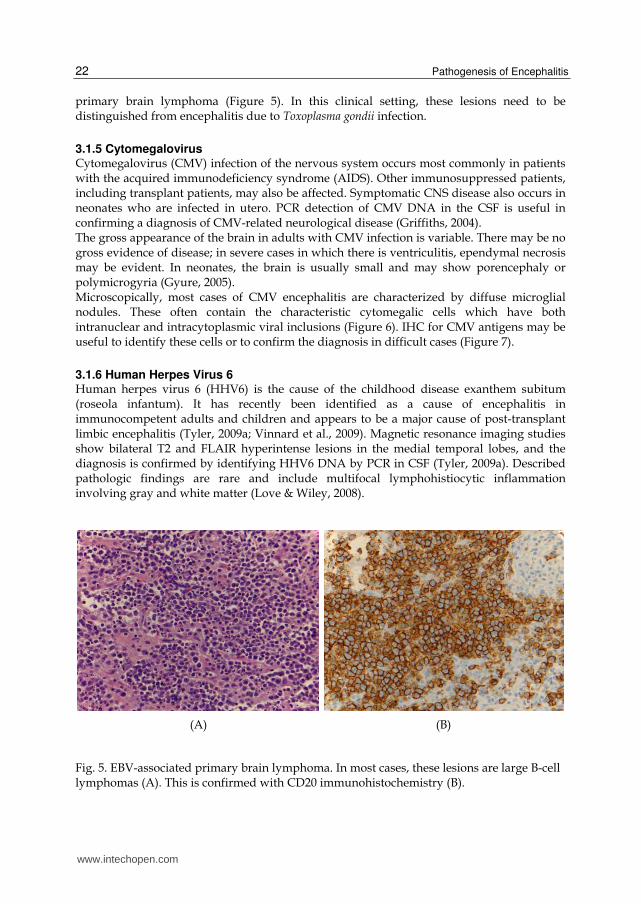
pathologic features depend in part on the immune status of the patient. In immunocompetent patients, bulbar encephalitis or transverse myelitis can occur when the virus spreads centrally toward the brainstem or spinal cord. Immunocompromised patients are more likely to develop disseminated disease. Vasculopathies caused by VZV can also occur during primary infection or after reactivation (Gilden et al., 2009). These affect both small and large arteries and lead to ischemic or hemorrhagic infarction. Useful diagnostic studies include magnetic resonance imaging and angiography with detection of anti-VZV IgG antibodies in cerebrospinal fluid. Pathologic findings are variable and include vasculitis, which may be granulomatous in large vessels, necrosis, and demyelination (Figure 4). Cowdry A intranuclear inclusions may be seen in glial cells.
Fig. 4. VZV vasculitis. A small vessel with fibrinoid necrosis and a mural inflammatory infiltrate is present in white matter.
3.1.4 Epstein-Barr Virus Encephalitis is a rare complication of Epstein-Barr virus (EBV) infection. In immunocompromised patients, this virus has been implicated in the development of
*
*
www.intechopen.com
Pathogenesis of Encephalitis 22
primary brain lymphoma (Figure 5). In this clinical setting, these lesions need to be distinguished from encephalitis due to Toxoplasma gondii infection.
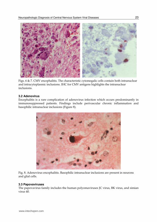
3.1.5 Cytomegalovirus Cytomegalovirus (CMV) infection of the nervous system occurs most commonly in patients with the acquired immunodeficiency syndrome (AIDS). Other immunosuppressed patients, including transplant patients, may also be affected. Symptomatic CNS disease also occurs in neonates who are infected in utero. PCR detection of CMV DNA in the CSF is useful in confirming a diagnosis of CMV-related neurological disease (Griffiths, 2004). The gross appearance of the brain in adults with CMV infection is variable. There may be no gross evidence of disease; in severe cases in which there is ventriculitis, ependymal necrosis may be evident. In neonates, the brain is usually small and may show porencephaly or polymicrogyria (Gyure, 2005). Microscopically, most cases of CMV encephalitis are characterized by diffuse microglial nodules. These often contain the characteristic cytomegalic cells which have both intranuclear and intracytoplasmic viral inclusions (Figure 6). IHC for CMV antigens may be useful to identify these cells or to confirm the diagnosis in difficult cases (Figure 7).
3.1.6 Human Herpes Virus 6 Human herpes virus 6 (HHV6) is the cause of the childhood disease exanthem subitum (roseola infantum). It has recently been identified as a cause of encephalitis in immunocompetent adults and children and appears to be a major cause of post-transplant limbic encephalitis (Tyler, 2009a; Vinnard et al., 2009). Magnetic resonance imaging studies show bilateral T2 and FLAIR hyperintense lesions in the medial temporal lobes, and the diagnosis is confirmed by identifying HHV6 DNA by PCR in CSF (Tyler, 2009a). Described pathologic findings are rare and include multifocal lymphohistiocytic inflammation involving gray and white matter (Love & Wiley, 2008).
(A) (B)
Fig. 5. EBV-associated primary brain lymphoma. In most cases, these lesions are large B-cell lymphomas (A). This is confirmed with CD20 immunohistochemistry (B).
www.intechopen.com
Neuropathologic Diagnosis of Central Nervous System Viral Diseases 23
Figs. 6 & 7. CMV encephalitis. The characteristic cytomegalic cells contain both intranuclear and intracytoplasmic inclusions. IHC for CMV antigens highlights the intranuclear inclusions.
3.2 Adenovirus Encephalitis is a rare complication of adenovirus infection which occurs predominantly in immunosuppressed patients. Findings include perivascular chronic inflammation and basophilic intranuclear inclusions (Figure 8).
Fig. 8. Adenovirus encephalitis. Basophilic intranuclear inclusions are present in neurons and glial cells.
3.3 Papovaviruses The papovavirus family includes the human polyomaviruses JC virus, BK virus, and simian virus 40.
www.intechopen.com

Pathogenesis of Encephalitis 24
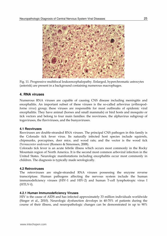
3.3.1 JC virus JC virus is the cause of progressive multifocal leukoencephalopathy (PML), a demyelinating disease that results from the infection of oligodendroglial cells by this virus. It occurs almost exclusively in immunocompromised patients, including those with AIDS. More recently, it has been associated with treatment with the monoclonal antibodies natalizumab, efalizumab, and rituximab (Tan & Koralnik, 2010; Tyler, 2009b; Weber, 2008). Patients present with non-specific symptoms including limb weakness, speech or visual deficits, cognitive abnormalities, and seizures. Therefore, the clinical differential diagnosis is broad and encompasses other AIDS-related illnesses including toxoplasmosis and CNS lymphoma. In multiple sclerosis (MS) patients treated with monoclonal antibodies, the lesions of PML must be distinguished from those of MS (Weber, 2008). The diagnosis is established by brain biopsy or by detection of JC virus DNA in the CSF by PCR. Grossly, one sees multiple foci of demyelination which are typically subcortical in location and have a predilection for the parieto-occipital region (Figure 9). Microscopically, there is myelin loss with a variable degree of inflammation including macrophages (Shishido-Hara, 2010). Infected oligodendroglial nuclei are enlarged with a ground-glass appearance (Figure 10). Bizarre-appearing astrocytes with lobulated, hyperchromatic nuclei may also be present and can be confused with neoplastic astrocytes (Figure 11). The presence of viral inclusions within oligodendrocyte nuclei can be confirmed with IHC for JC virus or simian virus 40, which cross reacts with JC virus (Crowder et al., 2005).
3.3.2 Simian virus 40 Simian virus 40 (SV40) DNA sequences have been identified in a variety of human neoplasms including brain tumors (Love & Wiley, 2008). The significance of this finding in relationship to the development of these lesions remains unclear.
Figs. 9 & 10. Progressive multifocal leukoencephalopathy. Gross findings include subcortical foci of myelin loss. Microscopically, enlarged oligodendrocyte nuclei have a ground-glass appearance.
www.intechopen.com
Neuropathologic Diagnosis of Central Nervous System Viral Diseases 25
Fig. 11. Progressive multifocal leukoencephalopathy. Enlarged, hyperchromatic astrocytes (asterisk) are present in a background containing numerous macrophages.
4. RNA viruses
Numerous RNA viruses are capable of causing CNS disease including meningitis and
encephalitis. An important subset of these viruses is the so-called arbovirus (arthropod-
borne virus) group; these viruses are responsible for most outbreaks of epidemic viral
encephalitis. They have animal (horses and small mammals) or bird hosts and mosquito or
tick vectors and belong to four main families: the reoviruses, the alphavirus subgroup of
togaviruses, the flaviviruses, and the bunyaviruses.
4.1 Reoviruses Reoviruses are double-stranded RNA viruses. The principal CNS pathogen in this family is the Colorado tick fever virus. Its naturally infected host species include squirrels, chipmunks, porcupines, deer mice, and wood rats; and the vector is the wood tick Dermacentor andersoni (Romero & Simonsen, 2008). Colorado tick fever is an acute febrile illness which occurs most commonly in the Rocky
Mountain region of North America. It is the second most common arboviral infection in the
United States. Neurologic manifestations including encephalitis occur most commonly in
children. The diagnosis is typically made serologically.
4.2 Retroviruses The retroviruses are single-stranded RNA viruses possessing the enzyme reverse
transcriptase. Human pathogens affecting the nervous system include the human
immunodeficiency viruses (HIV-1 and HIV-2) and human T-cell lymphotropic virus I
(HTLV-I).
4.2.1 Human Immunodeficiency Viruses HIV is the cause of AIDS and has infected approximately 33 million individuals worldwide (Singer et al., 2010). Neurologic dysfunction develops in 40-70% of patients during the course of their illness, and neuropathologic changes can be demonstrated in up to 90%
*
www.intechopen.com

Pathogenesis of Encephalitis 26
brains from this patient population (Anthony & Bell, 2008; Boissé et al., 2008). These findings include opportunistic infections; primary CNS lymphomas; lesions thought to be directly related to the HIV virus including aseptic meningitis, HIV-associated neurocognitive disorders/HIV encephalitis, and vacuolar myelopathy; and disorders related to antiretroviral therapy. Commonly seen opportunistic infections in AIDS patients include the viral infections PML and cytomegalovirus encephalitis (illustrated above), the fungal infection cryptococcosis (Figure 12), and parasitic infection due to Toxoplasma gondii (Figure 13). These must be distinguished from primary CNS lymphomas, also illustrated above.
Figs. 12 & 13. Opportunistic infections in AIDS. Cryptococcal meninginitis may be diagnosed by finding budding yeasts in CSF. Toxoplasma encephalitis is characterized by cysts and trophozoites in a background containing microglial nodules.
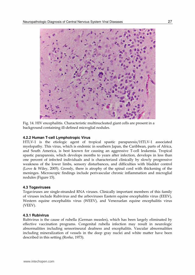
A significant number of AIDS patients develop neurocognitive disorders that are not attributable to opportunistic infections. The term HIV-associated neurocognitive disorder (HAND) has been applied to a number of clinical syndromes including HIV-associated dementia (HAD, also referred to as AIDS dementia complex or HIV encephalopathy), mild neurocognitive disorder (MND), and asymptomatic neurocognitive impairment (Boissé et al., 2008; Letendre et al., 2010; Singer et al., 2010). Many individuals with cognitive and motor abnormalities exhibit a pathologic picture which has been termed HIV encephalitis that is characterized by diffuse or multifocal accumulations of microglia including ill-defined microglial nodules. Characteristic macrophage-derived multinucleated cells are diagnostic when observed in this setting (Figure 14). Vacuolar myelopathy is an uncommon manifestation of AIDS. Patients present with leg weakness, spastic paraparesis, ataxia, and incontinence. Pathologic findings consist of vacuolation of white matter tracts in the lateral and posterior columns of the spinal cord. This disorder may be difficult to distinguish from subacute combined degeneration of the spinal cord associated with vitamin B12 deficiency. The introduction of highly active antiretroviral therapy (HAART) in very immunosuppressed AIDS patients may result in an abrupt clinical deterioration termed the immune reconstitution inflammatory syndrome (IRIS). This syndrome is thought to be secondary to the sudden activation of and increase in CD4-positive T cells resulting in a heightened immune response with host-mediated inflammatory cell damage, resulting in new neurologic deficits or worsening of prior signs and symptoms (McCombe et al., 2009). Neuropathologic findings include widespread chronic inflammatory infiltrates in the brain adjacent to areas of pre-existing infection as well as demyelination.
www.intechopen.com
Neuropathologic Diagnosis of Central Nervous System Viral Diseases 27
Fig. 14. HIV encephalitis. Characteristic multinucleated giant cells are present in a background containing ill-defined microglial nodules.
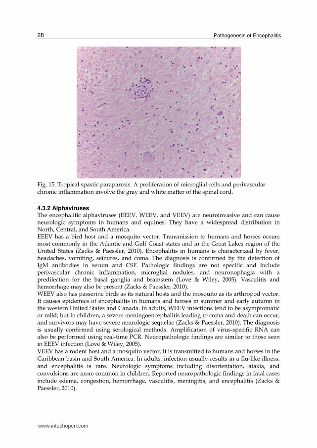
4.2.2 Human T-cell Lymphotropic Virus HTLV-1 is the etiologic agent of tropical spastic paraparesis/HTLV-1 associated myelopathy. This virus, which is endemic in southern Japan, the Caribbean, parts of Africa, and South America, is best known for causing an aggressive T-cell leukemia. Tropical spastic paraparesis, which develops months to years after infection, develops in less than one percent of infected individuals and is characterized clinically by slowly progressive weakness of the lower limbs, sensory disturbances, and difficulties with bladder control (Love & Wiley, 2005). Grossly, there is atrophy of the spinal cord with thickening of the meninges. Microscopic findings include perivascular chronic inflammation and microglial nodules (Figure 15).
4.3 Togaviruses Togaviruses are single-stranded RNA viruses. Clinically important members of this family of viruses include Rubivirus and the arboviruses Eastern equine encephalitis virus (EEEV), Western equine encephalitis virus (WEEV), and Venezuelan equine encephalitis virus (VEEV).
4.3.1 Rubivirus Rubivirus is the cause of rubella (German measles), which has been largely eliminated by effective vaccination programs. Congenital rubella infection may result in neurologic abnormalities including sensorineural deafness and encephalitis. Vascular abnormalities including mineralization of vessels in the deep gray nuclei and white matter have been described in this setting (Rorke, 1973).
www.intechopen.com
Pathogenesis of Encephalitis 28
Fig. 15. Tropical spastic paraparesis. A proliferation of microglial cells and perivascular chronic inflammation involve the gray and white matter of the spinal cord.
4.3.2 Alphaviruses The encephalitic alphaviruses (EEEV, WEEV, and VEEV) are neuroinvasive and can cause neurologic symptoms in humans and equines. They have a widespread distribution in North, Central, and South America. EEEV has a bird host and a mosquito vector. Transmission to humans and horses occurs most commonly in the Atlantic and Gulf Coast states and in the Great Lakes region of the United States (Zacks & Paessler, 2010). Encephalitis in humans is characterized by fever, headaches, vomiting, seizures, and coma. The diagnosis is confirmed by the detection of IgM antibodies in serum and CSF. Pathologic findings are not specific and include perivascular chronic inflammation, microglial nodules, and neuronophagia with a predilection for the basal ganglia and brainstem (Love & Wiley, 2005). Vasculitis and hemorrhage may also be present (Zacks & Paessler, 2010). WEEV also has passerine birds as its natural hosts and the mosquito as its arthropod vector. It causes epidemics of encephalitis in humans and horses in summer and early autumn in the western United States and Canada. In adults, WEEV infections tend to be asymptomatic or mild; but in children, a severe meningoencephalitis leading to coma and death can occur, and survivors may have severe neurologic sequelae (Zacks & Paessler, 2010). The diagnosis is usually confirmed using serological methods. Amplification of virus-specific RNA can also be performed using real-time PCR. Neuropathologic findings are similar to those seen in EEEV infection (Love & Wiley, 2005). VEEV has a rodent host and a mosquito vector. It is transmitted to humans and horses in the Caribbean basin and South America. In adults, infection usually results in a flu-like illness, and encephalitis is rare. Neurologic symptoms including disorientation, ataxia, and convulsions are more common in children. Reported neuropathologic findings in fatal cases include edema, congestion, hemorrhage, vasculitis, meningitis, and encephalitis (Zacks & Paessler, 2010).
www.intechopen.com

Neuropathologic Diagnosis of Central Nervous System Viral Diseases 29
4.4 Flaviviruses Flaviviruses are enveloped, single-stranded RNA viruses. Most are arboviruses that produce a variety of diseases in humans. Japanese encephalitis virus (JEV) is the most common cause of acute viral encephalitis worldwide and causes 10,000 to 15,000 deaths annually (Granerod et al., 2010; Tyler, 2009a). The geographic area where it is endemic has been increasing, and encephalitis due to this virus is prevalent throughout Eastern and Southern Asia and the Pacific Rim (Misra & Kalita, 2010). Its hosts include water birds and pigs, and mosquitoes are its vector. In endemic areas, Japanese encephalitis is a disease of children. JEV may produce asymptomatic infections, a non-specific febrile illness, aseptic meningitis, or severe encephalitis. Grossly, lesions are mainly restricted to gray matter and consist of petechiae or focal hemorrhages. Microscopic findings, which are most prominent in the diencephalon and mesencephalon, include congestion and hemorrhage, thrombus formation with associated necrosis, and widespread chronic inflammation with neuronophagia (Love & Wiley, 2005; Misra and Kalita, 2010). St. Louis encephalitis virus (SLEV) is present throughout the United States and produces infections in late summer and early autumn. It has a bird host and a mosquito vector. Most SLEV virus infections are subclinical; symptomatic encephalitis is most common in patients over the age of 60 years. Neuropathologic findings are those of a non-necrotizing encephalitis which may be particularly marked in the thalamus and midbrain (Love & Wiley, 2005). West Nile virus (WNV) is the most common cause of epidemic meningoencephalitis in North America and the leading cause of arboviral encephalitis in the United States. Recent epidemics in Algeria, Morocco, Tunisia, Italy, France, Romania, Israel, and Russia have also been associated with severe human disease including neurologic complications (Rossi et al., 2010). WNV cycles between bird hosts and mosquito vectors; humans and horses are incidental hosts. Transmission may also occur via transfusion of blood products and via organ transplantation from infected donors. Approximately one of 150 patients develops CNS disease; risk factors for neuroinvasive disease are older age and immunosuppression. CNS manifestations include meningitis, encephalitis, and acute flaccid paralysis/poliomyelitis. The diagnosis in most cases is confirmed by detecting WNV-specific antibodies in serum or CSF. The brain and spinal cord are grossly normal in most cases of WNV meningoencephalitis. Microscopically, one sees perivascular chronic inflammation, microglial nodules, and variable neuronal loss (Figure 16). Neuronophagia and foci of necrosis are sometimes present. Gray matter is more severely affected than white matter, and the inflammatory infiltrate is typically most prominent in the brainstem (Gyure, 2009). Tick-borne encephalitis virus (TBEV) is the cause of tick-borne encephalitis. This potentially
fatal neurologic infection occurs predominantly in Europe and Asia. TBEV has wild
mammalian hosts, particularly rodents, and its vector is the Ixodid tick. Symptoms of acute
tick-borne encephalitis range from mild meningitis to a severe meningoencephalitis
characterized by muscle weakness. The diagnosis is made by demonstrating TBEV-specific
antibodies or by detecting viral RNA in serum and CSF (Mansfield et al., 2009).
Neuropathologic findings in fatal cases include chronic inflammation with microglial
activation affecting gray matter structures including those of the spinal cord, especially the
anterior horns (Love & Wiley, 2005).
Other flaviviruses that can affect the nervous system include Murray Valley encephalitis virus, louping ill virus, Powasan virus, Kyasanur Forest disease virus, and Omsk hemorrhagic fever virus (LaSala & Holbrook, 2010; Romero & Simonsen, 2008).
www.intechopen.com

Pathogenesis of Encephalitis 30
Fig. 16. WNV encephalomyelitis. Microglial nodules are associated with variable neuronal loss.
4.5 Picornaviruses Picornaviruses are single-stranded RNA viruses. Important human pathogens include poliovirus and other enteroviruses.
4.5.1 Poliovirus Despite the overall success of the Global Poliomyelitis Eradication Initiative of the World Health Organization, poliovirus remains endemic in Afghanistan, India, Nigeria, and Pakistan (Dutta, 2008). Polio outbreaks attributed to circulating vaccine-derived poliovirus have also occurred throughout the world (Tyler, 2009a). In most infected patients, a non-specific gastroenteritis develops. Paralytic disease occurs in one to two percent of patients and is characterized by a flaccid paralysis with muscle wasting and hyporeflexia. A late complication termed postpolio syndrome, which occurs 30-40 years after the acute illness, is characterized by progressive weakness associated with decreased muscle bulk and pain. In most cases, the brain and spinal cord are grossly normal, but in severe cases, vascular congestion, petechial hemorrhages, and necrosis may be present. Chronically, there is atrophy of the anterior nerve roots. Microscopically, there is perivascular chronic inflammation and neuronophagia involving the anterior horns of the spinal cord. Neutrophils may be present in some cases.
4.5.2 Enteroviruses Other enteroviruses have been associated with a poliomyelitis-like syndrome. Enterovirus 71, a cause of hand-foot-and-mouth disease, has caused numerous outbreaks of disease in the Southeast Asia and Pacific region (Johnson & Power, 2008; Tyler, 2009a). In addition to the poliomyelitis-like syndrome, neurologic manifestations include meningitis, cerebellitis,
www.intechopen.com
Neuropathologic Diagnosis of Central Nervous System Viral Diseases 31
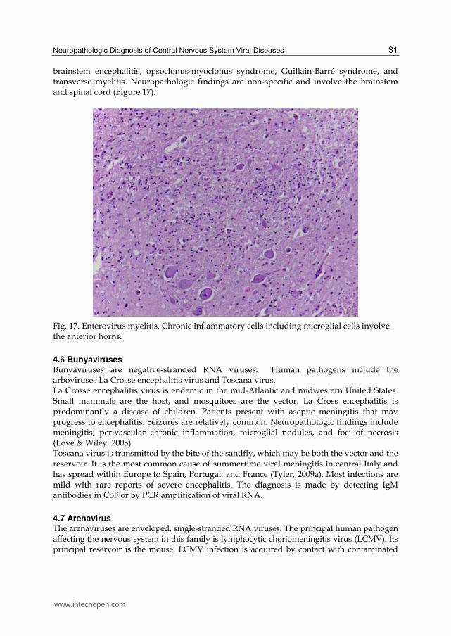
brainstem encephalitis, opsoclonus-myoclonus syndrome, Guillain-Barré syndrome, and transverse myelitis. Neuropathologic findings are non-specific and involve the brainstem and spinal cord (Figure 17).
Fig. 17. Enterovirus myelitis. Chronic inflammatory cells including microglial cells involve the anterior horns.
4.6 Bunyaviruses Bunyaviruses are negative-stranded RNA viruses. Human pathogens include the arboviruses La Crosse encephalitis virus and Toscana virus. La Crosse encephalitis virus is endemic in the mid-Atlantic and midwestern United States. Small mammals are the host, and mosquitoes are the vector. La Cross encephalitis is predominantly a disease of children. Patients present with aseptic meningitis that may progress to encephalitis. Seizures are relatively common. Neuropathologic findings include meningitis, perivascular chronic inflammation, microglial nodules, and foci of necrosis (Love & Wiley, 2005). Toscana virus is transmitted by the bite of the sandfly, which may be both the vector and the reservoir. It is the most common cause of summertime viral meningitis in central Italy and has spread within Europe to Spain, Portugal, and France (Tyler, 2009a). Most infections are mild with rare reports of severe encephalitis. The diagnosis is made by detecting IgM antibodies in CSF or by PCR amplification of viral RNA.
4.7 Arenavirus The arenaviruses are enveloped, single-stranded RNA viruses. The principal human pathogen affecting the nervous system in this family is lymphocytic choriomeningitis virus (LCMV). Its principal reservoir is the mouse. LCMV infection is acquired by contact with contaminated
www.intechopen.com
Pathogenesis of Encephalitis 32
fomites, via inhalation of aerosolized virus, via organ transplantation, or transplacentally when a pregnant woman acquires a primary infection (Bonthius, 2009; Tyler, 2009a). Typically, patients present with a non-specific viral syndrome followed by symptoms of an aseptic/viral meningitis. Some patients develop a more severe neurologic illness including transverse myelitis, Guillain-Barré syndrome, hydrocephalus, or encephalitis. Congenital LCMV leads to chorioretinitis with scarring, macrocephaly secondary to a non-communicating hydrocephalus, or microcephaly with periventricular calcifications. Other reported findings include periventricular cysts, porencephaly, encephalomalacia, intraparenchymal calcifications, cerebellar hypoplasia, and disturbances in neuronal migration (Bonthius, 2009).
4.8 Paramyxoviruses Paramyxoviruses are single-stranded RNA viruses. Members of this family causing CNS
disease are the measles virus (Morbillivirus) and the recently emerging henipaviruses,
Hendra and Nipah.
4.8.1 Morbillivirus Measles virus can cause CNS complications early after acute measles (acute postinfectious
measles encephalitis) or after years of viral persistence (subacute sclerosing panencephalitis)
(Reuter & Schneider-Schaulies, 2010). These disorders are rare in developed countries
because of widespread immunization against measles.
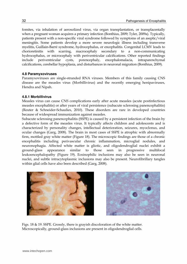
Subacute sclerosing panencephalitis (SSPE) is caused by a persistent infection of the brain by
a defective form of the measles virus. It typically affects children and adolescents and is
characterized by personality changes, intellectual deterioration, seizures, myoclonus, and
ocular changes (Garg, 2008). The brain in most cases of SSPE is atrophic with abnormally
firm, mottled gray white matter (Figure 18). The microscopic findings are those of a chronic
encephalitis including perivascular chronic inflammation, microglial nodules, and
neuronophagia. Affected white matter is gliotic, and oligodendroglial nuclei exhibit a
ground-glass appearance similar to those seen in progressive multifocal
leukoencephalopathy (Figure 19). Eosinophilic inclusions may also be seen in neuronal
nuclei, and subtle intracytoplasmic inclusions may also be present. Neurofibrillary tangles
within glial cells have also been described (Garg, 2008).
Figs. 18 & 19. SSPE. Grossly, there is grayish discoloration of the white matter. Microscopically, ground-glass inclusions are present in oligodendroglial cells.
www.intechopen.com
Neuropathologic Diagnosis of Central Nervous System Viral Diseases 33
4.8.2 Hendra and Nipah viruses Hendra and Nipah viruses are relatively recently identified members of the genus Henipavirus causing outbreaks in Australia and Asia, respectively. Their natural host is the fruit bat. Bat-to-human transmission may be direct or via intermediate hosts such as the horse or pig (Wong, 2010). Hendra virus infection may manifest as either a pulmonary or neurological syndrome consisting of confusion, motor deficits, and seizures. The diagnosis is based on the finding of IgM and IgG antibodies in the serum and CSF (Wong, 2010). CNS disease in Nipah virus infection follows a prodrome of fever, nausea, myalgia, and dizziness. Neurologic signs and symptoms include reduced deep tendon reflexes, segmental myoclonus, nuchal rigidity, seizures, cerebellar ataxia, and brainstem signs. A relapsing or late-onset encephalitis occurs in some patients (Johnson & Power, 2008; Tyler, 2009b; Wong; 2010). The neuropathologic findings in these two viral infections are similar and consist of vasculitis with associated vascular thrombosis and foci of necrosis. Perivascular chronic inflammation, microglial nodules, and neuronophagia are also present. Intranuclear and intracytoplasmic inclusions are seen predominantly in neurons near areas of vasculitis or necrosis (Tyler, 2009b; Wong, 2010).
4.9 Orthomyxoviruses The orthomyxoviruses are single-stranded RNA viruses. The influenza viruses are part of
this family and can cause CNS dysfunction via a number of different mechanisms including
influenza-associated encephalopathy, febrile seizures, Reye syndrome, postinfluenzal
encephalitic Parkinson disease, and encephalitis lethargica (Wang et al., 2010). Influenza A
viruses have the potential to cause pandemics, as illustrated recently by the emergence of a
novel influenza A (H1N1) virus of swine origin (Maritz et al., 2010).
Influenza-associated encephalopathy (IAE) is an uncommon complication with high
mortality characterized by typical flu symptoms with associated CNS manifestations
including seizure, alteration in consciousness, decreased cognitive function, abnormal
behavior, and change in mental status that occurs most commonly in children. Imaging
studies show a necrotizing encephalopathy with associated cerebral edema (Lyon et al.,
2010; Wang et al., 2010; Webster et al., 2010). Risk factors for the development of severe
disease in the recent H1N1 pandemic include age less than two years, pregnancy, chronic
pulmonary disease, immunosuppression, and metabolic disorders including diabetes
mellitus and obesity (Maritz et al., 2010). Most fatal cases are the result of pulmonary
complications.
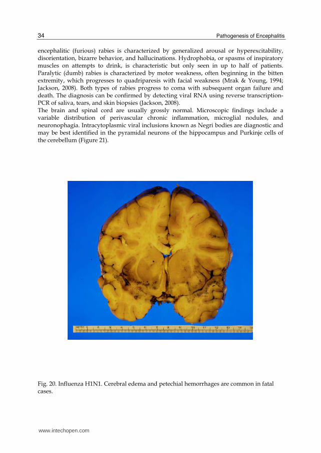
Gross brain findings in fatal cases of influenza H1N1 infection include cerebral edema,
dusky discoloration of gray matter structures, and petechial hemorrhages (Figure 20).
Microscopic findings include diffuse anoxic changes thought to be secondary to systemic
disease; most patients do not have a meningoencephalitis (Milyani et al., 2010).
4.10 Rhabdoviruses The principal human pathogen in the rhabdovirus family is rabies virus. It is usually transmitted from the bite of a rabid animal, but rare cases have been transmitted by aerosols in laboratories or caves with large numbers of bats and by organ transplantation (Jackson, 2008; Tyler, 2009a). There are two clinical forms of rabies. The more common form,
www.intechopen.com
Pathogenesis of Encephalitis 34
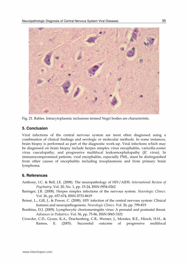
encephalitic (furious) rabies is characterized by generalized arousal or hyperexcitability, disorientation, bizarre behavior, and hallucinations. Hydrophobia, or spasms of inspiratory muscles on attempts to drink, is characteristic but only seen in up to half of patients. Paralytic (dumb) rabies is characterized by motor weakness, often beginning in the bitten extremity, which progresses to quadriparesis with facial weakness (Mrak & Young, 1994; Jackson, 2008). Both types of rabies progress to coma with subsequent organ failure and death. The diagnosis can be confirmed by detecting viral RNA using reverse transcription-PCR of saliva, tears, and skin biopsies (Jackson, 2008). The brain and spinal cord are usually grossly normal. Microscopic findings include a variable distribution of perivascular chronic inflammation, microglial nodules, and neuronophagia. Intracytoplasmic viral inclusions known as Negri bodies are diagnostic and may be best identified in the pyramidal neurons of the hippocampus and Purkinje cells of the cerebellum (Figure 21).
Fig. 20. Influenza H1N1. Cerebral edema and petechial hemorrhages are common in fatal cases.
www.intechopen.com
Neuropathologic Diagnosis of Central Nervous System Viral Diseases 35
Fig. 21. Rabies. Intracytoplasmic inclusions termed Negri bodies are characteristic.
5. Conclusion
Viral infections of the central nervous system are most often diagnosed using a combination of clinical findings and serologic or molecular methods. In some instances, brain biopsy is performed as part of the diagnostic work-up. Viral infections which may be diagnosed on brain biopsy include herpes simplex virus encephalitis, varicella-zoster virus vasculopathy, and progressive multifocal leukoencephalopathy (JC virus). In immunocompromised patients, viral encephalitis, especially PML, must be distinguished from other causes of encephalitis including toxoplasmosis and from primary brain lymphoma.
6. References
Anthony, I.C. & Bell, J.E. (2008). The neuropathology of HIV/AIDS. International Review of
Psychiatry. Vol. 20, No. 1, pp. 15-24, ISSN 0954-0262
Baringer, J.R. (2008). Herpes simplex infections of the nervous system. Neurologic Clinics.
Vol. 26, pp. 657-674, ISSN 0733-8619
Boissé, L., Gill, J., & Power, C. (2008). HIV infection of the central nervous system: Clinical
features and neuropathogenesis. Neurologic Clinics. Vol. 26, pp. 799-819
Bonthius, D.J. (2009). Lymphocytic choriomeningitis virus: A prenatal and postnatal threat.
Advances in Pediatrics. Vol. 56, pp. 75-86, ISSN 0065-3101
Crowder, C.D., Gyure, K.A., Drachenberg, C.B., Werner, J., Morales, R.E., Hirsch, H.H., &
Ramos, E. (2005). Successful outcome of progressive multifocal
www.intechopen.com
Pathogenesis of Encephalitis 36
leukoencephalopathy in a renal transplant patient. American Journal of
Transplantation. Vol. 5, pp. 1151-1158
Dutta, A. (2008). Epidemiology of poliomyelitis – Options and update. Vaccine. Vol. 26, pp.
5767-5773, ISSN 0264-410X
Garg, R.K. (2008). Subacute sclerosing panencephalitis. Journal of Neurology. Vol. 255, pp.
1861-1871
Gilden, D., Cohrs, R.J., Mahalingam, R., & Nagel, M.A. (2009). Varicella zoster virus
vasculopathies: Diverse clinical manifestations, laboratory features, pathogenesis,
and treatment. Lancet Neurology. Vol. 8, pp. 731-740
Granerod, J., Tam, C.C., Crowcroft, N.S., Davies, N.W.S., Borchert, M., & Thomas, S.L.
(2010). Challenge of the unknown: A systematic review of acute encephalitis in
non-outbreak situations. Neurology. Vol. 75. pp. 924-932
Griffiths, P. (2004). Cytomegalovirus infection of the central nervous system. Herpes. Vol. 11,
pp. 95A-104A
Gyure, K.A. (2005). Infections, In: Neuropathology, Prayson, R.A. (ed.), pp.287-338, Elsevier
Inc., ISBN 0-443-06658-2, Philadelphia, PA
Gyure, K.A. (2009). West Nile virus infections. Journal of Neuropathology and Experimental
Neurology. Vol. 68, No. 10, pp. 1053-1060
Jackson, A.C. (2008). Rabies. Neurologic Clinics. Vol. 26, pp. 717-726, ISSN 0733-8619
Johnson, R.T. & Power, C. (2008). Emerging issues in neurovirology: New viruses,
diagnostic tools, and therapeutics. Neurologic Clinics. Vol. 26, pp. 855-864, ISSN
0733-8619
LaSala, P.R. & Holbrook, M. (2010). Tick-borne flaviviruses. Clinics in Laboratory Medicine.
Vol. 30, pp. 221-235, ISSN 0272-2712
Letendre, S.L., Ellis, R.J., Ances, B.M., & McCutchan, J.A. (2010). Neurologic complications
of HIV disease and their treatment. Topics in HIV Medicine. Vol. 18, No. 2, pp. 45-
55
Lo, M.K., & Rota, P.A. (2008). The emergence of Nipah virus, a highly pathogenic
paramyxovirus. Journal of Clinical Virology. Vol. 43, pp. 396-400, ISSN 1386-6532
Love, S. & Wiley, C.A. (2008). Viral infections, In: Greenfield’s Neuropathology, Love, S., Louis,
D.N., & Ellison, D.W. (eds.), pp. 1275-1389, Edward Arnold (Publishers) Ltd, ISBN
978-0-340-90682-8
Lyon, J.B., Remigio, C., Milligan, T., & Deline, C. (2010). Acute necrotizing
encephalopathy in a child with H1N1 influenza infection. Pediatric Radiology. Vol.
40, pp. 200-205
Mansfield, K.L., Johnson, N., Phipps, L.P., Stephenson, J.R., Fooks, A.R., & Solomon, T.
(2009). Tick-borne encephalitis virus – A review of an emerging zoonosis. Journal of
General Virology. Vol. 90, pp. 1781-1794
Maritz, J., Maree, L., Preiser, W. (2010). Pandemic influenza A (H1N1): The experience of
the first 6 months. Clinical Chemistry and Laboratory Medicine. Vol. 48, No. 1, pp.
11-21
McCombe, J.A., Auer, R.N., Maingat, F.G., Houston, S., Gill, M.J., & Power, C. (2009).
Neurologic immune reconstitution inflammatory syndrome in HIV/AIDS:
Outcome and epidemiology. Neurology. Vol. 72, pp. 835-841
www.intechopen.com
Neuropathologic Diagnosis of Central Nervous System Viral Diseases 37
Milyani, W., Skitarelic, K., Bafakih, F., Gyure, K. (2010) Neuropathologic findings in a fatal
case of H1N1 influenza virus infection (abstract). Journal of Neuropathology and
Experimental Neurology. Vol. 69, p. 555
Misra, U.K. & Kalita, J. (2010). Overview: Japanese encephalitis. Progress in Neurobiology. Vol.
91, pp. 108-120, ISSN 0301-0082
Mrak, R.E., Young, L. (1994). Rabies encephalitis in humans: Pathology, pathogenesis and
pathophysiology. Journal of Neuropathology and Experimental Neurology. Vol. 53, No.
1, pp. 1-10
Reuter, D. & Schneider-Schaulies, D. (2010). Measles virus infection of the CNS: Human
disease, animal models, and approaches to therapy. Medical Microbiology and
Immunology. Vol. 199, pp. 261-271
Romero, J.R. & Simonsen, K.A. (2008). Powassan encephalitis and Colorado tick fever.
Infectious Disease Clinics of North America. Vol. 22, pp. 545-559, ISSN 0891-5520
Rorke, L. (1973). Nervous system lesions in the congenital rubella syndrome. Archives of
Otolaryngology. Vol. 98, pp. 249-251
Rossi, S.L., Ross, T.M., & Evans, J.D. (2010). West Nile virus. Clinics in Laboratory Medicine.
Vol. 30, pp. 47-65, ISSN 0272-2712
Singer, E.J., Valdes-Sueiras, M., Commins, D., & Levine, A. (2010). Neurologic presentations
of AIDS. Neurologic Clinics. Vol. 28, pp. 253-275, ISSN 0733-8619
Shishido-Hara, Y. (2010). Progressive multifocal leukoencephalopathy and promyelocytic
leukemia nuclear bodies: A review of clinical, neuropathological, and virological
aspects of JC virus-induced demyelinating disease. Acta Neuropathologica. Vol. 120,
pp. 403-417
Steiner, I., Budka, H., Chaudhuri, A., Koskiniemi, M., Sainio, K., Salonen, O., & Kennedy,
P.G.E. (2010). Viral meningoencephalitis: A review of diagnostic methods and
guidelines for management. European Journal of Neurology. Vol. 17, pp. 999-1009,
ISSN 1468-1331
Tan, C.S. & Koralnik, I.J. (2010). Progressive multifocal leukoencephalopathy and other
disorders caused by JC virus: Clinical features and pathogenesis. Lancet Neurology.
Vol. 9, pp. 425-437
Tyler, K.L. (2009). Emerging viral infections of the central nervous system: Part 1. Archives of
Neurology. Vol. 66, No. 8, pp. 939-948
Tyler, K.L. (2009). Emerging viral infections of the central nervous system: Part 2. Archives of
Neurology. Vol. 66, No. 9, pp. 1065-1074
Vinnard, C., Barton, T., Jerud, E., & Blumberg, E. (2009). A report of human herpesvirus 6-
associated encephalitis in a solid organ transplant recipient and a review of
previously published cases. Liver Transplantation. Vol. 15, pp. 1242-1246
Wang, G.F., Weizhong, L., & Li, K. (2010). Acute encephalopathy and encephalitis caused by
influenza virus infection. Current Opinion in Neurology. Vol. 23, pp. 305-311, ISSN
1350-7540
Weber, T. (2008). Progressive multifocal leukoencephalopathy. Neurologic Clinics. Vol. 26,
pp. 833-854, ISSN 0733-8619
www.intechopen.com
Pathogenesis of Encephalitis 38
Webster, R.I., Hazelton, B., Suleiman, J., Macartney, K., Kesson, A., & Dale, R.C. (2010).
Severe encephalopathy with swine origin influenza A H1N1 infection in childhood:
Case reports. Neurology. Vol. 74, pp. 1077-1078.
Wong, K.T. Emerging epidemic viral encephalitides with a special focus on henipaviruses.
(2010). Acta Neuropathologica. Vol. 120, pp. 317-325
Zacks, M.A. & Paessler S. (2010). Encephalitic alphaviruses. Veterinary Microbiololgy. Vol.
140, pp. 281-286, ISSN 0378-1135
www.intechopen.com
Pathogenesis of EncephalitisEdited by Dr. Daisuke Hayasaka
ISBN 978-953-307-741-3Hard cover, 344 pagesPublisher InTechPublished online 09, December, 2011Published in print edition December, 2011
InTech EuropeUniversity Campus STeP Ri Slavka Krautzeka 83/A 51000 Rijeka, Croatia Phone: +385 (51) 770 447 Fax: +385 (51) 686 166www.intechopen.com
InTech ChinaUnit 405, Office Block, Hotel Equatorial Shanghai No.65, Yan An Road (West), Shanghai, 200040, China
Phone: +86-21-62489820 Fax: +86-21-62489821
Many infectious agents, such as viruses, bacteria, and parasites, can cause inflammation of the centralnervous system (CNS). Encephalitis is an inflammation of the brain parenchyma, which may result in a moreadvanced and serious disease meningoencephalitis. To establish accurate diagnosis and develop effectivevaccines and drugs to overcome this disease, it is important to understand and elucidate the mechanism of itspathogenesis. This book, which is divided into four sections, provides comprehensive commentaries onencephalitis. The first section (6 chapters) covers diagnosis and clinical symptoms of encephalitis with someneurological disorders. The second section (5 chapters) reviews some virus infections with the outlines ofinflammatory and chemokine responses. The third section (7 chapters) deals with the non-viral causativeagents of encephalitis. The last section (4 chapters) discusses the experimental model of encephalitis. Thedifferent chapters of this book provide valuable and important information not only to the researchers, but alsoto the physician and health care workers.
How to referenceIn order to correctly reference this scholarly work, feel free to copy and paste the following:
Kymberly A. Gyure (2011). Neuropathologic Diagnosis of Central Nervous System Viral Diseases,Pathogenesis of Encephalitis, Dr. Daisuke Hayasaka (Ed.), ISBN: 978-953-307-741-3, InTech, Available from:http://www.intechopen.com/books/pathogenesis-of-encephalitis/neuropathologic-diagnosis-of-central-nervous-system-viral-diseases

© 2011 The Author(s). Licensee IntechOpen. This is an open access articledistributed under the terms of the Creative Commons Attribution 3.0License, which permits unrestricted use, distribution, and reproduction inany medium, provided the original work is properly cited.