Neuromuscular Disease Stacy Rudnicki, MD Department of Neurology.
57
Neuromuscular Disease Stacy Rudnicki, MD Department of Neurology
-
Upload
ashlynn-maxwell -
Category
Documents
-
view
221 -
download
1
Transcript of Neuromuscular Disease Stacy Rudnicki, MD Department of Neurology.

Neuromuscular Disease
Stacy Rudnicki, MD
Department of Neurology
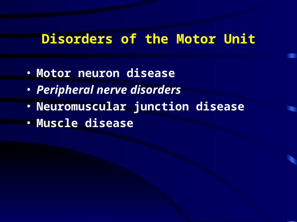
Disorders of the Motor Unit
• Motor neuron disease• Peripheral nerve disorders• Neuromuscular junction disease• Muscle disease
Motor Neuron Disease
• Diseases that can involve Betz cells of the motor cortex, the lower CN motor nuclei, the CST, and/or the anterior horn cells– Amyotrophic Lateral Sclerosis (ALS)– Progressive bulbar palsy (PBP)– Progressive muscular atrophy (PMA), spinal
muscular atrophy (SMA)– Primary lateral sclerosis

Distinctions b/w Types of MND
UMN LMN
ALS yes yes
PLS yes no
PMA/SMA no yes
PBP* yes yes
*limited to bulbar musculature

ALS
• Loss of motor neurons in the cortex, brainstem and spinal cord
• Mix of upper motor neuron and lower motor neuron findings– Weakness, atrophy, fasciculations– Slurred speech, difficulty swallowing,
shortness of breath• Can start in any extremity or the bulbar
musculature• Relentlessly progressive

ALS
• 50 % dead in 3 years, 80% dead in 5 years, 5-10% live more than 10 years
• Death usually from respiratory failure• Etiology still only theoretical
– Excess glutamate– Oxidative stress– Free radicals– Mitochondrial dysfunction
ALS
• 5-10% have inherited disease– Superoxide dismutase (SOD-1) gene defect in
about 20% of inherited ALS• Different mutations in the same gene associated
with differences in age of onset, rate of progression

Treatment in ALS
• Riluzole– Antiglutamate agent– Prolonged survival - modest benefits– Only agent with proven efficacy
• Many other agents tried– Other antiglutamatergic meds, trophic factors,
immunosuppressants, vitamins E & C (antixoidants)
• Supportive care

Primary Lateral Sclerosis (PLS)
• Pure upper motor neuron disease• Primary findings are related to spasticity and
pathologically brisk reflexes• Weakness is present - but the spasticity is
usually more disabling• Slowly progressive - in general, less bulbar
involvement

Progressive Bulbar Palsy• “Bulb” refers to the brainstem
• Sxs
– Slurring of voice (dysarthria)
– Difficulty chewing
– Difficulty swallowing (dysphagia)
– Shortness of breath - with exertion, later at rest
• Signs
– Weak, fasciculating tongue
– Jaw and/or neck weakness
– Decreased forced vital capacity
– Brisk jaw jerk
– Bulbar affect

Progressive Muscular Atrophy (PMA)
• Since it’s a lower motor neuron disease only, weakness with decreased reflexes and normal to decreased tone
• In general, a more slowly progressive disease• Bulbar musculature may be spared until late in
disease
The MNDs are a spectrum, and PMA, PLS and PBP can all evolve into ALS

Spinal Muscular Atrophy (SMA)
• Most common form of inherited MND - autosomal recessive
• Age of onset – Infancy - Werdnig Hoffman disease– Adolescence - Kugelberg Welander disease– Late onset
• Survival motor neuron gene with a modifier gene that effects onset age

Peripheral Nerve Disorders
• Mononeuropathy– Pattern of weakness and sensory loss conforms
to the distribution of a single nerve• Carpal tunnel syndrome
• Peroneal palsy at the fibular head
• Mononeuritis multiplex– Multiple nerves affected in a random pattern
• Acute onset, frequently painful
• Diabetes mellitus, vasculitis
• Polyneuropathy (peripheral neuropathy)– Distal, symmetric

Polyneuropathies
• Can affect different types of fibers– Autonomic– Motor – Sensory
• Large well myelinated
• Small poorly myelinated or unmyelinated
Symptoms of a Polyneuropathy
• Sensory symptoms– Start in feet, move proximally– Hand sxs appear when LE sxs up to knees– Positive
• Pins and needles
• Tingling
• Burning
– Negative• Numbness
• Deadness
• “Like I’m walking with thick socks on”

Polyneuropathy Symptoms, cont
• Motor– Weakness first in feet
• Tripping
• Turn ankles
– Progress to weakness in hands• Trouble opening jars
• Trouble turning key in lock
Polyneuropathy: Signs
• Distal sensory loss– Large fiber– Small fiber
• Distal weakness and atrophy• Decreased or absent reflexes
– Ankle jerks lost first

Stocking glove sensory loss

Classification of Polyneuropathies• By types of fibers involved
– Pure sensory– Sensory motor– Pure motor– Autonomic
• By pathology– Demyelinating– Axonal– Mixed
• By tempo– Acute– Subacute– Chronic

Acute Polyneuropathies
• Guillain Barre Syndrome• Porphyria
– Neuropathy, psychiatric disorder, unexplained GI complaints
• Toxins– Glue sniffing (n-hexane)– Arsenic

Guillain Barre Syndrome
• Most common cause of rapidly progressive weakness
• Demyelinating neuropathy• Ascending weakness which may include cranial
neuropathies• Exam reveals symmetric weakness with
areflexia and large fiber sensory loss• Bowel and bladder usually preserved

Guillain Barre Syndrome, cont• Respiratory failure can be precipitous• Other causes of morbidity and mortality
– Autonomic instability– DVT– Infection
• Immune mediated, may be post infectious• Treatment
– Plasma exchange– Intravenous immunoglobulin

Subacute Polyneuropathies
• Vasculitis– Can be isolated to peripheral nerves or part
of a more systemic process• Paraneoplastic
– May be presenting symptom of the cancer• Chronic inflammatory demyelinating
polyneuropathy– With or without a gammopathy
• Toxins• Drug

Chronic Polyneuropathies
• Metabolic– Diabetes mellitus– Chronic renal failure– Chronic liver failure– Thyroid disease
• Nutritional– B12 deficiency
• Infections– HIV– Leprosy
• Inherited

Evaluation of a Polyneuropathy
• Tempo
• Lab work
• Nerve conduction study/electromyography
– Distinguishes between axonal and demyelinating
– Helps ascertain severity
• Nerve biopsy
– Frequently non-diagnostic
– Can establish the dx in certain disorders, such as vasculitis and amyloidosis

Neuromuscular Diseases Part 2

Disorders of the Neuromuscular Junction

NMJ
• Pre-synaptic– Lambert Eaton myasthenic syndrome– Botulism
• Post-synaptic– Myasthenia Gravis


Myasthenia Gravis
• Antibody that alters the acetylcholine receptor– Binding– Blocking– Modulating
• Antibody detected in– 50% of pts with pure ocular MG– 90-95% of pts with generalized MG

Clinical Manifestation of MG
• Sxs worsen with exercise, end of day (Fatigue)• Ocular
– Droopy eyelids (ptosis)– Double vision (diplopia)
• Extremity weakness– Arms > legs
• Bulbar– Dysarthria– Dysphagia
• Respiratory– Shortness of breath

Approach to treating MG
• Remove any exacerbating factors– Infections, medication, endocrine disease
• Acetylcholinesterase inhibitors• Plasma exchange/ intravenous immunoglobulin• Thymectomy• Immunosuppressants
– Prednisone– Imuran (azathioprin)

Botulism• Presynaptic part of the NMJ• Prevents release of acetylcholine• Food borne
– Infants at particular risk • Features
– Weakness, may be profound– Autonomic system dysfunction– Pupillary involvement
• Dx: – Nerve conduction studies– Stool culture
• Rx: Antitoxin, supportive

Lambert Eaton Myasthenic Syndrome (LEMS)
• Presynaptic disorder of the NMJ
• Voltage gates calcium channel antibodies impede release of acetylcholine
• Weakness -
– more of LE than UE
– bulbar and ocular muscles less often involved
• decreased reflexes - post tetanic potentiation?
• ANS involvement

LEMS
• Associated with a cancer in the majority of patients (paraneoplastic)
• Underlying cancer may be previously unrecognized
• Small cell lung cancer the most common• Rx :
– Underlying cancer– Guanidine– 3,4 diamino pyradine

Myopathies

Clinical Manifestations of Myopathies
• Proximal muscle weakness– Waddling gait– Difficulty climbing stairs– Trouble lifting arms over head
• Cramps with the metabolic myopathies• Myalgias with the inflammatory myopathies• Swallowing and breathing difficulties, when
present, are usually late
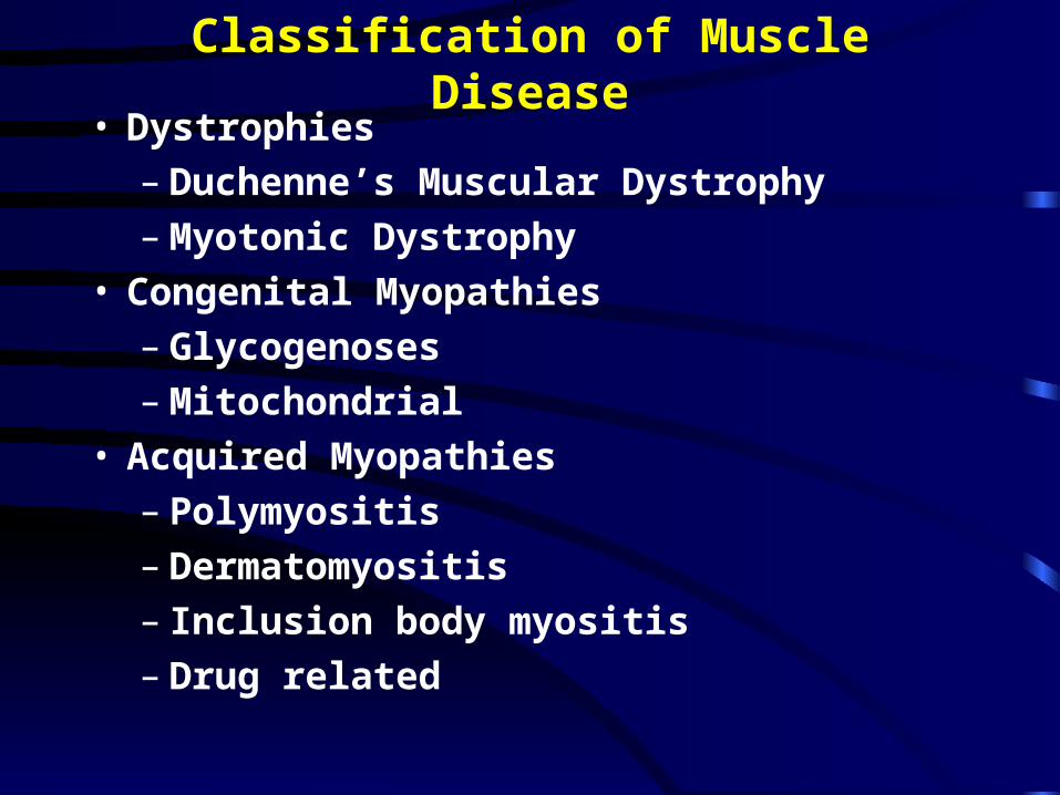
Classification of Muscle Disease• Dystrophies
– Duchenne’s Muscular Dystrophy– Myotonic Dystrophy
• Congenital Myopathies– Glycogenoses– Mitochondrial
• Acquired Myopathies– Polymyositis– Dermatomyositis– Inclusion body myositis– Drug related
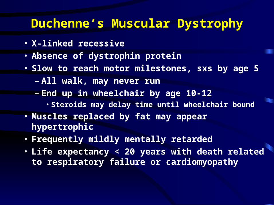
Duchenne’s Muscular Dystrophy
• X-linked recessive • Absence of dystrophin protein• Slow to reach motor milestones, sxs by age 5
– All walk, may never run– End up in wheelchair by age 10-12
• Steroids may delay time until wheelchair bound
• Muscles replaced by fat may appear hypertrophic• Frequently mildly mentally retarded• Life expectancy < 20 years with death related to
respiratory failure or cardiomyopathy

Myotonic Dystrophy• Most common of the dystrophies• Autosomal dominant• Age of onset varies• Myotonia -
– Failure of relaxation of the muscle following contraction
• Neuromuscular features– Distal weakness– Ptosis– Facial weakness – Tongue weakness - dysarthria and dysphagia– Involvement of respiratory muscles

Myotonic Dystrophy Involvement outside the NM system
• Heart– Conduction block
• Decreased fertility, undescended testicles• Diabetes mellitus• Mild MR

Idiopathic Inflammatory Myopathies
• Polymyositis (PM)• Inclusion body myositis (IBM)• Dermatomyositis (DM)
• Together - incidence ~ 1:100,000

The typical patient
• Sent with– Fatigue– Weakness– Muscle aches and pains– Elevated CPK
• May also get the pt with the dx of PM who failed steroids– May or may not have had a muscle bx

Clinical Features of Polymyositis• May occur at any age but rare under 18• Subacute onset of proximal > distal weakness
including neck flexor weakness– Rare to see it isolated to proximal muscles
• Dystrophy
• Muscle pain and tenderness– Seen in 50%
• Facial weakness uncommon• Respiratory involvement
– Mostly in pts with severe, unresponsive disease


Polymyositis
• Slightly increased risk of cancer– Bladder, lung, lymphoma
• Biopsy of muscle confirms diagnosis• Treatment with immunosuppression
– Prednisone– Methotrexate

Dermatomyositis• Affects children (Ages 5-15) as well as adults• Females more affected than males• Subacute onset of proximal > distal weakness• Facial weakness in severe cases• Dysphagia in 1/3• Fulminant cases
– Rhabdo– ARF
• Skin changes - present in 60%, frequently first– Skin changes without muscle sxs– Muscle disease without skin changes

Skin Changes in DM
• Heliotropic rash of eyelids• Erythematous rash on malar regions, neck,
shoulders, or extensor surfaces of arms/legs• Gottron’s papules
– Red, raised, scaly lesion over extensor surfaces of PIP and DIP
• Nail changes– Thickend cracked nail beds– Dilated capillary loops


Associated Conditions with DM
• Malignancy– Increased, particularly in adults over 40– Risk greatest within 5 yrs of dx of DM– Most common cancers:
• Ovarian• Lung• Pancreatic• Colorectal
• Joint disease– Arthritis– Arthralgias

Associated Condtions, cont
• GI tract– Dysphagia– Delayed gastric emptying– Intestinal ulceration/perforation
• Lungs– Interstitial lung disease
• Heart - more common in DM than PM– Conduction problems– Cardiopmyopathy
• Overlap CTD

Etiology and Treatment of DM
• Also Immune mediated• Identical to PM except
– IVIG is effective in DM but not PM• Prednisone is still considered first line of choice
in many, but consider IVIG as first agent – if pt very young – has medical reasons to avoid steroids

Inclusion Body Myositis (IBM)
• Now thought to be the most common idiopathic inflammatory myopathy in adults
• Prevalence ~5 per million• Age onset - over 50• 3 males: 1 female• Majority sporadic

IBM - clinical features• Indolent onset
– Sxs up to 10 yrs before medical care sought• Typical pattern of weakness - majority but not all
pts– Asymmetric– Wrist and finger flexors– Quadriceps
• In contrast to PM– Dysphagia (40-60%) and facial weakness (30%)
more common

IBM
• Etiology unclear• “aging” of muscle?• Do not respond to immunosuppression

Evaluation of the Patient with Suspected Muscle Disease
• Lab– Muscle enzymes (CPK, aldolase)– Erythrocyte sedimentation rate (ESR or sed rate)
if suspect inflammatory disease– Genetic test
• Duchenne’s• Myotonic dystrophy
• EMG/NCS• Muscle biopsy
• May provide a definitive diagnosis

Extremity CN Reflexes Sensation
Weakness
ALS Random yes Increased Normal
Polyneuro- Distal> rare Decreased Lost distally >
pathy Proximal distally proximally
LEMS LE > UE rare Decreased or Normal
Prox>distal absent
MG UE>LE yes Normal or dec Normal
+/-prox>distal
Myopathy Prox>distal occ Normal or dec Normal