Neurohumoral control gastrointestinal motility ROZ‰
17
Neurohumoral control of gastrointestinal motility C. ROZÉ Laboratoire de Biologie et Physiologie des Cellules digestives, (FRA 49 INSERM), Faculté de Médecine X Bichat, 16, rue H.-Huchard, 75018 Paris, France, et Université P.-et-M.-Curie Summary. The progress of gastrointestinal physiology is presently dominated by the concept of neuroendocrine regulation and the control of motility is in keeping with this concept. Progress in biochemistry, immunocytochemistry and radioimmunoassay in the past few years has produced a whole new collection of peptide molecules extracted from the digestive tract. Two facts are evident from these new data : (i) many peptides in the gastro- intestinal tract are present in both endocrine cells and nerve fibers, and (ii) many peptides are found in both the brain and the gastrointestinal tract. There is thus a large wave of interest in the so-called brain-gut axis and in the relations between brain and gut and between nerves and hormones. The motile system of the gastrointestinal tract is a complex whole having several organizational levels. The morphological, fixed structure is a« cable » network comprising smooth muscle and intrinsic and extrinsic nerves. Control feedback loops may be evidenced at all levels-muscular, intrinsic, ganglionic and central. Local concentrations of circulating (endocrine) or paracrine peptides may act as modulators of this basic pattern by contribu- ting to the depolarization or the hyperpolarization of smooth muscle cell membranes. The mode of action of these peptides reveals at least two different kinds of receptor sites : one on the smooth muscle cells and the other on the axons or cell bodies of intramural excita- tory or inhibitory neurons. In addition, central nervous system effects may centrally modu- late the long pathway reflexes travelling in the main vagosympathetic trunks. It is no longer possible to separate completely the neural from the hormonal in studying motility control. The whole neuroendocrine control complex must be investigated. Introduction. The changing picture of gastrointestinal physiology is dominated at present by the neuroendocrine regulation concept, and digestive motility does not escape this influence. At the end of the 19th century, nervous control of digestion seemed to be demons- trated by the work of Pavlov. In 1902, Bayliss and Starling discovered secretin and digestive hormonology made such great strides that, except for obvious vagal effect on the stomach, nervous system influence was practically forgotten by the new diges- tive endocrinologists.
Transcript of Neurohumoral control gastrointestinal motility ROZ‰
Neurohumoral control of gastrointestinal motility
C. ROZÉLaboratoire de Biologie et Physiologie des Cellules digestives,(FRA 49 INSERM), Faculté de Médecine X Bichat,16, rue H.-Huchard, 75018 Paris, France,et Université P.-et-M.-Curie
Summary. The progress of gastrointestinal physiology is presently dominated by theconcept of neuroendocrine regulation and the control of motility is in keeping with thisconcept.
Progress in biochemistry, immunocytochemistry and radioimmunoassay in the pastfew years has produced a whole new collection of peptide molecules extracted from thedigestive tract. Two facts are evident from these new data : (i) many peptides in the gastro-intestinal tract are present in both endocrine cells and nerve fibers, and (ii) many peptidesare found in both the brain and the gastrointestinal tract. There is thus a large wave ofinterest in the so-called brain-gut axis and in the relations between brain and gut andbetween nerves and hormones.
The motile system of the gastrointestinal tract is a complex whole having severalorganizational levels. The morphological, fixed structure is a« cable » network comprisingsmooth muscle and intrinsic and extrinsic nerves. Control feedback loops may be evidencedat all levels-muscular, intrinsic, ganglionic and central. Local concentrations of circulating(endocrine) or paracrine peptides may act as modulators of this basic pattern by contribu-ting to the depolarization or the hyperpolarization of smooth muscle cell membranes. Themode of action of these peptides reveals at least two different kinds of receptor sites : oneon the smooth muscle cells and the other on the axons or cell bodies of intramural excita-
tory or inhibitory neurons. In addition, central nervous system effects may centrally modu-late the long pathway reflexes travelling in the main vagosympathetic trunks. It is no longerpossible to separate completely the neural from the hormonal in studying motility control.The whole neuroendocrine control complex must be investigated.
Introduction.
The changing picture of gastrointestinal physiology is dominated at present bythe neuroendocrine regulation concept, and digestive motility does not escape thisinfluence.
’
At the end of the 19th century, nervous control of digestion seemed to be demons-trated by the work of Pavlov. In 1902, Bayliss and Starling discovered secretin anddigestive hormonology made such great strides that, except for obvious vagal effecton the stomach, nervous system influence was practically forgotten by the new diges-tive endocrinologists.
Due to progress in biochemical methods of extracting, analyzing and synthesiz-ing peptides, the structure of a whole new series of peptide molecules from the diges-tive tract has been successively determined. Besides the usual three main hormones(gastrin : Gregory et al., 1964 ; secretin : Mutt, Jorpes and Magnusson, 1970 ; chole-cystokinin : Mutt and Jorpes, 1971), another series of molecules has been success-fully sequenced and sometimes synthesized : e.g. VIP (vasoactive intestinal peptide,Mutt and Said, 1974), GIP (gastric inhibitory peptide, or better, glucose-dependentinsulinotropic peptide, Brown and Dryburgh, 1971), motilin (Brown, Cook and Dry-burgh, 1973) and pancreatic polypeptide (Hazlewood et al., 1973 ; Lin, Chance andEvans, 1973). Since all the gastrointestinal hormones known were peptides, it was
supposed that all peptides isolated in the digestive tract were hormones.Due to progress in immunocytochemistry, radioimmunoassay and radiorecep-
tor assay, two facts have emerged which support this supposition : (i) some gastroin-testinal peptides are not only present in endocrine cells but also in nerve cells, (ii)some peptides are present in both the digestive tract and the central nervous system.
In spite of the work of Von Euler and Gaddum (1931) on substance-P, that pep-tide was only sequenced in 1971 from brain extracts (Chang, Leeman and Niall) andin 1973 from intestinal extracts (Studer, Trzeciak and Lergier). The discovery of so-matostatin in gastrointestinal and pancreatic D-cells in 1975 led physiologists to studybrain-gut relations (the brain-gut axis) and also revived an interest in the effects of thenervous system on the gut, even if those effects were sometimes peptide-independent.
Several families of peptides common to both the central nervous system (CNS)and the digestive tract (DT) may be distinguished. The first ones isolated and sequencedfrom both the brain and the DT were substance P (Chang, Leeman and Niall, 1971 ;Studer, Trzeciak and Lergier, 1973) and neurotensin (Carraway and Leeman, 1975 ;Kitabgi, Carraway and Leeman, 1976).
Other peptides first isolated in the DT, then found in the brain by immunoassayare CCK (Dockray, 1976), VIP (Bryant et al., 1976), motilin (Yanaihara et al., 1978).In the reverse, isolated brain peptides have been found in the DT by immunoassay :somatostatin (Arimura et al., 1975), enkephalin (Polak et al., 1977), TRH (Morley et al.,1977).
Bombesin, a special case, was first isolated in amphibian skin and then found in.the brain (Brown et al., 1978) and the DT (Erspamer et al., 1978).
Due to the rapid progress in this field, this review is necessarily incomplete andinexact because peptides often have several molecular forms, complicating their
analysis, as in the case of somatostatin which has various origins (Spiess and Vale,1978 ; Pradayrol, Chayvialle and Mutt, 1978 ; Chayvialle et al., 1979).
This dual origin raises several questions as to how peptides function as chemi-
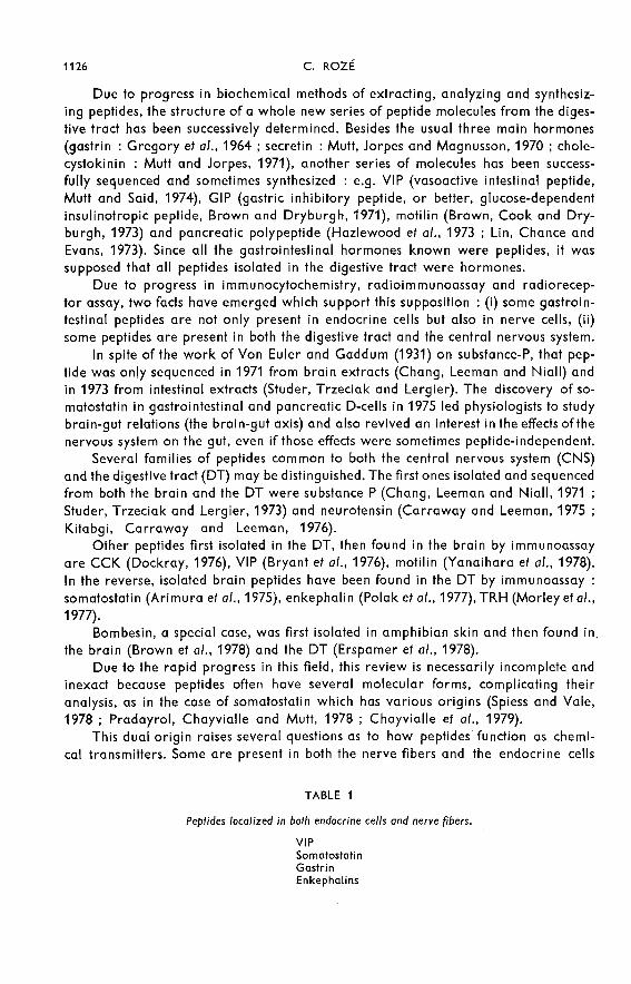
cal transmitters. Some are present in both the nerve fibers and the endocrine cells
(table 1) ; most have measurable circulating levels in physiological conditions
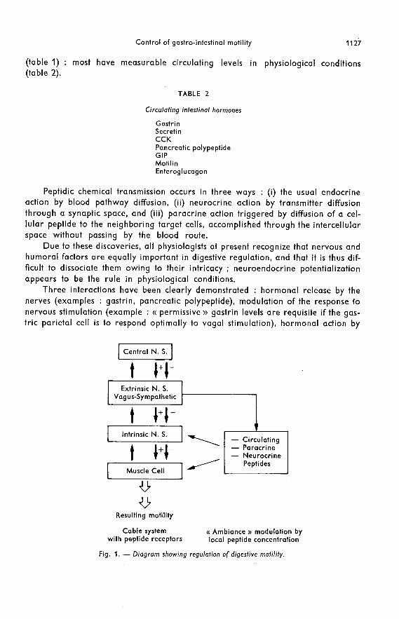
(table 2).
Peptidic chemical transmission occurs in three ways : (i) the usual endocrineaction by blood pathway diffusion, (ii) neurocrine action by transmitter diffusionthrough a synaptic space, and (iii) paracrine action triggered by diffusion of a cel-lular peptide to the neighboring target cells, accomplished through the intercellularspace without passing by the blood route.
Due to these discoveries, all physiologists at present recognize that nervous andhumoral factors are equally important in digestive regulation, and that it is thus dif-ficult to dissociate them owing to their intricacy ; neuroendocrine potentializationappears to be the rule in physiological conditions.
Three interactions have been clearly demonstrated : hormonal release by thenerves (examples : gastrin, pancreatic polypeptide), modulation of the response fonervous stimulation (example : « permissive » gastrin levels are requisite if the gas-tric parietal cell is to respond optimally to vagal stimulation), hormonal action by

nerve fiber intermediary (example : relaxation of the lower esophageal sphincter -LES - induced by octo-CCK in the cat and in humans).
The system regulating digestive motility is thus a complex one (see diagram infig. 1). The anatomic neuromuscular whole is a fixed system which we will call a
« cable » system with its sensorimotor inhibitors and excitors and its successive orga-nizational levels which are at least three in number. The intestinal smooth musclehas autonomous myogenic activity and its own receptors. Its innervation includes the
intrinsic wall plexus, the coeliac and mesenteric ganglia, which integrate some data,and the central nervous system with its cables of vagal and sympathetic pathways.This basic structure is under hormonal control, each peptide theoretically being ableto act at each of the preceding organizational levels, including participation in pos-sible synaptic transmission.
Each effector is thus controlled by the local result of the sum of the concentrationsof the numerous agonists and antagonists (peptides or neurotransmitters) present,each of which is often only relatively specific for the effector receptors because thepeptide structures, although different, have common partial sequences. We havecalled this « ambiance » modulation, indicating by this rather vague term the manyfactors - such as circulating peptide level, distribution of blood flow, binding bynon-specific elimination sites, presence of other agonists or antagonists, receptorturnover - which could play a role in the local regulation of a peptidic agonistnear an effector site.
Such complexity explains why present studies often only describe or analyze anaction and do not present a synthesis of the whole. It also justifies the large numberof research teams which, stunned by the complexity of this problem in vivo, prefer tostudy very simple preparations, isolated in a test-tube, although they may be spendingtime on theoretical studies of peptide receptor binding having only a secondary impor-tance in physiological conditions in vivo.
We shall try to analyze briefly the levels of this « cable » system and its peptidicmodulation.
Myogenic activity or muscular level.
The intestinal smooth muscle cells are spindle-shaped and mononucleated ; ascompared to the striated muscle cells, their contractile filaments are poorly organized.Moreover, they have a spontaneously oscillating membrane potential which conditionsa high degree of spontaneous, myogenic, independent rhythmic activity.
Some smooth muscles (stomach of carnivores such as the pig, monkey and man)show « plateau » action potentials, including rapid depolarization followed by slowrepolarization. The plateau may be overloaded with rapid oscillations, but this phe-nomenon is not constant and the amplitude of slow depolarization conditions the con-tractile force (see Gonella, 1978). Other muscles such as those of the small intestineand some in the stomach and colon show slow, permanent oscillations, or slow waves,which are only accompanied by contractions when they are overloaded with spikepotentials. In both cases, the contractile machinery is only activated when depolariza-tion reaches a sufficient level (spike or plateau). The maximum result is that contrac-
tions appear at the same rhythm as the periodic oscillations of the membrane potential.

It is probable that most intestinal smooth muscle cells have the intrinsic propertyof oscillating spontaneously. A cell, however, does not oscillate alone because it is
connected to others by low-resistance junctions ; the whole can thus function physio-logically as a syncitium. Morphologically, these are gap junctions, also called nexus)>.They aid in conducting the stimulus from one cell to another, and explain why poten-tials with a shape and polarity comparable to those recorded by intracellular micro-electrodes can be recorded with extracellular electrodes.
The gastrointestinal smooth muscle thus acts as a network of coupled oscillators.All the properties of intestinal slow waves can be reproduced by simulating this prin-ciple (Sarna, Daniel and Kingma, 1971, 1972). The oscillator with the highest intrinsicfrequency usually imposes its rhythm on the neighboring cells, thus explaining thefrequency plateaus and decreasing frequency gradients found from the duodenum tothe ileum. However, in some cases, oscillator coupling is more complicated, as shownin table 3 from Daniel (1977).
The origin of slow waves has usually been reported in the longitudinal layer ofthe small intestine, but it might also be found in some cells of the circular layer (Tay-lor, Daniel and Tomita, 1975). Spontaneous oscillations in the circular layer havebeen demonstrated in the cat colon (Christensen, Caprilli and Lund, 1969). Sinceoscillations can be recorded in both muscle layers, they must pass from one layer tothe other. The existence of direct junctions has not been clearly demonstrated ; it is
thus probable that the myenteric nervous plexus plays an important role in couplingthe two muscles. Three types of intestinal movement ― pendular, segmental andperistaltic - may be distinguished according to the variations in this coupling (seeGonella, 1971).
Intestinal smooth muscle is provided with a series of receptors for neurotrans-mitters and gastrointestinal peptides. Table 4 shows the receptors found in a parti-cularly well known muscular zone, the lower oesophageal sphincter. While most of

the usual neurotransmitter receptors are beginning to be well identified, those of thegastrointestinal hormones are far from definition and need further investigation.
Intrinsic innervation.
This innervation consists of groups of cell bodies constituting the intramural
ganglia ; these ganglia are interconnected exclusively by abundant, unmyelinatedaxons connecting the cell bodies. This whole forms the myenteric (Auerbach’s) andthe submucosal (Meissner’s) plexus of the wall.

Some of the intramural neurons innervate the smooth muscles, while others are
sensory neurons. A majority are interneurons establishing complex connections be-
tween the successive segments of the intestine.
The motor neurons innervating the smooth muscle do not terminate in neuromus-cular junctions similar to those of the striated muscle. The fibers simply run near thesmooth muscle cells without really contacting them. The neurotransmitters are storedas vesicles in the nodes distributed along the axon near the muscle cells. Moreover,nerve fiber density is low in relation to that of the muscle, indicating that most musclefibers are not in the direct vicinity of a motor axon varicosity. The neurotransmitterreleased near a muscle fiber will probably excite several fibers, one after the other,through the intercellular gap junctions in the muscle.
Using electromyographical methods for recording neuromuscular excitatoryjunction potentials (depolarization) or inhibitory junction potentials (hyperpolariza-tion), two main types of motor terminals are determined : excitatory terminals withacetylcholine as a transmitter and inhibitory terminals with one or more unknown,non-adrenergic neurotransmitters. These fibers could be purinergic (Burnstock, 1972,1979), ATP-consuming, peptidergic, or sometimes dopaminergic (Valenzuela, 1976).Localized principally in Auerbach’s plexus, the intrinsic integration system consistsof a neuronal chain distally inhibiting the circular muscle in response to stretching.The nature of the interneurons in the chain is unknown but it is probably cholinergicsince nicotine blockade inhibits distal chain transmission.
On the basis of intracellular microelectrode recordings, Hirst, Holman and
McKirdy (1975) proposed a nervous circuit pattern for the descending inhibition
causing peristalsis. Their results are limited morphologically and cannot be genera-lized to include all mammalian intestines having myogenic control systems becausethe experimental material chosen (the guinea-pig small intestine) is an exception tothe rule since its smooth muscle does not generate slow waves and it has neurogenicelectrical activity.
However, their work described a descending contractile inhibition as well as adescending stimulus, occurring only after some time-lag. The descending inhibitoryroute seems to pass entirely through the myenteric plexus, while part of the descend-ing excitatory route would go by way of the submucosal plexus.
Descending inhibitory circuits have also been described in the rabbit small intes-tine (Daniel and Taylor, 1975) and in the esophagus and at the gastroduodenal junc-tion (Daniel, 1977). Generalizing the system implies that intrinsic distal inhibition
is the same throughout the gut. Its determination depends on the existence or absenceof an active basal tone in the zone considered. When there is no basal tone, relaxa-
tion cannot occur ; only induced contractions can be inhibited by descending inhibi-tory pathways.
Neurotransmitters of the intramural nervous system.
Many synapses in the intramural nervous system are cholinergic and correspondto excitatory routes. There are probably several other neurotransmitters interveningin the inhibitory pathways. ATP, referred to as an important neurotransmitter of
inhibitory neurons, probably exists but does not always have a predominant influence

(Stockley, 1978). The existence of serotoninergic neurons may be considered as cer-tain. These neurons have a specific 5HT receptor (a specific 5HT-binding protein),contain tryptophane hydroxylase permitting 5HT synthesis from tryptophane andsurvive up to 3 weeks in organ culture (Gershon and Dreyfus, 1977), thus provingthat their cell bodies are localized in the gut wall. It appears that they act by inhibitingthe cholinergic excitatory pathway (Bulbring and Gershon, 1967 ; Gonella, 1978).
Besides these substances, there are many peptides in plexus nerve fibers (Polakand Bloom, 1978 ; Hokfelt et al., 1978), but arguments in favor of their physiologicalrole are still very limited at the present time.
A rich, probably intrinsic, network of VIP-immunoreactive nerves is found throu-ghout the intestinal wall (Larsson et al., 1976). VIP has been identified mainly in thecell bodies of the submucosal plexus, in the whole fiber network near the musclecells and around the ganglionic cells of Auerbach’s and Meissner’s plexus.
Substance-P neurons have also been reported in the muscle layer and aroundganglionic cell bodies of myenteric plexus (Pearse and Polak, 1975). The highestconcentrations are found in the duodenum and the colon.
Somatostatin and Leu-enkephalin fibers are reported in the myenteric plexus.The persistence in organ culture of fibers reacting with antibodies raised againstsubstance-P, somatostatin and Leu-enkephalin strongly suggests that the cell bodiesof these neurons are to be found in the gut wall (Hokfelt et al., 1978).
Sensory neurons.
The functioning of the intrinsic nervous system implies the perception of sensorysignals triggering control systems. Mechanoreceptors are found in the gut whichadapt slowly or rapidly ; some electrophysiological recordings have found cellswhich might fulfill this function in the intramural ganglia. When it was establishedthat the peristaltic reflex of the guinea-pig intestine was maintained after removal ofthe mucosa, the idea prevailed that mechanoreceptors were probably grouped « inseries » with the circular fibers and were thus closely associated with them. Daniel(1977) proposed « intermediary » cells as candidates for this function in the dog ;these cells appearing to be glial cells in Schwann’s sheath, but forming many gapjunctions with the muscle cells and among themselves, would also have contact withthe axons and varicosities of the myenteric plexus, and might sometimes containactin-type filaments or neurotubules. The future will tell if they are mechanoreceptorsor not.
Fiber and cell body receptors of the intrinsic nervous system.
The cell bodies and presynaptic elements of intrinsic nervous system neuronsall have receptors which have not all been identified. Naturally, there are acetyl-choline, noradrenalin and serotonin receptors. As these substances are mainlyreleased near the receptors by the neural route, they usually act as ordinary neuro-transmitters. However, circulating plasma catecholamines may reach a modulatorylevel acceptable to the accessible receptors.

Neural gastrointestinal peptide receptors probably also exist in the intrinsic
plexus. The circulating level of a peptide, or the rate of its paracrine release, thenbecomes important in modulating a nervous process, and peptidic action wouldbe effected by the intermediary of intrinsic nervous system fibers. Peptides usuallyactivate excitatory cholinergic mechanisms, but there are other possibilities. Amongthe peptides activating cholinergic fibers, motilin acts on the stomach (Tani et at.,
1978) and the LES (Meissner et a/., 1976) and CCK affects the small intestine (Gonella,Salducci and Monges, 1973), while gastrin action on the LES is controversial, someauthors reporting an atropine-sensitive mechanism and others none. Szurszewski
(1975) has shown the action of gastrin on the intrinsic plexus of the dog gastric antrum.Octo-CCK would act on non-adrenergic inhibitory fibers of the cat LES (Behar andBiancani, 1977) and bombesin on adrenergic postganglionic fibers releasing nore-pinephrine (Mukhopadhyay and Kunnemann, 1979).
These investigations, in full progress, are far from being concluded.
Extrinsic nervous system.
The extramural innervation is composed of parasympathetic and sympatheticfibers and includes motor efferent fibers and sensory afferent ones.
Parasympathetic.
The cell bodies of the parasympathetic efferent axons using the vagal pathway isin the brainstem and that of the axons using the pelvic nerve route is in the sacral
segments of the spinal cord.
All the preganglionic axons, whether excitatory or inhibitory, are cholinergic.In the intestinal wall, these axons synapse with intramural neurons whether they arecholinergic excitatory or non-adrenergic inhibitory (NAI). Both types of postganglio-nic parasympathetic neurons act directly on the smooth fibers.
Vagal control of motor function is greater in the proximal part of the digestivetract (esophagus, stomach, upper small intestine) than in the distal part. However, alllevels are somewhat independent from vagal control. Thus, in the smooth part ofthe cat, monkey and opossum esophagus, the main functions of peristalsis and LESrelaxation may be obtained by stretching the isolated or extrinsically denervated eso-phagus. Cervical vagotomy induces motor disorders but does not abolish the esopha-geal peristalsis induced by stretching.
The distribution of efferent vagal fibers in the stomach has been thoroughlystudied (Sarna and Daniel, 1975). The proximal fibers do not affect the slow or rapidelectrical activity of the stomach. Like the chief cells and parietal cells of the fundic
mucosa, they probably innervate the fundic muscle, inducing receptive relaxationor fundic contraction.
Further down, the segmental branches entering the gastric wall by the anteriorand posterior nerves of Latarjet are segmentally distributed on the stomach. If theyare severed sequentially from the proximal to the distal zone of the antrum, the con-tractions induced by stimulation disappear sequentially. Latarjet’s nerves show someanterior-posterior cross-innervation by preganglionic fibers. The postganglionic
neurons, on the contrary, are short and do not show this type of cross-innervationbetween the two sides of the stomach.
Sympathetic nervous system.The cell bodies of the preganglionic efferent neurons of the sympathetic nervous
system are in the dorsal and lumbar segments of the spinal cord. Their axon passesalong the sympathetic ganglion column situated on either side of the spinal cord,then by thoracic and lumbar splanchnic nerves to synapse within the coeliac andmesenteric ganglia. The preganglionic neuron is cholinergic, while the postganglio-nic neuron is noradrenergic.
In most cases, the stimulation of noradrenergic fibers induces hyperpolarizationof smooth muscle fibers and blockade of the cholinergic excitatory pathway ; thiscorresponds to the usual cholinergic-adrenergic balance. Noradrenergic innervationseems more developed in the circular layer (Furness and Costa, 1974). Moreover,many noradrenergic fibers innervate the myenteric plexus. It would appear, however,that sympathetic terminals do not act directly on the cell body of neurons which arecholinergic activators, but inhibit acetylcholine release at a presynaptic site. lntracel-lular recording techniques have not evidenced enteric neurons whose membrane
potential is modified either by noradrenaline or by sympathetic fiber stimulation(Gonella, 1978).
Sphincter innervation, particularly that of the lower esophagus sphincter, is
different. Alpha adrenergic excitatory receptors are present on the smooth musclecells of the LES and induce contraction. On the other hand, it has been recently shownin the cat that sympathetic stimulation and noradrenaline enhance cholinergic LESeffects, probably by stimulating cholinergic myenteric neurons (Gonella, Niel and
Roman, 1979 ; Niel, Gonella and Roman, 1979). This effect could not be demonstratedin the opossum (Fournet, Snape and Cohen, 1979).
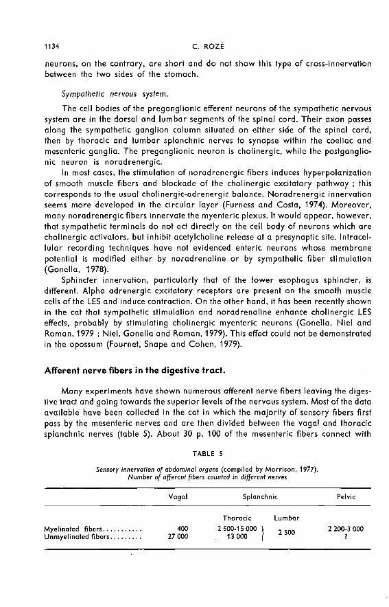
Afferent nerve fibers in the digestive tract.
Many experiments have shown numerous afferent nerve fibers leaving the diges-tive tract and going towards the superior levels of the nervous system. Most of the dataavailable have been collected in the cat in which the majority of sensory fibers firstpass by the mesenteric nerves and are then divided between the vagal and thoracicsplanchnic nerves (table 5). About 30 p. 100 of the mesenteric fibers connect with

an efferent neuron in the inferior mesenteric and coeliac ganglia, thus constitutinga short reflex pathway. The rest of the afferent fibers pass directly by the lumbarsplanchnic nerve and the pelvic nerves.
Studies of degeneration show that most afferent neurons are situated in the entericplexus. The cell body of some however is in the spinal ganglia.
To give an idea of the extent of the sensory fibers, more than 80 p. 100 of the fibersof the abdominal vagus in the cat and rabbit are afferent. A small percentage of theseare medium-sized, myelinated fibers, while most are unmyelinated (Morrison, 1977).
The morphology of sensory DT terminals is not well known. Some of the fibers
which may be considered as sensory are : small unmyelinated fibers going from themuscularis mucosae to the submucosal plexus ; fibers penetrating the muscularis muco-sae, reaching the mucosa and terminating near the epithelial cells ; networks of verysmall fibers with varicosities, present in the longitudinal muscles (cat stomach) ;spindle-shaped endings with terminal nodes penetrating the gastric smooth muscle(tension : Morrison, 1977). The duodenal zone is particularly rich in small fibers
which are probably sensory.The most studied receptors are sensitive to gut tension and stretching.Stretching causes various rapid motor, secretory and behavioural responses
(inhibition of food intake). Electrophysiological methods can be used to record theresponses of the individual fibers connected to a receptor.
The mechanoreceptors described are of two types. Some are found in the muscle
layers and are known as tension receptors « in series » with the muscle. They havebeen described in the stomach and small intestine as slowly adapting and connectedto vagal or splanchnic fibers (see Mei, 1970 ; Leek, 1972 ; Paintal, 1973). Other mecha-noreceptors found in the mucosal layers are stimulated by contractions of the mus-cularis mucosae and by tactile stimulation of the mucosa. They discharge irregularlyand adapt rapidly (Paintal, 1957).
There are many chemoreceptors in the duodenum which are pH, glucose oramino acid-sensitive (Iggo, 1957 ; Davison, 1972 ; Sharma and Nasset, 1962).
Recently, intestinal vagal glucoreceptors were studied in the cat (Mei, 1978).These receptors are connected to slowly adapting neurons corresponding to type-Cfibers. They respond rapidly when the intestine is perfused with glucose or othersugars, are not sensitive to mechanical stimulation and do not seem to respond direc-tly to the osmotic pressure of solutions, although showing increased response withthe concentration. Their structure is unknown but they are probably situated nearthe mucosal cells. Some complexely branched fibers in the villi could represent glu-cosensitive endings.
Vagal thermoreceptors have also been described (El Ouazzani and Mei, 1979).
Role of prevertebral ganglia.
The prevertebral ganglia of the autonomic nervous system (solar plexus ganglia,inferior mesenteric ganglion) were considered for a long time as simple relays on theefferent sympathetic pathway destined for the abdominal organs.
Recently, however, intracellular neuronal activity recorded in the inferior mesen-teric ganglion has shown that some postganglionic neurons receive synaptic influences

from the colon, and even from the more oral zones of the digestive tract as well asfrom the central nervous system (Szurszewski and Weems, 1976). This autonomicganglion would have three different functions : multiplication of efferent impulses,integration of afferent impulses and participation in peripheral reflex activity. It
should be noted that since the same ganglionic neuron receives both centrifugal andcentripetal impulses from the central nervous system by the peripheral reflex route,it is able to integrate these data (Szurszewski, 1977).
All autonomic ganglia do not function in the same way, and electrophysiologicaltechniques are useful in determining whether a ganglionic cell receives one or morepreganglionic fibers. In the hamster pelvic ganglia, each neuron receives only one tofour preganglionic fibers, each fiber forming an « obligatory » synapse, i.e. an impulseon only one preganglionic fiber always triggers a postganglionic impulse. Moreover,many cells of that ganglion receive sympathetic (hypogastric nerve) as well as para-sympathetic (pelvic nerve) afferent impulses. The functional significance of this pro-cess is unknown. Finally, the ganglion cell of these pelvic ganglia acts simply as a relay.
In contrast to these data, other prevertebral ganglia behave differently. Studieson the guinea-pig (Crowcroft and Szurszewski, 1971) or cat using cellular responsesof neurons of the inferior mesenteric ganglion obtained by microelectrodes, showthat each neuron receives impulses from at least 40 different identifiable preganglio-nic fibers. Most of these fibers form «weak» synapses, individually insufficient forreleasing an action potential. The action potential occurs on the postganglionic axononly when the sympathetic potentials accumulate in space or time. Integration is thus
a function of these neurons.
The neurons of the inferior mesenteric ganglion also receive sensory impulsesfrom the colon through the lumbar colic nerves. These impulses from slowly adaptingmechanoreceptors, whose discharge rhythm seems correlated with the state of endo-luminal pressure and propulsive motor activity, are inhibited when tetrodotoxin is
perfused into the ’colon (Szurszewski, 1977). These mechanoreceptors constitute
the afferent branch of a reflex loop whose efferent is the noradrenergic postganglio-nic neuron. This neuron acts (i) on the intramural excitatory neurons by decreasingtheir acetylcholine release, (ii) on the muscle by reducing contractile activity and (iii)on the mechanoreceptors themselves by desensitizing them (Szurszewski and Weems,1976).
A last function of the prevertebral ganglia would be to connect the functions ofthe different zones of the digestive tract. This has been shown between the distal andproximal colon by the cross-innervation of inferior and superior mesenteric ganglia.
Role of the central nervous system.
This role is evident in view of the high number of-afferent impulses taking thevagosympathetic routes, and the marked effects of stimulation of the efferent fibersdestined for the digestive tract. However, there are few data on the way in whichinformation is treated and transformed in the central nervous system.
The hypothalamic glucoreceptors causing central motor stimulation have beenstudied thoroughly. This system is easily modulated by insulin hypoglycemia or by2-deoxyglucose (2-DG) which, in competition with glucose, produces intra-cellular

cytoglucopenic in spite of the hyperglycemia following nervous stimulation of theadrenals.
2-DG has many effects on the digestive tract, stimulating secretion, motility andhyperphagic response and activating hyperglycemia and hypothermia (Muller,Cocchi and Forni, 1971 ; Muller et a/., 1974). The motor effects of 2-DG are particu-larly evident in the stomach where they are partially correlated with secretory effects(Boiselle, Rozé and Vatier, 1977).
The existence of a pathway connecting the hypothalamus and the vagal nerveindicates that there may be many modulating factors.
A pharmacological study of the modulation of this pathway has produced posi-tive results on gastric and pancreatic secretion (Rozé et al., 1978, 1979) and it shouldalso be fruitful as regards motility (Burks, 1978).
The central effects of morphine on gastrointestinal motility have been studied byinjecting doses too low to be active in systemic injection into the cerebral ventriclesof the conscious cat. This drug induced vomiting and also triggered a characteristicsequence of intestinal myoelectrical activity showing an intense burst of spike poten-tials moving in the aboral-oral direction (Stewart, Burks and Weisbrodt, 1977a). Theeffect is not specific to morphine since it can also be obtained after intraventricular
injections of apomorphine and adrenaline. In all cases, the process is controlled bythe emetic center.
Moreover, intraventricular injection of smaller doses of morphine, which do notproduce vomiting and are inactive by the systemic route, increases spike potentialsat all levels of the small intestine (Stewart, Weisbrodt and Burks, 1977b). This effectis inhibited by small doses of naloxone by the IVT route, indicating that morphine mayproduce intestinal contractions by acting on cerebral structures (Bueno and Rucke-busch, 1978). These motor changes may affect transit ; in the rat doses of morphine,which would be ineffective when given by the systemic route, inhibit small intestinetransit when given by IVT (Parolaro, Sala and Gori, 1977 ; Stewart, Weisbrodt andBurks, 1977c). Transit inhibition is reduced by vagotomy and by naltrexone given byI VT.
It is certain that opiates modify intestinal motility by direct organ effect. However,both central and peripheral mechanisms may play a role in vivo.
The actions reported with morphine encourage research on endogenous opioidpeptides, endorphins and enkephalins (Konturek, 1978). Pascaud et al. (1979) recentlyshowed a decrease in antral contractions and an increase in colic motility in the ratunder the effect of p-endorphin, Met-enkephalin and the long-acting analog, D-AlaMet-enkephalinamide, when given by IVT.
Other substances probably also affect central motility. Injecting small amountsof TRH (thyrotropin-releasing hormone) by IVT causes intense stimulation of colonicmotility (Smith et al., 1977).
As emphasized previously, the fact that the brain and the gut have commonpeptides, such as somatostatin, VIP, neurotensin, CCK-like, gastrin-like and sub-stance P, is a reason for continuing toexplore the central mechanisms of the brain-gutrelationship.
journ6es Ingestion-Digestion-Absorptionde l’Association fron5aise de Nutrition,Paris, 15-16 novembre 1979.
Résumé. Le panorama changeant de la physiologie gastro-intestinale est actuellementdominé par le concept de régulation neuro-endocrine, et les phénomènes moteurs dans letube digestif n’échappent pas à cette règle.
A la fin du XIXE siècle le contrôle nerveux de la digestion paraît évident à la suitedes travaux de Pavlov. Puis en 1902 Bayliss et Starling découvrent la sécrétine et l’hormo-nologie digestive prend un tel essor que l’influence du système nerveux se trouve pratique-ment oubliée des nouveaux endocrinologues digestifs. Il faut pratiquement attendre 1975avec la mise en évidence de la somatostatine dans les cellules D gastro-intestinales et pan-créatiques pour qu’explose subitement une énorme vague d’intérêt immunochimique,embryologique et physiologique pour les peptides présents à la fois dans le cerveau et letube digestif (somatostatine, VIP, gastrine, octo-CCK, neurotensine, enkephalines...). Deplus certains de ces peptides sont trouvés à la fois dans des fibres nerveuses et des cellulesde type endocrine, posant de nouvelles questions sur les origines et le fonctionnement de cesystème neuro-endocrine. Il faut admettre qu’un peptide hormonal peut agir au moins detrois façons différentes : comme neuro-transmetteur peptidergique, comme substance àaction locale dite paracrine, comme hormone circulante.
Grâce à ces découvertes, tous les physiologistes sont obligés de reconnaître que lesfacteurs nerveux et les facteurs humoraux sont d’une égale importance dans la régulationdigestive, et qu’ils sont de plus difficilement dissociables en raison de leur intrication : lasituation de potentialisation neuro-endocrine paraît en effet de règle dans les conditionsphysiologiques.
La régulation de la motricité digestive n’échappe pas à cette loi générale. Le tableauest cependant complexe. Le muscle lisse intestinal possède son activité myogène autonomeet ses récepteurs propres. Son innervation comprend les plexus de la paroi (intrinsèque),les ganglions du plexus solaire et mésentérique inférieur qui intègrent certaines informa-tions et le niveau du système nerveux central, « câblé » par les voies vagale et sympathique.Sur cette structure se greffent les actions hormonales, chaque peptide ayant en théorie lapossibilité d’agir à chacun des niveaux d’organisation précédents, y compris en participantà une éventuelle transmission synaptique.
Chaque effecteur se trouve ainsi contrôlé par le résultat local de la somme des concen-trations des nombreux agonistes et antagonistes présents, peptides ou neurotransmetteurs,dont chacun n’est souvent que relativement spécifique des récepteurs de l’effecteur, comptetenu des parentés partielles de structure des peptides mis en jeu.
L’étude analytique des différents facteurs agissant sur le muscle gastro-intestinal est
nécessaire avant de construire le tableau de synthèse, qui seul représentera la physiologie.Dans la plupart des cas nous n’en sommes encore qu’à la première partie, analytique, de ceprogramme. En tout cas, une séparation complète des facteurs nerveux et des facteurshumoraux n’est plus possible : leur interaction est permanente sous forme d’une régulationneuro-endocrine.
Références
ARIMURA A., SATO H., DUPONT A., NISHI N., SCHALLY A. V., 1975. Somatostatin : Abundance
of immunoreactive hormone in rat stomach and pancreas. Science, 189, 1007-1009.BEHAR J., BIANCANI P., 1977. Effect of cholecystokinin-octapeptide on lower esophageal sphincter.
Gastroenterology, 73, 57-61.BOISELLE J. C., ROZÉ C., VATIER J., 1977. Etude comparative de la motricité et de la sécrétion
gastriques stimulées par le 2-deoxy-D-glucose chez le chien. Gastroenterol. clin. 6iol., 1,345-352.
BROWN J. C., COOK M. A., DRYBURGH J. R., 1973. Motilin, a gastric motor stimulating polypep-tide : The complete amino acid sequence. Can. J. Biochem., 51, 533-537.
BROWN J. C., DRYBURGH J. R., 1971. A gastric inhibitory polypeptide. Il. The complete aminoacid sequence. Can. J. Biochem., 49, 867-872.
BROWN M., RIVIER J., KOBAYASHI R., VALE W., 1978. Neurotensin-like and bombesin-like pep-tides : CNS distribution and actions, 550-558. In Bloom S. R., Gut Hormones, Churchill Living-stone, Edinburgh.
BRYANT M. G., POLAK J. M., MODLIN L, BLOOM S. R., ALBUQUERQUE R. H., PEARSE A. G. E.,1976. Possible dual role for vasoactive intestinal peptide as gastrointestinal hormone andneurotransmitter substance. Lancet, 1, 991-993.
BUENO L., RUCKEBUSCH Y., 1978. Origine centrale de l’action excito-motrice de l’intestin par lamorphine. C. R. Soc. Biol., 172, 972-977.
BULBRING E., GERSHON M. D., 1967. 5-hydroxytryptamine participation in the vagal inhibitoryinnervation of the stomach. J. Physiol. Lond., 192, 823-846.
BURKS T. F., 1978. Central sites of action of gastrointestinal drugs. Gastroenterology, 74, 322-324.BURNSTOCK G., 1972. Purinergic nerves. Pharmacol. Rev., 24, 509-581.BURNSTOCK G., 1979. Non-adrenergic, non-cholinergic enteric neurones : Their roles and inter-
actions with classical nerves, 481-492. In ROSSELIN G., FROMAGEOT P., BONFILS S.,Hormone receptors in digestion and nutrition, Elsevier/North Holland Biomed, Press, Amsterdam,New York.
CARRAWAY R., LEEMAN S. E., 1975. The amino-acid sequence of a hypothalamic peptide, neuro-tensin. J. 6iol. Chem., 250, 1907-1911.
CHANG M. M., LEEMAN S. E., NIALL H. D., 1971. Amino-acid sequence of substance-P. Nature,
(New Siol.), 232, 86-87.
CHAYVIALLE J. A., DESCOS F., MIYATA M., RAYFORD P. L., THOMPSON J. C., 1979. Molecularforms of somatostatin in acetic extracts of pancréas and gastro-intestinal mucosa, 109-114.In ROSSELIN G., FROMAGEOT P., BONFILS S. Hormone receptors in digestion and nutrition,Elsevier/North Holland Biomed. Press.
CHRISTENSEN J., CAPRILLI R., LUND G. F., 1969. Electric slow waves in circular muscle of cat
colon. Am. J. Physiol., 217, 771-776.CROWCROFT P. J., SZURSZEWSKI J. H., 1971. A study of the inferior mesenteric and pelvic
ganglia of guinea-pigs with intracellular electrodes. J. Physiol London, 219, 421-441.DANIEL E. E., 1977. Nerves and motor activity of the gut, 154-196. In Brooks F. P., EVERS P. W.
Nerves and the gut, C. B. Slack Inc, Thorofare, N. J.DANIEL E. E., TAYLOR G. S., 1975. Junction potentials and control of motility of the small intestine.
Proc. 5th int. Symp. on Gi motility, G. VANTRAPPEN Ed., Typoff Press, Herentals Belgium,213-218.
DAVISON J. J., 1972. Response of single vagal afférent fibres to mechanical and chemical stimulationof the gastric and duodenal mucosa in cats. J. exp. Physiol., 57, 405-416.
DOCKRAY G. J., 1976. Immunochemical evidence of cholecystokinin-like peptides in brain. Nature264, 568-570.
EL OUAZZANI T., MEI N., 1979. Mise en évidence électrophysiologique des thermorécepteursvagaux dans la région gastro-intestinale, leur rôle dans la régulation de la motricité digestive.Expl. Brain Res., 34, 419-434.
ERSPAMER V., MELCHIORRI P., FALCONIERI-ERSPAMER C., NEGRI L., 1978. Polypeptides of theamphibian skin active on the gut and their mammalian counterparts, 51-64. In SPERANZA V.,BASSO N., LEZOCHE E., GROSSMAN M. 1. ; Gastrointestinal hormones and pathology of thedigestive system, Plenum Press, New York.
FOURNET J., SNAPE W. J., COHEN S., 1979. The cholinergic component of basal lower esophagealsphincter pressure. 7th int. Symp, on gastrointestinal motility, Iowa City, Abstr. Vol., p. 4.
FURNESS J. B., COSTA M., 1974. The adrenergic innervation of the gastro-intestinal tract. Ergebn.Physiol., 69, 1-51.
GERSHON M. D., DREYFUS C. F., 1977. Serotonergic neurons in the mammalian gut, 197-206. InBROOKS F. P., EVERS P. W., Nerves and the gut. CBS Inc. Thorofare, N. J.
GONELLA J., 1971. Etude électromyographique des contractions segmentaires et péristaltiques duduodénum de lapin. P(lügers Arch. 322, 217-234.
GONELLA J., 1978. La motricité digestive et sa régulation nerveuse. J. Physiol. Paris, 74, 131-
140.
GONELLA J., NIEL J. P., ROMAN C., 1979. Sympathetic control of lower aesophageal sphinctermotility in the cat. J. Physiol London, 287, 177-190.
GONELLA J., SALDUCCI J., MONGES H., 1973. Mode d’action de la cholecystokinine pancreozy-mine sur le duodenum de lapin in vitro. Etude électromyographique. Biol. Gastroentérol Paris,6, 365-366.

GOYAL R. K., RATTAN S., 1978. Neurohumoral, hormonal and drug receptors for the lower
esophageal sphincter. Gastroenterology, 74, 598-619.GREGORY H., HARDY P. M., JONES D. S., 1964. Structure of gastrin. Nature, 204, 931-933.HAZLEWOOD R. L., TURNER S. D., KIMMELJ. R., POLLOCK H. G., 1973. Spectrum effects of a new
polypeptide (third hormone ?) isolated from the chicken pancreas. Gen. comp. Endocrinol., 21,485-497.
HIRST G. D. S., HOLMAN M. E., McKIRDY H. C., 1975. Two descending pathways activated bydistension of guinea pig small intestine J. Physiol. London, 244, 113-127.
HOKFELTT., SCHULTZBERG M., JOHANSSON O., LJUNGDAHL A., ELFVIN L., ELDE R., TERENIUSL., NILSSON G., SAID S., GOLDSTEIN M., 1978. Central and peripheral peptide producingneurons, 423-433. In BLOOM S. R., Gut hormones, Churchill Livingstone, Edinburgh.
IGGO A., 1957. Gastric mucosal chemoreceptors with vagal afferent fibres in the cat. Q. J. expl.Physiol., 42, 398-409.
KITABGI P., CARRAWAY R., LEEMAN S. E.,1976. isolation of a tridecapeptide from bovine intestinaltissue and its partial characterization as neurotensin. J. biol. Chem., 251, 7053-7058.
KONTUREK S. J.,1978. Endogenous opiates and the digestive system. Scan d, J. Gastroent.,13, 257-261.LARSSON L. L, FAHRENKRUG J., SCHAFFALITZKY de MUCKADELL O., SUNDLER F., HAKAN-
SON R., REHFELD J. F., 1976. Localisation of vasoactive intestinal polypeptide (VIP) tocentral and peripheral neurons. Proc. nal. Acad. Sci. USA., 73, 3197-3200.
LEEK B. F., 1972. Abdominal visceral receptors, 113-160. In NEIL E., Handbook of sensory physiology,vol. III/1, Enteroceptors, Springer Verlag Berlin, Heidelberg. New York.
LIN T. M., CHANCE R. E., EVANS D., 1973. Stimulatory and inhibitory actions of a bovine pancreaticpolypeptide (BPP) on gastric and pancreatic secretions of dogs. Gastroenterology, 64, 865.
MEI N., 1970. Mécanorécepteurs vagaux digestifs chez le chat. Expl. Brain Res., 11, 480-501.MEI N.,1978. Vagal glucoreceptors in the small intestine of the cat. J. Physiol. London, 282, 485-506.MEISSNER A. J., BOWES K. L., ZWICK R., DANIEL E. E., 1976. Effect of motilin on the lower oeso-
phageal sphincter. Gut, 17, 925-932.MORLEY J. E., GARVIN T. J., PEKARY A. E., HERSHMAN J. M., 1977. Thyrotropin-releasing hor-
mone in the gastrointestinal tract. Biochem. biophys. Res. Commun., 79, 314-318.MORRISON J. F. B., 1977. The afferent innervation of the gastrointestinal tract, 297-326. In BROOKS
F. P., EVERS P. W., SLACK C. B., Nerves and the gut. Thorofare, N. J.MUKHOPADHYAY A. K., KUNNEMANN M., 1979. Mechanism of lower cesophageal sphincter
stimulation by bombesin in the opossum. Gastroenterology, 76, 1409-1414.MULLER E. E., COCCHI D., FORNI A., 1971. A central site for the hyperglycemic action of 2-deoxy
D glucose in mouse and rat. Life Sci., 10, 1057-1067.MULLER E. E., PECILE A,, COCCHI D., OLGIATI V. R., 1974. Hyperglycemic or feeding response to
glucoprivation and hypothalamic glucoreceptors. Amer. J. Physiol., 226, 1100-1109.MUTT V., JORPES E., 1971. Hormonal polypeptides of the upper intestine. Biochem J., 125, 57P-58P.MUTT V., JORPES J. E., MAGNUSSON S., 1970. Structure of secretin. The amino acid sequence. Eur.
J. Biochem., 15, 513-519.MUTT V., SAID S. 1., 1974. Structure of the porcine vasoactive intestinal octacosapeptide. The amino
acid sequence. Use of kallikrein in its détermination. Eur. 1. Biochem., 42, 581-589.NIEL J. P., GONELLA J., ROMAN C., 1979. Evidence of a cholinergic mechanism in the sympathetic
control of cat’s LES. 7th int. Symp. on gastrointestinal motility, Iowa City, Abstr. Vol., p. 5.PAINTAL A. S., 1957. Responses from mucosal mechano-receptors in the small intestine of the cat.
J. Physiol., 139, 353-368.PAINTAL A. S., 1973. Vagal sensory receptors and their reflex effects. Physiol. Rev., 53, 159-227.PAROLARO D., SALA M., GORI E.,1977. Effect of intracerebroventricular administration of morphine
upon intestinal motility in rat and its antagonism with naloxone. Europ. J. Pharmacol., 46,329-338.
PASCAUD X. B., GENTON M. G., REMOND G., VINCENT M., 1979. Effects of morphinomimeticpeptides upon gut motility in anesthesized rats. Europ. J. Pharmacol. (submitted for publication).
PEARSE A. G. E., POLAK J. M., 1975. Immunocytochemical localisation of substance P in mammalianintestine. Histochemistry, 41, 373-375.
POLAK J. M., BLOOM S. R., 1978. Peptidergic innervation of the gastrointestinal tract. Adv. expl.Med. Biol., 106, 27-49.
POLAK J. M., BLOOM S. R., SULLIVAN S. N., FACER P., PEARSE A. G. E., 1977. Enkephalin-likeimmunoreactivity in the human gastrointestinal tract. Lancet, 1, 972-974.
PRADAYROL L., CHAYVIALLE J., MUTT V.,1978. Pig duodenal somatostatin : extraction and purifi-cation. Metabolism, 27, suppl. 1, 1197-1200.
ROZÉ C., CHARIOT J., LA TOUR J. de, SOUCHARD M., VAILLE C.,1979. Endorphin and enkephalineffects on basal and vagally stimulated pancreatic secretion in rats, 513-516. ln ROSSELIN G.,FROMAGEOT P., BONFILS S. Hormone receptors in digestion and nutrition, Elsevier/NorthHolland Biomed Press, Amsterdam, New York.
ROZÉ C., CHARIOT J., LA TOUR J. de, SOUCHARD M., VAILLE C., DEBRAY C., 1978. Methadone
blockade of 2-deoxyglucose-induced pancreatic secretion in the rat. Evidence for a centralsite of action. Gastroenterology, 74, 215-220.
SARNA S. K., DANIEL E. E., 1975. Vagal control of gastric electrical control activity and motility.Gastroenterology, 68, 301-308.
SARNA S. K., DANIEL E. E., KINGMA Y. J., 1971. Stimulation of slow-wave electrical activity of smallintestine. Am. J. Physiol., 221, 166-175.
SARNA S. K., DANIEL E. E., KINGMA Y. J., 1972. Stimulation of electrical control activity of the sto-mach by an array of relaxation oscillators. Am. J. Dig. Dis., 17, 299-310.
SHARMA K. N., NASSET E. S., 1962. Electrical activity in mesenteric nerves after perfusion of gutlumen. Am. J. Physiol., 202, 725-730.
SMITH J. R., LA HANN T. R., CHESNUT R. M., CARINO M. A., HORITA A., 1977. Thyrotropin-releasing hormone : Stimulation of colonic activity following intracerebroventricular adminis-tration. Science, 196, 660-662.
SPIESS J., VALE W., 1978. Evidence for larger forms of somatostatin in pigeon pancreas and rat brain.Metabolism, 27, suppl. 1, 1175-1178.
STEWART J. J., BURKS T. F., WEISBRODT N. W., 1977a. Intestinal myoelectric activity after acti-vation of the central emetic mechanism. Am. J. Physiol., 233, E131-E137.
STEWART J. J., WEISBRODT N. W., BURKS T. F., 19776. Centrally mediated intestinal stimulation
by morphine. J. Pharmacol. exp. Ther., 202, 174-181.STEWART J. J., WEISBRODT N. W., BURKS T. F., 1977c. Centrally mediated inhibition of intestinal
transit by morphine. Pharmacologist, 19, 146.STOCKLEY H., 1978. 2.2’ Pyridylisatogen antagonizes adenosine 5’-triphosphate but not nerve-me-
diated relaxations in human isolated taenia coli, 145-150. In DUTHIE H. L., Gastrointestinal
motility in health and disease, MTP Press Ltd, Lancester-England.STUDER R. O., TRZECIAK A., LERGIER W., 1973. Isolierung und Aminosaüresequenz von Substans
P aus Pferdedarum. Helv. chim. Acta, 56, 860-866.SZURSZEWSKI J. H.,1975. Mechanism of action of pentagastrin and acetylcholine on the longitudinal
muscle of the canine antrum. J. Physiol. London, 252, 335-361.SZURSZEWSKI J. H., 1977. Toward a new view of prevertebral ganglion, 244-260. In BROOKS F. P.
EVERS P. W., SLACK C. B. Nerves and the gut, Thorofare, N. J.SZURSZEWSKI J. H., WEEMS W. A., 1976. A study of peripheral input to and its control by post-
ganglionic neurons of the inferior mesenteric ganglion. J. Physiol. London, 256, 541-556.TANI M., SUGAWARA K., KATOH M., MANABE M., AKASAKA Y., KAWAI K., 1978. Effect of syn-
thetic motilin on gastric motility. Nippon Heikat. Gakkai Zasshi, 14, 55-62.TAYLOR G. S., DANIEL E. E., TOMITA T., 1975. Origin and mechanism of intestinal slow waves.
In 51h int. Symp. on Gi mofilify, Leuven, Belgium, G. VANTRAPPEN, H. 0. AGG Eds, TypoffPress Herentals, Belgium, 102-106.
VALENZUELA J. E., 1976. Dopamine as a possible neuro-transmitter in gastric relaxation. Gastro-enterology, 71, 1019-1022.
VON EULER V. S., GADDUM J. H., 1931. An unidentified depressor substance in certain tissue
extracts. J. Physiol. London, 72, 74-87.YANAIHARA C., SATO H., YANAIHARA N., NARUSE S., FORSSMANN W. G., HELMSTAEDTER V.,
FUJITA T., YAMAGUCHI K., ABE K., 1978. Motilin, substance P and somatostatin-likeimmunoreactivities in extracts from dog, tupaia and monkey brain and GI tract, 269-283. InSPERANZA V., BASSO N., LEZOCHE E., GROSSMAN M. 1. Gastrointestinof hormones and
pathology of the digestive system. Plenum Press, N. Y.