Neurogenic Syncope Dharmen Shah MD Piedmont Healthcare Neurology and Sleep Medicine.
41
Neurogenic Syncope Dharmen Shah MD Piedmont Healthcare Neurology and Sleep Medicine
-
Upload
joy-hensley -
Category
Documents
-
view
216 -
download
2
Transcript of Neurogenic Syncope Dharmen Shah MD Piedmont Healthcare Neurology and Sleep Medicine.
Neurogenic Syncope
Dharmen Shah MDPiedmont Healthcare
Neurology and Sleep Medicine
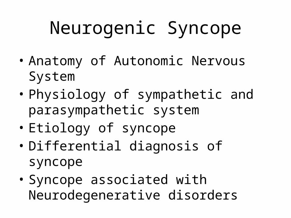
Neurogenic Syncope
• Anatomy of Autonomic Nervous System• Physiology of sympathetic and
parasympathetic system• Etiology of syncope• Differential diagnosis of syncope• Syncope associated with Neurodegenerative
disorders
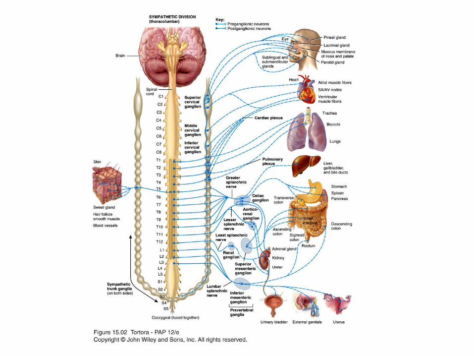
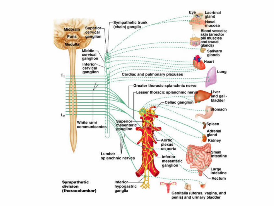
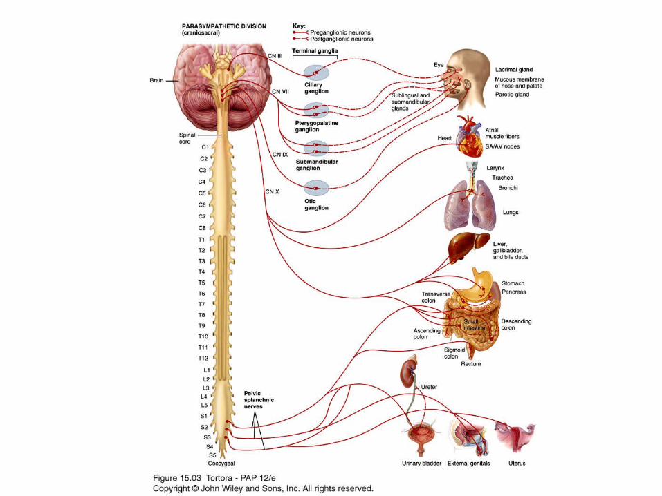
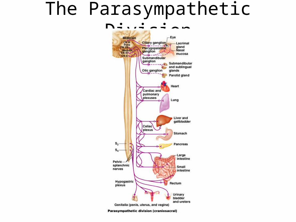
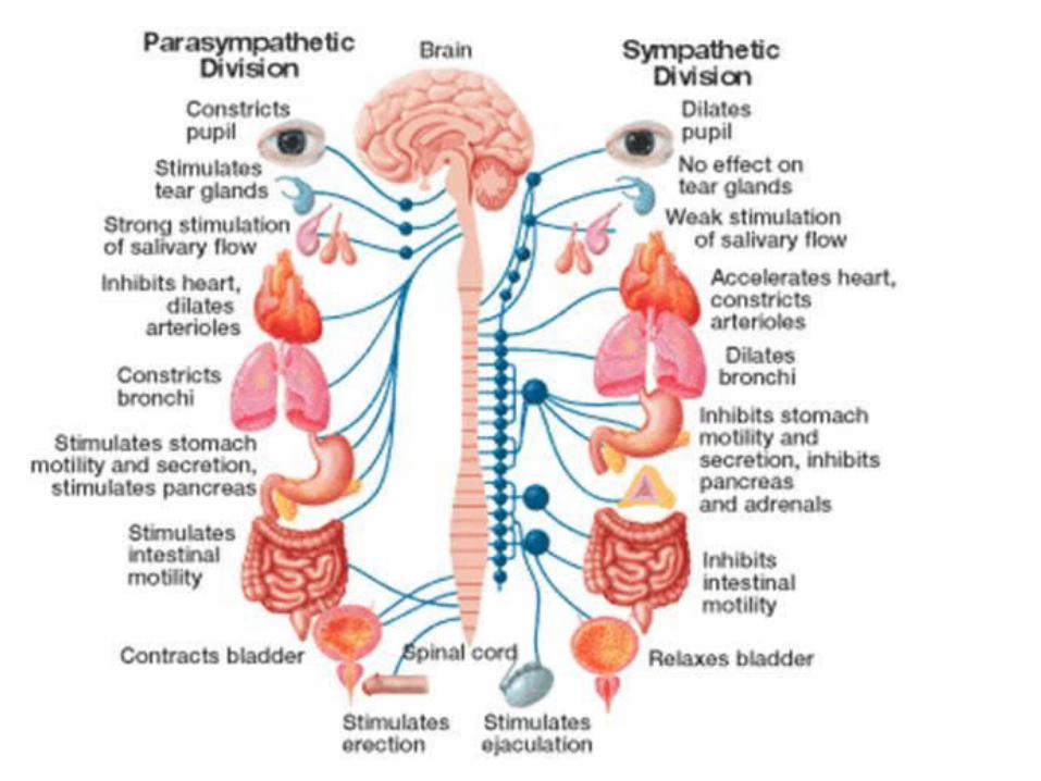
The Parasympathetic Division
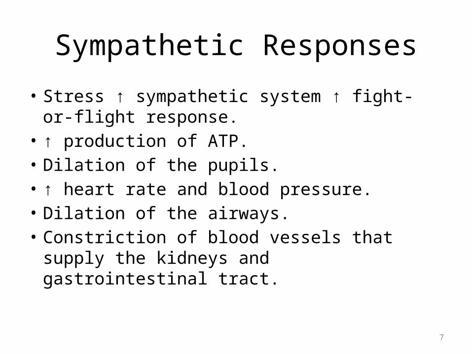
Sympathetic Responses
• Stress ↑ sympathetic system ↑ fight-or-flight response.
• ↑ production of ATP.• Dilation of the pupils.• ↑ heart rate and blood pressure.• Dilation of the airways.• Constriction of blood vessels that supply the
kidneys and gastrointestinal tract.
7
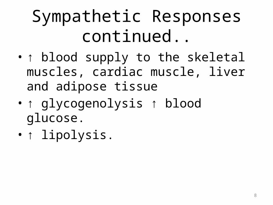
Sympathetic Responses continued..
• ↑ blood supply to the skeletal muscles, cardiac muscle, liver and adipose tissue
• ↑ glycogenolysis ↑ blood glucose.• ↑ lipolysis.
8
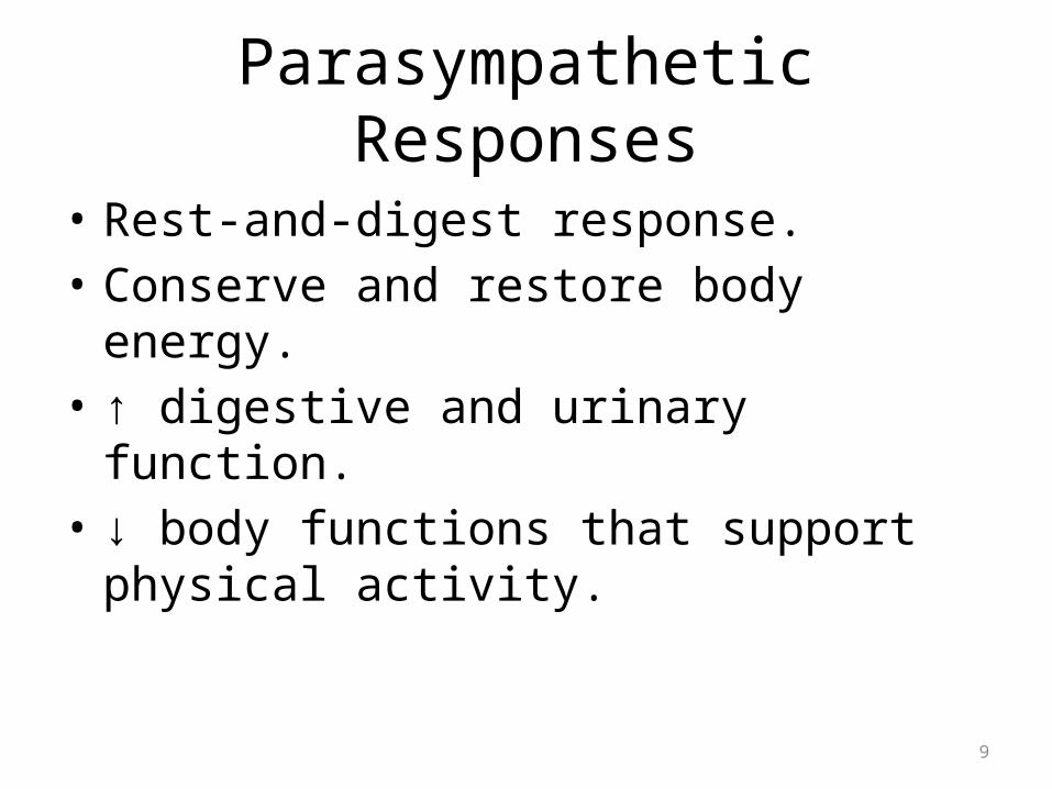
Parasympathetic Responses
• Rest-and-digest response.• Conserve and restore body energy.• ↑ digestive and urinary function.• ↓ body functions that support physical
activity.
9
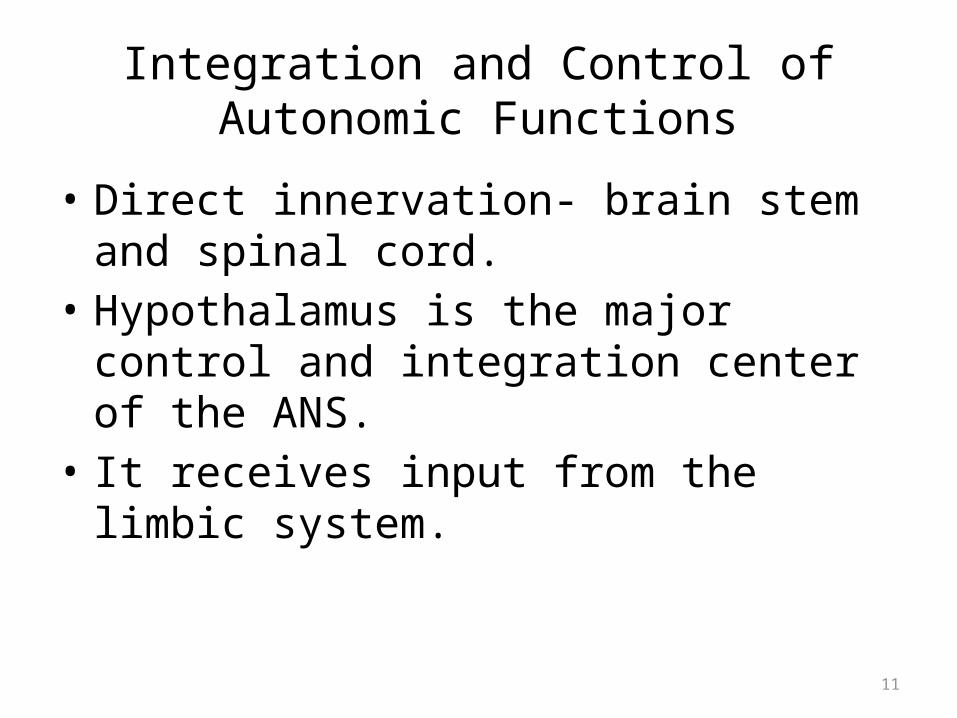
Integration and Control of Autonomic Functions
• Direct innervation- brain stem and spinal cord.• Hypothalamus is the major control and
integration center of the ANS.• It receives input from the limbic system.
11
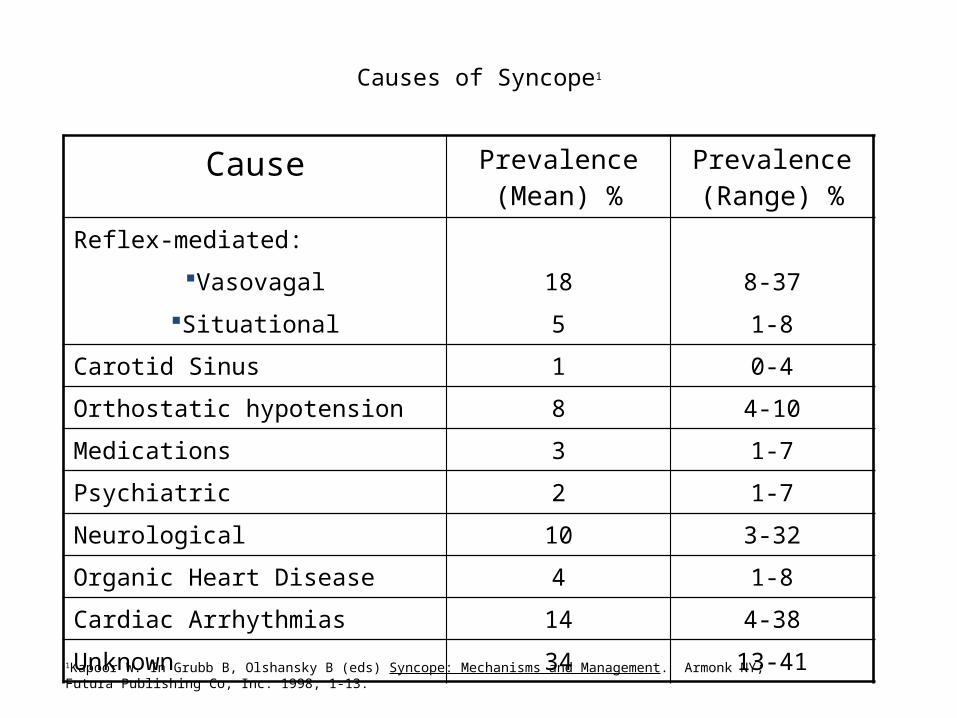
Cause Prevalence (Mean) %
Prevalence (Range) %
Reflex-mediated:
Vasovagal 18 8-37
Situational 5 1-8
Carotid Sinus 1 0-4
Orthostatic hypotension 8 4-10
Medications 3 1-7
Psychiatric 2 1-7
Neurological 10 3-32
Organic Heart Disease 4 1-8
Cardiac Arrhythmias 14 4-38
Unknown 34 13-41
Causes of Syncope1
1Kapoor W. In Grubb B, Olshansky B (eds) Syncope: Mechanisms and Management. Armonk NY; Futura Publishing Co, Inc: 1998; 1-13.
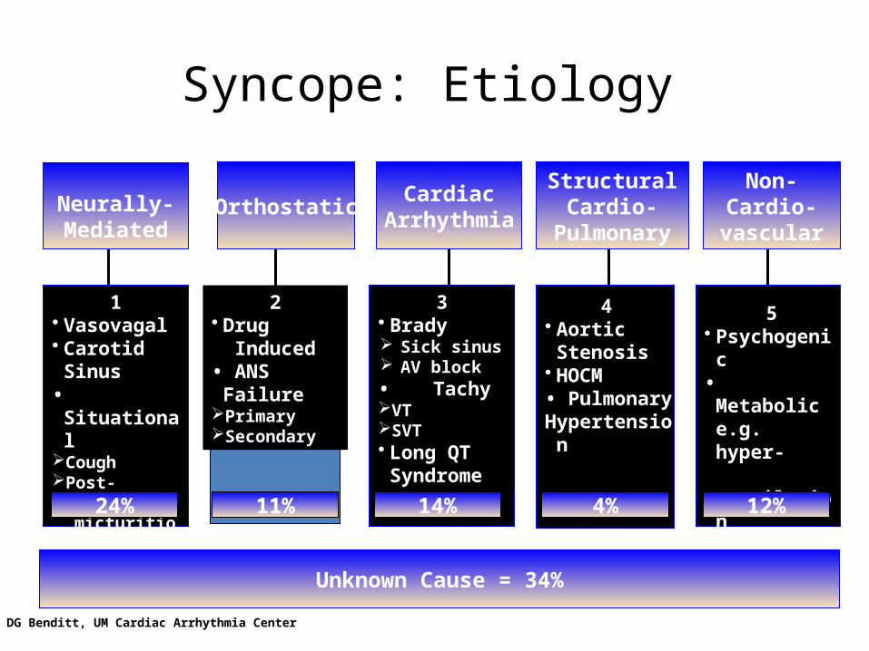
Syncope: Etiology
Orthostatic CardiacArrhythmia
StructuralCardio-
Pulmonary
*
1• Vasovagal• Carotid Sinus•SituationalCoughPost-
micturition
2• Drug Induced• ANS
FailurePrimarySecondary
3• Brady Sick sinus AV block•TachyVTSVT• Long QT
Syndrome
4 • Aortic Stenosis• HOCM• PulmonaryHypertension
5• Psychogenic•Metabolic
e.g. hyper-ventilation
• Neurological
Non-Cardio-vascular
Neurally-Mediated
Unknown Cause = 34%
24% 11% 14% 4% 12%
DG Benditt, UM Cardiac Arrhythmia Center
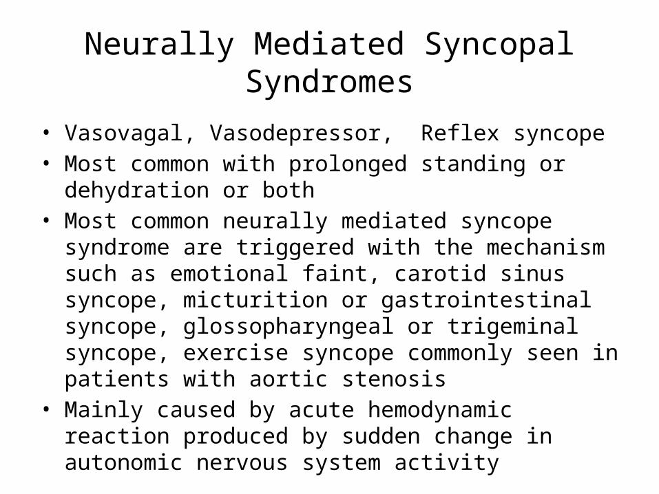
Neurally Mediated Syncopal Syndromes
• Vasovagal, Vasodepressor, Reflex syncope• Most common with prolonged standing or dehydration or
both• Most common neurally mediated syncope syndrome are
triggered with the mechanism such as emotional faint, carotid sinus syncope, micturition or gastrointestinal syncope, glossopharyngeal or trigeminal syncope, exercise syncope commonly seen in patients with aortic stenosis
• Mainly caused by acute hemodynamic reaction produced by sudden change in autonomic nervous system activity
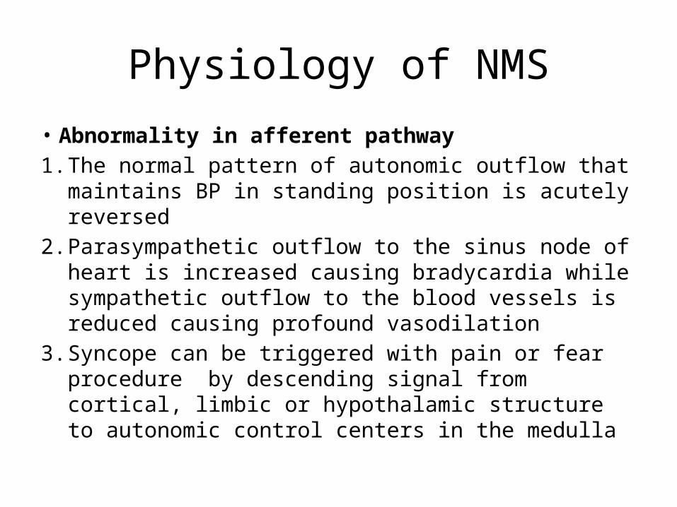
Physiology of NMS
• Abnormality in afferent pathway1. The normal pattern of autonomic outflow that
maintains BP in standing position is acutely reversed2. Parasympathetic outflow to the sinus node of heart is
increased causing bradycardia while sympathetic outflow to the blood vessels is reduced causing profound vasodilation
3. Syncope can be triggered with pain or fear procedure by descending signal from cortical, limbic or hypothalamic structure to autonomic control centers in the medulla
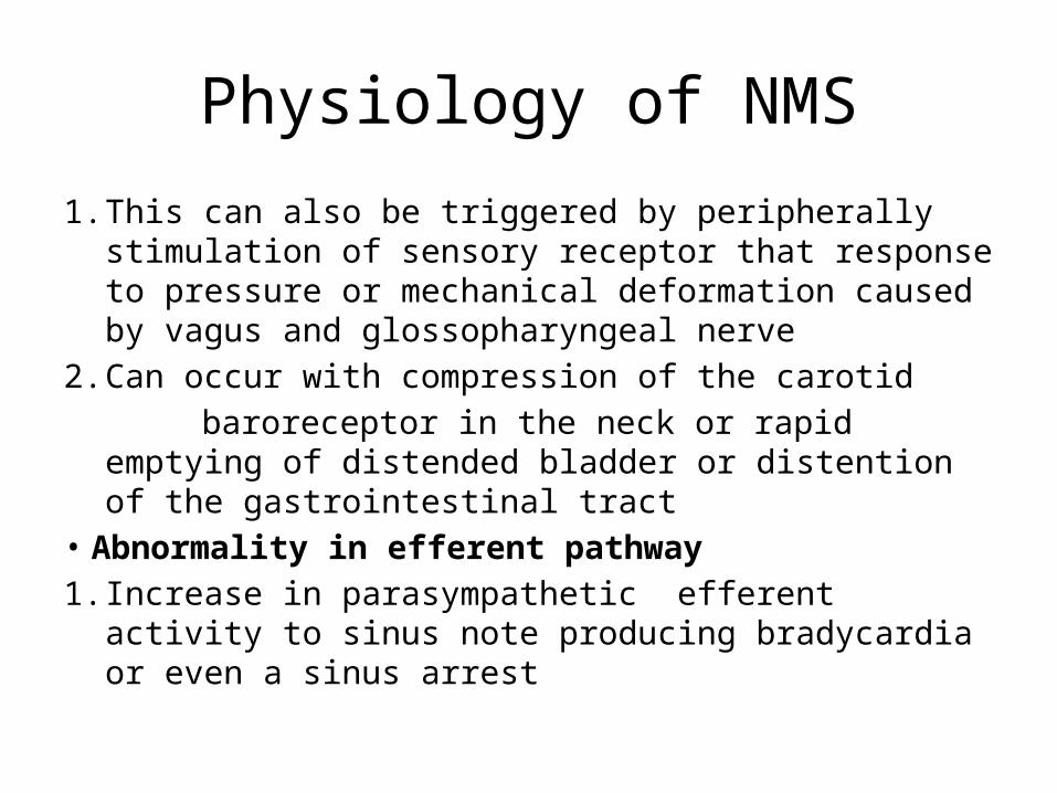
Physiology of NMS
1. This can also be triggered by peripherally stimulation of sensory receptor that response to pressure or mechanical deformation caused by vagus and glossopharyngeal nerve
2. Can occur with compression of the carotid baroreceptor in the neck or rapid emptying of distended
bladder or distention of the gastrointestinal tract• Abnormality in efferent pathway1. Increase in parasympathetic efferent activity to sinus
note producing bradycardia or even a sinus arrest
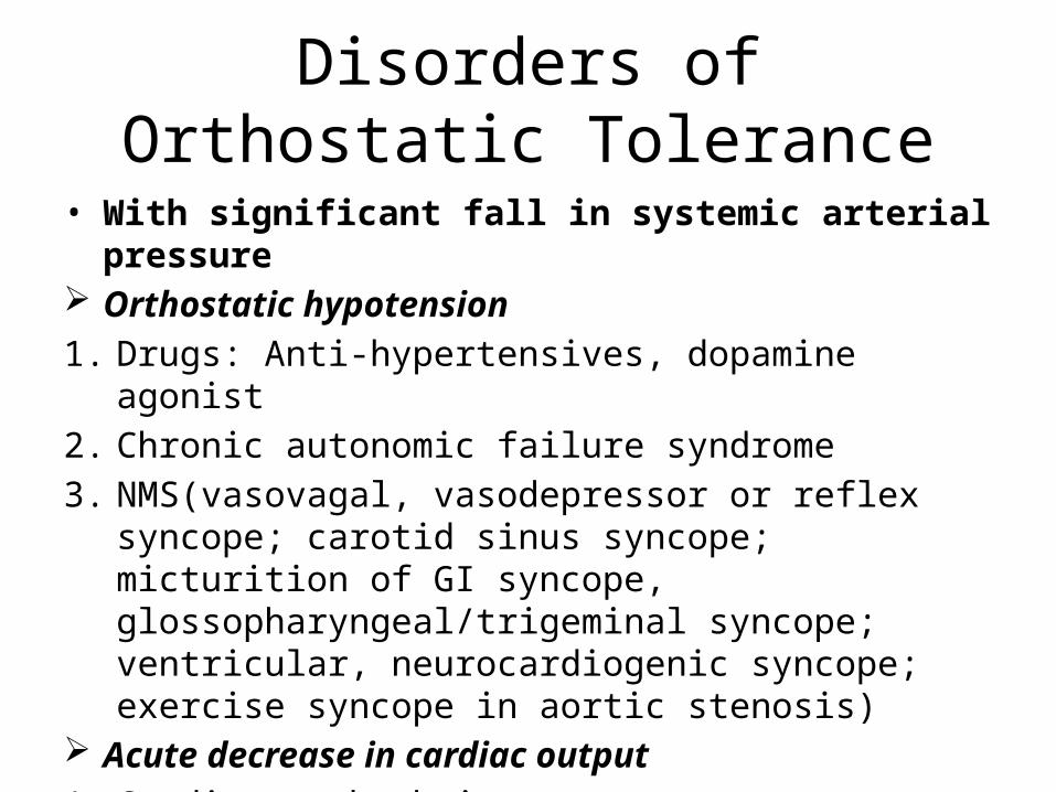
Disorders of Orthostatic Tolerance
• With significant fall in systemic arterial pressure Orthostatic hypotension1. Drugs: Anti-hypertensives, dopamine agonist2. Chronic autonomic failure syndrome3. NMS(vasovagal, vasodepressor or reflex syncope; carotid
sinus syncope; micturition of GI syncope, glossopharyngeal/trigeminal syncope; ventricular, neurocardiogenic syncope; exercise syncope in aortic stenosis)
Acute decrease in cardiac output1. Cardiac arrhythmia2. Pulmonary embolism
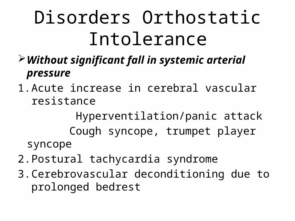
Disorders Orthostatic Intolerance
Without significant fall in systemic arterial pressure
1. Acute increase in cerebral vascular resistance Hyperventilation/panic attack Cough syncope, trumpet player syncope2. Postural tachycardia syndrome3. Cerebrovascular deconditioning due to
prolonged bedrest
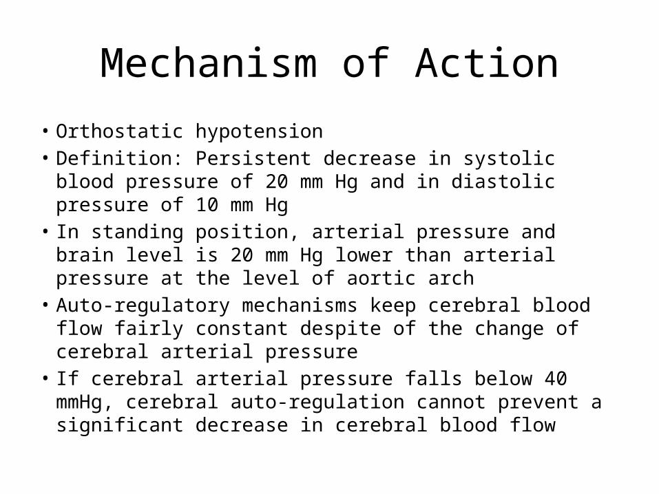
Mechanism of Action
• Orthostatic hypotension• Definition: Persistent decrease in systolic blood pressure of
20 mm Hg and in diastolic pressure of 10 mm Hg • In standing position, arterial pressure and brain level is 20
mm Hg lower than arterial pressure at the level of aortic arch• Auto-regulatory mechanisms keep cerebral blood flow fairly
constant despite of the change of cerebral arterial pressure• If cerebral arterial pressure falls below 40 mmHg, cerebral
auto-regulation cannot prevent a significant decrease in cerebral blood flow
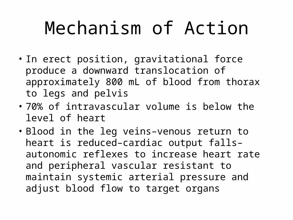
Mechanism of Action
• In erect position, gravitational force produce a downward translocation of approximately 800 mL of blood from thorax to legs and pelvis
• 70% of intravascular volume is below the level of heart
• Blood in the leg veins–venous return to heart is reduced–cardiac output falls–autonomic reflexes to increase heart rate and peripheral vascular resistant to maintain systemic arterial pressure and adjust blood flow to target organs
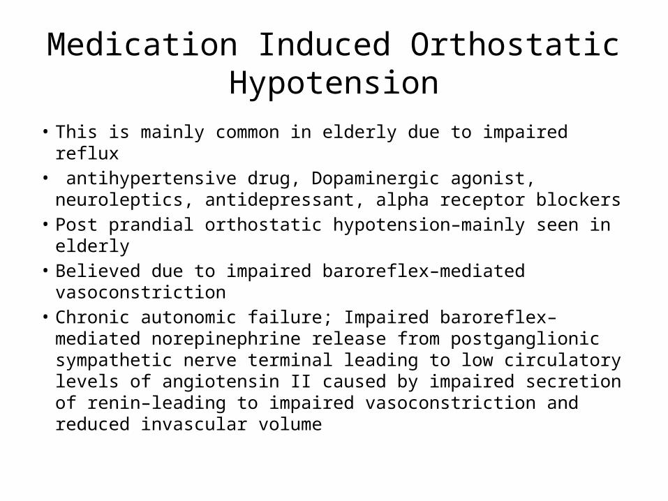
Medication Induced Orthostatic Hypotension
• This is mainly common in elderly due to impaired reflux• antihypertensive drug, Dopaminergic agonist, neuroleptics,
antidepressant, alpha receptor blockers• Post prandial orthostatic hypotension–mainly seen in elderly• Believed due to impaired baroreflex–mediated
vasoconstriction• Chronic autonomic failure; Impaired baroreflex–mediated
norepinephrine release from postganglionic sympathetic nerve terminal leading to low circulatory levels of angiotensin II caused by impaired secretion of renin–leading to impaired vasoconstriction and reduced invascular volume

Orthostatic Intolerance Without Significant Hypotension
• Cerebral perfusion pressure equals cerebral arterial pressure minus intracranial pressure
• Causes for increased ICP1. Panic attacks: Hyperventilation contributing to
hypercapnia2. Repetitive coughing: Increased intra-thoracic and
intra-abdominal pressure transmitted via the great veins of cranial vault causing transient elevation of ICP
3. Playing a wind instrument 4. Straining to defecate
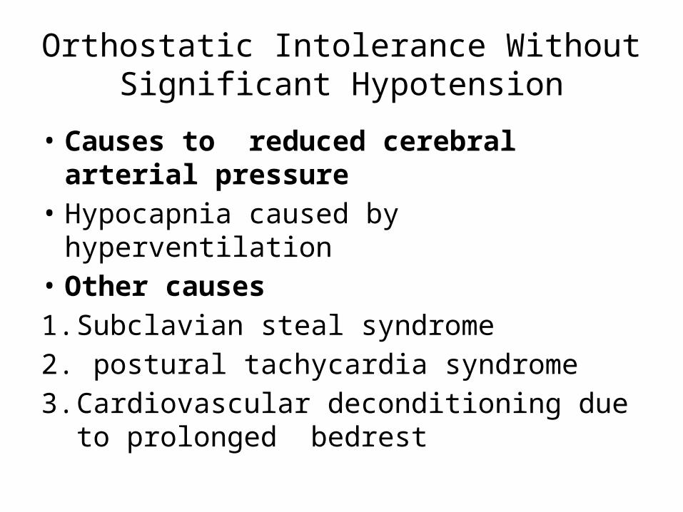
Orthostatic Intolerance Without Significant Hypotension
• Causes to reduced cerebral arterial pressure• Hypocapnia caused by hyperventilation• Other causes1. Subclavian steal syndrome2. postural tachycardia syndrome3. Cardiovascular deconditioning due to
prolonged bedrest

Clinical Differentiation
• Orthostatic hypotension with normal autonomic reflexes
while standing position drop in systolic pressure with marked reflex tachycardia• Orthostatic hypotension with sympathetic failure while standing position drop in both systolic and diastolic pressure with minimal or
little increase in the heart rate
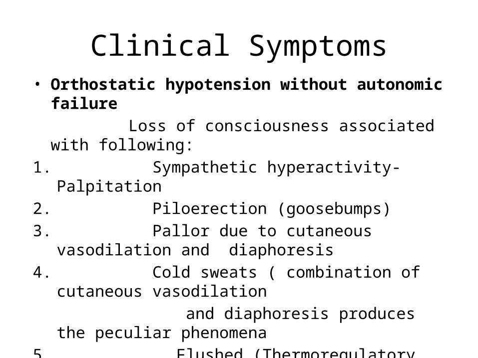
Clinical Symptoms• Orthostatic hypotension without autonomic failure Loss of consciousness associated with following:1. Sympathetic hyperactivity- Palpitation2. Piloerection (goosebumps)3. Pallor due to cutaneous vasodilation and diaphoresis4. Cold sweats ( combination of cutaneous vasodilation and diaphoresis produces the peculiar phenomena5. Flushed (Thermoregulatory sweating occurs along the skin vasodilation to dissipate heat and the person appear red or flushed)6. Blurred vision ( caused by pupillary dilation)
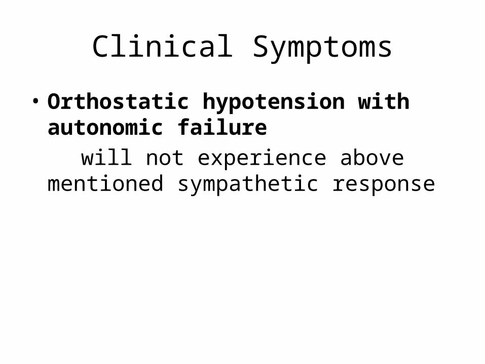
Clinical Symptoms
• Orthostatic hypotension with autonomic failure
will not experience above mentioned sympathetic response
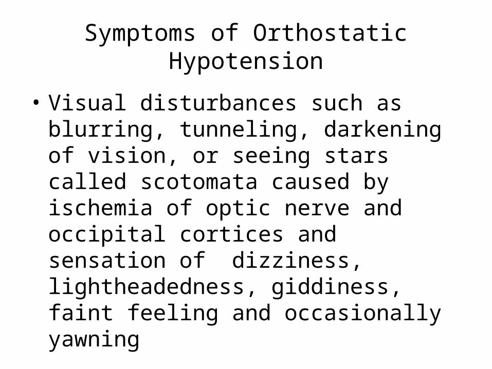
Symptoms of Orthostatic Hypotension
• Visual disturbances such as blurring, tunneling, darkening of vision, or seeing stars called scotomata caused by ischemia of optic nerve and occipital cortices and sensation of dizziness, lightheadedness, giddiness, faint feeling and occasionally yawning
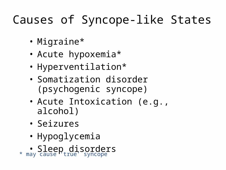
Causes of Syncope-like States• Migraine*• Acute hypoxemia*• Hyperventilation*• Somatization disorder (psychogenic syncope)• Acute Intoxication (e.g., alcohol)• Seizures• Hypoglycemia• Sleep disorders
* may cause ‘true’ syncope
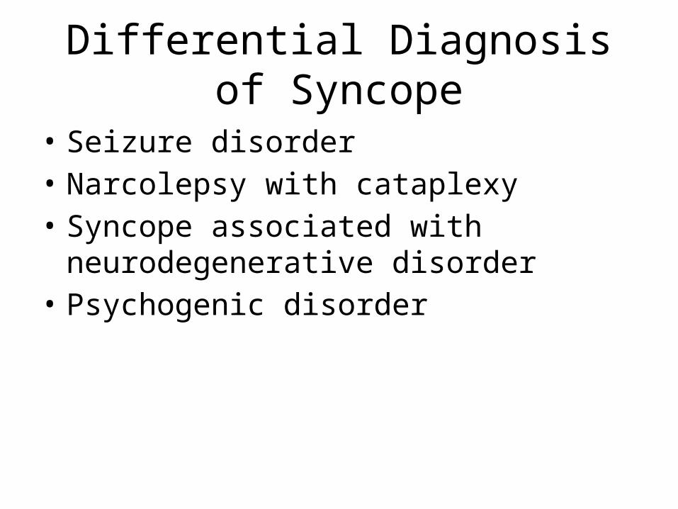
Differential Diagnosis of Syncope
• Seizure disorder• Narcolepsy with cataplexy• Syncope associated with neurodegenerative
disorder• Psychogenic disorder
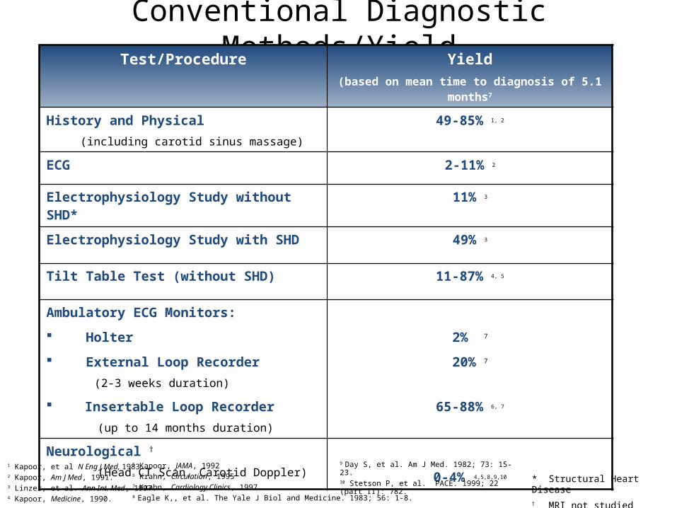
Conventional Diagnostic Methods/YieldTest/Procedure Yield
(based on mean time to diagnosis of 5.1 months7
History and Physical (including carotid sinus massage)
49-85% 1, 2
ECG 2-11% 2
Electrophysiology Study without SHD* 11% 3
Electrophysiology Study with SHD 49% 3
Tilt Table Test (without SHD) 11-87% 4, 5
Ambulatory ECG Monitors:
Holter 2% 7
External Loop Recorder(2-3 weeks duration)
20% 7
Insertable Loop Recorder(up to 14 months duration)
65-88% 6, 7
Neurological †
(Head CT Scan, Carotid Doppler) 0-4% 4,5,8,9,10
* Structural Heart Disease† MRI not studied
1 Kapoor, et al N Eng J Med, 1983.2 Kapoor, Am J Med, 1991.3 Linzer, et al. Ann Int. Med, 1997.4 Kapoor, Medicine, 1990.
5 Kapoor, JAMA, 19926 Krahn, Circulation, 19957 Krahn, Cardiology Clinics, 1997.8 Eagle K,, et al. The Yale J Biol and Medicine. 1983; 56: 1-8.
9 Day S, et al. Am J Med. 1982; 73: 15-23.10 Stetson P, et al. PACE. 1999; 22 (part II): 782.
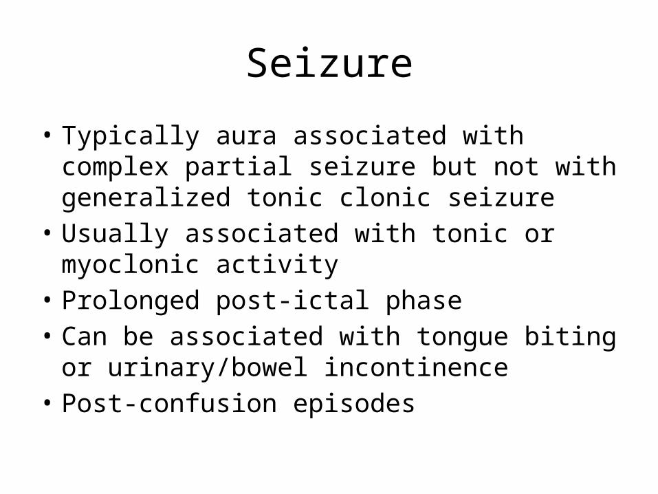
Seizure
• Typically aura associated with complex partial seizure but not with generalized tonic clonic seizure
• Usually associated with tonic or myoclonic activity
• Prolonged post-ictal phase• Can be associated with tongue biting or
urinary/bowel incontinence• Post-confusion episodes

Narcolepsy with Cataplexy
• Abrupt onset of REM sleep leading to cataplexy• Abrupt atonia of muscle associated with REM
sleep• Usually associated with clinical history of excessive
daytime sleepiness• Can be triggered with sudden stimulation• Last for few seconds to minutes• No post-ictal state• No other prodromal aura
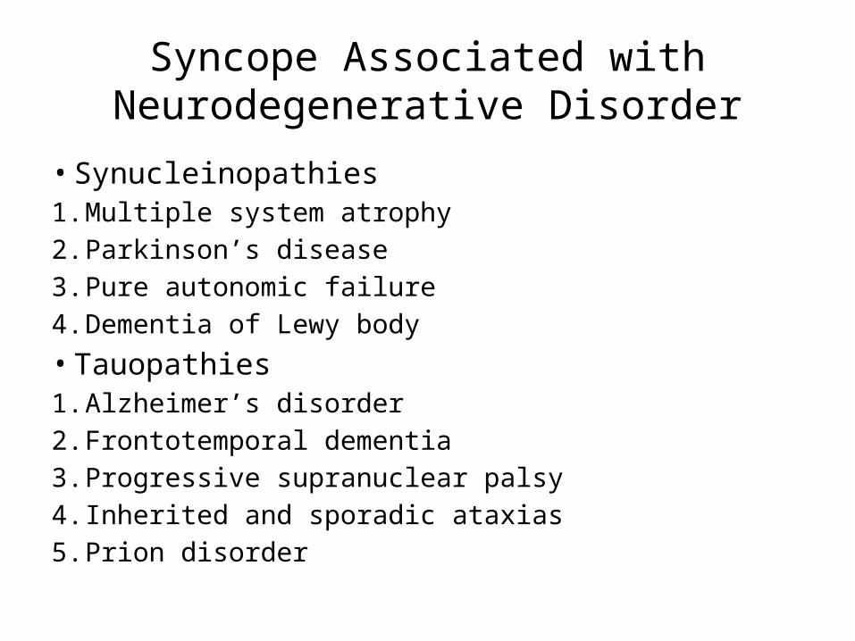
Syncope Associated with Neurodegenerative Disorder
• Synucleinopathies1. Multiple system atrophy2. Parkinson’s disease3. Pure autonomic failure4. Dementia of Lewy body• Tauopathies1. Alzheimer’s disorder2. Frontotemporal dementia3. Progressive supranuclear palsy4. Inherited and sporadic ataxias5. Prion disorder
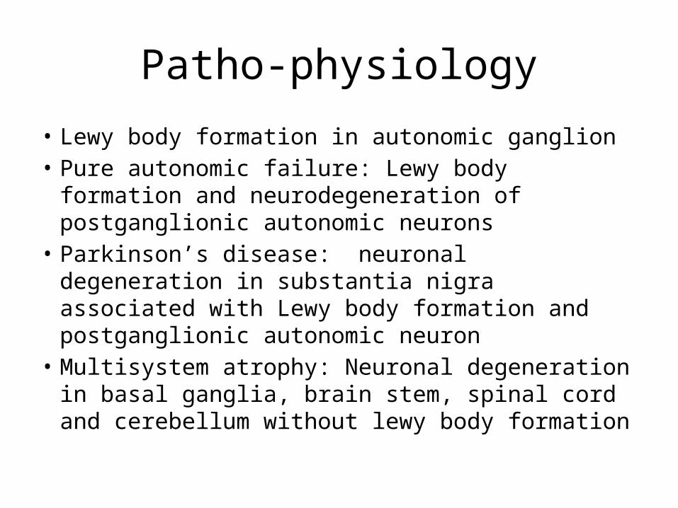
Patho-physiology
• Lewy body formation in autonomic ganglion• Pure autonomic failure: Lewy body formation and
neurodegeneration of postganglionic autonomic neurons
• Parkinson’s disease: neuronal degeneration in substantia nigra associated with Lewy body formation and postganglionic autonomic neuron
• Multisystem atrophy: Neuronal degeneration in basal ganglia, brain stem, spinal cord and cerebellum without lewy body formation
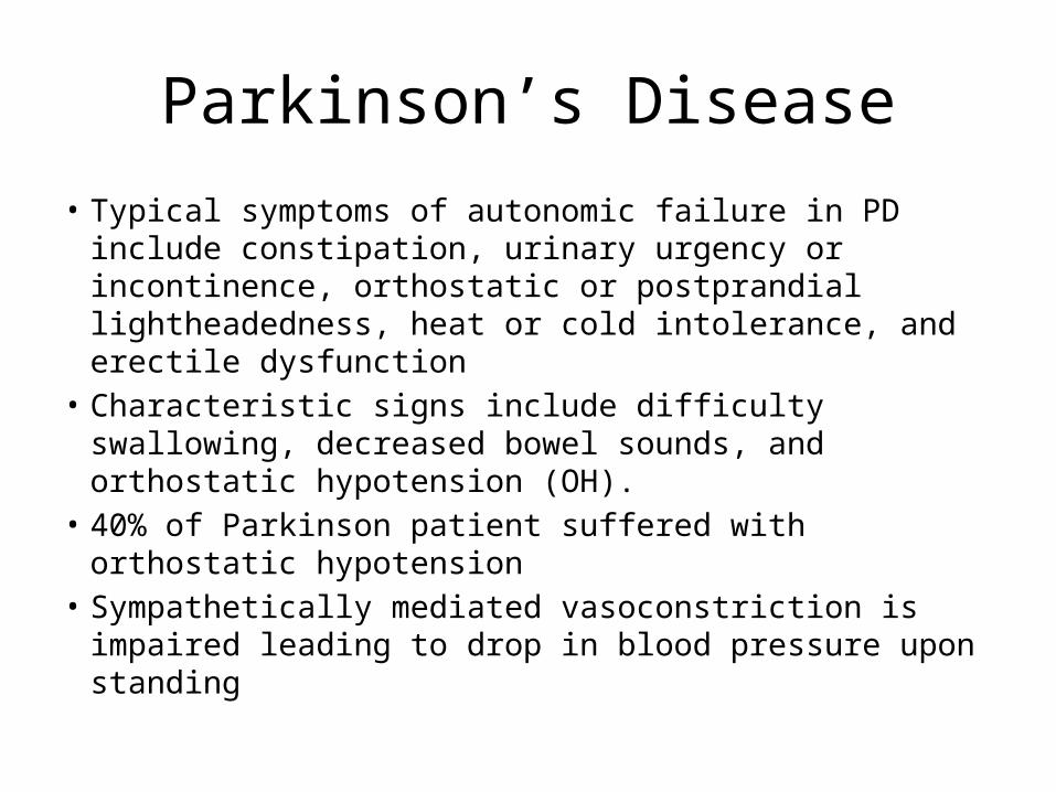
Parkinson’s Disease
• Typical symptoms of autonomic failure in PD include constipation, urinary urgency or incontinence, orthostatic or postprandial lightheadedness, heat or cold intolerance, and erectile dysfunction
• Characteristic signs include difficulty swallowing, decreased bowel sounds, and orthostatic hypotension (OH).
• 40% of Parkinson patient suffered with orthostatic hypotension
• Sympathetically mediated vasoconstriction is impaired leading to drop in blood pressure upon standing
Parkinson’s Disease
• Due to neuronal degeneration there is a reduction of neurotransmitters norepinephrine and precursor of norepinephrine
• Reduced cardiac sympathetic denervation• Reduced intensity of postganglionic noradrenergic nerve fiber
to heart• Dopamine in periphery act as a diuretic and leads to vaso-
dilation• Patients with Parkinson’s disease who suffered with autonomic
failure and reduced cardiac sympathetic denervation are increase risk of syncope associated with levodopa treatment
Dementia of Lewy Body
• Fluctuations in alertness• Cognition, and visual hallucinations are core features of DLB. • Cognitive or psychiatric manifestations at initial presentation,
but they may also present with parkinsonian features alone.• Autonomic features typically occur after the development of
cognitive changes, but DLB may also present with parkinsonism or autonomic dysfunction, or both, without significant cognitive or psychiatric abnormalities.
• Chronic autonomic failure is virtually universal in DLB. Urinary incontinence and constipation are very common (Horimoto et al, 2003). Neurogenic OH also is common and can precede cognitive and motor deficits by several years
Multiple System Atrophy
• MSA–C, MSA–p• Progressive disorder with life expectancy of 6-9
years• Cerebellar type: significant gait and limb ataxia• Parkinson type: Resting tremor, bradykinesia,
stooging, reduced arm swing• Other symptoms includes dysarthria, dystonia,
stridor of voice, pseudobulbar affect, postural instability, myoclonus, rapid decline in motor activity
Alzheimer’s Disease
• Sympathetic noradrenergic neurons are intact• normal plasma norepinephrine level
Fronto-temporal Dementia
• Cortical atrophy in frontal and temporal region• Dementia associated with aphasia, personality disorder• mild autonomic dysfunction include sialorrhea,
hyperhidrosis,urinary frequency orincontinence, heat intolerance, erectile dysfunction, or dry eyes or mouth, but there are no reports of OH, suggesting that sympathetic noradrenergic outflow to blood vessels is intact. Impaired cardiac vagal control, abnormal pupillary accommodation, and sudomotor dysfunction have been noted
Conclusion
• Detailed clinical history • Conventional diagnostic testing helpful in
Cardiogenic syncope• Neurologic examination to help differentiate
Neurodegenerative disorder• Treatment of underlying neurologic disorder
and supportive measures