
Neurocysticercosis
41
Neurocyscticercosis Dr. Yusuf Imran M.D Pediatrics J.N Medical College, Aligarh, India
-
Upload
dr-yusuf-imran-jnmc-amu -
Category
Health & Medicine
-
view
167 -
download
1
Transcript of Neurocysticercosis
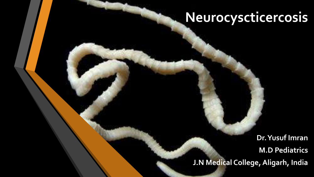
Neurocyscticercosis
Dr. Yusuf Imran
M.D Pediatrics
J.N Medical College, Aligarh, India

Introduction
Neurocysticercosis (NCC) is the infection of the CNS by the larval stage of thepork tapeworm Taenia solium.
Infection may develop in any organ- CNS (parenchyma, subarachnoid spaces,ventricles and spinal cord), eyes and muscles are the most commonly involved.
Most common manifestation is epilepsy but can have several other neurologicalmanifestations.
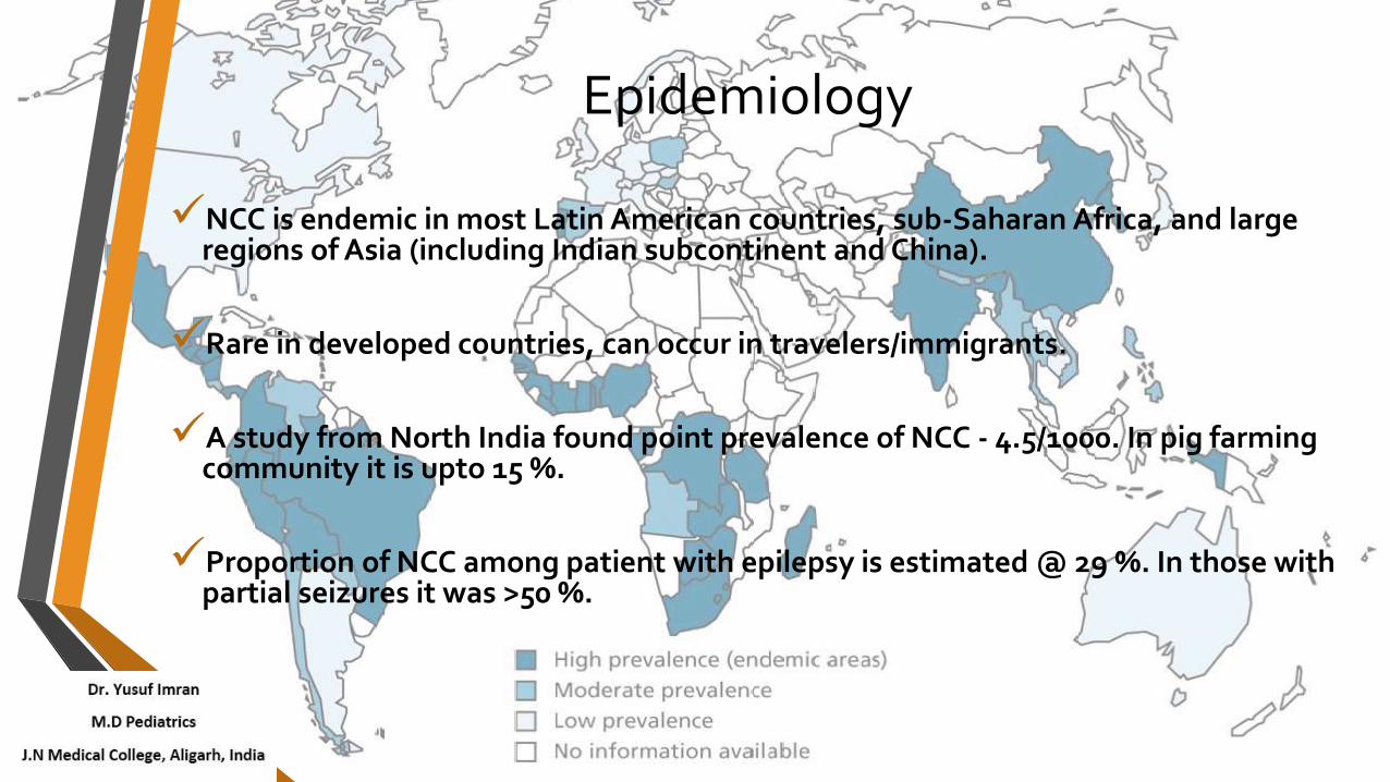
Epidemiology
NCC is endemic in most Latin American countries, sub-Saharan Africa, and large regions of Asia (including Indian subcontinent and China).
Rare in developed countries, can occur in travelers/immigrants.
A study from North India found point prevalence of NCC - 4.5/1000. In pig farming community it is upto 15 %.
Proportion of NCC among patient with epilepsy is estimated @ 29 %. In those with partial seizures it was >50 %.
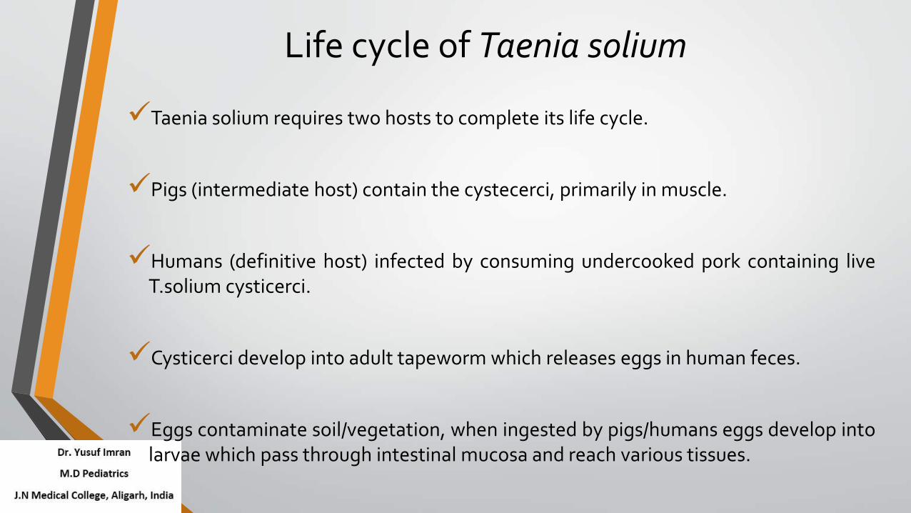
Life cycle of Taenia solium
Taenia solium requires two hosts to complete its life cycle.
Pigs (intermediate host) contain the cystecerci, primarily in muscle.
Humans (definitive host) infected by consuming undercooked pork containing liveT.solium cysticerci.
Cysticerci develop into adult tapeworm which releases eggs in human feces.
Eggs contaminate soil/vegetation, when ingested by pigs/humans eggs develop intolarvae which pass through intestinal mucosa and reach various tissues.

Lif
e c
ycl
e o
f Taen
iaso
lium

Growth stages of Taenia solium:
A-Infective T.solium eggs, B-Larva or cysticercus, C-Evaginating cysticercus, D-Tapeworm scolex, E-Tapeworm strobilaL
ife
cy
cle
of T
ae
nia
soliu
m

Modes of infection
Sources of infection- persons with Taeniasis (acquired from pork).
Transmission ways-
not spread from person to person directly
persons with Taeniasis will shed tapeworm eggs in their bowel movements

Modes of infection
Infection can happen by accidentally swallowing pork tapeworm eggs
Through-
drinking contaminated water or food
by putting contaminated fingers to mouth (external autoinfection)
by internal autoinfection

Disease spectrum of T.solium
Taeniasis = adult tapeworm in small intestine
• Usually asymptomatic (eggs or proglottids in feces)
• Vague abdominal symptoms
Cysticercosis = T. solium larvae in human tissues (eg, muscle)
• Usually asymptomatic
• Painless subcutaneous nodules in arms and chest
Neurocysticercosis (NCC) = cysts in the central nervous system
• Most severe manifestation

Etiopathogenesis
Clinical expression, management and prognosis of NCC varies depending onthe number of CNS lesions, their stage, size, location and host inflammatoryresponse.
Studies have identified a number of mechanisms used by the cycticercus tomodulate host’s immune response.
• Protease inhibitor- Taeniaestatin
• Sulfated polysaccharides
• Parasite paramyosin
• Prostaglandins
• Stimulate antibody production

Etiopathogenesis
Although, cysticerci reach mature size within a few weeks, there a periodof several years between exposure and onset of symptoms.
When parasite degenerates there is a brisk inflammatory response.
The seizures are thought to result not from the parasitic infection per se,but from the host response.

Types of Neurocysticercosis
1. Intraparenchymal NCC
Most common form, seen at grey white matter junction.
Single or Multiple.
Range in size from a few mm to 1 to 2 cm.
Commonly seen in children >5 years but can occur in toddlers and infants.

Types of Neurocysticercosis
Intraparenchymal NCC cont…
Seizures are most common manifestation of intraparenchymal NCC.
1/3rd may have associated headache and vomiting.
Papilloedema occurs in 2 to 7 %.
Neurological deficits seen in 4 to 6 %.
Seizures respond well to monotherapy.
Cysticercal Encephalitis- results from large number of cysticeci in brain parenchyma with diffuse inflammation and edema.

Types of Neurocysticercosis
The parenchymal cysts evolve through 4 stages-
The vesicular cyst
Colloidal stage
Granular nodular stage
Nodular calcified stage

Sta
ge
s o
f In
tra
pa
ren
chy
ma
lNC
C
Types of Neurocysticercosis
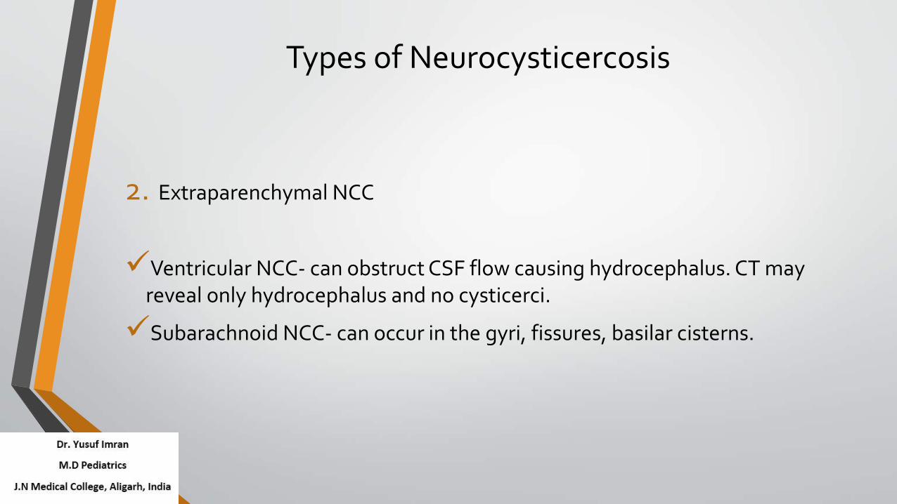
2. Extraparenchymal NCC
Ventricular NCC- can obstruct CSF flow causing hydrocephalus. CT may reveal only hydrocephalus and no cysticerci.
Subarachnoid NCC- can occur in the gyri, fissures, basilar cisterns.

Ty
pe
s o
f N
eu
rocy
stic
erc
osi
s
Types of Neurocysticercosis
Other forms of Cysticercosis
Spinal Cysticercosis (intramedullary/extramedullary)
Ocular Cysticercosis
In Muscles
In Subcutaneous tissue
Other organs
A single patient may have multiple types and locations of cysticercus.

Ty
pe
s o
f Cy
stic
erc
osi
s

Clinical Manifestations
Asymptomatic.
Most common manifestation is Seizure (focal, secondary generalized orgeneralized). (80%)
Headache is common (unilateral/bilateral)- may reflect raised ICT or vasculitis.
Focal neurological deficits (16%)
Symptoms/signs of raised ICT-nausea, vomiting, altered mental status, visualchanges, dizziness, cerebral edema. (12%)
Neurocognitive defects- leaning disability, psychosis, depression. (5%)

Imaging
CT
Parenchymal cysts- usually appear as single, small (<20mm) with ring/diskenhancement and eccentric hyper dense scolex. Multiple lesions give ‘starrysky’ appearance. (colloidal stage).
Enhancement indicates inflammation. Live vesicular cysts are non-enhancing.
Extra-parenchymal cysts- may show hydrocephalus, enhancement oftenctorium and basal cisterns and occasionally infarcts.

ImagingMRI
Superior to CT in detecting cysts in ventricles, posterior fossa, brainstem,small cysts.
Small calcified lesions may be missed.
Magnetization transfer images (MT) and magnetization transfer ratio (MTR),recovery (FLAIR) and fast imaging employing steady-state acquisition(FIESTA) sequences for lesions not visible on routine MRI.

Serological Tests
Seropositivity depends on parasite load and endemicity. False positive and falsenegative results can occur.
False-negative result:
Single lesions
Calcification
False-positive:
Other parasitic infections
High percentage of false positive for patients from endemic area

Serological Tests
EITB assay- uses lentil lectin purified glycoprotein antigens (LLGP) to detectantibodies to T soliumin in serum. Sensitivity 98% (multiple parasites), 50-70 %(solitary cysticercus).
Detection of anticysticercal antibodies in the CSF by ELISA.
Detection of circulating parasitic antigens in serum by ELISA with monoclonalantibodies is experimental.
In patients with reliable diagnosis of NCC by imaging studies, immunological test is notrequired, since a negative test will not discard a NCC.

Other lab tests…
Eosinophilia may occur.
CSF-
Usually done to rule out other causes.
Can be normal in inactive disease.
Moderate pleocytosis (mostly mononuclear cells; upto 300/mm3), increasedprotein (50-300 mg/dl). Correlate with disease activity and whether or not theparasites are located in sub-arachnoid space.

Other lab tests…
Biopsy of subcutaneous nodules.
Radiographs of skeletal muscles.
Specific coproantigen detection by ELISA for screening for T solium carriers.
Stool examination for T.solium eggs has poor sensitivity.
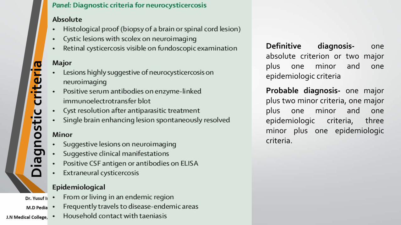
Dia
gn
ost
ic c
rite
ria
Definitive diagnosis- oneabsolute criterion or two majorplus one minor and oneepidemiologic criteria
Probable diagnosis- one majorplus two minor criteria, one majorplus one minor and oneepidemiologic criteria, threeminor plus one epidemiologiccriteria.
Differential diagnosis
Tuberculoma
(presence of raised ICT, progressivefocal neurodeficit, size >20mm,lobulated irregular shape, midline shift& marked edema, lesions at base ofbrain)
Special MRI sequences – diffusionweighted MRI and proton magneticresonance spectroscopy (MRS) arebeing tried.
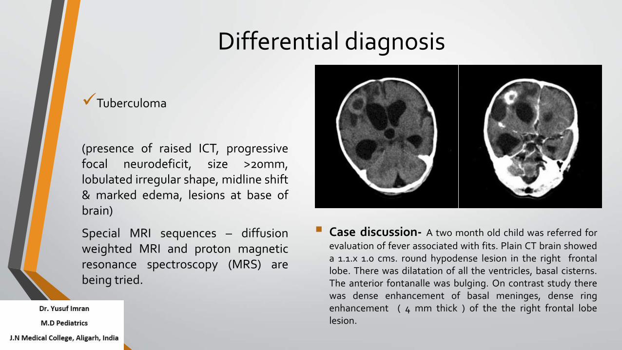
Case discussion- A two month old child was referred for
evaluation of fever associated with fits. Plain CT brain showeda 1.1.x 1.0 cms. round hypodense lesion in the right frontallobe. There was dilatation of all the ventricles, basal cisterns.The anterior fontanalle was bulging. On contrast study therewas dense enhancement of basal meninges, dense ringenhancement ( 4 mm thick ) of the the right frontal lobelesion.

Differential diagnosis
Microabscesses
Toxoplasmosis
Fungal lesions
Low grade astrocytoma
Cystic cerebral metastasis
CNS lymphoma

Management
Emergency care
Manage seizure activity
Supportive care (A-B-C)
Monitor, and correct metabolic abnormalities
Anticonvulsants are effective.
Evidence of increased ICP- Steroids, osmotic agents, and/or diuretics
Initiate proper diagnostic procedures
Blood work and imaging

ManagementIntraparenchymal NCC- Symptomatic treatment, anti-parasite treatment or
surgery ?
Calcified cysts only- antiepileptic, analgesic, and anti-inflammatory drugs; forseizures relapses, repeat imaging looking for peri-calcification oedema. AED for at least2yrs since last seizure. No anti-parasite drugs.
*One or more cystic or degenerating lesions- antiepileptic, analgesic, and anti-inflammatory drugs; antiparasitic treatment under hospital conditions with steroidtreatment. Discontinue AED in single lesions after they resolve (without calcification).
*Level 1 evidence
Cysticercal encephalitis- Manage intracranial hypertension; do not use antiparasiticdrugs.

Management
Asymptomatic parenchymal lesions
• Prophylactic AED not justified in calcified lesions without seizures.
• Viable cysts- Give prophylactic AED when antiparasite treatment is alsoplanned.
Repeat neuroimaging after 3-6 m to document lesion resolution. Repeatcourse of cysticidal therapy if persistent lesion.

Management
Drugs:
• Albendazole- 15mg/kg/day in 2-3 divided doses for 2-4 weeks. Shortercourses of 3 to 14 days tried in single lesions.
• Praziquintal- 50mg/kg/day, less effective than albendazole.
• Combinations of two antiparasitic drugs- increased cyst clearance inmultiple lesions.
• Steroids-
Dexamethasone- 0.1 mg/kg/day i.v starting one day before antiparasitedrugs, give for 1 to 2 weeks then taper.
Prednisolone- 1 to 2 mg/kg/day.

Ma
na
ge
me
nt

ManagementExtraparenchymal NCC

Management
Other forms of NCC
Spinal- Intramedullary cysts are treated with surgery, albendazole withsteroids is being tried. Subarachnoid cysts can migrate so imaging is donejust before surgery.
Ocular- Surgical management is the standard of care.
Prognosis
Single lesions- good prognosis, disappears in >60% at 6m.
Seizure recurrence is 10-20% with single lesions, multiple lesions havemore frequent seizures.
Prognosis is poorer in cysticercal encephalitis and extraparenchymalNCC.

Prevention
T solium infection is one of a few diseases targeted for focalelimination and eventual eradication by the International Task Forcefor Disease Eradication.
Public education, proper hygiene, provision for toilets.
Safe handling of meat, strict animal husbandry and meet inspectionsprocedures.

Prevention
Mass deworming of population with Niclosamide or Praziquintal.
Mass vaccination of pigs and treatment of pigs with Oxfendazole.
Community interventions reduce rate of epilepsy in hyper-endemic areas.

Conclusion
NCC is a common cause of seizures and other neurological manifestationsand needs to be considered in D/D of many neurological conditions.
Treatment with cycticidal therapy leads to reduction in seizure frequencyand faster resolution of lesions.
Children with a single or few lesions have a good outcome.
Prevention of NCC is important and feasible.

Thank you


![Clinical Diagnoses of Neurocysticercosis · Clinical Diagnoses of Neurocysticercosis 281 extraparenchymal location [88%), in comparison with the parenchymal location (10%). [12] When](https://static.fdocuments.us/doc/165x107/5e76ff60412a36576f46bf82/clinical-diagnoses-of-neurocysticercosis-clinical-diagnoses-of-neurocysticercosis.jpg)


