Neurocryptococcoma: Curative Treatment with Surgical...
6
Proceedings of UCLA Healthcare -VOLUME 14 (2010)- CLINICAL VIGNETTE Neurocryptococcoma: Curative Treatment with Surgical and Medical Modalities and 9 Year Follow up Maria I. Romanova, M.D. 1 Introduction Cryptococcus is encapsulated yeast capable of causing invasive infection in humans. Although the vast majority of patients with cryptococcosis are immunocompromised, there are infrequent reports of disseminated infection in healthy individuals. We encountered an unusual case of multiple tumor-like cryptococcal brain lesions in immunocompetent host. Case presentation A 65-year-old male with history of hypertension and cerebrovascular accident presented with nausea, projectile vomiting and gait abnormality. Three months prior to admission, he noticed change in taste. Subsequently, he developed infrequent sudden bouts of vomiting without preceding nausea following the meals. His gait became unsteady; he veered to the left when attempting to walk straight. He had a history of stroke 5 years prior with residual mild right hand numbness. He also had hypertension and prostatic hypertrophy. He had no prior history of infection. His medications included felodipine 5 mg, terazosin 5 mg and aspirin 81 mg once a day. He was born and raised in Kentucky, and had lived in Southern California for most of his adult life. He worked as an interior designer with significant dust exposure, but no known contact with birds or other animals. He was in a monogamous homosexual relationship for many years. His family history was notable for lung cancer and hypertension. Physical examination revealed normal temperature, blood pressure and pulse. Head, neck, chest, heart, abdominal and extremity examination was normal. No rashes were noted. Neurological evaluation revealed inability to perform tandem walk and near miss on right finger to nose testing. Sensation to light touch in the right hand was slightly diminished. Otherwise, strength and sensation were normal. No cranial nerve abnormality or other cerebellar signs were elicited. Routine complete and biochemical blood tests revealed mild leukopenia with normal differential. Magnetic resonance imaging (MRI) of the brain revealed an ill-defined, lobulated, rim enhancing 4 cm mass in the right cerebellar hemisphere with moderate surrounding edema and central necrosis (Fig. 1). The mass caused distortion of the fourth ventricle and aqueduct of Sylvus, as well as compression of the brain stem, early herniation of the cerebellar tonsils and post- obstructive hydrocephalus. A second 2 cm rim- enhancing lesion was noted in the right parietal lobe (Fig.2), with another 1 cm mass in the right frontal lobe. Also noted was periventricular deep white matter signal abnormality consistent with microvascular ischemic disease. A differential diagnosis of metastatic malignancy and intracranial infection was considered, and the patient was started on intravenous steroids and anti-seizure medications prophylactically. Serological blood tests for Toxoplasma, Cryptococcus, Cysticercus, Histoplasma and Coccidioides immitis were obtained.T-4 lymphocyte panel revealed decrease in number of CD4 positive cells to 159 (normal 400-1500 cells/uL). HIV test along with HIV p24 antigen and viral PCR were negative. Chest X-ray was normal. Computer tomography of the thorax, abdomen and pelvis did not reveal evidence of malignancy. Lumbar puncture was considered but not performed due to high risk for brain herniation. Tests for HTLV I and II, fungal blood cultures, tumor antigen Ca 19-9 and alpha fetoprotein were negative. Serum cryptococcal antigen testing was strongly positive (Table 1). Antifungal therapy with intravenous amphotericin B 40 mg IV and flucytosine 2 gm
Transcript of Neurocryptococcoma: Curative Treatment with Surgical...

Proceedings of UCLA Healthcare -VOLUME 14 (2010)-
CLINICAL VIGNETTE
Neurocryptococcoma: Curative Treatment with Surgical and Medical Modalities and 9 Year Follow up
Maria I. Romanova, M.D.
1
Introduction Cryptococcus is encapsulated yeast capable of causing invasive infection in humans. Although the vast majority of patients with cryptococcosis are immunocompromised, there are infrequent reports of disseminated infection in healthy individuals. We encountered an unusual case of multiple tumor-like cryptococcal brain lesions in immunocompetent host. Case presentation A 65-year-old male with history of hypertension and cerebrovascular accident presented with nausea, projectile vomiting and gait abnormality. Three months prior to admission, he noticed change in taste. Subsequently, he developed infrequent sudden bouts of vomiting without preceding nausea following the meals. His gait became unsteady; he veered to the left when attempting to walk straight. He had a history of stroke 5 years prior with residual mild right hand numbness. He also had hypertension and prostatic hypertrophy. He had no prior history of infection. His medications included felodipine 5 mg, terazosin 5 mg and aspirin 81 mg once a day. He was born and raised in Kentucky, and had lived in Southern California for most of his adult life. He worked as an interior designer with significant dust exposure, but no known contact with birds or other animals. He was in a monogamous homosexual relationship for many years. His family history was notable for lung cancer and hypertension. Physical examination revealed normal temperature, blood pressure and pulse. Head, neck, chest, heart, abdominal and extremity examination was normal. No rashes were noted. Neurological evaluation revealed inability to perform tandem walk and near miss on right finger to nose testing. Sensation to light touch in the right hand was slightly diminished. Otherwise, strength and sensation were normal.
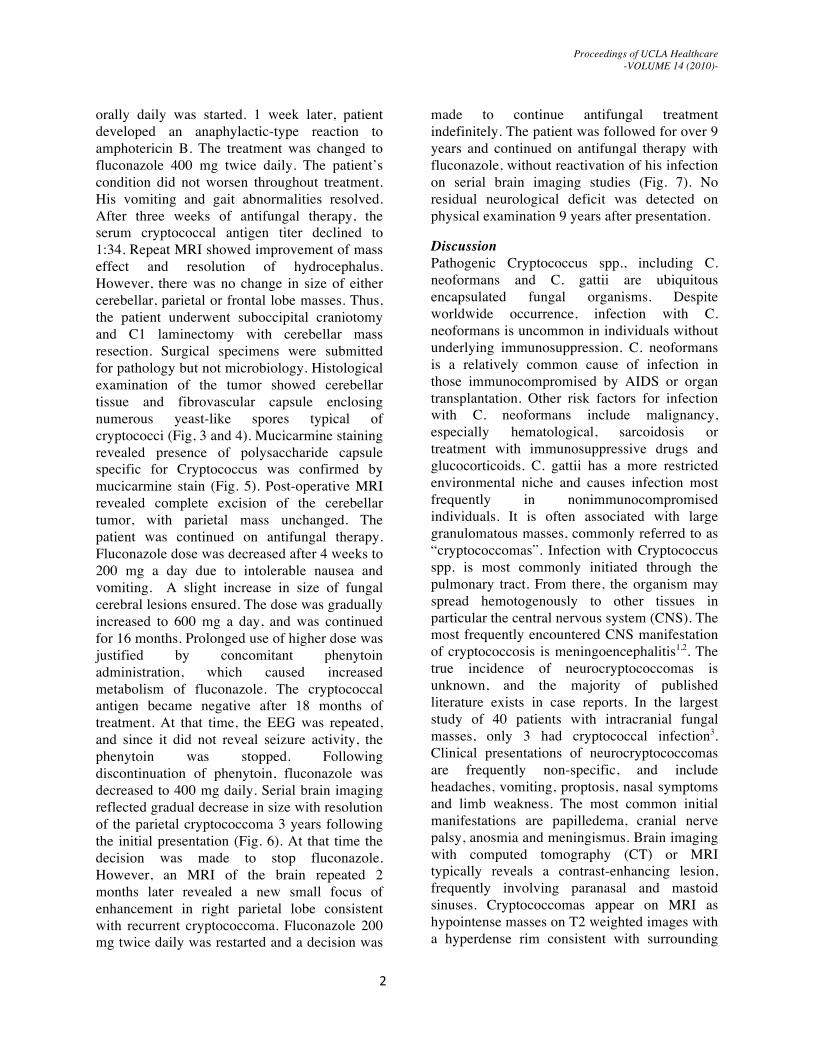
No cranial nerve abnormality or other cerebellar signs were elicited. Routine complete and biochemical blood tests revealed mild leukopenia with normal differential. Magnetic resonance imaging (MRI) of the brain revealed an ill-defined, lobulated, rim enhancing 4 cm mass in the right cerebellar hemisphere with moderate surrounding edema and central necrosis (Fig. 1). The mass caused distortion of the fourth ventricle and aqueduct of Sylvus, as well as compression of the brain stem, early herniation of the cerebellar tonsils and post-obstructive hydrocephalus. A second 2 cm rim-enhancing lesion was noted in the right parietal lobe (Fig.2), with another 1 cm mass in the right frontal lobe. Also noted was periventricular deep white matter signal abnormality consistent with microvascular ischemic disease. A differential diagnosis of metastatic malignancy and intracranial infection was considered, and the patient was started on intravenous steroids and anti-seizure medications prophylactically. Serological blood tests for Toxoplasma, Cryptococcus, Cysticercus, Histoplasma and Coccidioides immitis were obtained.T-4 lymphocyte panel revealed decrease in number of CD4 positive cells to 159 (normal 400-1500 cells/uL). HIV test along with HIV p24 antigen and viral PCR were negative. Chest X-ray was normal. Computer tomography of the thorax, abdomen and pelvis did not reveal evidence of malignancy. Lumbar puncture was considered but not performed due to high risk for brain herniation. Tests for HTLV I and II, fungal blood cultures, tumor antigen Ca 19-9 and alpha fetoprotein were negative. Serum cryptococcal antigen testing was strongly positive (Table 1). Antifungal therapy with intravenous amphotericin B 40 mg IV and flucytosine 2 gm
Proceedings of UCLA Healthcare -VOLUME 14 (2010)-
2
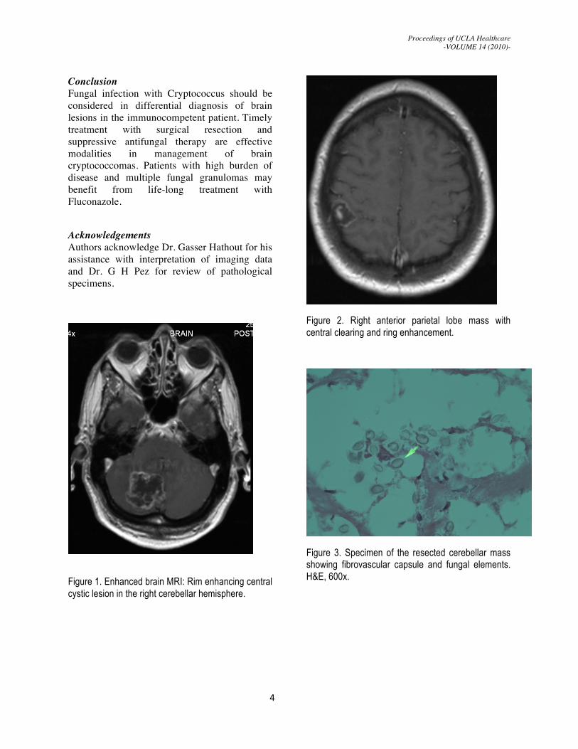
orally daily was started. 1 week later, patient developed an anaphylactic-type reaction to amphotericin B. The treatment was changed to fluconazole 400 mg twice daily. The patient’s condition did not worsen throughout treatment. His vomiting and gait abnormalities resolved. After three weeks of antifungal therapy, the serum cryptococcal antigen titer declined to 1:34. Repeat MRI showed improvement of mass effect and resolution of hydrocephalus. However, there was no change in size of either cerebellar, parietal or frontal lobe masses. Thus, the patient underwent suboccipital craniotomy and C1 laminectomy with cerebellar mass resection. Surgical specimens were submitted for pathology but not microbiology. Histological examination of the tumor showed cerebellar tissue and fibrovascular capsule enclosing numerous yeast-like spores typical of cryptococci (Fig, 3 and 4). Mucicarmine staining revealed presence of polysaccharide capsule specific for Cryptococcus was confirmed by mucicarmine stain (Fig. 5). Post-operative MRI revealed complete excision of the cerebellar tumor, with parietal mass unchanged. The patient was continued on antifungal therapy. Fluconazole dose was decreased after 4 weeks to 200 mg a day due to intolerable nausea and vomiting. A slight increase in size of fungal cerebral lesions ensured. The dose was gradually increased to 600 mg a day, and was continued for 16 months. Prolonged use of higher dose was justified by concomitant phenytoin administration, which caused increased metabolism of fluconazole. The cryptococcal antigen became negative after 18 months of treatment. At that time, the EEG was repeated, and since it did not reveal seizure activity, the phenytoin was stopped. Following discontinuation of phenytoin, fluconazole was decreased to 400 mg daily. Serial brain imaging reflected gradual decrease in size with resolution of the parietal cryptococcoma 3 years following the initial presentation (Fig. 6). At that time the decision was made to stop fluconazole. However, an MRI of the brain repeated 2 months later revealed a new small focus of enhancement in right parietal lobe consistent with recurrent cryptococcoma. Fluconazole 200 mg twice daily was restarted and a decision was
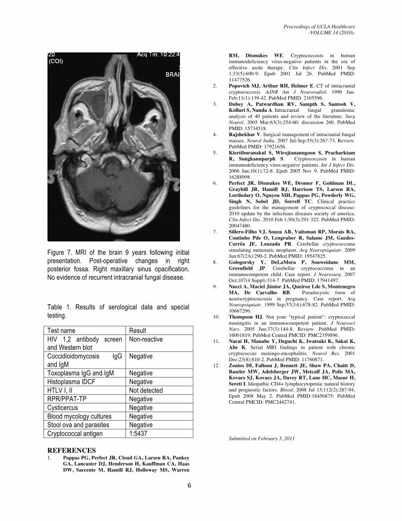
made to continue antifungal treatment indefinitely. The patient was followed for over 9 years and continued on antifungal therapy with fluconazole, without reactivation of his infection on serial brain imaging studies (Fig. 7). No residual neurological deficit was detected on physical examination 9 years after presentation.
Discussion Pathogenic Cryptococcus spp., including C. neoformans and C. gattii are ubiquitous encapsulated fungal organisms. Despite worldwide occurrence, infection with C. neoformans is uncommon in individuals without underlying immunosuppression. C. neoformans is a relatively common cause of infection in those immunocompromised by AIDS or organ transplantation. Other risk factors for infection with C. neoformans include malignancy, especially hematological, sarcoidosis or treatment with immunosuppressive drugs and glucocorticoids. C. gattii has a more restricted environmental niche and causes infection most frequently in nonimmunocompromised individuals. It is often associated with large granulomatous masses, commonly referred to as “cryptococcomas”. Infection with Cryptococcus spp. is most commonly initiated through the pulmonary tract. From there, the organism may spread hemotogenously to other tissues in particular the central nervous system (CNS). The most frequently encountered CNS manifestation of cryptococcosis is meningoencephalitis1,2. The true incidence of neurocryptococcomas is unknown, and the majority of published literature exists in case reports. In the largest study of 40 patients with intracranial fungal masses, only 3 had cryptococcal infection3. Clinical presentations of neurocryptococcomas are frequently non-specific, and include headaches, vomiting, proptosis, nasal symptoms and limb weakness. The most common initial manifestations are papilledema, cranial nerve palsy, anosmia and meningismus. Brain imaging with computed tomography (CT) or MRI typically reveals a contrast-enhancing lesion, frequently involving paranasal and mastoid sinuses. Cryptococcomas appear on MRI as hypointense masses on T2 weighted images with a hyperdense rim consistent with surrounding

Proceedings of UCLA Healthcare -VOLUME 14 (2010)-
3
vasogenic edema. Although this appearance may distinguish cryptococcus from other fungal pathogens, careful differential diagnosis and serological confirmation is necessary4. Fungal granulomas can be caused by Aspergillus, Mucor, Coccidioides, Histoplasma, Blastomycosis, Paracoccidioidomycosis, Candida and dematiaceous fungi. Among tumors, a similar picture can be caused by glioma, lymphoma, metastatic cancer or meningioma. Other infectious pathogens like Toxoplasma, Cysticercus, Mycobacterium tuberculosis and pyogenic bacteria forming abscess can cause similar appearing lesions on brain scans. Neurosarcoidosis also needs to be considered in a patient with likely features. The diagnosis of neurocryptococcoma can be confirmed by microbiologic recovery of fungus from cerebrospinal fluid or blood. Elevated serum or cerebrospinal fluid (CSF) titers of cryptococcal antigen are seen in 70-90% of patients5. In unclear cases with negative cryprococcal antigen and fungal CSF analysis brain biopsy is often required since the differential is so broad. Treatment with antifungal agents is warranted in all patients with neurocryptococcoma. Untreated infection is uniformly fatal. The optimal treatment algorithm was published in practice guidelines in year 20106. For cerebral cryptococcomas, standard induction therapy consists at least 6 weeks of amphotericin B and flucytosine. This should be followed by consolidation and maintenance therapy with oral fluconazole (400-800 mg/day) for 6-18 months. Surgery is also recommended for large (>3cm), accessible lesions with mass effect. Our patient demonstrated many unique aspects of this rare disease. Despite multiple brain space occupying lesions, brain stem compression and high serum cryptococcal antigen titers, his clinical symptoms and signs were very mild, and definitely delayed proper diagnosis. Indolent presentation of cryptococcal infection was likely related to little inflammation the organism caused by the surrounding capsule. Presence of multiple lesions is also unusual for cryptococcoma. We found very few reports of cerebellar neurocryptococcoma2,7-9. Posterior fossa lesions of this size frequently manifest with rapid neurological deterioration. His
inability to tolerate amphotericin and initially high doses of fluconazole caused additional challenge in his management. It dictated urgent surgical treatment, and prolonged antifungal therapy. Concomitant administration of dilantin for seizure prophylaxis required attention to the fluconazole dosage. The complete resolution of remaining fungal masses took 3 years of therapy. Quick relapse of cryptococcal lesions in our case may be caused by high virulence of the fungus, unique susceptibility of the host or lack of good initial therapy with amphotericin B and flucytosine. We decided that our patient will require a lifelong maintenance therapy. The outcomes of CNS cryptococcosis after discontinuation of therapy vary. At least one case report described fatal relapse in immunocompetent patient only 3 months after the treatment was stopped10. This case gave us an opportunity to observe natural history of treated fungal infection over long time period. Serial MRI scans revealed gradual resolution of rim enhancement after initiation of antifungal therapy. The prior area of necrotizing granuloma in the parietal lobe eventually disappeared completely, and the resection cavity was replaced by scar tissue. Similar changes have been described in a prior report, but over a 2-year period11. Finally, CD4+ T cell lymphopenia detected at the time of initial presentation resolved after 3 months of therapy. Initially HIV infection was suspected given the low CD4+ T cell count. However, this was ruled out by negative tests for HIV antibodies, antigen and RNA. Subsequently, idiopatic CD4 lymphopenia was considered12. Diagnosis of idiopathic CD+ lymphopenia requires persistent CD+ T-cell counts less than 300 cells/microL on 2 separate analysis 1-3 months apart, in absence of infection with HIV-1 or any other cause of immunodeficiency. However, our patient did not meet diagnostic criteria for this disease, and on repeat testing his CD4+ T cell count returned to normal. The etiology of his transient CD4+ T cell lymphopenia remains obscure. Patient did not have prior history of infection, and remained essentially symptom free in his long follow up period, eliminating our suspicion for either cellular or humoral immunodeficiency.
Proceedings of UCLA Healthcare -VOLUME 14 (2010)-
4
Conclusion Fungal infection with Cryptococcus should be considered in differential diagnosis of brain lesions in the immunocompetent patient. Timely treatment with surgical resection and suppressive antifungal therapy are effective modalities in management of brain cryptococcomas. Patients with high burden of disease and multiple fungal granulomas may benefit from life-long treatment with Fluconazole.
Acknowledgements Authors acknowledge Dr. Gasser Hathout for his assistance with interpretation of imaging data and Dr. G H Pez for review of pathological specimens.
Figure 1. Enhanced brain MRI: Rim enhancing central cystic lesion in the right cerebellar hemisphere.
Figure 2. Right anterior parietal lobe mass with central clearing and ring enhancement.
Figure 3. Specimen of the resected cerebellar mass showing fibrovascular capsule and fungal elements. H&E, 600x.

Proceedings of UCLA Healthcare -VOLUME 14 (2010)-
5
Figure 4. Cerebellar tissue with massive number of yeast-like elements typical for Cryptococcus. H&E, 400x.
Figure 5. Mucicarmine stains the polysaccharide capsule red.
Figure 6. MRI of the brain demonstrating resolution of the right parietal mass 3 years after initial presentation.
Proceedings of UCLA Healthcare -VOLUME 14 (2010)-
6
Figure 7. MRI of the brain 9 years following initial presentation. Post-operative changes in right posterior fossa. Right maxillary sinus opacification. No evidence of recurrent intracranial fungal disease.
Table 1. Results of serological data and special testing.
Test name Result HIV 1,2 antibody screen and Western blot
Non-reactive
Coccidioidomycosis IgG and IgM
Negative
Toxoplasma IgG and IgM Negative Histoplasma IDCF Negative HTLV I, II Not detected RPR/PPAT-TP Negative Cysticercus Negative Blood mycology cultures Negative Stool ova and parasites Negative Cryptococcal antigen 1:5437 REFERENCES 1. Pappas PG, Perfect JR, Cloud GA, Larsen RA, Pankey
GA, Lancaster DJ, Henderson H, Kauffman CA, Haas DW, Saccente M, Hamill RJ, Holloway MS, Warren
RM, Dismukes WE. Cryptococcosis in human immunodeficiency virus-negative patients in the era of effective azole therapy. Clin Infect Dis. 2001 Sep 1;33(5):690-9. Epub 2001 Jul 26. PubMed PMID: 11477526.
2. Popovich MJ, Arthur RH, Helmer E. CT of intracranial cryptococcosis. AJNR Am J Neuroradiol. 1990 Jan-Feb;11(1):139-42. PubMed PMID: 2105596.
3. Dubey A, Patwardhan RV, Sampth S, Santosh V, Kolluri S, Nanda A. Intracranial fungal granuloma: analysis of 40 patients and review of the literature. Surg Neurol. 2005 Mar;63(3):254-60; discussion 260. PubMed PMID: 15734518.
4. Rajshekhar V. Surgical management of intracranial fungal masses. Neurol India. 2007 Jul-Sep;55(3):267-73. Review. PubMed PMID: 17921656.
5. Kiertiburanakul S, Wirojtananugoon S, Pracharktam R, Sungkanuparph S. Cryptococcosis in human immunodeficiency virus-negative patients. Int J Infect Dis. 2006 Jan;10(1):72-8. Epub 2005 Nov 9. PubMed PMID: 16288998.
6. Perfect JR, Dismukes WE, Dromer F, Goldman DL, Graybill JR, Hamill RJ, Harrison TS, Larsen RA, Lortholary O, Nguyen MH, Pappas PG, Powderly WG, Singh N, Sobel JD, Sorrell TC. Clinical practice guidelines for the management of cryptococcal disease: 2010 update by the infectious diseases society of america. Clin Infect Dis. 2010 Feb 1;50(3):291-322. PubMed PMID: 20047480.
7. Sillero-Filho VJ, Souza AB, Vaitsman RP, Morais RA, Coutinho Pde O, Lengruber R, Salame JM, Guedes-Corrêa JF, Louzada PR. Cerebellar cryptococcoma simulating metastatic neoplasm. Arq Neuropsiquiatr. 2009 Jun;67(2A):290-2. PubMed PMID: 19547825.
8. Gologorsky Y, DeLaMora P, Souweidane MM, Greenfield JP. Cerebellar cryptococcoma in an immunocompetent child. Case report. J Neurosurg. 2007 Oct;107(4 Suppl):314-7. PubMed PMID: 17941497.
9. Nucci A, Maciel Júnior JA, Queiroz Lde S, Montenegro MA, De Carvalho RB. Pseudocystic form of neurocryptococcosis in pregnancy. Case report. Arq Neuropsiquiatr. 1999 Sep;57(3A):678-82. PubMed PMID: 10667296.
10. Thompson HJ. Not your "typical patient": cryptococcal meningitis in an immunocompetent patient. J Neurosci Nurs. 2005 Jun;37(3):144-8. Review. PubMed PMID: 16001819; PubMed Central PMCID: PMC2359896.
11. Narai H, Manabe Y, Deguchi K, Iwatsuki K, Sakai K, Abe K. Serial MRI findings in patient with chronic cryptococcus meningo-encephalitis. Neurol Res. 2001 Dec;23(8):810-2. PubMed PMID: 11760871.
12. Zonios DI, Falloon J, Bennett JE, Shaw PA, Chaitt D, Baseler MW, Adelsberger JW, Metcalf JA, Polis MA, Kovacs SJ, Kovacs JA, Davey RT, Lane HC, Masur H, Sereti I. Idiopathic CD4+ lymphocytopenia: natural history and prognostic factors. Blood. 2008 Jul 15;112(2):287-94. Epub 2008 May 2. PubMed PMID:18456875; PubMed Central PMCID: PMC2442741.
Submitted on February 3, 2011