NeurobiologyofDisease NOS2 ...
8
Neurobiology of Disease NOS2 Gene Deficiency Protects from Sepsis-Induced Long-Term Cognitive Deficits Marc Weberpals, 1 Michael Hermes, 1 S. Hermann, 2 Markus P. Kummer, 1 Dick Terwel, 1 Alexander Semmler, 1 Meike Berger, 3 Michael Scha ¨fers, 4 and Michael T. Heneka 1 1 Department of Neurology, University of Bonn, 53127 Bonn, Germany, and Departments of 2 Nuclear Medicine and 3 Physiology I and 4 European Institute of Molecular Imaging, University of Mu ¨nster, 48149 Mu ¨nster, Germany To date, long-term consequences of septic encephalopathy on cerebral metabolism, cognition, learning, and memory capabilities and factors involved are poorly understood. In this study, we used a murine sepsis model to demonstrate that bacterial lipopolysaccharide (LPS) causes long-term cognitive deficits in mice. Two months after LPS treatment, wild-type mice committed more working and reference memory errors than controls. The behavioral impairment was independent of the cerebral glucose uptake as evidenced by 18 F-Fluordeoxyglucose small animal positron emission tomography. In contrast, mice deficient for the inducible nitric oxide synthase gene (NOS2/) did not show any cognitive changes when challenged with LPS. Immunohistochemical analysis demonstrated that LPS did not lead to neuronal cell death but caused sustained microglial activation in wild-type as compared to NOS2/ mice. Expression analysis showed that LPS-treated NOS2/ mice had lower brain mRNA levels for proinflammatory factors compared with wild-type mice. Expression analysis demonstrated distinct changes in the content of synaptic proteins in wild-type mice, which were not observed in the NOS2/ mice. Together, this data set outlines the importance of the NOS2 activation for long-term cerebral changes after severe sepsis. Introduction Sepsis-induced brain dysfunction receives increasing attention, since it has become evident that it directly causes brain damage and is well correlated with the rate of morbidity and mortality of intensive care unit patients (Sprung et al., 1990; Sharshar et al., 2004; Nguyen et al., 2006). Of note, up to 71% of septic patients develop septic encephalopathy (SE), a potentially irreversible acute cerebral dysfunction (Pine et al., 1983; Sprung et al., 1990; Wilson and Young, 2003). SE is caused by systemic inflammation evoked by the immune response to bacterial lipopolysaccharide (LPS) or other endo- toxic bacterial cell wall components in the absence of direct brain infection. The acute sepsis-induced syndrome is clinically char- acterized by slowing of mental processes, impaired attention, memory dysfunction, delirium, or coma. The pathogenesis of SE is likely to be influenced by various factors including the systemic and local generation of proinflammatory cytokines, activation of microglia, alterations of the cerebral microcirculation, neuro- transmitter imbalances, and a negative impact of sepsis-induced peripheral organ failure (Wilson and Young, 2003). Evidence from rodent models suggest that the peripheral ad- ministration of LPS either as single injection or given repeatedly affects learning and memory functions (Arai et al., 2001; Shaw et al., 2001; Sparkman et al., 2005). In line with these findings, LPS has been demonstrated to impair long-term potentiation (LTP), a key cellular process of learning and memory consolidation (Malenka and Nicoll, 1999), in vitro and in vivo (Commins et al., 2001; Jo et al., 2001; Hennigan et al., 2007). However, these in- vestigations are likely to reflect the immediate effects of SE on brain functions, since the experimental paradigms applied were of rather acute nature, exposing animals or brain slices to LPS directly or just hours before the respective analysis. In contrast, the long-term consequences of sepsis on learning and memory have not been studied intensively in humans and rodent models until relatively recently (Barichello et al., 2005a,b; Hopkins and Jackson, 2006; Semmler et al., 2007). Based on previous observations, which revealed sepsis-induced upregulation of the inducible isoform of nitric oxide synthase (NOS2) in the brain in vitro and in vivo (Wong et al., 1996; Suzuki et al., 1998; Semmler et al., 2005) and the fact that NOS2 inhibi- tion prevented neuronal apoptosis in a model of acute sepsis (Semmler et al., 2005), we hypothesized that NO may also be involved in sepsis-induced long-term cognitive deficits. Materials and Methods Animals and experimental procedures Wild-type C57BL/6 and NOS2 knock-out mice (Laubach et al., 1995) were both from The Jackson Laboratory. C57BL/6 and NOS2 knock outs were crossed resulting in NOS2 heterozygous mice. These mice were subsequentially crossed resulting in C57BL/6 wild-type and ho- mozygous NOS2 knock-out mice, which were then used for the study. To induce sepsis, wild-type and NOS2/ mice received a single intraperitoneal injection containing 5 mg/kg of LPS (0127:B8, Esche- richia coli; Sigma) dissolved in 1 ml PBS or, as control, PBS only, at 3 Received July 8, 2009; revised Sept. 16, 2009; accepted Sept. 17, 2009. This study was supported by a grant from the Deutsche Forschungsgemeinschaft to M.T.H. (HE 3350/4-1, 4-2) and the Interdisciplinary Centre for Clinical Research Mu ¨nster, core unit Small Animal PET (S.H., M.S.). Correspondence should be addressed to Dr. Michael T. Heneka, Department of Neurology, Clinical Neuroscience, Sigmund-Freud-Strasse 33, 53127 Bonn, Germany. E-mail: [email protected]. DOI:10.1523/JNEUROSCI.3238-09.2009 Copyright © 2009 Society for Neuroscience 0270-6474/09/2914177-08$15.00/0 The Journal of Neuroscience, November 11, 2009 • 29(45):14177–14184 • 14177
Transcript of NeurobiologyofDisease NOS2 ...
Neurobiology of Disease
NOS2 Gene Deficiency Protects from Sepsis-InducedLong-Term Cognitive Deficits
Marc Weberpals,1 Michael Hermes,1 S. Hermann,2 Markus P. Kummer,1 Dick Terwel,1 Alexander Semmler,1
Meike Berger,3 Michael Schafers,4 and Michael T. Heneka1
1Department of Neurology, University of Bonn, 53127 Bonn, Germany, and Departments of 2Nuclear Medicine and 3Physiology I and 4European Institute ofMolecular Imaging, University of Munster, 48149 Munster, Germany
To date, long-term consequences of septic encephalopathy on cerebral metabolism, cognition, learning, and memory capabilities andfactors involved are poorly understood. In this study, we used a murine sepsis model to demonstrate that bacterial lipopolysaccharide(LPS) causes long-term cognitive deficits in mice. Two months after LPS treatment, wild-type mice committed more working andreference memory errors than controls. The behavioral impairment was independent of the cerebral glucose uptake as evidenced by18F-Fluordeoxyglucose small animal positron emission tomography. In contrast, mice deficient for the inducible nitric oxide synthasegene (NOS2�/�) did not show any cognitive changes when challenged with LPS. Immunohistochemical analysis demonstrated that LPSdid not lead to neuronal cell death but caused sustained microglial activation in wild-type as compared to NOS2�/� mice. Expressionanalysis showed that LPS-treated NOS2�/� mice had lower brain mRNA levels for proinflammatory factors compared with wild-typemice. Expression analysis demonstrated distinct changes in the content of synaptic proteins in wild-type mice, which were not observedin the NOS2�/� mice. Together, this data set outlines the importance of the NOS2 activation for long-term cerebral changes after severesepsis.
IntroductionSepsis-induced brain dysfunction receives increasing attention,since it has become evident that it directly causes brain damageand is well correlated with the rate of morbidity and mortality ofintensive care unit patients (Sprung et al., 1990; Sharshar et al.,2004; Nguyen et al., 2006). Of note, up to 71% of septic patientsdevelop septic encephalopathy (SE), a potentially irreversibleacute cerebral dysfunction (Pine et al., 1983; Sprung et al., 1990;Wilson and Young, 2003).
SE is caused by systemic inflammation evoked by the immuneresponse to bacterial lipopolysaccharide (LPS) or other endo-toxic bacterial cell wall components in the absence of direct braininfection. The acute sepsis-induced syndrome is clinically char-acterized by slowing of mental processes, impaired attention,memory dysfunction, delirium, or coma. The pathogenesis of SEis likely to be influenced by various factors including the systemicand local generation of proinflammatory cytokines, activation ofmicroglia, alterations of the cerebral microcirculation, neuro-transmitter imbalances, and a negative impact of sepsis-inducedperipheral organ failure (Wilson and Young, 2003).
Evidence from rodent models suggest that the peripheral ad-ministration of LPS either as single injection or given repeatedlyaffects learning and memory functions (Arai et al., 2001; Shaw et
al., 2001; Sparkman et al., 2005). In line with these findings, LPShas been demonstrated to impair long-term potentiation (LTP),a key cellular process of learning and memory consolidation(Malenka and Nicoll, 1999), in vitro and in vivo (Commins et al.,2001; Jo et al., 2001; Hennigan et al., 2007). However, these in-vestigations are likely to reflect the immediate effects of SE onbrain functions, since the experimental paradigms applied wereof rather acute nature, exposing animals or brain slices to LPSdirectly or just hours before the respective analysis. In contrast,the long-term consequences of sepsis on learning and memoryhave not been studied intensively in humans and rodent modelsuntil relatively recently (Barichello et al., 2005a,b; Hopkins andJackson, 2006; Semmler et al., 2007).
Based on previous observations, which revealed sepsis-inducedupregulation of the inducible isoform of nitric oxide synthase(NOS2) in the brain in vitro and in vivo (Wong et al., 1996; Suzukiet al., 1998; Semmler et al., 2005) and the fact that NOS2 inhibi-tion prevented neuronal apoptosis in a model of acute sepsis(Semmler et al., 2005), we hypothesized that NO may also beinvolved in sepsis-induced long-term cognitive deficits.
Materials and MethodsAnimals and experimental proceduresWild-type C57BL/6 and NOS2 knock-out mice (Laubach et al., 1995)were both from The Jackson Laboratory. C57BL/6 and NOS2 knockouts were crossed resulting in NOS2 heterozygous mice. These micewere subsequentially crossed resulting in C57BL/6 wild-type and ho-mozygous NOS2 knock-out mice, which were then used for the study.To induce sepsis, wild-type and NOS2�/� mice received a singleintraperitoneal injection containing 5 mg/kg of LPS (0127:B8, Esche-richia coli; Sigma) dissolved in 1 ml PBS or, as control, PBS only, at 3
Received July 8, 2009; revised Sept. 16, 2009; accepted Sept. 17, 2009.This study was supported by a grant from the Deutsche Forschungsgemeinschaft to M.T.H. (HE 3350/4-1, 4-2)
and the Interdisciplinary Centre for Clinical Research Munster, core unit Small Animal PET (S.H., M.S.).Correspondence should be addressed to Dr. Michael T. Heneka, Department of Neurology, Clinical Neuroscience,
Sigmund-Freud-Strasse 33, 53127 Bonn, Germany. E-mail: [email protected]:10.1523/JNEUROSCI.3238-09.2009
Copyright © 2009 Society for Neuroscience 0270-6474/09/2914177-08$15.00/0
The Journal of Neuroscience, November 11, 2009 • 29(45):14177–14184 • 14177

months of age (Fig. 1 A). A subset of mice was killed after 24 h toinvestigate brain mRNA levels of cytokines; the remaining animalswere housed then in groups of four under standard conditions at atemperature of 22°C and a 12 h light/dark cycle with ad libitum accessto standard food (Altromin) and tap water. Before behavioral studies,which were performed in the active phase of the animals, mice werehoused for at least 2 weeks on a reversed light/dark cycle (lights off8:00 A.M., on 8 P.M.). Animals were first tested in an open-fieldassessment, followed by an eight-arm radial maze (Fig. 1 A). Animalcare and handling was performed according to the declaration ofHelsinki and approved by the local ethical committees.
Behavioral assessmentOpen-field exploration. Mice were placed into the center of a dimly lit(20 –30 lux) chamber of the open-field apparatus (44 � 44 � 30 cm).Movements of the animals were tracked by an automatic monitoringsystem (TSE) for 15 min. Horizontal (distance traveled) and verticalactivity (number of rears) were evaluated. The experiment was repeatedon three consecutive days. Behavioral data were analyzed by two-wayANOVA (within factor, repetition; between factor, group) followed byStudent–Newman–Keuls (SNK) post hoc test.
Eight-arm radial maze. Learning and memory testing for each mousewas conducted in an eight-arm maze constructed of wood and ele-vated 50 cm from the floor. Each of the arms was 60 cm long and 6 cmwide and extended from an octagonal central platform, 10 cm across.Food cups, 1 cm deep, were placed 2 cm from the end of each arm. Thetesting room contained several visual cues outside of the maze andwas dimly lit while sessions were in progress. Initially, the mice weretrained for 3 d. During each training session, the mouse was placed onthe center platform and allowed to move freely in the maze to obtain
food pellets, which were presented in all the eight arms, for a period of10 min. From day 4 on the mice were tested one session per day, for atotal of 14 d. During the test sessions, four randomly selected armswere baited with 1 pellet of food each; the same arms were baitedthroughout the experiment. The mouse was allowed to move freely inthe maze until it had collected the 4 food pellets or until 10 min hadpassed, whichever occurred first. Three parameters were evaluated:(1) re-entry into baited arms that had been visited during the session(working memory errors); (2) entries into unbaited arms (referencememory errors); (3) total arm entries needed for to collect all pelletsin the maze. The task was considered learned when the working mem-ory error was zero, and the average reference memory error was oneor less than one in three successive sessions (Olton, 1987). Behavioraldata were analyzed by two-way ANOVA (between factor, group;within factor, trial) followed by SNK test.
Small animal positron emission tomographyPositron emission tomography (PET) was performed on a 32-modulequadHIDAC scanner (Oxford Positron Systems) dedicated to small animalimaging. The scanner has an effective resolution of 0.7 mm (full-width athalf-maximum) in the transaxial and axial directions when using an iterativeresolution recovery reconstruction algorithm (Schafers et al., 2005).
Animals were anesthetized using isoflurane and 10 MBq of 18F-Fluordeoxyglucose ( 18F-FDG) in 100 �l saline were injected intrave-nously. After a 30 min interval, animals were placed in the PET scanner,and list mode data were acquired for 15 min. While being anesthetized,animals were placed on a heating pad to maintain normal body temper-ature and vital signs (heart rate, breathing, body temperature) were mon-itored. Acquisition data were reconstructed into a single-image volumewith voxel size of 0.4 � 0.4 � 0.4 mm 3.
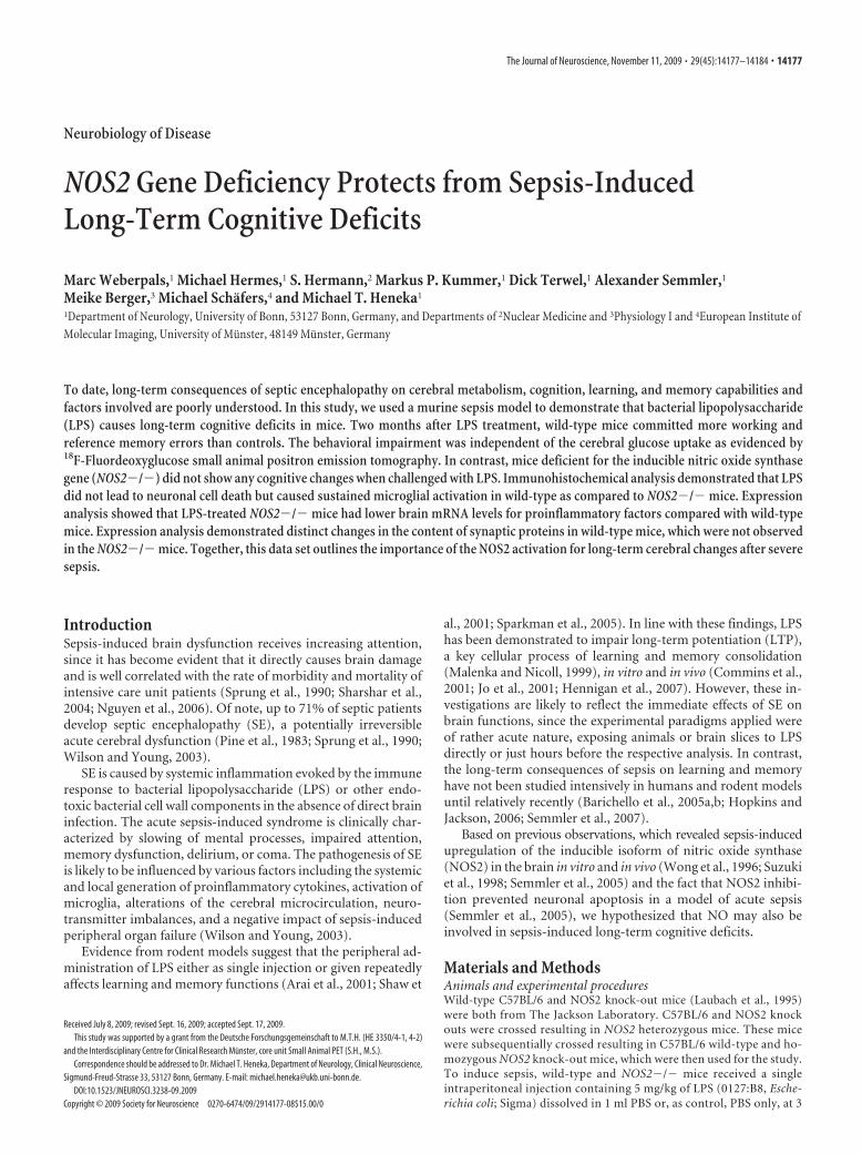
Figure 1. Peripheral LPS administration causes sustained NOS2 expression in the brain of C57BL/6 mice. A, LPS treatment schedule including the time points for the different analyses. OF, Openfield; RAM, radial arm maze. B, Western blot detection of NOS2 in the hippocampus (HC) and frontal cortex (FC) over the experimental period of 8 weeks. C, Comparison of NOS2 expression in LPSand vehicle (PBS)-injected mice at 1 and 8 weeks from whole-brain lysates. Densitometrical quantification verified a significant and sustained LPS effect on NOS2. Each lane represents a single animal(mean � SEM, n � 6, student’s t test, *p � 0.05; **p � 0.01). D, DAB immunostaining against NOS2 in an LPS-treated mouse (top). Scale bar, 50 �m. Representative confocal immunostainingof NOS2 and CD11b revealing colocalization (bottom). Scale bar, 10 �m.
14178 • J. Neurosci., November 11, 2009 • 29(45):14177–14184 Weberpals et al. • Sepsis-Induced Cognitive Dysfunction
ImmunohistochemistrySerial sagittal sections were cut (10 �m) from cryo-conserved hemi-spheres (Leica Cryostat CM 3050S), embedded in tissue-freezing medium(Jung/Leica Microsystems; #0201-08926) and mounted (MicroscopeSlides; #K0123b). After drying for 30 min at ambient temperature, forfixation slides were incubated in 4% paraformaldehyde (Roti Histofix;4% #P087.4, Roth) for 20 min. Blocking of nonspecific binding wasachieved by 1 h incubation in 5% normal goat serum (Linaris; #S-1000).Between the steps, slides were washed three times for 5 min in PBST.Immunostaining was performed overnight by incubation at 4°C with thefollowing primary antibodies: (1) pAb rabbit-anti-glial fibrillary acidicprotein (1:900 in 2% normal goat serum in PBST; DAKO; Z0334); (2) ratmAb #MCA 711 against murine CD11b (CD11b, 1:250; Serotec); and (3)mouse mAb #MAB 377 against neuronal nuclei (NeuN; 1:500; MilliporeBioscience Research Reagents). Afterward, slides were incubated withAlexa-Fluor 594 goat-anti-rat secondary antibodies hosted in goat for 1 h(1:800 in PBST; Invitrogen #A11036 and #A11020). Again, slides werewashed with PBST between the steps. Finally, the slides were coverslippedin Mowiol 4-88 (Calbiochem/VWR #475904) and stored at �20°C in thedark until microscopy was performed. Double immunostaining foriNOS and CD11b was performed on cryofixed sections as describedabove. Sections were dried at ambient temperature for 1 h and then fixedin 4% PFA or methanol for 15 min at ambient temperature. After wash-ing with PBS, the double staining was performed by adding first antibod-ies followed by overnight incubation at 4°C. The following antibodieswere used: rat mAb #MCA 711 against murine CD11b (1:250; Serotec)rabbit pAb against iNOS, 32030 (1:150; Transduction Laboratories).
The goat secondary antibodies (dichlorotriazinylaminofluorescein-conjugated anti-rabbit 1:150; Texas Red-conjugated anti-rat 1:80; Jack-son Immuno Research Laboratories) were applied sequentially afterwashing in PBS. Negative controls included nonspecific IgG instead ofprimary antibodies, preabsorption with respective cognate peptides(150 –200 �g of peptide/ml of antibody-working solution), and omis-sion of the secondary antibody.
Confocal laser-scanning microscopyDouble-labeled specimens were analyzed with a confocal laser-scanningmicroscope (Multiprobe 2001; Invitrogen) equipped with an Ar/Kr laserwith balanced emission at 488, 568, and 647 nm. Images were acquired atusing a 40� objective. To achieve an optimal signal-to-noise ratio foreach fluorophore, sequential scanning with 568 and 488 nm was used.Images were processed using ImageSpace 3.10 software (Invitrogen) on aSilicon Graphics power series 310GTX work station. Original sectionseries were subjected to Gaussian filtration to reduce noise and enhanceweakly but specifically labeled parts. Original and filtered sections wereprojected on one plane using a maximum-intensity algorithm and insome cases using depth-coding and surface-rendering algorithms.
Quantification of immunohistochemistryFor quantitative image analysis of hippocampal immunostaining, serialsagittal sections of one hemisphere were collected (lateral position �0.5to �2.25 from bregma). GFAP, CD11b, and NeuN immunostainingwere evaluated on sagittal brain sections of six animals from each group.For each animal, antigens were detected in 10 parallel sections having adistance of 70 mm to each other and showing both the hippocampus andcortex. In each section, staining in the hippocampus and the frontalcortex were evaluated. The stained area was determined and given aspercentage of the respective brain region. All images were acquired onBX-61 microscope (Olympus), equipped with a digital camera (F-ViewII; Olympus), with identical exposure time. Image analysis was per-formed using Cell P (Olympus). For each animal, average values from allsections were determined. Data were analyzed by one-way ANOVA andTukey’s post hoc tests using SYSTAT software (Systat).
Real-time PCRNeocortical, hippocampal, and cerebellar tissue was dissected from onehemisphere on a cold glass plate. RNA was extracted from these tissuesusing Trizol (Life Technologies Invitrogen). Total RNA was quantifiedspectrophotometrically and reverse transcribed using the RevertAid FirstStrand cDNA Synthesis kit (Fermentas) according to the manufacturer’s
instructions. PCR was performed as described previously (Heneka et al.,2002) using the following primers: TNF� forward 5-GCA CAG AAAGCA TGA TCC GC-3 and TNF� reverse 5-TGT CTT TGA GAT CCATGC CG-3, Il-1� forward 5-CCTGTGTAATGAAAGACGGC-3 andIl-1� reverse 5-AAGGGA GCTCCTTCACA TGC-3, RANTES forward5-GTG CCC ACG TCA AGG AGT ATT TCT-3 and RANTES reverse5-GAC CGA GTG GGA GTA GGG GAT TAC-3, GFAP forward 5-TCCGCG GCA CGA ACG AGT C-3 and GFAP reverse 5-CAC CAT CCCGCA TCT CCA CAG TCT-3, GAPDH forward 5-TCA CCA GGG CTGCCA TTT GC-3 and GAPDH reverse 5-GAC TCC ACG ACA TAC TCAGC-3. PCR was performed on cDNA generated from individual animalsin each group. PCR conditions were 35 cycles of denaturation at 95°C for30 s, annealing at 63°C for 45 s, and extension at 72°C for 45 s using athermal cycler (Biometra Personal T; Biometra). PCR products wereseparated by agarose gel electrophoresis and detected using an AlphaIno-tech imaging system, and band intensities determined using ImageJ. Datawere analyzed by ANOVA with Tukey’s post hoc test (Systat).
Synaptosomal preparationWhole-brain hemispheres were homogenized in 9 volumes of 50 mM
Tris-acetate, pH 7.4, 5 mM NaF, 2 mM Na3VO4, 5 mM Na4P2O7, 1 mM
PMSF, protease inhibitor mixture (1:500; Sigma) by 15 slow up-and-down strokes using a Potter-Evehjem homogenizer at 700 rpm (FisherScientific; clearance 0.15 mm). Fractions of 1 ml were centrifuged in 1.5ml tubes at 800 � g for 5 min. The supernatant was centrifuged at16,000 � g for 20 min. The resulting pellet was resuspended in 300 mlhomogenization buffer and loaded on a gradient consisting of 300 ml 1.4M sucrose and 500 ml 1.0 M sucrose. The gradient was centrifuged at 9100g for 10 min without brake. The synaptosomes were collected from the1.4 M sucrose layer with a Pasteur pipette. Synaptosomes were suspendedin 0.4 M sucrose, pelleted at 16,000 � g for 20 min and resuspended inRIPA for protein determination and Western blot.
Western blottingSynaptosomal proteins (20 �g) were separated in 4 –12% NuPAGE gels,transferred to Immobilon-P polyvinylidene fluoride membranes and in-cubated with antibodies directed against a set of presynaptic and postsyn-aptic protein targets including synaptotagmin (Sigma; S2177, 1:500),synaptobrevin (Synaptic Systems; #104211, 1:500), munc-18 (BD Bio-sciences; #610336, 1:500), PSD-95 (Cell Signaling Technologies; #2507,1:1000), synaptophysin (Millipore Bioscience Research Reagents;#MAB5258, 1:500), and CaMKII (Santa Cruz; #sc-9035, 1:500). To visualizeimmunoreactions, blots were incubated with ECL reagent (GE Healthcare,Pharmacia), and digital images were obtained with the ChemiDoc System(Bio-Rad). Signal intensities were determined using ImageJ. Data were ana-lyzed by ANOVA and Tukey’s post hoc tests (Systat).
ResultsPeripheral administration of LPS at 3 months rapidly lead to signsof sickness behavior including reduced exploration, grooming,decreased motor activity, and anhedonia without an obvious dif-ference between NOS2�/� and wild-type mice. In line with this,two animals in each group (wild type and NOS2�/�) died inresponse to LPS challenge within the first 24 h (supplemental Fig.3B, available at www.jneurosci.org as supplemental material);thereafter, no further deaths occurred, and all animals surviveduntil the end of the experiment. LPS injection caused a sustainedincrease of NOS2 expression which is detectable in the hip-pocampus and frontal cortex of wild-type mice after 2 months(Fig. 1B).
When compared with vehicle injection, LPS did significantlyincrease NOS2 expression at 1 and 8 weeks after the initial im-mune challenge (Fig. 1C). Immunohistologically, the majority ofthe NOS2-positive cells were found in close vicinity to bloodvessels at both time points (Fig. 1D). Double immunostainingand subsequent confocal analysis revealed that most NOS2-positive cells were also immunoreactive for CD11b, suggesting
Weberpals et al. • Sepsis-Induced Cognitive Dysfunction J. Neurosci., November 11, 2009 • 29(45):14177–14184 • 14179

that NOS2 was expressed by cells from themyeloid lineage and most likely by micro-glia or invaded macrophages (Fig. 1D).
Since previous observations (Semmleret al., 2005, 2007) suggested a role of NOS2expression in the brain’s response to en-dotoxemia, we aimed to determine whethernitric oxide is involved in LPS causing long-term impairment of memory and cognitionafter a single LPS challenge. Thus, wild-typeand NOS2�/� mice that had previouslybeen challenged with LPS or control ve-hicle were analyzed at 2 month by firstopen-field analysis and afterward by ra-dial arm maze test.
Open-field testing assessed generalmotor behavior of the mice including in-activity time, corner time, center time aswell as rearing, grooming, urination, anddefecation. While there was a general ten-dency of the NOS2�/� mice to spend lesstime in the corner and more in the center,this phenomenon did not reach the levelof statistical significance and was not in-fluenced by LPS administration. Like-wise, all other parameters did not showdifferences (supplemental Fig. 1, avail-able at www.jneurosci.org as supple-mental material).
For the eight-arm maze test, the behav-ioral parameters included the total num-ber of trials needed to complete the task,working, and reference memory errors.Wild-type mice and NOS2�/� mice didnot show any differences when treatedwith control vehicle. LPS treatment re-sulted in a worse performance on all be-havioral measures examined in wild-typemice, which was not observed in NOS2�/�animals (Fig. 2). This result suggests thatNOS2-derived nitric oxide generationinduced by peripheral administration ofLPS contributes to sustained memorydysfunction.
Since we have previously shown thatacute sepsis can affect cerebral glucoseutilization (Semmler et al., 2008), 18F-FDG–PET analysis was performed in thehippocampus, cortex, and striatum to de-termine whether the observed behavioralchanges would be accompanied by alter-ations of cerebral glucose uptake (Fig. 3).Although there was a consistent tendencyof LPS to reduce cerebral glucose utiliza-tion in all brain areas evaluated, this didnot reach the level of statistical signifi-cance ( p � 0.058, ANOVA followed byTukey test). Of note, there was no suchtendency in NOS2�/� mice.
To assess the degree of morphologicalchanges in the hippocampus CD11b, GFAP,and NeuN was immunohistochemicallyassessed in serial sections with a defined
Figure 2. LPS-induced long-term memory impairment. Radial arm maze test: wild-type mice (wt) challenged with LPS(wt�LPS) needed more trials and committed more errors during the test compared to NOS2�/� (NOS2�/� �LPS). A, Totaltrials (mean � SEM, n � 10 –12, ANOVA, F � 5.73, Tukey post hoc analysis, *p � 0.05; ***p � 0.001 wt�LPS vs wt,NOS2�/�, NOS2�/� �LPS). B, Working memory errors (mean � SEM, n � 10 –12, ANOVA, F � 3.56, Tukey post hocanalysis, *p � 0.05 wt�LPS vs wt, NOS2�/�, NOS2�/� �LPS). C, Reference memory errors (mean � SEM, n � 10 –12,ANOVA, F � 10.80, Tukey post hoc analysis, ***p � 0.001 wt�LPS vs wt, NOS2�/�, NOS2�/� �LPS).
Figure 3. Analysis of cerebral glucose uptake 2 months after sepsis. A, Shown are five representative transversal 18F-FDG–PETbrain slices of wild-type and NOS2�/� mice treated with vehicle or LPS. Corresponding region-of-interest (ROI) masks aredisplayed below. B, Quantification of 18F-FDG uptake (relative to the cerebellum) did not reveal any significant differencesbetween groups in all brain areas examined including the frontal cortex (FC), the striatum (St), and hippocampus (HC). Thearrow exemplarily indicates noncerebral uptake of 18F-FDG in retro-orbital Harderian glands routinely seen with all PETstudies (mean � SEM, n � 5).
14180 • J. Neurosci., November 11, 2009 • 29(45):14177–14184 Weberpals et al. • Sepsis-Induced Cognitive Dysfunction
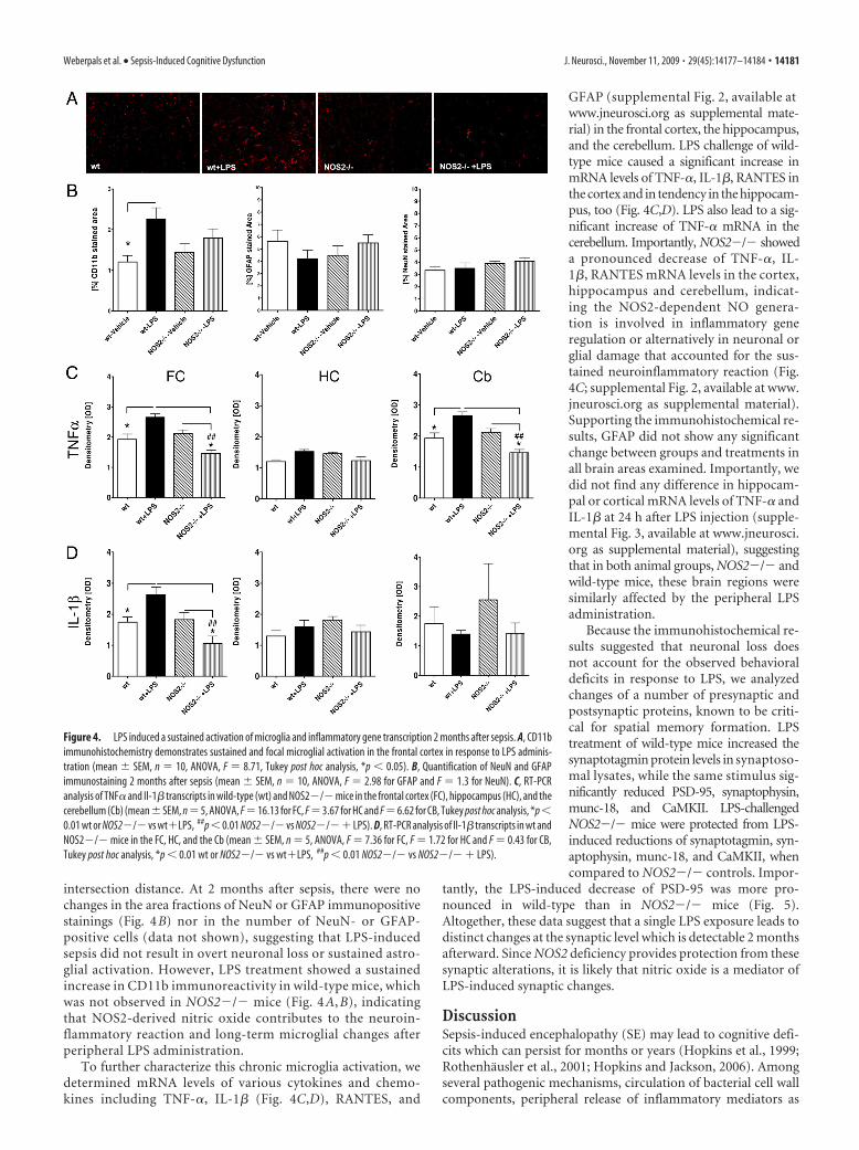
intersection distance. At 2 months after sepsis, there were nochanges in the area fractions of NeuN or GFAP immunopositivestainings (Fig. 4B) nor in the number of NeuN- or GFAP-positive cells (data not shown), suggesting that LPS-inducedsepsis did not result in overt neuronal loss or sustained astro-glial activation. However, LPS treatment showed a sustainedincrease in CD11b immunoreactivity in wild-type mice, whichwas not observed in NOS2�/� mice (Fig. 4 A, B), indicatingthat NOS2-derived nitric oxide contributes to the neuroin-flammatory reaction and long-term microglial changes afterperipheral LPS administration.
To further characterize this chronic microglia activation, wedetermined mRNA levels of various cytokines and chemo-kines including TNF-�, IL-1� (Fig. 4C,D), RANTES, and
GFAP (supplemental Fig. 2, available atwww.jneurosci.org as supplemental mate-rial) in the frontal cortex, the hippocampus,and the cerebellum. LPS challenge of wild-type mice caused a significant increase inmRNA levels of TNF-�, IL-1�, RANTES inthe cortex and in tendency in the hippocam-pus, too (Fig. 4C,D). LPS also lead to a sig-nificant increase of TNF-� mRNA in thecerebellum. Importantly, NOS2�/� showeda pronounced decrease of TNF-�, IL-1�, RANTES mRNA levels in the cortex,hippocampus and cerebellum, indicat-ing the NOS2-dependent NO genera-tion is involved in inflammatory generegulation or alternatively in neuronal orglial damage that accounted for the sus-tained neuroinflammatory reaction (Fig.4C; supplemental Fig. 2, available at www.jneurosci.org as supplemental material).Supporting the immunohistochemical re-sults, GFAP did not show any significantchange between groups and treatments inall brain areas examined. Importantly, wedid not find any difference in hippocam-pal or cortical mRNA levels of TNF-� andIL-1� at 24 h after LPS injection (supple-mental Fig. 3, available at www.jneurosci.org as supplemental material), suggestingthat in both animal groups, NOS2�/� andwild-type mice, these brain regions weresimilarly affected by the peripheral LPSadministration.
Because the immunohistochemical re-sults suggested that neuronal loss doesnot account for the observed behavioraldeficits in response to LPS, we analyzedchanges of a number of presynaptic andpostsynaptic proteins, known to be criti-cal for spatial memory formation. LPStreatment of wild-type mice increased thesynaptotagmin protein levels in synaptoso-mal lysates, while the same stimulus sig-nificantly reduced PSD-95, synaptophysin,munc-18, and CaMKII. LPS-challengedNOS2�/� mice were protected from LPS-induced reductions of synaptotagmin, syn-aptophysin, munc-18, and CaMKII, whencompared to NOS2�/� controls. Impor-
tantly, the LPS-induced decrease of PSD-95 was more pro-nounced in wild-type than in NOS2�/� mice (Fig. 5).Altogether, these data suggest that a single LPS exposure leads todistinct changes at the synaptic level which is detectable 2 monthsafterward. Since NOS2 deficiency provides protection from thesesynaptic alterations, it is likely that nitric oxide is a mediator ofLPS-induced synaptic changes.
DiscussionSepsis-induced encephalopathy (SE) may lead to cognitive defi-cits which can persist for months or years (Hopkins et al., 1999;Rothenhausler et al., 2001; Hopkins and Jackson, 2006). Amongseveral pathogenic mechanisms, circulation of bacterial cell wallcomponents, peripheral release of inflammatory mediators as
Figure 4. LPS induced a sustained activation of microglia and inflammatory gene transcription 2 months after sepsis. A, CD11bimmunohistochemistry demonstrates sustained and focal microglial activation in the frontal cortex in response to LPS adminis-tration (mean � SEM, n � 10, ANOVA, F � 8.71, Tukey post hoc analysis, *p � 0.05). B, Quantification of NeuN and GFAPimmunostaining 2 months after sepsis (mean � SEM, n � 10, ANOVA, F � 2.98 for GFAP and F � 1.3 for NeuN). C, RT-PCRanalysis of TNF� and Il-1� transcripts in wild-type (wt) and NOS2�/� mice in the frontal cortex (FC), hippocampus (HC), and thecerebellum (Cb) (mean�SEM, n�5, ANOVA, F�16.13 for FC, F�3.67 for HC and F�6.62 for CB, Tukey post hoc analysis, *p�0.01 wt or NOS2�/� vs wt�LPS, ##p�0.01 NOS2�/� vs NOS2�/�� LPS). D, RT-PCR analysis of Il-1� transcripts in wt andNOS2�/� mice in the FC, HC, and the Cb (mean � SEM, n � 5, ANOVA, F � 7.36 for FC, F � 1.72 for HC and F � 0.43 for CB,Tukey post hoc analysis, *p � 0.01 wt or NOS2�/� vs wt�LPS, ##p � 0.01 NOS2�/� vs NOS2�/� � LPS).
Weberpals et al. • Sepsis-Induced Cognitive Dysfunction J. Neurosci., November 11, 2009 • 29(45):14177–14184 • 14181

well as the stimulation of innate immunemechanisms within the brain are consid-ered to contribute to SE. Additionally, thenegative impact of peripheral organ fail-ure in response to sepsis is likely to affectbrain function.
In rodent models, sepsis induced byperipheral injection of LPS has been shownto activate microglial cells (Buttini et al.,1996) and thus mimics the microglia acti-vation observed in brains from patientswho died from sepsis (Lemstra et al.,2007). LPS injection can therefore be usedto model the effect of a defined concentra-tion of this immunostimulant and thesubsequent induction of neuroinflamma-tory events. In the present study, a singleinjection of a high dose of LPS caused sus-tained microglial activation, which wasdetectable up to 8 weeks after the initialimmune challenge. Similarly, persistentneuroinflammation has been described in amodel of chronic intracerebroventricularLPS infusion in rats, where microglia activa-tion was observed at 37 d after cessation ofLPS exposure (Hauss-Wegrzyniak et al.,2000). Although there are important differ-ences between both models, these findingscollectively support the hypothesis that LPScan lead to a long-lasting activation of theinnate immune system, independently ofthe route of exposure. Importantly, thisneuroinflammatory reaction was accompa-nied by a significant increase of NOS2 ex-pression, predominantly located in thevicinity of blood vessels.
LPS administration in rodents leadsrapidly to behavioral changes character-ized by a reduction of food and water in-take, less social interactions, decreasedlocomotor activity and exploration, sum-marized as “sickness behavior.” In thepresent study, there was no obvious dif-ference between the experimental groupsregarding this phenomenon. However,this initial sickness behavior was notquantified, and a more detailed analysis ofthis very initial phase may have detectedmore subtle changes. Nevertheless, thenumber of LPS-induced deaths, as well asthe transcription of two key cytokines,TNF-� and IL-1� in the hippocampusand frontal cortex, was not different between NOS2-deficient andwild-type mice. Several studies suggest that peripheral LPS ad-ministration also causes learning and memory impairment inmice (Arai et al., 2001; Sparkman et al., 2005). While in thesestudies the LPS administration was performed daily just hoursbefore the behavioral tests, a further study found that a singleinjection of LPS was sufficient to cause persistent spatial memorydeficits which were detectable over the entire test period of 11 d(Shaw et al., 2001). Extending these observations, the presentstudy investigates locomotion and spatial memory changes at 2months after LPS treatment, a time point where no symptoms of
sickness behavior were detectable anymore. Mice immunochal-lenged with LPS showed impaired spatial memory performancein the eight-arm radial maze as compared to vehicle-treated con-trols. Based on the observation that LPS administration causedsustained induction of NOS2 expression in the brain and thepreviously reported in vivo detection of nitric oxide by electronparamagnetic resonance in the brain of LPS-treated rats (Suzukiet al., 1998), we used NOS2-deficient mice to address the func-tional role of inflammation-induced nitric oxide generation. Im-portantly, NOS2 deficiency protected mice from LPS-inducedmemory dysfunction, suggesting that NOS2-derived nitric oxide
Figure 5. Alterations of synaptic key proteins in wild-type and NOS2�/� mice at 2 months after sepsis. A, Synaptosomalpreparations from the whole-brain hemispheres if individual mice were separated by SDS gel electrophoresis and immunoblottedfor the synaptic proteins synaptotagmin, synaptobrevin, munc-18, PSD-95, synaptophysin and CaMKII. B, Densitometrical analysisof the immunoblots from A (n � 4, mean � SEM, ANOVA, F � 19.06 for synaptotagmin, F � 10.77 for synaptobrevin, F � 10.5for munc18, F � 119.0 for PSD-95, F � 12.14 for synaptophysin, F � 18.68 for CaMKII, Tukey post hoc analysis *p � 0.05, **p �0.01, ***p � 0.001).
14182 • J. Neurosci., November 11, 2009 • 29(45):14177–14184 Weberpals et al. • Sepsis-Induced Cognitive Dysfunction

contributes to neurocognitive changes induced by inflammation.This assumption is further supported by the observation that inhi-bition of NOS2 by aminoguanidine attenuated learning and mem-ory deficits in a four-vessel occlusion model of cerebral ischemia(Mori et al., 2001). In line with this, it has been shown that hip-pocampal LTP, a major indicator of synaptic efficacy underlyinglearning and memory formation (Malenka and Nicoll, 1999;Malenka and Bear, 2004), was reduced by LPS in vitro and in vivo(Cunningham et al., 1996; Commins et al., 2001; Jo et al., 2001).Importantly, inhibition of NOS2 has been found to prevent inflam-matory suppression of LTP in similar models (Mori et al., 2001;Togashi et al., 2001; Wang et al., 2004). Together, these data suggestthat inflammation-induced, NOS2-derived nitric oxide can causelearning and memory dysfunction.
Immunostimulated nitric oxide production has been shownto exert detrimental effects by a variety of mechanisms includingthe induction of apoptosis, inhibition of mitochondrial respira-tion, protein nitrosylation, and potentiation of excitotoxicity(Hewett et al., 1994; Nicotera et al., 1999; Moncada and Bolanos,2006; Brown, 2007; Calabrese et al., 2007). More specifically, ni-tric oxide has been shown to affect neighboring cell survivalthrough DNA damage, poly-ADP ribose synthetase activation,and subsequent energy depletion (Zhang et al., 1994). Further-more, NO is able to cause p53 accumulation (Forrester et al.,1996) and mediates the release of apoptosis-inducing factorevoked by systemic LPS administration (Czapski et al., 2007).The effects of nitric oxide and reactive nitrogen species on mito-chondrial function are complex and include the reversible inhi-bition of cytochrome oxidase inhibition of complex I and II, theinduction of mitochondrial permeability transition, and produc-tion of radical oxygen species from mitochondria (Brown andBorutaite, 2004). Using a higher concentration of LPS, we previ-ously observed neuronal cell death within the hippocampus(Semmler et al., 2005, 2007). However, we did not detect neuro-nal loss which could account for the observed behavioral pheno-type. In keeping with this, no significant change of corticalglucose utilization was detectable at 2 months after LPS admin-istration. This further substantiates that neither overt neuronalloss nor altered neuronal metabolism mediates the impairedlearning and memory behavior in response to LPS treatment.
In contrast, analysis of the brain innate immune system re-vealed a subtle but sustained activation of microglia that wasdetected in wild-type mice but not in NOS2-deficient animalsand was paralleled by a distinct regulation of cytokine mRNAlevels, including those of TNF� and IL-1� mRNAs. It remainsunclear whether nitric oxide directly affected the transcription ofthe TNF� and IL-1� genes as previously described (Kroncke,2003) or caused cellular damage which in turn accounted for thesustained activation of microglia and cytokine transcription.Given the reported negative impact of TNF� and IL-1� on learn-ing and memory function, as well as on LTP (Tancredi et al.,1992; Oitzl et al., 1993; Gibertini et al., 1995), the reduced levels ofthese cytokines in NOS2-deficient mice may partly explain theobserved protection. As a further possibility, NOS2 deficiency hasbeen shown to generally protect from septic shock and organfailure (Wei et al., 1995), and thus, a reduced level of peripheraltoxins and cytokines could also contribute to neuroprotection.
Based on the many findings that synapses are particularly vul-nerable in neurodegenerative conditions, we next studied the ef-fects of LPS and NOS2 deficiency on the synaptosome and moreprecisely on the regulation of synaptic proteins, shown to havekey functions for learning and memory. LPS induced distinctalterations at the synaptic protein level, and more specifically a
significant reduction of PSD-95, CaMKII, and synaptophysinmay also contribute to the observed memory deficits, given therole of these proteins in synaptic plasticity (Silva et al., 1992;O’Brien et al., 1998; Janz et al., 1999; Fukunaga and Miyamoto,2000). Interestingly, a previous in vitro study found that microglial-derived nitric oxide impairs the anterograde axonal transport ofsynaptophysin, a mechanism which may be involved in the sig-nificant reduction of synaptophysin observed in the presentstudy (Stagi et al., 2005).
The reduction of PSD-95 is of particular importance, sincethis protein is a central player at the postsynaptic density by scaf-folding specialized large membrane complexes consisting in thecase of PSD-95 of NMDA, neuroligin, and �-adrenergic recep-tors. In line with the findings presented in this study, mice lackingPSD-95 show a severely impaired spatial memory performance(Migaud et al., 1998; Kim and Sheng, 2004).
Importantly, NOS2 deficiency protected from these synapticchanges. It remains unclear by which molecular mechanisms ni-tric oxide causes the observed synaptic alterations and moreoverif those are ultimately responsible for the observed phenotype. Itcan, however, be speculated that NOS2 deficiency protects fromsynaptic alterations by interfering with more than one detrimen-tal pathomechanism.
Together, this study highlights the impact of severe sepsis onbrain function and reveals that sepsis can cause persistent learn-ing and memory deficits in mice. Sustained microglial activationmay represent a valuable therapeutic target in patients sufferingfrom severe sepsis. To exclude compensatory responses due toNOS2 deficiency and to test a potential therapeutic benefit, futurestudies should include treatment protocols with NOS2-selectiveinhibitors. In particular, inflammatory-induced and nitric oxide-mediated alterations of synapses should be further investigated tounravel the underlying molecular mechanisms, since specificprotection of synapses from immunostimulated changes mayprove to be beneficial.
ReferencesArai K, Matsuki N, Ikegaya Y, Nishiyama N (2001) Deterioration of spatial
learning performances in lipopolysaccharide-treated mice. Jpn J Pharma-col 87:195–201.
Barichello T, Martins MR, Reinke A, Feier G, Ritter C, Quevedo J, Dal-PizzolF (2005a) Cognitive impairment in sepsis survivors from cecal ligationand perforation. Crit Care Med 33:221–223; discussion 262–263.
Barichello T, Martins MR, Reinke A, Feier G, Ritter C, Quevedo J, Dal-PizzolF (2005b) Long-term cognitive impairment in sepsis survivors. CritCare Med 33:1671.
Brown GC (2007) Mechanisms of inflammatory neurodegeneration: iNOSand NADPH oxidase. Biochem Soc Trans 35:1119 –1121.
Brown GC, Borutaite V (2004) Inhibition of mitochondrial respiratorycomplex I by nitric oxide, peroxynitrite and S-nitrosothiols. BiochimBiophys Acta 1658:44 – 49.
Buttini M, Limonta S, Boddeke HW (1996) Peripheral administration oflipopolysaccharide induces activation of microglial cells in rat brain. Neu-rochem Int 29:25–35.
Calabrese V, Mancuso C, Calvani M, Rizzarelli E, Butterfield DA, Stella AM(2007) Nitric oxide in the central nervous system: neuroprotection ver-sus neurotoxicity. Nat Rev Neurosci 8:766 –775.
Commins S, O’Neill LA, O’Mara SM (2001) The effects of the bacterial en-dotoxin lipopolysaccharide on synaptic transmission and plasticity in theCA1-subiculum pathway in vivo. Neuroscience 102:273–280.
Cunningham AJ, Murray CA, O’Neill LA, Lynch MA, O’Connor JJ (1996)Interleukin-1 beta (IL-1 beta) and tumour necrosis factor (TNF) inhibitlong-term potentiation in the rat dentate gyrus in vitro. Neurosci Lett203:17–20.
Czapski GA, Cakala M, Chalimoniuk M, Gajkowska B, Strosznajder JB(2007) Role of nitric oxide in the brain during lipopolysaccharide-evoked systemic inflammation. J Neurosci Res 85:1694 –1703.
Weberpals et al. • Sepsis-Induced Cognitive Dysfunction J. Neurosci., November 11, 2009 • 29(45):14177–14184 • 14183

Forrester K, Ambs S, Lupold SE, Kapust RB, Spillare EA, Weinberg WC,Felley-Bosco E, Wang XW, Geller DA, Tzeng E, Billiar TR, Harris CC(1996) Nitric oxide-induced p53 accumulation and regulation of induc-ible nitric oxide synthase expression by wild-type p53. Proc Natl Acad SciU S A 93:2442–2447.
Fukunaga K, Miyamoto E (2000) A working model of CaM kinase II activityin hippocampal long-term potentiation and memory. Neurosci Res38:3–17.
Gibertini M, Newton C, Klein TW, Friedman H (1995) Legionellapneumophila-induced visual learning impairment reversed by anti-interleukin-1-beta. Proc Soc Exp Biol Med 210:7–11.
Hauss-Wegrzyniak B, Vraniak PD, Wenk GL (2000) LPS-induced neuroin-flammatory effects do not recover with time. Neuroreport 11:1759 –1763.
Hennigan A, Trotter C, Kelly AM (2007) Lipopolysaccharide impairs long-term potentiation and recognition memory and increases p75NTR ex-pression in the rat dentate gyrus. Brain Res 1130:158 –166.
Hewett SJ, Csernansky CA, Choi DW (1994) Selective potentiation ofNMDA-induced neuronal injury following induction of astrocytic iNOS.Neuron 13:487– 494.
Hopkins RO, Jackson JC (2006) Long-term neurocognitive function aftercritical illness. Chest 130:869 – 878.
Hopkins RO, Weaver LK, Pope D, Orme JF, Bigler ED, Larson-Lohr V(1999) Neuropsychological sequelae and impaired health status in survi-vors of severe acute respiratory distress syndrome. Am J Resp Crit CareMed 160:50 –56.
Janz R, Sudhof TC, Hammer RE, Unni V, Siegelbaum SA, Bolshakov VY(1999) Essential roles in synaptic plasticity for synaptogyrin I and synap-tophysin I. Neuron 24:687–700.
Jo JH, Park EJ, Lee JK, Jung MW, Lee CJ (2001) Lipopolysaccharide inhibitsinduction of long-term potentiation and depression in the rat hippocam-pal CA1 area. Eur J Pharmacol 422:69 –76.
Kim E, Sheng M (2004) PDZ domain proteins of synapses. Nat Rev Neuro-sci 5:771–781.
Kroncke KD (2003) Nitrosative stress and transcription. Biol Chem 384:1365–1377.
Laubach VE, Shesely EG, Smithies O, Sherman PA (1995) Mice lacking in-ducible nitric oxide synthase are not resistant to lipopolysaccharide-induced death. Proc Natl Acad Sci U S A 92:10688 –10692.
Lemstra AW, Groen in’t Woud JC, Hoozemans JJ, van Haastert ES, Rozemul-ler AJ, Eikelenboom P, van Gool WA (2007) Microglia activation insepsis: a case-control study. J Neuroinflammation 4:4.
Malenka RC, Bear MF (2004) LTP and LTD: an embarrassment of riches.Neuron 44:5–21.
Malenka RC, Nicoll RA (1999) Long-term potentiation–a decade of progress?Science 285:1870–1874.
Migaud M, Charlesworth P, Dempster M, Webster LC, Watabe AM, Makhin-son M, He Y, Ramsay MF, Morris RG, Morrison JH, O’Dell TJ, Grant SG(1998) Enhanced long-term potentiation and impaired learning in micewith mutant postsynaptic density-95 protein. Nature 396:433– 439.
Moncada S, Bolanos JP (2006) Nitric oxide, cell bioenergetics and neurode-generation. J Neurochem 97:1676 –1689.
Mori K, Togashi H, Ueno KI, Matsumoto M, Yoshioka M (2001) Amino-guanidine prevented the impairment of learning behavior and hippocam-pal long-term potentiation following transient cerebral ischemia. BehavBrain Res 120:159 –168.
Nguyen DN, Spapen H, Su F, Schiettecatte J, Shi L, Hachimi-Idrissi S, Huygh-ens L (2006) Elevated serum levels of S-1000 protein and neuron-specific enolase are associated with brain injury in patients with severesepsis and septic shock. Crit Care Med 34:1967–1974.
Nicotera P, Bernassola F, Melino G (1999) Nitric oxide (NO), a signalingmolecule with a killer soul. Cell Death Differ 6:931–933.
O’Brien RJ, Lau LF, Huganir RL (1998) Molecular mechanisms of gluta-mate receptor clustering at excitatory synapses. Curr Opin Neurobiol8:364 –369.
Oitzl MS, van Oers H, Schobitz B, de Kloet ER (1993) Interleukin-1-beta,but not interleukin-6, impairs spatial navigation learning. Brain Res613:160 –163.
Olton DS (1987) The radial arm maze as a tool in behavioral pharmacology.Physiol Behav 40:793–797.
Pine RW, Wertz MJ, Lennard ES, Dellinger EP, Carrico CJ, Minshew BH(1983) Determinants of organ malfunction or death in patients withintra-abdominal sepsis. A discriminant analysis. Arch Surg 118:242–249.
Rothenhausler HB, Ehrentraut S, Stoll C, Schelling G, Kapfhammer HP(2001) The relationship between cognitive performance and employ-ment and health status in long-term survivors of the acute respiratorydistress syndrome: results of an exploratory study. Gen Hosp Psychiatry23:90 –96.
Schafers KP, Reader AJ, Kriens M, Knoess C, Schober O, Schafers M (2005)Performance evaluation of the 32-module quadHIDAC small-animalPET scanner. J Nucl Med 46:996 –1004.
Semmler A, Okulla T, Sastre M, Dumitrescu-Ozimek L, Heneka MT (2005)Systemic inflammation induces apoptosis with variable vulnerability ofdifferent brain regions. J Chem Neuroanat 30:144 –157.
Semmler A, Frisch C, Debeir T, Ramanathan M, Okulla T, Klockgether T,Heneka MT (2007) Long-term cognitive impairment, neuronal loss andreduced cortical cholinergic innervation after recovery from sepsis in arodent model. Exp Neurol 204:733–740.
Semmler A, Hermann S, Mormann F, Weberpals M, Paxian SA, Okulla T,Schafers M, Kummer MP, Klockgether T, Heneka MT (2008) Sepsiscauses neuroinflammation and concomitant decrease of cerebral metab-olism. J Neuroinflammation 5:38
Sharshar T, Annane D, de la Grandmaison GL, Brouland JP, HopkinsonNS, Francoise G (2004) The neuropathology of septic shock. Brain Pathol14:21–33.
Shaw KN, Commins S, O’Mara SM (2001) Lipopolysaccharide causes defi-cits in spatial learning in the watermaze but not in BDNF expression in therat dentate gyrus. Behav Brain Res 124:47–54.
Silva AJ, Paylor R, Wehner JM, Tonegawa S (1992) Impaired spatial-learning in alpha-calcium-calmodulin kinase-II mutant mice. Science257:206 –211.
Sparkman NL, Kohman RA, Scott VJ, Boehm GW (2005) Bacterialendotoxin-induced behavioral alterations in two variations of the Morriswater maze. Physiol Behav 86:244 –251.
Sprung CL, Peduzzi PN, Shatney CH, Schein RM, Wilson MF, Sheagren JN,Hinshaw LB (1990) Impact of encephalopathy on mortality in the sepsissyndrome. Crit Care Med 18:801– 806.
Stagi M, Dittrich PS, Frank N, Iliev AI, Schwille P, Neumann H (2005)Breakdown of axonal synaptic vesicle precursor transport by microglialnitric oxide. J Neurosci 25:352–362.
Suzuki Y, Fujii S, Numagami Y, Tominaga T, Yoshimoto T, Yoshimura T(1998) In vivo nitric oxide detection in the septic rat brain by electronparamagnetic resonance. Free Rad Res 28:293–299.
Tancredi V, D’Arcangelo G, Grassi F, Tarroni P, Palmieri G, Santoni A, EusebiF (1992) Tumor-necrosis-factor alters synaptic transmission in rat hip-pocampal slices. Neurosci Lett 146:176 –178.
Togashi H, Mori K, Itoh Y, Matsumoto M, Ueno K, Ohashi S, Otani H,Yoshioka M (2001) Involvement of interleukin-1 beta/nitric oxide path-way in the postischemic impairment of long-term potentiation of the rathippocampus. Neurosci Lett 313:133–136.
Wang Q, Rowan MJ, Anwyl R (2004) �-Amyloid-mediated inhibition ofNMDA receptor-dependent long-term potentiation induction involvesactivation of microglia and stimulation of inducible nitric oxide synthaseand superoxide. J Neurosci 24:6049 – 6056.
Wei XQ, Charles IG, Smith A, Ure J, Feng GJ, Huang FP, Xu D, Muller W,Moncada S, Liew FY (1995) Altered immune-responses in mice lackinginducible nitric-oxide synthase. Nature 375:408 – 411.
Wilson JX, Young GB (2003) Progress in clinical neurosciences: sepsis-associated encephalopathy: evolving concepts. Can J Neurol Sci 30:98 –105.
Wong ML, Rettori V, al-Shekhlee A, Bongiorno PB, Canteros G, McCann SM,Gold PW, Licinio J (1996) Inducible nitric oxide synthase gene expres-sion in the brain during systemic inflammation. Nat Med 2:581–584.
Zhang J, Dawson VL, Dawson TM, Snyder SH (1994) Nitric-oxide activa-tion of poly(ADP-ribose) synthetase in neurotoxicity. Science 263:687–689.
14184 • J. Neurosci., November 11, 2009 • 29(45):14177–14184 Weberpals et al. • Sepsis-Induced Cognitive Dysfunction