NeurobiologyofDisease FamilialFrontotemporalDementia ... ·...
12
Neurobiology of Disease Familial Frontotemporal Dementia-Associated Presenilin-1 c.548G>T Mutation Causes Decreased mRNA Expression and Reduced Presenilin Function in Knock-In Mice Hirotaka Watanabe, 1 Dan Xia, 1,2 Takahisa Kanekiyo, 3 Raymond J. Kelleher III, 2,4 and Jie Shen 1,4 1 Center for Neurologic Diseases, Department of Neurology, Brigham and Women’s Hospital, Boston, Massachusetts 02115, 2 Center for Human Genetic Research, Department of Neurology, Massachusetts General Hospital, Boston, Massachusetts 02114, 3 Department of Neuroscience, Mayo Clinic, Jacksonville, Florida 32224, and 4 Program in Neuroscience, Harvard Medical School, Boston, Massachusetts 02115 Mutations in the presenilin-1 (PSEN1) gene are associated with familial Alzheimer’s disease and frontotemporal dementia (FTD). Inter- estingly, neuropathological analysis of a Belgian FTD family carrying a PSEN1 c.548G>T mutation confirmed neurodegeneration in the absence of amyloid plaques. To investigate the impact of the c.548G>T mutation on presenilin-1 (PS1) function in vivo, we introduced this mutation into the genomic Psen1 locus. The resulting c.548G>T knock-in (KI) mice are viable but express markedly lower levels of Psen1 mRNA and protein in the brain. This reduction is due to production of aberrantly spliced transcripts lacking either exon 6 or exons 6 and 7 and their subsequent degradation via non-sense-mediated decay (NMD); inhibition of NMD by cycloheximide treatment stabi- lized these transcripts and restored the level of Psen1 mRNA in KI/KI brains. Interestingly, the reduction of Psen1 mRNA expression and the degradation of aberrant Psen1 splice products occur exclusively in the brain but not in other tissues. Consistent with decreased Psen1 expression, -secretase activity was strongly reduced in the cerebral cortex of KI mice, as measured by de novo -secretase-mediated cleavage of APP and Notch. Moreover, PS1 expressed from Psen1 cDNA carrying the c.548G>T mutation displayed normal -secretase activity in cultured cells, indicating that the corresponding p.183GV amino acid substitution does not affect -secretase activity. Finally, Psen1 c.548G>T KI/KI ;Psen2 / mice exhibited mild spatial memory deficits in the Morris water maze task. Together, our findings demonstrate that the c.548G>T mutation results in a brain-specific loss of presenilin function due to decreased Psen1 mRNA expression. Introduction Frontotemporal dementia (FTD) is the second most common form of dementia following Alzheimer’s disease (AD) (Sjo ¨gren and Andersen, 2006). FTD is a clinically diverse syndrome char- acterized by profound behavioral changes and degeneration of the frontal and anterior temporal cortex (Neary et al., 2005). Due to the clinical and neuropathological heterogeneity, FTD com- prises a number of related disorders with overlapping but distinct features, including Pick’s disease and frontotemporal lobar de- generation. For example, tau pathology is present in some FTD cases, whereas others lack tau deposition. A large proportion (20 –50%) of FTD cases have a familial component, and muta- tions in microtubule-associated protein tau and progranulin genes are the most frequent genetic causes (Galimberti and Scarpini, 2010). Interestingly, mutations in the presenilin-1 (PSEN1) and PSEN2 genes, which are the major cause of familial AD, have also been implicated in FTD (Mendez and McMurtray, 2006). Since the first identification of the PSEN1 c.338TC mutation in cases of familial dementia with prominent clinical frontotemporal fea- tures (Raux et al., 2000), 10 mutations in the PSEN genes have been associated with clinical diagnoses of FTD, in some cases accompanied by frontotemporal atrophy and frontotemporal hypoperfusion on neuroimaging studies (Rippon et al., 2003; Dermaut et al., 2004; Halliday et al., 2005; Zekanowski et al., 2006; Bernardi et al., 2009; de Bot et al., 2009; Marcon et al., 2009; Gallo et al., 2010; Borroni et al., 2011). Moreover, some patho- genic PSEN1 mutations can cause neuropathological changes consistent with coexisting Pick’s disease and AD (Ikeda et al., 1996; Halliday et al., 2005). Interestingly, several of these muta- tions reside at the exon/intron boundaries and, therefore, may affect PSEN1 splicing in addition to causing mis-sense substitu- tions (Raux et al., 2000; Dermaut et al., 2004; Borroni et al., 2011). Thus, mutations in PSEN may result in overlapping clinical and neuropathological manifestations of AD and FTD, and func- tional changes of presenilin (PS) may underlie common patho- genesis of both dementias. To investigate how PSEN mutations may be associated with FTD, we chose the PSEN1 c.548GT mutation, which was Received Jan. 20, 2012; accepted Feb. 19, 2012. Author contributions: H.W., R.J.K., and J.S. designed research; H.W., D.X., and T.K. performed research; H.W. analyzed data; H.W., R.J.K., and J.S. wrote the paper. This work was supported by National Institutes of Health Grants R01NS041783 and RC2AG036614 (to J.S.) and grants from the Alzheimer’s Association (to R.J.K., J.S.). We thank H. Zhao and X. Zou for breeding and genotyping the mice, and other laboratory members for helpful discussions. We also thank T. Iwatsubo and T. Tomita for pTrcHis2A-C100-FmH and pTrcHis2A-N102-FmH plasmids, B. DeStrooper for Psen double-knock-out mouse embry- onic fibroblasts, P. Davies for phospho-specific tau antibodies (PHF1 and CP13), and A. Takashima for total tau antibody (J.M.). The authors declare no competing financial interests. Correspondence should be addressed to Dr. Jie Shen, Center for Neurologic Diseases, Brigham and Women’s Hospital, Program in Neuroscience, Harvard Medical School, 77 Avenue Louis Pasteur, New Research Building 636E, Boston, MA 02115. E-mail: [email protected]. DOI:10.1523/JNEUROSCI.0317-12.2012 Copyright © 2012 the authors 0270-6474/12/325085-12$15.00/0 The Journal of Neuroscience, April 11, 2012 • 32(15):5085–5096 • 5085
Transcript of NeurobiologyofDisease FamilialFrontotemporalDementia ... ·...

Neurobiology of Disease
Familial Frontotemporal Dementia-Associated Presenilin-1c.548G>T Mutation Causes Decreased mRNA Expressionand Reduced Presenilin Function in Knock-In Mice
Hirotaka Watanabe,1 Dan Xia,1,2 Takahisa Kanekiyo,3 Raymond J. Kelleher III,2,4 and Jie Shen1,4
1Center for Neurologic Diseases, Department of Neurology, Brigham and Women’s Hospital, Boston, Massachusetts 02115, 2Center for Human GeneticResearch, Department of Neurology, Massachusetts General Hospital, Boston, Massachusetts 02114, 3Department of Neuroscience, Mayo Clinic,Jacksonville, Florida 32224, and 4Program in Neuroscience, Harvard Medical School, Boston, Massachusetts 02115
Mutations in the presenilin-1 (PSEN1) gene are associated with familial Alzheimer’s disease and frontotemporal dementia (FTD). Inter-estingly, neuropathological analysis of a Belgian FTD family carrying a PSEN1 c.548G>T mutation confirmed neurodegeneration in theabsence of amyloid plaques. To investigate the impact of the c.548G>T mutation on presenilin-1 (PS1) function in vivo, we introducedthis mutation into the genomic Psen1 locus. The resulting c.548G>T knock-in (KI) mice are viable but express markedly lower levels ofPsen1 mRNA and protein in the brain. This reduction is due to production of aberrantly spliced transcripts lacking either exon 6 or exons6 and 7 and their subsequent degradation via non-sense-mediated decay (NMD); inhibition of NMD by cycloheximide treatment stabi-lized these transcripts and restored the level of Psen1 mRNA in KI/KI brains. Interestingly, the reduction of Psen1 mRNA expression andthe degradation of aberrant Psen1 splice products occur exclusively in the brain but not in other tissues. Consistent with decreased Psen1expression, �-secretase activity was strongly reduced in the cerebral cortex of KI mice, as measured by de novo �-secretase-mediatedcleavage of APP and Notch. Moreover, PS1 expressed from Psen1 cDNA carrying the c.548G>T mutation displayed normal �-secretaseactivity in cultured cells, indicating that the corresponding p.183G�V amino acid substitution does not affect�-secretase activity. Finally, Psen1c.548G>TKI/KI;Psen2�/� mice exhibited mild spatial memory deficits in the Morris water maze task. Together, our findings demonstrate thatthe c.548G>T mutation results in a brain-specific loss of presenilin function due to decreased Psen1 mRNA expression.
IntroductionFrontotemporal dementia (FTD) is the second most commonform of dementia following Alzheimer’s disease (AD) (Sjogrenand Andersen, 2006). FTD is a clinically diverse syndrome char-acterized by profound behavioral changes and degeneration ofthe frontal and anterior temporal cortex (Neary et al., 2005). Dueto the clinical and neuropathological heterogeneity, FTD com-prises a number of related disorders with overlapping but distinctfeatures, including Pick’s disease and frontotemporal lobar de-generation. For example, tau pathology is present in some FTDcases, whereas others lack tau deposition. A large proportion(�20 –50%) of FTD cases have a familial component, and muta-
tions in microtubule-associated protein tau and progranulin genesare the most frequent genetic causes (Galimberti and Scarpini,2010). Interestingly, mutations in the presenilin-1 (PSEN1) andPSEN2 genes, which are the major cause of familial AD, have alsobeen implicated in FTD (Mendez and McMurtray, 2006). Sincethe first identification of the PSEN1 c.338T�C mutation in casesof familial dementia with prominent clinical frontotemporal fea-tures (Raux et al., 2000), �10 mutations in the PSEN genes havebeen associated with clinical diagnoses of FTD, in some casesaccompanied by frontotemporal atrophy and frontotemporalhypoperfusion on neuroimaging studies (Rippon et al., 2003;Dermaut et al., 2004; Halliday et al., 2005; Zekanowski et al.,2006; Bernardi et al., 2009; de Bot et al., 2009; Marcon et al., 2009;Gallo et al., 2010; Borroni et al., 2011). Moreover, some patho-genic PSEN1 mutations can cause neuropathological changesconsistent with coexisting Pick’s disease and AD (Ikeda et al.,1996; Halliday et al., 2005). Interestingly, several of these muta-tions reside at the exon/intron boundaries and, therefore, mayaffect PSEN1 splicing in addition to causing mis-sense substitu-tions (Raux et al., 2000; Dermaut et al., 2004; Borroni et al., 2011).Thus, mutations in PSEN may result in overlapping clinical andneuropathological manifestations of AD and FTD, and func-tional changes of presenilin (PS) may underlie common patho-genesis of both dementias.
To investigate how PSEN mutations may be associatedwith FTD, we chose the PSEN1 c.548G�T mutation, which was
Received Jan. 20, 2012; accepted Feb. 19, 2012.Author contributions: H.W., R.J.K., and J.S. designed research; H.W., D.X., and T.K. performed research; H.W.
analyzed data; H.W., R.J.K., and J.S. wrote the paper.This work was supported by National Institutes of Health Grants R01NS041783 and RC2AG036614 (to J.S.) and
grants from the Alzheimer’s Association (to R.J.K., J.S.). We thank H. Zhao and X. Zou for breeding and genotypingthe mice, and other laboratory members for helpful discussions. We also thank T. Iwatsubo and T. Tomita forpTrcHis2A-C100-FmH and pTrcHis2A-N102-FmH plasmids, B. DeStrooper for Psen double-knock-out mouse embry-onic fibroblasts, P. Davies for phospho-specific tau antibodies (PHF1 and CP13), and A. Takashima for total tauantibody (J.M.).
The authors declare no competing financial interests.Correspondence should be addressed to Dr. Jie Shen, Center for Neurologic Diseases, Brigham and Women’s
Hospital, Program in Neuroscience, Harvard Medical School, 77 Avenue Louis Pasteur, New Research Building 636E,Boston, MA 02115. E-mail: [email protected].
DOI:10.1523/JNEUROSCI.0317-12.2012Copyright © 2012 the authors 0270-6474/12/325085-12$15.00/0
The Journal of Neuroscience, April 11, 2012 • 32(15):5085–5096 • 5085
originally identified in familial FTD pa-tients with neuropathological confirma-tion of Pick’s-type tauopathy in theabsence of amyloid deposition (Dermautet al., 2004). Since the c.548G�T muta-tion resides at the last nucleotide of exon6, we generated knock-in (KI) mice inwhich the c.548G�T mutation was intro-duced into the genomic Psen1 locus. Thec.548G�TKI/KI mice are viable, but Psen1mRNAs are significantly decreased selec-tively in the brain due to aberrant exonskipping and subsequent degradation ofaberrantly spliced transcripts by non-sense-mediated mRNA decay. Accordingly,�-secretase activity is reduced in theKI brain, as measured by �-secretase-mediated cleavage of two physiologicalsubstrates, Notch and amyloid precursorprotein (APP). However, PS1 expressedfrom full-length Psen1 c.548G�T mRNAdisplayed normal �-secretase activity whentested in cultured cells. Furthermore,Psen1KI/KI;Psen2 �/� mice exhibited asignificant deficit in spatial referencememory. Together, these findings dem-onstrate that the c.548G�T mutationcauses brain-specific reduction of Psen1mRNA expression and PS function inthe maintenance of �-secretase activityand memory.
Materials and MethodsGeneration of Psen1 c.548G�T KI mice. For thePsen1 c.548G�T KI construct, a 2.49 kb left-arm fragment and a 3.15 kb right-arm frag-ment surrounding exon 6 were amplified byPCR using BAC (bacterial artificial chromo-some) DNA harboring the mouse Psen1 gene(clone RP23–330F11, Children’s HospitalOakland Research Institute) as a template.The primer sequences are 5�-TACCGCGGAATGGGATGTGTGTGTTGGGATGC-3� and5�-TGGCGGCCGCATGTGAGAATCCTGGGTGCAGTC-3� for the left-arm (the italic se-quences are for SacII and NotI, respectively),and 5�-GCGTCGACAAGTATGTGCTGATCCCCAAAGC-3� and 5�-GCAAGCTTAAGTGCTGGGATTACAGGAGGAC-3� for the right-arm (the italic sequences are for SalI and HindIIIrecognition sites, respectively). The c.548G�Tpathogenic mutation (encoding a mis-sense mu-tation, p.183G�V) and c.546A�G humanizednucleotide change (a silent mutation for Leuat the amino acid residue 182) were introducedby site-directed mutagenesis into exon 6 inthe right-arm fragment (see Fig. 1 A) (onlyc.548G�T mutation will be described hereaf-ter). The left- and right-arm fragments werethen subcloned into the SacII–NotI site and theSalI–HindIII site of the PGKneolox2DTA plas-mid (gift of P. Soriano, Mount Sinai School ofMedicine, New York, NY), respectively, to pro-duce the KI targeting vector (Fig. 1B), which wasthen confirmed by extensive restriction diges-tions and sequencing. The linearized targeting
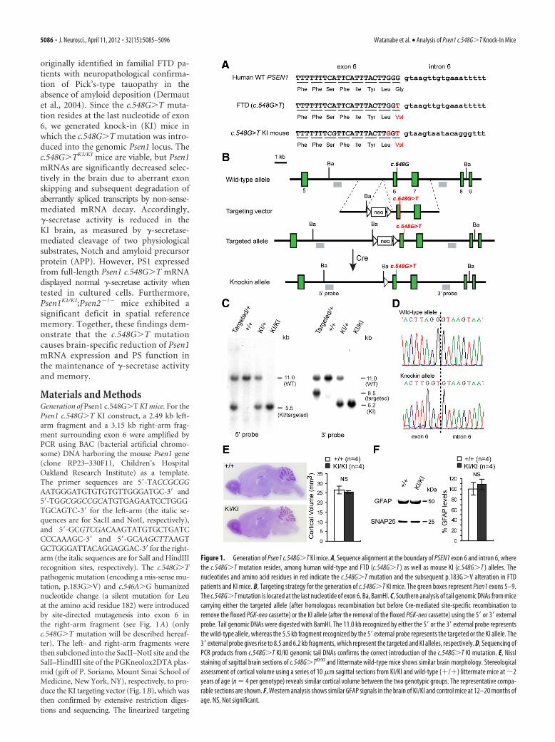
Figure 1. Generation of Psen1 c.548G�T KI mice. A, Sequence alignment at the boundary of PSEN1 exon 6 and intron 6, wherethe c.548G�T mutation resides, among human wild-type and FTD (c.548G�T ) as well as mouse KI (c.548G�T ) alleles. Thenucleotides and amino acid residues in red indicate the c.548G�T mutation and the subsequent p.183G�V alteration in FTDpatients and KI mice. B, Targeting strategy for the generation of c.548G�T KI mice. The green boxes represent Psen1 exons 5–9.The c.548G�T mutation is located at the last nucleotide of exon 6. Ba, BamHI. C, Southern analysis of tail genomic DNAs from micecarrying either the targeted allele (after homologous recombination but before Cre-mediated site-specific recombination toremove the floxed PGK-neo cassette) or the KI allele (after the removal of the floxed PGK-neo cassette) using the 5� or 3� externalprobe. Tail genomic DNAs were digested with BamHI. The 11.0 kb recognized by either the 5� or the 3� external probe representsthe wild-type allele, whereas the 5.5 kb fragment recognized by the 5� external probe represents the targeted or the KI allele. The3� external probe gives rise to 8.5 and 6.2 kb fragments, which represent the targeted and KI alleles, respectively. D, Sequencing ofPCR products from c.548G�T KI/KI genomic tail DNAs confirms the correct introduction of the c.548G�T KI mutation. E, Nisslstaining of sagittal brain sections of c.548G�TKI/KI and littermate wild-type mice shows similar brain morphology. Stereologicalassessment of cortical volume using a series of 10 �m sagittal sections from KI/KI and wild-type (�/�) littermate mice at �2years of age (n � 4 per genotype) reveals similar cortical volume between the two genotypic groups. The representative compa-rable sections are shown. F, Western analysis shows similar GFAP signals in the brain of KI/KI and control mice at 12–20 months ofage. NS, Not significant.
5086 • J. Neurosci., April 11, 2012 • 32(15):5085–5096 Watanabe et al. • Analysis of Psen1 c.548G�T Knock-In Mice
vector by XhoI was electroporated into MKV6.5 ES cells (gift of R. Jaenisch,Whitehead Institute, Cambridge, MA) and selected with 150 �g/ml G418(Invitrogen) for 4–5 d. Genomic DNAs were purified from a total of 360independent ES clones and were subjected to Southern analysis using the 5�probe. For Southern analysis, genomic DNAs were digested with BamHI.Among 52 clones that tested positive for proper homologous recombinationin the 5� arm, 3 ES clones (3–9-H, 4–1-F, 7–11-A) were expanded andsubjected to further Southern analysis using the 5�, 3�, and neomycin probes(Fig. 1B). We confirmed that homologous recombination correctly oc-curred in both the 5� and 3� homologous regions in these three clones. Togenerate Psen1 c.548G�T KI mice, the targeted ES cells (clones 4–1-F and7–11-A) were injected into C57BL/6 blastocysts, and the resulting male chi-meric mice were mated with C57BL/6J-129 F1 mice. Mice transmitting thetargeted allele were further crossed to �CaMKII-Cre male transgenic mice(Yu et al., 2001) to excise the floxed phosphoglycerate-kinase-neomycin (PGK-neo) cassette, as the �CaMKII promoter allows the Cre transgene to expressweakly in male germ cells (Ignotz and Suarez, 2005). The resulting progeny(KI heterozygous mice) were intercrossed to obtain homozygous KI mice forfurther characterization. Correct homologous and Cre-mediated site-specific recombination events were further confirmed by Southern analysiswith tail genomic DNAs using the external 5�, 3�, and neo probes, and bysequencing to detect the presence of the c.548G�T mutation in genomicDNAs. Since offspring from both targeted ES clones 4–1-F and 7–11-A wereindistinguishable, we used the KI mice derived from the ES clone 4–1-F forfurther characterization. In subsequent generations, mice were genotyped
routinely by PCR, and the c.548G�T KI allelewas detected by size shift due to the remainingloxP and construct sequence between exons5 and 6 using the following primers:5�-TGGTGAGAGCTCAGCAGGTAAG-3� and5�-TGCTTTCTAGTTGTCCTTCGTCG-3�. The410 bp band represents the wild-type allele,whereas the 529 bp band represents the KI al-lele. Psen1 c.548G�TKI/KI;Psen2 �/� mice weregenerated by breeding Psen1 c.548G�T KImice with Psen2 �/� mice, which exhibited nodetectable phenotypes (Steiner et al., 1999).The genetic background of all the mice used inthis study was C57BL/6J 129 F1 hybrid. All pro-cedures relating to animal care and treatmentconformed to the institutional and NIHguidelines.
qRT-PCR and Northern analyses. TotalRNAs were purified with TRI reagent (Sigma)according to manufacturer’s protocol, treatedwith DNase I, and reverse transcribed in thepresence of random hexamers and SuperScriptIII Reverse Transcriptase (Invitrogen). PCRswere performed using SYBR Green PCR Mas-ter Mix with a 7500 Fast Real-Time PCR Sys-tem (Applied Biosystems) using cDNA andgene-specific primers. Reactions were per-formed in triplicate, and threshold cycle valueswere normalized to those of Gapdh. Theprimer pairs used in this study were as follows:5�-TGGCCACCATCAAATCAG-3� and 5�-TCATGATGGCCGCATTCAG-3� for Psen1; and5�-TTGTCTCCTGCGACTTCA-3� and 5�-TCCACCACCCTGTTGCTGTA-3� for Gapdh.These primer pairs were confirmed not to giverise to primer dimers during the PCR. ForNorthern analysis, 10–20 �g of total RNA wasseparated in formaldehyde agarose gels andtransferred to nylon membranes (GE Health-care). Hybridization was performed using [�-32P] dCTP-labeled probes specific for each gene.Almost the entire coding region of the mousePsen1 gene was used for a Psen1-specific probe soas to detect any possible transcriptional/post-transcriptional variant from the Psen1 gene.
Western analysis. Dissected cortices (at 1–2 months for c.548G�T KImice) were homogenized in RIPA buffer [50 mM Tris-Cl, pH 7.6, 150 mM
NaCl, 0.5 mM EDTA, 1% NP40, 0.5% sodium deoxycholate, 0.1% SDS,protease inhibitor cocktail (Sigma), 1 mM PMSF]. Equal amounts(10 – 40 �g per lane) of proteins were separated in NuPAGE gels (Invit-rogen) and transferred to nitrocellulose membranes. The membraneswere blocked in 5% nonfat milk/TBS for 1 h and incubated with specificprimary antibodies as follows: rabbit anti-PS1 N-terminal fragments(NTFs; catalog #529591, Calbiochem); rabbit anti-PS1 C-terminal frag-ments (CTFs; catalog #529592, Calbiochem); rabbit anti-Pen2 (catalog#36 –7100, Zymed); rabbit anti-nicastrin (NCT; N1660, Sigma); rabbitanti-Aph1 (PA1–2010, Thermo science); mouse anti-�-tubulin (T6199,Sigma); mouse anti-phospho-tau (PHF1 and CP13, gift of P. Davies,Albert Einstein College of Medicine, New York, NY); rabbit anti-total tau(JM, gift of A. Takashima, RIKEN, Saitama, Japan); rabbit anti-Notch1(V1744) (catalog #2421, Cell Signal Tech); and mouse anti-SNAP25(MAB331, Millipore Bioscience Research Reagents). The membrane wasthen incubated with IRDye 800CW or IRDye 680-labeled secondary an-tibodies (LI-COR Bioscience). Signals were developed and quantifiedwith an Odyssey Infrared Imaging System (LI-COR Bioscience).
Administration of cycloheximide. Drug administration was performedas described previously (Contet et al., 2007). Briefly, cycloheximide(Sigma) or saline was intraperitoneally administered at 1 h intervals (200
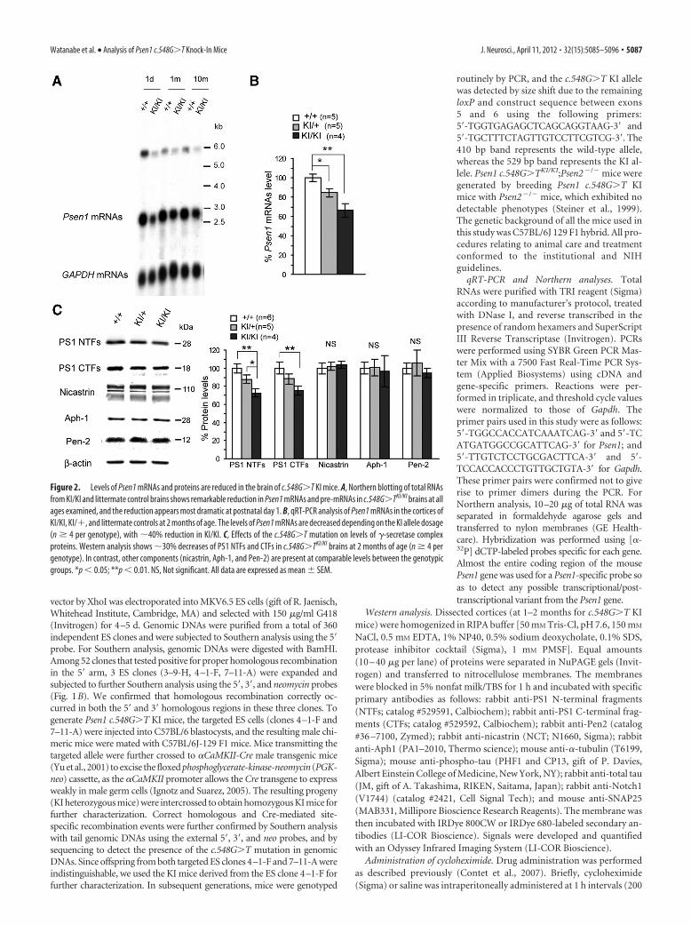
Figure 2. Levels of Psen1 mRNAs and proteins are reduced in the brain of c.548G�T KI mice. A, Northern blotting of total RNAsfrom KI/KI and littermate control brains shows remarkable reduction in Psen1 mRNAs and pre-mRNAs in c.548G�TKI/KI brains at allages examined, and the reduction appears most dramatic at postnatal day 1. B, qRT-PCR analysis of Psen1 mRNAs in the cortices ofKI/KI, KI/�, and littermate controls at 2 months of age. The levels of Psen1 mRNAs are decreased depending on the KI allele dosage(n � 4 per genotype), with �40% reduction in KI/KI. C, Effects of the c.548G�T mutation on levels of �-secretase complexproteins. Western analysis shows �30% decreases of PS1 NTFs and CTFs in c.548G�TKI/KI brains at 2 months of age (n � 4 pergenotype). In contrast, other components (nicastrin, Aph-1, and Pen-2) are present at comparable levels between the genotypicgroups. *p � 0.05; **p � 0.01. NS, Not significant. All data are expressed as mean � SEM.
Watanabe et al. • Analysis of Psen1 c.548G�T Knock-In Mice J. Neurosci., April 11, 2012 • 32(15):5085–5096 • 5087
mg/kg body weight). Tissues were dissectedand frozen for further molecular analysis 4 hafter the first injection.
Cell-based �-secretase assay. Psen-deficientmouse embryonic fibroblasts (MEFs) weretransfected with equal amounts (1 ng) of ex-pression vectors encoding wild-type or variousmutant PS1 along with an N-terminally trun-cated Notch1 construct (NE, gift of A. Goate,Washington University School of Medicine, St.Louis, MO) as described previously (Heilig etal., 2010). The amount of plasmid DNA usedwas derived from a dose– curve analysis using0.125, 0.25, 0.5, 1.0, 2.0, 4.0, and 8.0 ng of Psen1wild-type cDNAs, and doses within the linearrange were 0.5 (0.5), 1.0 (1) and 2.0 (2)ng. A 1.7 kb mouse Psen1 coding sequence wasamplified with primers 5�-GATCTCGAGTTCGAGGTCTTTAGGCAGCTTG-3� and 5�-AGTGCGGCCGCTGCTGCAGCGATGGATGTTGG-3� (italics indicates XhoI and NotIsequences, respectively), and subcloned intoXhoI-NotI sites of pCI expression vector (In-vitrogen). The corresponding mutations ofeach mutant PS1 were introduced by eithersubcloning or site-directed mutagenesis. Cellswere harvested with RIPA buffer 24 h aftertransfection, and Western analysis was per-formed using anti-Notch1 (V1744) antibody(catalog #2421, Cell Signal Tech). Data are nor-malized to �-tubulin, and three independentexperiments were quantified.
In vitro �-secretase assay. �-Secretase-mediated de novo amyloid-� (A�) generationwas measured using a method described previ-ously (Takahashi et al., 2003). Briefly, the cor-tices at 1–2 months of age were homogenizedin homogenization buffer (20 mM PIPES, pH7.0, 140 mM KCl, 0.25 M sucrose, 5 mM EGTA)using a glass/Teflon tissue grinder. The homoge-nates were centrifuged at 800 g for 10 min toremove nuclei and cell debris. The postnuclearsupernatants were recentrifuged at 100,000 gfor 1 h, and the resulting pellets were washed with0.1 M sodium carbonate, pH 11.4, and then cen-trifuged again. The membrane pellets weresolubilized with 1% 3-[(3-cholamidopropyl)dimethylammonio]-2-hydroxy-1-propanesulfonate (CHAPSO) in homogeniza-tion buffer for 1 h on ice and then centrifuged at100,000 g for 1 h, and finally the resulting sol-uble fractions were saved at �80°C until used as crude �-secretase fractions.For in vitro �-secretase assay, the CHAPSO-soluble microsomal proteinswere mixed with assay buffer [10 mM HEPES, pH 7.3, 150 mM NaCl, 1 mM
EDTA, complete protease inhibitor cocktail (Roche), 5 mM 1,10-phenanthroline, 5 mg/ml phosphoramidon, and 0.1% (w/v) phosphatidyl-choline], and incubated with recombinant C100-FmH or N102-FmH (1–2�M) as a �-secretase substrate at 37°C for 14 h. To quantify de novo A�generation, samples were subjected to ELISA specific for A�40 and A�42.Specific �-secretase activity was obtained by subtracting the values ob-tained from a reaction conducted in the presence of �-secretase in-hibitor (III-31C, Sigma). For Western analysis of in vitro �-secretaseassay, specific signals of cleaved substrate were normalized by thesignals of SNAP25. For recombinant C100-FmH and N102-FmH,which are tagged with Flag-Myc-Histidine, bacterial strain DH5� wastransformed with pTrcHis2A-C100-FmH and pTrcHis2A-N102-FmH plasmids (gifts of T. Iwatsubo and T. Tomita, University ofTokyo, Tokyo, Japan), respectively, and induced with 0.1 mM Isopro-pyl �-D-1-thiogalactopyranoside for 3 h. Each recombinant protein
was purified with Ni 2�-chelated HiTrap Chelating HP column (GEHealthcare).
ELISA. Monoclonal antibodies directed against the C terminus ofA�40 (11A50-B10, Covance) or A�42 (12F4, Covance) were usedfor specific capture of A� species. A biotinylated monoclonalsecondary antibody (4G8, Covance) recognizing A� residues 17–24was used for detection of both A�x-40 and A�x-42 with a reportersystem of streptavidin-conjugated alkaline phosphatase (Promega)and AttoPhos reagent (Promega). Fluorescence was measured withexcitation at 444 nm and emission at 555 nm by a Synergy HT micro-plate reader (BioTek). A�40 and A�42 synthetic peptide standards(BioPeptide) were included in each analysis for quantifying A� levels,which were expressed as the picomolar concentration of A�x-40 orA�x-42. For endogenous mouse A� ELISA, mouse cortices were ho-mogenized in 0.2% diethylamine and centrifuged at 100,000 g. Theresulting supernatants were neutralized with Tris-Cl, pH 6.8, anddirectly used for A� measurement. Monoclonal antibodies were usedfor capture of A�40 (266) and A�42 (21F12), and biotinylated mono-
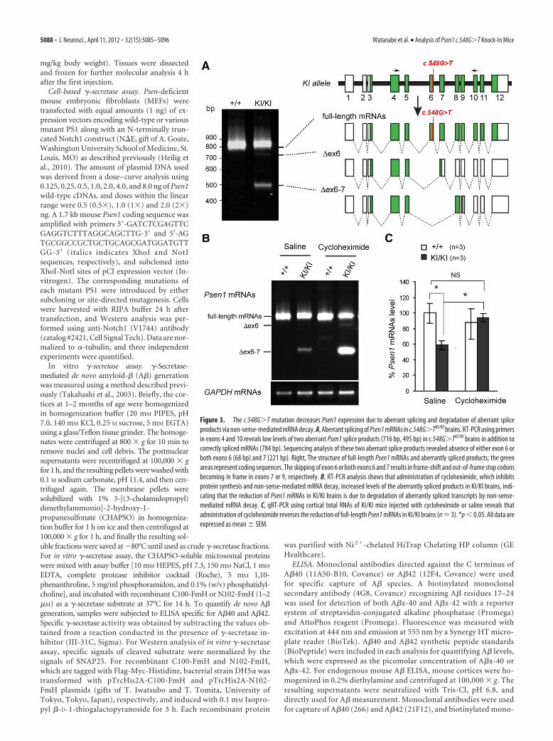
Figure 3. The c.548G�T mutation decreases Psen1 expression due to aberrant splicing and degradation of aberrant spliceproducts via non-sense-mediated mRNA decay. A, Aberrant splicing of Psen1 mRNAs in c.548G�TKI/KI brains. RT-PCR using primersin exons 4 and 10 reveals low levels of two aberrant Psen1 splice products (716 bp, 495 bp) in c.548G�TKI/KI brains in addition tocorrectly spliced mRNAs (784 bp). Sequencing analysis of these two aberrant splice products revealed absence of either exon 6 orboth exons 6 (68 bp) and 7 (221 bp). Right, The structure of full-length Psen1 mRNAs and aberrantly spliced products; the greenareas represent coding sequences. The skipping of exon 6 or both exons 6 and 7 results in frame-shift and out-of-frame stop codonsbecoming in frame in exons 7 or 9, respectively. B, RT-PCR analysis shows that administration of cycloheximide, which inhibitsprotein synthesis and non-sense-mediated mRNA decay, increased levels of the aberrantly spliced products in KI/KI brains, indi-cating that the reduction of Psen1 mRNAs in KI/KI brains is due to degradation of aberrantly spliced transcripts by non-sense-mediated mRNA decay. C, qRT-PCR using cortical total RNAs of KI/KI mice injected with cycloheximide or saline reveals thatadministration of cycloheximide reverses the reduction of full-length Psen1 mRNAs in KI/KI brains (n � 3). *p � 0.05. All data areexpressed as mean � SEM.
5088 • J. Neurosci., April 11, 2012 • 32(15):5085–5096 Watanabe et al. • Analysis of Psen1 c.548G�T Knock-In Mice
clonal secondary antibodies were used for detection of A�40 (2G3)and A�42 (266). Endogenous A� concentration was calculated todivide the total A� amount by the total protein amount in the cortexfraction.
Behavioral analysis. The Morris water maze is a circular pool 160 cm indiameter. Mice were housed in a standard 12 h light/dark cycle. Beforetesting, the experimenter handles each mouse for 5 min/d for 5 d. Duringthe hidden platform training, the platform (10 cm in diameter) was keptsubmerged under water and maintained in the same position. Eachmouse was given four trials daily with a maximum duration of 90 sseparated by a minimum of 15 min. If the mouse did not find the hiddenplatform, it was guided to the platform and allowed to remain on it for15 s. The swimming of the mice was monitored using an automatedtracking system (HVS Image). A group of mice aged 6 – 8 months (Psen1c.548G�TKI/KI;Psen2 �/� and Psen1 �/�;Psen2 �/�) or 18 –20 months(Psen1 c.548G�TKI/KI and Psen1 �/�) were trained in the hidden plat-form task for 12 d. Twenty-four hours after the last training (day 12), thehidden platform was removed and a 90 s probe trial was performed to testa spatial reference memory. The mice were released from all four quad-rants in a pseudorandom manner during the training and the probe trial.Finally, the visible cue task with four trials was performed to verify visualand swimming ability using the platform marked by a yellow object. Theexperimenters were blind to the genotypes of the mice.
Histological analysis. Brains were perfused with PBS, fixed in 4% para-formaldehyde for 3 h at 4°C, and processed for paraffin embedding.Paraffin-embedded sagittal sections were serially cut at 10 �m. Sectionsin every 40 slides were deparaffinized, dehydrated, and stained with 0.5%Cresyl Violet (Sigma), and analyzed for brain volumes by BioQuant im-age analysis software. For immunohistochemistry, paraffin-embeddedbrain sections were deparaffinized, alcohol dehydrated, and immuno-stained with monoclonal antibodies raised against phosphorylated(pSer396/pSer404) tau (PHF1, gift of P. Davies, Albert Einstein Collegeof Medicine, New York, NY). After specific signals were developed byVectastain Elite ABC kit and DAB peroxidase substrate, the sections werelightly counterstained with hematoxylin. The signals were analyzed usinga BX50 microscope system (Olympus).
Data analysis. Statistical analyses were per-formed using one-way ANOVA or two-tailedunpaired Student’s t test for all the compari-sons in the behavioral and biochemical results.A p value of �0.05 was considered significant.All the data were described as mean � SEM.
ResultsGeneration of Psen1 c.548G>T KI miceTo investigate the pathogenic mechanismunderlying the Psen1 c.548G�T muta-tion, we generated a KI mouse in whichthe c.548G�T mutation was introducedinto Psen1 exon 6 by homologous recom-bination. The targeting vector includesthe 5� homologous region (2.49 kb),floxed PGK-neo cassette and the 3� ho-mologous region (3.15 kb), in which thec.548G�T mutation was introduced intothe last nucleotide of exon 6 (Fig. 1A,B).The ES cells carrying the proper homolo-gous recombination events in the 5� and3� homologous regions were confirmedby Southern analysis using the 5� and 3�external probes, respectively (Fig. 1B;data not shown), and were injected intomouse blastocysts to generate chimericmice, which were then used to generateheterozygous mice carrying the targetedallele (targeted/�). Southern analysis oftail genomic DNAs of these mice con-
firmed that homologous recombination and germline transmis-sion occurred correctly, as demonstrated by the presence of twobands representing the wild-type allele (11.0 kb for both 5� and 3�external probes) and the targeted allele (5.5 kb for the 5� externalprobe, 8.5 kb for the 3� external probe) (Fig. 1C). The floxedPGK-neo selection cassette was removed by crossing the F1 micetransmitted with the targeted allele to Cre-expressing transgenicmice so as to avoid its possible transcriptional interference onPsen1 expression (Fig. 1B). Southern analysis of tail genomicDNAs from the resulting mice confirmed the deletion of thefloxed PGK-neo selection cassette using the 3� external probe(Fig. 1C). The targeted allele following the removal of the floxedPGK-neo cassette is termed the KI allele (Fig. 1B,C). AdditionalSouthern analysis using the neo probe further confirmed the lackof the floxed PGK-neo cassette as well as the absence of randominsertion of the targeting vector in the KI mice (data not shown).The correct introduction of the c.548G�T mutation was alsoconfirmed by sequencing (Fig. 1D).
The c.548G�TKI/KI homozygous mice were born in Men-delian ratio and were fertile, and adult KI/KI mice were grosslynormal in appearance compared with littermate controls. His-tological analysis of brain morphology shows that KI/KIbrains were indistinguishable from littermate controls (Fig.1 E). Using stereological methods, we measured cortical vol-ume in Nissl-stained series sagittal sections and found similarcortical volumes between KI/KI and littermate controls evenat 20 months of age (Fig. 1 E). Western analysis of GFAP,which is elevated accompanying astrogliosis (Beglopoulos etal., 2004; Saura et al., 2004), indicated no increases in thecerebral cortex of KI/KI mice at 12–20 months of age (Fig. 1 F).These results suggest that Psen1 c.548G�TKI/KI mice do notdevelop age-dependent neurodegeneration.
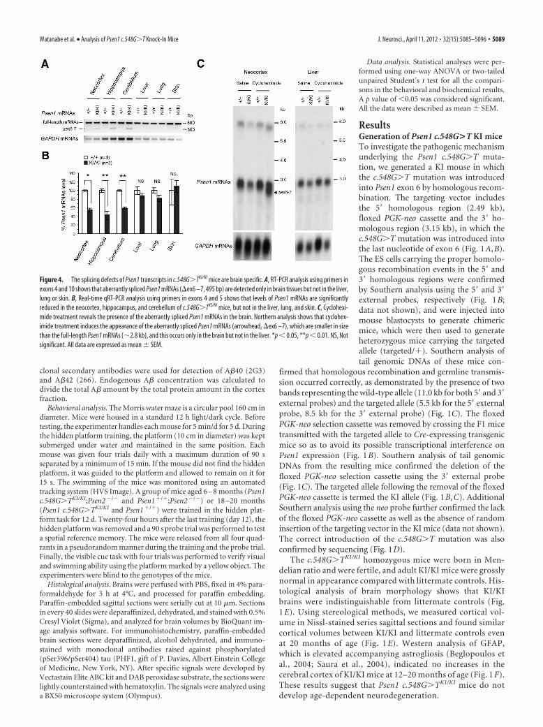
Figure 4. The splicing defects of Psen1 transcripts in c.548G�TKI/KI mice are brain specific. A, RT-PCR analysis using primers inexons 4 and 10 shows that aberrantly spliced Psen1 mRNAs (ex6 –7, 495 bp) are detected only in brain tissues but not in the liver,lung or skin. B, Real-time qRT-PCR analysis using primers in exons 4 and 5 shows that levels of Psen1 mRNAs are significantlyreduced in the neocortex, hippocampus, and cerebellum of c.548G�TKI/KI mice, but not in the liver, lung, and skin. C, Cyclohexi-mide treatment reveals the presence of the aberrantly spliced Psen1 mRNAs in the brain. Northern analysis shows that cyclohex-imide treatment induces the appearance of the aberrantly spliced Psen1 mRNAs (arrowhead, ex6 –7), which are smaller in sizethan the full-length Psen1 mRNAs (�2.8 kb), and this occurs only in the brain but not in the liver. *p � 0.05, **p � 0.01. NS, Notsignificant. All data are expressed as mean � SEM.
Watanabe et al. • Analysis of Psen1 c.548G�T Knock-In Mice J. Neurosci., April 11, 2012 • 32(15):5085–5096 • 5089
Levels of Psen1 mRNAs and proteinsare reduced in c.548G>T KI miceSince the c.548G�T mutation resides onthe splice junction of exon 6 and intron 6,which likely disrupts normal splicing of itstranscripts, we evaluated Psen1 mRNA ex-pression in c.548G�T KI mice. Northernanalysis revealed that levels of Psen1mRNAs are significantly reduced in thecerebral cortex of KI/KI mice throughouttheir life (from postnatal day 1 to 10months) (Fig. 2A). qRT-PCR also con-firmed the reduction of Psen1 mRNAs inc.548G�T KI mice in a KI allele dose-dependent manner, with a �40% reduc-tion in KI/KI mice (Fig. 2B). Westernanalysis disclosed �30% reduction in lev-els of PS1 NTFs and CTFs in total corticallysates (Fig. 2C). Given that levels of Psen1mRNAs correlated with levels of PS1 pro-teins in Psen1�/� and Psen1�/� mice(Shen et al., 1997), the discrepancy in thereduction of Psen1 mRNAs (�40%) andproteins (�30%) in c.548G�TKI/KI micemay reflect a mechanism suggested in aprevious report (Lee et al., 1997), in whichmutant PS1 tends to accumulate in trans-genic mouse brains. In contrast, the pro-tein levels of other �-secretase complexcomponents—NCT, Aph-1, and Pen-2—were similar among the three genotypicgroups (Fig. 2C).
The presence of aberrant Psen1 spliceproducts in c.548G>T KI mice and theirdegradation by non-sense-mediateddecayWe next investigated the mechanism un-derlying reduced levels of Psen1 mRNAsin the brain of KI/KI mice. The location ofthe G-to-T transversion responsible forthe c.548G�T mutation suggested thatsplicing of Psen1 transcripts might be dis-turbed in KI/KI mice (Fig. 1A), becauseproper splicing requires the exon–intronjunction to conform to the consensus se-quences (Cartegni et al., 2002). Indeed,RT-PCR using primers in exons 4 and 10followed by sequencing showed that inaddition to normal splice products con-taining all exons 4 –10, two aberrant spliceproducts lacking either exon 6 or exons 6and 7 were produced in c.548G�TKI/KI
brains (Fig. 3A). Since the skipping ofexon 6 or both exons 6 and 7 results inframe shift and the use of downstream premature terminationcodons that are normally out of frame, we then evaluated whetherthese abnormal transcripts are degraded by NMD mechanisms,which constitute a surveillance system to prevent production oftruncated proteins (Cartegni et al., 2002). We used cyclohexi-mide, a potent inhibitor of protein synthesis and non-sense-mediated decay, to determine whether blockade of NMD wouldrestore the levels of aberrant splice products that are normally
degraded by NMD. Indeed, cycloheximide treatment enhancedthe stability of the aberrantly spliced transcripts and drasticallyincreased levels of the splice product lacking exons 6 and 7 inKI/KI brains (Fig. 3B). Quantitative RT-PCR analysis alsoshowed that cycloheximide treatment fully rescued the reductionof Psen1 mRNA levels in KI/KI brains (Fig. 3C). These resultsreveal that in addition to the glycine-to-valine conversion at theamino acid residue 183 (p.183G�V), the c.548G�T mutation
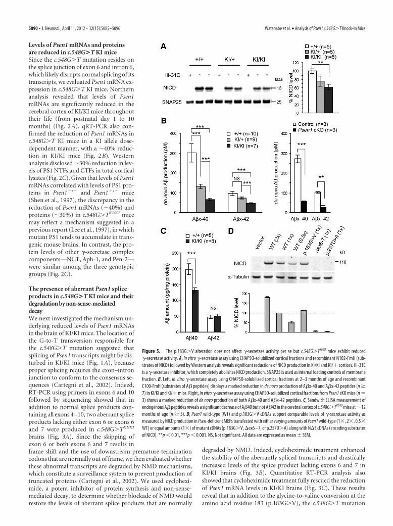
Figure 5. The p.183G�V alteration does not affect �-secretase activity per se but c.548G�TKI/KI mice exhibit reduced�-secretase activity. A, In vitro �-secretase assay using CHAPSO-solubilized cortical fractions and recombinant N102-FmH (sub-strates of NICD) followed by Western analysis reveals significant reductions of NICD production in KI/KI and KI/� cortices. III-31Cis a �-secretase inhibitor, which completely abolishes NICD production. SNAP25 is used as internal loading controls of membranefraction. B, Left, In vitro �-secretase assay using CHAPSO-solubilized cortical fractions at 2–3 months of age and recombinantC100-FmH (substrates of A� peptides) displays a marked reduction in de novo production of A�x-40 and A�x-42 peptides (n �7) in KI/KI and KI/� mice. Right, In vitro �-secretase assay using CHAPSO-solubilized cortical fractions from Psen1 cKO mice (n �3) shows a marked reduction of de novo production of both A�x-40 and A�x-42 peptides. C, Sandwich ELISA measurement ofendogenous A� peptides reveals a significant decrease of A�40 but not A�42 in the cerebral cortex of c.548G�TKI/KI mice at �12months of age (n � 5). D, Psen1 wild-type (WT) and p.183G�V cDNAs support comparable levels of �-secretase activity asmeasured by NICD production in Psen-deficient MEFs transfected with either varying amounts of Psen1 wild-type (1, 2, 0.5WT) or equal amounts (1) of mutant cDNAs (p.183G�V, ex6 –7, or p.257D�A) along with NE cDNAs (encoding substratesof NICD). **p � 0.01, ***p � 0.001. NS, Not significant. All data are expressed as mean � SEM.
5090 • J. Neurosci., April 11, 2012 • 32(15):5085–5096 Watanabe et al. • Analysis of Psen1 c.548G�T Knock-In Mice
disrupts proper splicing of a portion of the Psen1 transcripts,resulting in the generation of aberrantly spliced products harbor-ing premature termination codons, which were rapidly degradedby NMD mechanisms, leading to a consequent �40% decrease inthe expression of full-length Psen1 mRNAs in KI/KI brains.
The reduction of Psen1 mRNAs in c.548G>T KI mice isbrain specificPS1 is ubiquitously expressed in many tissues (Lee et al., 1996),and reduced Psen dosage has been associated with skin disordersin humans and mice (Xia et al., 2001; Tournoy et al., 2004; Kelle-her and Shen, 2010; Wang et al., 2010; Pink et al., 2011). But inFTD patients, the c.548G�T mutation gives rise to a brain-specific phenotype with no clinical remarks in other tissues (Der-maut et al., 2004). We therefore examined whether the c.548G�Tmutation affects Psen1 splicing and expression in non-neural tis-sues. Interestingly, RT-PCR showed no evidence of aberrantsplicing in the liver, lung, and skin of KI/KI mice, whereas theaberrantly spliced mRNAs were readily detected in the neocortex,hippocampus, and cerebellum (Fig. 4A). As a result, levels ofPsen1 mRNAs are unaltered in the liver, lung, and skin, as shownby quantitative RT-PCR (Fig. 4B). Additionally, we also exam-ined expression levels of Psen1 mRNAs in splenocytes, whichcontain broad spectrums of lymphocyte cell lineages, becauseNotch, a substrate of �-secretase, is involved in regulation of
lymphocyte cell development (Pui et al.,1999; Radtke et al., 1999). Northern anal-ysis showed that expression levels of Psen1mRNAs are not changed in splenocytes inKI/KI mice (data not shown). Moreover,cycloheximide treatment results in an ac-cumulation of aberrantly spliced Psen1transcripts in the brain but not in the liverof c.548G�TKI/KI mice (Fig. 4C). These re-sults demonstrate that the c.548G�T mu-tation causes aberrant splicing andsubsequent reduction of Psen1 expressiononly in the brain but not in non-neuraltissues.
Decreases of �-secretase activities in thebrain of c.548G>T KI miceTo determine the effect of the c.548G�Tmutation on �-secretase activity, we per-formed in vitro �-secretase assays usingdetergent-solubilized cerebral corticalfractions as the sensitive, direct method(Takahashi et al., 2003). Using CHAPSO-solubilized fractions from the cerebralcortex of c.548G�TKI/KI, c.548G�TKI/�,and wild-type littermate control miceat 2–3 months of age, we found that�-secretase activity is reduced in KI/�mice and further decreased in KI/KI mice,as measured by de novo production ofNotch intracellular domain (NICD) fromrecombinant N102-FmH (substrates ofNICD) (Fig. 5A). Production of NICDwas abolished by a specific �-secretase in-hibitor, III-31C (Fig. 5A).
APP is the other well established phys-iological substrate of �-secretase and�-secretase-mediated cleavages give rise
to A�40 and A�42 peptides (De Strooper et al., 1998). We thenperformed the same in vitro �-secretase assay using CHAPSO-solubilized cortical fractions of c.548G�TKI/KI, c.548G�TKI/�, andwild-type mice, and found that de novo generation of A�x-40 andA�x-42 is drastically reduced in KI mice depending on the KIallele dosage (Fig. 5B). As a control, we also used cortices of Psen1conditional KO (cKO) mice, and, similar to our prior finding (Yuet al., 2001), de novo generation of A�x-40 and A�x-42 is robustlyreduced in Psen1 cKO mice (Fig. 5B). In addition, we confirmeda decrease of de novo generation of total A� by Western blot (datanot shown). We further performed sandwich ELISA to measuresteady-state levels of endogenous A� peptides. Interestingly,measurement of the peptides showed decreased levels of A�40but unchanged levels of A�42 in the cerebral cortex ofc.548G�TKI/KI mice at 12 months of age (Fig. 5C). The differencebetween de novo A� production and steady-state A� levels islikely due to the effect of A� turnover in vivo (Wang et al., 2006).
Although the reduction of �-secretase activities in c.548G�TKI mice is consistent with decreased Psen1 mRNA levels in thesemice, the c.548G�T mutation also results in a substitution ofglycine with valine at amino acid residue 183. To determinewhether the p.183G�V conversion itself alters �-secretase activ-ity, we transfected vectors expressing either wild-type at varyingamounts (1, 2, 0.5) or various mutant Psen1 cDNAs (1) andtruncated Notch1 (NE) into Psen-deficient MEFs (Herreman et al.,
Figure 6. The Psen1 c.548G�T allele does not cause tau pathology in KI/KI mice. A, Phosphorylation status of tau protein inKI/KI cerebral cortices. Total cortical protein lysates were subject to Western analysis for phosphorylated tau (PHF1 and CP13) andtotal tau. The signals of phosphorylated tau were divided by those of total tau. B, The representative photos of immunohistochem-istry for phospho-tau (PHF1). There is no significant change of tau phosphorylation in the neocortex and hippocampal areas CA1and CA3. Scale bar, 10 �m. NS, Not significant. All data are expressed as mean � SEM.
Watanabe et al. • Analysis of Psen1 c.548G�T Knock-In Mice J. Neurosci., April 11, 2012 • 32(15):5085–5096 • 5091

2000). One day after the transfection, wecollected cell lysates and performed Westernanalysis. Wild-type and p.183G�V PS1 dis-played similar levels of �-secretase activity,as measured by production of NICD,whereas PS1 bearing a p.257D�A substitu-tion, which abolishes �-secretase activity,was unable to produce detectable NICD(Fig. 5D). The Psen1 mutant cDNA lackingexons 6 and 7 was similarly devoid of�-secretase activity (Fig. 5D). These resultssuggest that full-length PS1 harboringthe p.183G�V alteration has normal�-secretase activity; thus, this change is un-likely to be pathogenic. Collectively, theseresults suggest that the pathogenic effect ofthe c.548G�T mutation is a consequence ofthe brain-specific reduction in Psen1 mRNAexpression.
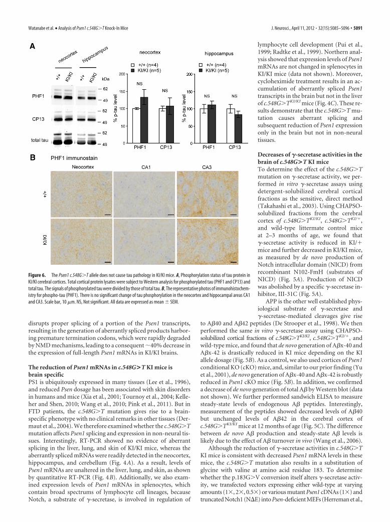
Absence of tau pathology in the brain ofc.548G>T KI miceIn addition to cerebral atrophy, FTD issometimes associated neuropathologicallywith tau pathology (Neary et al., 2005).Neuropathological analysis of the patientcarrying the PSEN1 c.548G�T mutation re-vealed Pick’s body and phosphorylated taustaining, in addition to severe frontotempo-ral atrophy and clear neuronal loss (Der-maut et al., 2004). Therefore, we nextexamined whether levels of tau and phos-phorylated status are elevated in the cerebralcortex of KI/KI mice. Western analysis usingrabbit polyclonal antibody for total taushowed similar levels of tau in the hip-pocampus and the neocortex betweenKI/KI and wild-type littermate mice (Fig.6A and data not shown). Using monoclonalantibodies PHF1 (specific for phosphory-lated Ser396/Ser404) and CP13 (specific forphosphorylated Ser202/Thr205), we foundno significant changes phospho-tau in theneocortex and hippocampus of KI/KI mice(Fig. 6A). Immunohistochemical analysisusing PHF1 antibodies further confirmedsimilar levels of phospho-tau in the neocor-tex and hippocampal areas CA1 and CA3regions of KI/KI mice compared with litter-mate controls at 20 months of age (Fig. 6B).These results show that the Psen1 c.548G�Tmutation does not cause tau pathology inmice, which may be due to the subtle effectof the mutation and the short lifespan ofmice.
Mild spatial memory impairment ofc.548G>T KI mice in the Psen2 �/�
backgroundWe previously reported that loss of prese-nilin function in the postnatal cerebralcortex impairs learning and memory in agene dosage-dependent manner (Yu et
Figure 7. The Psen1 c.548G�T KI/KI mice in the Psen2-null background exhibit mild spatial memory impairment. A–D, Psen1c.548G�TKI/KI and littermate wild-type control mice at 18 –20 months of age were subjected to training in the hidden platformwater maze for 12 d (four trials a day) followed by four trials of training in the visible platform version of the task. The escape latency(A) and swimming speed (B) of KI/KI mice (n � 5) are similar to that of littermate controls (n � 7) during the 12 d training phaseof the hidden platform water maze task. C, KI/KI and littermate control mice also displayed similar quadrant occupancy in thepost-training probe trial administered 24 h after the final training session. D, The escape latency in the visible platform version ofthe task is normal in KI/KI mice. E–H, Psen1 c.548G�T KI/KI;Psen2 �/� and littermate control mice at 6 – 8 months of age weresubjected to training in the hidden platform water maze for 12 d (four trials a day) followed by four trials of training in the visibleplatform version of the task. E, F, The escape latency (F � 0.032; df � 1, 17; p � 0.8601) and swimming speed (F � 3.549; df �1,17; p � 0.0768) of Psen1 c.548G�T KI/KI;Psen2 �/� mice (n � 11) are similar as littermate controls (n � 8) during the 12 dtraining phase in the hidden platform. G, However, during the probe trial 24 h after the final training session at day 12, Psen1c.548G�T KI/KI;Psen2 �/� mice show significant reduction in quadrant occunpancy ( p � 0.0267), suggesting mild impairment ofspatial memory. H, Psen1 c.548G�T KI/KI;Psen2 �/� mice performed normally in the visible cue task. AL, Adjacent left quadrant;T, target quadrant; AR, adjacent right quadrant; OP, opposite quadrant. All data are expressed as mean � SEM. *p � 0.05.
5092 • J. Neurosci., April 11, 2012 • 32(15):5085–5096 Watanabe et al. • Analysis of Psen1 c.548G�T Knock-In Mice
al., 2001; Saura et al., 2004). Whereas Psen2�/� mice exhibitednormal learning and memory, conditional inactivation of Psen1and Psen1/2 in postnatal cerebral cortex caused mild and severememory deficits, respectively. To determine whether thec.548G�T mutation impairs presenilin function in cognition, wetested hippocampus-dependent spatial learning and memory us-ing the Morris water maze task. Psen1 c.548G�TKI/KI and wild-type littermate control mice were given four trials a day for 12 d,and they performed similarly during the 12 d learning phase (Fig.7A,B, latency and swim speed) and in the probe trial 24 h after thefinal training session (Fig. 7C, quadrant occupancy). Psen1c.548G�TKI/KI mice also did not show any deficit in the visiblecue task (Fig. 7D). We next examined Psen1 c.548G�TKI/KI andlittermate control mice in the Psen2-null background, becausePsen1 cKO mice exhibit more severe learning and memory defi-cits in the absence of PS2 compensation (Saura et al., 2004).During the 12 d training phase, ANOVA did not show a signifi-cant genotype effect in escape latency (Fig. 7E) (F � 0.709; df �1,17; p � 0.4114), path length (F � 0.032; df � 1,17; p � 0.8601),and swim speed (Fig. 7F) (F � 3.549; df � 1,17; p � 0.0768), butPsen1 c.548G�TKI/KI;Psen2�/� mice exhibited significantlylower quadrant occupancy during the probe trial administered24 h after the last training session (Fig. 7G; p � 0.05), suggesting
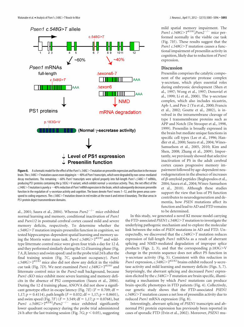
mild spatial memory impairment. ThePsen1 c.548G�TKI/KI;Psen2�/� mice per-formed normally in the visible cue task(Fig. 7H). These results suggest that thePsen1 c.548G�T mutation causes a func-tional impairment of presenilin activity incognition, likely due to reduction of Psen1expression.
DiscussionPresenilin comprises the catalytic compo-nent of the aspartate protease complex�-secretase, which plays essential rolesduring embryonic development (Shen etal., 1997; Wong et al., 1997; Donoviel etal., 1999; Li et al., 2000). The �-secretasecomplex, which also includes nicastrin,Aph-1, and Pen-2 (Yu et al., 2000; Franciset al., 2002; Goutte et al., 2002), is in-volved in the intramembrane cleavage oftype I transmembrane proteins such asAPP and Notch (De Strooper et al., 1998,1999). Presenilin is broadly expressed inthe brain but mediate unique functions inspecific cell types (Lee et al., 1996; Han-dler et al., 2000; Saura et al., 2004; Wines-Samuelson et al., 2005, 2010; Kim andShen, 2008; Zhang et al., 2009). Impor-tantly, we previously showed that selectiveinactivation of PS in the adult cerebralcortex causes progressive memory im-pairment followed by age-dependent neu-rodegeneration in the absence of increasesof �-amyloid peptides (Beglopoulos et al.,2004; Saura et al., 2004; Wines-Samuelsonet al., 2010). Although these studiessupport the view that loss of PS functioncontributes to neurodegeneration and de-mentia, how PSEN mutations affect itsfunction and lead to AD and FTD remainsto be determined.
In this study, we generated a novel KI mouse model carryingthe FTD-associated PSEN1 c.548G�T mutation to investigate theunderlying pathogenic mechanism and to explore the molecularlink between the roles of PSEN mutations in AD and FTD. Un-expectedly, we discovered that the c.548G�T mutation reducesexpression of full-length Psen1 mRNAs as a result of aberrantsplicing and NMD-mediated degradation of improper spliceproducts (Figs. 2, 3), and that the corresponding p.183G�Vchange in the protein sequence does not affect PS function in�-secretase activity (Fig. 5). Consistent with this reduction inPsen1 expression, c.548G�TKI/KI brains exhibit reduced �-secre-tase activity and mild learning and memory deficits (Figs. 5, 7).Surprisingly, the aberrant splicing and decreased Psen1 expres-sion elicited by the c.548G�T mutation are brain specific, illumi-nating a mechanism by which Psen1 mutations can producebrain-specific phenotypes in FTD patients (Fig. 4). Collectively,our genetic study shows that the FTD-associated PSEN1c.548G�T mutation causes a net loss of presenilin activity due toreduced Psen1 mRNA expression (Fig. 8).
Interestingly, aberrant splicing of PSEN1 transcripts and ab-normal PS1 protein expression has previously been reported incases of sporadic FTD (Evin et al., 2002). Moreover, PSEN1 mu-
Figure 8. A schematic model for the effect of the Psen1 c.548G�T mutation on presenilin expression and function in the mousebrain. The c.548G�T mutation causes exon skipping in �40% of Psen1 transcripts, which were degraded by non-sense-mediateddecay mechanisms. The remaining �60% Psen1 transcripts were spliced properly into full-length Psen1 c.548G�T mRNAs,producing PS1 proteins containing the p.183G�V variant, which exhibit normal �-secretase activity. Thus, the net effect of thec.548G�T mutation is purely a �40% reduction of Psen1 mRNA expression in the brain, which subsequently decreases presenilinfunction in the regulation of �-secretase activity and cognition. The boxes denote Psen1 exons 1–12, and the green areas corre-spond to coding sequences. The c.548G�T mutation shown in red resides at the exon 6 and intron 6 boundary. The blue areas inPS1 protein depict transmembrane domains.
Watanabe et al. • Analysis of Psen1 c.548G�T Knock-In Mice J. Neurosci., April 11, 2012 • 32(15):5085–5096 • 5093
tations identified in other FTD pedigrees also reside at exon–intron boundaries, possibly leading to splicing defects (Raux etal., 2000; Borroni et al., 2011). Thus, failure of proper PSEN1mRNA splicing and consequent reduction of PS1 expressioncould represent a common mechanism underlying both sporadicand familial FTD. Supporting the plausibility of such a mecha-nism in FTD pathogenesis, it has been estimated that �15% of allpoint mutations causing human genetic disease result in anmRNA splicing defect (Krawczak et al., 1992; Liu et al., 2001).
Another intriguing discovery of our study is the brain speci-ficity of the splicing defect caused by the c.548G�T mutation,sparing other tissues from detrimental effects associated with re-duced PSEN1 expression. Recent studies have shown an associa-tion between haploinsufficiency of �-secretase componentproteins and familial acne inversa (Kelleher and Shen, 2010;Wang et al., 2010; Li et al., 2011; Pink et al., 2011). However, ourmolecular analysis of c.548G�T KI mice showed that levels ofPsen1 mRNAs are only reduced in the brain (Fig. 4). Furtheranalysis revealed that aberrant splicing and subsequent degrada-tion of transcripts from the c.548G�T KI allele occur only in thebrain but not in other tissues. The detailed molecular mechanismby which aberrant splice products are produced in a brain-specific manner remains to be determined. One possible expla-nation could be the existence of brain-specific alternative splicingfactors (Dredge et al., 2001). A brain-restricted expression of aspecific splicing factors might render the splicing machinery lesstolerant of the G � T transversion at the splice donor site, leadingto skipping of this exon only in the brain.
While the effect of the c.548G�T mutation on the overall levelof Psen1 mRNAs is modest, its effect on �-secretase activity mea-sured by de novo production of NICD or A� peptides in KI brainsis surprisingly robust (Fig. 5). For example, the production ofA�40 peptides in c.548G�TKI/KI brains is reduced by �70%.These results indicate that �-secretase activity is very sensitive tochanges in presenilin dosage, suggesting that small reductions ofpresenilin expression may be sufficient to produce substantialdeficits in its essential functions (e.g., at the synapse). However,in vivo steady-state levels of A� peptides are less affected (Fig. 5),suggesting that a compensatory reduction in A� turnover mayaccompany the reduced A� production in KI mice. Consistentwith our earlier findings (Yu et al., 2001; Saura et al., 2004), wefound that Psen1 c.548G�TKI/KI mice in the Psen2-null back-ground exhibit mild deficits in spatial learning and memory (Fig.7). Not surprisingly, the spatial memory deficits exhibited byPsen1 c.548G�TKI/KI;Psen2�/� mice was subtler than those ofPsen1 cKO mice, in which PS1 is completely inactivated in excit-atory neurons of the adult cerebral cortex, whereas Psen condi-tional double-knock-out mice exhibit more dramatic memoryimpairment (Yu et al., 2001; Saura et al., 2004).
It has been long debated whether PSEN mutations cause atoxic gain of function or a loss of essential functions normallyperformed by presenilin (Shen and Kelleher, 2007). PSEN muta-tions associated with familial AD often lead to selective increasesof the longer and more amyloidogenic A�42 peptides, which hasbeen taken as evidence for the toxic gain-of-function mechanism.However, shortly after the identification of the PSEN mutationsin familial AD, such mutations were found to reduce its biologicalfunction and �-secretase activity in invertebrate models and cellculture systems (Levitan et al., 1996; Song et al., 1999; Seidner etal., 2006). More recently, a clinical PSEN1 mutation conferring acomplete loss of PS1 function and �-secretase activity has beendescribed (Heilig et al., 2010). These findings are consistent withour prior work showing that complete inactivation of presenilin
function in the adult mouse brain causes dementia and progres-sive neurodegeneration, two key features common to AD andFTD (Saura et al., 2004; Wines-Samuelson et al., 2010). Thus,impairment of essential PS functions in neuronal survival andmemory may be a common property of PSEN mutations in bothof these neurodegenerative dementias.
Our findings demonstrate that the FTD-associated c.548G�Tmutation decreases Psen1 mRNA expression, leading to a partial lossof presenilin and �-secretase function. However, the amino acidsubstitution derived from the c.548G�T mutation p.183G�V doesnot affect �-secretase activity. Thus, the net effect of the c.548G�Tmutation is limited to the reduction of Psen1 mRNA and proteinexpression, indicating that a pure loss of PSEN expression is patho-genic. This loss of PS expression without altered PS protein activityoffers a possible mechanism to account for the development of neu-rodegeneration and dementia in the absence of A� deposition inFTD. It is presently unclear whether other PSEN mutations that havebeen associated with FTD also affect PS expression, although thelocalization of some FTD-associated PSEN mutations at splice junc-tions (e.g., c.1129A�T, c.338T�C) raises the possibility that similarmechanisms may be at play (Ikeda et al., 1996; Raux et al., 2000;Binetti et al., 2003; Portet et al., 2003; Rippon et al., 2003; Halliday etal., 2005; Zekanowski et al., 2006; Bernardi et al., 2009; de Bot et al.,2009; Marcon et al., 2009; Gallo et al., 2010; Borroni et al., 2011).Future studies will be needed to determine the effects on presenilinfunction and �-secretase activity of other FTD-associated PSENmutations, which include mutations situated at the splice con-sensus sites [PSEN1: c.1129A�T (p.377R�W) and c.338T�C(p.113L�P)] and mutations whose localization suggests simple mis-sense substitution (e.g., PSEN1: p.139M�V, p.146M�L, p.226L�F,p.233M�L, p.260A�V, p.412V�I, p.424L�H, p.424L�R; PSEN2:p.62R�H, p.122T�R, p.231Y�C, p.239M�V). These studies mayhelp us understand why some of the same PSEN1 mutations areassociated with patients who were initially diagnosed clinically as ADor FTD (Ikeda et al., 1996; Portet et al., 2003; Rippon et al., 2003;Halliday et al., 2005; Zekanowski et al., 2006). Our findings implythat transgenic approaches in cell culture or mice may not provide acomplete picture of the impact of PSEN mutations on PS functionand �-secretase activity; rather, analysis of PSEN mutations in thecontext of the genomic locus will be important to evaluate potentialeffects on mRNA splicing and post-transcriptional regulation ofPSEN expression. Whether the pure loss of PSEN expression identi-fied in this study constitutes a common pathogenic mechanism bywhich PSEN mutations cause FTD remains to be determined.
ReferencesBeglopoulos V, Sun X, Saura CA, Lemere CA, Kim RD, Shen J (2004) Re-
duced beta-amyloid production and increased inflammatory responses inpresenilin conditional knock-out mice. J Biol Chem 279:46907– 46914.
Bernardi L, Tomaino C, Anfossi M, Gallo M, Geracitano S, Costanzo A, ColaoR, Puccio G, Frangipane F, Curcio SA, Mirabelli M, Smirne N, Iapaolo D,Maletta RG, Bruni AC (2009) Novel PSEN1 and PGRN mutations inearly-onset familial frontotemporal dementia. Neurobiol Aging30:1825–1833.
Binetti G, Signorini S, Squitti R, Alberici A, Benussi L, Cassetta E, Frisoni GB,Barbiero L, Feudatari E, Nicosia F, Testa C, Zanetti O, Gennarelli M,Perani D, Anchisi D, Ghidoni R, Rossini PM (2003) Atypical dementiaassociated with a novel presenilin-2 mutation. Ann Neurol 54:832– 836.
Borroni B, Pilotto A, Bonvicini C, Archetti S, Alberici A, Lupi A, GennarelliM, Padovani A (2011) Atypical presentation of a novel Presenilin 1R377W mutation: sporadic, late-onset Alzheimer disease with epilepsyand frontotemporal atrophy. Neurol Sci. Advance online publication.Retrieved August 6, 2011. doi:10.1007/s10072– 011-0714 –1.
Cartegni L, Chew SL, Krainer AR (2002) Listening to silence and under-standing nonsense: exonic mutations that affect splicing. Nat Rev Genet3:285–298.
5094 • J. Neurosci., April 11, 2012 • 32(15):5085–5096 Watanabe et al. • Analysis of Psen1 c.548G�T Knock-In Mice
Contet C, Dierich A, Kieffer BL (2007) Knock-in mice reveal nonsense-mediated mRNA decay in the brain. Genesis 45:38 – 43.
de Bot ST, Kremer HP, Dooijes D, Verbeek MM (2009) CSF studies facili-tate DNA diagnosis in familial Alzheimer’s disease due to a presenilin-1mutation. J Alzheimers Dis 17:53–57.
Dermaut B, Kumar-Singh S, Engelborghs S, Theuns J, Rademakers R, SaerensJ, Pickut BA, Peeters K, van den Broeck M, Vennekens K, Claes S, Cruts M,Cras P, Martin JJ, Van Broeckhoven C, De Deyn PP (2004) A novelpresenilin 1 mutation associated with Pick’s disease but not beta-amyloidplaques. Ann Neurol 55:617– 626.
De Strooper B, Saftig P, Craessaerts K, Vanderstichele H, Guhde G, AnnaertW, Von Figura K, Van Leuven F (1998) Deficiency of presenilin-1 in-hibits the normal cleavage of amyloid precursor protein. Nature391:387–390.
De Strooper B, Annaert W, Cupers P, Saftig P, Craessaerts K, Mumm JS,Schroeter EH, Schrijvers V, Wolfe MS, Ray WJ, Goate A, Kopan R (1999)A presenilin-1-dependent gamma-secretase-like protease mediates re-lease of Notch intracellular domain. Nature 398:518 –522.
Donoviel DB, Hadjantonakis AK, Ikeda M, Zheng H, Hyslop PS, Bernstein A(1999) Mice lacking both presenilin genes exhibit early embryonic pat-terning defects. Genes Dev 13:2801–2810.
Dredge BK, Polydorides AD, Darnell RB (2001) The splice of life: alternativesplicing and neurological disease. Nat Rev Neurosci 2:43–50.
Evin G, Smith MJ, Tziotis A, McLean C, Canterford L, Sharples RA, Cappai R,Weidemann A, Beyreuther K, Cotton RG, Masters CL, Culvenor JG(2002) Alternative transcripts of presenilin-1 associated with frontotem-poral dementia. Neuroreport 13:917–921.
Francis R, McGrath G, Zhang J, Ruddy DA, Sym M, Apfeld J, Nicoll M,Maxwell M, Hai B, Ellis MC, Parks AL, Xu W, Li J, Gurney M, Myers RL,Himes CS, Hiebsch R, Ruble C, Nye JS, Curtis D (2002) aph-1 and pen-2are required for Notch pathway signaling, gamma-secretase cleavage ofbetaAPP, and presenilin protein accumulation. Dev Cell 3:85–97.
Galimberti D, Scarpini E (2010) Genetics and biology of Alzheimer’s diseaseand frontotemporal lobar degeneration. Int J Clin Exp Med 3:129 –143.
Gallo M, Tomaino C, Puccio G, Frangipane F, Curcio SA, Bernardi L, Ger-acitano S, Anfossi M, Mirabelli M, Colao R, Vasso F, Smirne N, MalettaRG, Bruni AC (2010) Novel MAPT Val75Ala mutation and PSEN2Arg62Hys in two siblings with frontotemporal dementia. Neurol Sci31:65–70.
Goutte C, Tsunozaki M, Hale VA, Priess JR (2002) APH-1 is a multipassmembrane protein essential for the Notch signaling pathway in Caeno-rhabditis elegans embryos. Proc Natl Acad Sci U S A 99:775–779.
Halliday GM, Song YJ, Lepar G, Brooks WS, Kwok JB, Kersaitis C, Gregory G,Shepherd CE, Rahimi F, Schofield PR, Kril JJ (2005) Pick bodies in afamily with presenilin-1 Alzheimer’s disease. Ann Neurol 57:139 –143.
Handler M, Yang X, Shen J (2000) Presenilin-1 regulates neuronal differen-tiation during neurogenesis. Development 127:2593–2606.
Heilig EA, Xia W, Shen J, Kelleher RJ 3rd (2010) A presenilin-1 mutationidentified in familial Alzheimer disease with cotton wool plaques causes anearly complete loss of gamma-secretase activity. J Biol Chem285:22350 –22359.
Herreman A, Serneels L, Annaert W, Collen D, Schoonjans L, De Strooper B(2000) Total inactivation of gamma-secretase activity in presenilin-deficient embryonic stem cells. Nat Cell Biol 2:461– 462.
Ignotz GG, Suarez SS (2005) Calcium/calmodulin and calmodulin kinase IIstimulate hyperactivation in demembranated bovine sperm. Biol Reprod73:519 –526.
Ikeda M, Sharma V, Sumi SM, Rogaeva EA, Poorkaj P, Sherrington R, Nee L,Tsuda T, Oda N, Watanabe M, Aoki M, Shoji M, Abe K, Itoyama Y, HiraiS, Schellenberg GD, Bird TD, St George-Hyslop PH (1996) The clinicalphenotype of two missense mutations in the presenilin I gene in Japanesepatients. Ann Neurol 40:912–917.
Kelleher RJ 3rd, Shen J (2010) Genetics. Gamma-secretase and human dis-ease. Science 330:1055–1056.
Kim WY, Shen J (2008) Presenilins are required for maintenance of neuralstem cells in the developing brain. Mol Neurodegener 3:2.
Krawczak M, Reiss J, Cooper DN (1992) The mutational spectrum of singlebase-pair substitutions in mRNA splice junctions of human genes: causesand consequences. Hum Genet 90:41–54.
Lee MK, Slunt HH, Martin LJ, Thinakaran G, Kim G, Gandy SE, Seeger M,Koo E, Price DL, Sisodia SS (1996) Expression of presenilin 1 and 2 (PS1and PS2) in human and murine tissues. J Neurosci 16:7513–7525.
Lee MK, Borchelt DR, Kim G, Thinakaran G, Slunt HH, Ratovitski T, MartinLJ, Kittur A, Gandy S, Levey AI, Jenkins N, Copeland N, Price DL, SisodiaSS (1997) Hyperaccumulation of FAD-linked presenilin 1 variants invivo. Nat Med 3:756 –760.
Levitan D, Doyle TG, Brousseau D, Lee MK, Thinakaran G, Slunt HH, SisodiaSS, Greenwald I (1996) Assessment of normal and mutant human pre-senilin function in Caenorhabditis elegans. Proc Natl Acad Sci U S A93:14940 –14944.
Li CR, Jiang MJ, Shen DB, Xu HX, Wang HS, Yao X, Zhang Y, Zhou WQ,Wang B (2011) Two novel mutations of the nicastrin gene in Chinesepatients with acne inversa. Br J Dermatol 165:415– 418.
Li YM, Xu M, Lai MT, Huang Q, Castro JL, DiMuzio-Mower J, Harrison T,Lellis C, Nadin A, Neduvelil JG, Register RB, Sardana MK, Shearman MS,Smith AL, Shi XP, Yin KC, Shafer JA, Gardell SJ (2000) Photoactivatedgamma-secretase inhibitors directed to the active site covalently labelpresenilin 1. Nature 405:689 – 694.
Liu HX, Cartegni L, Zhang MQ, Krainer AR (2001) A mechanism for exonskipping caused by nonsense or missense mutations in BRCA1 and othergenes. Nat Genet 27:55–58.
Marcon G, Di Fede G, Giaccone G, Rossi G, Giovagnoli AR, Maccagnano E,Tagliavini F (2009) A novel Italian presenilin 2 gene mutation withprevalent behavioral phenotype. J Alzheimers Dis 16:509 –511.
Mendez MF, McMurtray A (2006) Frontotemporal dementia-like pheno-types associated with presenilin-1 mutations. Am J Alzheimers Dis OtherDemen 21:281–286.
Neary D, Snowden J, Mann D (2005) Frontotemporal dementia. LancetNeurol 4:771–780.
Pink AE, Simpson MA, Brice GW, Smith CH, Desai N, Mortimer PS, BarkerJN, Trembath RC (2011) PSENEN and NCSTN mutations in familialhidradenitis suppurativa (Acne Inversa). J Invest Dermatol131:1568 –1570.
Portet F, Dauvilliers Y, Campion D, Raux G, Hauw JJ, Lyon-Caen O, CamuW, Touchon J (2003) Very early onset AD with a de novo mutation inthe presenilin 1 gene (Met 233 Leu). Neurology 61:1136 –1137.
Pui JC, Allman D, Xu L, DeRocco S, Karnell FG, Bakkour S, Lee JY, KadeschT, Hardy RR, Aster JC, Pear WS (1999) Notch1 expression in early lym-phopoiesis influences B versus T lineage determination. Immunity11:299 –308.
Radtke F, Wilson A, Stark G, Bauer M, van Meerwijk J, MacDonald HR, AguetM (1999) Deficient T cell fate specification in mice with an inducedinactivation of Notch1. Immunity 10:547–558.
Raux G, Gantier R, Thomas-Anterion C, Boulliat J, Verpillat P, Hannequin D,Brice A, Frebourg T, Campion D (2000) Dementia with prominentfrontotemporal features associated with L113P presenilin 1 mutation.Neurology 55:1577–1578.
Rippon GA, Crook R, Baker M, Halvorsen E, Chin S, Hutton M, Houlden H,Hardy J, Lynch T (2003) Presenilin 1 mutation in an african americanfamily presenting with atypical Alzheimer dementia. Arch Neurol60:884 – 888.
Saura CA, Choi SY, Beglopoulos V, Malkani S, Zhang D, ShankaranarayanaRao BS, Chattarji S, Kelleher RJ 3rd, Kandel ER, Duff K, Kirkwood A,Shen J (2004) Loss of presenilin function causes impairments of mem-ory and synaptic plasticity followed by age-dependent neurodegenera-tion. Neuron 42:23–36.
Seidner GA, Ye Y, Faraday MM, Alvord WG, Fortini ME (2006) Modelingclinically heterogeneous presenilin mutations with transgenic Drosoph-ila. Curr Biol 16:1026 –1033.
Shen J, Bronson RT, Chen DF, Xia W, Selkoe DJ, Tonegawa S (1997) Skel-etal and CNS defects in Presenilin-1-deficient mice. Cell 89:629 – 639.
Shen J, Kelleher RJ 3rd (2007) The presenilin hypothesis of Alzheimer’sdisease: evidence for a loss-of-function pathogenic mechanism. Proc NatlAcad Sci U S A 104:403– 409.
Sjogren M, Andersen C (2006) Frontotemporal dementia–a brief review.Mech Ageing Dev 127:180 –187.
Song W, Nadeau P, Yuan M, Yang X, Shen J, Yankner BA (1999) Proteolyticrelease and nuclear translocation of Notch-1 are induced by presenilin-1and impaired by pathogenic presenilin-1 mutations. Proc Natl Acad SciU S A 96:6959 – 6963.
Steiner H, Duff K, Capell A, Romig H, Grim MG, Lincoln S, Hardy J, Yu X,Picciano M, Fechteler K, Citron M, Kopan R, Pesold B, Keck S, Baader M,Tomita T, Iwatsubo T, Baumeister R, Haass C (1999) A loss of function
Watanabe et al. • Analysis of Psen1 c.548G�T Knock-In Mice J. Neurosci., April 11, 2012 • 32(15):5085–5096 • 5095
mutation of presenilin-2 interferes with amyloid beta-peptide productionand notch signaling. J Biol Chem 274:28669 –28673.
Takahashi Y, Hayashi I, Tominari Y, Rikimaru K, Morohashi Y, Kan T, Nat-sugari H, Fukuyama T, Tomita T, Iwatsubo T (2003) Sulindac sulfide isa noncompetitive gamma-secretase inhibitor that preferentially reducesAbeta 42 generation. J Biol Chem 278:18664 –18670.
Tournoy J, Bossuyt X, Snellinx A, Regent M, Garmyn M, Serneels L, Saftig P,Craessaerts K, De Strooper B, Hartmann D (2004) Partial loss of prese-nilins causes seborrheic keratosis and autoimmune disease in mice. HumMol Genet 13:1321–1331.
Wang B, Yang W, Wen W, Sun J, Su B, Liu B, Ma D, Lv D, Wen Y, Qu T, ChenM, Sun M, Shen Y, Zhang X (2010) Gamma-secretase gene mutations infamilial acne inversa. Science 330:1065.
Wang R, Wang B, He W, Zheng H (2006) Wild-type presenilin 1 protectsagainst Alzheimer disease mutation-induced amyloid pathology. J BiolChem 281:15330 –15336.
Wines-Samuelson M, Handler M, Shen J (2005) Role of presenilin-1 in cor-tical lamination and survival of Cajal-Retzius neurons. Dev Biol277:332–346.
Wines-Samuelson M, Schulte EC, Smith MJ, Aoki C, Liu X, Kelleher RJ 3rd,Shen J (2010) Characterization of age-dependent and progressive corti-cal neuronal degeneration in presenilin conditional mutant mice. PLoSOne 5:e10195.
Wong PC, Zheng H, Chen H, Becher MW, Sirinathsinghji DJ, TrumbauerME, Chen HY, Price DL, Van der Ploeg LH, Sisodia SS (1997) Presenilin
1 is required for Notch1 and DII1 expression in the paraxial mesoderm.Nature 387:288 –292.
Xia X, Qian S, Soriano S, Wu Y, Fletcher AM, Wang XJ, Koo EH, Wu X,Zheng H (2001) Loss of presenilin 1 is associated with enhancedbeta-catenin signaling and skin tumorigenesis. Proc Natl Acad SciU S A 98:10863–10868.
Yu G, Nishimura M, Arawaka S, Levitan D, Zhang L, Tandon A, Song YQ,Rogaeva E, Chen F, Kawarai T, Supala A, Levesque L, Yu H, Yang DS,Holmes E, Milman P, Liang Y, Zhang DM, Xu DH, Sato C, Rogaev E,Smith M, Janus C, Zhang Y, Aebersold R, Farrer LS, Sorbi S, Bruni A,Fraser P, St George-Hyslop P (2000) Nicastrin modulates presenilin-mediated notch/glp-1 signal transduction and betaAPP processing. Na-ture 407:48 –54.
Yu H, Saura CA, Choi SY, Sun LD, Yang X, Handler M, Kawarabayashi T,Younkin L, Fedeles B, Wilson MA, Younkin S, Kandel ER, Kirkwood A,Shen J (2001) APP processing and synaptic plasticity in presenilin-1conditional knockout mice. Neuron 31:713–726.
Zekanowski C, Golan MP, Krzysko KA, Lipczynska-Lojkowska W, Filipek S,Kowalska A, Rossa G, Peplonska B, Styczynska M, Maruszak A, Religa D,Wender M, Kulczycki J, Barcikowska M, Kuznicki J (2006) Two novelpresenilin 1 gene mutations connected with frontotemporal dementia-like clinical phenotype: genetic and bioinformatic assessment. Exp Neurol200:82– 88.
Zhang C, Wu B, Beglopoulos V, Wines-Samuelson M, Zhang D, Dragatsis I,Sudhof TC, Shen J (2009) Presenilins are essential for regulating neu-rotransmitter release. Nature 460:632– 636.
5096 • J. Neurosci., April 11, 2012 • 32(15):5085–5096 Watanabe et al. • Analysis of Psen1 c.548G�T Knock-In Mice