Neurobiology of Development, - Columbia Universityphys.columbia.edu/~rtw/Lecture I.pdf ·...
25
Click to edit Master subtitle style 10/15/09 Neurobiology of Development, Diseases and Behavior. Lecture I by Neela Zareen [email protected] Laboratory of Lloyd Greene, PhD
Transcript of Neurobiology of Development, - Columbia Universityphys.columbia.edu/~rtw/Lecture I.pdf ·...

Click to edit Master subtitle style
10/15/09
Neurobiology of Development, Diseases and Behavior.
Lecture Iby Neela [email protected] of Lloyd Greene, PhD

10/15/09
Hallmarks of early brain science
v Greek physician Galen believed that the nervous system were glands that transmit fluids from the brain and spinal cord to the periphery.
v The Italian physiologist Luigi Galvani (late 1700s) discovered that muscles and nervous tissue produce electricity. A number of clever Germans (DuBois-Reymond, Mueller, von Helmholtz) follow up and demonstrate electrical activity of neurons effect activity of adjecent neurons.
v Pharmacologists (esp Bernard, Ehrlich, Langley) show that drugs recognize specific target receptors on the cell membrane chemical basis of neural communication.

10/15/09
Franz Joseph GallØ German physician and neuroanatomist
Ø Proposed 3 new theories:
Ø All behavior emanate from the brain
Ø Specific functions are controlled by specific cortical areas.
Ø Each of these areas grew as its function was exercised, therefore, one can interpret areas that are most developed by reading bumps on the skull (phrenology)

10/15/09
Pierre Flourens
v In 1820 applied Gall’s theory to experimentation.
v By lesioning areas of rabbit and pigeon brains he could find no specific deficit in individual behaviors .
v This led to the concept of the aggregate-field view of the brain all brain regions perform all functions.
“All perceptions, all volitions occupy the same seat in these (cerebral) organs; the faculty of perceiving, of conceiving, of willing merely constitutes therefore a faculty which is essentially one.”

10/15/09
Pierre Paul Broca
Ø Suggested that studies to identify localization of behaviors in the brain should be based on clinical lesions and not on bumpy heads.
Ø 1861 he describes a patient named Leborgne with aphasia (inability to speak). Postmortem analysis of the man yielded a lesion in the posterior prefrontal cortex (now known as Broca’s area).
Ø Follow up with eight other patients yielded similar results.

10/15/09
Karl WernickeØ Described another type of aphasia: the
patient could speak but not understand language.
Ø Identified a lesion in the posterior temporal lobe (now known as Wernicke’s Area).
Ø Later Wernicke’s area found to be in a larger association cortex that integrates many sensory modalities in to perception.
Broca and Wernicke’s work (along with others) lent evidence for a new model of brain function, the distributed processing specific functions are carried out by specific brain areas.
10/15/09
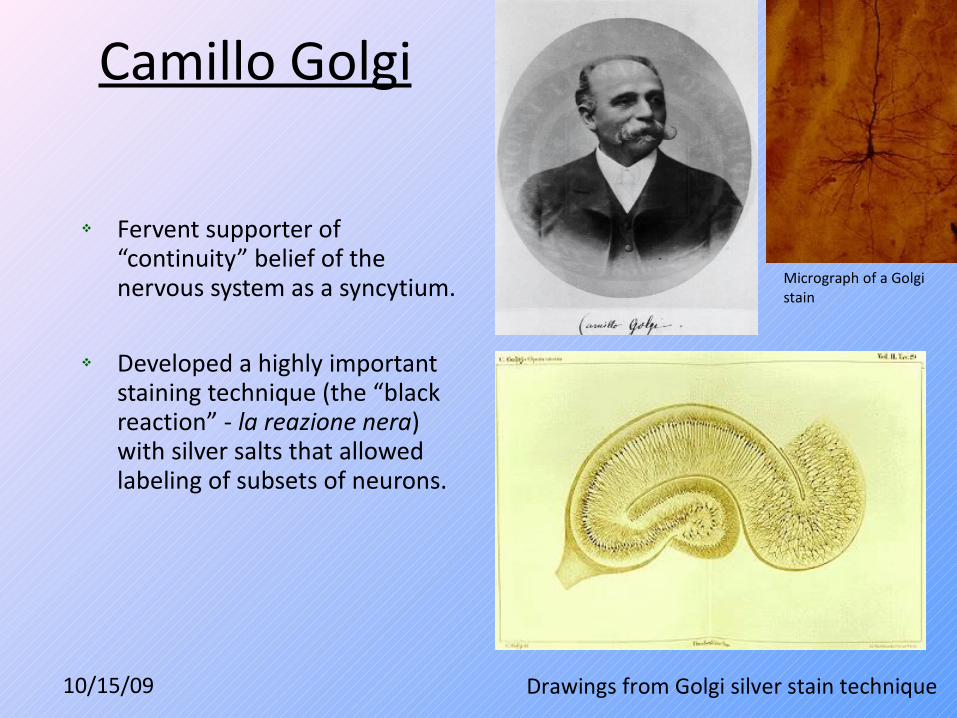
Camillo Golgi
v Fervent supporter of “continuity” belief of the nervous system as a syncytium.
v Developed a highly important staining technique (the “black reaction” - la reazione nera) with silver salts that allowed labeling of subsets of neurons.
Drawings from Golgi silver stain technique
Micrograph of a Golgi stain
10/15/09
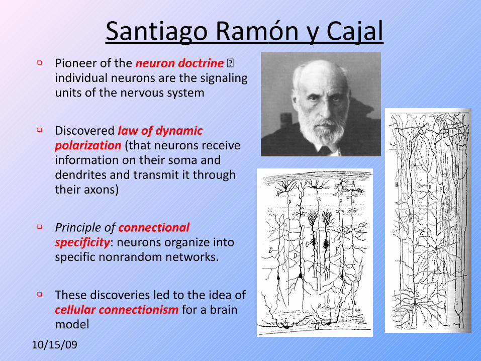
Santiago Ramón y Cajalq Pioneer of the neuron doctrine
individual neurons are the signaling units of the nervous system
q Discovered law of dynamic polarization (that neurons receive information on their soma and dendrites and transmit it through their axons)
q Principle of connectional specificity: neurons organize into specific nonrandom networks.
q These discoveries led to the idea of cellular connectionism for a brain model

10/15/09
Organization of the Human Brain
10/15/09
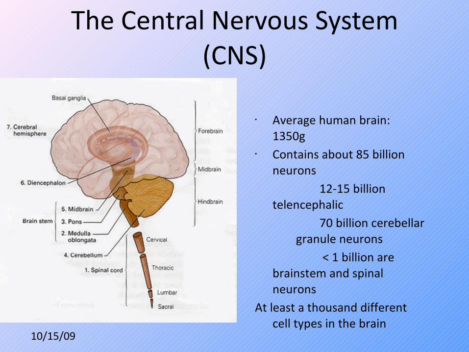
The Central Nervous System (CNS)
• Average human brain: 1350g
• Contains about 85 billion neurons
12-15 billion telencephalic
70 billion cerebellar granule neurons
< 1 billion are brainstem and spinal neurons
At least a thousand different cell types in the brain

10/15/09
1. Spinal Cord
• Divided into four segments: cervical, thoracic, lumbar, sacral.
• Acts as an assimilation, information processing and relay center between the periphery (skin and skeletal muscles) and the rostral CNS areas (brain).
2. Medula Oblongata
• Primary center for autonomic regulation: digestion, breathing, and heart rate.
• Lies rostral of spinal cord.
10/15/09
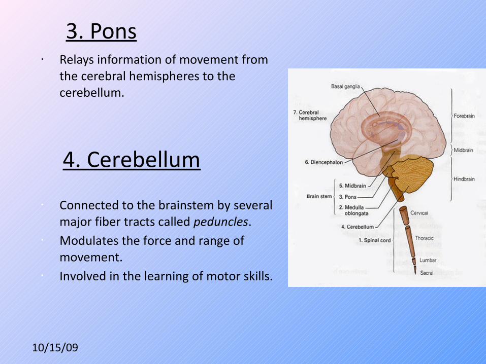
3. Pons• Relays information of movement from
the cerebral hemispheres to the cerebellum.
4. Cerebellum
• Connected to the brainstem by several major fiber tracts called peduncles.
• Modulates the force and range of movement.
• Involved in the learning of motor skills.

10/15/09
5. Midbrain• Controls diverse sensory and motor
functions (ie eye movement, coordination of visual/auditory reflexes).
• Thalamus: Functions as the relay center for most sensory modalities arriving at the visual cortex from the rest of the nervous system.
• Hypothalamus: Regulates the autonomic nervous system via hormone production and release. Affects and regulates blood pressure, heart rate, hunger, thirst, sexual arousal, and the sleep/wake cycle.
Segregated into two areas:
6. Diencephalon
10/15/09
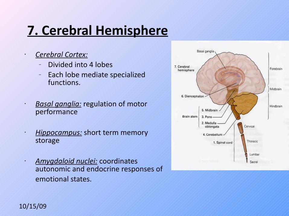
7. Cerebral Hemisphere• Cerebral Cortex:
– Divided into 4 lobes– Each lobe mediate specialized
functions.
• Basal ganglia: regulation of motor performance
• Hippocampus: short term memory storage
• Amygdaloid nuclei: coordinates autonomic and endocrine responses of emotional states.

10/15/09
Lobes of the Cerebral Hemispheresv Frontal lobe: personality, emotional
coloring, foresight, complex decision, motor activity. Represents highest level of neural evolution.
v Parietal lobe: somatic sensation,
forming a body image, and relating one’s body image with extrapersonal space
v Temporal lobe: hearing, learning, memory and emotion.
v Occipital lobe: processing of visual input and coordination of eye movement. Break images into components such as form, color and movement and transmit the images to the temporal lobe (form and color) and the parietal lobe (analysis of movement).

10/15/09
Neurons are Highly Polymorphic
• Neurons possess a dizzying arrays of shapes, sizes and characteristics that have been specified by evolution to fulfill their specialized role in the networks they are a part of.
• Different classes of neurons use certain neurotransmitters to propagate their signal downstream in the network. This is a property of their fate.
10/15/09
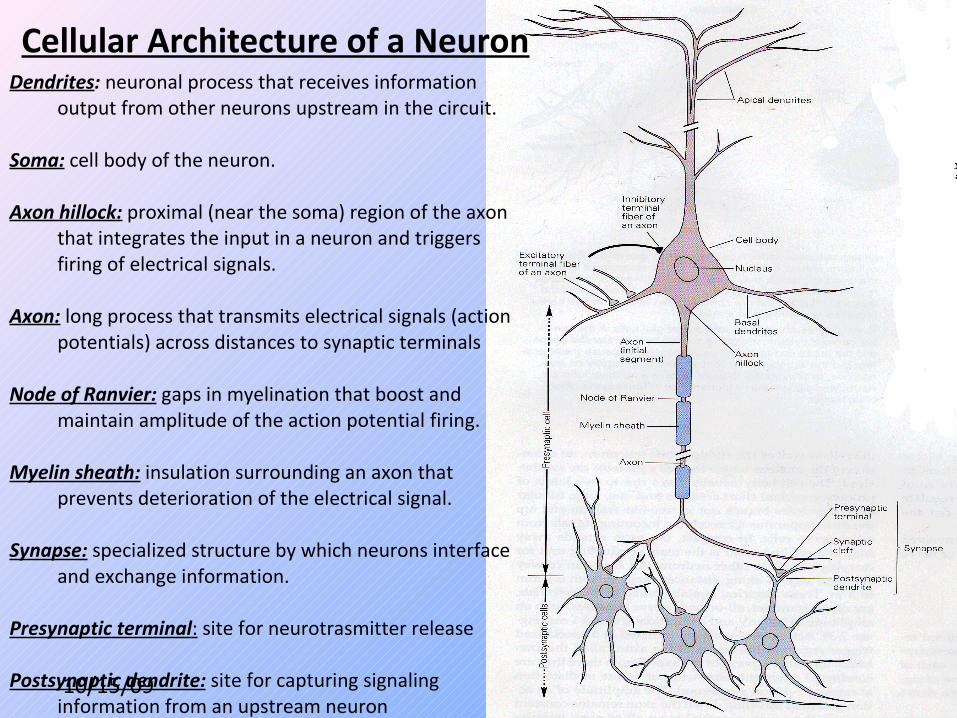
Cellular Architecture of a NeuronDendrites: neuronal process that receives information
output from other neurons upstream in the circuit.
Soma: cell body of the neuron.
Axon hillock: proximal (near the soma) region of the axonthat integrates the input in a neuron and triggersfiring of electrical signals.
Axon: long process that transmits electrical signals (actionpotentials) across distances to synaptic terminals
Node of Ranvier: gaps in myelination that boost andmaintain amplitude of the action potential firing.
Myelin sheath: insulation surrounding an axon thatprevents deterioration of the electrical signal.
Synapse: specialized structure by which neurons interfaceand exchange information.
Presynaptic terminal: site for neurotrasmitter release Postsynaptic dendrite: site for capturing signaling
information from an upstream neuron

10/15/09
Division of the Nervous System
1. CNS (Central Nervous System): Brain and spinal cord. Behavior.
1. PNS (Peripheral Nervous System): connects the CNS to the limbs and organs.
a. Somatic – voluntary movement.
b. Autonomic – involuntary actions.
c. Sensory – perception (visual, hearing, taste, touch, smell, etc).

10/15/09
Autonomic Effects
• stimulates heartbeat• raises blood pressure • dilates the pupils • dilates the trachea and the bronchi• stimulates the conversion of liver
glycogen into glucose • shunts blood away from the skin
and viscera to the skeletal muscles, brain, and heart
• inhibits peristalsis in the gastrointestinal (GI) tract
• inhibits contraction of the bladder and rectum
• slowing down of the heartbeat • lowering of blood pressure • constriction of the pupils • increased blood flow to the skin and
viscera • peristalsis of the GI tract
Sympathetic Nervous System Parasympathetic activation

10/15/09
Peripheral/Autonomic Nervous System (PNS)

10/15/09
Contrast between PNS and CNS
• Axon myelinated by Schwann cells
• Regenerative capacity
• Either cholinergic (parasympathetic) or adrenergic/noradrenergic (sympathetic)
• Axon myelinated by oligodendrocytes
• Regeneration incompetent
• Utilize wide array of neurotransmitters (Opioids, serotonin, noradrenaline, substance P, GABA, etc.)
• More diverse array of cell types.
PNS CNS
10/15/09
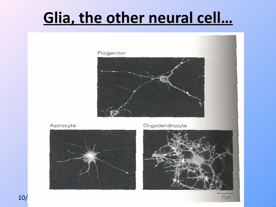
Glia, the other neural cell…

10/15/09
The Five Kinds of Glia
• Schwann Cell: myelination in the PNS• Oligodendrocyte: myelination in the CNS• Microglia:
– phagocytotic cells– Activated and recruited during injury, infection, and siezure.
• Astrocyte: – establish blood-brain barrier– transport nutrients to neurons– uptake neurotransmitters to enhance transmission of signals– maintain [K+] around neurons to upkeep electrochemical gradient.
• Radial Glia: – function as embryonic neural stem cells. – Act as a scaffold to guide differentiating neurons up to the surface.

10/15/09
Glia at Work

10/15/09
Astrocytes Form a Blood-Brain Barrier