Neural Systems Underlying Spatial Language in American ...lcn.salk.edu/publications/2002/Emmorey -...
13
Neural Systems Underlying Spatial Language in American Sign Language Karen Emmorey,* ,1 Hanna Damasio,* , † Stephen McCullough,* Thomas Grabowski,† Laura L. B. Ponto,† Richard D. Hichwa,† and Ursula Bellugi* *Salk Institute for Biological Studies, La Jolla, California 92037; and †University of Iowa, Iowa City, Iowa 52242 Received August 28, 2001 A [ 15 O]water PET experiment was conducted to in- vestigate the neural regionsengagedin processing constructions unique to signed languages: classifier predicates in which the position of the hands in sign- ing spaceschematically representsspatial relations among objects. Ten deaf nativesigners viewed line drawings depicting a spatial relation between twoob- jects (e.g., a cup on a table) and were asked eitherto produce a classifier construction or an American Sign Language (ASL) preposition that described thespatial relation orto name thefigure object (colored red). Compared to naming objects, describing spatial rela- tionships with classifier constructionsengaged thesu- pramarginal gyrus (SMG) within bothhemispheres. Compared to naming objects, naming spatial relations with ASL prepositionsengaged only the right SMG. Previous research indicates thatretrieval of English prepositionsengages both right and left SMG, but more inferiorly than for ASL classifier constructions. Compared to ASL prepositions, naming spatial rela- tions with classifier constructionsengaged left infe- riortemporal (IT) cortex, a region activated when namingconcrete objects in either ASL or English. Left IT may beengaged because the handshapes in classi- fier constructionsencode information about object type (e.g., flat surface). Overall, the resultssuggest more right hemisphere involvement when expressing spatial relations in ASL, perhaps because signing space is used to encode thespatial relationship be- tween objects. © 2002 Elsevier Science (USA) INTRODUCTION For more than a century we have known thatthe left hemisphere of the human brain is critical for producing and comprehending spoken language. Damage to peri- sylvian areas within the left hemisphere produces var- ious types of aphasia, whereas damage to homologous areas within the right hemisphere does not generally produce aphasic symptoms, such as effortful speech, phonological or morphological errors, or difficulty un- derstanding words or sentences. Similarly, research over the past 2 decades has indicated thatthe left cerebral hemisphere is also critical to processing signed languages. Damage to perisylvian areas within the left, but notthe right hemisphere lead to sign language aphasias, and the general dichotomy be- tween anterior–posterior lesions and nonfluent–fluent aphasias holds for signed language as well (forreviews see Corina, 1998, and Hickok and Bellugi, 2000). Although sign aphasia does not result from right- hemisphere damage, evidence indicates that some signers with right hemisphere damageexhibit a spe- cific impairment in the topographic use of signing space (Poizner et al., 1987; Emmorey et al., 1995; Em- morey, 1996). In American Sign Language (ASL), as well as in other signed languages, signing space can function topographically to represent spatial relations among objects. Signing space is the term used for the three-dimensional space in front of the signer, extend- ing from the waistto the forehead, where signs can be articulated. Signersschematize thisspace to represent physical space, as well as to represent abstract concep- tual structure (see Emmorey, 2001). For most locative expressions in ASL, there is a schematic correspon- dence between the location of the hands in signing space and the position of physical objects in the world. When describing spatial relations in ASL, the identity of each object is first indicated by a lexical sign (e.g., HOUSE, BIKE 2 ). The location of the objects, their ori- entation, and their spatial relation vis-a-vis one an- other is indicated by where the appropriate classifier signs are articulated. Figure 1 provides a simple illus- 1 To whom reprint requestsshould be addressed at Laboratory for Cognitive Neuroscience, The Salk Institute for BiologicalStudies, 10010 North Torrey Pines Rd., La Jolla, CA 92037. E-mail: emmorey@salk.edu. 2 Words in capitalletters represent English glosses for ASL signs. Multiword glosses connected by hyphens are used when more than one English word is required to translate a single sign. English translations are given in quotes. NeuroImage 17, 812– 824 (2002) doi:10.1006/nimg.2002.1187 812 1053-8119/02 $35.00 © 2002 Elsevier Science (USA) All rights reserved.
Transcript of Neural Systems Underlying Spatial Language in American ...lcn.salk.edu/publications/2002/Emmorey -...
Neural Systems Underlying Spatial Languagein American Sign Language
Karen Emmorey,* ,1 Hanna Damasio,* ,† Stephen McCullough,* Thomas Grabowski,†Laura L. B. Ponto,† Richard D. Hichwa,† and Ursula Bellugi*
*Salk Institute for Biological Studies, La Jolla, California 92037; and †University of Iowa, Iowa City, Iowa 52242
Received August 28, 2001
A [15O]water PET experiment was conducted to in-vestigate the neural regions engaged in processingconstructions unique to signed languages: classifierpredicates in which the position of the hands in sign-ing space schematically represents spatial relationsamong objects. Ten deaf native signers viewed linedrawings depicting a spatial relation between two ob-jects (e.g., a cup on a table) and were asked either toproduce a classifier construction or an American SignLanguage (ASL) preposition that described the spatialrelation or to name the figure object (colored red).Compared to naming objects, describing spatial rela-tionships with classifier constructions engaged the su-pramarginal gyrus (SMG) within both hemispheres.Compared to naming objects, naming spatial relationswith ASL prepositions engaged only the right SMG.Previous research indicates that retrieval of Englishprepositions engages both right and left SMG, butmore inferiorly than for ASL classifier constructions.Compared to ASL prepositions, naming spatial rela-tions with classifier constructions engaged left infe-rior temporal (IT) cortex, a region activated whennaming concrete objects in either ASL or English. LeftIT may be engaged because the handshapes in classi-fier constructions encode information about objecttype (e.g., flat surface). Overall, the results suggestmore right hemisphere involvement when expressingspatial relations in ASL, perhaps because signingspace is used to encode the spatial relationship be-tween objects. © 2002 Elsevier Science (USA)
INTRODUCTION
For more than a century we have known that the lefthemisphere of the human brain is critical for producingand comprehending spoken language. Damage to peri-
sylvian areas within the left hemisphere produces var-ious types of aphasia, whereas damage to homologousareas within the right hemisphere does not generallyproduce aphasic symptoms, such as effortful speech,phonological or morphological errors, or difficulty un-derstanding words or sentences. Similarly, researchover the past 2 decades has indicated that the leftcerebral hemisphere is also critical to processingsigned languages. Damage to perisylvian areas withinthe left, but not the right hemisphere lead to signlanguage aphasias, and the general dichotomy be-tween anterior–posterior lesions and nonfluent–fluentaphasias holds for signed language as well (for reviewssee Corina, 1998, and Hickok and Bellugi, 2000).Although sign aphasia does not result from right-
hemisphere damage, evidence indicates that somesigners with right hemisphere damage exhibit a spe-cific impairment in the topographic use of signingspace (Poizner et al., 1987; Emmorey et al., 1995; Em-morey, 1996). In American Sign Language (ASL), aswell as in other signed languages, signing space canfunction topographically to represent spatial relationsamong objects. Signing space is the term used for thethree-dimensional space in front of the signer, extend-ing from the waist to the forehead, where signs can bearticulated. Signers schematize this space to representphysical space, as well as to represent abstract concep-tual structure (see Emmorey, 2001). For most locativeexpressions in ASL, there is a schematic correspon-dence between the location of the hands in signingspace and the position of physical objects in the world.When describing spatial relations in ASL, the identityof each object is first indicated by a lexical sign (e.g.,HOUSE, BIKE2). The location of the objects, their ori-entation, and their spatial relation vis-a-vis one an-other is indicated by where the appropriate classifiersigns are articulated. Figure 1 provides a simple illus-
1 To whom reprint requests should be addressed at Laboratory forCognitive Neuroscience, The Salk Institute for Biological Studies,10010 North Torrey Pines Rd., La Jolla, CA 92037. E-mail:[email protected].
2 Words in capital letters represent English glosses for ASL signs.Multiword glosses connected by hyphens are used when more thanone English word is required to translate a single sign. Englishtranslations are given in quotes.
NeuroImage 17, 812–824 (2002)doi:10.1006/nimg.2002.1187
8121053-8119/02 $35.00© 2002 Elsevier Science (USA)All rights reserved.
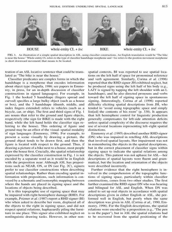
tration of an ASL locative sentence that could be trans-lated as “The bike is near the house.”
Classifier predicates are complex forms in which thehandshape is a morpheme that encodes informationabout object type (Supalla, 1986; see papers in Emmo-rey, in press, for an in-depth discussion of classifierconstructions in signed languages). For example, inFig. 1 the hooked 5 handshape (fingers spread andcurved) specifies a large bulky object (such as a houseor box), and the 3 handshape (thumb, middle, andindex fingers extended) refers to vehicles (such as abicycle, car, or ship). The first and third signs of Fig. 1are nouns that refer to the ground and figure objects,respectively (the sign for BIKE is made with the righthand, while the left hand holds the classifier sign re-ferring to the house). This ordering of figure andground may be an effect of the visual–spatial modalityof sign languages (Emmorey, 1996). For example, topresent a scene visually by drawing a picture, theground object tends to be drawn first, and then thefigure is located with respect to the ground. Thus, ifdrawing a picture of a bike next to a house, most peopledraw the house first. Crucially, the spatial relationshipexpressed by the classifier construction in Fig. 1 is notencoded by a separate word as it would be in Englishwith the preposition near. Although ASL has preposi-tions such as NEAR, ON, or IN (see Fig. 2C), signersprefer to use classifier constructions when describingspatial relationships. Rather than encoding spatial in-formation with prepositions, such information is con-veyed by a schematic and isomorphic mapping betweenwhere the hands are placed in signing space and thelocations of objects being described.
It is this topographic use of signing space that maybe impaired with right hemisphere damage (RHD). Forexample, Poizner et al. (1987) report a RHD signer (BI)who when asked to describe her room, displaced all ofthe objects to the right in signing space, and did notrespect spatial relations, haphazardly piling the furni-ture in one place. This signer also exhibited neglect onnonlinguistic drawing tasks. However, in other non-
spatial contexts, BI was reported to use spatial loca-tions on the left half of space for pronominal referenceand verb agreement. Similarly, Corina et al. (1996)reported that the RHD signer JH exhibited neglect, buthe produced signs using the left half of his body (e.g.,LAZY is signed by tapping the left shoulder with an Lhandshape), and he also directed pronouns and verbstoward the left half of signing space in spontaneoussigning. Interestingly, Corina et al. (1996) reporteddifficulty eliciting spatial descriptions from JH, whotended to “avoid using topographic space and simplylist[ed] the contents of his room” (p. 338). It appearsthat left hemisphere control for linguistic productiongenerally compensates for left-side attention deficitsunless spatial complexity of the discourse requires theuse of several locations representing left–right spatialdistinctions.
Emmorey et al. (1995) described another RHD signer(DN) who was impaired in retelling ASL descriptionsthat involved spatial layouts. Her impairment was notin remembering the objects in the spatial descriptions,but in the correct placement of classifier signs withinsigning space to indicate the spatial relations amongthe objects. This patient was not aphasic for ASL—herdescriptions of spatial layouts were fluent and gram-matical, but the location and orientation of the objectswere described incorrectly.
Further evidence that the right hemisphere is in-volved in the comprehension of the topographic func-tions of signing space, particularly within classifierconstructions, comes from two other studies. The firstfurther examined the RHD signer DN who was hearingand bilingual for ASL and English. When DN wasasked to set up real objects in accordance with spatialdescriptions given in either English or ASL, she per-formed well in English, but poorly when the samedescription was given in ASL (Corina et al., 1990; Em-morey, 1996). For the English descriptions, the spatialrelations were encoded by a preposition (e.g., “The penis on the paper”), but in ASL the spatial relations hadto be recovered from the spatial positioning of the
FIG. 1. An illustration of a simple spatial description in ASL, using classifier constructions. An English translation would be “The bikeis near the house.” Whole-entity CL refers to the type of classifier handshape morpheme and !loc refers to the position movement morpheme(a short downward movement) that means to be located.
813NEURAL SYSTEMS UNDERLYING SPATIAL LANGUAGE
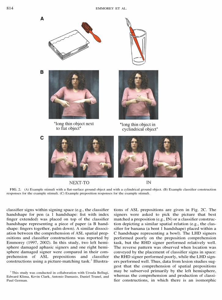
classifier signs within signing space (e.g., the classifierhandshape for pen (a 1 handshape: fist with indexfinger extended) was placed on top of the classifierhandshape representing a piece of paper (a B hand-shape: fingers together, palm down). A similar dissoci-ation between the comprehension of ASL spatial prep-ositions and classifier constructions was reported byEmmorey (1997, 2002). In this study, two left hemi-sphere damaged aphasic signers and one right hemi-sphere damaged signer were compared in their com-prehension of ASL prepositions and classifierconstructions using a picture-matching task.3 Illustra-
tions of ASL prepositions are given in Fig. 2C. Thesigners were asked to pick the picture that bestmatched a preposition (e.g., IN) or a classifier construc-tion depicting a similar spatial relation (e.g., the clas-sifier for banana (a bent 1 handshape) placed within aC handshape representing a bowl). The LHD signersperformed poorly on the preposition comprehensiontask, but the RHD signer performed relatively well.The reverse pattern was observed when location wasconveyed by the placement of classifier signs in space:the RHD signer performed poorly, while the LHD sign-ers performed well. Thus, data from lesion studies sug-gest that the comprehension of spatial prepositionsmay be subserved primarily by the left hemisphere,whereas the comprehension and production of classi-fier constructions, in which there is an isomorphic
3 This study was conducted in collaboration with Ursula Bellugi,Edward Klima, Kevin Clark, Antonio Damasio, Daniel Tranel, andPaul German.
FIG. 2. (A) Example stimuli with a flat-surface ground object and with a cylindrical ground object. (B) Example classifier constructionresponses for the example stimuli. (C) Example preposition responses for the example stimuli.
814 EMMOREY ET AL.
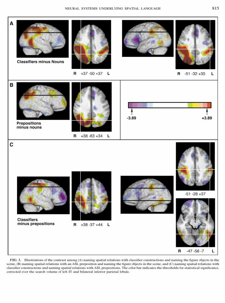
FIG. 3. Illustrations of the contrast among (A) naming spatial relations with classifier constructions and naming the figure objects in thescene, (B) naming spatial relations with an ASL preposition and naming the figure objects in the scene, and (C) naming spatial relations withclassifier constructions and naming spatial relations with ASL prepositions. The color bar indicates the thresholds for statistical significance,corrected over the search volume of left IT and bilateral inferior parietal lobule.
815NEURAL SYSTEMS UNDERLYING SPATIAL LANGUAGE

mapping between the location of the hands in signingspace and the locations of objects in physical space,may be subserved by structures within the right hemi-sphere.
We do not propose that the comprehension and pro-duction of classifier constructions are solely subservedby the right hemisphere. Structures within left hemi-sphere language areas are also very likely to be in-volved in the production and comprehension of classi-fier expressions. For example, a right visual field (lefthemisphere) advantage was found when classifier ex-pressions were presented in a hemifield experiment inwhich signers decided whether a motion or locativepredicate contained a target classifier handshape (Em-morey, 1998). In addition, left lesioned aphasic signersproduce grammatical and sublexical errors when pro-ducing spatial descriptions with classifier construc-tions, although the topographic use of signing space isintact (Poizner et al., 1987). Finally, the linguistic com-plexity of classifier constructions would predict lefthemisphere involvement. Linguistic constraints spec-ify which classifier handshapes can be combined withina locative expression, how scale and perspective areexpressed, and the lexical semantics of classifier hand-shapes within particular expressions. Mastery of clas-sifier constructions is not attained until late in child-hood by signing children (Newport and Meier, 1985;Schick, 1990), and these constructions are notoriouslydifficult for second-language learners to acquire.
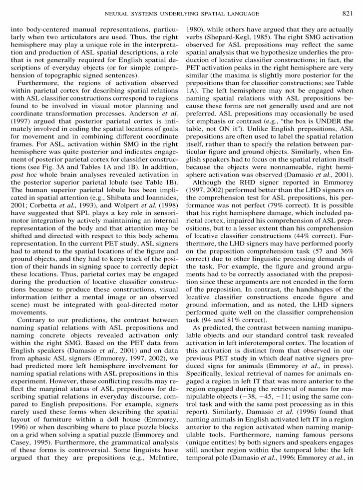
With respect to the expression of spatial relations byhearing English speakers, recent positron emission to-mography (PET) data indicate left and right hemi-sphere involvement for the production of English prep-ositions (Damasio et al., 2001). Damasio et al. (2001)presented English speakers with pictures depicting aspatial relation either between two objects (e.g., a cupon a table) or between two abstract drawings (jaggedmeaningless shapes from Atteneave, 1957). Subjectswere asked either to name the figure object (e.g., thecup) which was colored red or to name the spatialrelationship with a preposition. For the abstract draw-ings, subjects were only asked to name the spatialrelation between the two objects (the figure object wassmaller and colored red). When naming of the figureobject (concrete manipulable entities) was subtractedfrom naming of spatial relations, activation in the leftsupramarginal gyrus (SMG) was observed (see Table 2under Results). Left parietal activation may reflectasymmetric engagement of the dorsal “where” systemwhich is involved in recognizing and identifying objectlocations (Ungerleider and Mishkin, 1982). Left supra-marginal activation may also reflect the engagement ofcortices involved in the lexical retrieval of locativeprepositions. For example, left lesioned aphasic pa-tients often have difficulty producing and comprehend-ing prepositions (Kemmerer and Tranel, 2000; see alsoFriederici, 1982).
In addition, when naming spatial relations betweenmeaningless objects was contrasted with either nam-ing concrete objects or naming spatial relations be-tween concrete (nameable) objects, activation in rightSMG was observed for hearing subjects (see Table 2;Damasio et al., 2001). Naming spatial relations be-tween meaningless shapes may have engaged the rightSMG because speakers had to analyze the spatial re-lation between objects in more detail and could not relyon automatic linguistic encoding. For these stimuli,speakers may have analyzed the spatial relation interms of coordinate spatial relations representationswhich specify more metric spatial properties, such asdistance. Kosslyn and colleagues have found that theright hemisphere encodes this type of spatial represen-tation more efficiently than the left (Kosslyn, 1987;Kosslyn et al., 1995).
Based on the lesion data from ASL signers and thelesion and PET data from English speakers, we makethe following predictions: (1) naming spatial relationswith ASL classifier constructions will engage the infe-rior parietal lobules bilaterally, (2) naming spatial re-lations with ASL prepositions will engage the left in-ferior parietal lobule, and (3) naming manipulableobjects will engage the left inferotemporal (IT) cortex(naming objects is the baseline control task for thisstudy). The third hypothesis is based on PET data fromEnglish speakers who were asked to name tools andother manipulable objects (Damasio et al., 1996).Damasio et al. (1996) found activation in the posteriorleft IT when English speakers were asked to namevarious tools and implements. This cortical area ishypothesized to mediate between the neural regionswhich support conceptual knowledge concerning toolsand those which support the linguistic representationsneeded for the production of the name of the tool(Damasio et al., 1996). We have no reason to suspectthat deaf people have a distinct conceptualization ofmanipulable objects compared to hearing people, andtherefore, we have no reason to predict differences inthe left temporal activation when retrieving signs fortools.
METHODS
Subjects
Ten right-handed, adult native deaf signers werestudied under a PET protocol using [15O]water. Thesubjects were 5 men and 5 women, aged 20–28, with 12years or more of formal education and were right-handed (handedness quotient of !90 or greater asmeasured by the Oldfield–Geschwind questionnaire).All participants had deaf parents and acquired ASL astheir first language from birth. All were profoundlydeaf (90-db loss or greater), and none had any historyof neurological or psychiatric disease. All participants
816 EMMOREY ET AL.
gave formal consent in accordance with federal andinstitutional guidelines.
Procedures
Image acquisition. All subjects underwent MRscanning in a General Electric Signa scanner operatingat 1.5 T, using the following protocol: SPGR 30, TR 24,TE 7, NEX 1, FOV 24 cm, matrix 256 " 192. Each ofthree individual 1NEX SPGR data sets was obtainedwith 124 contiguous coronal slices with thickness of1.5–1.7 mm and interpixel distance of 0.94 mm. Theslice thickness varied so as to be adjusted to the size ofthe brain and the head in order to sample the entirebrain, while avoiding wrap artifacts. The three individ-ual data sets were coregistered post hoc with Auto-mated Image Registration (AIR 3.03) to produce a sin-gle data set, of enhanced quality, with pixel dimensionsof 0.7 mm in plane and 1.5 mm between planes(Holmes et al., 1998). The MR sequences were recon-structed for each subject in 3D using Brainvox(Damasio and Frank, 1992; Frank et al., 1997). Extra-cerebral voxels were edited away manually. The MRscans were used to confirm the absence of structuralabnormalities, to plan the PET slice orientation, and todelineate regions of interest a priori.
PET-Brainvox (Damasio et al., 1994; Grabowski etal., 1995) was used to plan the PET slice orientationparallel to the long axis of the temporal lobes, so thatthe PET acquisition volume included the temporallobes and the inferior parietal lobules in all subjects.Talairach space was constructed directly for each sub-ject via user identification of the anterior and posteriorcommissures and the midsagittal plane in Brainvox.An automated planar search routine defined thebounding box and a piecewise linear transformationwas used (Frank et al., 1997), as defined in the Ta-lairach atlas (Talairach and Tournoux, 1988). AfterTalairach transformation, the MR data sets werewarped (AIR 5th-order nonlinear algorithm) to an atlasspace constructed by averaging 50 normal Talairach-transformed brains, rewarping each brain to the aver-age, and finally averaging them again (analogous tothe procedure described in Woods et al., 1999). Forsimplicity, we will henceforth refer to this standardspace as “Talairach space.” The Talairach-transformed3D scans of all 10 subjects were averaged. The searchvolume, encompassing the left inferotemporal corticesand the bilateral inferior parietal lobules (the supra-marginal and angular gyri), was traced on the aver-aged brain, so as to establish the limits and the size ofthe search volume.
PET data were acquired with a General Electric4096 Plus body tomograph (G.E. Medical Systems, Mil-waukee, WI), yielding 15 transaxial slices with a nom-inal interslice interval of 6.5 mm. For each injection, 50mCi of [15O] water was administered as a bolus through
a venous catheter. Arterial blood sampling was notperformed.
Each subject received eight injections containing 50mCi of [15O]water.
Experimental tasks. Each subject performed fourtasks, twice each. The tasks were as follows: (1) pro-duction of a classifier construction denoting the spatialrelation between two objects (mostly manipulable ob-jects) depicted by line drawings in which the figureobject was colored red (interstimulus interval [ISI]1.5 s; see Figs. 2A and 2B); (2) production of ASLprepositions denoting the spatial relation between ob-jects (ISI 1.5 s; see Fig. 2C); (3) production of ASL signsdenoting the red-shaded manipulable objects in thestimuli presented in (1) (the control task for (1) and (2);ISI 1.5 s); and (4) an orientation judgment performedon the faces of unknown persons requiring the re-sponse YES if the face was in the canonical position(up) and NO if the face was inverted (the control taskfor (3); ISI 1.0 s).
For the control task (4), subjects made a signed re-sponse, but no naming was involved. This task waschosen as the baseline task for naming objects (3) be-cause it has been used in our previous word and signretrieval experiments (Emmorey et al., in press;Damasio et al., 1996, 2001). Using the same controltask consistently allows us to explore the retrieval ofwords/signs for different conceptual categories andacross separate subject groups.
When producing classifier constructions in task (1),the left hand represented the ground object (either aflat or a cylindrical object), and the right hand indi-cated the location of the figure object, as illustrated inFig. 2B. The configuration of the right hand dependedupon the nature of the figure object, e.g., a 1 handshapefor long thin objects, an F handshape (thumb and indexfinger touch, remaining fingers are extended) for smallflat round objects. For one injection, the left hand wasalways in a B hand configuration (fingers together,palm down) indicating a flat surface, and the groundobjects could all be represented by a B classifier hand-shape. For the second injection, the left hand was al-ways in a C hand configuration (fingers together andcurved, palm facing right) indicating a cylindrical ob-ject, and the ground objects were all cylindrical. Theleft hand remained relatively static, while the righthand was placed on top of, next to, behind, in front of,under, above, or inside of the left hand, dependingupon the spatial relation described. Prior to each injec-tion, subjects were told which hand configurationshould be used to represent the ground object. Subjectswere told not to name either the figure or the groundobject, but to produce only the classifier predicate thatexpressed the spatial relation depicted in the picture.Subjects performed the classifier production task asthe first task in the experimental session.
817NEURAL SYSTEMS UNDERLYING SPATIAL LANGUAGE
When producing prepositions, object names, and theyes/no response for the control task, subjects signedwith their right hand in a natural whisper mode sothat the hand did not contact the face. One-handedsigning is natural for whispering and also occurs dur-ing everyday signing (e.g., when one hand is occupied).The majority of signed responses involved only theright hand (this was also true for classifier construc-tions because the left hand remained in the same con-figuration for each stimuli set).
Data analysis. Reconstructed images of the distri-bution of radioactive counts from each injection werecoregistered with each other using Automated ImageRegistration (AIR 3.03, Roger Woods, UCLA). 3D MRand the mean coregistered PET data were also coreg-istered using PET-Brainvox and Automated ImageRegistration (Woods et al., 1993). PET data were Ta-lairach-transformed as described above, masked to thecoregistered MRI brain contour to exclude extracere-bral voxels, and then smoothed with an isotropic16-mm gaussian kernel by Fourier transformation,complex multiplication, and reverse Fourier transfor-mation. The final calculated image resolution was 18 "18 " 18 mm.
PET data were analyzed with a pixelwise linearmodel which estimated coefficients for global activity(covariable) and task and block/subject effects (classi-fication variables) (Friston et al., 1995; Grabowski etal., 1996). We searched for increases in adjusted meanactivity in images of t statistics generated for each ofthe planned contrasts. Critical t values were calculatedusing gaussian random field theory for t statistics(Worsley et al., 1992; Worsley, 1994).
The planned contrasts were as follows:
(a) To address the hypothesis that naming spatialrelations with classifier constructions will engagestructures in the inferior parietal lobules bilaterally,
naming of the figure objects was subtracted from nam-ing spatial relations with classifier constructions.(b) To address the hypothesis that naming spatial
relations with prepositions will engage the left inferiorparietal lobule, naming of the figure objects was sub-tracted from naming spatial relations with preposi-tions.(c) To address how naming spatial relations with
classifier constructions differs from naming spatial re-lations with prepositions, these tasks were subtractedfrom each other.(d) To address the hypothesis that naming manipu-
lable concrete objects will engage left IT, the standardcontrol task (task 4) was subtracted from naming thefigure objects.
RESULTS
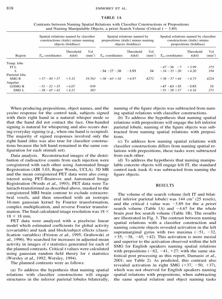
The volume of the search volume (left IT and bilat-eral inferior parietal lobule) was 144 cm3 (25 resels),and the critical t value was #3.89 for the a priorisearch volume (Table 1A) and #4.67 for the wholebrain post hoc search volume (Table 1B). The resultsare illustrated in Fig. 3. The contrast between namingspatial relations with a classifier construction andnaming concrete objects revealed activation in the leftsupramarginal gyrus with two maxima (#51, #32,!35; #38, #45, !42). This activation is more mesialand superior to the activation observed within the leftSMG for English speakers naming spatial relationswith prepositions (#62, #41, !27; with the same sta-tistical post processing as this report, Damasio et al.,2001; see Table 2). As predicted, this contrast alsorevealed activation in right SMG (!37, #50, !37),which was not observed for English speakers namingspatial relations with prepositions, when subtractingthe same spatial relation and object naming tasks
TABLE 1A
Contrasts between Naming Spatial Relations with Classifier Constructions or Prepositionsand Naming Manipulable Objects, a priori Search Volume (Critical t # 3.89)
Region
Spatial relations named by classifierconstructions (italic) minus naming
objects (boldface)
Spatial relations named byprepositions (italic) minus naming
objects (boldface)
Spatial relations named by classifierconstructions (italic) minus
prepositions (boldface)
T88 coordinatesThreshold
t(dof)Vol
(mm3) T88 coordinatesThreshold
t(dof)Vol
(mm3) T88 coordinatesThreshold
t(dof)Vol
(mm3)
Temp. lobeIT L #47 #56 #7 !3.99 375
L #34 #27 #20 #3.93 34 #34 #33 #20 !4.20 194Parietal lobe
SMG R !37 #50 !37 !5.32 19,763 !38 #63 !34 !4.97 8272 !38 #37 !44 !4.73 4224Angular
G/SMG R #51 #32 !35 !4.07 939 !47 #63 !35 #3.93 51SMG L #38 #45 !42 !4.21 383 #51 #28 !37 !4.16 371
818 EMMOREY ET AL.
(Damasio et al., 2001); however, right hemisphere ac-tivation was observed for English speakers when theynamed spatial relations between abstract drawings(see Table 2).
Contrary to our predictions, the contrast betweennaming spatial relations with ASL prepositions andnaming concrete objects revealed no significant activa-tion in the left hemisphere and only activation in theright angular gyrus/SMG (!38, #63, !34). The directcontrast between naming spatial relations with classi-fier constructions and with prepositions revealed bilat-eral activation in the supramarginal gyrus for classi-fier constructions (#51, #28, !37; !38, #37, !44). Inaddition, this contrast revealed activation in left ITwith two maxima (#47, #56, #7; #34, #33, #20) whennaming spatial relations with classifier constructions
(Table 1A). Finally, the contrast revealed greater acti-vation in right angular gyrus (just bordering SMG)when naming spatial relations with ASL prepositions(!47, #63, !35).
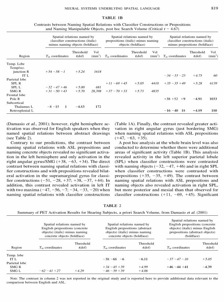
A post hoc analysis at the whole brain level was alsoconducted to determine whether there were additionalareas of significant activity (Table 1B). This analysisrevealed activity in the left superior parietal lobule(SPL) when classifier constructions were contrastedwith naming objects (#32, #47, !46) and in right SPLwhen classifier constructions were contrasted withprepositions (!35, #35, !49). The contrast betweennaming spatial relations with ASL prepositions andnaming objects also revealed activation in right SPL,but more posterior and mesial than that observed forclassifier constructions (!11, #69, !45). Significant
TABLE 1B
Contrasts between Naming Spatial Relations with Classifier Constructions or Prepositionsand Naming Manipulable Objects, post hoc Search Volume (Critical t # 4.67)
Region
Spatial relations named byclassifier constructions (italic)
minus naming objects (boldface)
Spatial relations named byprepositions (italic) minus naming
objects (boldface)
Spatial relations named byclassifier constructions (italic)minus prepositions (boldface)
T88 coordinatesThreshold
t(dof)Vol
(mm3) T88 coordinatesThreshold
t(dof)Vol
(mm3) T88 coordinatesThreshold
t(dof)Vol
(mm3)
Temp. LobeTemp/occ.
junction R !54 #58 #1 !5.24 1618IT L #34 #33 #23 !4.75 60
Parietal lobeSPL R !11 #69 !45 !5.05 4410 !35 #35 !49 !5.28 6139SPL L #32 #47 !46 !5.00 681SMG R !31 #50 !43 !5.70 28,398 !37 #70 !33 !5.73 4835
Frontal lobePole R !38 !52 !9 #4.91 1033
SubcorticalThalamus L #8 #15 1 #4.83 172
Retrosplenial L #16 #40 18 !4.89 180
TABLE 2
Summary of PET Activation Results for Hearing Subjects, a priori Search Volume, from Damasio et al. (2001)
Region
Spatial relations named byEnglish prepositions (concreteobjects) (italic) minus namingconcrete objects (boldface)
Spatial relations named byEnglish prepositions (abstractobjects) (italic) minus namingconcrete objects (boldface)
Spatial relations named byEnglish prepositions (concreteobjects) (italic) minus Englishprepositions (abstract objects)
(boldface)
T88 coordinatesThreshold
t(dof) T88 coordinatesThreshold
t(dof) T88 coordinatesThreshold
t(dof)
Temp. lobeIT L #38 #68 #6 #6.11 #37 #47 #10 !5.05
Parietal lobeSMG R !34 #45 !39 !4.99 !46 #44 !41 #4.39SMG L #62 #41 !27 !4.29 #46 #39 !39 !4.06
Note. The contrast in column 2 was not reported in the original study and is reported here to provide additional data relevant to thecomparison between English and ASL.
819NEURAL SYSTEMS UNDERLYING SPATIAL LANGUAGE

activation within superior parietal cortex was not ob-served for English speakers with the post hoc analysiscontrasting naming spatial relations and naming con-crete entities (Damasio et al., 2001). A few other re-gions of activation were also observed with the post hocanalysis (see Table 1B).
Finally, when naming concrete manipulable objectsand the control task were contrasted, we observed ac-tivation in left IT with two maxima (#50, #53, #6;#27, #38, #13), left SMG (#34, #66, !40), and rightangular gyrus (!31, #80, !27). As predicted, the acti-vation in left IT was similar to that found for Englishspeakers naming tools (#50, #50, #11; #29, #28, #19;subtracting the same control task and with the samestatistical post processing as in this report, Damasio etal., 1996).
DISCUSSION
Production of ASL classifier constructions that spec-ified the spatial relation between two objects engagedthe parietal lobe within both left and right hemi-spheres. The activation within left parietal cortex wassimilar to that observed for English speakers in theparallel study by Damasio et al. (2001). However, forASL classifier constructions, activation within the leftsupramarginal gyrus was superior and mesial to theactivation observed for English speakers (see Tables1A and 2). Although the contrast between naming spa-tial relations and naming objects did not reveal signif-icant activation within the right hemisphere for En-glish speakers, Damasio et al. (2001) found evidence ofactivation in the right SMG when speakers namedspatial relations between abstract nonnameable ob-jects. Furthermore, the activation peak within theright SMG for ASL signers was quite similar to thatseen in English speakers (!46, #44, !41; using thesame post processing procedures as in this report).
For ASL, we propose that naming spatial relationswith classifier constructions engages the right hemi-sphere even when concrete nameable objects are in-volved because signing space is used topographically.As discussed in the Introduction, there is a schematicand analogue mapping between the location of thehands in signing space and the location of physicalobjects described by classifier constructions. Signersmust analyze the spatial relation between objects inenough detail to place their hands in an analogousposition in signing space. ASL does not encode metriccontrasts, but the locative information expressed byclassifier constructions is analogue rather than cate-gorical, and locations in signing space are not morphe-mic representations (Liddell, 1990, 2000; Emmoreyand Herzig, in press). In contrast, the locative informa-tion expressed by prepositions is categorical, and prep-ositions constitute a closed class set of morphemes inEnglish.
In a related study, MacSweeney et al. (in press) usedfMRI to investigate the neural areas engaged whendeaf and hearing users of British Sign Language com-prehended sentences that used space topographically(e.g., “The cat sat on the bed”) compared to BSL sen-tences that did not (e.g., “The man telephoned thewoman”). The topographic sentences generally in-volved classifier constructions, while the nonlocativesentences did not. Their results did not show moreright hemisphere activation for processing topographicsentences compared to nonlocative sentences. Rather,the results revealed greater activation in left inferiorparietal cortex when comprehending topographic BSLsentences. Importantly, when MacSweeney et al. (inpress) translated their topographic and nontopo-graphic BSL sentences into English and presentedthem audiovisually to hearing nonsigners, they foundno differences in parietal activation in the left hemi-sphere for the two sentence types. This finding sug-gests that the comprehension of signed sentences thatuse space topographically engages parietal structureswithin the left hemisphere that may be uniquely re-quired for processing signed language. The Talairachcoordinates for the activation in left parietal cortex(#54, #37, !37; these coordinates represent centroidsof 3D clusters; MacSweeney et al., in press) were nearlyidentical to the activation maxima observed in ourstudy for the contrast between producing classifier con-structions and naming the figure objects (#51, #32,!35; see Table 1A). Thus, left parietal cortex may beengaged during both the comprehension and the pro-duction of sentences or constructions that involve thetopographic use of signing space.
The lack of right hemisphere activation when com-prehending signed sentences that used space topo-graphically is most likely attributable to the taskdemands of the MacSweeney et al. (in press) study.Specifically, the BSL signers were asked to press abutton when they detected a semantic anomaly. In ourPET study, as well as in the lesion studies discussed inthe introduction (Corina et al., 1990; Emmorey, 1997;Poizner et al., 1987), subjects were asked to translatethe spatial relation between the hands in signing spaceinto another spatial representation (i.e., the location ofphysical objects in nonsigning space). The right hemi-sphere may be specifically engaged when spatial loca-tions in signing space must be related to another rep-resentation of spatial locations either in the form of amental image (e.g., when describing a room from mem-ory) or in the form of physical objects (e.g., objects in amanipulation task or pictures in a picture-matchingtask). A reasonable hypothesis suggested by Corina(1998) is that the difficulties that right hemispheredamaged patients exhibit in producing and compre-hending classifier constructions and the topographicfunctions of signing space may stem from a more gen-eral problem with encoding external spatial relations
820 EMMOREY ET AL.
into body-centered manual representations, particu-larly when two articulators are used. Thus, the righthemisphere may play a unique role in the interpreta-tion and production of ASL spatial descriptions, a rolethat is not generally required for English spatial de-scriptions of everyday objects (or for simple compre-hension of topographic signed sentences).
Furthermore, the regions of activation observedwithin parietal cortex for describing spatial relationswith ASL classifier constructions correspond to regionsfound to be involved in visual motor planning andcoordinate transformation processes. Anderson et al.(1997) argued that posterior parietal cortex is inti-mately involved in coding the spatial locations of goalsfor movement and in combining different coordinateframes. For ASL, activation within SMG in the righthemisphere was quite posterior and indicates engage-ment of posterior parietal cortex for classifier construc-tions (see Fig. 3A and Tables 1A and 1B). In addition,post hoc whole brain analyses revealed activation inthe posterior superior parietal lobule (see Table 1B).The human superior parietal lobule has been impli-cated in spatial attention (e.g., Shibata and Ioannides,2001; Corbetta et al., 1993), and Wolpert et al. (1998)have suggested that SPL plays a key role in sensori-motor integration by actively maintaining an internalrepresentation of the body and that attention may beshifted and directed with respect to this body schemarepresentation. In the current PET study, ASL signershad to attend to the spatial locations of the figure andground objects, and they had to keep track of the posi-tion of their hands in signing space to correctly depictthese locations. Thus, parietal cortex may be engagedduring the production of locative classifier construc-tions because to produce these constructions, visualinformation (either a mental image or an observedscene) must be integrated with goal-directed motormovements.
Contrary to our predictions, the contrast betweennaming spatial relations with ASL prepositions andnaming concrete objects revealed activation onlywithin the right SMG. Based on the PET data fromEnglish speakers (Damasio et al., 2001) and on datafrom aphasic ASL signers (Emmorey, 1997, 2002), wehad predicted more left hemisphere involvement fornaming spatial relations with ASL prepositions in thisexperiment. However, these conflicting results may re-flect the marginal status of ASL prepositions for de-scribing spatial relations in everyday discourse, com-pared to English prepositions. For example, signersrarely used these forms when describing the spatiallayout of furniture within a doll house (Emmorey,1996) or when describing where to place puzzle blockson a grid when solving a spatial puzzle (Emmorey andCasey, 1995). Furthermore, the grammatical analysisof these forms is controversial. Some linguists haveargued that they are prepositions (e.g., McIntire,
1980), while others have argued that they are actuallyverbs (Shepard-Kegl, 1985). The right SMG activationobserved for ASL prepositions may reflect the samespatial analysis that we hypothesize underlies the pro-duction of locative classifier constructions; in fact, thePET activation peaks in the right hemisphere are verysimilar (the maxima is slightly more posterior for theprepositions than for classifier constructions; see Table1A). The left hemisphere may not be engaged whennaming spatial relations with ASL prepositions be-cause these forms are not generally used and are notpreferred. ASL prepositions may occasionally be usedfor emphasis or contrast (e.g., “the box is UNDER thetable, not ON it”). Unlike English prepositions, ASLprepositions are often used to label the spatial relationitself, rather than to specify the relation between par-ticular figure and ground objects. Similarly, when En-glish speakers had to focus on the spatial relation itselfbecause the objects were nonnameable, right hemi-sphere activation was observed (Damasio et al., 2001).Although the RHD signer reported in Emmorey
(1997, 2002) performed better than the LHD signers onthe comprehension test for ASL prepositions, his per-formance was not perfect (79% correct). It is possiblethat his right hemisphere damage, which included pa-rietal cortex, impaired his comprehension of ASL prep-ositions, but to a lesser extent than his comprehensionof locative classifier constructions (44% correct). Fur-thermore, the LHD signers may have performed poorlyon the preposition comprehension task (57 and 36%correct) due to other linguistic processing demands ofthe task. For example, the figure and ground argu-ments had to be correctly associated with the preposi-tion since these arguments are not encoded in the formof the preposition. In contrast, the handshapes of thelocative classifier constructions encode figure andground information, and as noted, the LHD signersperformed quite well on the classifier comprehensiontask (94 and 81% correct).As predicted, the contrast between naming manipu-
lable objects and our standard control task revealedactivation in left inferotemporal cortex. The location ofthis activation is distinct from that observed in ourprevious PET study in which deaf native signers pro-duced signs for animals (Emmorey et al., in press).Specifically, lexical retrieval of names for animals en-gaged a region in left IT that was more anterior to theregion engaged during the retrieval of names for ma-nipulable objects (#38, #45, #11; using the same con-trol task and with the same post processing as in thisreport). Similarly, Damasio et al. (1996) found thatnaming animals in English activated left IT in a regionanterior to the region activated when naming manip-ulable tools. Furthermore, naming famous persons(unique entities) by both signers and speakers engagesstill another region within the temporal lobe: the lefttemporal pole (Damasio et al., 1996; Emmorey et al., in
821NEURAL SYSTEMS UNDERLYING SPATIAL LANGUAGE
press). Damasio et al. (1996) hypothesize that thesenonclassical language areas within the left hemispheremediate between neural regions which support concep-tual knowledge about objects and regions which sup-port the phonemic representations needed for the pro-duction of the name of an object. Naming of distincttypes of objects engages distinct neural areas becausethe neural mapping of conceptual knowledge is hypoth-esized to be determined by the physical characteristicsof objects and by our sensorimotoric interaction withthese objects inter alia, which of course differ for ani-mals and tools (see Damasio and Damasio, 1994). Theresults of this study and our previous study (Emmoreyet al., in press) indicate that signed languages exhibitthe same neural organization for lexical retrievalwithin nonclassical language areas as has been ob-served for spoken language (at least with respect toconcrete entities).
The direct contrast between naming spatial relationswith classifier constructions and with prepositions alsorevealed activation within the posterior left IT for clas-sifier constructions (see Table 1A). We interpret thisactivation as a reflection of the fact that handshapewithin these constructions encodes information aboutobject type. For prepositions, handshape is lexicallyspecified and does not change with the nature of thefigure or ground objects. For example, the B handshapeof the sign NEXT-TO does not specify a flat surfaceprominent figure object (see Fig. 2C). In contrast, whendeaf signers produce classifier constructions to indicatespatial relationships, they must choose the appropriatehandshape for each figure object. For example, longthin objects such as pencils or fishing poles require a 1handshape, while cylindrical objects such as cups orbowls require a C handshape. Thus, signers had torecognize and interpret details about the figure object’sshape and other properties in order to produce thecorrect classifier handshape. We propose that a (sub-conscious) retrieval of this information underlies theactivation observed in the posterior left IT when clas-sifier constructions were contrasted with prepositionsbecause retrieving a preposition does not require suchcomplex processing of the figure object (linguistically,figure objects are treated as points for both ASL andEnglish prepositions, see Emmorey, 1996; Talmy,1983).
In conclusion, the production of ASL classifier con-structions that express spatial relationships engagesneural areas within both left and right parietal cortex.Parietal regions of the cortex in both hemispheres havelong been known to be involved in the attention to andperception of the spatial location of physical objects inthe environment (e.g., Posner and Petersen, 1990; Un-gerleider and Mishkin, 1982). With respect to lan-guage, parietal regions may be uniquely engaged dur-ing the production (and comprehension) of spatiallanguage in signed languages, particularly for locative
classifier constructions in which the location of thesigner’s hands in space specifies the spatial relationbetween objects. Furthermore, right parietal cortexmay be specifically engaged when external spatial re-lations must be translated into body-centered manualrepresentations in which each articulator representsan object within the spatial relation. The nature ofspatial language differs quite dramatically from spa-tial language in spoken languages where single closedclass elements (i.e., prepositions or locative affixes)denote spatial relations. And it is precisely within thisdomain where we find variation between the neuralsystems underlying speech and sign production. In con-trast, the results from this study in conjunction withthose of our previous studies (Emmorey et al., in press;Damasio et al., 1996) indicate that the neural systemsinvolved in the retrieval of ASL signs denoting concreteentities within distinct conceptual categories (i.e., an-imals, tools, and famous persons) are remarkably sim-ilar to those underlying the retrieval of spoken Englishwords denoting the same types of entities. Thus, whennaming concrete entities, the neural structures thatmediate language output are the same regardless ofthe mode of output, either speech or sign. However,when expressing spatial relationships, the visual–spa-tial modality of signed languages has an impact on theneural systems that underlie language production.
ACKNOWLEDGMENTS
This research was supported by a grant from the National Insti-tute on Deafness and other Communicative Disorders, 1 P50 DC03189, awarded to the University of Iowa and the Salk Institute forBiological Studies. We thank Samuel Hawk, Amy Hoshina, KathyJones, and Jon Spradling for their help in conducting the study. Weparticularly thank all of the deaf subjects who participated in thestudy.
REFERENCES
Anderson, R. A., Snyder, L. H., Bradley, D. C., and Xing, J. 1997.Multimodal representation of space in the posterior parietal cortexand its use in planning movements. Annu. Rev. Neurosci. 20:303–330.
Atteneave, F. 1957. Physical determinants of the judged complexityof shapes. J. Exp. Psychol. 55: 221–227.
Corbetta, M., Miezin, F. M., Shulman, G. L., and Petersen, S. E.1993. A PET study of visuospatial attention. J. Neurosci. 13:1202–1226.
Corina, D. P. 1998. Aphasia in users of signed languages. In Aphasiain Atypical Populations (P. Coppens, Y. Lebrun, and A. Basso,Eds.), pp. 261–310. Erlbaum, Mahwah, NJ.
Corina, D. P. Bellugi, U., Kritchevsky, M., O Grady-Batch, L., andNorman, F. 1990. Spatial relations in signed versus spoken lan-guage: Clues to right parietal functions. In Academy of AphasiaAbstracts, 1–3, Baltimore.
Corina, D., Kritchevsky, M., and Bellugi, U. 1996. Visual languageprocessing and unilateral neglect: Evidence from American SignLanguage. Cog. Neuropsychol. 13(3): 321–356.
822 EMMOREY ET AL.
Damasio, A., and Damasio, H. 1994. Cortical systems for retrieval ofconcrete knowledge: The convergence zone framework. In Large-Scale Neuronal Theories of the Brain (C. Koch, Ed.), pp. 61–74.MIT Press, Cambridge, MA.
Damasio, H., and Frank, R. 1992. Three-dimensional in vivo map-ping of brain lesions in humans. Arch. Neurol. 49: 137–143.
Damasio, H., Grabowski, T. J., Frank, R., Knosp, B., Hichwa, R. D.,Watkins, G. L., and Ponto, L. L. B. 1994. PET-Brainvox, a tech-nique for neuroanatomical analysis of positron emission tomogra-phy images. In Quantification of Brain Function (K. Uemura, N. A.Lassen, T. Jones, and I. Kanno, Eds.), pp. 465–474. Elsevier,Amsterdam.
Damasio, H., Grabowski, T. J., Tranel, D., Ponto, L. L. B., Hichwa,R. D., and Damasio, A. R. 2001. Neural correlates of namingactions and of naming spatial relations. NeuroImage 13: 1053–1064.
Damasio, H., Grabowski, T. J., Tranel, D., Hichwa, R., and Damasio,A. R., 1996. A neural basis for lexical retrieval. Nature 380: 499–505.
Emmorey, K. 1996. The confluence of space and language in signedlanguages. In Language and Space (P. Bloom, M. Peterson, L.Nadel, and M. Garrett, Eds.), pp. 171–209. MIT Press, Cambridge,MA.
Emmorey, K. 1997. The neural substrates for spatial cognition andlanguage: Insights from sign language. In Proceedings of the Nine-teenth Cognitive Science Society Meeting, p. 908. Erlbaum, Mah-wah, NJ.
Emmorey, K. 1998. Some consequences of using signing space torepresent physical space. In Abstracts from the Theoretical Issuesin Sign Language Research Meeting, p. 23. Gallaudet Univ. Press,Washington, DC.
Emmorey, K. 2001. Space on hand: The exploitation of signing spaceto illustrate abstract thought. In Spatial Schemas and AbstractThought (M. Gattis, Ed.), pp. 147–174. MIT Press, Cambridge,MA.
Emmorey, K. 2002. Language, Cognition, and the Brain: Insightsfrom Sign Language Research. Erlbaum, Mahwah, NJ.
Emmorey, K. ed. (in press). Perspectives on Classifier Constructionsin Sign Languages. Erlbaum, Mahwah, NJ.
Emmorey, K., and Casey, S. 1995. A comparison of spatial languagein English and American Sign Language. Sign Language Stud. 88:225–288.
Emmorey, K., Corina, D., and Bellugi, U. 1995. Differential process-ing of topographic and referential functions of space. In Language,Gesture, and Space (K. Emmorey and J. Reilly, Eds.), pp. 43–62.Erlbaum, Hillsdale, NJ.
Emmorey, K., Grabowski, R., McCullough, S., Damasio, H, Ponto, L,Hichwa, R., and Bellugi, U. Neural systems underlying lexicalretrieval for sign language. Neuropsychologia, in press.
Emmorey, K., and Herzig, M. In press. Categorical versus gradientproperties of classifier constructions in ASL. In Perspectives onClassifier Constructions in Signed Languages (K. Emmorey, Ed.).Erlbaum, Mahwah, NJ.
Frank R. J., Damasio H., and Grabowski, T. J. 1997. Brainvox: Aninteractive, multimodal visualization and analysis system for neu-roanatomical imaging. NeuroImage 5: 13–30.
Friederici, A. D. 1982. Syntactic and semantic processes in aphasicdeficits: The availability of prepositions. Brain Language 15: 259–258.
Friston, K. J., Holmes, A. P., Worsley, K. J., Poline, J.-B., Frith,C. D., and Frackowiak, R. S. J. 1995. Statistical parametric mapsin functional imaging: A general linear approach. Hum. BrainMapping 2: 189–210.
Grabowski, T. J., Damasio, H., Frank, R., Hichwa, R., Boles Ponto,L. L., and Watkins, G. L. A. 1995. new technique for PET slice
orientation and MRI-PET coregistration. Hum. Brain Mapping 2:123–133.
Grabowski, T. J., Frank, R. J., Brown, C. K., Damasio, H., BolesPonto, L. L., Watkins, G. L., and Hichwa, R. D. 1996. Reliability ofPET activation across statistical methods, subject groups, andsample sizes. Hum. Brain Mapping 4: 23–46.
Hickok, G., and Bellugi, U. 2000. The signs of aphasia. In Handbookof Neuropsychology, 2nd ed. (F. Boller and J. Grafman, Eds.),Elsevier, Amsterdam.
Holmes, C. J., Hoge, R., Collins, L., Woods, R. P., Evans, A. C., andToga, A. W. 1998. Enhancement of MR images using registra-tion for signal averaging. J. Comput. Assist. Tomogr. 22: 324–333.
Kemmerer, D., and Tranel, D. 2000. A double dissociation betweenlinguistic and perceptual representations of spatial relationships.Cog. Neuropsychol. 17(5): 393–414.
Kosslyn, S. M. 1987. Seeing and imagining in the cerebral hemi-spheres: A computational approach. Psychol. Rev. 94: 148–175.
Kosslyn, S. M., Maljkovic, V., Hamilton, S. E., Horwitz, G., andThompson, W. L. 1995. Two types of image generation: Evidencefor left- and right-hemisphere processes. Neuropsychologia 33:1485–1510.
Liddell, S. 1990. Four functions of a locus: Re-examining the struc-ture of space in ASL. In Sign Language Research, TheoreticalIssues (C. Lucas, Ed.), pp. 176–198. Gallaudet College Press,Washington, DC.
Liddell, S. 2000. Indicating verbs and pronouns: Pointing away fromagreement. In The Signs of Language Revisited: An Anthology toHonor Ursula Bellugi and Edward Klima (K. Emmorey and H.Lane, Eds.), pp. 303–329. Erlbaum, Mahwah, NJ.
MacSweeney, M., Woll, B., Campbell, R., Calvert, G., McGuire, P.,David, A. S., Simmons, A., and Brammer, M. J. In press. Neuralcorrelates of British Sign Language comprehension: Spatial pro-cessing demands of topographic language. J. Cogn. Neurosci.
McIntire, M. 1980. Locatives in American Sign Language. Unpub-lished Ph.D. Dissertation, University of California, Los Angeles.
Newport, E. L., and Meier, R. P. 1985. The acquisition of AmericanSign Language. In The Cross-Linguistic Study of Language Acqui-sition (D. Slobin, Ed.), pp. 881–938. Erlbaum, Hillsdale, NJ.
Poizner, H., Klima, E. S., and Bellugi, U., 1987. What the HandsReveal about the Brain. MIT Press, Cambridge, MA.
Posner, M. I., and Petersen, S. E. 1990. The attention system of thehuman brain. Annu. Rev. Neurosci. 13: 25–42.
Schick, B. 1990. The effects of morphosyntactic structure on theacquisition of classifier predicates in ASL. In Sign Language Re-search: Theoretical Issues (C. Lucas. Ed.), pp. 358–371. GallaudetUniv. Press, Washington, DC.
Shepard-Kegl, J. 1985. Locative Relations in American Sign Lan-guage Word Formation, Syntax, and Discourse. Unpublished Ph.D.dissertation, Massachusetts Institute of Technology.
Shibata, T., and Ioannides, A. A. 2001. Contributions of the humansuperior parietal lobule to spatial selection process: An MEGstudy. Brain Res. 897: 165–168.
Supalla, T. 1986. The classifier system in American Sign Language.In Noun Classification and Categorization (C. Craig, ed.). JohnBenjamins, Amsterdam.
Talairach, J., and Tournoux, P. 1988. Co-planar Stereotaxic Atlas ofthe Human Brain. Thieme, New York.
Talmy, L. 1983. How language structures space. In Spatial Orienta-tion: Theory, Research, and Application (H. Pick & L. Acredolo,Eds.). Plenum, New York.
Ungerleider, L. G., and Mishkin, M. 1982. Two cortical visual sys-tems. In Analysis of Visual Behavior (D. J. Ingle, M. A. Goodale,
823NEURAL SYSTEMS UNDERLYING SPATIAL LANGUAGE

and R. J. W. Mansfield, Eds.), pp. 549–589. MIT Press, Cambridge,MA.
Wolpert, D. M., Goodbody, S. J., and Husain, M. 1998. Maintaininginternal representations: The role of the human superior parietallobe. Nat. Neurosci. 1(6): 529–533.
Woods, R. P., Daprett, M., Sicotte, N. L., Toga, A. W., and Mazziotta,J. C., 1999. Creation and use of a Talairach-compatible atlas foraccurate, automated, nonlinear intersubject registration, and analy-sis of functional imaging data. Hum. Brain Mapping 8: 73–79.
Woods, R. P., Mazziotta, J. C., and Cherry, S. R., 1993. MRI-PETregistration with automated algorithm. J. Comput. Assist. To-mogr. 17: 536–546.
Worsley, K. J., Evans, A. C., Marrett, S., and Neelin, P., 1992. Athree dimensional statistical analysis for CBF activation studies inhuman brain. J. Cereb. Blood Flow Metab. 12: 900–918.
Worsley, K. J. 1994. Local maxima and the expected Euler charac-teristic of excursion sets of c2,F, and t fields. Adv. Appl. Probability26: 13–42.
824 EMMOREY ET AL.