![Neural Discrete Representation Learningpapers.nips.cc/paper/7210-neural-discrete-representation-learning.pdf · variables [27]. Using discrete variables in deep learning has proven](https://static.fdocuments.us/doc/165x107/5f9fc45cad3f5378060015cb/neural-discrete-representation-variables-27-using-discrete-variables-in-deep.jpg)
Neural representation of visual concepts in people born blind
12
ARTICLE Neural representation of visual concepts in people born blind Ella Striem-Amit 1,2 , Xiaoying Wang 3 , Yanchao Bi 3 & Alfonso Caramazza 1,4 How do we represent information without sensory features? How are abstract concepts like “freedom”, devoid of external perceptible referents, represented in the brain? Here, to address the role of sensory information in the neural representation of concepts, we used fMRI to investigate how people born blind process concepts whose referents are imper- ceptible to them because of their visual nature (“rainbow”, “red”). Activity for these concepts was compared to that of sensorially-perceptible referents (“rain”), classical abstract concepts (“justice”) and concrete concepts (“cup”), providing a gradient between fully concrete and fully abstract concepts in the blind. We find that anterior temporal lobe (ATL) responses track concept perceptibility and objecthood: preference for imperceptible object concepts was found in dorsal ATL, for abstract (non-object, non-referential) concepts in lateral ATL, and for perceptible concepts in medial ATL. These findings point to a new division-of-labor among aspects of ATL in representing conceptual properties that are abstract in different ways. https://doi.org/10.1038/s41467-018-07574-3 OPEN 1 Department of Psychology, Harvard University, Cambridge, MA 02138, USA. 2 Department of Psychology, Ben Gurion University of the Negev, Beer-Sheva 8410501, Israel. 3 State Key Laboratory of Cognitive Neuroscience and Learning & IDG/McGovern Institute for Brain Research, Beijing Normal University, Beijing 100875, China. 4 Center for Mind/Brain Sciences, University of Trento, 38068 Rovereto, Italy. These authors contributed equally: Ella Striem-Amit, Xiaoying Wang. Correspondence and requests for materials should be addressed to E.S-A. (email: [email protected]) or to Y.B. (email: [email protected]) NATURE COMMUNICATIONS | (2018)9:5250 | https://doi.org/10.1038/s41467-018-07574-3 | www.nature.com/naturecommunications 1 1234567890():,;
Transcript of Neural representation of visual concepts in people born blind

ARTICLE
Neural representation of visual concepts in peopleborn blindElla Striem-Amit 1,2, Xiaoying Wang3, Yanchao Bi3 & Alfonso Caramazza1,4
How do we represent information without sensory features? How are abstract concepts like
“freedom”, devoid of external perceptible referents, represented in the brain? Here, to
address the role of sensory information in the neural representation of concepts, we used
fMRI to investigate how people born blind process concepts whose referents are imper-
ceptible to them because of their visual nature (“rainbow”, “red”). Activity for these concepts
was compared to that of sensorially-perceptible referents (“rain”), classical abstract concepts
(“justice”) and concrete concepts (“cup”), providing a gradient between fully concrete and
fully abstract concepts in the blind. We find that anterior temporal lobe (ATL) responses
track concept perceptibility and objecthood: preference for imperceptible object concepts
was found in dorsal ATL, for abstract (non-object, non-referential) concepts in lateral ATL,
and for perceptible concepts in medial ATL. These findings point to a new division-of-labor
among aspects of ATL in representing conceptual properties that are abstract in different
ways.
https://doi.org/10.1038/s41467-018-07574-3 OPEN
1 Department of Psychology, Harvard University, Cambridge, MA 02138, USA. 2 Department of Psychology, Ben Gurion University of the Negev, Beer-Sheva8410501, Israel. 3 State Key Laboratory of Cognitive Neuroscience and Learning & IDG/McGovern Institute for Brain Research, Beijing Normal University,Beijing 100875, China. 4 Center for Mind/Brain Sciences, University of Trento, 38068 Rovereto, Italy. These authors contributed equally: Ella Striem-Amit,Xiaoying Wang. Correspondence and requests for materials should be addressed to E.S-A. (email: [email protected])or to Y.B. (email: [email protected])
NATURE COMMUNICATIONS | (2018) 9:5250 | https://doi.org/10.1038/s41467-018-07574-3 |www.nature.com/naturecommunications 1
1234
5678
90():,;

How do we represent concepts that extend beyond ourperceptual experience, concepts like “freedom” and “jus-tice”, which have no clear external referent? And how do
blind people represent concepts such as rainbow, whose referentis perceptible only visually and comprised of colors, which areuniquely visual qualia?
Various studies have addressed the neural correlates ofconcrete and abstract concepts1–4. Because concrete concepts,like “cup”, have perceptible features, such as shape, size andcolor, whereas abstract concepts, like “freedom”, lack sensoryfeatures, it has been proposed that the latter type of conceptsrely more heavily on semantic or verbal information5,6. Theinvestigation of how abstract concepts are represented has beenconsidered an important way to understand knowledge repre-sentation in the brain. Traditionally, this has been tested bycomparing brain responses to abstract and concrete words. Thiscomparison has revealed large-scale networks of regions asso-ciated with abstract concepts involving language and morebroadly multimodal processing areas and concrete conceptsinvolving modality-specific areas2,7–9. Among these areas, theleft anterior temporal lobe (ATL) has been deemed to play acentral role in the representation and retrieval of semantic andconceptual information1,4,7,9–11.
However, there are additional differences between abstractand concrete concepts beyond the existence of external sensoryreferents. Abstract concepts tend to be learned later in life, to beless familiar12,13, and some of them refer to social or emotionalcontents14,15. The latter factor, emotional responses associatedwith particular concepts, has been argued to provide an emo-tional (internal) “sensory” referent for some concepts16. Theinvolvement of differential emotional arousal for different wordsmay be thought to provide sensorially perceptible features forcertain domains of abstract concepts, contributing to an ongoingdebate about the role of sensory features in conceptrepresentation3,17–21. Furthermore, abstract words differ fromconcrete ones in their linguistic properties, in that they are moreambiguous and their interpretation depends more on context-dependent variation22,23. Therefore, the difference betweenclassical abstract and concrete concepts in terms of their sensoryfeatures is confounded by additional factors. Furthermore,abstract and concrete concepts differ in an additional importantdimension, beyond their sensory perceptibility: their merereferentiality. Concrete concepts generally refer to externalobjects or referents which can be “pointed” to in the world,whereas abstract concepts do not. Nevertheless, these twodimensions are nearly impossible to be teased apart in mostcircumstances as most referents are intrinsically sensible.
How can the effects of sensory perceptibility and experience, aswell as that of referentiality/ objecthood, be tested then? Here wetake a unique approach to overcome the various confounds listedand investigate the roles of these conceptual dimensions directly,by using a special population that does not have access to sen-sorially perceptible referents for otherwise concrete object con-cepts, thereby eliminating the confounds mentioned above. Inthis study we chose to focus on the effect of imperceptibility. Tothis aim we studied a group of people born blind as they werepresented with concepts that have both object referents andsensory-accessible features (“cup”); concepts that have externalreferents but are perceivable through vision alone, and thus aresensorially-inaccessible referents to the congenitally blind(“rainbow”); and abstract concepts without referents or sensoryfeatures, which do not refer to emotional or social relations(“freedom”). This gradient of concepts between fully concreteobject and fully abstract non-object concepts in the blind allowsus to separate sensory components from those of objecthood andstudy their neural correlates.
ResultsAbstract concept preference in the brain. Two experiments(Experiment 1, block design; Experiment 2, event-related design)were conducted to inspect how abstract information, in particularthose aspects relating to imperceptibility (“rainbow” vs. “rain” incongenitally blind) and objecthood (“rainbow” vs. “freedom”), isrepresented in the brain.
To explore the effect of these factors within the hypothesizednetwork involved in processing abstract information, we firstlocalized brain preference for classical abstract concepts, chosencarefully as to not arouse strong emotional responses (seeSupplementary Table 2). We plotted the preferential response toabstract concepts in the data from Experiment 1. Similar toprevious reports7,8, abstract concepts (“freedom”, compared toconcrete every-day objects that are similarly familiar to the blind;“cup”; see Fig. 1a two right-most columns) evoked significantactivation in multiple regions, mainly left-lateralized, in thecombined subject group (Fig. 1b; for similar findings in eachgroup separately and the reverse contrast see SupplementaryFig. 1). These included the inferior frontal lobe, superior temporalsulcus and anterior temporal lobe (ATL), both in the anteriorsuperior temporal plane, as well as below it towards the temporalpole. These regions did not show a significant difference betweenthe groups (See Supplementary Fig. 2), supporting the validity ofusing the blind group to study the representation of abstractconcepts. A more stringent contrast, in which the abstractconcepts condition was further required to also elicit significantpositive activation (abstract > concrete AND abstract > baseline),limited this network to the left hemisphere, and within the ATL,mainly to the dorsal and lateral aspects (Fig. 1c).
Imperceptibility – dorsal ATL. We then investigated which ofthose regions showing preference for abstract concepts weresensitive to the absence of sensory information, as opposed tosensitivity to the existence of external referents or to other con-founding factors. To do so, we examined brain activity in peopleblind from birth (Table 1) for concepts that have external refer-ents in the world, but are perceptible only through the visualsensory modality, and are thus imperceptible to a congenitallyblind person (e.g. “rainbow”). For the blind, these stimuli do nothave sensory correlates for their defining characteristics. Wecompared these concepts to other concepts from the same con-tent domain (in the case of rainbow, astral/weather phenomena)which also have external referents with sensory features in othersenses, and are thus sensorially available, perceptible, to the blind(for example, “rain”; sensory perceptibility was rated by blindsubjects; see methods). Imperceptible and perceptible conceptswere chosen from three different content domains to avoiddomain-specific effects: astral/weather phenomena (e.g. “rain-bow” vs. “rain”), scenes (“island” vs. “beach”) and object features(colors vs. shapes, e.g. “red” vs. “square”). Importantly, theimperceptibility comparison – ANOVA of the full design, com-paring the perceptible and imperceptible concepts across domains(Fig. 1a, comparing dark red and blue across the first three left-most columns) – did not significantly differ in any of the variouspotentially confounding factors: general concreteness/abstract-ness, imageability, age of acquisition, familiarity, semanticdiversity, emotional valence or arousal (mixed effects ANOVA,F(1,58)= 0.1, p= 0.76, η2= 0.0017, for stimuli ratings see Sup-plementary Table 1, for complete post hoc t test results seeSupplementary Table 2).
Given that the imperceptible concepts are sensorially inacces-sible only to the blind, we expected regions differentially engageddue to the sensory imperceptibility of concepts to show differentresponses for the blind and sighted subjects in our experimental
ARTICLE NATURE COMMUNICATIONS | https://doi.org/10.1038/s41467-018-07574-3
2 NATURE COMMUNICATIONS | (2018) 9:5250 | https://doi.org/10.1038/s41467-018-07574-3 | www.nature.com/naturecommunications
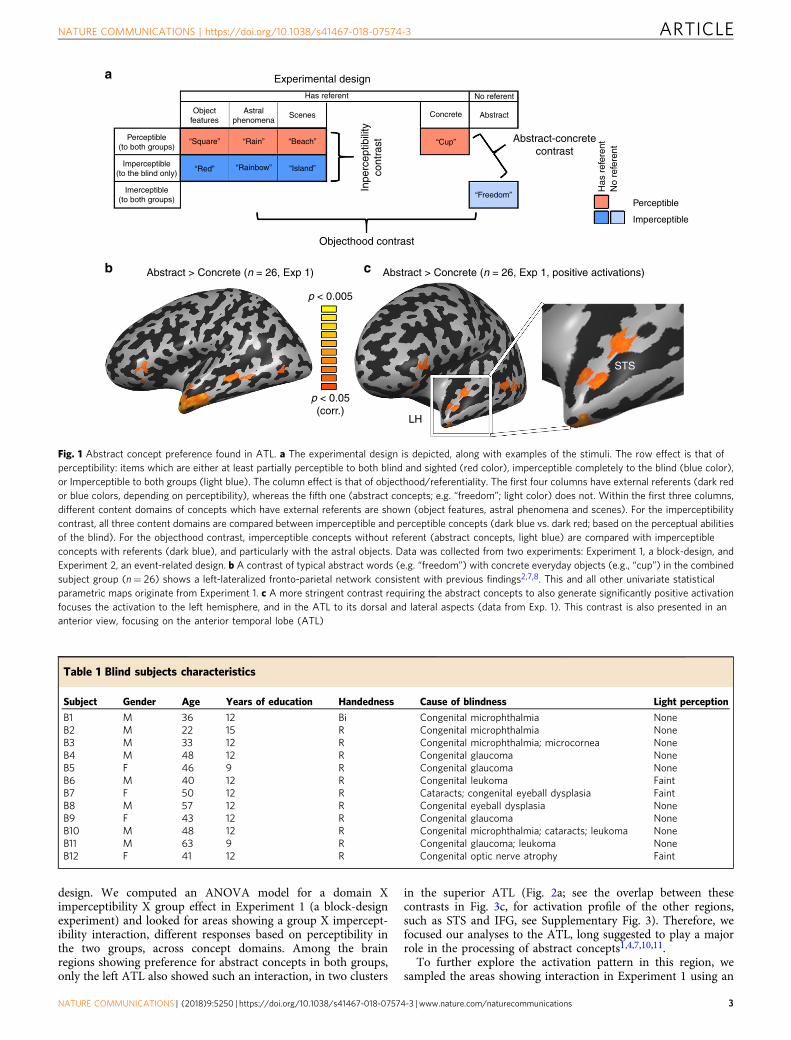
design. We computed an ANOVA model for a domain Ximperceptibility X group effect in Experiment 1 (a block-designexperiment) and looked for areas showing a group X impercept-ibility interaction, different responses based on perceptibility inthe two groups, across concept domains. Among the brainregions showing preference for abstract concepts in both groups,only the left ATL also showed such an interaction, in two clusters
in the superior ATL (Fig. 2a; see the overlap between thesecontrasts in Fig. 3c, for activation profile of the other regions,such as STS and IFG, see Supplementary Fig. 3). Therefore, wefocused our analyses to the ATL, long suggested to play a majorrole in the processing of abstract concepts1,4,7,10,11.
To further explore the activation pattern in this region, wesampled the areas showing interaction in Experiment 1 using an
Table 1 Blind subjects characteristics
Subject Gender Age Years of education Handedness Cause of blindness Light perception
B1 M 36 12 Bi Congenital microphthalmia NoneB2 M 22 15 R Congenital microphthalmia NoneB3 M 33 12 R Congenital microphthalmia; microcornea NoneB4 M 48 12 R Congenital glaucoma NoneB5 F 46 9 R Congenital glaucoma NoneB6 M 40 12 R Congenital leukoma FaintB7 F 50 12 R Cataracts; congenital eyeball dysplasia FaintB8 M 57 12 R Congenital eyeball dysplasia NoneB9 F 43 12 R Congenital glaucoma NoneB10 M 48 12 R Congenital microphthalmia; cataracts; leukoma NoneB11 M 63 9 R Congenital glaucoma; leukoma NoneB12 F 41 12 R Congenital optic nerve atrophy Faint
Experimental design
Inpe
rcep
tibili
tyco
ntra
st
Objectfeatures
“Square”
“Red” “Rainbow”
“Rain” “Beach”
“Island”
“Cup”
“Freedom”
Abstract-concretecontrast
Objecthood contrast
Abstract > Concrete (n = 26, Exp 1) Abstract > Concrete (n = 26, Exp 1, positive activations)
p < 0.005
p < 0.05(corr.)
LH
STS
Concrete
No referent
AbstractAstralphenomena
Scenes
Perceptible(to both groups)
Imerceptible(to both groups)
Imperceptible(to the blind only)
Has referent
a
b c
Perceptible
Imperceptible
Has
ref
eren
tN
o re
fere
nt
Fig. 1 Abstract concept preference found in ATL. a The experimental design is depicted, along with examples of the stimuli. The row effect is that ofperceptibility: items which are either at least partially perceptible to both blind and sighted (red color), imperceptible completely to the blind (blue color),or Imperceptible to both groups (light blue). The column effect is that of objecthood/referentiality. The first four columns have external referents (dark redor blue colors, depending on perceptibility), whereas the fifth one (abstract concepts; e.g. “freedom”; light color) does not. Within the first three columns,different content domains of concepts which have external referents are shown (object features, astral phenomena and scenes). For the imperceptibilitycontrast, all three content domains are compared between imperceptible and perceptible concepts (dark blue vs. dark red; based on the perceptual abilitiesof the blind). For the objecthood contrast, imperceptible concepts without referent (abstract concepts, light blue) are compared with imperceptibleconcepts with referents (dark blue), and particularly with the astral objects. Data was collected from two experiments: Experiment 1, a block-design, andExperiment 2, an event-related design. b A contrast of typical abstract words (e.g. “freedom”) with concrete everyday objects (e.g., “cup”) in the combinedsubject group (n= 26) shows a left-lateralized fronto-parietal network consistent with previous findings2,7,8. This and all other univariate statisticalparametric maps originate from Experiment 1. c A more stringent contrast requiring the abstract concepts to also generate significantly positive activationfocuses the activation to the left hemisphere, and in the ATL to its dorsal and lateral aspects (data from Exp. 1). This contrast is also presented in ananterior view, focusing on the anterior temporal lobe (ATL)
NATURE COMMUNICATIONS | https://doi.org/10.1038/s41467-018-07574-3 ARTICLE
NATURE COMMUNICATIONS | (2018) 9:5250 | https://doi.org/10.1038/s41467-018-07574-3 |www.nature.com/naturecommunications 3

additional independent data set: Experiment 2, an event-relateddesign with the same participants scanned in a separate session.Henceforward, all univariate map analyses originate from datafrom Experiment 1, and the bar plots of activity provideconfirmatory evidence from Experiment 2.
In the anterior cluster of the interaction map, in left anteriordorsal ATL (cluster labeled adATL), we find that the interactionmanifested in heightened activity in the blind group forimperceptible concepts across the three content domains (Fig. 2b,data sampled from the independent experiment 2 in adATL). Fordetail of the exploration of the posterior cluster (labeled pdATL inFig. 2a), which does not show an activation pattern consistentwith an overall effect of imperceptibility in the blind, see Supple-mentary Notes and Supplementary Figure 6. A post hoc contrastof imperceptible concepts as compared to the perceptible
counterparts in the blind in adATL (in Experiment 1 data)showed a significant effect of imperceptibility, in a slightly moredorsal part of ATL, in the anterior superior temporal plane(Fig. 2c). The same area was found also as a main effect in animperceptibility X domain ANOVA model analysis in the blind(Supplementary Figure 6C). Specifically, when inspecting thissuperior ATL imperceptibility cluster in the data from Experi-ment 2, it showed a main effect of imperceptibility in the blind (F(1,11)= 8.63, p < 0.05; see sampled data in Fig. 2d), a significantpreference for imperceptible concepts, but no domain effect (p >0.4) or interaction (p > 0.61). That is, sensory perceptibility affectsthis region independently of content domains. Importantly, thesighted group showed no such effect in this region (all effects andinteraction p > 0.82), and a combined ANOVA with both groupsin this ROI in Experiment 2 revealed an imperceptibility X group
Group × Imperceptibilityinteraction (Exp 1)
ROI analysis: adATLBlind (Exp 2)
ROI analysis: Imperceptibile concept preference clusterBlind (Exp 2)
Imperceptibile > PerceptibleBlind (Exp 1)
0.35
pdATL
adATL
0.25
0.2
0.15
0.05
0Feature
GLM
par
amet
er e
stim
ate
(bet
a)G
LM p
aram
eter
est
imat
e (b
eta)
Astral Scene
Feature Astral Scene Feature Astral Scene
Feature Astral Scene
0.1
0.3
0.25
0.2
0.15
0.05
0
0.1
0.3
* * *
**
0.25
0.2
0.15
0.05
0
0.1
0.3
0.35
Sighted (Exp 2)
Sighted (Exp 2)
PerceptibleImperceptible
0.25
0.2
0.15
0.05
0
0.1
0.3
a b
c d
Fig. 2 Imperceptible concepts processing is supported by the left dorsal ATL. a To probe for the effect of sensory feature perceptibility, we compared brainactivity in people blind from birth and sighted controls, in response to concepts which have external referents in the world, but are perceptible only throughthe visual sensory modality, and are thus imperceptible to a blind person (e.g. “rainbow”) as compared to concepts whose referents are sensoriallyperceptible also to the blind (through other modalities; e.g., “rain”). An area which is sensitive to imperceptibility of concepts should respond differently inthe two groups for this contrast, as visually-dominant concepts are fully perceptible to the sighted subjects. The ANOVA effect of Group X Imperceptibilityinteraction across content domains shows two clusters in dorsal ATL which respond differently in the blind and sighted to the presented words based ontheir perceptibility (adATL and pdATL; data from Exp. 1). b The anterior cluster shown in Fig. 2A, labeled adATL, shows a preference for imperceptibleconcepts across concept domains (object features, astral phenomena and scenes) only in the blind group (data from independent Exp. 2). Error barsrepresent standard error of the difference between means for the perceptible and imperceptible words in each content domain. Asterisks representstatistically significant difference between perceptible and imperceptible concepts (paired t test, t(22) > 3.505, p < 0.05, Bonferroni corrected for multiplecomparisons). c Preferential activation for imperceptible vs perceptible concepts in the blind group, affects the left dorsal ATL (data from Exp. 1). d Thedorsal ATL cluster showing preferential activation for imperceptible concepts (shown in Fig. 2C) shows a preference for imperceptible concepts acrossconcept domains (object features, astral phenomena and scenes) only in the blind group (data from independent Exp. 2). Error bars represent standarderror of the difference between means for the perceptible and imperceptible words in each content domain. In addition to the significant mainimperceptibility effect in the blind, asterisks represent statistically significant difference between perceptible and imperceptible concepts (paired t test, t(22) > 3.505, p < 0.05, Bonferroni corrected for multiple comparisons)
ARTICLE NATURE COMMUNICATIONS | https://doi.org/10.1038/s41467-018-07574-3
4 NATURE COMMUNICATIONS | (2018) 9:5250 | https://doi.org/10.1038/s41467-018-07574-3 | www.nature.com/naturecommunications
interaction (F(1,24)= 5.46, p < 0.05), supporting the absence ofvisual experience as the factor behind the imperceptible/perceptible category differences.
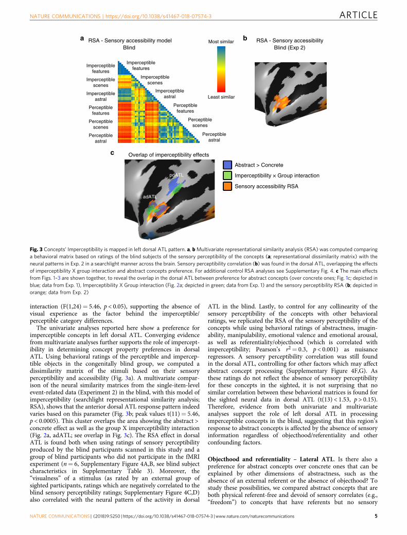
The univariate analyses reported here show a preference forimperceptible concepts in left dorsal ATL. Converging evidencefrom multivariate analyses further supports the role of impercept-ibility in determining concept property preferences in dorsalATL. Using behavioral ratings of the perceptible and impercep-tible objects in the congenitally blind group, we computed adissimilarity matrix of the stimuli based on their sensoryperceptibility and accessibility (Fig. 3a). A multivariate compar-ison of the neural similarity matrices from the single-item-levelevent-related data (Experiment 2) in the blind, with this model ofimperceptibility (searchlight representational similarity analysis;RSA), shows that the anterior dorsal ATL response pattern indeedvaries based on this parameter (Fig. 3b; peak values t(11)= 5.46,p < 0.0005). This cluster overlaps the area showing the abstract >concrete effect as well as the group X imperceptibility interaction(Fig. 2a, adATL; see overlap in Fig. 3c). The RSA effect in dorsalATL is found both when using ratings of sensory perceptibilityproduced by the blind participants scanned in this study and agroup of blind participants who did not participate in the fMRIexperiment (n= 6, Supplementary Figure 4A,B, see blind subjectcharacteristics in Supplementary Table 3). Moreover, the“visualness” of a stimulus (as rated by an external group ofsighted participants, ratings which are negatively correlated to theblind sensory perceptibility ratings; Supplementary Figure 4C,D)also correlated with the neural pattern of the activity in dorsal
ATL in the blind. Lastly, to control for any collinearity of thesensory perceptibility of the concepts with other behavioralratings, we replicated the RSA of the sensory perceptibility of theconcepts while using behavioral ratings of abstractness, imagin-ability, manipulability, emotional valence and emotional arousal,as well as referentiality/objecthood (which is correlated withimperceptibility; Pearson’s r2= 0.3, p < 0.001) as nuisanceregressors. A sensory perceptibility correlation was still foundin the dorsal ATL, controlling for other factors which may affectabstract concept processing (Supplementary Figure 4F,G). Asthese ratings do not reflect the absence of sensory perceptibilityfor these concepts in the sighted, it is not surprising that nosimilar correlation between these behavioral matrices is found forthe sighted neural data in dorsal ATL (t(13) < 1.53, p > 0.15).Therefore, evidence from both univariate and multivariateanalyses support the role of left dorsal ATL in processingimperceptible concepts in the blind, suggesting that this region’sresponse to abstract concepts is affected by the absence of sensoryinformation regardless of objecthood/referentiality and otherconfounding factors.
Objecthood and referentiality – Lateral ATL. Is there also apreference for abstract concepts over concrete ones that can beexplained by other dimensions of abstractness, such as theabsence of an external referent or the absence of objecthood? Tostudy these possibilities, we compared abstract concepts that areboth physical referent-free and devoid of sensory correlates (e.g.,“freedom”) to concepts that have referents but no sensory
Most similarRSA - Sensory accessibility modelBlind
Overlap of imperceptibility effects
RSA - Sensory accessibilityBlind (Exp 2)
a b
c
Least similar
Abstract > Concrete
Imperceptibility × Group interaction
Sensory accessibility RSA
pdATL
adATL
Imperceptiblefeatures
Imperceptiblefeatures
Perceptiblefeatures
Perceptiblefeatures
Perceptiblescenes
Perceptiblescenes
Perceptibleastral
Perceptibleastral
Imperceptiblescenes
Imperceptiblescenes
Imperceptibleastral
Imperceptibleastral
Fig. 3 Concepts’ Imperceptibility is mapped in left dorsal ATL pattern. a, bMultivariate representational similarity analysis (RSA) was computed comparinga behavioral matrix based on ratings of the blind subjects of the sensory perceptibility of the concepts (a; representational dissimilarity matrix) with theneural patterns in Exp. 2 in a searchlight manner across the brain. Sensory perceptibility correlation (b) was found in the dorsal ATL, overlapping the effectsof imperceptibility X group interaction and abstract concepts preference. For additional control RSA analyses see Supplementary Fig. 4. c The main effectsfrom Figs. 1–3 are shown together, to reveal the overlap in the dorsal ATL between preference for abstract concepts (over concrete ones; Fig. 1c; depicted inblue; data from Exp. 1), Imperceptibility X Group interaction (Fig. 2a; depicted in green; data from Exp. 1) and the sensory perceptibility RSA (b; depicted inorange; data from Exp. 2)
NATURE COMMUNICATIONS | https://doi.org/10.1038/s41467-018-07574-3 ARTICLE
NATURE COMMUNICATIONS | (2018) 9:5250 | https://doi.org/10.1038/s41467-018-07574-3 |www.nature.com/naturecommunications 5
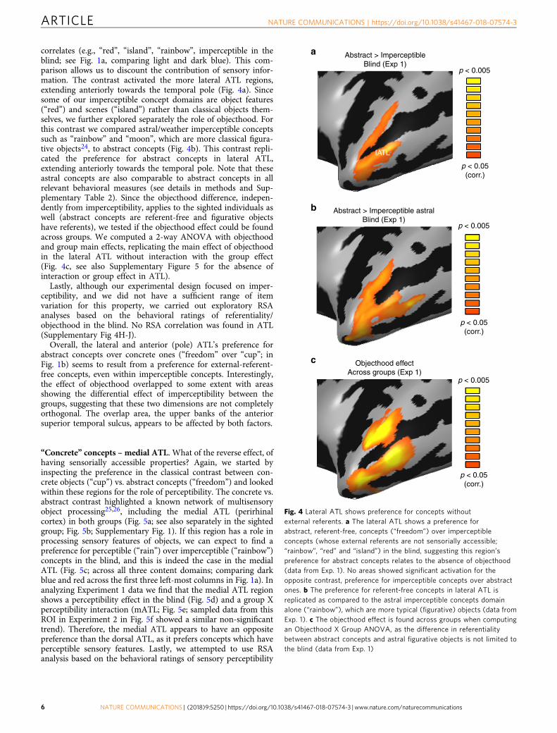
correlates (e.g., “red”, “island”, “rainbow”, imperceptible in theblind; see Fig. 1a, comparing light and dark blue). This com-parison allows us to discount the contribution of sensory infor-mation. The contrast activated the more lateral ATL regions,extending anteriorly towards the temporal pole (Fig. 4a). Sincesome of our imperceptible concept domains are object features(“red”) and scenes (“island”) rather than classical objects them-selves, we further explored separately the role of objecthood. Forthis contrast we compared astral/weather imperceptible conceptssuch as “rainbow” and “moon”, which are more classical figura-tive objects24, to abstract concepts (Fig. 4b). This contrast repli-cated the preference for abstract concepts in lateral ATL,extending anteriorly towards the temporal pole. Note that theseastral concepts are also comparable to abstract concepts in allrelevant behavioral measures (see details in methods and Sup-plementary Table 2). Since the objecthood difference, indepen-dently from imperceptibility, applies to the sighted individuals aswell (abstract concepts are referent-free and figurative objectshave referents), we tested if the objecthood effect could be foundacross groups. We computed a 2-way ANOVA with objecthoodand group main effects, replicating the main effect of objecthoodin the lateral ATL without interaction with the group effect(Fig. 4c, see also Supplementary Figure 5 for the absence ofinteraction or group effect in ATL).
Lastly, although our experimental design focused on imper-ceptibility, and we did not have a sufficient range of itemvariation for this property, we carried out exploratory RSAanalyses based on the behavioral ratings of referentiality/objecthood in the blind. No RSA correlation was found in ATL(Supplementary Fig 4H-J).
Overall, the lateral and anterior (pole) ATL’s preference forabstract concepts over concrete ones (“freedom” over “cup”; inFig. 1b) seems to result from a preference for external-referent-free concepts, even within imperceptible concepts. Interestingly,the effect of objecthood overlapped to some extent with areasshowing the differential effect of imperceptibility between thegroups, suggesting that these two dimensions are not completelyorthogonal. The overlap area, the upper banks of the anteriorsuperior temporal sulcus, appears to be affected by both factors.
“Concrete” concepts –medial ATL. What of the reverse effect, ofhaving sensorially accessible properties? Again, we started byinspecting the preference in the classical contrast between con-crete objects (“cup”) vs. abstract concepts (“freedom”) and lookedwithin these regions for the role of perceptibility. The concrete vs.abstract contrast highlighted a known network of multisensoryobject processing25,26, including the medial ATL (perirhinalcortex) in both groups (Fig. 5a; see also separately in the sightedgroup; Fig. 5b; Supplementary Fig. 1). If this region has a role inprocessing sensory features of objects, we can expect to find apreference for perceptible (“rain”) over imperceptible (“rainbow”)concepts in the blind, and this is indeed the case in the medialATL (Fig. 5c; across all three content domains; comparing darkblue and red across the first three left-most columns in Fig. 1a). Inanalyzing Experiment 1 data we find that the medial ATL regionshows a perceptibility effect in the blind (Fig. 5d) and a group Xperceptibility interaction (mATL; Fig. 5e; sampled data from thisROI in Experiment 2 in Fig. 5f showed a similar non-significanttrend). Therefore, the medial ATL appears to have an oppositepreference than the dorsal ATL, as it prefers concepts which haveperceptible sensory features. Lastly, we attempted to use RSAanalysis based on the behavioral ratings of sensory perceptibility
Abstract > ImperceptibleBlind (Exp 1)
Abstract > Imperceptible astralBlind (Exp 1)
Objecthood effectAcross groups (Exp 1)
IATL
a
b
c
p < 0.005
p < 0.05(corr.)
p < 0.005
p < 0.05(corr.)
p < 0.005
p < 0.05(corr.)
Fig. 4 Lateral ATL shows preference for concepts withoutexternal referents. a The lateral ATL shows a preference forabstract, referent-free, concepts (“freedom”) over imperceptibleconcepts (whose external referents are not sensorially accessible;“rainbow”, “red” and “island”) in the blind, suggesting this region’spreference for abstract concepts relates to the absence of objecthood(data from Exp. 1). No areas showed significant activation for theopposite contrast, preference for imperceptible concepts over abstractones. b The preference for referent-free concepts in lateral ATL isreplicated as compared to the astral imperceptible concepts domainalone (“rainbow”), which are more typical (figurative) objects (data fromExp. 1). c The objecthood effect is found across groups when computingan Objecthood X Group ANOVA, as the difference in referentialitybetween abstract concepts and astral figurative objects is not limited tothe blind (data from Exp. 1)
ARTICLE NATURE COMMUNICATIONS | https://doi.org/10.1038/s41467-018-07574-3
6 NATURE COMMUNICATIONS | (2018) 9:5250 | https://doi.org/10.1038/s41467-018-07574-3 | www.nature.com/naturecommunications

by the blind and ratings of visual perceptibility by the sighted.While a small area in the medial ATL displayed RSA correlationfor both analyses, this cluster was not sufficiently significant tosurvive the multiple comparisons correction.
Different functional networks for aspects of ATL. Given thedifferent functional roles we find for different regions of ATL, wefurther tested if this dissociation of preferences would alsomanifest in having different network connectivity patterns, basedon resting-state data acquired from the same participants.
We first tested the dissociation between the dorsal and lateralATL, which appear to represent different attributes of abstractconcepts (imperceptibility and non-objecthood, respectively). Wecomputed resting-state functional connectivity (RSFC) fromseeds at the peaks of the cluster showing the group Ximperceptibility interaction in the dorsal ATL (adATL; Fig. 2a)and the peak of the cluster showing the abstract > imperceptibleconcepts in the lateral ATL (lATL; Fig. 4a) in the sighted group.Despite their difference in functional preferences, the dorsal andlateral ATL seem to belong largely to the same functionalnetwork, which includes large parts of the dorsolateral ATL andinferior frontal lobe (Fig. 6a; note the prevalence of shared RSFCmarked in yellow). The spatial overlap of the activation (see detailabove) and shared network suggest that these regions may be partof the same system for the processing of semantic, non-sensoriallyderived information. Similar connectivity patterns are found inthe blind group (see Supplementary Fig. 7A,C,D), with few areasshowing group differences in RSFC. This suggests that the blindbrain is not differently wired in these regions, again supportingthe validity of using the blind group to investigate ATL.
We investigated the potential dissociation in brain functionalnetworks between the dorsal and medial ATL, which showcontrasting roles regarding perceptibility. We plotted their jointand partial functional connectivity (RSFC; Fig. 6b) based onseeds from the peaks of the cluster showing the group Ximperceptibility interaction in the dorsal ATL (adATL; shown inFig. 2a; also used for Fig. 6a) and of the cluster showing thegroup X imperceptibility interaction in medial ATL (mATL;shown in Fig. 5e). The partial RSFC shows that the medial ATLis better connected to multisensory object-related regions in thefrontal lobe, parietal lobe, as well as in the ventral visual cortex.This connectivity profile is consistent with the literature linkingmedial structures in ATL, mainly the perirhinal cortex, as themechanism of sensory feature integration of object features27–29.In contrast, the dorsal ATL is more strongly connected to thelateral and anterior ATL towards the temporal pole, as well as tothe inferior frontal lobe, parts of the language network.Therefore, the RSFC analysis also supports the distinct roles ofthese subregions of ATL. The connectivity profiles of these two
Concrete > Abstractn = 26, both groups (Exp 1)
Concrete > AbstractSighted (Exp 1)
Perceptible > ImperceptibleBlind (Exp 1)
p < 0.005
p < 0.05(corr.)
a
b c
d e
f
Feature Astral Scene0
0.1
0.2
Feature Astral Scene0
0.1
0.2* *
mATL
GLM
par
amet
er e
stim
ate
(bet
a)
Imperceptibility effectBlind (Exp 1)
Group × Imperceptibilityinteraction (Exp 1)
ROI analysis: mATLBlind (Exp 2) Sighted (Exp 2)
PerceptibleImperceptible
Fig. 5 Perceptible concepts processing is supported by the medial ATL. a Acontrast of concrete everyday objects as compared with typical abstractwords in the combined subject group (n= 26, in a; see b for sighted groupseparately) shows a network of regions associated with multisensory objectperception (data from Exp. 1). In the ATL, the medial ATL shows preferencefor processing concrete objects. b The contrast of concrete everydayobjects as compared with typical abstract words in the sighted groupreplicates the effect of both groups (panel A) in medial ATL (data from Exp.1). c The contrast for perceptible vs. imperceptible concepts in the blindshows medial ATL prefers perceptible concepts (data from Exp. 1). dMedialATL shows a perceptibility effect in the blind group across content domains(2-way ANOVA, perceptibility and content domain; data from Exp. 1). e Theperceptibility effect differs between the groups in medial ATL (clusterlabeled mATL), as evident from a group X imperceptibility interaction (datafrom Exp. 1). f Data sampled from mATL (the cluster shown in e) in theindependent Experiment 2 replicates the preference for perceptible (e.g.,“rain”) over imperceptible (“rainbow”) concepts in the blind in medial ATL,although the concepts are perceptible via non-visual modalities. Error barsrepresent standard error of the difference between means for theperceptible and imperceptible words in each content domain. Asterisksrepresent statistically significant difference between perceptible andimperceptible concepts (paired t test, t(22) > 3.505, p < 0.05, Bonferronicorrected for multiple comparisons)
NATURE COMMUNICATIONS | https://doi.org/10.1038/s41467-018-07574-3 ARTICLE
NATURE COMMUNICATIONS | (2018) 9:5250 | https://doi.org/10.1038/s41467-018-07574-3 |www.nature.com/naturecommunications 7
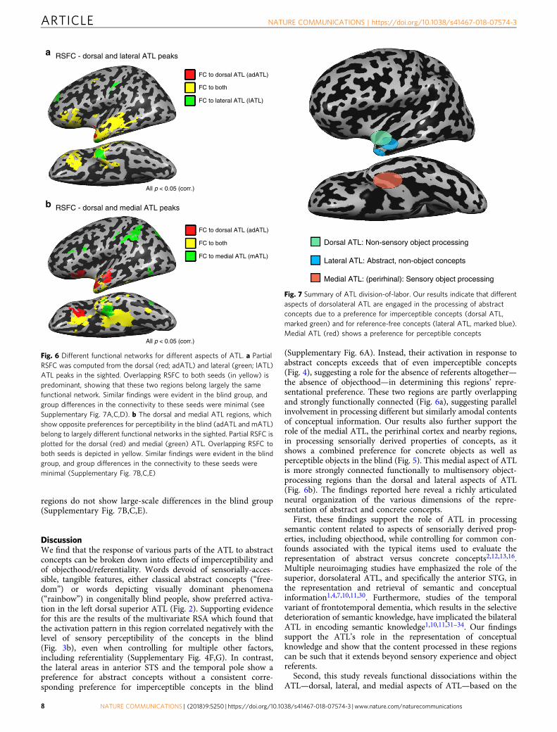
regions do not show large-scale differences in the blind group(Supplementary Fig. 7B,C,E).
DiscussionWe find that the response of various parts of the ATL to abstractconcepts can be broken down into effects of imperceptibility andof objecthood/referentiality. Words devoid of sensorially-acces-sible, tangible features, either classical abstract concepts (“free-dom”) or words depicting visually dominant phenomena(“rainbow”) in congenitally blind people, show preferred activa-tion in the left dorsal superior ATL (Fig. 2). Supporting evidencefor this are the results of the multivariate RSA which found thatthe activation pattern in this region correlated negatively with thelevel of sensory perceptibility of the concepts in the blind(Fig. 3b), even when controlling for multiple other factors,including referentiality (Supplementary Fig. 4F,G). In contrast,the lateral areas in anterior STS and the temporal pole show apreference for abstract concepts without a consistent corre-sponding preference for imperceptible concepts in the blind
(Supplementary Fig. 6A). Instead, their activation in response toabstract concepts exceeds that of even imperceptible concepts(Fig. 4), suggesting a role for the absence of referents altogether—the absence of objecthood—in determining this regions’ repre-sentational preference. These two regions are partly overlappingand strongly functionally connected (Fig. 6a), suggesting parallelinvolvement in processing different but similarly amodal contentsof conceptual information. Our results also further support therole of the medial ATL, the perirhinal cortex and nearby regions,in processing sensorially derived properties of concepts, as itshows a combined preference for concrete objects as well asperceptible objects in the blind (Fig. 5). This medial aspect of ATLis more strongly connected functionally to multisensory object-processing regions than the dorsal and lateral aspects of ATL(Fig. 6b). The findings reported here reveal a richly articulatedneural organization of the various dimensions of the repre-sentation of abstract and concrete concepts.
First, these findings support the role of ATL in processingsemantic content related to aspects of sensorially derived prop-erties, including objecthood, while controlling for common con-founds associated with the typical items used to evaluate therepresentation of abstract versus concrete concepts2,12,13,16.Multiple neuroimaging studies have emphasized the role of thesuperior, dorsolateral ATL, and specifically the anterior STG, inthe representation and retrieval of semantic and conceptualinformation1,4,7,10,11,30. Furthermore, studies of the temporalvariant of frontotemporal dementia, which results in the selectivedeterioration of semantic knowledge, have implicated the bilateralATL in encoding semantic knowledge1,10,11,31–34. Our findingssupport the ATL’s role in the representation of conceptualknowledge and show that the content processed in these regionscan be such that it extends beyond sensory experience and objectreferents.
Second, this study reveals functional dissociations within theATL—dorsal, lateral, and medial aspects of ATL—based on the
RSFC - dorsal and lateral ATL peaks
FC to dorsal ATL (adATL)
All p < 0.05 (corr.)
All p < 0.05 (corr.)
FC to lateral ATL (IATL)
FC to dorsal ATL (adATL)
FC to both
FC to both
FC to medial ATL (mATL)
a
b RSFC - dorsal and medial ATL peaks
Fig. 6 Different functional networks for different aspects of ATL. a PartialRSFC was computed from the dorsal (red; adATL) and lateral (green; lATL)ATL peaks in the sighted. Overlapping RSFC to both seeds (in yellow) ispredominant, showing that these two regions belong largely the samefunctional network. Similar findings were evident in the blind group, andgroup differences in the connectivity to these seeds were minimal (seeSupplementary Fig. 7A,C,D). b The dorsal and medial ATL regions, whichshow opposite preferences for perceptibility in the blind (adATL and mATL)belong to largely different functional networks in the sighted. Partial RSFC isplotted for the dorsal (red) and medial (green) ATL. Overlapping RSFC toboth seeds is depicted in yellow. Similar findings were evident in the blindgroup, and group differences in the connectivity to these seeds wereminimal (Supplementary Fig. 7B,C,E)
Dorsal ATL: Non-sensory object processing
Lateral ATL: Abstract, non-object concepts
Medial ATL: (perirhinal): Sensory object processing
Fig. 7 Summary of ATL division-of-labor. Our results indicate that differentaspects of dorsolateral ATL are engaged in the processing of abstractconcepts due to a preference for imperceptible concepts (dorsal ATL,marked green) and for reference-free concepts (lateral ATL, marked blue).Medial ATL (red) shows a preference for perceptible concepts
ARTICLE NATURE COMMUNICATIONS | https://doi.org/10.1038/s41467-018-07574-3
8 NATURE COMMUNICATIONS | (2018) 9:5250 | https://doi.org/10.1038/s41467-018-07574-3 | www.nature.com/naturecommunications
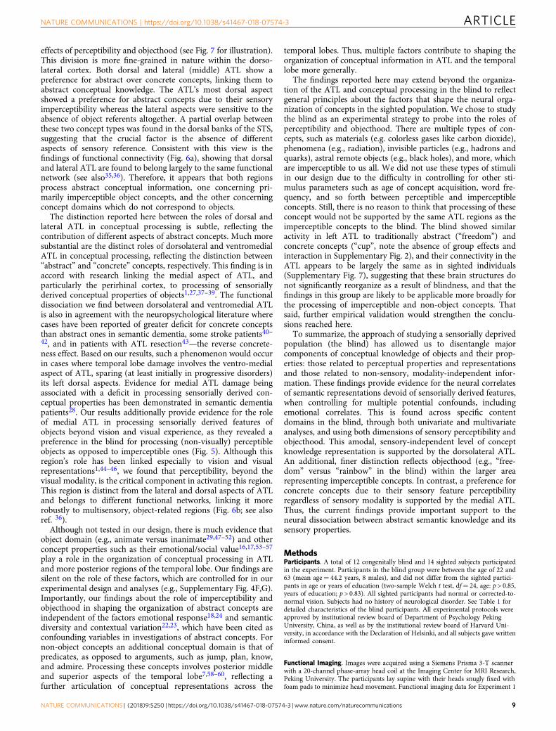
effects of perceptibility and objecthood (see Fig. 7 for illustration).This division is more fine-grained in nature within the dorso-lateral cortex. Both dorsal and lateral (middle) ATL show apreference for abstract over concrete concepts, linking them toabstract conceptual knowledge. The ATL’s most dorsal aspectshowed a preference for abstract concepts due to their sensoryimperceptibility whereas the lateral aspects were sensitive to theabsence of object referents altogether. A partial overlap betweenthese two concept types was found in the dorsal banks of the STS,suggesting that the crucial factor is the absence of differentaspects of sensory reference. Consistent with this view is thefindings of functional connectivity (Fig. 6a), showing that dorsaland lateral ATL are found to belong largely to the same functionalnetwork (see also35,36). Therefore, it appears that both regionsprocess abstract conceptual information, one concerning pri-marily imperceptible object concepts, and the other concerningconcept domains which do not correspond to objects.
The distinction reported here between the roles of dorsal andlateral ATL in conceptual processing is subtle, reflecting thecontribution of different aspects of abstract concepts. Much moresubstantial are the distinct roles of dorsolateral and ventromedialATL in conceptual processing, reflecting the distinction between“abstract” and “concrete” concepts, respectively. This finding is inaccord with research linking the medial aspect of ATL, andparticularly the perirhinal cortex, to processing of sensoriallyderived conceptual properties of objects1,27,37–39. The functionaldissociation we find between dorsolateral and ventromedial ATLis also in agreement with the neuropsychological literature wherecases have been reported of greater deficit for concrete conceptsthan abstract ones in semantic dementia, some stroke patients40–42, and in patients with ATL resection43—the reverse concrete-ness effect. Based on our results, such a phenomenon would occurin cases where temporal lobe damage involves the ventro-medialaspect of ATL, sparing (at least initially in progressive disorders)its left dorsal aspects. Evidence for medial ATL damage beingassociated with a deficit in processing sensorially derived con-ceptual properties has been demonstrated in semantic dementiapatients28. Our results additionally provide evidence for the roleof medial ATL in processing sensorially derived features ofobjects beyond vision and visual experience, as they revealed apreference in the blind for processing (non-visually) perceptibleobjects as opposed to imperceptible ones (Fig. 5). Although thisregion’s role has been linked especially to vision and visualrepresentations1,44–46, we found that perceptibility, beyond thevisual modality, is the critical component in activating this region.This region is distinct from the lateral and dorsal aspects of ATLand belongs to different functional networks, linking it morerobustly to multisensory, object-related regions (Fig. 6b; see alsoref. 36).
Although not tested in our design, there is much evidence thatobject domain (e.g., animate versus inanimate29,47–52) and otherconcept properties such as their emotional/social value16,17,53–57
play a role in the organization of conceptual processing in ATLand more posterior regions of the temporal lobe. Our findings aresilent on the role of these factors, which are controlled for in ourexperimental design and analyses (e.g., Supplementary Fig. 4F,G).Importantly, our findings about the role of imperceptibility andobjecthood in shaping the organization of abstract concepts areindependent of the factors emotional response18,24 and semanticdiversity and contextual variation22,23, which have been cited asconfounding variables in investigations of abstract concepts. Fornon-object concepts an additional conceptual domain is that ofpredicates, as opposed to arguments, such as jump, plan, know,and admire. Processing these concepts involves posterior middleand superior aspects of the temporal lobe7,58–60, reflecting afurther articulation of conceptual representations across the
temporal lobes. Thus, multiple factors contribute to shaping theorganization of conceptual information in ATL and the temporallobe more generally.
The findings reported here may extend beyond the organiza-tion of the ATL and conceptual processing in the blind to reflectgeneral principles about the factors that shape the neural orga-nization of concepts in the sighted population. We chose to studythe blind as an experimental strategy to probe into the roles ofperceptibility and objecthood. There are multiple types of con-cepts, such as materials (e.g. colorless gases like carbon dioxide),phenomena (e.g., radiation), invisible particles (e.g., hadrons andquarks), astral remote objects (e.g., black holes), and more, whichare imperceptible to us all. We did not use these types of stimuliin our design due to the difficulty in controlling for other sti-mulus parameters such as age of concept acquisition, word fre-quency, and so forth between perceptible and imperceptibleconcepts. Still, there is no reason to think that processing of theseconcept would not be supported by the same ATL regions as theimperceptible concepts to the blind. The blind showed similaractivity in left ATL to traditionally abstract (“freedom”) andconcrete concepts (“cup”, note the absence of group effects andinteraction in Supplementary Fig. 2), and their connectivity in theATL appears to be largely the same as in sighted individuals(Supplementary Fig. 7), suggesting that these brain structures donot significantly reorganize as a result of blindness, and that thefindings in this group are likely to be applicable more broadly forthe processing of imperceptible and non-object concepts. Thatsaid, further empirical validation would strengthen the conclu-sions reached here.
To summarize, the approach of studying a sensorially deprivedpopulation (the blind) has allowed us to disentangle majorcomponents of conceptual knowledge of objects and their prop-erties: those related to perceptual properties and representationsand those related to non-sensory, modality-independent infor-mation. These findings provide evidence for the neural correlatesof semantic representations devoid of sensorially derived features,when controlling for multiple potential confounds, includingemotional correlates. This is found across specific contentdomains in the blind, through both univariate and multivariateanalyses, and using both dimensions of sensory perceptibility andobjecthood. This amodal, sensory-independent level of conceptknowledge representation is supported by the dorsolateral ATL.An additional, finer distinction reflects objecthood (e.g., “free-dom” versus “rainbow” in the blind) within the larger arearepresenting imperceptible concepts. In contrast, a preference forconcrete concepts due to their sensory feature perceptibilityregardless of sensory modality is supported by the medial ATL.Thus, the current findings provide important support to theneural dissociation between abstract semantic knowledge and itssensory properties.
MethodsParticipants. A total of 12 congenitally blind and 14 sighted subjects participatedin the experiment. Participants in the blind group were between the age of 22 and63 (mean age= 44.2 years, 8 males), and did not differ from the sighted partici-pants in age or years of education (two-sample Welch t test, df= 24, age: p > 0.85,years of education; p > 0.83). All sighted participants had normal or corrected-to-normal vision. Subjects had no history of neurological disorder. See Table 1 fordetailed characteristics of the blind participants. All experimental protocols wereapproved by institutional review board of Department of Psychology PekingUniversity, China, as well as by the institutional review board of Harvard Uni-versity, in accordance with the Declaration of Helsinki, and all subjects gave writteninformed consent.
Functional Imaging. Images were acquired using a Siemens Prisma 3-T scannerwith a 20-channel phase-array head coil at the Imaging Center for MRI Research,Peking University. The participants lay supine with their heads snugly fixed withfoam pads to minimize head movement. Functional imaging data for Experiment 1
NATURE COMMUNICATIONS | https://doi.org/10.1038/s41467-018-07574-3 ARTICLE
NATURE COMMUNICATIONS | (2018) 9:5250 | https://doi.org/10.1038/s41467-018-07574-3 |www.nature.com/naturecommunications 9
were comprised of four functional runs, each containing 251 continuous whole-brain functional volumes that were acquired with a simultaneous multi-slice (SMS)sequence supplied by Siemens: slice planes scanned along the rectal gyrus, 64 slices,phase encoding direction from posterior to anterior; 2 mm thickness; 0.2 mm gap;multi-band factor= 2; TR= 2000 ms; TE= 30 ms; FA= 90°; matrix size= 112 ×112; FOV= 224 × 224 mm; voxel size= 2 × 2 × 2 mm.
Functional imaging data for the single-item-level event-related Experiment 2were comprised of eight functional runs, each containing 209 continuous whole-brain functional volumes using the same sequence parameters as the block-designscans. The functional scans were conducted in oblique slices to overcome some ofthe susceptibility artifacts affecting the ATL. Temporal signal-to-noise ratio (tSNR,the ratio of the average signal intensity to the signal standard deviation) maps werecalculated and averaged across subjects for each group to assess data quality(Supplementary Fig. 8). tSNR maps show signal coverage over the anteriortemporal lobes at acceptable levels for existing scan durations61 (tSNR > 70throughout ATL), though lowest at the temporal pole and could thus lead to lowerdetection power in that area. T1-weighted anatomical images were acquired using a3D MPRAGE sequence: 192 sagittal slices; 1 mm thickness; TR= 2530 ms; TE=2.98 ms; inversion time= 1100 ms; FA= 7°; FOV= 256 × 224 mm; voxel size=0.5 × 0.5 × 1 mm, interpolated; matrix size= 512 × 448.
Experimental paradigm and stimuli. The stimuli for the experiment were spokenwords, each a two-character word in Mandarin Chinese, belonging to eight conceptcategories (see Fig. 1a): abstract concepts (e.g., “freedom”), concrete everydayobjects (e.g., “cup”), and three additional content domains, astral/weather phe-nomena, scenes and object features (shape and color names). Those three domainshad two different categories each, one which is perceptible through non-visualsenses (e.g., “rain”, “beach” and “square”, respectively) and the other which isperceptible only visually (e.g., “rainbow”, “island” and “red”, respectively), andtherefore imperceptible to the blind. We used three different content domains fortesting the effect of perceptibility such that domain-specific effects would be neg-ligible. Broadly, the visually-dominant categories are those that fit the definition of“figurative”, following the distinction between operative and figurative objects24.Operative objects, used for the perceivable categories here, are defined as thosewhich were relatively discrete and separate from the surrounding context, andeasily available to several sense modalities. Figurative elements, in contrast, arethose which did not meet these criteria but were nonetheless picturable and knownprimarily by their visual configuration. For scenes, operative, perceptible sceneswere chosen such that their defining characteristics can be explored non-visually(e.g. “beach”) and figurative, imperceptible, ones chosen to be too large for theiroverall configuration or defining features to be perceived in non-visual sensorymodalities (e.g., “island”). Perceptibility ratings (the extent to which the words haveassociated sensory information) of the stimuli for the astral and scene categorieswere collected prior to the experiment by an independent group of six early-onsetblind subjects without visual memory (characteristics detailed in SupplementaryTable 3, subjects BS1-6) who could not participate in the fMRI study due to MRsafety issues or difficulty to reach the scanning site. These subjects did not differ ineducation from the main sample of blind participants (two-sample Welch t test, t(6)= 0.18, p < 0.86). Additionally, all the perceptible and imperceptible conceptswere rated by the blind fMRI subjects for their sensory perceptibility severalmonths after the scan, and were confirmed to be significantly different for all threecategories (2-way ANOVA, significant perceptible-imperceptible difference acrossall three categories; F(1,54)= 344, p < 0.00001, η2= 0.69, post hoc Welch t testscorrected for multiple comparisons for each category perceptible-imperceptibledifference two-sample t test t(22) > 3.32, p < 0.005 in all three cases).
Each category included 10 words, matched as best as possible for imageability,age of acquisition (AoA), familiarity and concreteness/abstractness, as assessed inan independent sample of 45 sighted Chinese subjects with similar levels ofeducation (see average stimulus ratings in Supplementary Table 1). Subjects wereintroduced with each word separately and asked to rate it, in a scale of 1–7 for thesecharacteristics, as reported previously62. Age of acquisition and familiarity areexpected to be similar in the sighted and blind subjects for these concepts; even forcolor concepts which are a uniquely visual qualia, blind adults have shownextensive familiarity63,64, such that they can create an approximated Newton colorwheel65, know the colors of everyday objects66,67 (in line with a generally intactvocabulary acquisition68) and only a sensitive similarity measure of one specificconcept category (fruit and vegetables) based on color proved to be affected byblindness66. Emotional valence and arousal levels were assessed in a similarmanner69 in Mandarin Chinese directly in the blind subjects, several months afterthe scan. Semantic diversity values were derived from previous literature22.Concrete objects and abstract concepts differed significantly in concreteness/abstractness, imageability and semantic diversity but not in AoA, familiarity,emotional arousal or valence (see detail for all statistical tests in SupplementaryTable 2). No figurative-operative (imperceptible-perceptible) condition pairsshowed significant difference in these parameters (post hoc t tests, corrected formultiple comparisons; see Supplementary Table 2). Importantly, the overallimperceptible vs. perceptible design (relevant for fMRI effect depicted in Fig. 2a, c,Fig. 5c, d, e) did not significantly differ (mixed-effects ANOVA, F(1,58)= 0.1, p=0.76, η2= 0.0017), nor did it significantly differ in post hoc contrasts in any of thebehavioral parameters (post hoc Welch t -tests, Bonferroni corrected, t(18) < 2.19,
p > 0.03, corrected α= 0.007). The comparison of the imperceptible astral conceptsto the abstract concepts (relevant for fMRI effect depicted in Fig. 4b) differed in theabstractness/ concreteness and imageability ratings but not in any other parameter(see Supplementary Table 2).
During Experiment 1, the participants kept their eyes closed and heard shortlists of words in a block design paradigm (8 s blocks with 8 words each, baselinebetween blocks 8 s). They were instructed to detect and respond to semantic catchtrials, a fruit name appearing within blocks (which occurred three times in eachrun; these blocks were removed from further analysis). Each run began with a 12 srest period. Each block contained words from one of the eight concept categories.
Experiment 2 was an item-level slow event-related design and was carried out toconduct representational similarity analysis (RSA;70), as well as to be used as anindependent data set for sampling ROI data (as the ROIs were defined from mapsplotted from Experiment 1). Experiment 2 was conducted at a different scanningsession on the same subjects. The stimuli were eight of the ten words theperceptible, imperceptible and abstract categories from the main, block-designexperiment stimuli. During each of the eight slow event-related runs, the subjectsheard each word once, in a random order, followed by a 5 s baseline period. Thesubjects task was, as in Experiment 1, to detect fruit names, trials which were notfurther analyzed.
Data analysis. Data analysis was performed using the Brain Voyager QX2.8 software package (Brain Innovation, Maastricht, Netherlands) using standardpreprocessing procedures. The first two images of each scan were excluded fromthe analysis because of non-steady state magnetization. Functional MRI datapreprocessing included head motion correction, slice scan time correction andhigh-pass filtering (cutoff frequency: 3 cycles/scan) using temporal smoothing inthe frequency domain to remove drifts and to improve the signal to noise ratio. Nodata included in the study showed translational motion exceeding 2 mm in anygiven axis, or had spike-like motion of more than 1 mm in any direction. Func-tional and anatomical datasets for each subject were aligned and fit to standardizedTalairach space71. Single subject data were spatially smoothed with a three-dimensional 6 mm full-width at half- maximum Gaussian in order to reduce inter-subject anatomical variability, and then grouped using a general linear model(GLM) in a hierarchical random effects analysis (RFX;72). Group analyses wereconducted for the blind and sighted group separately (e.g. Supplementary Fig. 1)and for the combined blind and sighted subject group (n= 26, e.g., Fig. 1b, c). AnANOVA model was computed for group, stimulus domain and perceptibility,including the perceptible and imperceptible stimuli for the object features, scenesand weather/astral phenomena (Figs. 2a and 5e; comparing dark red and dark bluethree left-most columns in Fig. 1a). An imperceptibility X domain ANOVA modelwere computed for the blind group separately to assess the direction of theinteraction with the group (Supplementary Fig. 6). The minimum significance levelof all results presented in this study was set to p < 0.05 corrected for multiplecomparisons, using the spatial extent method73 (a set-level statistical inferencecorrection). This was done based on the Monte Carlo simulation approach,extended to 3D datasets using the threshold size plug-in for BrainVoyager QX. Thecorrection was applied in the entire cortex for the abstract vs. concrete contrast(and vice versa; e.g., Figs. 1b and 5a) and for the group X imperceptibility inter-action, as well as for supplementary analyses in figures presented on entire corticalhemispheres. The correction was applied in the anatomically defined left ATL (thetemporal lobe anterior to Heschl’s gyrus) for the rest of the analyses which focusedon this region. To assess the different conditions contribution to the impercept-ibility effects, we also sampled the activation GLM parameter estimates for eachgroup and experimental condition several regions of interest. These ROIs weredefined from contrasts/voxel-wise effects from Experiment 1, and sampled fromthe independent Experiment 2.
RSA70 from the event-related data was computed as using CoSMoMVPA, antoolbox in MATLAB (MathWorks, Natick, MA)74. Dissimilarity matrices werebuilt from behavioral ratings of the stimuli for their sensory perceptibility, as ratedby the blind fMRI subjects several months after the scan (Fig. 3a). Searchlightpattern correlation analysis was computed for the unsmoothed neural patterns inthe blind and sighted controls, based on the median ratings of the blind group. Themean Fisher-transformed correlation for each participant was entered into a two-tailed one-sample t test against the correlation expected by chance (0) for eachgroup. The resulting map (Fig. 3b) was corrected for multiple comparisons usingthe spatial extent method, as described above. A similar analysis was applied basedon behavioral ratings of sensory perceptibility collected from the blind subjectswho did not participate in the fMRI experiment (Supplementary Fig. 4A,B; SubjectsBS1-BS6 in Supplementary Table 3), and based on behavioral ratings of visualperceptibility of the concepts as rated by an independent group of sightedparticipants (S3 C,D; n= 45). To control for any collinearity of the sensoryperceptibility of the concepts with other behavioral ratings, we replicated the RSAof the sensory perceptibility of the concepts (as rated by the fMRI blindparticipants) while using behavioral ratings of abstractness, imaginability,manipulability, emotional valence, emotional arousal and referentiality (see below)as nuisance regressors (Supplementary Fig. 4F,G). RSA based on ratings ofreferentiality/objecthood collected from the blind fMRI participants and threeadditional congenitally blind people (total n= 15; see additional subjectcharacteristics in Supplementary Table 3), while using behavioral ratings ofperceptibility, abstractness, imaginability, manipulability, emotional valence and
ARTICLE NATURE COMMUNICATIONS | https://doi.org/10.1038/s41467-018-07574-3
10 NATURE COMMUNICATIONS | (2018) 9:5250 | https://doi.org/10.1038/s41467-018-07574-3 | www.nature.com/naturecommunications
emotional arousal as nuisance regressors. Referentiality was defined as the extent towhich each concept describes something that could be pointed out in the externalworld (Supplementary Fig. 4H).
Functional connectivity data analysis and MRI acquisition. In addition to task-based data, a data set of spontaneous BOLD fluctuations for the investigation ofintrinsic (rest state75) functional connectivity was collected while the blind andsighted subjects lay in the scanner without any external stimulation or task. Datawas comprised of one functional run, containing 240 continuous whole-brainfunctional volumes that were acquired with the same EPI sequence and parametersas the task experiments. The first two images of each scan were excluded from theanalysis because of non-steady state magnetization. After registration to individualanatomies in Talairach space, ventricles and white matter signal were sampledusing a grow-region function embedded in the Brain Voyager from a seed in eachindividual brain. Using MATLAB ventricle and white matter time-courses wereregressed out of the data and the resulting time course was filtered to the frequencyband-width of 0.1–0.01 Hz. The resulting data were then imported back ontoBrainVoyager for further analyses. Single subject data were spatially smoothed witha three-dimensional 6 mm half-width Gaussian. Seed regions-of-interest (ROIs)were defined from the group-level analyses of the task-data from Experiment 1.Resting state functional connectivity (RSFC) was computed from the following: (1)a cluster showing group X imperceptibility interaction in the dorsal ATL (Fig. 2a;adATL), (2) a cluster showing group X imperceptibility interaction in medial ATL(Fig. 5e; mATL), (3) a cluster in lateral anterior ATL showing preference to con-cepts without external referents (“freedom”) over imperceptible astral ones(“rainbow”; Fig. 4a; lATL). Individual time courses from these seed ROIs weresampled from each of the sighted participants, z-normalized and used as individualpredictors in group random-effect GLM analysis. Partial correlation was alsocomputed for seeds 1 and 2 (Fig. 6b), and seeds 1 and 3 (Fig. 6a), to observe thecommon and separate networks to which these seeds belong. The results werecorrected for multiple comparisons using the spatial extent method within theentire cortex as detailed above. Identical analysis was conducted in the blind groupseparately, and the joint group data was used to compute the difference in RSFCbetween the groups (sighted RSFC > blind RSFC, p < 0.05 corrected for multiplecomparisons; see Supplementary Fig. 7).
Data availabilityThe data that support the findings of this study are available from the corre-sponding authors upon reasonable request.
Received: 29 March 2018 Accepted: 7 November 2018
References1. Lambon Ralph, M. A., Patterson, K. & Rogers, T. T. The neural and
computational bases of semantic cognition. Nat. Rev. Neurosci. 18, 42–55(2017).
2. Hoffman, P., Binney, R. J. & Lambon Ralph, M. A. Differing contributions ofinferior prefrontal and anterior temporal cortex to concrete and abstractconceptual knowledge. Cortex 63, 250–266 (2015).
3. Shallice, T. & Cooper, R. P. Is there a semantic system for abstract words?Front. Hum. Neurosci. 7, 175 (2013).
4. Simmons, W. K. & Martin, A. The anterior temporal lobes and the functionalarchitecture of semantic memory. J. Int. Neuropsychol. Soc. 15, 645–649(2009).
5. Paivio, A. Imagery and Verbal Processes (Holt, Rinehart and Winston, Oxford,1971).
6. Beauvois, M. F. The neuropsychology of cognitive function - Optic aphasia: aprocess of interaction between vision and language. Philos. Trans. R. Soc.Lond. Biol. Sci. 298, 35–47 (1982).
7. Binder, J. R., Desai, R. H., Graves, W. W. & Conant, L. L. Where Is thesemantic system? a critical review and meta-analysis of 120 functionalneuroimaging studies. Cereb. Cortex 19, 2767–2796 (2009).
8. Wang, J., Conder, J. A., Blitzer, D. N. & Shinkareva, S. V. Neuralrepresentation of abstract and concrete concepts: a meta-analysis ofneuroimaging studies. Hum. Brain. Mapp. 31, 1459–1468 (2010).
9. Noppeney, U. & Price, C. J. Retrieval of abstract semantics. Neuroimage 22,164–170 (2004).
10. Patterson, K., Nestor, P. J. & Rogers, T. T. Where do you know what youknow? The representation of semantic knowledge in the human brain. Nat.Rev. Neurosci. 8, 976–987 (2007).
11. Mirman, D. et al. Neural organization of spoken language revealed by lesion-symptom mapping. Nat. Commun. 6, 6762 (2015).
12. Carroll, J. B. & White, M. N. Word frequency and age of acquisition asdeterminers of picture-naming latency. Q. J. Exp. Psychol. 25, 85–95 (1973).
13. Feyereisen, P., Borght, V. D. & Seron, X. The operativity effect in naming: a re-analysis. Neuropsychologia 26, 401–415 (1988).
14. Kousta, S. T., Vigliocco, G., Vinson, D. P., Andrews, M. & Del Campo, E. Therepresentation of abstract words: why emotion matters. J. Exp. Psychol. Gen.140, 14–34 (2011).
15. Wiemer‐Hastings, K. & Xu, X. Content differences for abstract and concreteconcepts. Cogn. Sci. 29, 719–736 (2005).
16. Vigliocco, G. et al. The neural representation of abstract words: the role ofemotion. Cereb. Cortex 24, 1767–1777 (2013).
17. Borghi, A. A. & Binkofski, F. Words As Social Tools: An Embodied View OnAbstract Concepts. (Springer, New York, US, 2014).
18. Barsalou, L. W. Grounded cognition. Annu. Rev. Psychol. 59, 617–645 (2008).19. Kiefer, M. & Pulvermüller, F. Conceptual representations in mind and brain:
theoretical developments, current evidence and future directions. Cortex 48,805–825 (2012).
20. Martin, A. GRAPES—Grounding representations in action, perception, andemotion systems: How object properties and categories are represented in thehuman brain. Psychon. Bull. Rev. 23, 979–990 (2016).
21. Leshinskaya, A. & Caramazza, A. For a cognitive neuroscience of concepts:moving beyond the grounding issue. Psychon. Bull. Rev. 23, 991–1001 (2016).
22. Hoffman, P., Lambon Ralph, M. & Rogers, T. Semantic diversity: a measure ofsemantic ambiguity based on variability in the contextual usage of words.Behav. Res. Methods 45, 718–730 (2013).
23. Schwanenflugel, P. J. & Shoben, E. J. Differential context effects in thecomprehension of abstract and concrete verbal materials. J. Exp. Psychol.Learn. Mem. Cogn. 9, 82 (1983).
24. Gardner, H. The contribution of operativity to naming capacity in aphasicpatients. Neuropsychologia 11, 213–220 (1973).
25. Lacey, S. & Sathian, K. Visuo-haptic multisensory object recognition,categorization and representation. Front. Psychol. 5, 00730 (2014).
26. Bizley, J. K. & Cohen, Y. E. The what, where and how of auditory-objectperception. Nat. Rev. Neurosci. 14, 693–707 (2013).
27. Coutanche, M. N. & Thompson-Schill, S. L. Creating concepts fromconverging features in human cortex. Cereb. Cortex 25, 2584–2593 (2014).
28. Hoffman, P., Evans, G. A. & Lambon Ralph, M. A. The anterior temporallobes are critically involved in acquiring new conceptual knowledge: evidencefor impaired feature integration in semantic dementia. Cortex 50, 19–31(2014).
29. Clarke, A. & Tyler, L. K. Understanding what we see: how we derive meaningfrom vision. Trends Cogn. Sci. 19, 677–687 (2016).
30. Wang, X. et al. Organizational principles of abstract words in the humanbrain. Cereb. Cortex 28, 4305–4318 (2017).
31. Bozeat, S., Lambon Ralph, M. A., Patterson, K., Garrard, P. & Hodges, J. R.Non-verbal semantic impairment in semantic dementia. Neuropsychologia 38,1207–1215 (2000).
32. Hodges, J. R., Patterson, K., Oxbury, S. & Funnell, E. Semantic dementia.Progressive fluent aphasia with temporal lobe atrophy. Brain 115(Pt 6),1783–1806 (1992).
33. Mesulam, M.-M., Thompson, C. K., Weintraub, S. & Rogalski, E. J. TheWernicke conundrum and the anatomy of language comprehension inprimary progressive aphasia. Brain 138, 2423–2437 (2015).
34. Bi, Y. et al. The role of the left anterior temporal lobe in language processingrevisited: evidence from an individual with ATL resection. Cortex 47, 575–587(2011).
35. Xu, Y., Lin, Q., Han, Z., He, Y. & Bi, Y. Intrinsic functional networkarchitecture of human semantic processing: modules and hubs. Neuroimage132, 542–555 (2016).
36. Jackson, R. L., Hoffman, P., Pobric, G. & Lambon Ralph, M. A. The semanticnetwork at work and rest: differential connectivity of anterior temporal lobesubregions. J. Neurosci. 36, 1490–1501 (2016).
37. Clarke, A. & Tyler, L. K. Object-specific semantic coding in human perirhinalcortex. J. Neurosci. 34, 4766–4775 (2014).
38. Martin, C. B., Douglas, D., Newsome, R. N., Man, L. L. Y. & Barense, M. D.Integrative and distinctive coding of visual and conceptual object features inthe ventral visual stream. eLife 7, e31873 (2018).
39. Tamura, K. et al. Conversion of object identity to object-general semanticvalue in the primate temporal cortex. Science 357, 687–692 (2017).
40. Warrington, E. K. The selective impairment of semantic memory. Q. J. Exp.Psychol. 27, 635–657 (1975).
41. Crutch, S. J. & Warrington, E. K. Abstract and concrete concepts havestructurally different representational frameworks. Brain 128, 615–627 (2005).
42. Papagno, C., Capasso, R. & Miceli, G. Reversed concreteness effect for nounsin a subject with semantic dementia. Neuropsychologia 47, 1138–1148 (2009).
43. Loiselle, M. et al. Comprehension of concrete and abstract words in patientswith selective anterior temporal lobe resection and in patients with selectiveamygdalo-hippocampectomy. Neuropsychologia 50, 630–639 (2012).
44. Rice, G. E., Lambon Ralph, M. A. & Hoffman, P. The roles of left versus rightanterior temporal lobes in conceptual knowledge: an ALE meta-analysis of 97functional neuroimaging studies. Cereb. Cortex 25, 4374–4391 (2015).
NATURE COMMUNICATIONS | https://doi.org/10.1038/s41467-018-07574-3 ARTICLE
NATURE COMMUNICATIONS | (2018) 9:5250 | https://doi.org/10.1038/s41467-018-07574-3 |www.nature.com/naturecommunications 11
45. Skipper, L. M., Ross, L. A. & Olson, I. R. Sensory and semantic categorysubdivisions within the anterior temporal lobes. Neuropsychologia 49,3419–3429 (2011).
46. Visser, M., Jefferies, E. & Lambon Ralph, M. A. Semantic processing in theanterior temporal lobes: a meta-analysis of the functional neuroimagingliterature. J. Cogn. Neurosci. 22, 1083–1094 (2009).
47. Caramazza, A. & Shelton, J. R. Domain-specific knowledge systems in thebrain the animate-inanimate distinction. J. Cogn. Neurosci. 10, 1–34 (1998).
48. Warrington, E. K. & Shallice, T. Category specific semantic impairments.Brain 107(Pt 3), 829–854 (1984).
49. Blundo, C., Ricci, M. & Miller, L. Category-specific knowledge deficit foranimals in a patient with herpes simplex encephalitis. Cogn. Neuropsychol. 23,1248–1268 (2006).
50. Malone, P. S., Glezer, L. S., Kim, J., Jiang, X. & Riesenhuber, M. Multivariatepattern analysis reveals category-related organization of semanticrepresentations in anterior temporal cortex. J. Neurosci. 36, 10089–10096(2016).
51. Bi, Y., Wang, X. & Caramazza, A. Object domain and modality in the ventralvisual pathway. Trends Cogn. Sci.20, 282–290 (2016).
52. Brambati, S. M. et al. The anatomy of category-specific object naming inneurodegenerative diseases. J. Cogn. Neurosci. 18, 1644–1653 (2006).
53. Mellem, M. S., Jasmin, K. M., Peng, C. & Martin, A. Sentence processing inanterior superior temporal cortex shows a social-emotional bias.Neuropsychologia 89, 217–224 (2016).
54. Lin, N. et al. Fine Subdivisions of the semantic network supporting socialand sensory–motor semantic processing. Cereb. Cortex 28, 2699–2710(2017).
55. Binney, R. J., Hoffman, P. & Lambon Ralph, M. A. Mapping the multiplegraded contributions of the anterior temporal lobe representational hub toabstract and social concepts: evidence from distortion-corrected fMRI. Cereb.Cortex 26, 4227–4241 (2016).
56. Zahn, R. et al. Social concepts are represented in the superior anteriortemporal cortex. Proc. Natl Acad. Sci. 104, 6430–6435 (2007).
57. Olson, I. R., McCoy, D., Klobusicky, E. & Ross, L. A. Social cognition and theanterior temporal lobes: a review and theoretical framework. Soc. Cogn. Affect.Neurosci. 8, 123–133 (2013).
58. Bedny, M. & Caramazza, A. Perception, action, and word meanings in thehuman brain: the case from action verbs. Ann. N. Y. Acad. Sci. 1224, 81–95(2011).
59. Martin, A., Haxby, J. V., Lalonde, F. M., Wiggs, C. L. & Ungerleider, L. G.Discrete cortical regions associated with knowledge of color and knowledge ofaction. Science 270, 102–105 (1995).
60. Damasio, H. et al. Neural correlates of naming actions and of naming spatialrelations. Neuroimage 13, 1053–1064 (2001).
61. Murphy, K., Bodurka, J. & Bandettini, P. A. How long to scan? Therelationship between fMRI temporal signal to noise ratio and necessary scanduration. Neuroimage 34, 565–574 (2007).
62. Barca, L., Burani, C. & Arduino, L. S. Word naming times andpsycholinguistic norms for Italian nouns. Behav. Res. Methods Instrum.Comput. 34, 424–434 (2002).
63. Bedny, M. & Saxe, R. Insights into the origins of knowledge from the cognitiveneuroscience of blindness. Cogn. Neuropsychol. 29, 56–84 (2012).
64. Landau, B. & Gleitman, L. R. Language and Experience: Evidence FromThe Blind Child. Vol. 8 (Harvard University Press, Cambridge, US,1985).
65. Marmor, G. S. Age at onset of blindness and the development of the semanticsof color names. J. Exp. Child Psychol. 25, 267–278 (1978).
66. Connolly, A. C., Gleitman, L. R. & Thompson-Schill, S. L. Effect of congenitalblindness on the semantic representation of some everyday concepts. Proc.Natl Acad. Sci. USA 104, 8241–8246 (2007).
67. Mills, A. E. Language Acquisition in the Blind Child: Normal and Deficient.(Croom Helm, 1983).
68. Bigelow, A. Early words of blind children. J. Child Lang. 14, 47–56 (2009).69. Bradley, M. M. & Lang, P. J. Affective norms for English words (ANEW):
Instruction Manual And Affective Ratings. Technical Report C-1 (Tthe centerfor research in psychophysiology, University of Florida, 1999).
70. Kriegeskorte, N., Mur, M. & Bandettini, P. Representational similarity analysis- connecting the branches of systems neuroscience. Front. Syst. Neurosci. 2, 4(2008).
71. Talairach, J. & Tournoux, P. Co-planar Stereotaxic Atlas of the Human Brain.(Thieme, New York, USA, 1988).
72. Friston, K. J., Holmes, A. P. & Worsley, K. J. How many subjects constitute astudy? Neuroimage 10, 1–5 (1999).
73. Friston, K. J., Worsley, K. J., Frackowiak, R. S. J., Mazziotta, J. C. & Evans, A.C. Assessing the significance of focal activations using their spatial extent.Hum. Brain. Mapp. 1, 210–220 (1993).
74. Oosterhof, N. N., Connolly, A. C. & Haxby, J. V. CoSMoMVPA: multi-modalmultivariate pattern analysis of neuroimaging data in Matlab / GNU Octave.Front. Neuroinform. 10, 27 (2016).
75. Biswal, B., Yetkin, F. Z., Haughton, V. M. & Hyde, J. S. Functionalconnectivity in the motor cortex of resting human brain using echo-planarMRI. Magn. Reson. Med. 34, 537–541 (1995).
AcknowledgementsWe are thankful to the blind subjects who participated in our experiment, and to HilaZadka for statistical advising. This work was supported by National Natural ScienceFoundation of China (31500882 to X.Y.W., 31671128 to Y.B.); Società Scienze MenteCervello–Fondazione Cassa di Risparmio di Trento e Rovereto, by a grant from theProvincia Autonoma di Trento, and by a Harvard Provostial postdoctoral fund (to A.C.);and by the European Union’s Horizon 2020 Research and Innovation Programme underMarie Sklodowska-Curie Grant Agreement 654837 and the Israel National PostdoctoralAward Program for Advancing Women in Science (to E.S-A.); National Program forSpecial Support of Top-notch Young Professionals (to Y.B.); the Fundamental ResearchFunds for the Central Universities (2017XTCX04, to Y.B.) and Interdisciplinary ResearchFunds of Beijing Normal University (to Y.B.).
Author contributionsE.S-A., X.W., Y.B. and A.C. designed research; X.W. performed research; E.S-A. analyzedthe data; and E.S-A., X.W., Y.B. and A.C. wrote the paper.
Additional informationSupplementary Information accompanies this paper at https://doi.org/10.1038/s41467-018-07574-3.
Competing interests: The authors declare no competing interests.
Reprints and permission information is available online at http://npg.nature.com/reprintsandpermissions/
Publisher’s note: Springer Nature remains neutral with regard to jurisdictional claims inpublished maps and institutional affiliations.
Open Access This article is licensed under a Creative CommonsAttribution 4.0 International License, which permits use, sharing,
adaptation, distribution and reproduction in any medium or format, as long as you giveappropriate credit to the original author(s) and the source, provide a link to the CreativeCommons license, and indicate if changes were made. The images or other third partymaterial in this article are included in the article’s Creative Commons license, unlessindicated otherwise in a credit line to the material. If material is not included in thearticle’s Creative Commons license and your intended use is not permitted by statutoryregulation or exceeds the permitted use, you will need to obtain permission directly fromthe copyright holder. To view a copy of this license, visit http://creativecommons.org/licenses/by/4.0/.
© The Author(s) 2018
ARTICLE NATURE COMMUNICATIONS | https://doi.org/10.1038/s41467-018-07574-3
12 NATURE COMMUNICATIONS | (2018) 9:5250 | https://doi.org/10.1038/s41467-018-07574-3 | www.nature.com/naturecommunications