Netter's Musculoskeletal Flash Cards, 1st
445
-
Upload
mirceastefan -
Category
Documents
-
view
176 -
download
15
description
netter's
Transcript of Netter's Musculoskeletal Flash Cards, 1st


Netter’sMusculoskeletal Flash Cards
Jennifer Hart, PA-C, ATC
Mark D. Miller, MD
University of Virginia

This page intentionally left blank

Netter’s Musculoskeletal Flash Cards
In a world dominated by electronics and gadgetry, learning from fl ash cards remains a reassuringly “tried and true” method of building knowledge. They taught us subtraction and multiplication tables when we were young, and here we use them to navigate the basics of musculoskeletal medicine. Netter illustrations are supplemented with clinical, radiographic, and arthroscopic images to review the most common musculoskeletal diseases. These cards provide the user with a steadfast tool for the very best kind of learning—that which is self directed.
“Learning is not attained by chance, it must be soughtfor with ardor and attended to with diligence.”
—Abigail Adams (1744–1818)
“It’s that moment of dawning comprehension I live for!”—Calvin (Calvin and Hobbes)
Jennifer Hart, PA-C, ATC Mark D. Miller, MD
Preface
1600 John F. Kennedy Blvd.Ste 1800Philadelphia, PA 19103-2899
NETTER’S MUSCULOSKELETAL FLASH CARDS ISBN: 978-1-4160-4630-1Copyright © 2008 by Saunders, an imprint of Elsevier Inc.
All rights reserved. No part of this book may be produced or transmitted in any form or by any means, electronic or mechanical, including photocopying, recording or any information storage and retrieval system, without permission in writing from the publishers. Permissions for Netter Art figures may be sought directly from Elsevier’s Health Science Licensing Department in Philadelphia PA, USA: phone 1-800-523-1649, ext. 3276 or (215) 239-3276; or e-mail [email protected].
NoticeKnowledge and best practice in this field are constantly changing. As new research and experience broaden our knowledge, changes in practice, treatment, and drug therapy may become necessary or appropriate. Readers are advised to check the most current information provided (i) on procedures featured or (ii) by the manufacturer of each product to be administered, to verify the recommended dose or formula, the method and duration of administration, and contraindications. It is the responsibility of the practitioner, relying on his or her own experience and knowledge of the patient, to make diagnoses, to determine dosages and the best treatment for each individual patient, and to take all appropriate safety precautions. To the fullest extent of the law, neither the Publisher nor the Authors assume any liability for any injury and/or damage to persons or property arising out of or related to any use of the material contained in this book. The Publisher
ISBN 978-1-4160-4630-1
Acquisitions Editor: Elyse O’GradyDevelopmental Editor: Marybeth ThielPublishing Services Manager: Linda Van PeltDesign Direction: Steve StaveIllustrations Manager: Karen GiacomucciMarketing Manager: Jason Oberacker
Printed in ChinaLast digit is the print number: 9 8 7 6 5 4
Working together to grow libraries in developing countries
www.elsevier.com | www.bookaid.org | www.sabre.org

Netter’s Musculoskeletal Flash Cards
Table of Contents
Section 1. The Shoulder and Upper Arm
Section 2. Elbow, Wrist, and Hand
Section 3. The Spine
Section 4. The Thorax and Abdomen
Section 5. The Pelvis, Hip, and Thigh
Section 6. The Knee and Lower Leg
Section 7. The Ankle and Foot

Discover the art of medicine!• 548 stunning, full page, hand-
painted illustrations bring anatomy to life.
• Painstaking revisions throughout enhance the precision of every detail.
• More diagnostic imaging and clinical illustrations translate basic science into practice.
• www.netteranatomy.com gives you online access to a plethora of ancillary material, including 90 plates from the book, human dissection videos, and much more.
Atlas of Human Anatomy, 4th EditionBy Frank Netter, MD. 2006. 640 pp. 548 ills. Soft cover book plus website access. ISBN: 978-1-4160-3385-1
To order your copy, please visit www.elsevierhealth.com
or your local medical bookstore.

Netter’s AnatomyFlash Cards, 3rd Edition(978-1-4377-1675-7)
Netter’s Advanced Headand Neck Flash Cards –Updated Edition(978-1-4557-4523-4)
Netter’s MusculoskeletalFlash Cards(978-1-4160-4630-1)
Netter’s NeuroscienceFlash Cards, 2nd Edition(978-1-4377-0940-7)

This page intentionally left blank

Netter’s Musculoskeletal Flash Cards
1 The Shoulder and Upper ArmPlates 1-1 to 1-22
Bony Anatomy
1-1 Bony Anatomy: Shoulder
Radiographic Anatomy
1-2 Radiographic Anatomy: Shoulder
Soft Tissue Anatomy
1-3 Soft Tissue Anatomy: Shoulder Joint
Muscles
1-4 Muscles: Shoulder (Anterior View)
1-5 Muscles: Shoulder and Upper Arm (Posterior
View)
1-6 Muscles: Rotator Cuff
1-7 Muscles: Upper Arm
Arteries and Nerves
1-8 Arteries: Shoulder and Upper Arm
1-9 Brachial Plexus
Physical Examination
1-10 Physical Examination: Shoulder Joint
Conditions
1-11 Conditions: Clavicle
1-12 Conditions: Scapula

The Shoulder and Upper Arm Table of Contents
1 The Shoulder and Upper ArmPlates 1-1 to 1-22
1-13 Conditions: Humerus
1-14 Conditions: Acromioclavicular Joint
1-15 Conditions: Subacromial Space
1-16 Conditions: Rotator Cuff
1-17 Conditions: Rotator Cuff
1-18 Conditions: Biceps Tendon
1-19 Conditions: Biceps Tendon
1-20 Conditions: Labrum and Shoulder
1-21 Conditions: Glenohumeral Joint Capsule
1-22 Conditions: Glenohumeral Joint

The Shoulder and Upper Arm 1-1
Bony Anatomy: Shoulder
1
2
3
109
11
4
567
12
8

The Shoulder and Upper Arm 1-1
Bony Anatomy: Shoulder
1. Body of the scapula 2. Glenoid 3. Coracoid process 4. Anatomical neck of the humerus 5. Greater tuberosity of the humerus 6. Lesser tuberosity of the humerus 7. Surgical neck of the humerus 8. Spine of the scapula 9. Clavicle10. Acromioclavicular (AC) joint11. Acromion12. Shaft of the humerus
Comment: The primary articulation of the shoulder joint is between the glenoid of the scapula and the head of the humerus (glenohumeral joint). Other articulations here include the acromioclavicular and the sternoclavicular joints. The bony anatomy does not provide much stability to the shoulder joint.
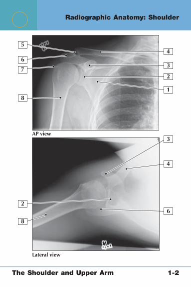
Radiographic Anatomy: Shoulder
1
2
2
3
4
4
5
3
6
6
7
8
8
AP view
Lateral view
The Shoulder and Upper Arm 1-2
Radiographic Anatomy: Shoulder
1. Body of the scapula2. Glenoid3. Coracoid process4. Clavicle5. Acromioclavicular (AC) joint6. Acromion7. Greater tuberosity of the humerus8. Shaft of the humerus
Comment: Anteroposterior and axillary views are the most common views of the shoulder, and both should always be ordered in cases of suspected dislocation.
The Shoulder and Upper Arm 1-2

Soft Tissue Anatomy: Shoulder Joint
8
12
Coronal section through joint
Shoulder joint, anterior view
3
4
5
10
7
4
6
9
11
12
The Shoulder and Upper Arm 1-3
Soft Tissue Anatomy: Shoulder Joint
1. Coracoclavicular ligaments (conoid and trapezoid) 2. Acromioclavicular ligament 3. Coracoacromial ligament 4. Supraspinatus tendon 5. Coracohumeral ligament 6. Subscapularis tendon 7. Long head of the biceps tendon 8. Joint capsule 9. Subdeltoid bursa10. Deltoid muscle11. Glenoid labrum12. Articular cartilage
Comment: The secondary stabilizers (ligaments, muscles, and joint capsule) provide most of the stability for the shoulder joint. The glenohumeral ligaments are really just thickenings of the glenohumeral joint capsule.
The Shoulder and Upper Arm 1-3

1
2
3
4
5
6
Muscles: Shoulder (Anterior View)
The Shoulder and Upper Arm 1-4
Muscles: Shoulder (Anterior View)
1. Pectoralis major muscle2. Trapezius muscle3. Deltoid muscle4. Cephalic vein5. Biceps brachii muscle6. Latissimus dorsi muscle
Deltoid MusclePectoralis Major Muscle
Latissimus Dorsi Muscle
Origin Clavicle, acromion, scapular spine
Medial clavicle and upper sternum
T6-L5 spinous processes
Insertion Deltoid tuberosity, humerus
Intertubercular groove of humerus
Intertubercular groove of humerus
Actions Primarily abduction, fl exion, extension
Arm adduction, assists rotation
Shoulder extension, adduction, and internal rotation
Innervation Axillary nerve (C5-6)
Medial and lateral pectoral nerves (C5-T1)
Thoracodorsal nerve
The Shoulder and Upper Arm 1-4

1
23
4
5
Muscles: Shoulder and Upper Arm (Posterior View)
The Shoulder and Upper Arm 1-5
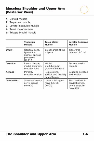
Muscles: Shoulder and Upper Arm (Posterior View)
1. Deltoid muscle2. Trapezius muscle3. Levator scapulae muscle4. Teres major muscle5. Triceps brachii muscle
Trapezius Muscle
Teres Major Muscle
Levator Scapulae Muscle
Origin Occipital bone, ligamentum nuchae, spinous processes C7-T12
Inferior angle of the scapula
Transverse process of C1-4
Insertion Lateral clavicle, medial acromion, scapular spine
Medial intertubercular groove of humerus
Superior medial scapula
Actions Primarily scapular rotation
Helps extend, adduct, and medially rotate the arm
Scapular elevation and rotation
Innervation Spinal accessory nerve (cranial nerve XI)
Lower subscapular nerve (C5-C6, C6-C7)
Third and fourth cervical nerves, dorsal scapular nerve (C5)
The Shoulder and Upper Arm 1-5

1
2
3
4
Posterior view
Anterior view
Muscles: Rotator Cuff
The Shoulder and Upper Arm 1-6

Musc
les: R
ota
tor C
uff
Supraspinatus Muscle
Infraspinatus Muscle
Teres Minor Muscle
Subscapularis Muscle
Origin Supraspinous fossa of scapula
Infraspinous fossa of scapula
Lateral border of the scapula
Subscapular fossa and lateral border of scapula
Insertion Greater tuberosity of humerus
Greater tuberosity of humerus
Greater tuberosity of humerus
Lesser tuberosity of humerus
Actions Shoulder abduction, external rotation
Shoulder external rotation
Shoulder external rotation and assists with adduction
Shoulder internal rotation and adduction
Innervation Suprascapular nerve (C5-6)
Suprascapular nerve (C5-6)
Axillary nerve (C5-6)
Subscapular nerves (C5-6)
1. Subscapularis muscle2. Supraspinatus muscle3. Infraspinatus muscle4. Teres minor muscle
The S
hould
er a
nd U
pper A
rm
1-6
1
1
2
23
3
4
Superficial layer Superficial layer
Deep layer
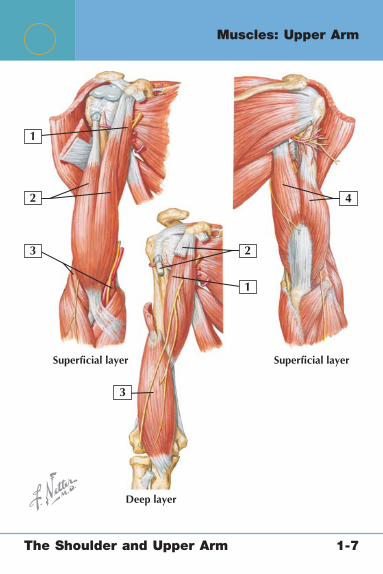
Muscles: Upper Arm
The Shoulder and Upper Arm 1-7
Musc
les: U
pper A
rm
Biceps Brachii Muscle (Long and Short Heads)
Triceps Brachii Muscle (Long, Lateral, and Medial Heads)
Coracobrachialis Muscle Brachialis Muscle
Origin Coracoid process (short head); supraglenoid tubercle of scapula (long head)
Infraglenoid tubercle of scapula (long head), posterior humerus (lateral head), posterior humerus inferior to radial groove (medial head)
Coracoid process of scapula
Distal anterior humerus
Insertion Radial tuberosity Posterior proximal olecranon
Medial aspect of midshaft of humerus
Tuberosity and anterior coronoid process of ulna
Actions Flexion and supination at elbow
Extension at the elbow
Shoulder fl exion and adduction
Elbow fl exion
Innervation Musculocutaneous nerve (C5-6)
Radial nerve (C7-8) Musculocutaneous nerve (C6-7)
Musculocutaneous nerve (C5-6), branch of radial nerve (C7)
1. Coracobrachialis muscle2. Biceps brachii muscle (long and short heads)3. Brachialis muscle4. Triceps brachii muscle (long, lateral)
The S
hould
er a
nd U
pper A
rm
1-7

1
2
3
4
121110
9
8
5
6 7
Anterior view
Arteries: Shoulder and Upper Arm
The Shoulder and Upper Arm 1-8
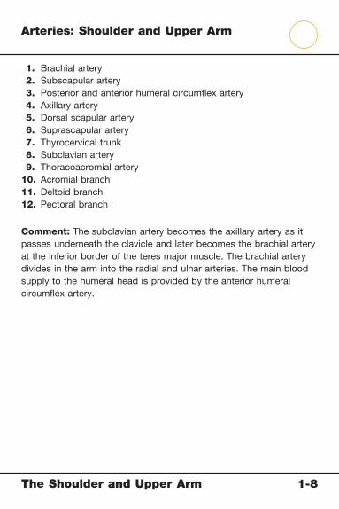
Arteries: Shoulder and Upper Arm
1. Brachial artery 2. Subscapular artery 3. Posterior and anterior humeral circumfl ex artery 4. Axillary artery 5. Dorsal scapular artery 6. Suprascapular artery 7. Thyrocervical trunk 8. Subclavian artery 9. Thoracoacromial artery10. Acromial branch11. Deltoid branch12. Pectoral branch
Comment: The subclavian artery becomes the axillary artery as it passes underneath the clavicle and later becomes the brachial artery at the inferior border of the teres major muscle. The brachial artery divides in the arm into the radial and ulnar arteries. The main blood supply to the humeral head is provided by the anterior humeral circumfl ex artery.
The Shoulder and Upper Arm 1-8

1
C5
C6
C7
C8
T1
2
3
4
5
16
17
18
19
20
21
13
14
15
1211
10
6
7
8
9
Brachial Plexus
The Shoulder and Upper Arm 1-9

Brachial Plexus
1. Roots 2. Trunks 3. Superior trunk 4. Middle trunk 5. Inferior trunk 6. Divisions (posterior and anterior) 7. Cords 8. Lateral cord 9. Lateral pectoral nerve10. Posterior cord11. Upper, middle, and lower subscapular nerves12. Medial cord13. Medial pectoral nerve14. Medial brachial cutaneous nerve15. Medial antebrachial cutaneous nerve16. Terminal branches17. Musculocutaneous nerve18. Axillary nerve19. Radial nerve20. Median nerve21. Ulnar nerve
Comment: The brachial plexus is formed by the nerve roots of C5, C6, C7, C8, and T1. Injuries typically occur when the plexus is stretched while the shoulder is depressed and the neck is laterally fl exed to the opposite side. A helpful mnemonic for the arrangement of the plexus (roots, trunks, divisions, cords, branches) is “Rob Taylor drinks cold beer.”
The Shoulder and Upper Arm 1-9
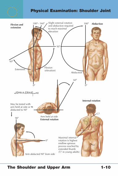
Arm held at sideExternal rotation
S1
T7
C7
Internal rotation
AbductionExtension
Flexion(elevation)
Flexion andextension
AbductionSlight external rotationand abduction requiredto reach maximalelevation
180�– 160�
90�
180�
60�
0�0�
60�
May be tested witharm held at side orabducted to 90�
90�
0�
Arm abducted 90� from side
Maximal internalrotation is highestmidline spinousprocess reached byextended thumb(T7 in young adults)
Physical Examination: Shoulder Joint
The Shoulder and Upper Arm 1-10
Physical Examination: Shoulder Joint
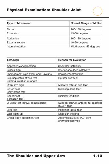
Type of Movement Normal Range of Motion
Flexion 160-180 degrees
Extension 45-60 degrees
Abduction 160-180 degrees
External rotation 40-60 degrees
Internal rotation Midthoracic: 55 degrees
Test/Sign Reason for Evaluation
Apprehension/relocation Shoulder instability
Sulcus sign Inferior shoulder instability
Impingement sign (Neer and Hawkins) Impingement/bursitis
Supraspinatus stress testExternal rotation strength
Rotator cuff tear
Drop arm sign Massive rotator cuff tear
Lift off testBelly press test
Subscapularis tear
Speed testYergason test
Bicipital tendinitis
O’Brien test (active compression) Superior labrum anterior to posterior (SLAP) tear
Jerk test Posterior labral tear
Wall push-up Scapular winging
Cross-body adduction test Acromioclavicular (AC) joint arthritis/osteolysis
The Shoulder and Upper Arm 1-10

1
2
43
5
6
Superior surface
Identify each condition
Inferior surface
Conditions: Clavicle
The Shoulder and Upper Arm 1-11
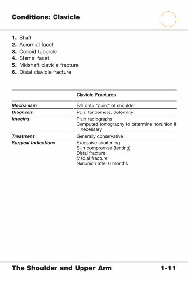
Conditions: Clavicle
1. Shaft2. Acromial facet3. Conoid tubercle4. Sternal facet5. Midshaft clavicle fracture6. Distal clavicle fracture
Clavicle Fractures
Mechanism Fall onto “point” of shoulder
Diagnosis Pain, tenderness, deformity
Imaging Plain radiographsComputed tomography to determine nonunion if
necessary
Treatment Generally conservative
Surgical Indications Excessive shorteningSkin compromise (tenting)Distal fractureMedial fractureNonunion after 6 months
The Shoulder and Upper Arm 1-11
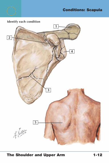
1
2
4
3
5
Identify each condition
Conditions: Scapula
The Shoulder and Upper Arm 1-12

Conditions: Scapula
1. Acromion fracture2. Coracoid process fracture3. Scapular body fracture4. Glenoid fracture5. Scapular winging
Scapular Fracture Scapular Winging
Mechanism Direct trauma Injury to the long thoracic nerve or cranial nerve XI
Diagnosis Anteroposterior, axillary, scapula Y radiographs, computed tomographic scan to further defi ne fracture pattern if necessary
Winging apparent with wall push-ups (weak serratus anterior)
Electromyography confi rms nerve injury
Classifi cation By area of involvement Primary, secondary, voluntary
Treatment Usually conservative Surgical open reduction and
internal fi xation (ORIF) indicated in cases of severely displaced fractures or “fl oating shoulder” (associated clavicle fracture)
Depends on cause of nerve injury, but winging frequently resolves spontaneously
The Shoulder and Upper Arm 1-12
1
Identify each condition
2
4
3
5
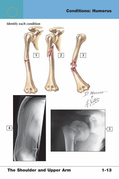
Conditions: Humerus
The Shoulder and Upper Arm 1-13

Conditions: Humerus
1. Transverse midshaft humerus fracture2. Oblique midshaft humerus fracture3. Comminuted midshaft humerus fracture4. Radiographic appearance of oblique midshaft humerus fracture5. Displaced proximal humerus fracture (anteroposterior [AP] view)
Midshaft Humerus Fracture Proximal Humerus Fracture
Mechanism Direct trauma Fall, direct trauma
Classifi cation By fracture type (transverse, oblique, comminuted)
By number of parts (greater tuberosity, lesser tuberosity, head, and shaft)
Imaging AP and lateral radiographs of the humerus
AP and axillary view of the shoulder
Treatment Conservative with protective brace if angulation <30 degrees
Conservative if one part or displacement <45 degrees
SurgicalIndications
Open fracture, associated forearm fracture, severe angulation, or pathological fracture
Surgical treatment is generally open reduction and internal fi xation (ORIF) but may include shoulder replacement in older patients with three- or four-part fractures
The Shoulder and Upper Arm 1-13

23
4
1
5 6
Conditions: Acromioclavicular Joint
The Shoulder and Upper Arm 1-14

Conditions: Acromioclavicular Joint
1. Coracoclavicular (CC) ligament2. Coracoacromial (CA) ligament3. Acromioclavicular (AC) ligament4. Type III AC separation5. Coracoclavicular distance6. Type IV AC separation
AC Separations
Mechanism Fall on “point” of shoulder
Diagnosis Local tenderness and deformity
Imaging Bilateral AC joint view, axillary of affected side
Grading I: AC sprainII: AC tear, intact CCIII: AC and CC tear (up to 100% displacement)IV: AC and CC tear (clavicle displaced posteriorly)V: AC and CC tear (over 100% displacement)VI: AC and CC tear (inferior displacement of clavicle)
Treatment Conservative for types I and IISurgical repair or reconstruction for symptomatic types IV,
and VTreatment for type III is controversial and depends on
individual patient circumstances
The Shoulder and Upper Arm 1-14

5
1 2
3 4
Identify each condition
Conditions: Subacromial Space
The Shoulder and Upper Arm 1-15
Conditions: Subacromial Space
1. Rotator cuff tendinitis2. Partial rotator cuff tear3. Partial rotator cuff tear and subacromial bursitis4. Calcifi c tendonitis5. Radiographic appearance of calcifi c tendinitis
Subacromial Bursitis and Rotator Cuff Tendonitis
Mechanism Overuse/impingement
Diagnosis Pain with overhead reaching, positive Neer and Hawkins impingement signs
Imaging Usually not necessaryPlain radiographs (anteroposterior, outlet, axillary) may show
calcifi c tendonitis
Treatment Generally conservative with nonsteroidal antiinfl ammatory drugs (NSAIDs), subacromial steroid injections, and rotator cuff strengthening
Arthroscopic débridement and acromioplasty for refractory cases
The Shoulder and Upper Arm 1-15
1
2
4
35
6 7
Identify each condition
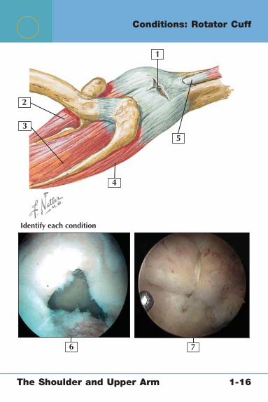
Conditions: Rotator Cuff
The Shoulder and Upper Arm 1-16
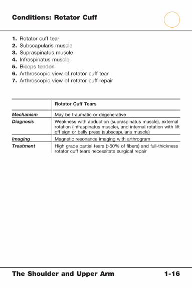
Conditions: Rotator Cuff
1. Rotator cuff tear2. Subscapularis muscle3. Supraspinatus muscle4. Infraspinatus muscle5. Biceps tendon6. Arthroscopic view of rotator cuff tear7. Arthroscopic view of rotator cuff repair
Rotator Cuff Tears
Mechanism May be traumatic or degenerative
Diagnosis Weakness with abduction (supraspinatus muscle), external rotation (infraspinatus muscle), and internal rotation with lift off sign or belly press (subscapularis muscle)
Imaging Magnetic resonance imaging with arthrogram
Treatment High grade partial tears (>50% of fi bers) and full-thickness rotator cuff tears necessitate surgical repair
The Shoulder and Upper Arm 1-16
1
1
2
2 4
4
3
6
5
Conditions: Rotator Cuff
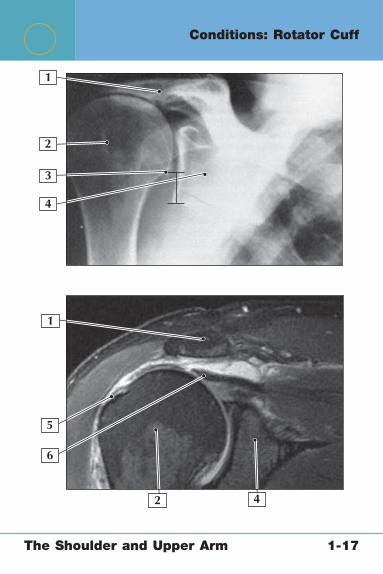
The Shoulder and Upper Arm 1-17

Conditions: Rotator Cuff
1. Acromion2. Humeral head3. Proximal migration of the humeral head4. Glenoid5. Rotator cuff insertion6. Rotator cuff tendon (retracted)
Rotator Cuff Arthropathy
Mechanism Rotator cuff tears that remain untreated which results in signifi cant retraction and fatty atrophy of the muscles
Diagnosis Weakness on examination, drop arm test, “horn blowers” sign
Imaging Plain radiographs show proximal migration of the humeral head, arthrographic magnetic resonance imaging demonstrates retraction and fatty atrophy
Treatment These tears are not repairableTreatment consists of conservative management initially and
later constrained hemiarthroplasty or reverse shoulder prosthesis
The Shoulder and Upper Arm 1-17

12
3
4
Identify the condition
Conditions: Biceps Tendon
The Shoulder and Upper Arm 1-18

Conditions: Biceps Tendon
1. Short head of the biceps, intact2. Long head of the biceps, torn3. Deltoid, refl ected4. Arthroscopic image of frayed biceps tendon
Rupture of the Long Head of the Biceps Biceps Tendinitis
Mechanism Trauma or fraying over time
Overuse
Diagnosis “Popeye” deformity Tenderness over the bicipital groove; positive Yergason and Speed tests
Imaging Not usually necessary Not necessary
Treatment Reassurance that this condition is really a cosmetic problem and does not lead to signifi cant strength loss
Initially conservative with injections of the biceps tendon sheath and physical therapy
Biceps tenotomy (releasing the long head) or biceps tenodesis (surgical fi xation in the groove) if conservative treatment fails
Arthroscopic image from Miller M, Cole B: Textbook of Arthroscopy. Philadelphia: WB Saunders, 2004.
The Shoulder and Upper Arm 1-18

1
3
2
54
Identify each condition
Joint opened:lateral view
Conditions: Biceps Tendon
The Shoulder and Upper Arm 1-19

Conditions: Biceps Tendon
1. Biceps tendon (long head)2. Superior glenoid labrum3. Glenoid articular cartilage4. Magnetic resonance imaging (MRI) of superior labrum anterior to
posterior (SLAP) tear5. Arthroscopic view of SLAP tear
SLAP Tears
Mechanism Most often compression, rotation/abduction, or traction on long head of the biceps
Diagnosis Positive O’Brien test is the classic physical examination fi nding
Imaging Arthrogram MRI confi rms diagnosis
Treatment Arthroscopic repair of the tear
Comment The biceps tendon anchors to the superior part of the glenoid labrum
SLAP tears are common in this location and are graded on severity and presence of disruption of the biceps anchor
The Shoulder and Upper Arm 1-19
1
2
3
4
5
76
Identify each condition
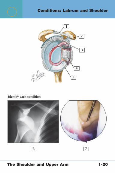
Conditions: Labrum and Shoulder
The Shoulder and Upper Arm 1-20

Conditions: Labrum and Shoulder
1. Coracoacromial ligament2. Coracohumeral ligament3. Superior glenohumeral ligament4. Middle glenohumeral ligament5. Inferior glenohumeral ligament6. Anterior shoulder dislocation (subcoracoid)7. Arthroscopic view of Bankart tear
Shoulder Instability
Mechanism For acute dislocations: trauma with arm in abducted, externally rotated position
Diagnosis Positive apprehension sign, positive relocation test, positive sulcus sign (may indicate multidirectional instability)
Imaging Anteroposterior and axillary radiographs for acute dislocationMay show bony injury such as Hill-Sachs lesion (impaction
fracture of humeral head during relocation)Arthrographic magnetic resonance imaging may show Bankart
lesion (classic labral injury of shoulder dislocation)
Treatment Generally conservative in nontraumatic cases and fi rst-time dislocations
Surgical Bankart repair and/or capsulorrhaphy (capsular imbrication and tightening)
Arthroscopic image from Miller M, Howard R, Plancher K: Surgical Atlas of Sports Medicine. Philadelphia: WB Saunders, 2003.
The Shoulder and Upper Arm 1-20

1
2
3
4
5
Identify the condition
Conditions: Glenohumeral Joint Capsule
The Shoulder and Upper Arm 1-21

Conditions: Glenohumeral Joint Capsule
1. Acromion2. Rotator cuff tendon (supraspinatus)3. Humeral head4. Capsular adhesions5. Arthroscopic view of shoulder with adhesive capsulitis (note
narrowed space and infl amed tissue)
Adhesive Capsulitis (Frozen Shoulder)
Cause Thickened fi brotic joint capsule
Mechanism Unknown but may follow trauma or surgery, or it may occur in patients with autoimmune disorders such as hypothyroidism and diabetes)
Diagnosis Key physical examination fi nding is loss of both active and passive motion
Imaging Imaging usually not necessaryArthrographic magnetic resonance imaging may demonstrate
decreased capsular volume
Treatment Physical therapy to emphasize motion is most effective after glenohumeral joint steroid injection
Arthroscopic lysis of adhesions and manipulation with anesthesia in refractory cases
The Shoulder and Upper Arm 1-21
1 2 3
4 5
Identify each condition
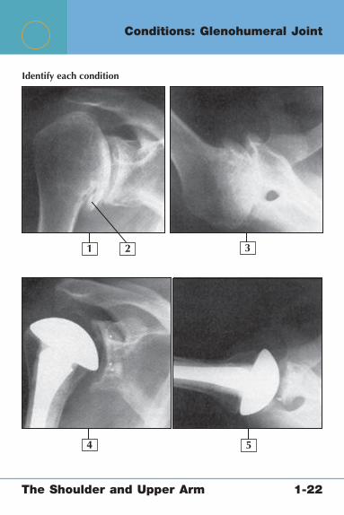
Conditions: Glenohumeral Joint
The Shoulder and Upper Arm 1-22
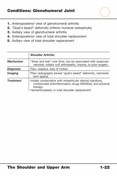
Conditions: Glenohumeral Joint
1. Anteroposterior view of glenohumeral arthritis2. “Goat’s beard” deformity (inferior humeral osteophyte)3. Axillary view of glenohumeral arthritis4. Anteroposterior view of total shoulder replacement5. Axillary view of total shoulder replacement
Shoulder Arthritis
Mechanism “Wear and tear” over time; can be associated with avascular necrosis, rotator cuff arthropathy, trauma, or prior surgery
Diagnosis Pain, crepitus, loss of motion
Imaging Plain radiographs (reveal “goat’s beard” deformity, narrowed joint space)
Treatment Initially conservative with intraarticular steroid injections, nonsteroidal antiinfl ammatory drugs (NSAIDs), and physical therapy
Hemiarthroplasty or total shoulder replacement
The Shoulder and Upper Arm 1-22

Netter’s Musculoskeletal Flash Cards
2 Elbow, Wrist, and HandPlates 2-1 to 2-44
Bony Anatomy
2-1 Bony Anatomy: Elbow
2-2 Bony Anatomy: Forearm
2-3 Bony Anatomy: Wrist and Hand
Radiographic Anatomy
2-4 Radiographic Anatomy: Elbow
2-5 Radiographic Anatomy: Hand
2-6 Radiographic Anatomy: Wrist
Joints
2-7 Elbow Joint
2-8 Wrist Joint
Muscles
2-9 Muscles: Superfi cial Anterior Forearm
2-10 Muscles: Superfi cial Anterior Forearm
2-11 Muscles: Deep Anterior Forearm
2-12 Muscles: Superfi cial Posterior Forearm
2-13 Muscles: Superfi cial Posterior Forearm
2-14 Muscles: Deep Posterior Forearm
2-15 Muscles: Deep Posterior Forearm
2-16 Muscles: Superfi cial Palmar Hand
2-17 Muscles: Deep Palmar Hand
2-18 Muscles: Hand and Fingers
2-19 Muscles and Tendons: Fingers
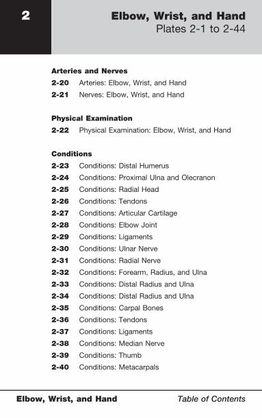
Elbow, Wrist, and Hand Table of Contents
2 Elbow, Wrist, and HandPlates 2-1 to 2-44
Arteries and Nerves
2-20 Arteries: Elbow, Wrist, and Hand
2-21 Nerves: Elbow, Wrist, and Hand
Physical Examination
2-22 Physical Examination: Elbow, Wrist, and Hand
Conditions
2-23 Conditions: Distal Humerus
2-24 Conditions: Proximal Ulna and Olecranon
2-25 Conditions: Radial Head
2-26 Conditions: Tendons
2-27 Conditions: Articular Cartilage
2-28 Conditions: Elbow Joint
2-29 Conditions: Ligaments
2-30 Conditions: Ulnar Nerve
2-31 Conditions: Radial Nerve
2-32 Conditions: Forearm, Radius, and Ulna
2-33 Conditions: Distal Radius and Ulna
2-34 Conditions: Distal Radius and Ulna
2-35 Conditions: Carpal Bones
2-36 Conditions: Tendons
2-37 Conditions: Ligaments
2-38 Conditions: Median Nerve
2-39 Conditions: Thumb
2-40 Conditions: Metacarpals

Netter’s Musculoskeletal Flash Cards
2 Elbow, Wrist, and HandPlates 2-1 to 2-44
2-41 Conditions: Phalanges
2-42 Conditions: Tendons of the Fingers
2-43 Conditions: Tendons of the Fingers
2-44 Conditions: Fingertip

This page intentionally left blank

Elbow, Wrist, and Hand 2-1
Bony Anatomy: Elbow
In extension: anterior view
In extension: posterior view
1
2
3
4
5
6
7
8 9
10
11
17
16
13
14
15
12

Elbow, Wrist, and Hand 2-1
Bony Anatomy: Elbow
1. Humerus 2. Lateral supracondylar ridge 3. Lateral epicondyle 4. Capitellum 5. Radial head 6. Radial neck 7. Radial tuberosity 8. Radius 9. Ulna10. Ulnar tuberosity11. Coronoid process12. Trochlea13. Medial epicondyle14. Coronoid fossa15. Medial supracondylar ridge16. Olecranon fossa17. Olecranon
Comment: The elbow is made up of three articulations: the humerus and ulna, the humerus and radius, and the ulna and radius. The two articulations with the humerus enable elbow extension; the radioulnar joint enables a degree of rotation.

Elbow, Wrist, and Hand 2-2
Right radius and ulnain supination: anterior view
Right radius and ulna inpronation: anterior view
1
2
3
45
6
7
8
9
10
11
12
Bony Anatomy: Forearm

Elbow, Wrist, and Hand 2-2
Bony Anatomy: Forearm
1. Radial head 2. Radial neck 3. Radial tuberosity 4. Olecranon 5. Trochlear notch 6. Coronoid process 7. Ulnar tuberosity 8. Radius 9. Ulna10. Interosseous membrane11. Styloid process of the ulna12. Styloid process of the radius
Comment: The ulna is the longer and more medial of the two bones of the forearm. The primary motions that involve the articulation of these two bones are pronation and supination. The two bones are connected by a thick fi brous interosseous membrane. The radius and ulna cross in the pronated position.

Elbow, Wrist, and Hand 2-3
1
2
3
4 5 6
7
8
9
10
11
12
13
Anterior(palmar)view
Bony Anatomy: Wrist and Hand
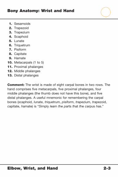
Elbow, Wrist, and Hand 2-3
Bony Anatomy: Wrist and Hand
1. Sesamoids 2. Trapezoid 3. Trapezium 4. Scaphoid 5. Lunate 6. Triquetrum 7. Pisiform 8. Capitate 9. Hamate10. Metacarpals (1 to 5)11. Proximal phalanges12. Middle phalanges13. Distal phalanges
Comment: The wrist is made of eight carpal bones in two rows. The hand comprises fi ve metacarpals, fi ve proximal phalanges, four middle phalanges (the thumb does not have this bone), and fi ve distal phalanges. A useful mnemonic for remembering the carpal bones (scaphoid, lunate, triquetrum, pisiform, trapezium, trapezoid, capitate, hamate) is “Simply learn the parts that the carpus has.”

Elbow, Wrist, and Hand 2-4
1
1
2
2
3
4
45
8
5
7
8
7
6
9
11
6
Lateral radiograph
Anteroposterior radiograph
3
10
Radiographic Anatomy: Elbow

Elbow, Wrist, and Hand 2-4
Radiographic Anatomy: Elbow
1. Humerus 2. Capitellum 3. Lateral epicondyle of humerus 4. Trochlea 5. Olecranon 6. Coronoid process of ulna 7. Radius 8. Ulna 9. Olecranon fossa10. Medial epicondyle of humerus11. Radial head
Comment: The standard elbow radiographs used to detect bony abnormalities of this joint include an anteroposterior (AP) view and a lateral view. Oblique views may also be helpful in identifying loose bodies or avulsion fractures. A useful mnemonic for remembering the ossifi cation order of the bones of the pediatric elbow is Captain Roy Makes Trouble On Leave (capitellum, radial head, medial epicondyle, trochlea, olecranon, lateral epicondyle).

Elbow, Wrist, and Hand 2-5
1
1
7
7
2
4
5
3
6
Radiographic Anatomy: Hand
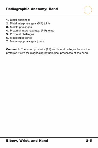
Elbow, Wrist, and Hand 2-5
Radiographic Anatomy: Hand
1. Distal phalanges2. Distal interphalangeal (DIP) joints3. Middle phalanges4. Proximal interphalangeal (PIP) joints5. Proximal phalanges6. Metacarpal bones7. Metacarpophalangeal joints
Comment: The anteroposterior (AP) and lateral radiographs are the preferred views for diagnosing pathological processes of the hand.

Elbow, Wrist, and Hand 2-6
1
2
4
7
8
5
3
611
910
Radiographic Anatomy: Wrist
Elbow, Wrist, and Hand 2-6
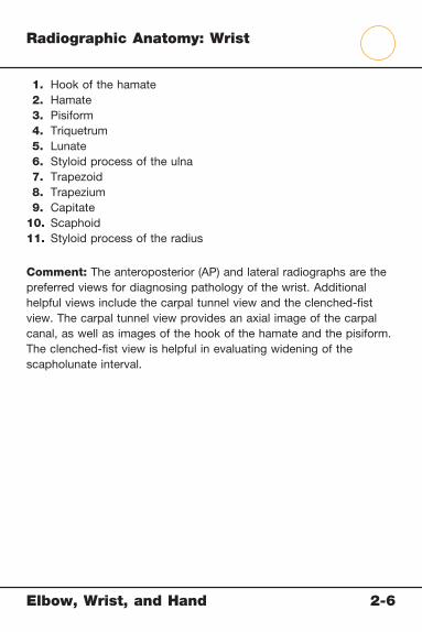
Radiographic Anatomy: Wrist
1. Hook of the hamate 2. Hamate 3. Pisiform 4. Triquetrum 5. Lunate 6. Styloid process of the ulna 7. Trapezoid 8. Trapezium 9. Capitate10. Scaphoid11. Styloid process of the radius
Comment: The anteroposterior (AP) and lateral radiographs are the preferred views for diagnosing pathology of the wrist. Additional helpful views include the carpal tunnel view and the clenched-fi st view. The carpal tunnel view provides an axial image of the carpal canal, as well as images of the hook of the hamate and the pisiform. The clenched-fi st view is helpful in evaluating widening of the scapholunate interval.
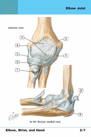
Elbow, Wrist, and Hand 2-7
1
23
4
5
6
7
8
9
Anterior view
In 90° flexion: medial view
Elbow Joint

Elbow, Wrist, and Hand 2-7
Elbow Joint
1. Ulnar collateral ligament2. Medial epicondyle of humerus3. Joint capsule4. Lateral epicondyle of humerus5. Radial collateral ligament6. Annular ligament7. Biceps tendon8. Triceps tendon9. Olecranon bursa
Comment: The ulnar collateral, radial collateral, and annular ligaments are the most important static stabilizers of the elbow joint. The anterior band of the ulnar collateral ligament is the strongest and resists valgus stress at the elbow. The radial collateral ligament is most important in posterolateral elbow stability, whereas the annular ligament is most important in stabilizing the radial head.

Elbow, Wrist, and Hand 2-8
1
9
2
28
7
6
11
12
4
4
5
7
3
8
10
Palmar view: Flexor retinaculum removed
Posterior (dorsal) view
Wrist Joint

Elbow, Wrist, and Hand 2-8
Wrist Joint
1. Palmar metacarpal ligaments 2. Ulnar collateral ligament 3. Palmar ulnocarpal ligament 4. Ulnar styloid 5. Palmar radioulnar ligament 6. Palmar radiocarpal ligament 7. Radial styloid 8. Radial collateral ligament 9. Capitotriquetral ligament10. Dorsal radioulnar ligament11. Dorsal radiocarpal ligament12. Dorsal carpometacarpal ligaments
Comment: The wrist joint comprises articulations between the distal radius and ulna and the fi rst row of carpal bones (scaphoid, lunate, and triquetrum). Dorsal and palmar ligaments support these articulations. Wrist arthroscopy allows a unique view of injuries to the ligaments and carpal bones.

Elbow, Wrist, and Hand 2-9
1
2
3
Muscles: Superfi cial Anterior Forearm

Elbow, Wrist, and Hand 2-9
Muscles: Superfi cial Anterior Forearm
1. Brachioradialis muscle2. Flexor carpi ulnaris muscle3. Flexor digitorum superfi cialis muscle
Brachioradialis Muscle
Flexor Digitorum Superfi cialis Muscle
Flexor Carpi Ulnaris Muscle
Origin Lateral aspect of the supracondylar ridge of the humerus
Medial epicondyle of humerus, ulnar collateral ligament, and coronoid process of ulna; superoanterior radius
Humeral head from medial epicondyle of humerus; ulnar head from medial border of olecranon and posterior ulna
Insertion Lateral aspect of the distal radius
Middle phalanges of the four fi ngers
Pisiform bone primarily but also the hook of the hamate and the base of the fi fth metacarpal
Actions Flexion of the elbow
Proximal interphalangeal (PIP) joint fl exion; assistance in fl exion of the elbow, wrist, and metacarpophalangeal (MCP) joints
Wrist fl exion and ulnar deviation
Innervation Radial nerve (C5-7)
Median nerve (C7, C8, and T1)
Ulnar nerve (C7-8)

Elbow, Wrist, and Hand 2-10
1
2
3
Muscles: Superfi cial Anterior Forearm
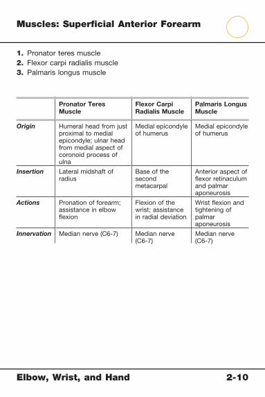
Elbow, Wrist, and Hand 2-10
Muscles: Superfi cial Anterior Forearm
1. Pronator teres muscle2. Flexor carpi radialis muscle3. Palmaris longus muscle
Pronator Teres Muscle
Flexor Carpi Radialis Muscle
Palmaris Longus Muscle
Origin Humeral head from just proximal to medial epicondyle; ulnar head from medial aspect of coronoid process of ulna
Medial epicondyle of humerus
Medial epicondyle of humerus
Insertion Lateral midshaft of radius
Base of the second metacarpal
Anterior aspect of fl exor retinaculum and palmar aponeurosis
Actions Pronation of forearm; assistance in elbow fl exion
Flexion of the wrist; assistance in radial deviation
Wrist fl exion and tightening of palmar aponeurosis
Innervation Median nerve (C6-7) Median nerve (C6-7)
Median nerve (C6-7)

Elbow, Wrist, and Hand 2-11
1
2
3
4
Muscles: Deep Anterior Forearm

Elb
ow
, Wrist, a
nd H
and
2-1
1
Musc
les: D
eep A
nte
rior F
ore
arm
1. Supinator muscle2. Flexor pollicis longus muscle3. Pronator quadratus muscle4. Flexor digitorum profundus muscle
Supinator MuscleFlexor Pollicis Longus Muscle
Pronator Quadratus Muscle
Flexor Digitorum Profundus Muscle
Origin Lateral epicondyle of humerus, radial collateral ligament, annular ligament of radioulnar joint, supinator fossa, and crest of the ulna
Anterior aspect of the radius
Anteromedial distal ulna
Anteromedial part of proximal three fourths of the ulna
Insertion Anterior, lateral, and posterior proximal radius
Base of the distal phalanx of the thumb
Distal part of the anterolateral radius
Bases of the distal interphalangeal (DIP) joints of the four fi ngers
Actions Supination Flexion of the thumb
Pronation Flexion of the DIP joints of the fi ngers
Innervation Radial nerve (C5-6) Median nerve (C8-T1)
Median nerve (C8-T1)
Medial part by the ulnar nerve (C8-T1); lateral part by the anterior interosseus branch of the median nerve (C8-T1)

Elbow, Wrist, and Hand 2-12
1
2
Muscles: Superfi cial Posterior Forearm
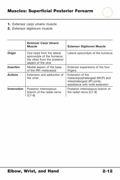
Elbow, Wrist, and Hand 2-12
Muscles: Superfi cial Posterior Forearm
1. Extensor carpi ulnaris muscle2. Extensor digitorum muscle
Extensor Carpi Ulnaris Muscle Extensor Digitorum Muscle
Origin One head from the lateral epicondyle of the humerus; the other from the posterior aspect of the ulna
Lateral epicondyle of the humerus
Insertion Medial aspect of the base of the fi fth metacarpal
Extensor expansions of the four fi ngers
Actions Extension and adduction of the wrist
Extension of the metacarpophalangeal (MCP) and interphalangeal (IP) joints; assistance with wrist extension
Innervation Posterior interosseous branch of the radial nerve (C7-8)
Posterior interosseous branch of the radial nerve (C7-8)

Elbow, Wrist, and Hand 2-13
1
2
3
Muscles: Superfi cial Posterior Forearm
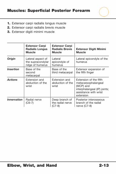
Elbow, Wrist, and Hand 2-13
Muscles: Superfi cial Posterior Forearm
1. Extensor carpi radialis longus muscle2. Extensor carpi radialis brevis muscle3. Extensor digiti minimi muscle
Extensor Carpi Radialis Longus Muscle
Extensor Carpi Radialis Brevis Muscle
Extensor Digiti Minimi Muscle
Origin Lateral aspect of the supracondylar ridge of humerus
Lateral epicondyle of humerus
Lateral epicondyle of the humerus
Insertion Base of the second metacarpal
Base of the third metacarpal
Extensor expansion of the fi fth fi nger
Actions Extension and abduction of the wrist
Extension and abduction of the wrist
Extension of the fi fth metacarpophalangeal (MCP) and interphalangeal (IP) joints; assistance with wrist extension
Innervation Radial nerve (C6-7)
Deep branch of the radial nerve (C7-8)
Posterior interosseous branch of the radial nerve (C7-8)

Elbow, Wrist, and Hand 2-14
1
2
Muscles: Deep Posterior Forearm

Elbow, Wrist, and Hand 2-14
Muscles: Deep Posterior Forearm
1. Anconeus muscle2. Extensor pollicis longus muscle
Anconeus Muscle Extensor Pollicis Longus Muscle
Origin Lateral epicondyle of the humerus
Posterior aspect of midshaft of the ulna
Insertion Lateral aspect of the olecranon and posterosuperior ulna
Base of the distal phalanx of the thumb
Actions Abduction of ulna during pronation
Extension of the distal phalanx of the thumb; assistance in thumb abduction
Innervation Radial nerve (C7-T1) Posterior interosseous branch of the radial nerve (C7-8)

Elbow, Wrist, and Hand 2-15
1
2
3
Muscles: Deep Posterior Forearm

Elbow, Wrist, and Hand 2-15
Muscles: Deep Posterior Forearm
1. Extensor indicis muscle2. Abductor pollicis longus muscle3. Extensor pollicis brevis muscle
Extensor Indicis Muscle
Abductor Pollicis Longus Muscle
Extensor Pollicis Brevis Muscle
Origin Posterior ulna just distal to the extensor pollicis longus origin
Posterior radius and ulna
Posterior radius
Insertion Extensor expansion of index fi nger
Base of the fi rst metacarpal
Base of the proximal phalanx of the thumb
Actions Extension of the index fi nger; assistance in wrist extension
Abduction, extension, and rotation of the thumb; assistance in abduction of the wrist
Extension of the proximal phalanx of the thumb
Innervation Posterior interosseous branch of the radial nerve (C7-8)
Posterior interosseous branch of the radial nerve (C7-8)
Posterior interosseous branch of the radial nerve (C7-8)
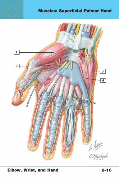
Elbow, Wrist, and Hand 2-16
1
2
4
3
Muscles: Superfi cial Palmar Hand
Elb
ow
, Wrist, a
nd H
and
2-1
6
Musc
les: S
uperfi c
ial P
alm
ar H
and
Abductor Pollicis Brevis Muscle
Flexor Pollicis Brevis Muscle
Abductor Digiti Minimi Muscle
Flexor Digiti Minimi Brevis Muscle
Origin Flexor retinaculum, scaphoid, trapezium
Superfi cial head from fl exor retinaculum and trapezium; deep head from fl oor of the carpal canal overlying the trapezoid and capitate
Pisiform and fl exor carpi ulnaris muscle
Flexor retinaculum and hook of the hamate
Insertion Lateral aspect of the base of the proximal phalanx of the thumb
Lateral aspect of fi rst metacarpal and base of proximal phalanx
Medial aspect of the base of the proximal phalanx of the fi fth fi nger
Medial aspect of the base of the proximal phalanx of the fi fth fi nger
Actions Abduction of thumb Flexion of the proximal phalanx of the thumb
Abduction of the fi fth fi nger
Flexion of the proximal phalanx of the fi fth fi nger
Innervation Recurrent branch of the median nerve (C8-T1)
Recurrent branch of the median nerve (C8-T1)
Deep branch of the ulnar nerve (C8-T1)
Deep branch of the ulnar nerve (C8-T1)
1. Abductor pollicis brevis muscle2. Flexor pollicis brevis muscle3. Abductor digiti minimi muscle4. Flexor digiti minimi brevis muscle
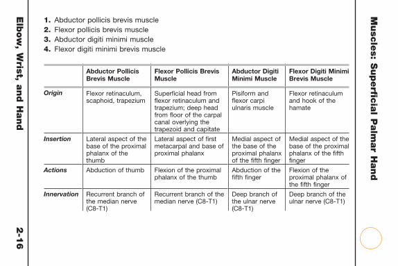
Elbow, Wrist, and Hand 2-17
1
2 3
Muscles: Deep Palmar Hand

Elbow, Wrist, and Hand 2-17
Muscles: Deep Palmar Hand
1. Opponens pollicis muscle2. Adductor pollicis muscle3. Opponens digiti minimi muscle
Opponens Pollicis Muscle
Adductor Pollicis Muscle
Opponens Digiti Minimi Muscle
Origin Flexor retinaculum and trapezium
Oblique head from the bases of second and third metacarpals and the capitate; transverse head from anterior aspect of the third metacarpal
Flexor retinaculum and hook of the hamate
Insertion Lateral aspect of the fi rst metacarpal
Base of the proximal phalanx of the thumb
Palmar surface of fi fth metacarpal
Actions Rotation of thumb to oppose fi ngers
Adduction of the proximal phalanx of the thumb
Abduction, fl exion, and rotation of the fi fth metacarpal
Innervation Recurrent branch of the median nerve (C8-T1)
Deep branch of the ulnar nerve (C8-T1)
Deep branch of the ulnar nerve (C8-T1)
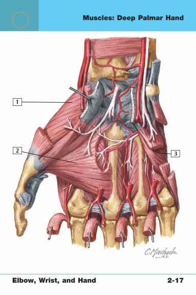
Elbow, Wrist, and Hand 2-18
1
3
2
Posterior (dorsal) view Anterior (palmar) view
Muscles: Hand and Fingers

Elb
ow
, Wrist, a
nd H
and
2-1
8
Musc
les: H
and a
nd F
ingers
1. Dorsal interosseous muscles2. Palmar interosseous muscles3. Lumbrical muscles
Dorsal Interosseous Muscles (Four)
Palmar Interosseous Muscles (Three) Lumbrical Muscles
Origin One head from each side of the metacarpal
Palmar aspects of the second, fourth, and fi fth metacarpals
Tendon of the fl exor digitorum profundus (lumbricales 1 and 2 from the lateral two tendons, and lumbricales 3 and 4 from the medial three tendons)
Insertion Base of the proximal phalanx and extensor expansion of fi ngers 2 to 4
Extensor expansions and bases of proximal phalanges of fi ngers 2, 4, and 5
Lateral aspect of the extensor expansions of fi ngers 2 to 5
Actions Finger abduction; assistance with fl exion at the metacarpophalangeal (MCP) joints; assistance with extension at the interphalangeal (IP) joints
Finger adduction; assistance with fl exion at the MCP joints; assistance with extension at the IP joints
Flexion of the MCP joints; extension of the IP joints
Innervation Deep branch of the ulnar nerve (C8-T1)
Deep branch of the ulnar nerve (C8-T1)
Fingers1 and 2 by the median nerve (C8-T1); 3 and 4 by a deep branch of the ulnar nerve (C8-T1)
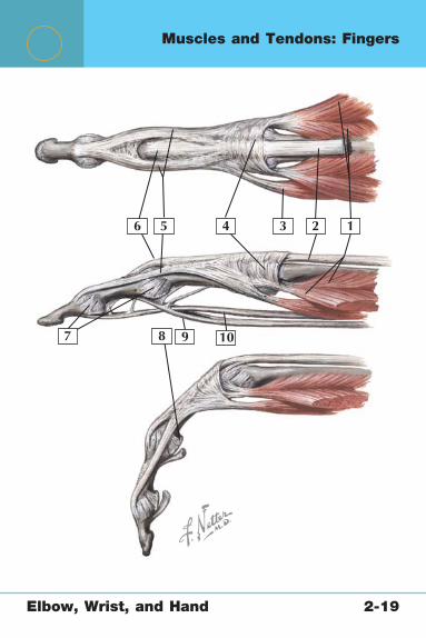
Elbow, Wrist, and Hand 2-19
15 36 24
7 8 109
Muscles and Tendons: Fingers
Elbow, Wrist, and Hand 2-19
Muscles and Tendons: Fingers
1. Interosseous muscles 2. Extensor digitorum longus tendon 3. Lumbrical muscle 4. Extensor expansion (hood) 5. Lateral bands 6. Central band 7. Collateral ligaments 8. Palmar ligament (plate) 9. Flexor digitorum superfi cialis muscle10. Flexor digitorum profundus muscle
Comment: Finger fl exion and extension occur through a complex system of pulleys, bands (sagittal and lateral), a central slip, a volar plate, and retinacular ligaments. The pulleys are really thickenings of the fl exor sheath and are important biomechanically to prevent “bowstringing” of the tendons.

Elbow, Wrist, and Hand 2-20
1
3
6
7
8
4
5
2
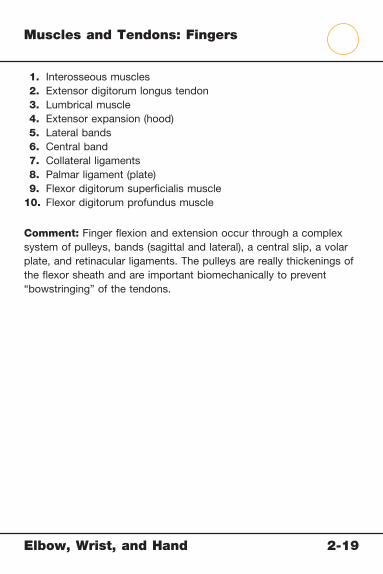
Arteries: Elbow, Wrist, and Hand
Elbow, Wrist, and Hand 2-20
Arteries: Elbow, Wrist, and Hand
1. Brachial artery2. Radial artery3. Ulnar artery4. Deep palmar branch of ulnar artery5. Superfi cial palmar arch (cut)6. Superfi cial palmar branch of radial artery7. Deep palmar arch8. Common palmar digital arteries
Comment: The radial artery passes over the pronator teres muscle and under the brachioradialis muscle. Distally, it separates into the palmar carpal, dorsal carpal, and superfi cial branches. It forms the deep palmar arch distally. The ulnar artery crosses between the fl exor digitorum superfi cialis and profundus muscles and forms four branches distally: the palmar and dorsal carpal branches, the deep palmar branch, and the superfi cial palmar arch.

Elbow, Wrist, and Hand 2-21
1
3
6
7
8
9
10
11
4
5
2
Nerves: Elbow, Wrist, and Hand

Elbow, Wrist, and Hand 2-21
Nerves: Elbow, Wrist, and Hand
1. Median nerve 2. Anterior interosseous nerve 3. Palmar branch of median nerve 4. Ulnar nerve 5. Dorsal branch of ulnar nerve 6. Palmar branch of ulnar nerve 7. Superfi cial branch of ulnar nerve 8. Deep branch of ulnar nerve 9. Radial nerve10. Posterior interosseous nerve11. Superfi cial branch of the radial nerve
Comment: The median nerve (C5-T1) passes anteriorly and centrally through the elbow to supply the anterior compartment of the forearm; then it passes into the hand through the carpal tunnel, where it provides innervation to the thumb, index, and middle fi ngers. The ulnar nerve (C7-T1) arises from the medial cord of the brachial plexus, passes the elbow just posterior to the medial epicondyle, and moves into the wrist through the Guyon canal. It supplies the fl exor digitorum profundus and carpi ulnaris muscles. It also has both motor and sensory supplies to the ulnar aspect of the palm. The radial nerve (C5-T1) divides into superfi cial and deep branches at the elbow and innervates the wrist extensors and sensation to the posterior forearm and dorsal hand (thumb, index fi nger, middle fi nger, and half of the ring fi nger).

Elbow, Wrist, and Hand 2-22
Distalpalmarcrease
Normal finger flexion iscomposite of flexion ofMP, PIP, and DIP jointsand allows fingertip totouch distal palmar crease.
Normal thumbopposition iscomposite ofmovements ofCMC, MP, andIP joints. Normalrange is to baseof little finger.
Limitation of finger flexion maybe quantified by measuringdistance from fingertip to distalpalmar crease.
Limitations ofthumboppositionmay bequantifiedby measuringdistance fromtip of thumbto base of little finger.
0�
Pronation
0�
85�75�
Supination
0�
Physical Examination: Elbow, Wrist, and Hand

Elbow, Wrist, and Hand 2-22
Physical Examination: Elbow, Wrist, and Hand
Movement Normal Range of Motion
Elbow fl exion 135-145 Degrees
Elbow extension 0 Degrees
Supination 85-90 Degrees
Pronation 80-90 Degrees
Wrist fl exion 80 Degrees
Wrist extension 70 Degrees
Radial deviation 20 Degrees
Ulnar deviation 30-45 Degrees
Finger (MCP) fl exion 90 Degrees
Finger (MCP) extension 30-45 Degrees
Thumb (MCP) fl exion 50 Degrees
Thumb (MCP) extension 0 Degrees
Thumb abduction 70 Degrees
Thumb adduction 0 Degrees
Test/Sign Reason for Evaluation
Valgus stress test Ulnar collateral ligament (UCL) tear
Varus stress test Lateral collateral ligament (LCL) tear
Tinel sign (elbow) Ulnar nerve dysfunction
Phalen testTinel sign (wrist)
Carpal tunnel syndrome
Finkelstein test DeQuervain tendonitis
Allen test Radial and ulnar artery function
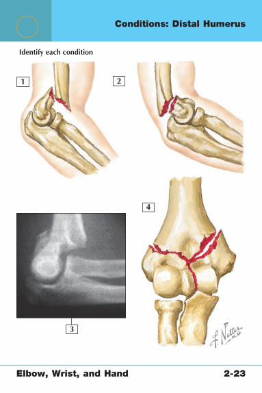
Elbow, Wrist, and Hand 2-23
1
Identify each condition
3
4
2
Conditions: Distal Humerus
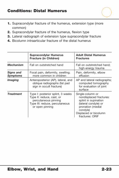
Elbow, Wrist, and Hand 2-23
Conditions: Distal Humerus
1. Supracondylar fracture of the humerus, extension type (more common)
2. Supracondylar fracture of the humerus, fl exion type3. Lateral radiograph of extension type supracondylar fracture4. Bicolumn intraarticular fracture of the distal humerus
Supracondylar Humerus Fracture (in Children)
Adult Distal Humerus Fractures
Mechanism Fall on outstretched hand Fall on outstretched hand; high-energy trauma
Signs and Symptoms
Focal pain, deformity, swelling; more common in children
Pain, deformity, elbow effusion
Imaging Anteroposterior (AP), lateral, and oblique radiographs (fat pad sign in occult fracture)
AP and lateral radiographs; computed tomography for evaluation of joint surface
Treatment Type I: posterior splint, 3 weeksType II: reduce, cast, or
percutaneous pinningType III: reduce, percutaneous
or open pinning
Single-column or nondisplaced fractures: splint in supination (lateral condyle) or pronation (medial condyle)
Displaced or bicolumn fractures: ORIF

Elbow, Wrist, and Hand 2-24
1
3
4
2
Identify each condition
Conditions: Proximal Ulna and Olecranon

Elb
ow
, Wrist, a
nd H
and
2-2
4
Conditio
ns: P
roxim
al U
lna
and O
lecra
non
1. Olecranon fracture2. Olecranon bursitis
3. Coronoid fracture4. Monteggia fracture
Olecranon Fracture Olecranon BursitisCoronoid Fracture
Monteggia Fracture
Mechanism Direct fall on elbow Usually direct trauma to the olecranon
Often associated with a posterior elbow dislocation
Direct blow or fall on the outstretched hand
Signs and Symptoms
Focal pain, swelling, deformity
Painless localized swelling over the olecranon
Pain, elbow deformity, joint effusion
Pain, tenderness, deformity
Imaging Anteroposterior (AP), lateral, oblique radiographs
Plain AP and lateral radiographs to evaluate for olecranon fracture
AP, lateral, oblique radiographs
AP/lateral of forearm, wrist, and elbow shows proximal ulna fracture and radial head dislocation
Treatment Nondisplaced fractures: cast in fl exion for 3 weeks, then begin range-of-motion exercises
Displaced fractures: open reduction, internal fi xation (ORIF) with tension band construct
Conservative with padding and compressive sleeve
Aspiration may speed recovery; an infected bursa necessitates surgical incision and drainage (I&D)
Types I and II fractures: hinged brace
More severe cases: ORIF
ORIF of ulna and reduction (closed or open) of the radial head
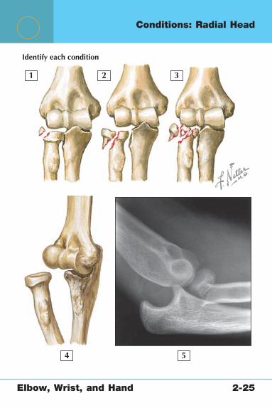
Elbow, Wrist, and Hand 2-25
1 3
4 5
2
Identify each condition
Conditions: Radial Head

Elbow, Wrist, and Hand 2-25
Conditions: Radial Head
1. Radial head fracture, type I: nondisplaced2. Type II: displaced3. Type III: comminuted4. Type IV: radial head fracture with associated elbow dislocation5. Radial head subluxation (nursemaid’s elbow)
Radial Head FracturesRadial Head Subluxation (Nursemaid’s Elbow)
Mechanism Fall on outstretched hand Pulled or swung by hand.
Signs andSymptoms
Focal tenderness, swelling Patients aged 2 to 4 years refuse to use arm; arm remains in fl exed/pronated position
Imaging Anteroposterior (AP), lateral, oblique radiographs (fat pad sign in occult fracture)
AP and lateral radiographs to rule out fracture
Treatment Displaced less than 3 mm or involving one third of the bone: splint and early range-of-motion exercises
More severe or comminuted fracture: open reduction, internal fi xation (ORIF) or radial head excision
Reduce by supinating and fl exing elbow
Radial head subluxation image from DeLee J, Drez D, Miller M: DeLee & Drez’s Orthopaedic Sports Medicine. Philadelphia: WB Saunders, 2002.

Elbow, Wrist, and Hand 2-26
2
3
4 5 6
7 8
1
In 90� flexion: lateral view
Conditions: Tendons

Elb
ow
, Wrist, a
nd H
and
2-2
6
1. Triceps brachii tendon (cut)2. Olecranon bursa3. Joint capsule4. Biceps brachii tendon (cut)5. Lateral epicondyle
Distal Biceps RuptureDistal Triceps Tendon Avulsion
Lateral Epicondylitis (Tennis Elbow)
Medial Epicondylitis (Golfer’s Elbow)
Mechanism Forceful eccentric overload of the biceps tendon
Deceleration force at the elbow; associated with chronic olecranon bursitis
Overuse commonly; may be associated with trauma
Overuse; may be associated with trauma
Signs and Symptoms
Pop, pain, deformity, loss of fl exion and supination strength
Posterior pain, palpable defect, weakness with elbow extension
Lateral epicondyle tenderness and pain with wrist extension
Medial epicondyle tenderness and pain with wrist fl exion and resisted pronation
Imaging Not usually necessary, but magnetic resonance imaging (MRI) helpful if diagnosis is in question
Plain anteroposterior (AP) and lateral radiographs
Not necessary unless history of trauma or suspicion of loose bodies
Not necessary unless history of trauma or suspicion of loose bodies
Treatment Surgical repair of the tendon
Surgical repair of the avulsion
Generally conservative with nonsteroidal antiinfl ammatory drugs (NSAIDs), activity modifi cation, elbow strap, and steroid injections
Surgery is reserved for refractory cases
Same as lateral epicondylitis
Arthroscopic image from Miller M, Cole B: Textbook of Arthroscopy. Philadelphia: WB Saunders, 2004. Radiograph from Miller M, Sekiya J: Core Knowledge in Orthopaedics: Sports Medicine. Philadelphia: WB Saunders, 2006.
6. Medial epicondyle7. Arthroscopic image of extensor carpi radialis brevis tear in
case of lateral epicondylitis8. Lateral radiograph of triceps tendon avulsion injury
Conditio
ns: T
endons
Elbow, Wrist, and Hand 2-27
1
3
4
5
2
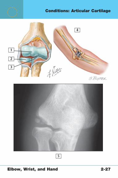
Conditions: Articular Cartilage

Elb
ow
, Wrist, a
nd H
and
2-2
7
Conditio
ns: A
rticula
r Cartila
ge
Panner’s Disease Osteochondritis Dissecans Osteoarthritis
Mechanism Idiopathic osteochondrosis
Focal vascular insuffi ciency Posttraumatic or age-related wear
Signs and Symptoms
Pain in a child aged 4 to 8 years
Pain, catching, and/or locking in a child aged 13 to 16 years
Primarily pain, but patient may complain of catching or locking sensation
Imaging Anteroposterior (AP) and lateral radiographs show involvement of entire capitellum
AP and lateral radiographs show lesion of the capitellum
Magnetic resonance imaging (MRI) to evaluate stability of fragment
AP and lateral radiographs show joint space narrowing and osteophytes
Treatment Conservative with rest and activity modifi cation, self-limited
If fragment is stable: conservative
If fragment if unstable or condition is refractory to other treatment: surgical débridement or fi xation
Initially conservative, with nonsteroidal antiinfl ammatory drugs (NSAIDs), activity modifi cation, steroid injections
Surgical treatment includes arthroscopic débridement or elbow arthroplasty
Arthroscopic image from Miller M, Cole B: Textbook of Arthroscopy. Philadelphia: WB Saunders, 2004.
1. Articular cartilage of the capitellum and trochlea2. Articular cartilage of the radial head3. Articular cartilage of the proximal ulna4. Total elbow replacement5. Anteroposterior (AP) radiograph showing osteochondritis dissecans of the capitellum
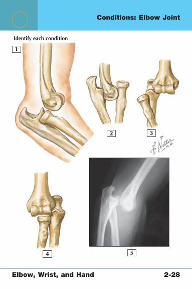
Elbow, Wrist, and Hand 2-28
1
2 3
4 5
Identify each condition
Conditions: Elbow Joint

Elbow, Wrist, and Hand 2-28
Conditions: Elbow Joint
1. Posterior elbow dislocation2. Divergent elbow dislocation3. Lateral elbow dislocation4. Medial elbow dislocation5. Lateral radiograph of posterolateral elbow dislocation
Elbow Dislocation
Mechanism Fall, high-energy trauma; more common in pediatric patients
Signs and Symptoms Pain, deformity, loss of ability to fl ex at elbow
Imaging Anteroposterior (AP), lateral, and oblique radiographs; rule out associated fractures; arteriography if vascular examination fi ndings are abnormal
Treatment Reduce immediately; splint 7 days; early range-of-motion exercises; surgery if persistent instability later
Comment Posterior and posterolateral dislocations are the most common

Elbow, Wrist, and Hand 2-29
1
345
6 7
2
Conditions: Ligaments

Elbow, Wrist, and Hand 2-29
Conditions: Ligaments
1. Joint capsule2. Annular ligament of the radius3. Radial collateral ligament4. Ulnar collateral ligament (UCL)5. Olecranon bursa6. Ulnar collateral ligament (UCL) tear7. Lateral collateral ligament (LCL) tear
UCL Injury LCL Injury
Mechanism Repetitive valgus stress (baseball pitch)
Often with elbow dislocation
Signs and Symptoms
Medial pain, ulnar neuropathy, valgus instability (although not always present)
Lateral pain, clicking/locking with full extension, posterolateral instability on examination
Imaging Arthrographic magnetic resonance imaging (MRI)
Arthrographic MRI
Treatment Conservative initially with ice, nonsteroidal antiinfl ammatory drugs (NSAIDs), activity modifi cation, and physical therapy
Surgical ligament reconstruction for refractory cases or in high- performing athletes
Conservative initially; surgical ligament reconstruction referral for refractory cases
Radiographs from Sanders TG, Miller MD: Imaging of the upper extremities. Clin Sports Med 25(3):395, Fig. 10 (UCL), 398, Fig. 14 (LCL), 2006.

Elbow, Wrist, and Hand 2-30
1
2
3
4
5
6
7
8
Conditions: Ulnar Nerve
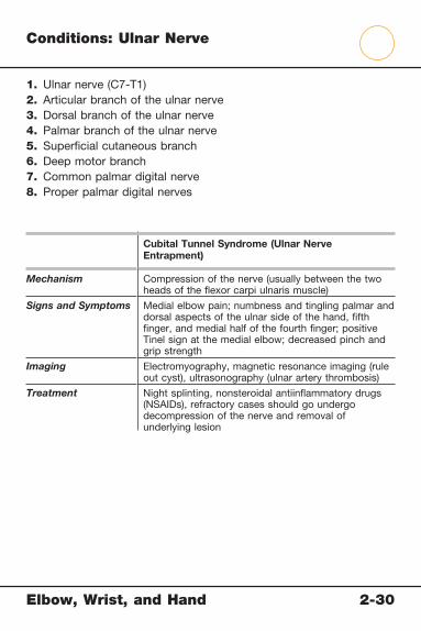
Elbow, Wrist, and Hand 2-30
Conditions: Ulnar Nerve
1. Ulnar nerve (C7-T1)2. Articular branch of the ulnar nerve3. Dorsal branch of the ulnar nerve4. Palmar branch of the ulnar nerve5. Superfi cial cutaneous branch6. Deep motor branch7. Common palmar digital nerve8. Proper palmar digital nerves
Cubital Tunnel Syndrome (Ulnar Nerve Entrapment)
Mechanism Compression of the nerve (usually between the two heads of the fl exor carpi ulnaris muscle)
Signs and Symptoms Medial elbow pain; numbness and tingling palmar and dorsal aspects of the ulnar side of the hand, fi fth fi nger, and medial half of the fourth fi nger; positive Tinel sign at the medial elbow; decreased pinch and grip strength
Imaging Electromyography, magnetic resonance imaging (rule out cyst), ultrasonography (ulnar artery thrombosis)
Treatment Night splinting, nonsteroidal antiinfl ammatory drugs (NSAIDs), refractory cases should go undergo decompression of the nerve and removal of underlying lesion
Elbow, Wrist, and Hand 2-31
123
4
5
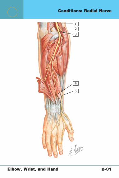
Conditions: Radial Nerve

Elbow, Wrist, and Hand 2-31
Conditions: Radial Nerve
1. Radial nerve2. Superfi cial branch of the radial nerve3. Deep branch of the radial nerve4. Posterior interosseus nerve5. Superfi cial branch of the radial nerve
Posterior Interosseous Nerve (PIN) Syndrome
Mechanism Compression of the radial nerve at the proximal border of the supinator muscle (radial head fracture, ganglion)
Signs and Symptoms Weakness of the thumb, fi nger extensors, extensor carpi ulnaris muscle; tenderness 5 cm distal to the lateral epicondyle
Imaging Electromyography, injection test
Treatment Surgical decompression of the nerve

Elbow, Wrist, and Hand 2-32
1
Identify each condition in 1 and 2
2
34
5
76
8
withC.A. Luce
Conditions: Forearm, Radius, and Ulna

Elbow, Wrist, and Hand 2-32
Conditions: Forearm, Radius, and Ulna
1. Radius and ulna fracture (“both bone forearm fracture”)2. X-ray view of both bone forearm fracture3. Extensor carpi radialis longus muscle4. Extensor carpi radialis brevis muscle5. Abductor pollicis longus muscle6. Extensor pollicis brevis muscle7. Extensor pollicis longus muscle8. Intersection syndrome
Radius and Ulna Fracture
Ulna Fracture(“Nightstick Fracture”) Intersection Syndrome
Mechanism High-energy trauma/fall
Direct blow to ulnar side of forearm
Overuse causing infl ammation of the crossing point between the abductor pollicis longus/extensor pollicis brevis muscles and the extensor carpi radialis longus and brevis muscles (fi rst and second dorsal compartment)
Signs and Symptoms
Pain, local edema, deformity
Focal pain, deformity less common
Forearm pain, crepitus (“squeakers”), commonly seen in rowers and weight lifters
Imaging Anteroposterior (AP) and lateral forearm radiographs
AP and lateral forearm radiographs
Not necessary
Treatment Open reduction, internal fi xation (ORIF) with plate and screws on both bones
Usually treated in cast; ORIF for severe displacement
Activity modifi cation, splinting, local steroid injection

Elbow, Wrist, and Hand 2-33
1 2 3 4
56
7
8
Identify each condition
Conditions: Distal Radius and Ulna

Elbow, Wrist, and Hand 2-33
Conditions: Distal Radius and Ulna
1. Frykman I: Extraarticular radius fracture2. Frykman II: Extraarticular radius fracture with ulnar styloid fracture3. Frykman III: Radiocarpal joint fracture4. Frykman IV: Radiocarpal joint and ulnar styloid fractures5. Frykman V: Radioulnar joint fracture6. Frykman VI: Radioulnar joint and ulnar styloid fractures7. Frykman VII: Radiocarpal and radioulnar joint fractures8. Frykman VIII: Radiocarpal, radioulnar, and ulnar styloid fractures
Colles Fracture
Mechanism Fall on the outstretched hand, causing dorsal displacement of the distal radius; most common in women older than 50
Signs and Symptoms Pain, swelling, deformity
Imaging Posteroanterior (PA) and lateral radiographs
Treatment For <10-degree change palmar tilt, <2-mm radial shortening, <5-degree change radial angle, >2-mm articular step-off: splint/cast
For unacceptable reduction: open reduction, internal fi xation (ORIF); also for young patients

Elbow, Wrist, and Hand 2-34
1
2
Identify each condition
Conditions: Distal Radius and Ulna

Elbow, Wrist, and Hand 2-34
Conditions: Distal Radius and Ulna
1. Barton fracture (dorsal or volar rim fracture)2. Galeazzi fracture (or Piedmont fracture)
Barton Fracture (Dorsal or Volar Rim Fracture)
Galeazzi Fracture (or Piedmont Fracture)
Smith Fracture (Reverse Colles Fracture)
Mechanism Fall on the outstretched hand
Fall on the outstretched hand; pronation (Galeazzi fracture); supination (reverse Galeazzi fracture)
Backwards fall on a fl exed wrist with volar displacement of the distal radius
Signs and Symptoms
Pain, swelling, deformity
Pain, swelling, deformity
Pain, swelling, deformity
Imaging Anteroposterior (AP) and lateral radiographs reveal dorsal or palmar lip fracture with associated subluxation of the carpus
AP and lateral radiographs reveal distal radial shaft fracture with associated radioulnar dislocation
AP and lateral radiographs
Classifi cation Classifi cation is descriptive (displaced/nondisplaced, dorsal/volar, angulated, etc)
Based on direction of radial head (BADO): I (anterior), II (posterior), III (lateral), IV (anterior with both bone forearm fracture)
Classifi cation is descriptive (displaced/nondisplaced, dorsal/volar, angulated, etc)
Treatment Open reduction, internal fi xation (ORIF)
Fracture of necessity: ORIF of radius, reduction of distal radioulnar joint
Closed reduction and splint/cast in supination
For unacceptable reduction: ORIF
Elbow, Wrist, and Hand 2-35
1
Identify each condition
2 3
4
5 6
withC.A. Luce
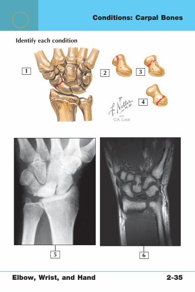
Conditions: Carpal Bones
Elbow, Wrist, and Hand 2-35
Conditions: Carpal Bones
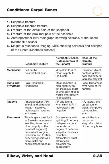
1. Scaphoid fracture2. Scaphoid tubercle fracture3. Fracture of the distal pole of the scaphoid4. Fracture of the proximal pole of the scaphoid5. Anteroposterior (AP) radiograph showing sclerosis of the lunate
(Kienböck disease)6. Magnetic resonance imaging (MRI) showing sclerosis and collapse
of the lunate (Kienböck disease)
Scaphoid Fracture
Kienböck Disease (Osteonecrosis of the Lunate)
Hook of the Hamate Fracture
Mechanism Fall on the outstretched hand
Idiopathic loss of blood supply to the lunate
Repeated direct contact (golfers, baseball batters, lacrosse players)
Signs and Symptoms
Pain, “snuffbox” tenderness
Most common in men aged 20 to 40; insidious onset of wrist pain that is worse with activity; mild swelling
Focal tenderness over hook of the hamate
Imaging Anteroposterior (AP), lateral, and scaphoid views; if negative, radiographs repeated in 1 week
AP and lateral wrist fi lms; MRI if radiographs are negative
AP, lateral, and carpal tunnel views; computed tomographic scan
Treatment Thumb spica cast for 6 to 8 weeks; nonunions (resulting from poor blood supply of the proximal fragment) necessitate surgical reduction and fi xation; better prognosis with more distal and nondisplaced fracture
Conservative with splinting if no bony collapse; surgery depends on degree of collapse (carpal arthrodesis, resection arthroplasty, wrist arthrodesis, proximal row carpectomy)
Immobilization by cast or surgical excision of the bony hook

Elbow, Wrist, and Hand 2-36
1
2
3
4
8 Identify the condition
7
6
5
Conditions: Tendons

Elbow, Wrist, and Hand 2-36
Conditions: Tendons
1. Extensor retinaculum2. Extensor carpi radialis brevis tendon3. Extensor carpi radialis longus tendon4. First dorsal interosseous muscle5. Insertion of the abductor pollicis longus tendon6. Insertion of the extensor pollicis brevis tendon7. Insertion of the extensor pollicis longus tendon8. Dorsal ganglion cyst
DeQuervain Tendinitis Ganglion Cysts
Mechanism Infl ammation or tenosynovitis of the abductor pollicis longus and extensor pollicis brevis fi rst dorsal compartment
Occult trauma
Signs and Symptoms
Radius-sided wrist pain, positive Finkelstein test result
Localized nodule on dorsal or volar wrist; transilluminates
Imaging Not necessary but can exclude other diagnoses
Posteroanterior (PA)/lateral wrist radiographs: negative
Treatment Conservative with activity modifi cation, nonsteroidal antiinfl ammatory drugs (NSAIDs), local steroid injections
For refractory cases: surgical release
Aspiration, cortisone injection, surgical excision including stalk

Elbow, Wrist, and Hand 2-37
1
3
45
6
7
8
9 10
2
Posterior (dorsal) view
Conditions: Ligaments

Elb
ow
, Wrist, a
nd H
and
2-3
7
1. Trapeziocapitate ligament 2. Trapeziotrapezoid ligament 3. Scapholunate ligament 4. Dorsal radiocarpal ligament 5. Arcuate ligament
Triangular Fibrocartilage Complex (TFCC) Tears
Scapholunate Ligament Injury
Lunotriquetral Ligament Injury
Mechanism Fall onto extended, pronated wrist Usually a fall resulting in hyperextension of a pronated wrist
Fall on outstretched hand
Signs and Symptoms
Ulnar sided wrist pain; tenderness between ulnar styloid and triquetrum (“ulnar snuffbox”)
Pain and anatomical “snuffbox” tenderness, positive Watson test result
Ulnar-sided pain worse with ulnar deviation; pain with ballottement of the lunotriquetral joint
Imaging Arthrographic magnetic resonance imaging (MRI)
Increased scapholunate interval (>2 mm) on posteroanterior (PA) clenched fi st radiograph and increased scapholunate angle (>70 degrees) on lateral radiograph
Increased lunotriquetral interval on PA radiographs; lateral radiograph scapholunate angle is normal or <40°
Treatment Nonsteroidal antiinfl ammatory drugs (NSAIDS), cortisone injections, splinting for degenerative cases initially
Arthroscopic débridement versus repair for degenerative and conservative lesions for which conservative treatment fails
Closed or open reduction and fi xation of the scapholunate joint; arthroscopy
Conservative initiallyFor refractory cases: joint
débridement
TFCC tear image from Tracy MR, Wiesler ER, Poehling GG: Arthroscopic management of triangular fi brocartilage tears in the athlete. Oper Tech Sports Med 14(2):97, 2006. Scapholunate ligament tear image from Miller M, Cole B: Textbook of Arthros-copy. Phiadelphia: WB Saunders, 2004, p 386, Fig. 39-5.
6. Dorsal radioulnar ligament 7. Triquetrohamate ligament 8. Dorsal intercarpal ligament 9. Triangular fi brocartilage complex tear10. Scapholunate ligament tear
Conditio
ns: L
igam
ents
Elbow, Wrist, and Hand 2-38
1
2
3
4
8
7
6
5
Conditions: Median Nerve
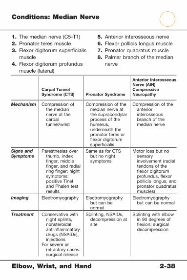
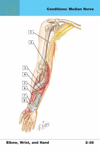
Elbow, Wrist, and Hand 2-38
Conditions: Median Nerve
Carpal Tunnel Syndrome (CTS) Pronator Syndrome
Anterior Interosseous Nerve (AIN) Compressive Neuropathy
Mechanism Compression of the median nerve at the carpal tunnel/wrist
Compression of the median nerve at the supracondylar process of the humerus, underneath the pronator teres or fl exor digitorum superfi cialis
Compression of the anterior interosseous branch of the median nerve
Signs and Symptoms
Paresthesias over thumb, index fi nger, middle fi nger, and radial ring fi nger; night symptoms; positive Tinel and Phalen test results
Same as for CTS but no night symptoms
Motor loss but no sensory involvement (radial tendons of the fl exor digitorum profundus, fl exor pollicis longus, and pronator quadratus muscles)
Imaging Electromyography Electromyography but can be normal
Electromyography but can be normal
Treatment Conservative with night splints, nonsteroidal antiinfl ammatory drugs (NSAIDs), injections
For severe or refractory cases: surgical release
Splinting, NSAIDs, decompression at site
Splinting with elbow in 90 degrees of fl exion; surgical decompression
1. The median nerve (C5-T1)2. Pronator teres muscle3. Flexor digitorum superfi cialis
muscle4. Flexor digitorum profundus
muscle (lateral)
5. Anterior interosseous nerve6. Flexor pollicis longus muscle7. Pronator quadratus muscle8. Palmar branch of the median
nerve

Elbow, Wrist, and Hand 2-39
1 2
3
4
Identify each condition
Conditions: Thumb
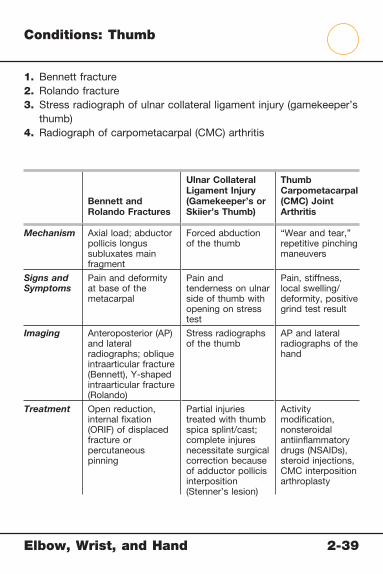
Elbow, Wrist, and Hand 2-39
Conditions: Thumb
1. Bennett fracture2. Rolando fracture3. Stress radiograph of ulnar collateral ligament injury (gamekeeper’s
thumb)4. Radiograph of carpometacarpal (CMC) arthritis
Bennett and Rolando Fractures
Ulnar Collateral Ligament Injury (Gamekeeper’s or Skiier’s Thumb)
Thumb Carpometacarpal (CMC) Joint Arthritis
Mechanism Axial load; abductor pollicis longus subluxates main fragment
Forced abduction of the thumb
“Wear and tear,” repetitive pinching maneuvers
Signs and Symptoms
Pain and deformity at base of the metacarpal
Pain and tenderness on ulnar side of thumb with opening on stress test
Pain, stiffness, local swelling/deformity, positive grind test result
Imaging Anteroposterior (AP) and lateral radiographs; oblique intraarticular fracture (Bennett), Y-shaped intraarticular fracture (Rolando)
Stress radiographs of the thumb
AP and lateral radiographs of the hand
Treatment Open reduction, internal fi xation (ORIF) of displaced fracture or percutaneous pinning
Partial injuries treated with thumb spica splint/cast; complete injures necessitate surgical correction because of adductor pollicis interposition (Stenner’s lesion)
Activity modifi cation, nonsteroidal antiinfl ammatory drugs (NSAIDs), steroid injections, CMC interposition arthroplasty

Elbow, Wrist, and Hand 2-40
1 2
3
Identify each condition
Conditions: Metacarpals

Elbow, Wrist, and Hand 2-40
Conditions: Metacarpals
1. Fracture of metacarpal neck2. Midshaft metacarpal fractures3. Radiographic image of fracture of the base of the fi fth metacarpal
Metacarpal Fractures
Boxer’s Fracture (Fracture of the Fifth Metacarpal Neck)
Mechanism Axial load Punching hard object
Signs and Symptoms
Pain, rotational deformity (closed fi st), local swelling and tenderness
Pain, rotational deformity (closed fi st), local swelling and tenderness
Imaging Anteroposterior (AP) and lateral radiographs of the hand
AP and lateral radiographs of the hand
Treatment If less than 10 to 15 degrees of angulation: reduction and cast
If more angulation or involvement of multiple metacarpals: open reduction, internal fi xation (ORIF), or percutaneous pinning
Reduction and ulnar gutter splint/cast with angulation of 40 to 50 degrees

Elbow, Wrist, and Hand 2-41
1
2
3
Identify each condition
Conditions: Phalanges
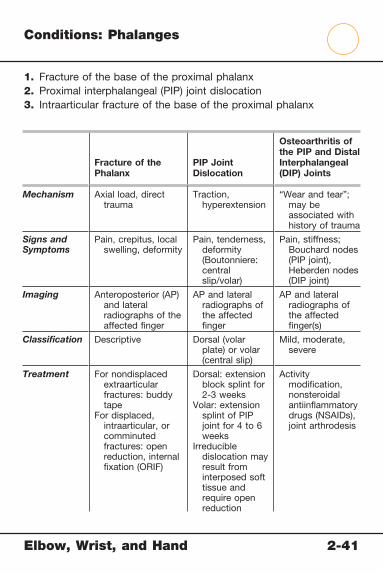
Elbow, Wrist, and Hand 2-41
Conditions: Phalanges
1. Fracture of the base of the proximal phalanx2. Proximal interphalangeal (PIP) joint dislocation3. Intraarticular fracture of the base of the proximal phalanx
Fracture of the Phalanx
PIP Joint Dislocation
Osteoarthritis of the PIP and Distal Interphalangeal (DIP) Joints
Mechanism Axial load, direct trauma
Traction, hyperextension
“Wear and tear”; may be associated with history of trauma
Signs and Symptoms
Pain, crepitus, local swelling, deformity
Pain, tenderness, deformity (Boutonniere: central slip/volar)
Pain, stiffness; Bouchard nodes (PIP joint), Heberden nodes (DIP joint)
Imaging Anteroposterior (AP) and lateral radiographs of the affected fi nger
AP and lateral radiographs of the affected fi nger
AP and lateral radiographs of the affected fi nger(s)
Classifi cation Descriptive Dorsal (volar plate) or volar (central slip)
Mild, moderate, severe
Treatment For nondisplaced extraarticular fractures: buddy tape
For displaced, intraarticular, or comminuted fractures: open reduction, internal fi xation (ORIF)
Dorsal: extension block splint for 2-3 weeks
Volar: extension splint of PIP joint for 4 to 6 weeks
Irreducible dislocation may result from interposed soft tissue and require open reduction
Activity modifi cation, nonsteroidal antiinfl ammatory drugs (NSAIDs), joint arthrodesis
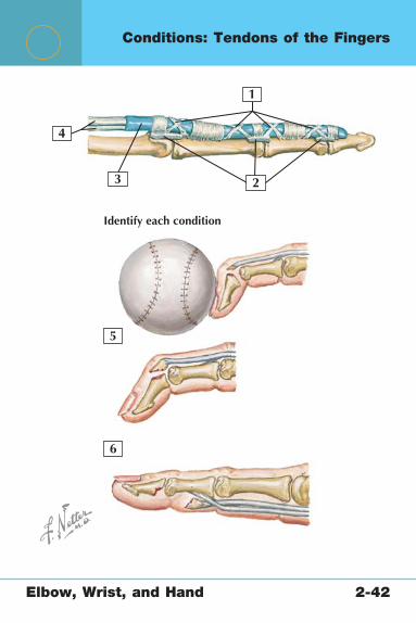
Elbow, Wrist, and Hand 2-42
1
23
4
5
6
Identify each condition
Conditions: Tendons of the Fingers

Elbow, Wrist, and Hand 2-42
Conditions: Tendons of the Fingers
1. Flexor tendon pulleys2. Palmar plates3. Tendon sheath4. Flexor digitorum tendons5. Mallet fi nger6. Jersey fi nger
Mallet Finger Jersey Finger
Mechanism Forced fl exion of extended fi nger, causing avulsion of extensor tendon from distal phalanx
Forced extension of fl exed fi nger (caught in jersey on tackle), causing avulsion of fl exor digitorum profundus from the base of PIP joint, usually of the ring fi nger
Signs and Symptoms
Pain, “dropped” distal phalanx (inability to extend at DIP joint)
Pain, inability to fl ex at the DIP joint
Imaging Anteroposterior (AP) and lateral radiographs of the fi nger can show avulsion fracture (bony mallet)
AP and lateral radiographs of the fi nger to exclude fracture
Treatment Immobilization in extension (stack splint) for 8 weeks; surgery if this fails
Surgical retrieval and repair of retracted tendon (within 10 days if retracted into palm)
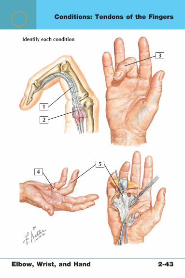
Elbow, Wrist, and Hand 2-43
1
2
3
45
Identify each condition
Conditions: Tendons of the Fingers

Elbow, Wrist, and Hand 2-43
Conditions: Tendons of the Fingers
1. Flexor tendon2. Stenosis of fl exor tendon sheath3. Trigger fi nger (stenosing tenosynovitis)4. Dupuytren’s nodules5. Dupuytren contracture
Trigger Finger (Stenosing Tenosynovitis) Dupuytren Disease
Mechanism Thickening of the fl exor tendon beneath A1 pulley
Contracture of the palmar fascia; incidence higher in men; genetic predisposition; northern European ancestry
Signs and Symptoms
Pain and catching with fi nger fl exion/extension; nodule in distal palm
Painless nodule in distal palm most common on ulnar side with eventual fl exion contracture of the MCP and later proximal interphalangeal (PIP) joints
Imaging Not necessary Not necessary
Treatment Steroid injections in the tendon sheath; surgical release of A1 pulley
Reassurance; surgical release if affects normal activities and prevents fi nger extension
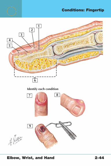
Elbow, Wrist, and Hand 2-44
21
3
4
5
9
7
6
8
Identify each condition
Conditions: Fingertip
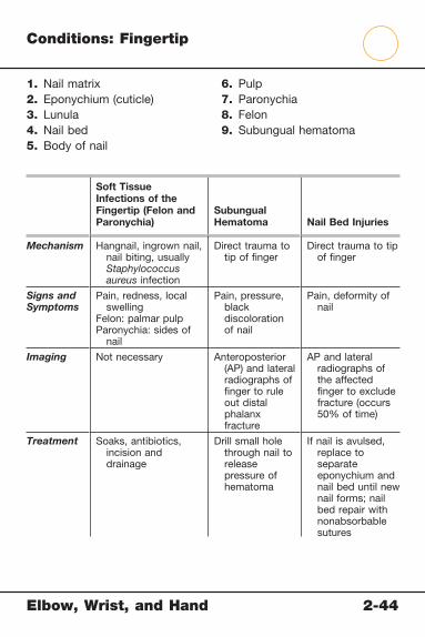
Elbow, Wrist, and Hand 2-44
Conditions: Fingertip
Soft Tissue Infections of the Fingertip (Felon and Paronychia)
Subungual Hematoma Nail Bed Injuries
Mechanism Hangnail, ingrown nail, nail biting, usually Staphylococcus aureus infection
Direct trauma to tip of fi nger
Direct trauma to tip of fi nger
Signs and Symptoms
Pain, redness, local swelling
Felon: palmar pulpParonychia: sides of
nail
Pain, pressure, black discoloration of nail
Pain, deformity of nail
Imaging Not necessary Anteroposterior (AP) and lateral radiographs of fi nger to rule out distal phalanx fracture
AP and lateral radiographs of the affected fi nger to exclude fracture (occurs 50% of time)
Treatment Soaks, antibiotics, incision and drainage
Drill small hole through nail to release pressure of hematoma
If nail is avulsed, replace to separate eponychium and nail bed until new nail forms; nail bed repair with nonabsorbable sutures
1. Nail matrix2. Eponychium (cuticle)3. Lunula4. Nail bed5. Body of nail
6. Pulp7. Paronychia8. Felon9. Subungual hematoma

Netter’s Musculoskeletal Flash Cards
3 The SpinePlates 3-1 to 3-39
Bony Anatomy
3-1 Bony Anatomy: Vertebral Column
3-2 Bony Anatomy: Cervical Spine
3-3 Bony Anatomy: Thoracic Spine
3-4 Bony Anatomy: Lumbar Spine
3-5 Bony Anatomy: Sacrum
Radiographic Anatomy
3-6 Radiographic Anatomy: Cervical Spine
3-7 Radiographic Anatomy: Lumbar Spine
Ligaments
3-8 Ligaments: Cervical Spine
3-9 Ligaments: Cervical Spine (C1 and C2)
3-10 Ligaments: Thoracolumbar Spine
Muscles
3-11 Muscles: Anterior Neck
3-12 Muscles: Anterior Neck
3-13 Muscles: Anterior Neck (Suprahyoid)
3-14 Muscles: Posterior Neck (Suboccipital Triangle)
3-15 Muscles: Posterior Neck (Suboccipital Triangle)
3-16 Muscles: Spine (Erector Spinae)
3-17 Muscles: Spine (Deep Dissection)
3-18 Muscles: Spine (Deep Dissection)
The Spine Table of Contents
3 The SpinePlates 3-1 to 3-39
Arteries and Nerves
3-19 Arteries of the Spine
3-20 Arteries of the Thoracolumbar Spine
3-21 Nerves of the Spine: Cervical Plexus
3-22 Nerves of the Spine: Lumbar Plexus
3-23 Nerves of the Spine: Sacral Plexus
Physical Examination
3-24 Physical Examination: Spine
Conditions
3-25 Conditions: Cervical Vertebrae (C1 and C2)
3-26 Conditions: Cervical Ligaments
3-27 Conditions: Spinal Cord
3-28 Conditions: Spinal Cord (Dermatomes)
3-29 Conditions: Spinal Cord (Myotomes)
3-30 Conditions: Neural Foramina and Central Canal
3-31 Conditions: Facet Joints
3-32 Conditions: Intervertebral Disc
3-33 Conditions: Pars Interarticularis
3-34 Conditions: Vertebral Bodies
3-35 Conditions: Vertebral Column
3-36 Conditions: Vertebral Column
3-37 Conditions: Vertebral Column
3-38 Conditions: Vertebral Column
3-39 Conditions: Sacrum
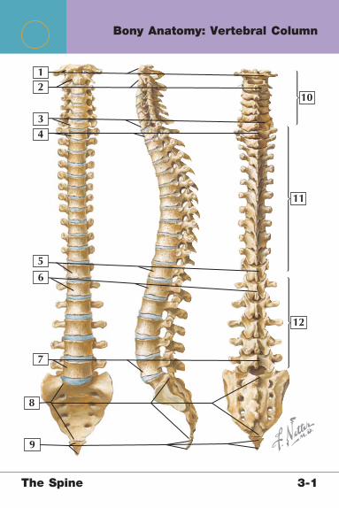
The Spine 3-1
Bony Anatomy: Vertebral Column
12
34
7
8
9
10
11
12
5
6
The Spine 3-1
Bony Anatomy: Vertebral Column
1. C1 (Atlas) 2. C2 (Axis) 3. C7 4. T1 5. T12 6. L1 7. L5 8. Sacrum 9. Coccyx10. Cervical vertebrae11. Thoracic vertebrae12. Lumbar vertebrae
Comment: The vertebral column is divided into the cervical, thoracic, and lumbar sections. The sacrum consists of 5 fused vertebrae. There is a normal kyphotic curve of the thoracic section and a normal lordotic curve of the cervical and lumbar sections.
12
3
4
57
6
88
9
10
11
10
Atlas (C1): superior view
Axis (C2): posterosuperior view
2nd cervical to 1st thoracic vertebrae:right lateral view
The Spine 3-2
Bony Anatomy: Cervical Spine

The Spine 3-2
Bony Anatomy: Cervical Spine
1. Anterior tubercle 2. Anterior arch 3. Transverse process 4. Transverse foramen 5. Superior articular facet 6. Posterior tubercle 7. Posterior arch 8. Dens 9. Inferior articular facet10. Spinous process11. C7
Comment: The cervical section of the spine is made up of 7 vertebrae. C1 (atlas) and C2 (axis) are ring-shaped vertebrae with no vertebral body or intervertebral disc. The other cervical vertebrae have a more standard anatomical appearance, with a body, transverse and spinous processes, pedicles, and laminae. The C7 vertebra is called the vertebrae prominens and is easily identifi ed because of its long spinous process.
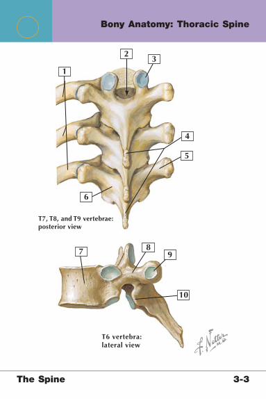
1
23
5
4
6
7 89
10
T7, T8, and T9 vertebrae:posterior view
T6 vertebra:lateral view
The Spine 3-3
Bony Anatomy: Thoracic Spine

The Spine 3-3
Bony Anatomy: Thoracic Spine
1. Ribs 2. Vertebral canal 3. Superior articular facet 4. Spinous processes 5. Transverse process 6. Lamina 7. Body 8. Pedicle 9. Transverse costal facet10. Inferior articular facet
Comment: The thoracic section of the spine is made up of 12 vertebrae. The anatomy is similar to that of the lumbar section; the primary difference is that the vertebral bodies and processes are smaller. Thoracic vertebrae also articulate with the thorax and have superior, inferior, and transverse costal facets for articulation with the ribs at those levels.
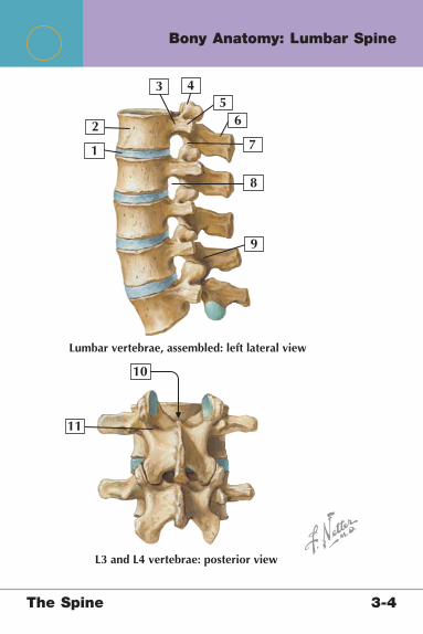
1
2
35
4
6
7
8
9
10
11
L3 and L4 vertebrae: posterior view
Lumbar vertebrae, assembled: left lateral view
The Spine 3-4
Bony Anatomy: Lumbar Spine
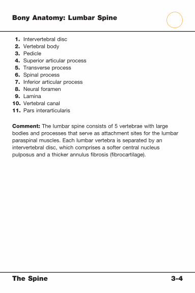
The Spine 3-4
Bony Anatomy: Lumbar Spine
1. Intervertebral disc 2. Vertebral body 3. Pedicle 4. Superior articular process 5. Transverse process 6. Spinal process 7. Inferior articular process 8. Neural foramen 9. Lamina10. Vertebral canal11. Pars interarticularis
Comment: The lumbar spine consists of 5 vertebrae with large bodies and processes that serve as attachment sites for the lumbar paraspinal muscles. Each lumbar vertebra is separated by an intervertebral disc, which comprises a softer central nucleus pulposus and a thicker annulus fi brosis (fi brocartilage).

1
2
3
5
4
67
89
10
11
Anterior inferior view
Posterior superior view
The Spine 3-5
Bony Anatomy: Sacrum

The Spine 3-5
Bony Anatomy: Sacrum
1. Lumbosacral articular surface 2. Sacral promontory 3. Linea terminalis 4. Sacral foramina 5. Transverse process of the coccyx 6. Coccyx 7. Transverse ridges 8. Sacral tuberosity 9. Lateral sacral crest10. Median sacral crest11. Intermediate sacral crest
Comment: The sacrum comprises 5 fused sacral vertebrae. Similarly, the coccyx comprises 4 fused coccygeal vertebrae. These structures provide strength and a site for muscle attachments of the spine and pelvis.
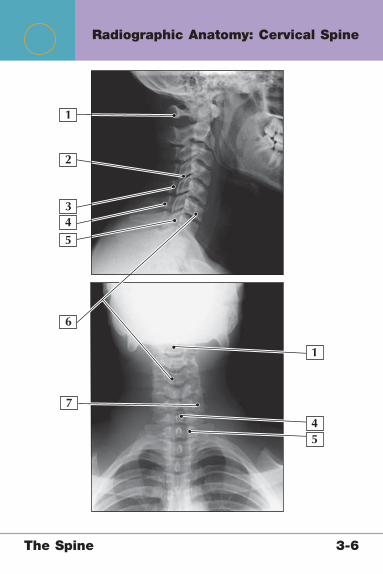
1
2
3
54
6
7
45
1
The Spine 3-6
Radiographic Anatomy: Cervical Spine

The Spine 3-6
Radiographic Anatomy: Cervical Spine
1. C12. Superior and inferior articular facets3. Lamina4. Spinous process5. C76. Vertebral body7. Transverse process
Comment: Anteroposterior (AP) and lateral radiographs are the views most commonly used to evaluate abnormalities of the cervical spine. Other views include oblique views, the Water’s view (C1 and C2), and fl exion/extension lateral views (to evaluate for stability of the cervical spine).

The Spine 3-7
1
2
3
8
9
10 11
12
2
4
4
56
7
7
Radiographic Anatomy: Lumbar Spine
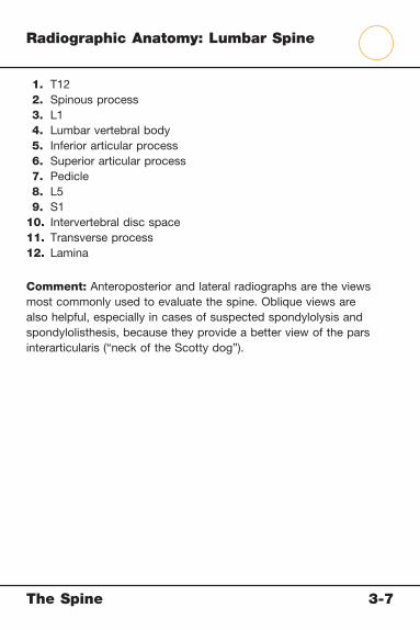
The Spine 3-7
Radiographic Anatomy: Lumbar Spine
1. T12 2. Spinous process 3. L1 4. Lumbar vertebral body 5. Inferior articular process 6. Superior articular process 7. Pedicle 8. L5 9. S110. Intervertebral disc space11. Transverse process12. Lamina
Comment: Anteroposterior and lateral radiographs are the views most commonly used to evaluate the spine. Oblique views are also helpful, especially in cases of suspected spondylolysis and spondylolisthesis, because they provide a better view of the pars interarticularis (“neck of the Scotty dog”).
The Spine 3-8
12
3
5 4
6
7
8
9
Right lateral view
Anterior view
Ligaments: Cervical Spine

The Spine 3-8
Ligaments: Cervical Spine
1. Anterior atlantooccipital membrane2. Atlantooccipital joint capsule3. Lateral atlantoaxial joint (capsule removed)4. Anterior longitudinal ligament5. Lateral atlantoaxial joint capsule6. Posterior atlantooccipital membrane7. Ligamenta fl ava8. Ligamentum nuchae9. Zygapophyseal joints
Comment: The cervical spine comprises multiple joints, including the atlantooccipital joint (C1 and occipital bone), which is an ellipsoid joint; the atlantoaxial joint (C1 and C2), which is a plane and pivot joint; the zygapophyseal joints (facet joints); and the intervertebral joints.
The Spine 3-9
1
2
Upper part of vertebral canal with spinous processes andparts of vertebral arches removed: posterior view
Principal part of tectorial membraneremoved: posterior view
3
5
4
6
789
10
Ligaments: Cervical Spine (C1 and C2)

The Spine 3-9
Ligaments: Cervical Spine (C1 and C2)
1. Posterior longitudinal ligament 2. Tectorial membrane 3. Atlantooccipital joint capsule 4. Lateral atlantoaxial joint capsule 5. Zygapophyseal joint capsule (C2-3) 6. Alar ligaments 7. Superior longitudinal band 8. Transverse ligament of the atlas 9. Inferior longitudinal band10. Cruciate ligament
Comment: The atlantoaxial joint (C1-2) is primarily a joint of rotation. The apical, alar, and cruciate ligaments provide stability to the joint. Of these, the transverse ligament of the atlas is biomechanically the strongest.
The Spine 3-10
1
2
3
5
4
6
7
8
9
10
Ligaments: Thoracolumbar Spine
The Spine 3-10
Ligaments: Thoracolumbar Spine
1. Anterior longitudinal ligament 2. Intervertebral discs 3. Supraspinous ligament 4. Interspinous ligament 5. Posterior longitudinal ligament 6. Ligamentum fl avum 7. Iliolumbar ligament 8. Dorsal sacroiliac ligament 9. Sacrospinous ligament10. Sacrotuberous ligament
Comment: The anterior and posterior longitudinal ligaments run along the anterior and posterior vertebral bodies the entire length of the spinal column and prevent excessive extension (anterior) and fl exion (posterior) of the spine. The supraspinous and interspinous ligaments run between each pair of spinous processes, and the ligamentum fl avum runs between the laminae.

The Spine 3-11
1
2
3
Muscles: Anterior Neck
The Spine 3-11
Muscles: Anterior Neck
1. Platysma2. Thyrohyoid muscle3. Sternocleidomastoid muscle
Platysma Thyrohyoid MuscleSternocleidomastoid Muscle
Origin Deltoid and pectoralis major fascia
Thyroid cartilage Manubrium of sternum (sternal head); medial superior clavicle (clavicular head)
Insertion Skin and mandible
Inferior hyoid bone Mastoid process of temporal bone and lateral occipital bone
Actions Depression of the jaw
Depression of hyoid and larynx
Flexion, rotation, and lateral fl exion of the cervical spine
Innervation Cranial nerve VII C1 via hypoglossal nerve (cranial nerve XII)
Spinal root of the accessory nerve (cranial nerve XI)

The Spine 3-12
1
23
Muscles: Anterior Neck

The Spine 3-12
Muscles: Anterior Neck
1. Omohyoid muscle2. Sternohyoid muscle3. Sternothyroid muscle
Omohyoid Muscle Sternohyoid Muscle
Sternothyroid Muscle
Origin Superior scapula (suprascapular notch)
Medial clavicle and manubrium of sternum
Posterior manubrium of sternum
Insertion Inferior hyoid bone
Body of hyoid bone Thyroid cartilage
Actions Depression of hyoid
Depression of hyoid Depression of hyoid and larynx
Innervation C1-3 C1-3 C1-3

The Spine 3-13
1
2
3
4
Muscles: Anterior Neck (Suprahyoid)

The S
pin
e
3-1
3
Musc
les: A
nte
rior N
eck (S
upra
hyo
id)
Digastric MuscleMylohyoid Muscle
Stylohyoid Muscle Scalene Muscles
Origin Mandible (anterior belly); mastoid (posterior belly)
Mandible Styloid process Transverse processes (C2-7)
Insertion Hyoid bone Hyoid bone Hyoid bone 1st and 2nd ribs
Actions Elevation of the hyoid bone; depression of the jaw
Elevation of the hyoid bone
Elevation of the hyoid bone
Elevation of ribs; fl exion, rotation, lateral fl exion of cervical spine
Innervation Anteriorly by cranial nerve V; posteriorly by cranial nerve VII
Cranial nerve V Cranial nerve VII
Ventral rami (C3-8)
1. Digastric muscle2. Mylohyoid muscle3. Stylohyoid muscle4. Scalene muscles (anterior, middle, posterior)

The Spine 3-14
1
2
3
Muscles: Posterior Neck (Suboccipital Triangle)
The Spine 3-14
Muscles: Posterior Neck (Suboccipital Triangle)
1. Longissimus capitis2. Semispinalis capitis and cervicis (thoracis, not pictured)3. Splenius capitis and cervicis
Longissimus Capitis
Semispinalis Capitis and Semispinalis Cervicis
Splenius Capitis and Splenius Cervicis
Origin Articular processes C4-7 and transverse processes T1-5
Transverse processes
Ligamentum nuchae (capitis)
Spinous processes T1-6 (cervicis)
Insertion Mastoid process Nuchal ridge (capitis)Spinous processes
(cervicis and thoracis)
Mastoid and nuchal line (capitis)
Transverse processes C1-4 (cervicis)
Actions Extension, rotation, lateral fl exion of neck
Extension and rotation of the spine
Lateral fl exion and rotation of the cervical spine
Innervation Dorsal rami of cervical nerves
Dorsal primary rami Dorsal rami of the inferior cervical nerves
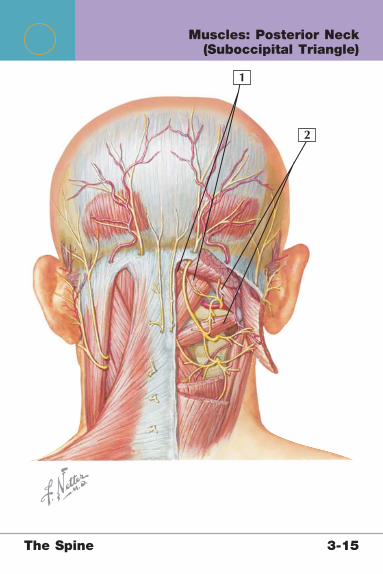
The Spine 3-15
1
2
Muscles: Posterior Neck (Suboccipital Triangle)
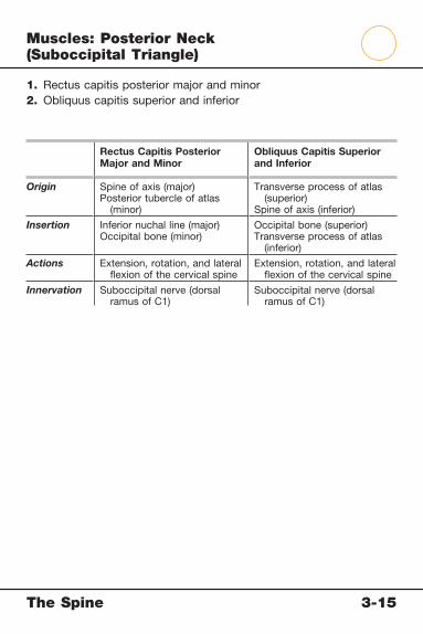
The Spine 3-15
Muscles: Posterior Neck (Suboccipital Triangle)
1. Rectus capitis posterior major and minor2. Obliquus capitis superior and inferior
Rectus Capitis Posterior Major and Minor
Obliquus Capitis Superior and Inferior
Origin Spine of axis (major)Posterior tubercle of atlas
(minor)
Transverse process of atlas (superior)
Spine of axis (inferior)
Insertion Inferior nuchal line (major)Occipital bone (minor)
Occipital bone (superior)Transverse process of atlas
(inferior)
Actions Extension, rotation, and lateral fl exion of the cervical spine
Extension, rotation, and lateral fl exion of the cervical spine
Innervation Suboccipital nerve (dorsal ramus of C1)
Suboccipital nerve (dorsal ramus of C1)

The Spine 3-16
1
2
3
Muscles: Spine (Erector Spinae)

The S
pin
e
3-1
6
Musc
les: S
pin
e (E
recto
r Spin
ae)
1. Iliocostalis2. Longissimus3. Spinalis
Iliocostalis Longissimus Spinalis
Origin Posterior iliac crest, sacrum, spinous processes of the inferior lumbosacral spine, and the supraspinous ligament
Posterior iliac crest, sacrum, spinous processes of the inferior lumbosacral spine, and the supraspinous ligament
Posterior iliac crest, sacrum, spinous processes of the inferior lumbosacral spine, and the supraspinous ligament
Insertion Angles of the lower ribs and cervical transverse processes
Ribs, transverse processes of the cervical and thoracic vertebrae, mastoid process of the temporal bone
Midcervical and upper thoracic vertebral spinous processes and the skull
Actions Vertebral column extension and lateral fl exion
Vertebral column extension and lateral fl exion
Vertebral column extension and lateral fl exion
Innervation Dorsal rami of the spinal nerves in each region
Dorsal rami of the spinal nerves in each region
Dorsal rami of the spinal nerves in each region
Comment The iliocostalis, longissimus, and spinalis muscles collectively form the erector spinae muscle group. Each set is separated into 3 groups on the basis of their location in the vertebral column: iliocostalis (cervicis, thoracis, lumborum), longissimus (capitis, cervicis, thoracis), and spinalis (capitis, cervicis, thoracis).

The Spine 3-17
1
2
3
Muscles: Spine (Deep Dissection)
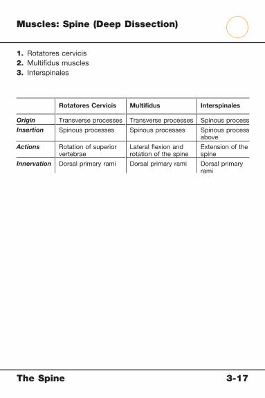
The Spine 3-17
Muscles: Spine (Deep Dissection)
1. Rotatores cervicis2. Multifi dus muscles3. Interspinales
Rotatores Cervicis Multifi dus Interspinales
Origin Transverse processes Transverse processes Spinous process
Insertion Spinous processes Spinous processes Spinous process above
Actions Rotation of superior vertebrae
Lateral fl exion and rotation of the spine
Extension of the spine
Innervation Dorsal primary rami Dorsal primary rami Dorsal primary rami

The Spine 3-18
1
2
Muscles: Spine (Deep Dissection)

The Spine 3-18
Muscles: Spine (Deep Dissection)
1. Levatores costarum2. Intertransversarii
Levatores Costarum Intertransversarii
Origin Transverse processes C7-T11 Transverse process
Insertion Posterior surface and angle of rib below muscle
Transverse process above muscle
Actions Elevation of ribs Lateral fl exion of the spine
Innervation Dorsal rami C7-T11 Dorsal primary rami

The Spine 3-19
Anterior view Posterior view
1
2
34
5
67 8
9
10
1112
13
Arteries of the Spine
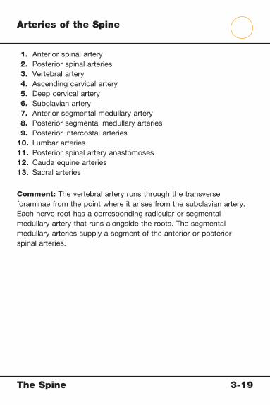
The Spine 3-19
Arteries of the Spine
1. Anterior spinal artery 2. Posterior spinal arteries 3. Vertebral artery 4. Ascending cervical artery 5. Deep cervical artery 6. Subclavian artery 7. Anterior segmental medullary artery 8. Posterior segmental medullary arteries 9. Posterior intercostal arteries10. Lumbar arteries11. Posterior spinal artery anastomoses12. Cauda equine arteries13. Sacral arteries
Comment: The vertebral artery runs through the transverse foraminae from the point where it arises from the subclavian artery. Each nerve root has a corresponding radicular or segmental medullary artery that runs alongside the roots. The segmental medullary arteries supply a segment of the anterior or posterior spinal arteries.

The Spine 3-20
Section through thoracic level:anterosuperior view
Arterial distribution: schema
12
2
3
3
45
5
67
8
910
Arteries of the Thoracolumbar Spine
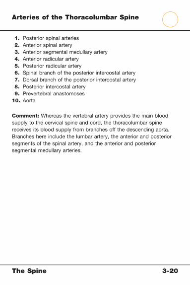
The Spine 3-20
Arteries of the Thoracolumbar Spine
1. Posterior spinal arteries 2. Anterior spinal artery 3. Anterior segmental medullary artery 4. Anterior radicular artery 5. Posterior radicular artery 6. Spinal branch of the posterior intercostal artery 7. Dorsal branch of the posterior intercostal artery 8. Posterior intercostal artery 9. Prevertebral anastomoses10. Aorta
Comment: Whereas the vertebral artery provides the main blood supply to the cervical spine and cord, the thoracolumbar spine receives its blood supply from branches off the descending aorta. Branches here include the lumbar artery, the anterior and posterior segments of the spinal artery, and the anterior and posterior segmental medullary arteries.

The Spine 3-21
1
1
2
6
3
3
4
4
5
5
6
7
8
9
9
10
11
12
Nerves of the Spine: Cervical Plexus

The Spine 3-21
Nerves of the Spine: Cervical Plexus
1. Accessory nerve (cranial nerve XI) 2. C2 spinal nerve 3. Lesser occipital nerve 4. Great auricular nerve 5. Hypoglossal nerve (cranial nerve XII) 6. Ansa cervicalis 7. C5 spinal nerve 8. Vagus nerve (cranial nerve X) 9. Phrenic nerve10. Brachial plexus11. Transverse cervical nerves12. Supraclavicular nerves
Comment: The cervical plexus (C1-4) comprises the lesser occipital nerve, great auricular nerve, transverse cervical nerve, supraclavicular nerve, ansa cervicalis, and phrenic nerve. The fi rst 4 of these provide only sensory input, whereas the ansa cervicalis provides motor input to the omohyoid, sternohyoid, and sternothyroid muscles. The phrenic nerve innervates the diaphragm.

The Spine 3-22
Anterior divisionPosterior division
1
1
2
2
6
3
34
4
5
5
6
7
8
9
8
9
Nerves of the Spine: Lumbar Plexus
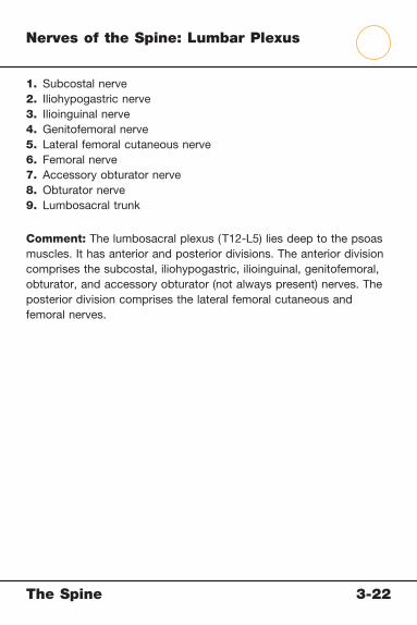
The Spine 3-22
Nerves of the Spine: Lumbar Plexus
1. Subcostal nerve2. Iliohypogastric nerve3. Ilioinguinal nerve4. Genitofemoral nerve5. Lateral femoral cutaneous nerve6. Femoral nerve7. Accessory obturator nerve8. Obturator nerve9. Lumbosacral trunk
Comment: The lumbosacral plexus (T12-L5) lies deep to the psoas muscles. It has anterior and posterior divisions. The anterior division comprises the subcostal, iliohypogastric, ilioinguinal, genitofemoral, obturator, and accessory obturator (not always present) nerves. The posterior division comprises the lateral femoral cutaneous and femoral nerves.

The Spine 3-23
1
1
2
6
3
3
4
4
5
5
6
7
7
2
10
101112
1112
8
8
9
9
Anterior division
Posterior division
Nerves of the Spine: Sacral Plexus

The Spine 3-23
Nerves of the Spine: Sacral Plexus
1. Lumbosacral trunk 2. Superior gluteal nerve 3. Inferior gluteal nerve 4. Nerve to the piriformis 5. Sciatic nerve 6. Nerve to quadratus femoris 7. Nerve to obturator internus 8. Posterior cutaneous nerve of the thigh 9. Perforating cutaneous nerve10. Pudendal nerve11. Nerve to levator ani and coccygeus muscles12. Perineal branch
Comment: The sacral plexus comprises nerve roots L4-S5. The anterior division (shown in green on the front) becomes the tibial nerve (L4-S3), the nerve to the quadratus femoris (L4-S1), the nerve to the obturator internus (L5-S2), the pudendal nerve (S2-S4), and the nerve to the coccygeus muscle (S3-S4). The posterior division (shown in yellow on the front) becomes the common peroneal nerve (L4-S2), the superior gluteal nerve (L4-S1), the inferior gluteal nerve (L5-S2), the nerve to the piriformis (S2), and the posterior femoral cutaneous nerve (S1-S3).

The Spine 3-24
Standing
Observe body build posture deformities pelvic obliquity spine alignmentPalpate for m. spasms trigger zones myofascial nodes sciatic n. tendernessCompress iliac crests for sacroiliac tenderness
Walking on heels(tests footand great toedorsiflexion)
Walking ontoes (tests calfmuscles)
Measurementof rib hump wtih scoliometer
Estimation of rib humpand evaluation of curveunwinding as patientturns trunk fromside to side
Test vertebral column for flexion extension side bending rotation
Physical Examination: Spine

The Spine 3-24
Physical Examination: Spine
Movement Normal Range of Motion
Cervical fl exion Chin within 3-4 cm of chest
Cervical extension 60 degrees
Cervical lateral fl exion 30-45 degrees
Cervical rotation 75-80 degrees
Lumbar fl exion 60-80 degrees
Lumbar extension 25-40 degrees
Lumbar lateral fl exion 40-60 degrees
Lumbar rotation 60-70 degrees
Test/Sign Reason for Evaluation
Spurling test Nerve root compression
Straight leg raise Sciatic nerve irritation
Single leg extension Spondylolysis/spondylolisthesis
Forward bend/rib hump Scoliosis
Valsalva test Space-occupying lesion (disc)

The Spine 3-25
1Identify the type of fracture
2
3
4
5
6
Conditions: Cervical Vertebrae (C1 and C2)
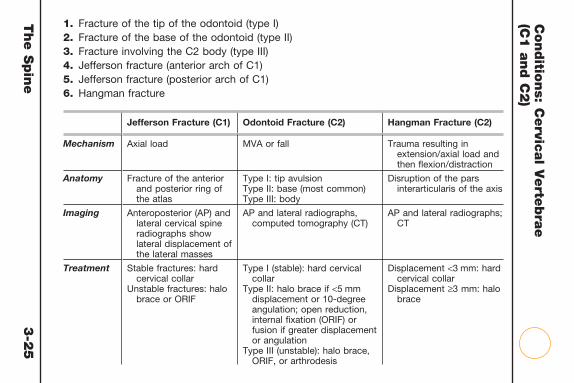
The S
pin
e
3-2
5
Conditio
ns: C
ervic
al V
erte
bra
e
(C1 a
nd C
2)
1. Fracture of the tip of the odontoid (type I)2. Fracture of the base of the odontoid (type II)3. Fracture involving the C2 body (type III)4. Jefferson fracture (anterior arch of C1)5. Jefferson fracture (posterior arch of C1)6. Hangman fracture
Jefferson Fracture (C1) Odontoid Fracture (C2) Hangman Fracture (C2)
Mechanism Axial load MVA or fall Trauma resulting in extension/axial load and then fl exion/distraction
Anatomy Fracture of the anterior and posterior ring of the atlas
Type I: tip avulsionType II: base (most common)Type III: body
Disruption of the pars interarticularis of the axis
Imaging Anteroposterior (AP) and lateral cervical spine radiographs show lateral displacement of the lateral masses
AP and lateral radiographs, computed tomography (CT)
AP and lateral radiographs; CT
Treatment Stable fractures: hard cervical collar
Unstable fractures: halo brace or ORIF
Type I (stable): hard cervical collar
Type II: halo brace if <5 mm displacement or 10-degree angulation; open reduction, internal fi xation (ORIF) or fusion if greater displacement or angulation
Type III (unstable): halo brace, ORIF, or arthrodesis
Displacement <3 mm: hard cervical collar
Displacement ≥3 mm: halo brace
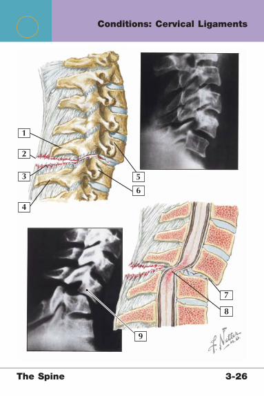
The Spine 3-26
1
2
3
4
5
9
6
8
7
Conditions: Cervical Ligaments

The Spine 3-26
Conditions: Cervical Ligaments
1. Spinous process C62. Supraspinous ligament (torn)3. Interspinous ligament (torn)4. Spinous process C75. Anterior body C66. Anterior body C77. Step-off C6 vertebral body on C78. Cord compression9. Step-off of more than 3 mm and angulation of more than 11
degrees, indicating instability
Cervical Sprain/Strain Cervical Spine Instability
Mechanism Stretch or whiplash injury to the neck
Traumatic (distraction and fl exion of the cervical spine), congenital (Down syndrome), or acquired (rheumatoid arthritis)
Signs and Symptoms
Pain and stiffness, focal tenderness, mild loss of motion, usually not associated with neurological symptoms
Cervical pain post-trauma; may be more insidious in chronic cases
Imaging Radiographs and magnetic resonance imaging (MRI) as needed to rule out fracture or neurological injury
Anteroposterior (AP), lateral, and odontoid radiographs show >3.5 mm step-off or >11-degree angulation
Treatment Rest, nonsteroidal antiinfl ammatory drugs (NSAIDs), activity modifi cation, physical therapy
Careful initial management after trauma; fi nal management may include halo brace or surgical fusion, depending on the cause and severity
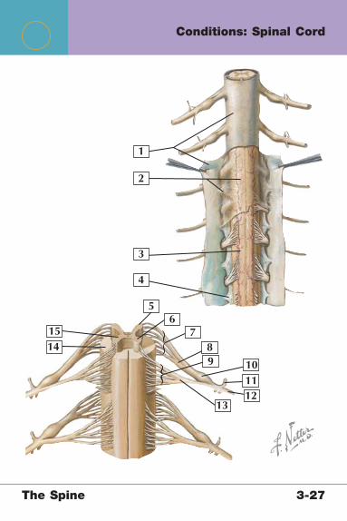
The Spine 3-27
1
2
3
4
5
1514
131211109
6
87
Conditions: Spinal Cord
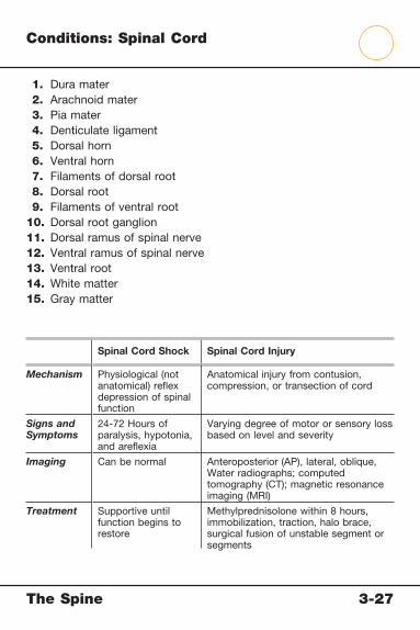
The Spine 3-27
Conditions: Spinal Cord
1. Dura mater 2. Arachnoid mater 3. Pia mater 4. Denticulate ligament 5. Dorsal horn 6. Ventral horn 7. Filaments of dorsal root 8. Dorsal root 9. Filaments of ventral root10. Dorsal root ganglion11. Dorsal ramus of spinal nerve12. Ventral ramus of spinal nerve13. Ventral root14. White matter15. Gray matter
Spinal Cord Shock Spinal Cord Injury
Mechanism Physiological (not anatomical) refl ex depression of spinal function
Anatomical injury from contusion, compression, or transection of cord
Signs and Symptoms
24-72 Hours of paralysis, hypotonia, and arefl exia
Varying degree of motor or sensory loss based on level and severity
Imaging Can be normal Anteroposterior (AP), lateral, oblique, Water radiographs; computed tomography (CT); magnetic resonance imaging (MRI)
Treatment Supportive until function begins to restore
Methylprednisolone within 8 hours, immobilization, traction, halo brace, surgical fusion of unstable segment or segments

The Spine 3-28
1
2
3
4
C2
C3
C4 C5
T1T2
T3
T4
T5T6
T7
T8
C6
C6
C8T1
C8C7
T9
T10
T11
T12L1
L2
L3
L4
S2
S3
L4 L5
S1
L5
L3
L2
S2S1
L5
5S4S3 S2
S1L5L4
L3L2L1
T12
Conditions: Spinal Cord (Dermatomes)

The Spine 3-28
Conditions: Spinal Cord (Dermatomes)
1. Cervical segments2. Thoracic segments3. Lumbar segments4. Sacral segments
Nerve Root Dermatome
C1 Top of skull
C2 Temple, occiput
C3 Neck, cheek
C4 Shoulder, axilla
C5 Deltoid patch
C6 Lateral forearm, thumb
C7 Middle fi nger
C8 Medial arm, small fi nger
T1 Medial elbow
T2 Axilla, pectoral region
T3-7 Upper thorax (T4, nipple)
T8-12 Abdomen and lower back (T10, umbilicus)
L1 Groin and lateral hip
L2 Anterior thigh
L3 Medial knee and leg
L4 Lateral thigh, medial leg
L5 Posterolateral thigh, lateral leg, dorsum foot
S1 Posterior thigh, leg, and heel
S2 Posterior thigh and leg
S3 Groin, medial thigh
S4 Perineum, genitals

The Spine 3-29
1
2
3
4
5
6
Identify the reflex
Conditions: Spinal Cord (Myotomes)
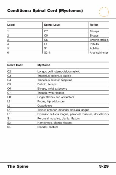
The Spine 3-29
Conditions: Spinal Cord (Myotomes)
Label Spinal Level Refl ex
1 C7 Triceps
2 C5 Biceps
3 C6 Brachioradialis
4 L4 Patellar
5 S1 Achilles
6 S2-4 Anal sphincter
Nerve Root Myotome
C2 Longus colli, sternocleidomastoid
C3 Trapezius, splenius capitis
C4 Trapezius, levator scapulae
C5 Deltoid, biceps
C6 Biceps, wrist extensors
C7 Triceps, wrist fl exors
C8 Finger fl exors and adductors
L2 Psoas, hip adductors
L3 Quadriceps
L4 Tibialis anterior, extensor hallucis longus
L5 Extensor hallucis longus, peroneal muscles, dorsifl exors
S1 Peroneal muscles, plantar fl exors
S2 Hamstrings, plantar fl exors
S4 Bladder, rectum
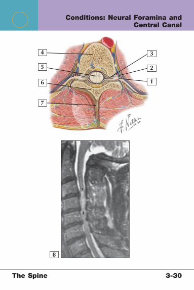
The Spine 3-30
1
2
34
5
6
7
8
Conditions: Neural Foramina and Central Canal
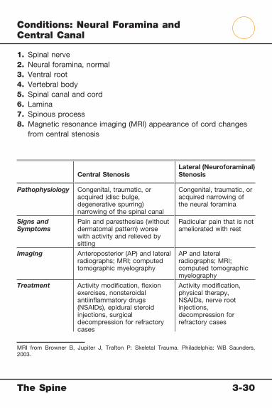
The Spine 3-30
Conditions: Neural Foramina and Central Canal
1. Spinal nerve2. Neural foramina, normal3. Ventral root4. Vertebral body5. Spinal canal and cord6. Lamina7. Spinous process8. Magnetic resonance imaging (MRI) appearance of cord changes
from central stenosis
Central StenosisLateral (Neuroforaminal) Stenosis
Pathophysiology Congenital, traumatic, or acquired (disc bulge, degenerative spurring) narrowing of the spinal canal
Congenital, traumatic, or acquired narrowing of the neural foramina
Signs and Symptoms
Pain and paresthesias (without dermatomal pattern) worse with activity and relieved by sitting
Radicular pain that is not ameliorated with rest
Imaging Anteroposterior (AP) and lateral radiographs; MRI; computed tomographic myelography
AP and lateral radiographs; MRI; computed tomographic myelography
Treatment Activity modifi cation, fl exion exercises, nonsteroidal antiinfl ammatory drugs (NSAIDs), epidural steroid injections, surgical decompression for refractory cases
Activity modifi cation, physical therapy, NSAIDs, nerve root injections, decompression for refractory cases
MRI from Browner B, Jupiter J, Trafton P: Skeletal Trauma. Philadelphia: WB Saunders, 2003.
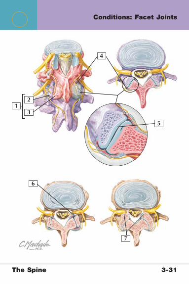
The Spine 3-31
12
3
4
5
6
7
Conditions: Facet Joints

The Spine 3-31
Conditions: Facet Joints
1. Facet (zygapophyseal) joint2. Superior articular process of vertebra below (purple)3. Inferior articular process of vertebra above (pink)4. Facet joint and capsule5. Articular cartilage of facet joint6. Synovial infl ammation with nerve root impingement7. Facet arthropathy with large osteophytes causing nerve root
impingement
Facet Syndrome
Pathophysiology Infl ammation or degeneration of facet joints
Mechanism Acute/injury-related or chronic degenerative process
Signs and Symptoms
Nonspecifi c pain that may radiate to buttocks, is worse with hyperextension/rotation, is worse in morning and after rest
Imaging Anteroposterior (AP), lateral, oblique radiographs show facet joint degenerative changes; magnetic resonance imaging (MRI) to rule out other soft tissue injury
Treatment Activity modifi cation, physical therapy, nonsteroidal antiinfl ammatory drugs (NSAIDs), facet joint injections; surgery (controversial)

The Spine 3-32
1
2
3
4
5
6
7
Identify each condition
8 9 10
11
Conditions: Intervertebral Disc
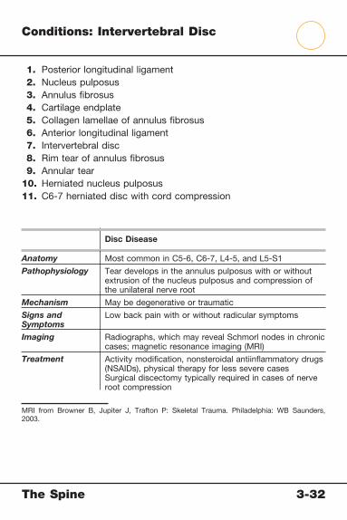
The Spine 3-32
Conditions: Intervertebral Disc
1. Posterior longitudinal ligament 2. Nucleus pulposus 3. Annulus fi brosus 4. Cartilage endplate 5. Collagen lamellae of annulus fi brosus 6. Anterior longitudinal ligament 7. Intervertebral disc 8. Rim tear of annulus fi brosus 9. Annular tear10. Herniated nucleus pulposus11. C6-7 herniated disc with cord compression
Disc Disease
Anatomy Most common in C5-6, C6-7, L4-5, and L5-S1
Pathophysiology Tear develops in the annulus pulposus with or without extrusion of the nucleus pulposus and compression of the unilateral nerve root
Mechanism May be degenerative or traumatic
Signs and Symptoms
Low back pain with or without radicular symptoms
Imaging Radiographs, which may reveal Schmorl nodes in chronic cases; magnetic resonance imaging (MRI)
Treatment Activity modifi cation, nonsteroidal antiinfl ammatory drugs (NSAIDs), physical therapy for less severe casesSurgical discectomy typically required in cases of nerve root compression
MRI from Browner B, Jupiter J, Trafton P: Skeletal Trauma. Philadelphia: WB Saunders, 2003.

The Spine 3-33
1
2
3
45
6
7 Identify the condition shown above
Identify the condition shown above8
Conditions: Pars Interarticularis
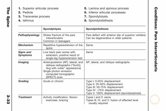
The S
pin
e
3-3
3
Conditio
ns: P
ars In
tera
rticula
ris1. Superior articular process2. Pedicle3. Transverse process4. Isthmus
5. Lamina and spinous process6. Inferior articular processes7. Spondylolysis8. Spondylolisthesis
Spondylolysis Spondylolisthesis
Pathophysiology Stress fracture of the pars interarticularis
Common in teenagers
Pars defect with anterior slip of superior vertebraCan be degenerative in older patients
Mechanism Repetitive hyperextension of the spine
Same
Signs and Symptoms
Low back pain worse with extension, positive result of single-leg hyperextension test
Same
Imaging Anteroposterior (AP), lateral, and oblique radiographs (“Scotty dog with collar” appearance)
Single photon emission computed tomographic (SPECT) scan
AP, lateral, and oblique radiographs
Grading Acute or chronic Type I: 0-25% displacementType II: 25-50% displacementType III: 50-75% displacementType IV: >75% displacementType V: 100% displacement (spondyloptosis)
Treatment Activity modifi cation, fl exion exercises, bracing
Types I and II: sameTypes III, IV, and V: fusion of affected level
usually required

The Spine 3-34
1
23
4
6
7
5
8
Conditions: Vertebral Bodies

The Spine 3-34
Conditions: Vertebral Bodies
1. Superior endplate2. Vertebral body3. Inferior endplate4. Compression fracture with spinal cord compression5. Radiographic appearance of compression fracture6. Burst fracture with anterior collapse7. Supraspinous and interspinous ligament injury8. Appearance of burst fracture on computed tomography (CT)
Compression Fracture Burst Fracture
Mechanism Vertebral body collapse from weakened bone (from osteoporosis or metastasis)
Trauma that axially loads spine (fall from a height)
Most common in T11-L2 but can occur at any spinal level
Signs and Symptoms
Focal back pain with or without neurological symptoms
Post-traumatic focal pain and tenderness
Imaging Anteroposterior (AP) and lateral radiographs, dual-energy x-ray absorptiometry (DEXA) (bone density)
AP and lateral radiographs, CT
Classifi cation Acute, chronic, pathologic Stable (neurologically intact with loss of <50% of height) or unstable (neurological defi cit and ≥50% loss of height)
Treatment Rest, brace, pain control; surgery if advanced kyphosis or neurological defi cit
Brace; if fracture is unstable, surgical decompression and fusion
Radiograph from Browner B, Jupiter J, Trafton P: Skeletal Trauma. Philadelphia: WB Saunders, 2003.
CT image from DeLee J, Drez D, Miller M: DeLee & Drez’s Orthopaedic Sports Medicine, 2nd ed. Philadelphia: WB Saunders, 2002.

The Spine 3-35
2
3
4
5
Lateral view
1 Identify the concept shown
Identify the condition
Conditions: Vertebral Column

The Spine 3-35
Conditions: Vertebral Column
1. Three-column concept of spinal stability (Denis)2. Posterior column3. Middle column4. Anterior column5. Chance fracture
Comment: The purpose of the three-column system is to determine whether fractures are stable or unstable. A fracture is determined to be stable if there is involvement of only one column. An unstable fracture should be suspected if the middle column is involved and is confi rmed by the involvement of all three columns.
• Anterior column: anterior longitudinal ligament (ALL) and anterior 2/3 of vertebral body and annulus
• Middle column: posterior 1/3 of vertebral body and annulus and posterior longitudinal ligament (PLL)
• Posterior column: pedicles, facets, spinous processes, interspinous and supraspinous ligaments
Chance Fracture
Mechanism Flexion/distraction injuryCommon in motor vehicle accidents
Anatomy Transverse fracture through spinous process into vertebral body; ALL is intact and acts as a hinge
Imaging Anteroposterior (AP) and lateral radiographs, computed tomography (CT)
Treatment Reduction and immobilization in thoracolumbosacral orthosis (TLSO) brace; surgery (rare)

The Spine 3-36
67�
63�
1
2
Identify the conditon shown
34
5
Conditions: Vertebral Column
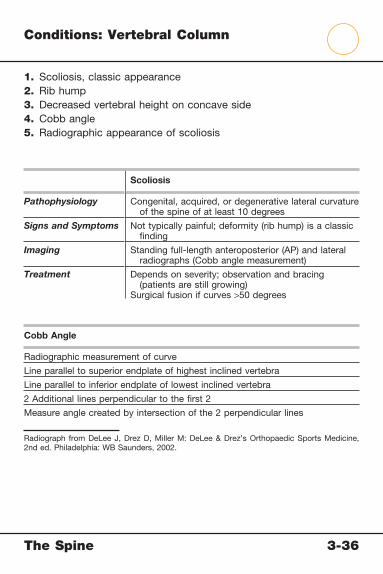
The Spine 3-36
Conditions: Vertebral Column
1. Scoliosis, classic appearance2. Rib hump3. Decreased vertebral height on concave side4. Cobb angle5. Radiographic appearance of scoliosis
Scoliosis
Pathophysiology Congenital, acquired, or degenerative lateral curvature of the spine of at least 10 degrees
Signs and Symptoms Not typically painful; deformity (rib hump) is a classic fi nding
Imaging Standing full-length anteroposterior (AP) and lateral radiographs (Cobb angle measurement)
Treatment Depends on severity; observation and bracing (patients are still growing)
Surgical fusion if curves >50 degrees
Cobb Angle
Radiographic measurement of curve
Line parallel to superior endplate of highest inclined vertebra
Line parallel to inferior endplate of lowest inclined vertebra
2 Additional lines perpendicular to the fi rst 2
Measure angle created by intersection of the 2 perpendicular lines
Radiograph from DeLee J, Drez D, Miller M: DeLee & Drez’s Orthopaedic Sports Medicine, 2nd ed. Philadelphia: WB Saunders, 2002.

The Spine 3-37
2
3
4
5
6
1
Identify each condition
Conditions: Vertebral Column

The Spine 3-37
Conditions: Vertebral Column
1. Cervical spine degeneration with hyperextension deformity2. Narrowed neural foramina secondary to degenerative changes3. Radiographic appearance of cervical spine osteoarthritis4. Lumbar spine osteoarthritis5. Anterior osteophytes6. Disc degeneration
Osteoarthritis of the Spine Myelopathy
Mechanism Degenerative (wear and tear); posttraumatic
Cord compression resulting from osteophytes, disc disease, or facet arthropathy
Signs and Symptoms
Pain that is worse with standing; motion loss may be present
Neurological symptoms only with cord or nerve root compression from osteophytes
Pain, neurological symptoms, occipital headaches
Imaging Anteroposterior (AP), lateral, and oblique radiographs
Radiographs, computed tomographic myelography, and/or magnetic resonance imaging (MRI)
Treatment Activity modifi cation, nonsteroidal antiinfl ammatory drugs (NSAIDs), physical therapy, selective injections
NSAIDs, physical therapy, traction, surgery (discectomy and arthrodesis)
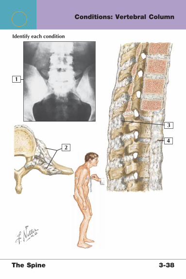
The Spine 3-38
2
3
4
1
Identify each condition
Conditions: Vertebral Column
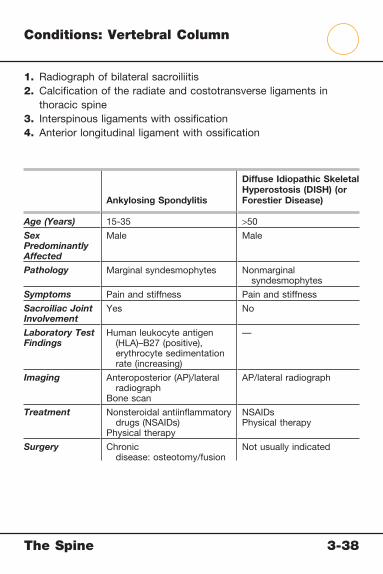
The Spine 3-38
Conditions: Vertebral Column
1. Radiograph of bilateral sacroiliitis2. Calcifi cation of the radiate and costotransverse ligaments in
thoracic spine3. Interspinous ligaments with ossifi cation4. Anterior longitudinal ligament with ossifi cation
Ankylosing Spondylitis
Diffuse Idiopathic Skeletal Hyperostosis (DISH) (or Forestier Disease)
Age (Years) 15-35 >50
Sex Predominantly Affected
Male Male
Pathology Marginal syndesmophytes Nonmarginal syndesmophytes
Symptoms Pain and stiffness Pain and stiffness
Sacroiliac Joint Involvement
Yes No
Laboratory Test Findings
Human leukocyte antigen (HLA)–B27 (positive), erythrocyte sedimentation rate (increasing)
—
Imaging Anteroposterior (AP)/lateral radiograph
Bone scan
AP/lateral radiograph
Treatment Nonsteroidal antiinfl ammatory drugs (NSAIDs)
Physical therapy
NSAIDsPhysical therapy
Surgery Chronic disease: osteotomy/fusion
Not usually indicated
The Spine 3-39
2
3
4
5
6
7
8
6
1
Conditions: Sacrum

The Spine 3-39
Conditions: Sacrum
1. L5 transverse processes2. L5 vertebral body3. Sacroiliac joint4. Sacrum5. Coccyx6. Fracture of the body of the sacrum7. Sacral promontory8. Fracture of the coccyx
Sacral Fractures Sacroiliac Joint Pain
Mechanism Trauma or stress reaction Muscle imbalance, postural changes, repetitive activity, trauma
Signs and Symptoms
Localized pain and tenderness, inability to sit
Pain/tenderness localized to joint; positive result of Gaenslen’s test
Imaging Anteroposterior (AP) and lateral radiographs, computed tomography (CT); rule out pelvic ring injury
Not necessary
Treatment Conservative for nondisplaced traumatic and stress fractures
Surgical fi xation for fractures displaced >1 cm
Nonsteroidal antiinfl ammatory drugs (NSAIDs), local injections

Netter’s Musculoskeletal Flash Cards
4 The Thorax and AbdomenPlates 4-1 to 4-17
Bony Anatomy
4-1 Bony Anatomy: Thorax
4-2 Bony Anatomy: Abdomen
Radiographic Anatomy
4-3 Radiographic Anatomy: Thorax
Muscles
4-4 Muscles: Anterior Thorax
4-5 Muscles: Anterior Thorax Wall
4-6 Muscles: Anterior Thorax Wall
4-7 Muscles: Superfi cial Layer of Posterior
Thorax
4-8 Muscles: Intermediate Layer of Posterior
Thorax
4-9 Muscles: Anterior Abdominal Wall
4-10 Muscles: Posterior Abdominal Wall
Arteries and Nerves
4-11 Intercostal Arteries and Nerves
4-12 Arteries: Thorax and Abdomen
4-13 Nerves: Thorax and Abdomen
Conditions
4-14 Conditions: Sternum
4-15 Conditions: Ribs
The Thorax and Abdomen Table of Contents
4 The Thorax and AbdomenPlates 4-1 to 4-17
4-16 Conditions: Sternoclavicular and Sternocostal
Joints
4-17 Conditions: Costochondral and Costovertebral
Joints
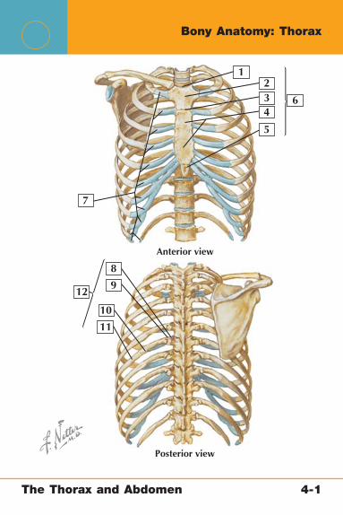
The Thorax and Abdomen 4-1
Bony Anatomy: Thorax
234
5
6
7
8
9
10
11
12
1
Anterior view
Posterior view

The Thorax and Abdomen 4-1
Bony Anatomy: Thorax
1. Jugular notch of sternum 2. Manubrium 3. Sternal angle 4. Body of sternum 5. Xiphoid process 6. Sternum 7. Costal cartilages 8. Head of the rib 9. Neck of the rib10. Angle of the rib11. Body of the rib12. Ribs
Comment: The thorax comprises 12 pairs of ribs. Each articulates with the corresponding vertebra posteriorly; ribs 1-7 articulate with the sternum anteriorly (true ribs). Other articulations of the thorax include the sternoclavicular joints and the costochondral joints.

2
3
4
5
6
7
8
9
10
1
Bony Anatomy: Abdomen
The Thorax and Abdomen 4-2

Bony Anatomy: Abdomen
1. Ribs 4-10 2. T11 3. Rib 11 4. Rib 12 5. Iliac crest 6. L5 7. Sacrum 8. Coccyx 9. Pubic symphysis10. Pubic ramus
Comment: The bony framework of the abdomen includes the ribs, the lower thoracic vertebrae, the lumbar vertebrae, the sacrum, and the pelvis. These structures provide important stability and sites of attachment for the muscles that make up the anterior and posterior abdominal wall.
The Thorax and Abdomen 4-2
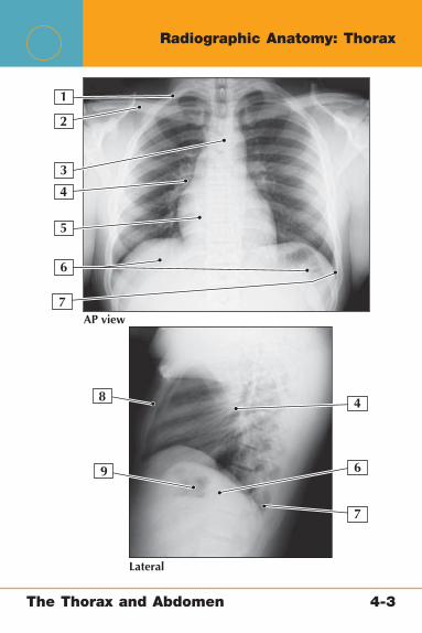
2
3
4
4
5
6
6
7AP view
Lateral
7
8
9
1
Radiographic Anatomy: Thorax
The Thorax and Abdomen 4-3

Radiographic Anatomy: Thorax
1. 1st Rib2. Clavicle3. Aortic knob4. Hilar nodes5. Cardiac shadow6. Diaphragm7. Costophrenic angle8. Sternum9. Gastric bubble
Comment: Anteroposterior (AP) and lateral views of the chest are helpful in evaluating a variety of conditions ranging from disorders of the lungs (pneumothorax, pneumonia), the heart (cardiomegaly, aortic dissection), and the musculoskeletal system (sternal fractures, rib fractures).
The Thorax and Abdomen 4-3

2
3
4
1
Muscles: Anterior Thorax
The Thorax and Abdomen 4-4

Musc
les: A
nte
rior T
hora
x1. Pectoralis major muscle2. Pectoralis minor muscle3. Serratus anterior muscle4. External intercostal muscles
Pectoralis Major Muscle
Pectoralis Minor Muscle
Serratus Anterior Muscle
External Intercostal Muscles
Origin Sternum (sternal head) and medial clavicle (clavicular head)
Ribs 3-5 Outer superior aspect of ribs 1-8
Inferior border of the fi rst 11 ribs
Insertion Intertubercular groove of the humerus
Coracoid process of the scapula
Vertebral border of the scapula
Superior border of the rib below
Actions Adduction and internal rotation of the humerus
Holds scapula to chest wall
Holds scapula to thoracic wall and laterally rotates the scapula to assist in shoulder abduction
Elevation of the ribs
Innervation Medial and lateral pectoral nerves (C5-6)
Medial pectoral nerve (C8-T1)
Long thoracic nerve (C5-7)
Intercostal nerves at each level
The T
hora
x and A
bdom
en
4-4
2
1
Muscles: Anterior Thorax Wall
The Thorax and Abdomen 4-5

Muscles: Anterior Thorax Wall
1. Diaphragm2. Transversus abdominis
Diaphragm Transversus Abdominis
Origin Xiphoid process (sternal portion), costal cartilages of lower 6 ribs (costal portion), and L1-3 vertebrae (lumbar portion)
Internal surface of ribs 7-12, thoracolumbar fascia, and iliac crest
Insertion Central tendon Linea alba and pubic crest
Actions Assists in respiration by pulling the central tendon down and forward
Compression of abdomen
Innervation Phrenic nerve (C3-5) Ventral rami of T7-L1
The Thorax and Abdomen 4-5
2
3
1
Muscles: Anterior Thorax Wall
The Thorax and Abdomen 4-6

Muscles: Anterior Thorax Wall
1. Internal intercostal muscles2. Innermost intercostal muscles3. Transversus thoracis muscle
Internal Intercostal Muscle
Innermost Intercostal Muscle
Transversus Thoracis Muscle
Origin Inferior border of each rib
Inferior border of each rib
Xiphoid process, inferior body of sternum, and adjacent costal cartilage
Insertion Superior border of rib below
Superior border of rib below
Costal cartilages 2-6
Actions Elevation of the ribs Elevation of the ribs
Elevation of the ribs
Innervation Intercostal nerves at each level
Intercostal nerves at each level
Intercostal nerves
The Thorax and Abdomen 4-6

2
3
4
1
Muscles: Superfi cial Layer of Posterior Thorax
The Thorax and Abdomen 4-7
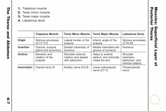
Musc
les: S
uperfi c
ial L
aye
r of
Poste
rior T
hora
x
1. Trapezius muscle2. Teres minor muscle3. Teres major muscle4. Latissimus dorsi
Trapezius Muscle Teres Minor Muscle Teres Major Muscle Latissimus Dorsi
Origin Spinous processes of C7-T12
Lateral border of the scapula
Inferior angle of the scapula
Spinous processes of T6-S5
Insertion Clavicle, scapula (spine and acromion)
Greater tuberosity of humerus
Medial intertubercular groove of humerus
Humerus
Actions Elevation and rotation of the scapula
Shoulder external rotation and assists with adduction
Helps to extend, adduct, and internally rotate the arm
Shoulder extension, adduction, and internal rotation
Innervation Cranial nerve XI Axillary nerve (C5-6) Lower subscapular nerve (C7-7)
Thoracodorsal nerve
The T
hora
x and A
bdom
en
4-7
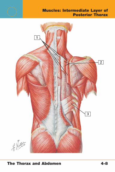
2
3
1
Muscles: Intermediate Layer of Posterior Thorax
The Thorax and Abdomen 4-8
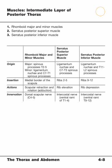
Muscles: Intermediate Layer of Posterior Thorax
1. Rhomboid major and minor muscles2. Serratus posterior superior muscle3. Serratus posterior inferior muscle
Rhomboid Major and Minor Muscles
Serratus Posterior Superior Muscle
Serratus Posterior Inferior Muscle
Origin Major: spinous processes T2-5
Minor: ligamentum nuchae and C7-T1 spinous processes
Ligamentum nuchae and C7-T3 spinous processes
Ligamentum nuchae and T11-L2 spinous processes
Insertion Medial border of the scapula
Ribs 2-5 Ribs 9-12
Actions Scapular retraction and rotation (adduction)
Rib elevation Rib depression
Innervation Dorsal scapular nerve (C4-5)
Intercostal nerve (ventral rami of T1-4)
Intercostal nerve (ventral rami T9-12)
The Thorax and Abdomen 4-8
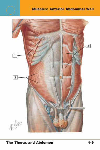
2
3
1
Muscles: Anterior Abdominal Wall
The Thorax and Abdomen 4-9
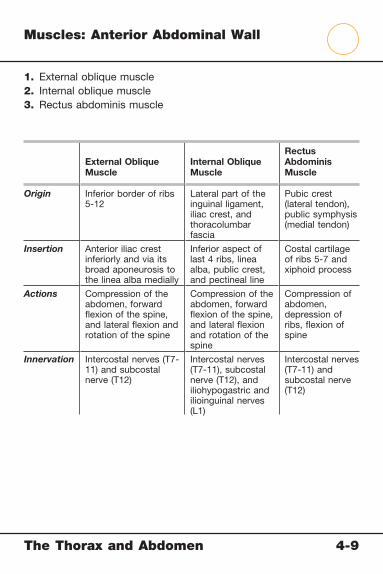
Muscles: Anterior Abdominal Wall
1. External oblique muscle2. Internal oblique muscle3. Rectus abdominis muscle
External Oblique Muscle
Internal Oblique Muscle
Rectus Abdominis Muscle
Origin Inferior border of ribs 5-12
Lateral part of the inguinal ligament, iliac crest, and thoracolumbar fascia
Pubic crest (lateral tendon), public symphysis (medial tendon)
Insertion Anterior iliac crest inferiorly and via its broad aponeurosis to the linea alba medially
Inferior aspect of last 4 ribs, linea alba, public crest, and pectineal line
Costal cartilage of ribs 5-7 and xiphoid process
Actions Compression of the abdomen, forward fl exion of the spine, and lateral fl exion and rotation of the spine
Compression of the abdomen, forward fl exion of the spine, and lateral fl exion and rotation of the spine
Compression of abdomen, depression of ribs, fl exion of spine
Innervation Intercostal nerves (T7-11) and subcostal nerve (T12)
Intercostal nerves (T7-11), subcostal nerve (T12), and iliohypogastric and ilioinguinal nerves (L1)
Intercostal nerves (T7-11) and subcostal nerve (T12)
The Thorax and Abdomen 4-9

23
4
1
Muscles: Posterior Abdominal Wall
The Thorax and Abdomen 4-10
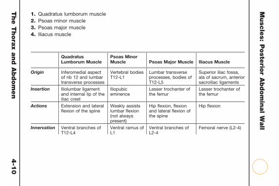
Musc
les: P
oste
rior A
bdom
inal W
all
1. Quadratus lumborum muscle2. Psoas minor muscle3. Psoas major muscle4. Iliacus muscle
Quadratus Lumborum Muscle
Psoas Minor Muscle Psoas Major Muscle Iliacus Muscle
Origin Inferomedial aspect of rib 12 and lumbar transverse processes
Vertebral bodies T12-L1
Lumbar transverse processes, bodies of T12-L5
Superior iliac fossa, ala of sacrum, anterior sacroiliac ligaments
Insertion Iliolumbar ligament and internal lip of the iliac crest
Iliopubic eminence
Lesser trochanter of the femur
Lesser trochanter of the femur
Actions Extension and lateral fl exion of the spine
Weakly assists lumbar fl exion (not always present)
Hip fl exion, fl exion and lateral fl exion of the spine
Hip fl exion
Innervation Ventral branches of T12-L4
Ventral ramus of L1
Ventral branches of L2-4
Femoral nerve (L2-4)
The T
hora
x and A
bdom
en
4-1
0
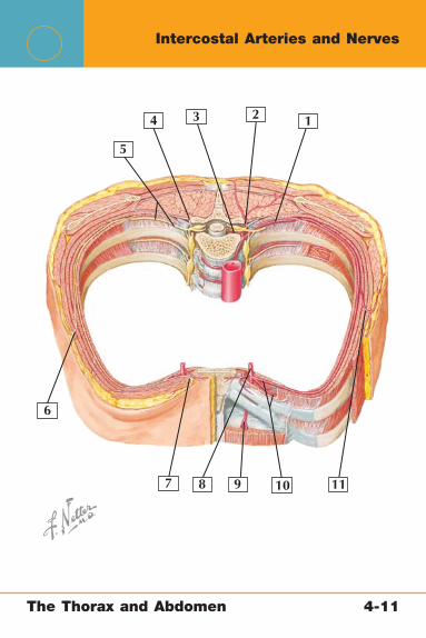
234
5
6
7 8 9 10 11
1
Intercostal Arteries and Nerves
The Thorax and Abdomen 4-11

Intercostal Arteries and Nerves
1. Posterior intercostal artery 2. Dorsal branch of the posterior intercostal artery 3. Spinal branch of the posterior intercostal artery 4. Dorsal ramus of thoracic nerve 5. Intercostal nerve (ventral ramus of thoracic nerve) 6. Lateral cutaneous branch of the intercostal nerve 7. Anterior cutaneous branch of the intercostal nerve 8. Internal thoracic artery 9. Superior epigastric artery10. Anterior intercostal arteries11. Lateral cutaneous branch of the posterior intercostal artery
Comment: Each intercostal space contains a large posterior intercostal artery and two smaller anterior intercostal arteries. The intercostal nerves are found in the same intercostal space and are also known as the ventral rami of T1-11. The intercostal arteries and nerves serve the intercostal muscles, which power respiration.
The Thorax and Abdomen 4-11

2
345
6
7
8
9
10
11
12
13
14
1
Arteries: Thorax and Abdomen
The Thorax and Abdomen 4-12

Arteries: Thorax and Abdomen
1. Pericardiacophrenic artery 2. Internal thoracic arteries 3. Subclavian artery 4. Axillary artery 5. Lateral thoracic artery 6. Thoracodorsal artery 7. Anterior intercostal arteries 8. Anastomoses with lower intercostal, subcostal, and lumbar
arteries 9. Ascending branch of the deep circumfl ex iliac artery10. Superfi cial epigastric artery11. Femoral artery12. Superior epigastric artery13. Inferior epigastric artery14. Musculophrenic arteries
Comment: The main blood supply to the anterior abdominal wall is provided by the superior and inferior epigastric and deep circumfl ex iliac arteries. The superior epigastric artery is a branch of the internal thoracic artery, whereas the inferior epigastric and deep circumfl ex iliac arteries are branches of the external iliac artery.
The Thorax and Abdomen 4-12

2
3
4
5
6
7
8
9
10
11
12
13
1
Nerves: Thorax and Abdomen
The Thorax and Abdomen 4-13

Nerves: Thorax and Abdomen
1. Medial brachial cutaneous nerve 2. Intercostobrachial nerve (T1-2) 3. Long thoracic nerve 4. Lateral cutaneous branches of the intercostal nerve (T2-11) 5. Anterior cutaneous branches of the intercostal nerve (T1-11) 6. Lateral cutaneous branch of the subcostal nerve (T12) 7. Lateral cutaneous branch of the iliohypogastric nerve (L1) 8. Anterior cutaneous branch of the subcostal nerve (T12) 9. Lateral femoral cutaneous nerve (L2-3)10. Anterior cutaneous branch of the iliohypogastric nerve (L1-2)11. Femoral branches of the genitofemoral nerve (L1-2)12. Ilioinguinal nerve (L1)13. Anterior branch of the iliohypogastric nerve (L1)
Comment: The nerve supply to the anterior abdominal wall is provided mainly by the ventral rami of the thoracic and lumbar nerves. In the upper abdomen, this supply is essentially an extension of the lower thoracic-derived intercostal and subcostal nerves. In the lower abdomen, this supply is derived mainly from the ilioinguinal and iliohypogastric nerves.
The Thorax and Abdomen 4-13

2
3
4
5
6
7
8
9
1
Conditions: Sternum
The Thorax and Abdomen 4-14

Conditions: Sternum
1. Sternal angle2. First costal cartilage3. Jugular notch4. Clavicular notch5. Manubrium6. Body of sternum (with fracture)7. Xiphisternal junction8. Xiphoid process9. Radiographic appearance of sternal fracture
Sternal Fracture
Mechanism Usually direct trauma (motor vehicle accident)
Signs and Symptoms
Pain, tenderness, and crepitus over the sternum
Imaging Lateral view of sternum; computed tomography (CT)
Treatment Usually symptomatic pain control but reduction if displaced
Radiograph from DeLee J, Drez D, Miller M: DeLee & Drez’s Orthopaedic Sports Medicine. Philadelphia: WB Saunders, 2002.
The Thorax and Abdomen 4-14

23
3
45
5
6
7
1
Conditions: Ribs
The Thorax and Abdomen 4-15
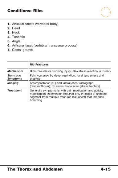
Conditions: Ribs
1. Articular facets (vertebral body)2. Head3. Neck4. Tubercle5. Angle6. Articular facet (vertebral transverse process)7. Costal groove
Rib Fractures
Mechanism Direct trauma or crushing injury; also stress reaction in rowers
Signs and Symptoms
Pain worsened by deep inspiration; focal tenderness and crepitus
Imaging Anteroposterior (AP) and lateral chest radiograph (pneumothorax); rib series; bone scan (stress fracture)
Treatment Generally symptomatic with pain medication and activity modifi cation; intervention required only in cases of unstable segment from multiple fractures (fl ail chest) that impedes breathing
The Thorax and Abdomen 4-15

2
3
4
5
6
7
8
1
Conditions: Sternoclavicular and Sternocostal Joints
The Thorax and Abdomen 4-16

Conditions: Sternoclavicular and Sternocostal Joints
1. Sternoclavicular ligament2. Clavicle3. Costoclavicular ligament4. 1st Rib5. Body of the sternum6. Sternocostal joint7. Manubrium of sternum8. Computed tomography (CT) of sternoclavicular joint dislocation
Sternoclavicular Joint InjuriesSternocostal Joint Injuries (Rib Dislocation)
Mechanism Traumatic dislocation, spontaneous atraumatic subluxation
Direct trauma, crush injury
Signs and Symptoms
Acute focal pain; palpable step-off
Focal pain and deformity, pain with inspiration; neurovascular integrity should be evaluated
Imaging Plain radiograph (serendipity view) and/or CT
Anteroposterior (AP) and lateral chest radiographs (usually involves joints 2-10)
Treatment Subluxations are treated conservatively
Dislocations necessitate closed reduction
Posterior dislocations may necessitate open reduction and ligament reconstruction
Generally conservative
Computed tomogram from Miller M, Sekiya JK: Core Knowledge in Orthopaedics: Sports Medicine. Philadelphia: Elsevier, 2006.
The Thorax and Abdomen 4-16
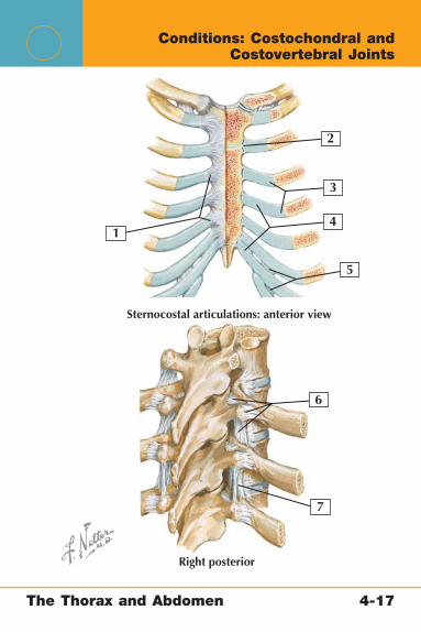
Sternocostal articulations: anterior view
Right posterior
2
3
4
5
6
7
1
Conditions: Costochondral and Costovertebral Joints
The Thorax and Abdomen 4-17

Conditions: Costochondral and Costovertebral Joints
1. Radiate sternocostal ligaments2. Sternocostal ligament3. Costochondral joint4. Costal cartilage5. Interchondral joint6. Costotransverse ligament7. Intertransverse ligament
Costochondral Injury (Rib Separation)
Costochondritis (Tietze Syndrome)
Costovertebral Joint Dislocation
Mechanism Direct trauma, crush injury
Infl ammatory process of the costochondral joint
Rare but from high-velocity trauma
Signs and Symptoms
Focal pain and deformity, pain with inspiration; neurovascular integrity should be evaluated
Focal pain and tenderness that is not exertional
Focal tenderness; other trauma extremely common
Imaging Anteroposterior (AP) and lateral chest radiographs
Usually none, but bone scan may help pinpoint injury
AP lateral radiograph, computed tomography (CT)
Treatment Generally conservative
Nonsteroidal antiinfl ammatory drugs (NSAIDs); corticosteroid injections in refractory cases
Usually conservative with focus on other injuries
The Thorax and Abdomen 4-17

Netter’s Musculoskeletal Flash Cards
5 The Pelvis, Hip, and ThighPlates 5-1 to 5-31
Bony Anatomy
5-1 Bony Anatomy: Pelvis
5-2 Bony Anatomy: Hip
5-3 Bony Anatomy: Femur
Radiographic Anatomy
5-4 Radiographic Anatomy: Pelvis and Hip
Ligaments
5-5 Ligaments: Pelvis
5-6 Ligaments: Hip Joint
Muscles
5-7 Muscles: Pelvis
5-8 Muscles: Anterior Hip and Thigh
5-9 Muscles: Anterior Hip and Thigh
5-10 Muscles: Anterior Hip and Thigh
5-11 Muscles: Posterior Hip and Thigh
5-12 Muscles: Posterior Hip and Thigh
Arteries and Nerves
5-13 Arteries and Nerves: Anterior Pelvis and Hip
5-14 Arteries and Nerves: Posterior Pelvis and Hip
5-15 Arteries: Hip Joint
The Pelvis, Hip, and Thigh Table of Contents
5 The Pelvis, Hip, and ThighPlates 5-1 to 5-31
Physical Examination
5-16 Physical Examination: Hip Joint
Conditions
5-17 Conditions: Pelvic Ring
5-18 Conditions: Acetabulum
5-19 Conditions: Femoral Head
5-20 Conditions: Proximal Femur
5-21 Conditions: Femoral Shaft
5-22 Conditions: Athlete’s Pelvis and Hip
5-23 Conditions: Hip Joint
5-24 Conditions: Hip Joint
5-25 Conditions: Hip Joint
5-26 Conditions: Hip Joint
5-27 Conditions: Muscles
5-28 Conditions: Sciatic Nerve
5-29 Conditions: Lateral Femoral Cutaneous Nerve
5-30 Conditions: Pediatric Hip
5-31 Conditions: Pediatric Hip

The Pelvis, Hip, and Thigh 5-1
Bony Anatomy: Pelvis
Female pelvis/female pelvic inlet: anterior view
Male pelvis/male pelvic inlet: anterior view
3
98
8
7
7
2
10
10
4
5
6
6
9
1
1
2
3
4
5

The Pelvis, Hip, and Thigh 5-1
Bony Anatomy: Pelvis
1. Sacroiliac joint 2. Arcuate line 3. Ischial spine 4. Pectineal line 5. Ischial tuberosity 6. Obturator foramen 7. Pubic symphysis 8. Pubic tubercle 9. Pelvic inlet10. Acetabulum
Comment: The pelvis is made up of two large “wings” that join with the sacrum (sacroiliac joints) proximally and each other (pubic symphysis) inferiorly. There are distinct structural differences between the male pelvis and the female pelvis: The female pelvis has more fl ared wings with a transversely oval pelvic inlet and a smaller pubic symphysis.
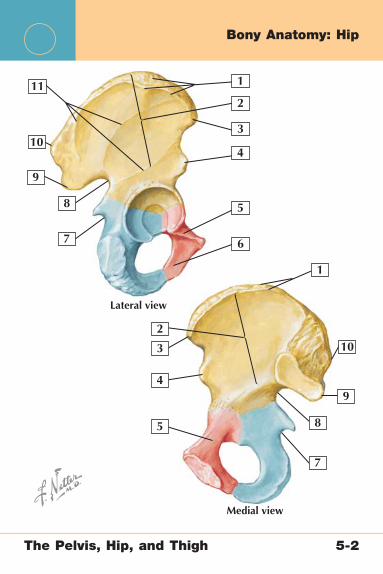
9
9
8
7
7
8
10
10
11
6
1
1
2
2
3
3
4
4
5
5
Lateral view
Medial view
Bony Anatomy: Hip
The Pelvis, Hip, and Thigh 5-2
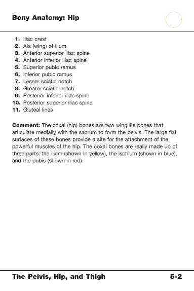
Bony Anatomy: Hip
1. Iliac crest 2. Ala (wing) of ilium 3. Anterior superior iliac spine 4. Anterior inferior iliac spine 5. Superior pubic ramus 6. Inferior pubic ramus 7. Lesser sciatic notch 8. Greater sciatic notch 9. Posterior inferior iliac spine10. Posterior superior iliac spine11. Gluteal lines
Comment: The coxal (hip) bones are two winglike bones that articulate medially with the sacrum to form the pelvis. The large fl at surfaces of these bones provide a site for the attachment of the powerful muscles of the hip. The coxal bones are really made up of three parts: the ilium (shown in yellow), the ischium (shown in blue), and the pubis (shown in red).
The Pelvis, Hip, and Thigh 5-2

9
87
10
11
6
1
2
34
5
Anterior view Posterior view
Bony Anatomy: Femur
The Pelvis, Hip, and Thigh 5-3
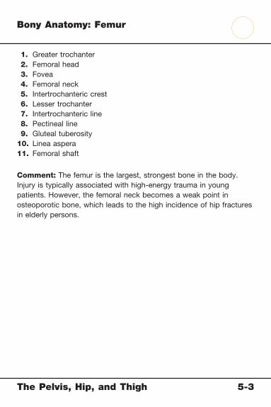
Bony Anatomy: Femur
1. Greater trochanter 2. Femoral head 3. Fovea 4. Femoral neck 5. Intertrochanteric crest 6. Lesser trochanter 7. Intertrochanteric line 8. Pectineal line 9. Gluteal tuberosity10. Linea aspera11. Femoral shaft
Comment: The femur is the largest, strongest bone in the body. Injury is typically associated with high-energy trauma in young patients. However, the femoral neck becomes a weak point in osteoporotic bone, which leads to the high incidence of hip fractures in elderly persons.
The Pelvis, Hip, and Thigh 5-3

7
8
9
10
11
6
1
1
2
3
4
5
AP pelvis
Lateral hip
Radiographic Anatomy: Pelvis and Hip
The Pelvis, Hip, and Thigh 5-4
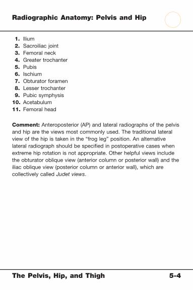
Radiographic Anatomy: Pelvis and Hip
1. Ilium 2. Sacroiliac joint 3. Femoral neck 4. Greater trochanter 5. Pubis 6. Ischium 7. Obturator foramen 8. Lesser trochanter 9. Pubic symphysis10. Acetabulum11. Femoral head
Comment: Anteroposterior (AP) and lateral radiographs of the pelvis and hip are the views most commonly used. The traditional lateral view of the hip is taken in the “frog leg” position. An alternative lateral radiograph should be specifi ed in postoperative cases when extreme hip rotation is not appropriate. Other helpful views include the obturator oblique view (anterior column or posterior wall) and the iliac oblique view (posterior column or anterior wall), which are collectively called Judet views.
The Pelvis, Hip, and Thigh 5-4

9
8 7
10
11
6
1
1
2
3
4
4
5
5
Anterior view
Posterior view
Ligaments: Pelvis
The Pelvis, Hip, and Thigh 5-5
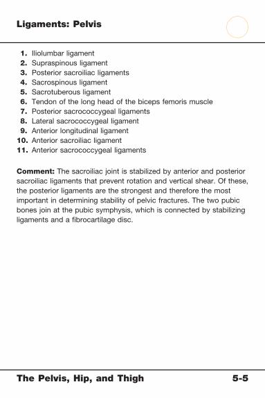
Ligaments: Pelvis
1. Iliolumbar ligament 2. Supraspinous ligament 3. Posterior sacroiliac ligaments 4. Sacrospinous ligament 5. Sacrotuberous ligament 6. Tendon of the long head of the biceps femoris muscle 7. Posterior sacrococcygeal ligaments 8. Lateral sacrococcygeal ligament 9. Anterior longitudinal ligament10. Anterior sacroiliac ligament11. Anterior sacrococcygeal ligaments
Comment: The sacroiliac joint is stabilized by anterior and posterior sacroiliac ligaments that prevent rotation and vertical shear. Of these, the posterior ligaments are the strongest and therefore the most important in determining stability of pelvic fractures. The two pubic bones join at the pubic symphysis, which is connected by stabilizing ligaments and a fi brocartilage disc.
The Pelvis, Hip, and Thigh 5-5

6
1
2
3
4
5
Anterior view
Posterior view
Ligaments: Hip Joint
The Pelvis, Hip, and Thigh 5-6
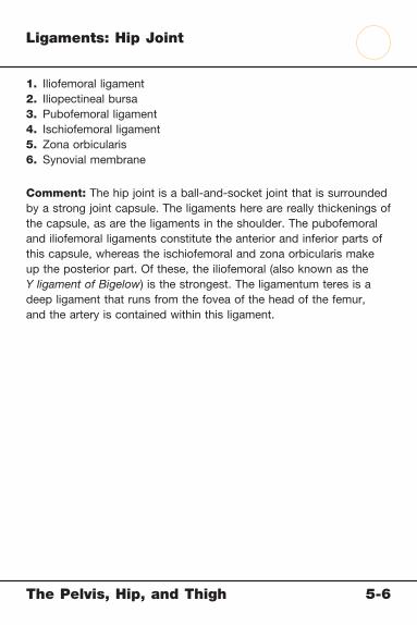
Ligaments: Hip Joint
1. Iliofemoral ligament2. Iliopectineal bursa3. Pubofemoral ligament4. Ischiofemoral ligament5. Zona orbicularis6. Synovial membrane
Comment: The hip joint is a ball-and-socket joint that is surrounded by a strong joint capsule. The ligaments here are really thickenings of the capsule, as are the ligaments in the shoulder. The pubofemoral and iliofemoral ligaments constitute the anterior and inferior parts of this capsule, whereas the ischiofemoral and zona orbicularis make up the posterior part. Of these, the iliofemoral (also known as the Y ligament of Bigelow) is the strongest. The ligamentum teres is a deep ligament that runs from the fovea of the head of the femur, and the artery is contained within this ligament.
The Pelvis, Hip, and Thigh 5-6
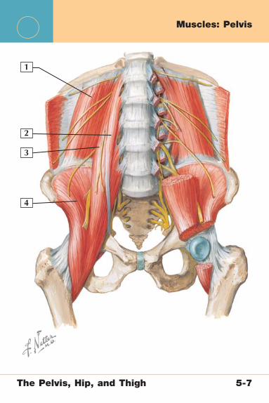
1
2
3
4
Muscles: Pelvis
The Pelvis, Hip, and Thigh 5-7

Musc
les: P
elvis
1. Quadratus lumborum muscle2. Psoas minor muscle3. Psoas major muscle4. Iliacus muscle
Quadratus Lumborum Muscle
Psoas Minor Muscle
Psoas Major Muscle Iliacus Muscle
Origin Inferomedial aspect of rib 12 and lumbar transverse processes
Vertebral bodies T12-L1
Lumbar transverse processes, bodies of T12-L5
Superior iliac fossa, ala of sacrum, anterior sacroiliac ligaments
Insertion Iliolumbar ligament and internal lip of the iliac crest
Iliopubic eminence Lesser trochanter of the femur
Lesser trochanter of the femur
Actions Extension and lateral fl exion of the spine
Weakly assists lumbar fl exion (not always present)
Hip fl exion, fl exion and lateral fl exion of the spine
Hip fl exion
Innervation Ventral branches of T12-L4
Ventral ramus of L1
Ventral branches of L2-L4
Femoral nerve (L2-L4)
The P
elvis, H
ip, a
nd T
hig
h
5-7

1
2
3
Muscles: Anterior Hip and Thigh
The Pelvis, Hip, and Thigh 5-8

Muscles: Anterior Hip and Thigh
1. Tensor fascia latae muscle2. Sartorius muscle3. Quadriceps femoris muscles
Tensor Fascia Latae Muscle Sartorius Muscle
Origin Anterior superior iliac spine Anterior superior iliac spine
Insertion Iliotibial band Proximal medial tibia
Actions Flexion, abduction, internal rotation of the hip
Flexion and external rotation of the hip
Innervation Superior gluteal nerve Femoral nerve
The Pelvis, Hip, and Thigh 5-8

1
2
3
Muscles: Anterior Hip and Thigh
The Pelvis, Hip, and Thigh 5-9

Muscles: Anterior Hip and Thigh
1. Pectineus muscle2. Adductor longus muscle3. Gracilis muscle
Pectineus MuscleAdductor Longus and Brevis Muscles Gracilis Muscle
Origin Pubis (pectineal line) Inferior pubis Pubic ramus
Insertion Femur (pectineal line) Linea aspera (longus)Pectineal line (brevis)
Proximal medial tibia
Actions Flexion and adduction of the hip
Adduction of the hip Flexion and adduction of the hip
Innervation Femoral nerve Obturator nerve Obturator nerve
The Pelvis, Hip, and Thigh 5-9

1
2
3
Muscles: Anterior Hip and Thigh
The Pelvis, Hip, and Thigh 5-10
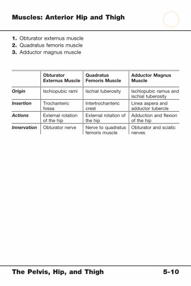
Muscles: Anterior Hip and Thigh
1. Obturator externus muscle2. Quadratus femoris muscle3. Adductor magnus muscle
Obturator Externus Muscle
Quadratus Femoris Muscle
Adductor Magnus Muscle
Origin Ischiopubic rami Ischial tuberosity Ischiopubic ramus and ischial tuberosity
Insertion Trochanteric fossa
Intertrochanteric crest
Linea aspera and adductor tubercle
Actions External rotation of the hip
External rotation of the hip
Adduction and fl exion of the hip
Innervation Obturator nerve Nerve to quadratus femoris muscle
Obturator and sciatic nerves
The Pelvis, Hip, and Thigh 5-10

1
2
3
Muscles: Posterior Hip and Thigh
The Pelvis, Hip, and Thigh 5-11
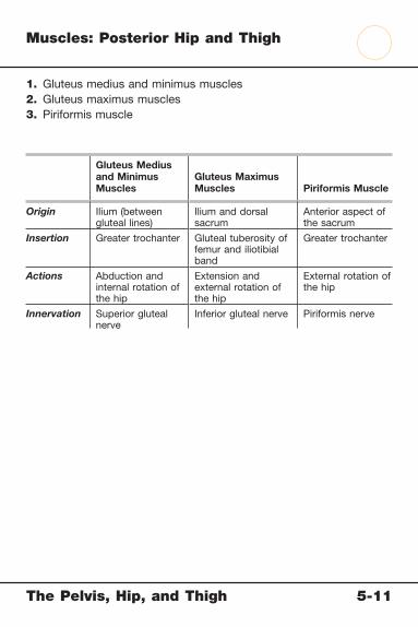
Muscles: Posterior Hip and Thigh
1. Gluteus medius and minimus muscles2. Gluteus maximus muscles3. Piriformis muscle
Gluteus Medius and Minimus Muscles
Gluteus Maximus Muscles Piriformis Muscle
Origin Ilium (between gluteal lines)
Ilium and dorsal sacrum
Anterior aspect of the sacrum
Insertion Greater trochanter Gluteal tuberosity of femur and iliotibial band
Greater trochanter
Actions Abduction and internal rotation of the hip
Extension and external rotation of the hip
External rotation of the hip
Innervation Superior gluteal nerve
Inferior gluteal nerve Piriformis nerve
The Pelvis, Hip, and Thigh 5-11
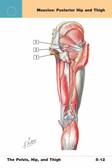
1
2
3
Muscles: Posterior Hip and Thigh
The Pelvis, Hip, and Thigh 5-12

Muscles: Posterior Hip and Thigh
1. Superior gemellus muscle2. Obturator internus muscle3. Inferior gemellus muscle
Superior and Inferior Gemellus Muscles
Obturator Internus Muscle
Origin Ischial spine (superior)Ischial tuberosity (inferior)
Ischiopubic rami and obturator membrane
Insertion Greater trochanter Greater trochanter
Actions External rotation of the hip External rotation of the hip
Innervation Nerve to obturator internus (superior)Nerve to quadratus femoris (inferior)
Nerve to obturator internus
The Pelvis, Hip, and Thigh 5-12

1
234
5
6
7
8 9
10
11
Arteries and Nerves: Anterior Pelvis and Hip
The Pelvis, Hip, and Thigh 5-13

Arteries and Nerves: Anterior Pelvis and Hip
1. Femoral vein 2. Femoral artery 3. External iliac vein 4. External iliac artery 5. Lateral femoral cutaneous nerve 6. Femoral nerve 7. Profunda femoris artery 8. Saphenous nerve 9. Descending genicular artery10. Cutaneous branch of the obturator nerve11. Obturator nerve (anterior and posterior branches)
Comment: The femoral triangle contains the femoral vein (most medial), artery, and nerve (most lateral). A useful mnemonic for this relation is “Lateral → Medial = NAVEL,” where “NAVEL” is nerve, artery, vein, empty space, lymphatics. The femoral artery passes between the adductor longus and the vastus medialis muscles before traveling to the knee, where it becomes the popliteal artery. The femoral nerve supplies innervation to the majority of the muscles of the anterior thigh, including the quadriceps, as it passes through the thigh.
The Pelvis, Hip, and Thigh 5-13

1
2
3
4
5
6
7
8
9
10
11 12
Arteries and Nerves: Posterior Pelvis and Hip
The Pelvis, Hip, and Thigh 5-14

Arteries and Nerves: Posterior Pelvis and Hip
1. Superior gluteal nerve and artery 2. Superior clunial nerves 3. Medial clunial nerves 4. Inferior gluteal artery and nerve 5. Pudendal nerve 6. Nerve to obturator internus 7. Posterior femoral cutaneous nerve 8. Inferior clunial nerves 9. Muscular branches of the sciatic nerve10. Sciatic nerve11. Tibial nerve12. Common peroneal nerve
Comment: The sciatic nerve (L4-S3) exits the greater sciatic foramen and travels distally, dividing into the common peroneal and tibial nerves. It supplies innervation to the hamstring muscles and part of the adductor magnus.
The Pelvis, Hip, and Thigh 5-14
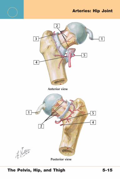
1
1
2
2
3
5
4
4
5
Anterior view
Posterior view
Arteries: Hip Joint
The Pelvis, Hip, and Thigh 5-15
Arteries: Hip Joint
1. Acetabular branch of the obturator artery2. Retinacular arteries (superior, anterior, and inferior)3. Anastomosis4. Lateral circumfl ex femoral artery5. Medial femoral circumfl ex artery
Comment: The foveal artery runs inside the ligamentum teres but is only minimally involved in the vascular supply of the femoral head. The primary source of vascularity here is from the terminal branch of the medial femoral circumfl ex artery. The location of this main artery puts it at risk with femoral neck fractures.
The Pelvis, Hip, and Thigh 5-15
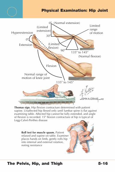
15˚
Thomas sign. Hip flexion contracture determined with patientsupine. Unaffected hip flexed only until lumbar spine is flat againstexamining table. Affected hip cannot be fully extended, and angleof flexion is recorded. 15� flexion contracture of hip is typical ofLegg-Calvé-Perthes disease
Roll test for muscle spasm. Patientrelaxed and supine on table. Examinerplaces hands on limb, gently rolls hipinto internal and external rotation,noting resistance
(Normal flexion)135� to 145�
135� to 145�
90�
(Limited flexion)
Limitedrangeof motion
30�
0�
0�
(Limitedextension)
(Normal extension)
Hyperextension
Flexion
Normal range ofmotion of knee joint
Extension
Physical Examination: Hip Joint
The Pelvis, Hip, and Thigh 5-16
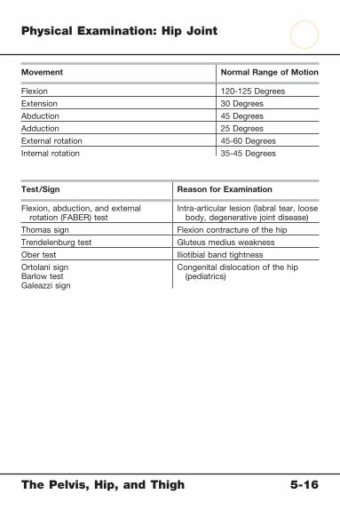
Physical Examination: Hip Joint
Movement Normal Range of Motion
Flexion 120-125 Degrees
Extension 30 Degrees
Abduction 45 Degrees
Adduction 25 Degrees
External rotation 45-60 Degrees
Internal rotation 35-45 Degrees
Test/Sign Reason for Examination
Flexion, abduction, and external rotation (FABER) test
Intra-articular lesion (labral tear, loose body, degenerative joint disease)
Thomas sign Flexion contracture of the hip
Trendelenburg test Gluteus medius weakness
Ober test Iliotibial band tightness
Ortolani signBarlow testGaleazzi sign
Congenital dislocation of the hip (pediatrics)
The Pelvis, Hip, and Thigh 5-16

2 3
4 5 6
7
1
Identify each condition
Conditions: Pelvic Ring
The Pelvis, Hip, and Thigh 5-17

Conditions: Pelvic Ring
1. Anteroposterior compression type I (APC-I)2. Anteroposterior compression type II (APC-II)3. Anteroposterior compression type III (APC-III)4. Lateral compression type I (LC-I)5. Lateral compression type II (LC-II)6. Lateral compression type III (LC-III)7. Vertical shear (Malgaigne fracture)
Young and Burgess Classifi cation
Fracture Type Fracture Pattern Stability Treatment
APC-I Symphysis diastasis <2 cm
Stable Conservative
APC-II Symphysis and anterior sacroiliac joint disruption
Stable Conservative
APC-III Symphysis and complete sacroiliac joint disruption
Unstable External fi xationOpen reduction, internal
fi xation (ORIF)
LC-I Rami + ipsilateral sacral compression
Stable Conservative
LC-II Rami + ipsilateral posterior ilium
Stable Conservative
LC-III Ipsilateral lateral compression + contralateral anteroposterior compression
Unstable External fi xationORIF
Vertical Shear Anterior and posterior involvement
Vertically unstable
External fi xationORIF
The Pelvis, Hip, and Thigh 5-17

2 3
4 5
6 7 8
9 10
1
Identify each condition
Conditions: Acetabulum
The Pelvis, Hip, and Thigh 5-18

Conditions: Acetabulum
Letournel Classifi cation of Simple Acetabular Fractures
1. Posterior wall 2. Posterior column 3. Anterior wall 4. Anterior column 5. Transverse
Letournel Classifi cation of Combined Acetabular Fractures
6. Posterior column/posterior wall 7. Transverse/posterior wall 8. T-fracture 9. Anterior column/posterior hemi-transverse10. Both columns
Acetabular Fractures
Mechanism High-energy trauma forcing femoral head into acetabulum
Signs and Symptoms
Pain, often with hip dislocation and/or gastrointestinal/genitourinary symptoms
Imaging Anteroposterior (AP)/lateral pelvis radiographs; computed tomography (CT)
Treatment Traction on ipsilateral side; open reduction, internal fi xation (ORIF) for unstable or displaced fractures
The Pelvis, Hip, and Thigh 5-18

5
6 7
12
3
4
Conditions: Femoral Head
The Pelvis, Hip, and Thigh 5-19

Conditions: Femoral Head
1. Femoral head2. Fovea3. Articular cartilage4. Line of synovial membrane attachment5. Femoral neck6. Subcapital femoral head fracture7. Magnetic resonance imaging (MRI) appearance of bilateral hip
avascular necrosis
Femoral Head Fractures Avascular Necrosis
Mechanism Flexion, abduction/adduction, internal/external rotation (with hip dislocation)
Microvascular injury often a result of trauma, chronic steroid use, or alcohol use.
Classifi cation Pipkin type I: below the foveaPipkin type II: above the foveaPipkin type III: type I or II plus
femoral neckPipkin type IV: type I or II plus
acetabulum
Based on the degree of bony involvement and severity of the patient’s symptoms
Signs and Symptoms
Pain, often external rotation of the foot
Groin pain, positive result of fl exion, abduction, and external rotation (FABER) test
Imaging Anteroposterior (AP) and lateral radiographs, postreduction computed tomography (CT)
AP and lateral radiographs of the hip often show joint destruction; MRI can confi rm the diagnosis
Treatment Pipkin types I and II fractures: conservative, protect weight-bearing
Pipkin types III and IV fractures: open reduction, internal fi xation (ORIF) or arthroplasty (older patients)
Core drilling of avascularized area, hip replacement
Image from Eustace S, Johnston C, O’Byrne J, et al: Sports Injuries. Philadelphia: Elsevier, 2007.
The Pelvis, Hip, and Thigh 5-19
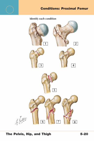
2
3 4
5
6 7 8
1
Identify each condition
Conditions: Proximal Femur
The Pelvis, Hip, and Thigh 5-20

Conditions: Proximal Femur
1. Intertrochanteric fracture: stable2. Intertrochanteric fracture: unstable3. Femoral neck fracture: type I4. Femoral neck fracture: type II5. Femoral neck fracture: type III6. Subtrochanteric fractures: type I7. Subtrochanteric fractures: type II8. Subtrochanteric fractures: type III
Fracture Type Classifi cation Treatment
Intertrochanteric Stable (2-part)Unstable (3- or 4-part)
Fixation with sliding hip screw
Hemiarthroplasty or total arthroplasty for primary failures
Femoral Neck (Garden Classifi cation)
Type I: impactedType II: nondisplacedType III: displaced <50%Type IV: signifi cantly displaced
Fixation with hip screwsHemiarthroplasty (older
patients, weak bone)
Subtrochanteric (Russell-Taylor Classifi cation)
Type I: nondisplacedType II: displaced, 2 partsType III: displaced, 3 partsType IV: comminuted, 4 partsType V: subtrochanteric and
intertrochanteric fracture
Intramedullary (IM) nail orFixed-angle device with
long sideplate
The Pelvis, Hip, and Thigh 5-20

2 3 4
5
1
Identify each condition
Conditions: Femoral Shaft
The Pelvis, Hip, and Thigh 5-21

Conditions: Femoral Shaft
1. Transverse femoral shaft fracture2. Spiral fracture3. Comminuted fracture4. Segmental fracture5. Radiographic appearance of comminuted femur fracture
Femoral Shaft Fractures Femoral Shaft Stress Fractures
Mechanism High-energy trauma Overuse; most common in runners
Signs and Symptoms
Pain, deformity, shock Pain with activity that improves with rest; positive fulcrum test result
Imaging Anteroposterior (AP) and lateral radiographs of the femur, include hip and knee
AP and lateral radiographs (often negative), magnetic resonance imaging (MRI), bone scan
Treatment Surgical treatment with intramedullary (IM) nail
Rest until symptoms resolve; fi xation with femoral nail if injury progresses to pending or complete fracture
The Pelvis, Hip, and Thigh 5-21

5
6
7
8
1 2 3
4
Conditions: Athlete’s Pelvis and Hip
The Pelvis, Hip, and Thigh 5-22
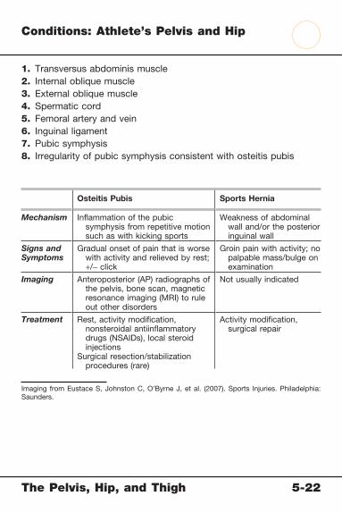
Conditions: Athlete’s Pelvis and Hip
1. Transversus abdominis muscle2. Internal oblique muscle3. External oblique muscle4. Spermatic cord5. Femoral artery and vein6. Inguinal ligament7. Pubic symphysis8. Irregularity of pubic symphysis consistent with osteitis pubis
Osteitis Pubis Sports Hernia
Mechanism Infl ammation of the pubic symphysis from repetitive motion such as with kicking sports
Weakness of abdominal wall and/or the posterior inguinal wall
Signs and Symptoms
Gradual onset of pain that is worse with activity and relieved by rest; +/− click
Groin pain with activity; no palpable mass/bulge on examination
Imaging Anteroposterior (AP) radiographs of the pelvis, bone scan, magnetic resonance imaging (MRI) to rule out other disorders
Not usually indicated
Treatment Rest, activity modifi cation, nonsteroidal antiinfl ammatory drugs (NSAIDs), local steroid injections
Surgical resection/stabilization procedures (rare)
Activity modifi cation, surgical repair
Imaging from Eustace S, Johnston C, O’Byrne J, et al. (2007). Sports Injuries. Philadelphia: Saunders.
The Pelvis, Hip, and Thigh 5-22
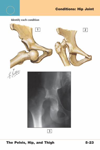
2
3
1
Identify each condition
Conditions: Hip Joint
The Pelvis, Hip, and Thigh 5-23

Conditions: Hip Joint
1. Anterior hip dislocation2. Posterior hip dislocation3. Radiographic appearance of posterior hip dislocation
Hip Dislocations
Mechanism Flexion, adduction, internal rotation (posterior, most common)Flexion, abduction, external rotation (anterior)
Signs and Symptoms
Pain, deformity, internally rotated foot
Imaging Anteroposterior (AP) and lateral radiographs, postreduction computed tomography (CT) to evaluate for occult fractures
Treatment Immediate reduction, protect weight-bearing for 4-6 weeks; recurrence rare
The Pelvis, Hip, and Thigh 5-23
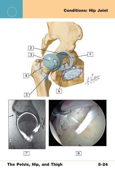
56
7 8
1
2
3
4
Conditions: Hip Joint
The Pelvis, Hip, and Thigh 5-24
Conditions: Hip Joint
1. Acetabular labrum2. Articular surface of acetabulum3. Articular cartilage of femoral head4. Neck of femur5. Ligament of head of femur6. Transverse acetabular ligament7. MRI appearance of anterior labral tear of the hip8. Arthroscopic image of labral tear of the hip
Labral Tears and Chondral Injuries of the Hip
Mechanism Traumatic or degenerative
Signs and Symptoms
Groin pain, catching and locking, positive result of fl exion, abduction, and external rotation (FABER) test
Imaging Arthrographic magnetic resonance imaging (MRI)
Treatment Nonsteroidal antiinfl ammatory drugs (NSAIDs), injections, hip arthroscopy with débridement, repair, or microfracture as indicated
Image from Miller M, Cole B: Textbook of Arthroscopy. Philadelphia: Elsevier, 2004.
The Pelvis, Hip, and Thigh 5-24

2 3
4
5 6
1
Identify each condition
Conditions: Hip Joint
The Pelvis, Hip, and Thigh 5-25

Conditions: Hip Joint
1. Osteophytes2. Articular cartilage wear3. Greater trochanter4. Lesser trochanter5. Hip arthritis appearance on anteroposterior (AP) pelvis radiograph6. Hip arthritis appearance on lateral radiograph
Hip Arthritis Trochanteric Bursitis
Mechanism Degenerative, posttraumatic Local trauma or repetitive friction
Signs and Symptoms
Groin pain, hip stiffness, positive result of fl exion, abduction, and external rotation (FABER) test
Local pain and tenderness over the greater trochanter
Imaging AP and lateral radiographs of the hip
Not usually necessary
Treatment Activity modifi cation, nonsteroidal antiinfl ammatory drugs (NSAIDs), steroid injections, hip arthroplasty
NSAIDs, steroid injections, surgical bursectomy rare
The Pelvis, Hip, and Thigh 5-25
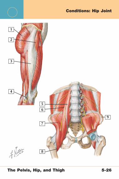
5
1
2
3
4
6
7
8
9
Conditions: Hip Joint
The Pelvis, Hip, and Thigh 5-26
Conditio
ns: H
ip J
oin
t1. Gluteus maximus muscle2. Tensor fascia latae muscle3. Iliotibial band4. Gerdy tubercle5. Psoas minor muscle6. Psoas major muscle7. Iliacus muscle8. Iliopsoas muscle insertion on lesser trochanter9. Iliac crest
Pathology Signs/Symptoms Imaging Treatment
External Snapping Hip
Iliotibial band or gluteal tendon snaps over greater trochanter
Lateral pain/snapping
Not needed Physical therapy, release or Z-plasty of iliotibial band
Internal Snapping Hip
Iliopsoas tendon over iliopectineal eminence
Groin pain Iliopsoas bursography or ultrasound
Physical therapyIliopsoas bursa injection
or iliopsoas release
Intra-Articular Snapping Hip
May be any intraarticular lesion such as labral tear or loose body
Mechanical catching in groin/joint
Arthrographic magnetic resonance imaging (MRI)
Hip arthroscopy
The P
elvis, H
ip, a
nd T
hig
h
5-2
6
5
6
1
2
3
4
Conditions: Muscles
The Pelvis, Hip, and Thigh 5-27
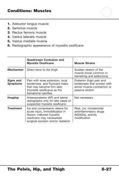
Conditions: Muscles
1. Adductor longus muscle2. Sartorius muscle3. Rectus femoris muscle4. Vastus lateralis muscle5. Vastus medialis muscle6. Radiographic appearance of myositis ossifi cans
Quadriceps Contusion and Myositis Ossifi cans Muscle Strains
Mechanism Direct blow to the thigh Sudden stretch of the muscle (most common in hamstring and adductors)
Signs and Symptoms
Pain with knee extension, local tenderness, and fl uctuant mass that may become fi rm later (myositis ossifi cans) as the hematoma calcifi es
Posterior thigh pain and tenderness that worsen with active muscle contraction or passive stretch
Imaging Anteroposterior (AP) and lateral radiographs only for late cases of suspected myositis ossifi cans
Not necessary
Treatment Ice and compression sleeve for acute injury, immobilization in fl exion; matured myositis ossifi cans may necessitate surgical excision and/or radiation
Rest, ice, nonsteroidal antiinfl ammatory drugs (NSAIDs), activity modifi cation
The Pelvis, Hip, and Thigh 5-27

5
6
1
2
3
4
Conditions: Sciatic Nerve
The Pelvis, Hip, and Thigh 5-28

Conditions: Sciatic Nerve
1. Sciatic nerve (L4-S3)2. Greater sciatic foramen3. Posterior cutaneous nerve of thigh4. Inferior cluneal nerves5. Tibial nerve6. Common peroneal nerve
Sciatica
Mechanism Irritation of sciatic nerve from compression as it passes through the greater sciatic foramen or underneath the piriformis muscle (piriformis syndrome)
Signs and Symptoms
Posterior radiating leg pain, numbness, paresthesias; positive result of straight leg raise test
Imaging Usually not necessary but may include magnetic resonance imaging (MRI) to rule out stenosis at the spinal level and/or electromyography
Treatment Nonsteroidal antiinfl ammatory drugs (NSAIDs), stretching program for piriformis muscle, activity modifi cation
The Pelvis, Hip, and Thigh 5-28

5
6
1
2
3
4
Conditions: Lateral Femoral Cutaneous Nerve
The Pelvis, Hip, and Thigh 5-29
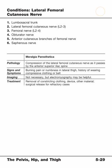
Conditions: Lateral Femoral Cutaneous Nerve
1. Lumbosacral trunk2. Lateral femoral cutaneous nerve (L2-3)3. Femoral nerve (L2-4)4. Obturator nerve5. Anterior cutaneous branches of femoral nerve6. Saphenous nerve
Meralgia Paresthetica
Pathology Compression of the lateral femoral cutaneous nerve as it passes by the anterior superior iliac spine
Signs and Symptoms
Burning pain or numbness in lateral thigh, history of wearing compressive clothing or belt
Imaging Not necessary, but electromyography may be helpful.
Treatment Removal of constricting clothing, device, other material;surgical release for refractory cases
The Pelvis, Hip, and Thigh 5-29
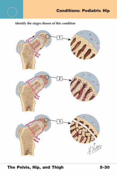
1
2
3
Identify the stages shown of this condition
Conditions: Pediatric Hip
The Pelvis, Hip, and Thigh 5-30
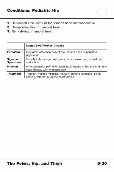
Conditions: Pediatric Hip
1. Decreased vascularity of the femoral head (osteonecrosis)2. Revascularization of femoral head3. Remodeling of femoral head
Legg-Calvé-Perthes Disease
Pathology Idiopathic osteonecrosis of the femoral head in pediatric population
Signs and Symptoms
Usually in boys aged 4-8 years, hip or knee pain, limited hip abduction
Imaging Anteroposterior (AP) and lateral radiographs of hip show femoral head density with crescent sign
Treatment Traction, muscle releases, range-of-motion exercises, Petrie casting, femoral or pelvic osteotomies
The Pelvis, Hip, and Thigh 5-30

1 2 3
4
Identify each condition
Conditions: Pediatric Hip
The Pelvis, Hip, and Thigh 5-31

Conditions: Pediatric Hip
1. Slipped capital femoral epiphysis (SCFE) with mild displacement (<30 degrees)
2. SCFE with moderate displacement (30-50 degrees)3. SCFE with severe displacement (>50 degrees)4. Radiographic appearance of SCFE
SCFE
Pathology Slippage of proximal femoral epiphysis
Signs and Symptoms
Patients aged 11-14; obese patients; hip or knee pain; limited abduction and internal rotation
Imaging Anteroposterior (AP) and lateral radiographs of hip
Treatment Make hip non–weight-bearing; percutaneous pinning for unstable lesions
The Pelvis, Hip, and Thigh 5-31

Netter’s Musculoskeletal Flash Cards
6 The Knee and Lower LegPlates 6-1 to 6-27
Bony Anatomy
6-1 Bony Anatomy: Knee
6-2 Bony Anatomy: Lower Leg
Radiographic Anatomy
6-3 Radiographic Anatomy: Knee
6-4 Radiographic Anatomy: Lower Leg
Muscles
6-5 Muscles: Anterior Thigh
6-6 Muscles: The Quadriceps
6-7 Muscles: Posterior Thigh
6-8 Muscles: Lower Leg (Superfi cial Dissection)
6-9 Muscles: Lower Leg (Superfi cial Dissection)
6-10 Muscles: Lower Leg (Posterior View)
6-11 Muscles: Lower Leg (Posterior View)
6-12 Muscles: Lower Leg (Posterior, Deep
Dissection)
Arteries and Nerves
6-13 Arteries: Knee and Lower Leg
6-14 Nerves: Lower Leg
Soft Tissue Anatomy
6-15 Soft Tissue Anatomy: Knee Joint
6-16 Soft Tissue Anatomy: Knee

The Knee and Lower Leg Table of Contents
6 The Knee and Lower LegPlates 6-1 to 6-27
Physical Examination
6-17 Physical Examination: Knee Joint and Lower Leg
Conditions
6-18 Conditions: Distal Femur
6-19 Conditions: Patella
6-20 Conditions: Tibia
6-21 Conditions: Cruciate Ligaments
6-22 Conditions: Collateral Ligaments
6-23 Conditions: Menisci
6-24 Conditions: Patellofemoral Joint
6-25 Conditions: Extensor Mechanism
6-26 Conditions: Articular Cartilage
6-27 Conditions: Lower Leg

The Knee and Lower Leg 6-1
Bony Anatomy: Knee
39
8
7
2
12
11
10
4
5
6
1
Anterior view
The Knee and Lower Leg 6-1
Bony Anatomy: Knee
1. Lateral femoral condyle 2. Lateral tibial condyle 3. Gerdy tubercle (insertion of iliotibial tract) 4. Head of fi bula 5. Neck of fi bula 6. Oblique line 7. Tibial tuberosity 8. Intercondylar eminence 9. Medial tibial condyle10. Medial femoral condyle11. Adductor tubercle12. Trochlear notch
Comment: The knee joint comprises three articulations: the femur and tibia, the femur and patella, and the tibia and fi bula. Stability of the joint is primarily through the ligaments, menisci, and joint capsule.

3
9
8
7
2
10
45
6
1
The Knee and Lower Leg 6-2
Bony Anatomy: Lower Leg

The Knee and Lower Leg 6-2
Bony Anatomy: Lower Leg
1. Lateral tibial condyle 2. Intercondylar eminence 3. Medial tibial condyle 4. Gerdy’s tubercle 5. Head of fi bula 6. Tibial tuberosity 7. Tibia 8. Fibula 9. Medial malleolus10. Lateral malleolus
Comment: The 2 bones of the lower leg are connected by a fi brous syndesmosis. They articulate both proximally at the knee and distally at the ankle. The tibia is the primary load-bearing bone of the lower leg. The fi bula is most important at the ankle, where its distal malleolus forms the lateral side of the ankle mortise. Proximally, it articulates with the tibia below the level of the knee.
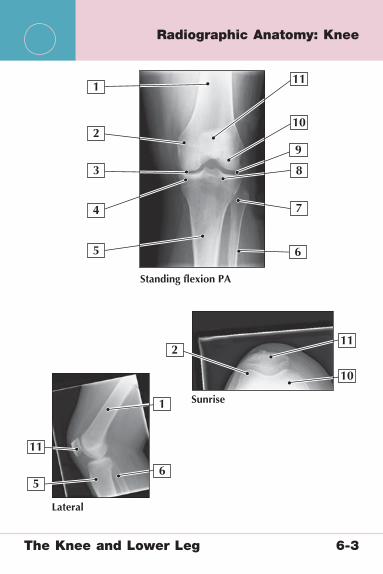
3
9
8
2
2
10
11
10
11
11
4
5
5
7
6
6
Standing flexion PA
Lateral
Sunrise
1
1
The Knee and Lower Leg 6-3
Radiographic Anatomy: Knee

The Knee and Lower Leg 6-3
Radiographic Anatomy: Knee
1. Femur 2. Medial femoral condyle 3. Medial joint space 4. Medial tibial plateau 5. Tibia 6. Fibula 7. Head of fi bula 8. Lateral tibial plateau 9. Lateral joint space10. Lateral femoral condyle11. Patella
Comment: Flexion weight-bearing radiographs are the most sensitive for knee arthritis. Sunrise views (with the knee in 30-45 degrees of fl exion) are useful for evaluating the patellofemoral joint. Other useful knee radiographs include the tunnel view (for osteochondritis dissecans [OCD], loose bodies), oblique views (for tibial plateau fractures), and stress views (for ligamentous or physeal injuries).
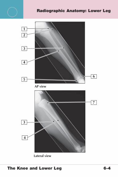
3
7
2
Lateral view
AP view
4
4
5
3
6
1
The Knee and Lower Leg 6-4
Radiographic Anatomy: Lower Leg
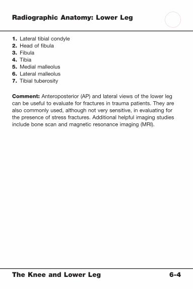
The Knee and Lower Leg 6-4
Radiographic Anatomy: Lower Leg
1. Lateral tibial condyle2. Head of fi bula3. Fibula4. Tibia5. Medial malleolus6. Lateral malleolus7. Tibial tuberosity
Comment: Anteroposterior (AP) and lateral views of the lower leg can be useful to evaluate for fractures in trauma patients. They are also commonly used, although not very sensitive, in evaluating for the presence of stress fractures. Additional helpful imaging studies include bone scan and magnetic resonance imaging (MRI).
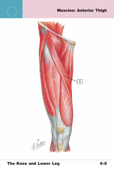
1
The Knee and Lower Leg 6-5
Muscles: Anterior Thigh
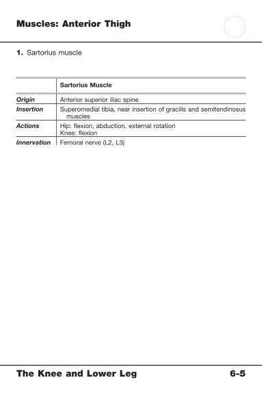
The Knee and Lower Leg 6-5
Muscles: Anterior Thigh
1. Sartorius muscle
Sartorius Muscle
Origin Anterior superior iliac spine
Insertion Superomedial tibia, near insertion of gracilis and semitendinosus muscles
Actions Hip: fl exion, abduction, external rotationKnee: fl exion
Innervation Femoral nerve (L2, L3)
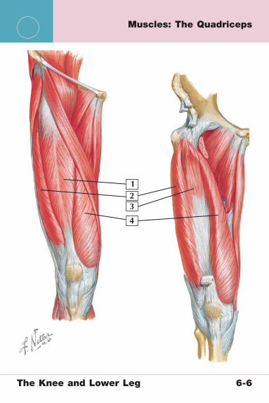
123
4
The Knee and Lower Leg 6-6
Muscles: The Quadriceps
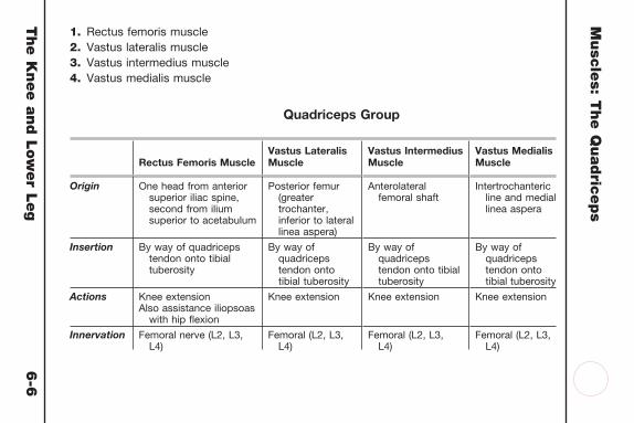
The K
nee a
nd L
ow
er L
eg
6-6
Musc
les: T
he Q
uadric
eps
1. Rectus femoris muscle2. Vastus lateralis muscle3. Vastus intermedius muscle4. Vastus medialis muscle
Quadriceps Group
Rectus Femoris MuscleVastus Lateralis Muscle
Vastus Intermedius Muscle
Vastus Medialis Muscle
Origin One head from anterior superior iliac spine, second from ilium superior to acetabulum
Posterior femur (greater trochanter, inferior to lateral linea aspera)
Anterolateral femoral shaft
Intertrochanteric line and medial linea aspera
Insertion By way of quadriceps tendon onto tibial tuberosity
By way of quadriceps tendon onto tibial tuberosity
By way of quadriceps tendon onto tibial tuberosity
By way of quadriceps tendon onto tibial tuberosity
Actions Knee extensionAlso assistance iliopsoas
with hip fl exion
Knee extension Knee extension Knee extension
Innervation Femoral nerve (L2, L3, L4)
Femoral (L2, L3, L4)
Femoral (L2, L3, L4)
Femoral (L2, L3, L4)
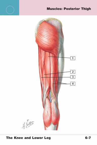
1
2
3
4
The Knee and Lower Leg 6-7
Muscles: Posterior Thigh

The K
nee a
nd L
ow
er L
eg
6-7
Musc
les: P
oste
rior T
hig
h1. Semitendinosus muscle2. Semimembranosus muscle3. Gracilis muscle4. Biceps femoris muscle (short and long head)
Semitendinosus Muscle
Semimembranosus Muscle
Gracilis Muscle*
Biceps Femoris Muscle (Short and Long Head)
Origin Ischial tuberosity Ischial tuberosity Body and inferior ramus of pubis
Long head: ischial tuberosity
Short head: linea aspera and supracondylar line of femur
Insertion Superomedial tibia Posteromedial aspect of the medial tibial condyle
Superior medial tibia
Lateral head of the fi bula
Actions Knee fl exion and internal rotation, hip extension
Knee fl exion and internal rotation, hip extension
Hip adduction, knee fl exion
Knee fl exion and external rotation; long head also acts as a hip extensor
Innervation Sciatic nerve, tibial division (L5, S1, and S2)
Sciatic nerve, tibial division (L5, S1, and S2)
Obturator nerve (L2, L3)
Long head: sciatic nerve, tibial division (L5, S1, and S2)
Short head: sciatic nerve, common peroneal division (L5, S1, and S2)
*Weak hip adductor; crosses the knee joint
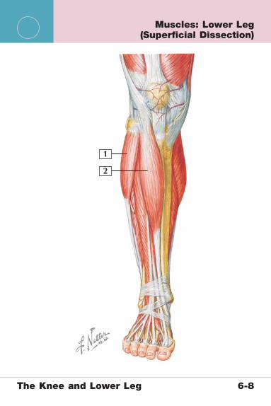
1
2
The Knee and Lower Leg 6-8
Muscles: Lower Leg (Superfi cial Dissection)

The Knee and Lower Leg 6-8
Muscles: Lower Leg (Superfi cial Dissection)
1. Peroneus longus muscle2. Tibialis anterior muscle
Peroneus Longus Muscle Tibialis Anterior Muscle
Origin Head and neck of fi bula Lateral side of proximal tibia
Insertion Base of 1st metatarsal Medial cuneiform and base of 1st metatarsal
Actions Foot eversion and assistance with plantar fl exion
Foot dorsifl exion and assistance with inversion
Innervation Superfi cial peroneal nerve (L5, S1, S2)
Deep peroneal nerve (L4, L5)
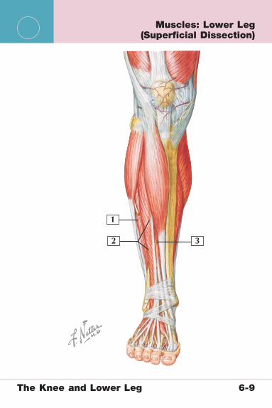
1
2 3
The Knee and Lower Leg 6-9
Muscles: Lower Leg (Superfi cial Dissection)
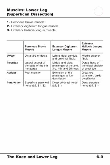
The Knee and Lower Leg 6-9
Muscles: Lower Leg (Superfi cial Dissection)
1. Peroneus brevis muscle2. Extensor digitorum longus muscle3. Extensor hallucis longus muscle
Peroneus Brevis Muscle
Extensor Digitorum Longus Muscle
Extensor Hallucis Longus Muscle
Origin Distal 2/3 of fi bula Lateral tibial condyle and proximal fi bula
Middle anterior fi bula
Insertion Lateral aspect of the base of the 5th metatarsal
Middle and distal phalanges of the 2nd, 3rd, 4th, and 5th toes
Dorsal base of the distal phalanx of great toe
Actions Foot eversion Extension of the phalanges, ankle dorsifl exion
Great toe extension, ankle dorsifl exion
Innervation Superfi cial peroneal nerve (L5, S1, S2)
Deep peroneal nerve (L5, S1)
Deep peroneal nerve (L5, S1)
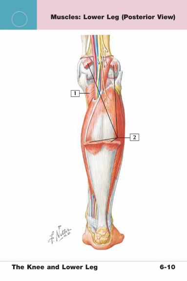
1
2
The Knee and Lower Leg 6-10
Muscles: Lower Leg (Posterior View)

The Knee and Lower Leg 6-10
Muscles: Lower Leg (Posterior View)
1. Popliteus muscle2. Gastrocnemius muscle
Popliteus Muscle Gastrocnemius Muscle
Origin Lateral aspect of lateral femoral condyle and lateral knee joint capsule
Lateral head from lateral aspect of lateral femoral condyle; medial head from posterior aspect of medial femoral condyle
Insertion Posterior tibia, just proximal to the soleal line
Via the Achilles tendon on the posterior calcaneus
Actions Leg fl exion and internal rotation at the knee, “unlocks” knee when standing
Ankle plantar fl exion, assistance with knee fl exion
Innervation Tibial nerve (L4, L5, S1) Tibial nerve (S1, S2)

1
2
3
The Knee and Lower Leg 6-11
Muscles: Lower Leg (Posterior View)

The Knee and Lower Leg 6-11
Muscles: Lower Leg (Posterior View)
1. Plantaris muscle2. Soleus muscle
Plantaris Muscle Soleus Muscle
Origin Lateral supracondylar line of femur and oblique popliteal ligament
Posterior aspect of proximal fi bula and medial tibia
Insertion Posterior calcaneus By way of Achilles tendon* onto the posterior calcaneus
Actions Assistance with ankle plantar fl exion and knee fl exion
Ankle plantar fl exion
Innervation Tibial nerve (S1-S2) Tibial nerve (S1, S2)
*The medial and lateral head of the gastrocnemius join with the soleus muscle to form the Achilles tendon, which then inserts on the posterior calcaneus.
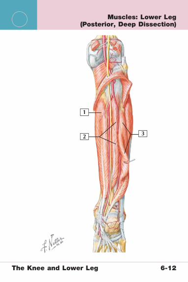
1
2 3
The Knee and Lower Leg 6-12
Muscles: Lower Leg (Posterior, Deep Dissection)
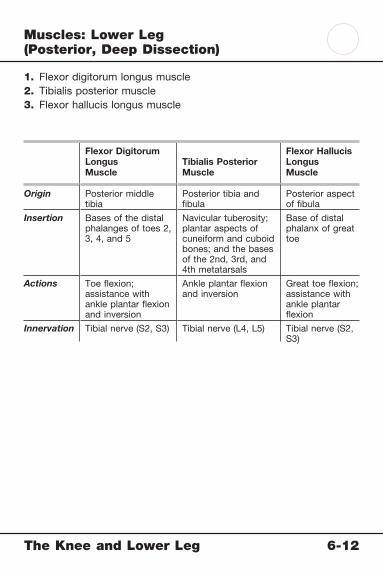
The Knee and Lower Leg 6-12
Muscles: Lower Leg (Posterior, Deep Dissection)
1. Flexor digitorum longus muscle2. Tibialis posterior muscle3. Flexor hallucis longus muscle
Flexor Digitorum LongusMuscle
Tibialis PosteriorMuscle
Flexor Hallucis LongusMuscle
Origin Posterior middle tibia
Posterior tibia and fi bula
Posterior aspect of fi bula
Insertion Bases of the distal phalanges of toes 2, 3, 4, and 5
Navicular tuberosity; plantar aspects of cuneiform and cuboid bones; and the bases of the 2nd, 3rd, and 4th metatarsals
Base of distal phalanx of great toe
Actions Toe fl exion; assistance with ankle plantar fl exion and inversion
Ankle plantar fl exion and inversion
Great toe fl exion; assistance with ankle plantar fl exion
Innervation Tibial nerve (S2, S3) Tibial nerve (L4, L5) Tibial nerve (S2, S3)

1
23
4
5
6
7
8
The Knee and Lower Leg 6-13
Arteries: Knee and Lower Leg
The Knee and Lower Leg 6-13
Arteries: Knee and Lower Leg
1. Inferior lateral genicular artery2. Superior lateral genicular artery3. Superior medial genicular artery4. Popliteal artery5. Inferior medial genicular artery6. Anterior tibial artery7. Posterior tibial artery8. Peroneal artery
Comment: The knee and lower leg are supplied by branches of the femoral artery. The popliteal artery passes through the popliteal fossa posteriorly and forms the geniculate arteries, which supply the knee. The anterior and posterior tibial arteries supply the muscles of the anterior and posterior compartments, respectively. The peroneal artery wraps around the head of the fi bula before passing distally to supply the peroneal muscles and the lateral compartment.
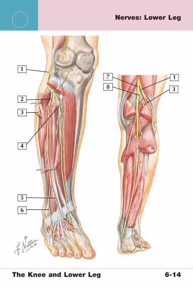
4
11
38
7
2
3
5
6
The Knee and Lower Leg 6-14
Nerves: Lower Leg

The Knee and Lower Leg 6-14
Nerves: Lower Leg
1. Common peroneal nerve2. Superfi cial peroneal nerve3. Lateral sural cutaneous nerve4. Deep peroneal nerve5. Medial dorsal cutaneous nerve6. Intermediate dorsal cutaneous nerve7. Tibial nerve8. Medial sural cutaneous nerve
Comment: The tibial nerve (L4-S3) passes through the midline of the popliteal space and runs between the heads of the gastrocnemius muscle as it travels to the foot. It provides sensation to the posterolateral calf and innervation to the posterior muscles of the lower leg. The common peroneal nerve crosses the knee joint passing between the biceps and the lateral head of the gastrocnemius muscle. It wraps around the fi bular head (where it is prone to injury with fractures) and then moves distally to supply the anterior and lateral compartments of the lower leg.
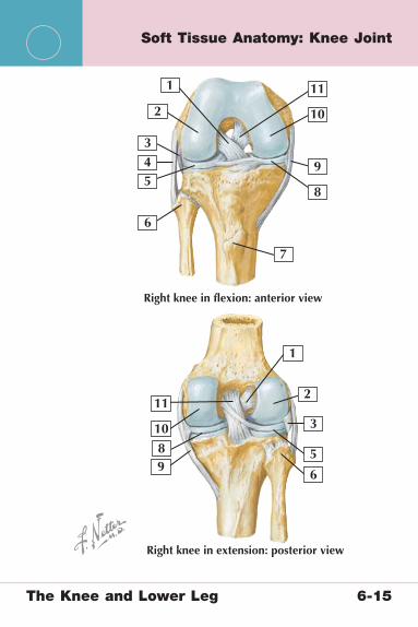
1
2
4 9
3
9
8
2
8
10
1
10
11
11
7
5
3
5
6
6
Right knee in flexion: anterior view
Right knee in extension: posterior view
The Knee and Lower Leg 6-15
Soft Tissue Anatomy: Knee Joint

The Knee and Lower Leg 6-15
Soft Tissue Anatomy: Knee Joint
1. Anterior cruciate ligament (ACL) 2. Lateral femoral condyle 3. Popliteus tendon 4. Lateral collateral ligament 5. Lateral meniscus 6. Head of the fi bula 7. Tibial tuberosity 8. Medial meniscus 9. Medial collateral ligament10. Medial femoral condyle11. Posterior cruciate ligament (PCL)
Comment: The anterior and posterior cruciate ligaments control anterior and posterior stability of the knee joint. Similarly, the medial and lateral collateral ligaments provide stability in those directions. The meniscus and capsule are important secondary stabilizers of this joint.
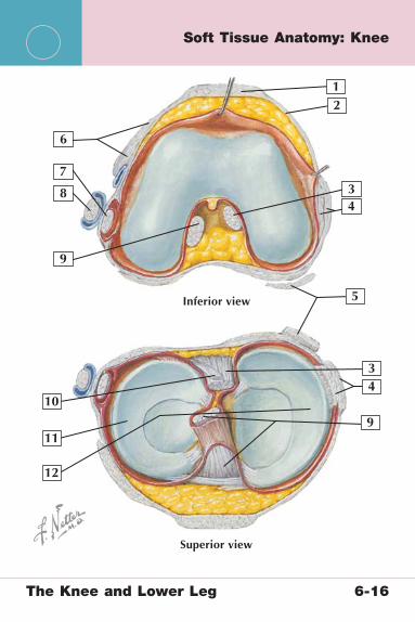
12
4
4
8
3
9
9
10
12
11
73
5
6
Superior view
Inferior view
The Knee and Lower Leg 6-16
Soft Tissue Anatomy: Knee

The Knee and Lower Leg 6-16
Soft Tissue Anatomy: Knee
1. Patella tendon 2. Medial patellofemoral ligament (MPFL) 3. Posterior cruciate ligament (PCL) 4. Medial collateral ligament 5. Semimembranosus tendon 6. Lateral patellar retinaculum and iliotibial band 7. Popliteus tendon 8. Lateral collateral ligament 9. Anterior cruciate ligament (ACL)10. Posterior meniscofemoral ligament11. Lateral meniscus12. Medial meniscus
Comment: The menisci are important secondary stabilizers of the knee joint. The medial meniscus is C-shaped, whereas the lateral meniscus is O-shaped.

Q angle formed byintersection of linesfrom anteriorsuperior iliac spineand from tibialtuberosity throughmidpoint of patella.Large Q anglepredisposes topatellar subluxation
Posterior sag sign.Leg drops backward
Apprehension (Fairbank)test As examinerdisplaces patellalaterally, patient feelspain and forcefullycontracts quadricepsfemoris muscle
Varus and valgus testsPatient supine on table,relaxed, leg over edgeof table, flexed about 30�
With one hand fixing thigh, examiner placesother hand just above ankle and applies valgusstress. Degree of mobility compared with thatof uninjured side, which is tested first. Forvarus stress test, direction of pressure reversed
Posterior drawer testProcedure same as foranterior drawer test,except that pressureon tibia is backwardinstead of forward
Lachman testWith patient’s knee bent 20�–30�,examiner’s hands grasp limb overdistal femur and proximal tibia.Tibia alternately pulled forwardand pushed backward.Movement of 5 mm or morethan that in normal limbindicates rupture of anteriorcruciate ligament
Q
The Knee and Lower Leg 6-17
Physical Examination: Knee Joint and Lower Leg

The Knee and Lower Leg 6-17
Physical Examination: Knee Joint and Lower Leg
Movement Normal Range of Motion
Flexion 135-140 Degrees
Extension 0 Degrees
Test/Sign Reason for Evaluation
Anterior drawer testLachman testPivot shift test
Anterior cruciate ligament (ACL) tear
Posterior drawer testSag signReverse pivot shift
Posterior cruciate ligament (PCL) tear
Dial test (rotary stability) Posterolateral corner injury
Valgus stress test Medial collateral ligament (MCL) tear
Varus stress test Lateral collateral ligament (LCL) tear
McMurray test Meniscal tear
Quadriceps (Q) angle measurement Patella tracking
Apprehension Patella instability
Patella grind Patellofemoral pain/arthritis
Homan sign Deep vein thrombosis
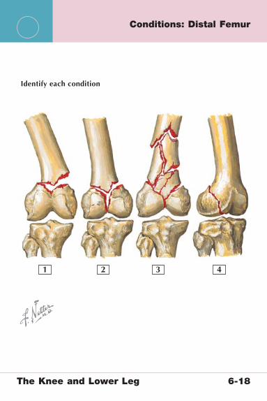
Identify each condition
1 2 43
The Knee and Lower Leg 6-18
Conditions: Distal Femur
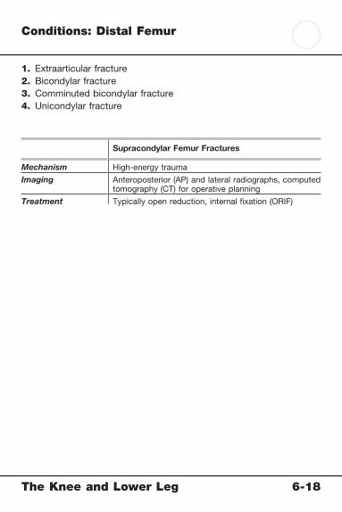
The Knee and Lower Leg 6-18
Conditions: Distal Femur
1. Extraarticular fracture2. Bicondylar fracture3. Comminuted bicondylar fracture4. Unicondylar fracture
Supracondylar Femur Fractures
Mechanism High-energy trauma
Imaging Anteroposterior (AP) and lateral radiographs, computed tomography (CT) for operative planning
Treatment Typically open reduction, internal fi xation (ORIF)

1 2
4
5
3
Identify each condition
The Knee and Lower Leg 6-19
Conditions: Patella
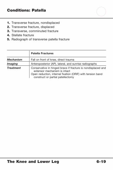
The Knee and Lower Leg 6-19
Conditions: Patella
1. Transverse fracture, nondisplaced2. Transverse fracture, displaced3. Transverse, comminuted fracture4. Stellate fracture5. Radiograph of transverse patella fracture
Patella Fractures
Mechanism Fall on front of knee, direct trauma
Imaging Anteroposterior (AP), lateral, and sunrise radiographs
Treatment Conservative in hinged brace if fracture is nondisplaced and extensor mechanism is intact
Open reduction, internal fi xation (ORIF) with tension band construct or partial patellectomy
1 2
4 5
6 7
3
Identify each condition
The Knee and Lower Leg 6-20
Conditions: Tibia
The Knee and Lower Leg 6-20
Conditions: Tibia
1. Type I tibial plateau fracture2. Type II tibial plateau fracture3. Type IV tibial plateau fracture4. Type V tibial plateau fracture5. Type VI tibial plateau fracture6. Open reduction, internal fi xation (ORIF) with medial buttress plate7. Transverse fracture of tibia and fi bula
Schatzker Fracture (Tibial Plateau) Description
Type I Lateral split
Type II Lateral split with depression
Type III Lateral depression without split
Type IV Medial depression
Type V Bicondylar
Type VI Split with extension into metadiaphysis
Comment: Tibial shaft fractures are described by type of fracture (transverse, displaced, open, closed) and are typically treated surgically with an intermedullary nail.
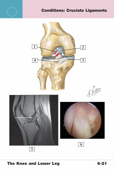
1
4 3
56
2
The Knee and Lower Leg 6-21
Conditions: Cruciate Ligaments
The Knee and Lower Leg 6-21
Conditions: Cruciate Ligaments
1. Anterior cruciate ligament (ACL) (ruptured)2. Posterior cruciate ligament (PCL)3. Medial meniscus4. Lateral meniscus5. Magnetic resonance imaging (MRI) appearance of normal ACL
and PCL6. Arthroscopic image of normal ACL
ACL Tear PCL Tear
Mechanism Noncontact pivot Hyperextension; direct blow to tibia of fl exed knee
Signs and Symptoms
Immediate swelling; “pop”; inability to continue playing; positive Lachman (“gold standard”), anterior drawer, and pivot shift test
Pain; swelling; positive posterior drawer test, sag sign, and quadriceps active test
Imaging Segond fracture on plain radiograph is pathognomonic, MRI confi rms
Stress radiographs; MRI confi rms
Treatment Reconstruction of ligament with tendon (autograft or allograft)
Usually conservative (grades I and II) unless combined ligamentous injury or grade III with persistent symptoms
Grade Posterior Drawer Test Result Treatment
I Anterior tibia anterior to femoral condyles Conservative
II Anterior tibia fl ush with femoral condyles Conservative
III Anterior tibia posterior to femoral condyles Often surgical reconstruction

1
2
4
5 6 7
Identify each condition
8 9
3
The Knee and Lower Leg 6-22
Conditions: Collateral Ligaments

The Knee and Lower Leg 6-22
Conditions: Collateral Ligaments
1. Lateral collateral ligament (LCL)2. Anterior cruciate ligament (ACL)3. Posterior cruciate ligament (PCL)4. Medial collateral ligament (MCL)5. Grade I MCL injury6. Grade II MCL injury7. Grade III MCL injury8. Combined ACL/MCL injury9. Magnetic resonance imaging (MRI) appearance of normal
collateral ligaments
MCL Tear LCL Tear
Mechanism Blow to lateral leg (forced valgus)
High-energy trauma, rarely isolated
Signs and Symptoms
Medial pain and tenderness over MCL origin/insertion, positive valgus stress test at 30 degrees (at 0 degrees, indicates combined injury)
Lateral pain, positive varus stress test result; evaluate for posterolateral corner injury (rotary instability)
Imaging Pellegrini-Stieda lesion (chronic MCL injury only); MRI confi rms
Anteroposterior (AP) and lateral radiographs; MRI confi rms
Treatment Hinged brace for 6 weeks; for chronic instability or combined injury: surgical reconstruction
If isolated, hinged braceFor combined injury: surgical
reconstruction

1 2
4 5 6
7 8
3
Identify each condition
The Knee and Lower Leg 6-23
Conditions: Menisci

The Knee and Lower Leg 6-23
Conditions: Menisci
1. Vertical tear2. Radial tear3. Oblique (“parrot-beak”) tear4. Bucket-handle tear5. Complex tear6. Discoid meniscus with tear7. Tear of posterior horn of the medial meniscus as seen on
magnetic resonance imaging (MRI)8. Arthroscopic appearance of meniscal tear
Meniscal Tears Discoid Meniscus*
Mechanism Twisting injury most common Normal variant but can tear owing to twisting injury
Signs and Symptoms
Pain, swelling, giving way, catching/locking, joint line tenderness, positive McMurray test (pain and click)
Same as normal meniscal tear, often presenting in adolescents
Imaging MRI confi rms diagnosis Anteroposterior (AP) and lateral radiographs may show joint space widening and cupping; MRI confi rms diagnosis
Treatment Arthroscopic partial meniscectomy most common; meniscal repair reserved for small, vertical, peripheral (red zone) tears where the blood supply can support healing
Saucerization/partial meniscectomy for torn meniscus; simple observation for incidentally noted discoid meniscus without tear
*Usually of the lateral meniscus, this involves a shape that is more rounded (“fi lled in”) than the usual C-shape of the meniscus, predisposing it to tears.
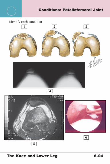
1 2
4
5
6
3Identify each condition
The Knee and Lower Leg 6-24
Conditions: Patellofemoral Joint

The Knee and Lower Leg 6-24
Conditions: Patellofemoral Joint
1. Normal patellofemoral alignment2. Lateral subluxation of the patella3. Medial patellofemoral ligament (MPFL) tear from patella
dislocation4. Sunrise view, demonstrating chronic patella subluxation (bilateral)5. Magnetic resonance imaging (MRI) of MPFL injury6. Arthroscopic image of patella chondral fl ap
Patellar Dislocation Patella Chondromalacia
Mechanism Direct blow resulting in lateral displacement
Almost always caused by quadriceps imbalance
Signs and Symptoms
Deformity, patellofemoral tenderness and laxity, medial pain over MPFL origin
Anterior knee pain that worsens with prolonged sitting or stair climbing, pseudolocking, patellofemoral tenderness, positive patella grind
Imaging Anteroposterior (AP), lateral, and sunrise views; MRI to evaluate MPFL tear and loose body
AP, lateral, and sunrise views;in rare cases, MRI to evaluate
for chondral fl ap in the presence of mechanical symptoms
Treatment Conservative with emphasis on quadriceps muscle strengthening
Surgical reconstructive procedures and MPFL reconstruction are later options
Quadriceps strengthening (emphasize vastus medialis oblique muscle), braces, nonsteroidal antiinfl ammatory drugs (NSAIDs), steroid injections
Sunrise view from Miller M, Cole B: Textbook of Arthroscopy. Philadelphia: Elsevier, 2004.MRI from Miller M, Howard R, Plancher K: Surgical Atlas of Sports Medicine. Philadelphia:
Elsevier, 2003.
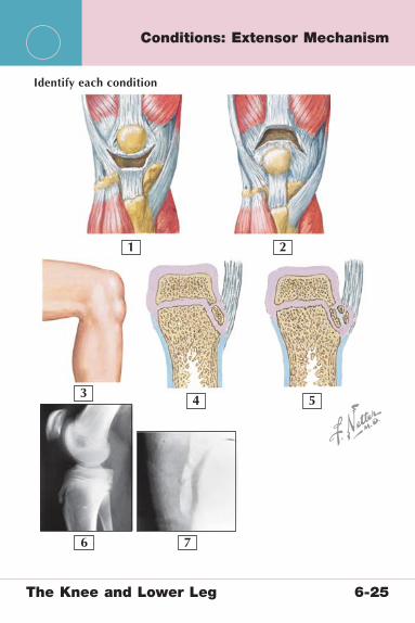
1 2
4 5
6 7
3
Identify each condition
The Knee and Lower Leg 6-25
Conditions: Extensor Mechanism

The Knee and Lower Leg 6-25
Conditions: Extensor Mechanism
1. Patella tendon rupture2. Quadriceps tendon rupture3. External appearance of Osgood-Schlatter disease4. Normal appearance of ossifying tibial tuberosity5. Osgood-Schlatter disease with additional ossifi cation at tuberosity6. Mild separation of the apophysis at the tendon insertion7. Fragmentation of the tibial tuberosity
Quadriceps/Patella Tendon Ruptures Osgood-Schlatter Disease
Mechanism Fall on fl exed kneePatella injury more common
before age 40; quadriceps injury more common after age 40
Traction apophysitis secondary to rapid bone growth, in comparison to soft tissue growth
Diagnosis Failed extensor mechanism (inability to hold extended knee against resistance)
Tenderness over tibial tuberosity; radiographs may reveal enlarged bony apophysis
Imaging Lateral radiographs may reveal patella alta (patella tendon rupture); magnetic resonance imaging (MRI) confi rms
AP and lateral radiographs may show separation or fragmentation of the apophysis
Treatment Surgical repair Condition is self-limited; therefore, treatment is conservative with Cho-Pat strap, rest, and activity modifi cation
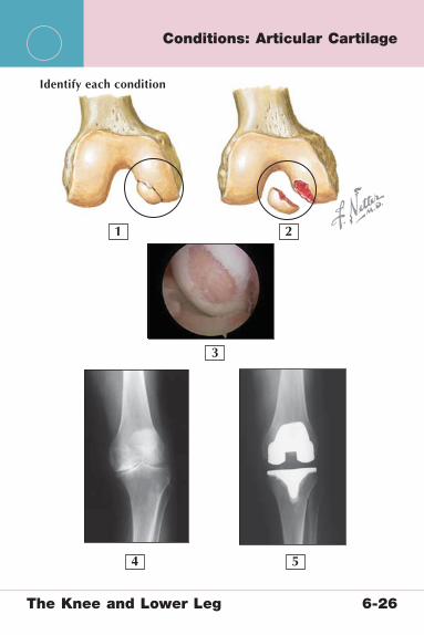
1 2
4 5
3
Identify each condition
The Knee and Lower Leg 6-26
Conditions: Articular Cartilage

The Knee and Lower Leg 6-26
Conditions: Articular Cartilage
1. Osteochondritis dissecans (OCD), nondisplaced2. OCD with displaced fragment3. Traumatic chondral lesion, arthroscopic view4. Osteoarthritis5. Total joint replacement
OCD and Traumatic Chondral Injury
Degenerative Joint Disease (DJD) of the Knee
Mechanism Chondral injury can be traumatic, but OCD is usually idiopathic and occurs in adolescents
Degenerative (wear and tear), or posttraumatic process
Signs and Symptoms
Pain, effusions, mechanical catching
Pain, walking intolerance, effusion, decreased range of motion
Imaging Anteroposterior (AP), lateral, and tunnel radiographs; magnetic resonance imaging (MRI) confi rms and helps with surgical planning
Standing fl exion PA, lateral, and sunrise radiographs show osteophytes and narrowed joint space
Treatment OCD: conservative while physes are still open, but surgical débridement with or without bone grafting and fi xation of lesion may be required later
Traumatic lesions usually necessitate débridement, microfractures, or chondral plug transfer
Activity modifi cation, nonsteroidal antiinfl ammatory drugs (NSAIDs), steroid or hyaluronic acid injections; unicompartmental or total knee replacement if conservative treatment fails
Radiograph from Miller M, Sekiya J: Core Knowledge in Orthopaedics: Sports Medicine. Philadelphia: Elsevier, 2006.

1
2
5
4
3
Identify each compartment
The Knee and Lower Leg 6-27
Conditions: Lower Leg

The Knee and Lower Leg 6-27
Conditions: Lower Leg
1. Anterior compartment• Tibialis anterior muscle• Extensor digitorum longus muscle• Extensor hallucis longus muscle• Anterior tibial artery and vein• Deep peroneal nerve
2. Lateral compartment• Peroneus longus muscle• Peroneus brevis muscle• Superfi cial peroneal nerve
3. Superfi cial posterior compartment• Soleus muscle• Gastrocnemius muscle• Plantaris muscle
4. Deep posterior compartment• Flexor digitorum longus muscle• Tibialis posterior muscle• Flexor hallucis longus muscle• Posterior tibial artery and nerve• Peroneal artery and nerve
5. Anterior tibial stress fracture
Comment: Both exertional compartment syndrome and tibial stress reactions are common in runners and manifest with exertional lower leg pain. Patients with compartment syndrome often have neurological complaints as well. Radiographs, bone scan, and magnetic resonance imaging (MRI) are all useful for diagnosing stress reactions, which are best treated with rest until symptoms resolve completely. Exertional compartment syndrome is diagnosed with measurement of compartment pressures after exertion (running on a treadmill) and is treated with surgical release of the affected compartments.
Netter’s Musculoskeletal Flash Cards
7 The Ankle and FootPlates 7-1 to 7-30
Bony Anatomy
7-1 Bony Anatomy: Distal Tibia and Fibula
7-2 Bony Anatomy: Foot
7-3 Bony Anatomy: Foot
Radiographic Anatomy
7-4 Radiographic Anatomy: Ankle
7-5 Radiographic Anatomy: Foot
Ligaments and Tendons
7-6 Ligaments: Ankle
7-7 Ligaments: Foot
7-8 Tendons and Muscles: Ankle and Foot
Muscles
7-9 Muscles: Superfi cial and First Muscle Layers of
Foot
7-10 Muscles: Second Muscle Layer of Foot
7-11 Muscles: Third Muscle Layer of Foot
7-12 Muscles: Fourth Muscle Layer of Foot
Arteries and Nerves
7-13 Arteries: Ankle and Foot
7-14 Nerves: Ankle and Foot (Peroneal Nerve)
7-15 Nerves: Ankle and Foot (Tibial Nerve)

The Ankle and Foot Table of Contents
7 The Ankle and FootPlates 7-1 to 7-30
Physical Examination
7-16 Physical Examination: Ankle Joint
Conditions
7-17 Conditions: Ankle Joint
7-18 Conditions: Ligaments of the Ankle
7-19 Conditions: Achilles Tendon
7-20 Conditions: Calcaneus
7-21 Conditions: Talus
7-22 Conditions: Talus
7-23 Conditions: Plantar Fascia and Arch
7-24 Conditions: Diabetic Foot
7-25 Conditions: Joints
7-26 Conditions: Tarsometatarsal Joints
7-27 Conditions: Metatarsals
7-28 Conditions: Nerves and Tarsal Tunnel
7-29 Conditions: Sesamoids and Metatarsophalangeal
Joints
7-30 Conditions: Toes

The Ankle and Foot 7-1
Bony Anatomy: Distal Tibia and Fibula
3
98
77
2
4
5
6
1

The Ankle and Foot 7-1
Bony Anatomy: Distal Tibia and Fibula
1. Shaft of fi bula2. Shaft of tibia3. Groove for tibialis posterior and digitorum longus tendons4. Medial malleolus5. Articular facet of the medial malleolus6. Inferior articular surface7. Lateral malleolus8. Articular facet of lateral malleolus9. Malleolar fossa of lateral malleolus
Comment: The tibia and fi bula articulate distally to form the superior aspect of the ankle mortise. They are connected by a thick fi brous syndesmotic membrane and the tibiofi bular ligament. The talus articulates with both the tibia and fi bula and sits between the medial and lateral malleoli.
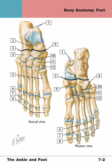
3
3
9
10
8
8
7
7
2
4
12
12
11
11
10
45
6
6
1
9
1
Dorsal view
Plantar view
The Ankle and Foot 7-2
Bony Anatomy: Foot

The Ankle and Foot 7-2
Bony Anatomy: Foot
1. Medial and lateral tubercles of talus 2. Sinus tarsi 3. Transverse tarsal joint 4. Cuboid 5. Metatarsal bones (5) 6. Proximal phalanges (5) 7. Middle phalanges (4) 8. Distal phalanges (5) 9. Navicular10. Lateral cuneiform11. Intermediate cuneiform12. Medial cuneiform
Comment: The foot articulates with the tibia and fi bula via the talus, which has no direct muscle attachments but is a site of attachment for several of the strong ligaments of the ankle joint.

32
3
7
7
2
4
11
109
4
5
6
6
8
1
1
Lateral view
Medial view
The Ankle and Foot 7-3
Bony Anatomy: Foot
The Ankle and Foot 7-3
Bony Anatomy: Foot
1. Talus 2. Talar head 3. Talar neck 4. Trochlea 5. Lateral process of talus 6. Posterior process of talus 7. Calcaneus 8. Base of the 5th metatarsal 9. Navicular bone10. Navicular tuberosity11. Sustentaculum tali
Comment: Weight is transmitted through the longitudinal arch of the foot, which is supported posteriorly by the calcaneus.
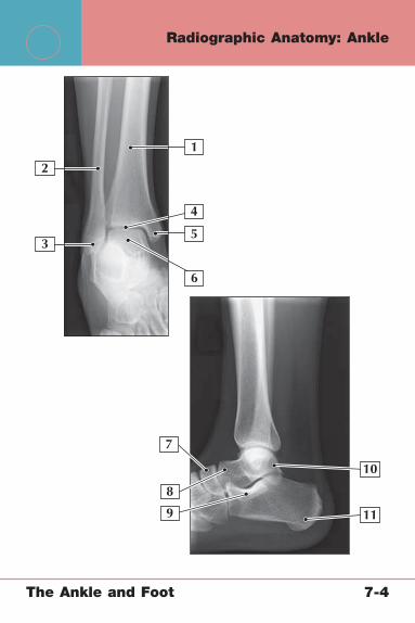
3
7
2
4
11
10
9
5
6
8
1
The Ankle and Foot 7-4
Radiographic Anatomy: Ankle
The Ankle and Foot 7-4
Radiographic Anatomy: Ankle
1. Tibia 2. Fibula 3. Lateral malleolus 4. Ankle mortise 5. Medial malleolus 6. Talus 7. Navicular bone 8. Head of talus 9. Sustentaculum tali10. Posterior process of talus11. Calcaneus
Comment: Anteroposterior (AP), lateral, and mortise views are the most commonly used to evaluate ankle injuries. External rotation stress views may be helpful for evaluating occult disruptions of the ankle mortise. The Ottawa ankle rules were designed to help emergency room practitioners decide when ankle radiographs should be ordered.
The Ottawa Ankle Rules
Tenderness of the medial malleolus
Tenderness on the lateral malleolus
Tenderness at the base of the 5th metatarsal
Navicular tenderness
Inability to bear weight immediately and in the emergency room

7
4
10119
5
6
3
2
1
8
The Ankle and Foot 7-5
Radiographic Anatomy: Foot
The Ankle and Foot 7-5
Radiographic Anatomy: Foot
1. Distal phalanges 2. Middle phalanges 3. Proximal phalanges 4. Medial sesamoid 5. Lateral sesamoid 6. Metatarsals 7. Talus 8. Navicular 9. Cuneiforms10. Base of the 5th metatarsal11. Calcaneus
Comment: Anteroposterior (AP) and lateral radiographs of the foot are the views most commonly used to evaluate pathology. These should be weight-bearing images in nontraumatic or chronic cases. Special views of the calcaneus can be helpful for diagnosing fracture of this bone.

3
8
7
2
4
10
9
5
6
1
The Ankle and Foot 7-6
Ligaments: Ankle

The Ankle and Foot 7-6
Ligaments: Ankle
1. Anterior and posterior talofi bular ligaments 2. Anterior and posterior tibiofi bular ligaments 3. Superior and inferior peroneal retinaculum 4. Achilles tendon (cut) 5. Calcaneofi bular ligament 6. Lateral talocalcaneal ligament 7. Peroneus longus and brevis muscles 8. Deltoid ligament 9. Medial talocalcaneal ligament10. Posterior talocalcaneal ligament
Comment: The lateral ligaments of the ankle (anterior and posterior talofi bular ligaments and calcaneofi bular ligament) are the ligaments most commonly injured with an inversion ankle sprain. The deltoid ligament on the medial side is much stronger, and the eversion mechanism of injuring it is less common.
3
8 7
2
2
6
54
11 10
12
9
78
6
1
Right foot: medial view
Right foot: lateral view
The Ankle and Foot 7-7
Ligaments: Foot

The Ankle and Foot 7-7
Ligaments: Foot
1. Interosseous talocalcaneal ligament 2. Dorsal talonavicular ligament 3. Calcaneonavicular ligament 4. Calcaneocuboid ligament 5. Dorsal cuboideonavicular ligaments 6. Dorsal cuneonavicular ligaments 7. Dorsal intercuneiform ligaments 8. Dorsal tarsometatarsal ligaments 9. Dorsal metatarsal ligaments10. Dorsal cuneocuboid ligament11. Dorsal calcaneocuboid ligament12. Long and short plantar ligaments
Comment: The intertarsal ligaments connect the subtalar and midtarsal joints to provide stability for the midfoot. There are also small dorsal and plantar ligaments of the tarsometatarsal (Lisfranc) joints, as well as the metatarsophalangeal and interphalangeal joints of the forefoot.

3
8
7
2
6
5
4
10
9
1
The Ankle and Foot 7-8
Tendons and Muscles: Ankle and Foot

The Ankle and Foot 7-8
Tendons and Muscles: Ankle and Foot
1. Tibialis anterior muscle and tendon 2. Extensor digitorum longus muscle and tendon 3. Extensor hallucis longus muscle and tendon 4. Peroneus longus tendon 5. Extensor digitorum brevis and extensor hallucis brevis muscles
(cut) 6. Peroneus tertius tendon 7. Soleus muscle 8. Achilles tendon 9. Calcaneal bursae10. Superior and inferior extensor retinaculum
Comment: The ankle fl exors cross the joint posteriorly; the tendons pass beneath the fl exor retinaculum, which prevents them from subluxating over the medial malleolus. Similarly, the ankle extensors cross the joint anteriorly and are supported by the superior and inferior extensor retinaculum.
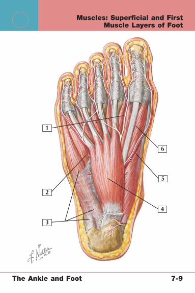
3
4
2
6
5
1
The Ankle and Foot 7-9
Muscles: Superfi cial and First Muscle Layers of Foot

The Ankle and Foot 7-9
Muscles: Superfi cial and First Muscle Layers of Foot
1. Lumbricals2. Flexor digiti minimi brevis muscle3. Abductor digiti minimi muscle4. Flexor digitorum brevis muscle5. Abductor hallucis muscle6. Flexor hallucis brevis muscle
Abductor Digiti Minimi Muscle
Flexor Digitorum Brevis Muscle
Abductor Hallucis Muscle
Origin Tuberosity and medial/lateral processes of calcaneus
Tuberosity and medial process of calcaneus
Tuberosity and medial process of the calcaneus
Insertion Base of 5th proximal phalanx
Middle phalanges of toes 2-5
Proximal phalanx of great toe
Actions Abduction of toe 5 Flexion of toes 2-5 Abduction of great toe
Innervation Lateral plantar nerve
Medial plantar nerve Medial plantar nerve

3
4
2
5
1
The Ankle and Foot 7-10
Muscles: Second Muscle Layer of Foot

The Ankle and Foot 7-10
Muscles: Second Muscle Layer of Foot
1. Lumbricals2. Flexor hallucis longus tendon3. Abductor hallucis muscle (cut)4. Flexor digitorum longus tendon5. Quadratus plantae muscle
Quadratus Plantae Muscle Lumbricals
Origin Plantar aspect of the calcaneus
Flexor digitorum longus tendons
Insertion Lateral aspect of the fl exor digitorum longus tendon
Proximal phalanges
Actions Toe fl exion (along with fl exor digitorum longus)
Metatarsophalangeal (MTP) joint fl exion, interphalangeal joint extension
Innervation Lateral plantar nerve 1 medial plantar nerve; 2-4 lateral plantar nerves

3
4
2
5
1
The Ankle and Foot 7-11
Muscles: Third Muscle Layer of Foot

The Ankle and Foot 7-11
Muscles: Third Muscle Layer of Foot
1. Flexor hallucis brevis muscle2. Adductor hallucis muscle3. Flexor digiti minimi brevis muscle4. Abductor digiti minimi muscle (cut)5. Flexor digitorum brevis muscle (cut)
Flexor Hallucis Brevis Muscle
Adductor Hallucis Muscle
Flexor Digiti Minimi Brevis Muscle
Origin Cuboid and lateral cuneiform
Oblique: base of metatarsals 2-4Transverse: lateral 4 metatarsals
Base of 5th metatarsal
Insertion Proximal phalanx of great toe
Proximal phalanx of great toe
Base of 5th proximal phalanx
Actions Flexion of great toe Adduction of great toe
Flexion of toe 5
Innervation Medial plantar nerve
Lateral plantar nerve
Lateral plantar nerve
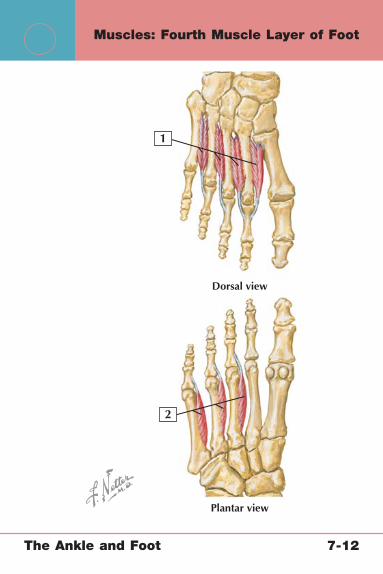
2
1
Dorsal view
Plantar view
The Ankle and Foot 7-12
Muscles: Fourth Muscle Layer of Foot

The Ankle and Foot 7-12
Muscles: Fourth Muscle Layer of Foot
1. Dorsal interosseous muscles (4)2. Plantar interosseous muscles (3)
Dorsal Interosseous Muscles Plantar Interosseous Muscles
Origin Shaft of adjacent metatarsal shafts
Medial aspect of metatarsal shafts 3-5
Insertion Proximal phalanges 2-5 Medial aspect of proximal phalanges 3-5
Actions Toe abduction Toe adduction
Innervation Lateral plantar nerve Lateral plantar nerve
Comment: Useful mnemonics for remembering these muscles are “PAD = plantar adducts” and “DAB = dorsal abducts.”
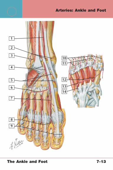
2
3
4
5
6
7
8
9
10
12
11
1314
1
The Ankle and Foot 7-13
Arteries: Ankle and Foot

The Ankle and Foot 7-13
Arteries: Ankle and Foot
1. Anterior tibial artery 2. Anterior medial malleolar artery 3. Anterior lateral malleolar artery 4. Dorsalis pedis artery 5. Medial tarsal arteries 6. Arcuate artery 7. Dorsal metatarsal arteries 8. Anterior perforating branches from plantar metatarsal arteries 9. Dorsal digital arteries10. Proper plantar digital arteries11. Common plantar digital arteries12. Plantar metatarsal arteries13. Deep plantar arch14. Lateral plantar artery
Comment: The anterior tibial, posterior tibial, and peroneal arteries form the branches that supply the foot and ankle. The posterior tibial and dorsalis pedis pulses are the easiest to assess with physical examination.
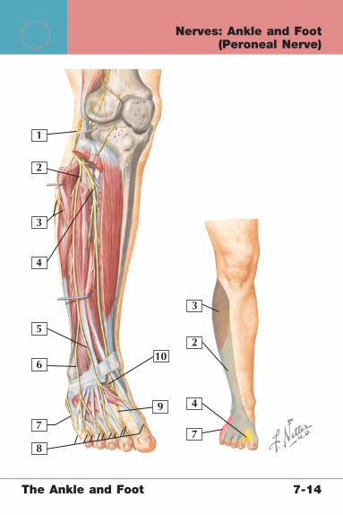
2
2
3
3
4
4
5
6
77
8
9
10
1
The Ankle and Foot 7-14
Nerves: Ankle and Foot (Peroneal Nerve)

The Ankle and Foot 7-14
Nerves: Ankle and Foot (Peroneal Nerve)
1. Common peroneal nerve (L4-S2) 2. Superfi cial peroneal nerve 3. Branches of the lateral sural cutaneous nerve 4. Deep peroneal nerve 5. Medial dorsal cutaneous nerve 6. Intermediate dorsal cutaneous nerve 7. Lateral dorsal cutaneous nerve 8. Dorsal digital nerves 9. Medial branch of deep peroneal nerve10. Lateral branch of deep peroneal nerve
Comment: The common peroneal nerve is derived from the posterior division of the sacral plexus (L4-S2). It divides into the superfi cial and deep peroneal nerves and supplies the primary innervation to the lateral lower leg and foot. The deep peroneal nerve supplies the tibialis anterior, extensor hallucis longus, extensor digitorum longus, and peroneus tertius muscles. The superfi cial peroneal nerve supplies the peroneus longus and brevis muscles.

2
3
4
5
6
7
8
9
10
13
12
11
1
The Ankle and Foot 7-15
Nerves: Ankle and Foot (Tibial Nerve)
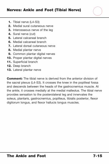
The Ankle and Foot 7-15
Nerves: Ankle and Foot (Tibial Nerve)
1. Tibial nerve (L4-S3) 2. Medial sural cutaneous nerve 3. Interosseous nerve of the leg 4. Sural nerve (cut) 5. Lateral calcaneal branch 6. Medial calcaneal branch 7. Lateral dorsal cutaneous nerve 8. Medial plantar nerve 9. Common plantar digital nerves10. Proper plantar digital nerves11. Superfi cial branch12. Deep branch13. Lateral plantar nerve
Comment: The tibial nerve is derived from the anterior division of the sacral plexus (L4-S3). It crosses the knee in the popliteal fossa and descends between the heads of the gastrocnemius muscle. At the ankle, it crosses medially at the medial malleolus. The tibial nerve provides sensation to the posterolateral leg and innervates the soleus, plantaris, gastrocnemius, popliteus, tibialis posterior, fl exor digitorum longus, and fl exor hallucis longus muscles.

Inversion0�0�
Eversion
Dorsiflexion
Inversion EversionFootAnkle
Plantar flexion
L4, 5 L5, S1
S1 , 2
L4, 5
Dorsalis pedis
The Ankle and Foot 7-16
Physical Examination: Ankle Joint
The Ankle and Foot 7-16
Physical Examination: Ankle Joint
Movement Normal Range of Motion
Dorsifl exion 20 Degrees
Plantar fl exion 45-50 Degrees
Subtalar eversion 5 Degrees
Subtalar inversion 5-10 Degrees
Test/Sign Reason for Evaluation
Anterior drawer testTalar tilt test
Ankle instability
Tinel sign Tarsal tunnel syndrome
Thompson test Achilles tendon rupture
“Too many toes” sign Acquired pes planus from posterior tibialis dysfunction
Metatarsal compression test Morton neuroma
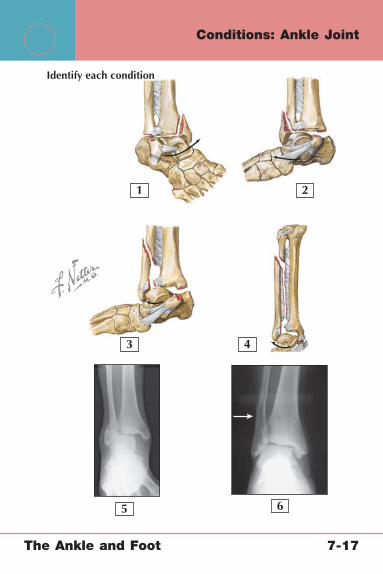
2
3 4
5 6
1
Identify each condition
The Ankle and Foot 7-17
Conditions: Ankle Joint

The Ankle and Foot 7-17
Conditions: Ankle Joint
1. Weber A fracture with medial malleolar fracture2. Weber B fracture with medial malleolar fracture3. Weber C fracture with medial malleolar fracture4. Maisonneuve fracture5. Weber B fracture6. Weber C fracture
Fractures of the Lateral Malleolus
Mechanism Pattern Treatment
Weber A Fracture
Avulsion Below level of mortise Usually nonoperative
Weber B Fracture
SupinationExternal rotation
At the level of mortise, may or may not involve medial malleolus or deltoid ligament
Open reduction, internal fi xation (ORIF) if unstable
Weber C Fracture
PronationExternal rotation
Above level of the mortise, usually involves medial structures
ORIF if talus displaced in the mortise
Maisonneuve Fracture
External rotation Proximal fi bula fractureLongitudinal tear of interosseous membrane
Syndesmotic screw; proximal fi bular treatment unnecessary
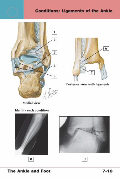
2
3
4
7
98
5
6
1
Posterior view with ligaments
Identify each condition
Medial view
The Ankle and Foot 7-18
Conditions: Ligaments of the Ankle

The Ankle and Foot 7-18
Conditions: Ligaments of the Ankle
1. Interosseous membrane2. Posterior tibiofi bular ligament3. Posterior inferior talofi bular ligament4. Calcaneofi bular ligament5. Deltoid ligament6. Anterior tibiofi bular ligament7. Anterior talofi bular ligament8. Radiographic appearance of Maisonneuve fracture9. Stress radiograph demonstrating lateral ankle instability
Acute Ankle Sprain “High Ankle Sprain”Chronic Ankle Instability
Mechanism Eversion/plantar fl exion (lateral: most common); inversion (medial)
Dorsifl exion, eversion Recurrent ankle sprains
Signs and Symptoms
Lateral ankle pain and tenderness, edema, ecchymosis
Lateral and syndesmotic tenderness, positive squeeze test
Subjective instability, positive result of anterior drawer test
Imaging Anteroposterior (AP), lateral, mortise radiographs to rule out fracture
AP, lateral, and mortise radiographs; may include proximal fi bula to rule out Maisonneuve fracture; external rotation stress radiograph
AP, lateral, mortise radiographs; stress radiographs; magnetic resonance imaging (MRI)
Treatment Conservative with rest, ice, compression, elevation, bracing, proprioception training
Same as for acute ankle sprain plus reassurance that recovery may take longer
Surgical stabilization (Broström procedure)

2
3
4
7
5
6
1
The Ankle and Foot 7-19
Conditions: Achilles Tendon

The Ankle and Foot 7-19
Conditions: Achilles Tendon
1. Gastrocnemius muscle2. Soleus muscle3. Achilles tendon4. Calcaneal tuberosity5. Retrocalcaneal bursa6. Achilles tendonitis (area of tenderness)7. Retrocalcaneal bursitis (area of tenderness)
Achilles Tendinitis and Retrocalcaneal Bursitis (Haglund Disease) Achilles Tendon Rupture
Mechanism Overuse, repetitive motion, poorly fi tted footwear
“Charge forward” pushing off the foot, jumping
Signs and Symptoms
Focal tenderness just proximal to calcaneal insertion, “pump bump,” intact but painful plantar fl exion and toe raises
Local pain/tenderness, “pop” or “kicked in calf” sensation, palpable tendon defect, inability to achieve plantar fl exion, positive Thompson test result
Imaging Usually not necessary Usually not necessary; magnetic resonance imaging (MRI) can confi rm tear
Treatment Conservative with nonsteroidal antiinfl ammatory drugs (NSAIDs), activity modifi cation, heel lift, or, for refractory cases, casting or fracture boot
Usually surgical repair (high rate of skin healing problems) but can be conservative with 8-12 weeks of casting with foot in plantar fl exion (higher rate of recurrence)

1
Identify each condition
5
4
3
2
6 7 8
9
The Ankle and Foot 7-20
Conditions: Calcaneus

The Ankle and Foot 7-20
Conditions: Calcaneus
1. Anterior, middle, and posterior talar articular surface2. Body of the calcaneus3. Tuberosity4. Lateral process of tuberosity5. Articular surface for the cuboid6. Fracture of the sustentaculum tali7. Extraarticular fracture of the body of the calcaneus8. Intraarticular fracture of the body of the calcaneus9. Split “tongue” fracture
Intraarticular Fracture Extraarticular Fracture
Mechanism Axial loadFall from height
Axial loadFall from height
Prognosis Worse Better
Treatment Open reduction, internal fi xation (ORIF)
Cast (ORIF if unstable)

2
3
4
5
19
Identify each condition
6
7
8
10 11 12
13
The Ankle and Foot 7-21
Conditions: Talus

The Ankle and Foot 7-21
Conditions: Talus
1. Head of talus 2. Artery of tarsal sinus 3. Neck of talus 4. Anterior tibial artery 5. Dorsalis pedis artery 6. Trochlea 7. Posterior tibial artery 8. Posterior process 9. Artery of tarsal canal10. Hawkins type I talar neck fracture11. Hawkins type II talar neck fracture12. Hawkins type III talar neck fracture13. Radiographic appearance of talar neck fracture
Talar Neck Fracture TreatmentAvascular Necrosis Risk
Hawkins Type I
Nondisplaced Closed reductionCast
Low
Hawkins Type II
Displaced 20%-50%Subtalar dislocation
Open reduction, internal fi xation (ORIF)
Early range-of-motion exercises
Intermediate
Hawkins Type III
Displaced 50%-100%Talar body dislocation
ORIFEarly range-of-motion
exercises
High
Hawkins Type IV
Talar head+/− body dislocation
(>100%)
ORIFEarly range-of-motion
exercises
High
2
3 4
5
1Identify each conditon
The Ankle and Foot 7-22
Conditions: Talus
The Ankle and Foot 7-22
Conditions: Talus
1. Medial subtalar dislocation2. Lateral subtalar dislocation3. Radiographic appearance of medial subtalar dislocation4. Magnetic resonance imaging (MRI) appearance of talar chondral
defect5. Arthroscopic appearance of talar chondral defect
Subtalar Dislocation (Basketball Foot)
Chondral Defects of the Talus/Osteochondritis Dissecans (OCD)
Mechanism High-energy trauma Usually posttraumatic process
Classifi cation Medial (most common) and lateral
Descriptive: based on size and location of lesion
Signs and Symptoms
Usually obvious deformity, pain, inability to bear weight
Pain and swelling with activity, with or without mechanical catching
Imaging Plain radiographs and computed tomography (CT) to rule out associated fracture (common)
MRI most sensitive for detecting and defi ning size and location
Treatment Immediate reduction; if dislocation is irreducible, suspect interposed tendon; immobilize for 6 weeks
Nonsteroidal antiinfl ammatory drugs (NSAIDs), steroid injections, arthroscopic débridement with or without microfracture, cartilage plug transfer (osteochondral autograft transfer system [OATS])

2
3
4
5
1
The Ankle and Foot 7-23
Conditions: Plantar Fascia and Arch

The Ankle and Foot 7-23
Conditions: Plantar Fascia and Arch
1. Digital slips of the plantar aponeurosis2. Plantar aponeurosis3. Tuberosity of the calcaneus4. Calcaneal fat pad5. Plantar fasciitis appearance on magnetic resonance imaging (MRI)
Plantar Fasciitis Pes Planus
Mechanism Overuse, repetitive microtrauma
Congenital, acquired (tibialis posterior tendon dysfunction)
Signs and Symptoms
Focal pain/tenderness over plantar aspect of heel, pain worse in morning with fi rst steps
May be symptomatic with aching pain or asymptomatic
Imaging Not usually necessary but may show calcaneal bone spur
Not usually necessary
Treatment Nonsteroidal antiinfl ammatory drugs (NSAIDs), injections, bracing/night splints, stretching, extracorporeal shock wave therapy, rarely surgical release
Flexible deformities: usually with calf stretches and orthotics; rigid: with failed conservative treatment require arthrodesis
MRI from Osborne J, Warner J, Fu F, et al: MRI-Arthroscopy Correlative Atlas. Philadelphia: WB Saunders, 1997.

2
1
Identify each condition
The Ankle and Foot 7-24
Conditions: Diabetic Foot
The Ankle and Foot 7-24
Conditions: Diabetic Foot
1. Classic deformity associated with Charcot joint2. Radiographic appearance of Charcot joint
Diabetic UlcersNeuropathic Arthropathy (Charcot Foot)
Mechanism Ischemic and/or neurotrophic with underlying infection
Progressive neuropathy from prolonged hyperglycemia
Signs and Symptoms
Often asymptomatic with varying degree of depth of tissue infection and necrosis
Decreased sensation, ulcers, edema, erythema/warmth, deformity
Imaging Magnetic resonance imaging (MRI) to rule out osteomyelitis; perfusion studies (ankle/brachial index, Doppler imaging)
Standing anteroposterior (AP), lateral, and oblique views (osseous destruction, subluxation)
Treatment Initially antibiotics and débridement, varying level amputations for advanced disease (based on vascular status)
Skin treatment, immobilizationSurgical débridement,
arthrodesis
2
3
4 5
1
Identify each condition
The Ankle and Foot 7-25
Conditions: Joints
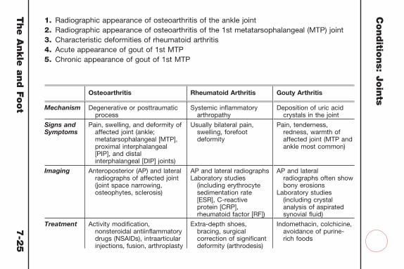
The A
nkle
and F
oot
7-2
5
Conditio
ns: J
oin
ts1. Radiographic appearance of osteoarthritis of the ankle joint2. Radiographic appearance of osteoarthritis of the 1st metatarsophalangeal (MTP) joint3. Characteristic deformities of rheumatoid arthritis4. Acute appearance of gout of 1st MTP5. Chronic appearance of gout of 1st MTP
Osteoarthritis Rheumatoid Arthritis Gouty Arthritis
Mechanism Degenerative or posttraumatic process
Systemic infl ammatory arthropathy
Deposition of uric acid crystals in the joint
Signs and Symptoms
Pain, swelling, and deformity of affected joint (ankle; metatarsophalangeal [MTP], proximal interphalangeal [PIP], and distal interphalangeal [DIP] joints)
Usually bilateral pain, swelling, forefoot deformity
Pain, tenderness, redness, warmth of affected joint (MTP and ankle most common)
Imaging Anteroposterior (AP) and lateral radiographs of affected joint (joint space narrowing, osteophytes, sclerosis)
AP and lateral radiographsLaboratory studies
(including erythrocyte sedimentation rate [ESR], C-reactive protein [CRP], rheumatoid factor [RF])
AP and lateral radiographs often show bony erosions
Laboratory studies (including crystal analysis of aspirated synovial fl uid)
Treatment Activity modifi cation, nonsteroidal antiinfl ammatory drugs (NSAIDs), intraarticular injections, fusion, arthroplasty
Extra-depth shoes, bracing, surgical correction of signifi cant deformity (arthrodesis)
Indomethacin, colchicine, avoidance of purine-rich foods

2 3
4
6 7
5
1
Identify each condition
The Ankle and Foot 7-26
Conditions: Tarsometatarsal Joints
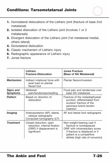
The Ankle and Foot 7-26
Conditions: Tarsometatarsal Joints
1. Homolateral dislocations of the Lisfranc joint (fracture of base 2nd metatarsal)
2. Isolated dislocation of the Lisfranc joint (involves 1 or 2 metatarsals)
3. Divergent dislocation of the Lisfranc joint (1st metatarsal medial, others lateral)
4. Dorsolateral dislocation5. Classic mechanism of Lisfranc injury6. Radiographic appearance of Lisfranc injury7. Jones fracture
Lisfranc Fracture-Dislocation
Jones Fracture(Base of 5th Metatarsal)
Mechanism Indirect rotational force with axial load on plantar fl exed foot
Plantar fl exion/inversion
Signs and Symptoms
Midtarsal pain/tenderness/swelling
Focal pain and tenderness over base 5th metatarsal
Pattern Based on direction of dislocation
Fracture of the metadiaphyseal junction, differentiated from avulsion fracture of the peroneus brevis tendon insertion
Imaging Anteroposterior (AP), lateral, oblique radiographs
Computed tomography (CT)
AP and lateral foot radiographs
Treatment Closed reduction; open reduction, internal fi xation (ORIF) if displacement is signifi cant
Non–weight-bearing cast if fracture is nondisplaced
ORIF with intramedullary screw if fracture is displaced or if patient is a competitive athlete (high rate of nonunion)

2
3
4
6
7
5
8
9
10
1
Identify each condition
The Ankle and Foot 7-27
Conditions: Metatarsals

The A
nkle
and F
oot
7-2
7
Conditio
ns: M
eta
tarsa
ls 1. Displaced oblique fracture, 2nd
metatarsal neck 2. Comminuted, non-displaced fracture,
base 1st metatarsal 3. Oblique fracture of the body, 3rd
metatarsal 4. Displaced transverse fracture of the
body, 4th metatarsal
Metatarsal and Phalangeal Fractures Freiberg Infarction Metatarsalgia
Mechanism Direct trauma, fall, stress fracture
Avascular necrosis of the 2nd metatarsal head caused by stress overload
Overuse especially related to impact activity
Signs and Symptoms
Focal tenderness, pain with weightbearing, ecchymosis
Pain with walking/running, localized tenderness
Gradual onset of pain over one or more metatarsal heads with or without symptoms of interdigital neuroma
Imaging Anteroposterior (AP), lateral, and oblique radiographs; magnetic resonance imaging (MRI) or bone scan for stress fractures
Standing AP, lateral, and oblique radiographs (fl attening of 2nd metatarsal head)
AP, lateral, and oblique radiographs to rule out fracture
Treatment Nondisplaced fracture: cast or fracture shoe
Displaced fracture: surgical open reduction, internal fi xation (ORIF)
Rest, padding, activity modifi cation
Activity modifi cation, metatarsal bar, orthotics, local digital nerve block
5. Fracture of the base of the 5th metatarsal (Jones fracture)
6. Avulsion fracture, base 5th metatarsal 7. Sesamoid fracture 8. Transverse fracture, 2nd proximal phalanx 9. Displaced fractures of 2nd and 3rd
metatarsal necks10. Freiberg infarction

7
65
1
2
4
3
8
The Ankle and Foot 7-28
Conditions: Nerves and Tarsal Tunnel

The Ankle and Foot 7-28
Conditions: Nerves and Tarsal Tunnel
1. Lateral plantar nerve2. Medial plantar nerve3. Common plantar digital nerves4. Tendinous sheath of fl exor hallucis longus5. Tendinous sheath of fl exor digitorum longus6. Tibialis posterior tendon and sheath7. Flexor retinaculum8. Posterior tibial artery and tibial nerve
Tarsal Tunnel Syndrome Interdigital (Morton) Neuroma
Pathophysiology Impingement of the posterior tibial neurovascular bundle as it passes under the fl exor retinaculum (tarsal tunnel)
Enlargement of the common digital nerve, usually in the 3rd web space, from degeneration, repetitive trauma, or constricting footwear
Signs and Symptoms
Diffuse pain in ankle and foot, tenderness over tarsal tunnel
Diffuse pain, dysesthesia, and paresthesias usually in toes 3 and 4; tenderness in 3rd web space
Imaging Radiographs are not usually helpful, but electromyography is useful
Radiographs to rule out fractureMRI is not necessary but can
confi rm
Treatment Footwear and activity modifi cation; surgical release for refractory cases
Wide-toe shoe, metatarsal bar or pad, steroid injection
Surgical excision of neuroma for refractory cases

2
3
4
6
7
5
8
1
The Ankle and Foot 7-29
Conditions: Sesamoids and Metatarsophalangeal Joints
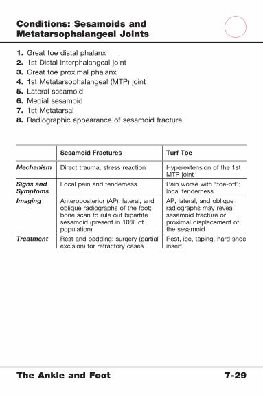
The Ankle and Foot 7-29
Conditions: Sesamoids and Metatarsophalangeal Joints
1. Great toe distal phalanx2. 1st Distal interphalangeal joint3. Great toe proximal phalanx4. 1st Metatarsophalangeal (MTP) joint5. Lateral sesamoid6. Medial sesamoid7. 1st Metatarsal8. Radiographic appearance of sesamoid fracture
Sesamoid Fractures Turf Toe
Mechanism Direct trauma, stress reaction Hyperextension of the 1st MTP joint
Signs and Symptoms
Focal pain and tenderness Pain worse with “toe-off”; local tenderness
Imaging Anteroposterior (AP), lateral, and oblique radiographs of the foot; bone scan to rule out bipartite sesamoid (present in 10% of population)
AP, lateral, and oblique radiographs may reveal sesamoid fracture or proximal displacement of the sesamoid
Treatment Rest and padding; surgery (partial excision) for refractory cases
Rest, ice, taping, hard shoe insert

2 3
4
6
9
7
5
8
1
Identify each condition
The Ankle and Foot 7-30
Conditions: Toes

The Ankle and Foot 7-30
Conditions: Toes
1. Distal interphalangeal (DIP) joint2. Proximal interphalangeal (PIP) joint3. Metatarsophalangeal (MTP) joint4. Plantar plate5. Collateral ligaments6. Hammer toe7. Mallet toe8. Claw toe9. Hallux valgus (bunion)
Lesser Toe DeformitiesAffectedJoint
JointDeformity
Mallet Toe DIP Flexion
Hammer Toe PIP Flexion
Claw Toe MTP Hyperextension
Hallux Valgus (Bunion)
Pathophysiology Increase in the valgus angle of the 1st MTP joint
Mechanism Multifactorial, including genetics, footwear, individual anatomy
Signs and Symptoms
Obvious deformity; redness/tenderness over medial MTP
Imaging Weight-bearing anteroposterior (AP) and lateral radiographs
Treatment Footwear and activity modifi cation with metatarsal osteotomy for refractory or severely deformed cases