Nervous tissue Department of Histology and Embryology Medical college in Three Gorges University.
106
Nervous tissue Department of Histology and Embryology Medical college in Three Gorges University
-
Upload
ashley-sharp -
Category
Documents
-
view
225 -
download
3
Transcript of Nervous tissue Department of Histology and Embryology Medical college in Three Gorges University.

Nervous tissue
Department of Histology and Embryology Medical college in Three Gorges University

Content Neuron Synapses Neuroglia Nerve fiber Nerve ending
Outline
More pay attention to the microstructure of
neuron and its classification
Master the microstructure of the synapses
Know the micrograph and function of glia
Familiar the microstructure of nerve fiber
Nervous tissue is composed of nerve cells
and neuroglia. The specialized cells that
constitute the functional units of the
nervous system are called neurons.
The function of neuron:generate nerve
impulses in response to stimuli and
transmit them along cellular processes.

Neuroglia or glia: neurons are supported by
a special kind of connective tissue within
the brain and spinal cord, that is called
neuroglia,it also located in the PNS.
Function: support, protect, connect

Central nervous system
Peripheral nervous system
Brain and spinal cord
The nerves and their associated ganglia.
Nervous system

Microstructure LM: EM: FunctionClassification
Neuron

1.Microstructure of neuron
Cell body
Axon
Dendrites

Neurons
Cell body
Processes or neurites
Dendrites
Axon
Nucleus
Cytoplasm
Cell membrane
The cell body,soma, is the part of neuron that
contains nucleus and surrounding cytoplasm,
also called perikaryon. It is the trophic center
of the neuron. The protein and enzymes
synthesis in this area.
Cell body: Perikaryon
Where is cell body

Cell body: Perikaryon
Position: only in grey matter in CNS which also contains dendrites and axons starting from or ending on the cell bodies,ganglia in PNS
Shape:They can be pyramidal, spherical, ovoid or pear-shaped.
Size: Measuring 5-150 um in diameter.
Microscopic examination
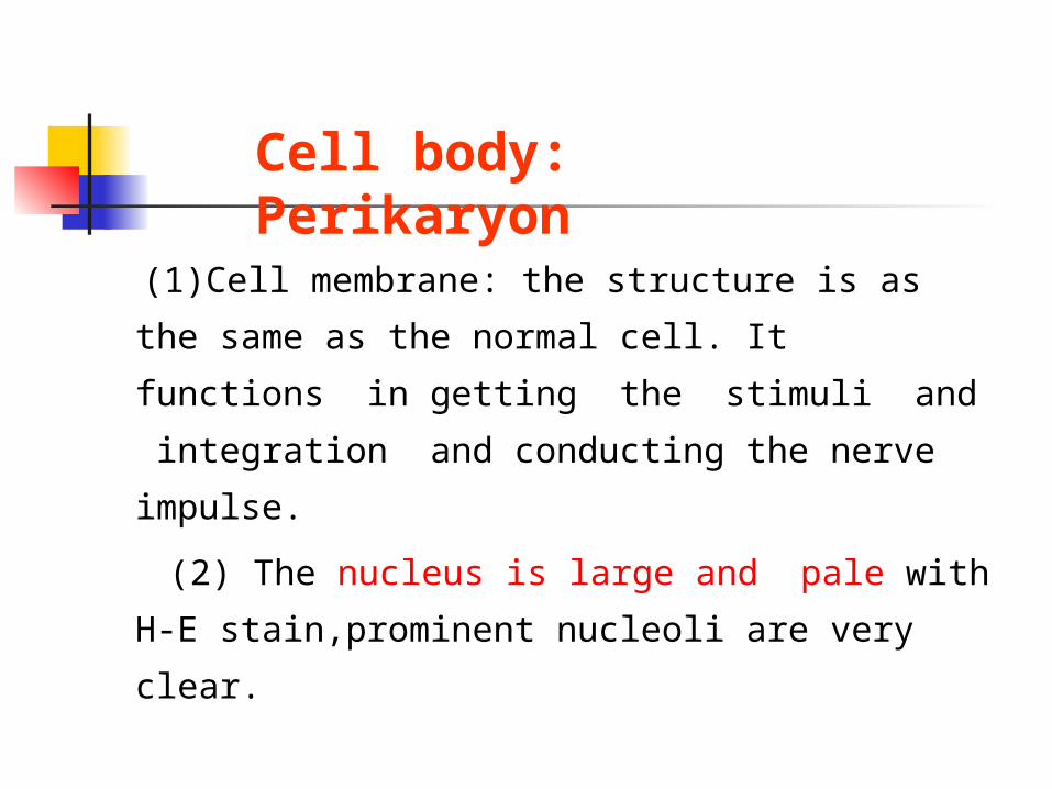
(1)Cell membrane: the structure is as the
same as the normal cell. It functions in
getting the stimuli and integration and
conducting the nerve impulse.
(2) The nucleus is large and pale with H-E
stain,prominent nucleoli are very clear.
Cell body: Perikaryon
nucleus
nucleolus

(3)Cytoplasm: the cytoplasm has some
distinctive characteristics not seen in other
cells. The cytoplasm is basophilic and
full of neurofibrils.
Cell body: Perikaryon

Nissl body
Neurofibril
H-E stains Silver nitrate

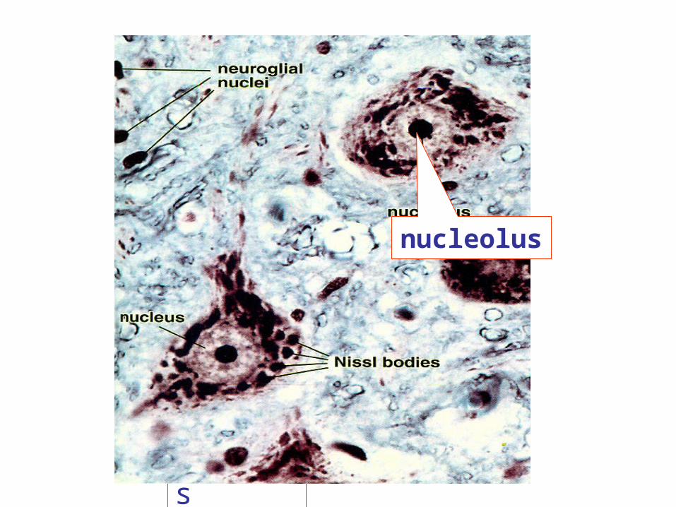
Nissl bodies: The cytoplasm shows the
presence of a granular material that stains
intensely with basic dyes; this material is the
Nissl substance (also called Nissl bodies or
granules).
Cell body: Perikaryon

细颗粒样的尼氏体
Nissil body

axon hilllock
Neurofibrils

Neurofibrils are thin black fibers
observed in LM with silver nitrate slides,
which is composed of microtubule and
filaments in EM.
Cell body: Perikaryon
EM


RER
ribosome
pigment
synapse
microtubemitochondria

EM: rough surfaced endoplasmic reticulum.
The presence of abundant granular endoplasmic
reticulum is an indication of the high level of
protein synthesis in neurons. Mitochondria,
SER,lysosomes,Golgi complexes,ribosome etc.
The proteins are needed for maintenance and repair, and for production of neurotransmitters and enzymes.

The processes arising from the cell body of
a neuron are called neurites.Most neurons
give off a number of short branching
processes called dendrites and one longer
process called an axon.
Neurites or processes
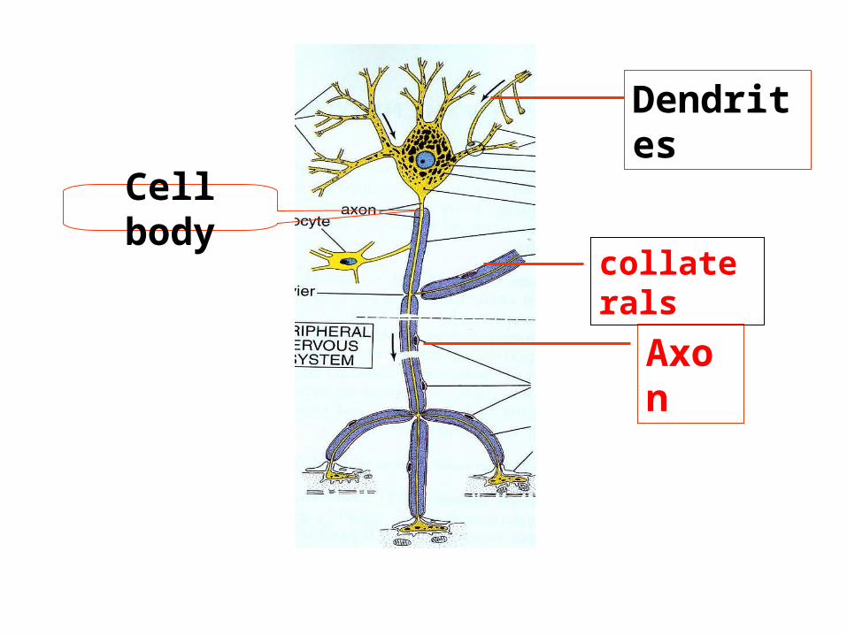
Cell body
Axon
Dendrites
collaterals

Nissl bodyNeurofibril
H-E stains Silver nitrate不同形态
Axon
Dendrites

The dendrites are characterized by the fact
that they terminate near the cell body. They
are irregular in thickness, and Nissl granules
extend into them. They bear numerous small
spines which are of variable shape.
Dendrites

Every neuron has only one long thin
process or axon which arises from a special
region or axon hillock, which is devoid of
Nissl bodies. It carries the impulse received
by the neuron to distant region.
Axon

axon hilllock

An axon may have not much branches than
that of dendrites. If branches, that arise near
the cell body and lie at right angles to the
axon are called collaterals. At its
termination the axon breaks up into a
number of fine branches called telodendria
which may end in small swellings (terminal
boutons.

The axon is identified according to the
axon hillock with LM.The part of the
axon just beyond the axon hillock is called
the initial segment.

Neurites or processes
Dendrites Axons
many one
short long
irregular in thickness uniform in diameter
Nissl granules No Nissl substance
spines axon hillock
impulse towards the soma away from the cell body

1) morphology
2) Axon hillock in LM
3) Dendrites have microtubule associated protein
(MAP-2 ) in immunocytochemically, not present in
axons.
Identification of dendrites from axon
distinguished from axons

2. Classification of neuron
Cell body
Axon
Dendrites
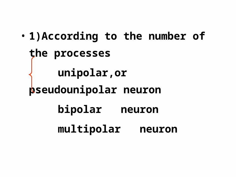
• 1)According to the number of the processes
unipolar,or pseudounipolar neuron
bipolar neuron
multipolar neuron

multipolar neuron
bipolar neuron
Unipolar neuron

Unipolar neuron
Spinal ganglia

Satellite cells
Unipolar neuron

Bipolar neuron
Retina of eye

axon hilllock
multipolar neuron

According to the size of cell body and the
length of axon: According to Cajal (1889):
Golgi type I neurons: long axon
Golgi type II neurons: short axon

Sense ( afferent )neurons:
Interneurons
Motor( efferent ) neurons
•According to their function:


• The classification of neurons:
• According to the number of process
The shape of the cell body is dependent on
the number of processes arising from it. The
most common type of neuron gives off several
processes from the cell body is, therefore,
multipolar. Some neurons have only one
axon and one dendrite and are bipolar.

Another type of neuron has a single process
(which is highly convoluted). After a very
short course this process divides into two.
One of the divisions represents the axon; the
other is functionally a dendrite, but its
structure is indistinguishable from that of an
axon. This neuron is described as unipolar,

but from a functional point of view it is to be
regarded as bipolar. (To avoid confusion on
this account this kind of neuron has been
referred to, in the past, as a pseudounipolar
neuron.
Depending on the shapes of their cell bodies some neurons are referred to as stellate (star shaped) or pyramidal.
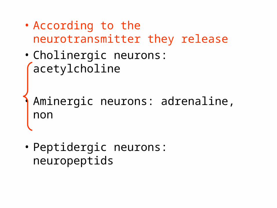
• According to the neurotransmitter they release
• Cholinergic neurons: acetylcholine
• Aminergic neurons: adrenaline, non
• Peptidergic neurons: neuropeptids

The Synapse 突触 :
Concept: Synapses are highly specialized
intercellular junctions which link the
neurons of each nervous pathway.
Similar intercellular junctions link
neurons and their effector cells such as
muscle fibers;where neurons synapse with
skeletal muscle they are referred to as
neuromuscular junction or motor end plate.

synapse
Processes
Cell body

• Classification of synapses:
According the constitution:
axodendritic synapse
axosomatic synapse
axoaxonal synapse
dendro-axonic
dendro-dendritic
somato-somatic synapse
somato-dendritic synapse

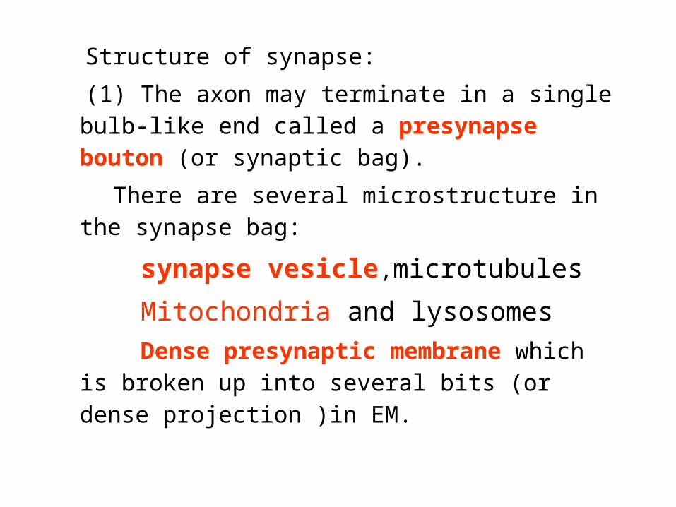
Structure of synapse:
(1) The axon may terminate in a single bulb-like end called a presynapse bouton (or synaptic bag).
There are several microstructure in the synapse bag:
synapse vesicle,microtubules
Mitochondria and lysosomes
Dense presynaptic membrane which is broken up into several bits (or dense projection )in EM.

(2) synapse cleft:
Delicate fibres or granular material may
be seen within the cleft. On either side of
the cleft there is a region of dense
cytoplasm.

(3)postsynaptic cleft: On the postsynaptic
side the dense Cytoplasm is continuous
and is associated with a meshwork of
filaments called the synaptic web. The
thickened areas of membrane on the
presynaptic and postsynaptic sides
constitute the active zone of a synapse.
Neurotransmission takes place through
this region.

The postsynaptic process may also
show membranous structures of various
shapes, microtubules, filaments and
endoplasmic reticulum.

presynaptic elements
postsynaptic elements
synaptic cleft

presynpatic membrane
visicle
mitochondria
postsynaptic membrane
presynaptic elemen
Synaptic cleft


Neuroglia

• Neuroglia:
Within the central nerve system:
Oligodendrocytes
astrocytes
microglia
ependymal cells


• Oligodendrocytes,small cells that are active
in the formation and maintenance of myelin
in the CNS
• Astrocytes, cells that provide physical and
metabolic support for the neurons of the
CNS, Astrocytes are of two types,
protoplasmic and fibrous


• Microglia, inconspicuous cells with
small,dark, elongated nuclei that possess
phagocytotic properties
• Ependymal cells, column-shaped cells that
line the ventricles of the brain and the
central canal of the spinal cord


• Peripheral neuroglia:
Schwann cells are active in the formation and maintenance of myelin in the PNS.
Satellite cells:The neuronal cell bodies of ganglia are surrounded by a layer of small cuboidal cells called satellite cells. (spinal and autonomic ganglia)

• Myelinated and non-myelinated nerve fibers
In the peripheral nervous system, all axon are
enveloped by highly specialized cells called
Schwann cells which provide both structure and
metabolic support. In general, small diameter
axon (e.g. those of the autonomic nervous
system and small pain fibers) are simply
enveloped by the cytoplasm of Schwann cells.

• These nerve fibers are said to be non-
myelinated. Large diameter fibers are
wrapped by a variable number of concentric
layers of the Schwann cell plasma membrane
forming a myelin sheath; such nerve fibers
are said to be myelinated.

• Within the central nerve system, myelination
is similar to that tin the peripheral nervous
system except that the myelin sheaths are
formed by cells called oligodendrocytes.

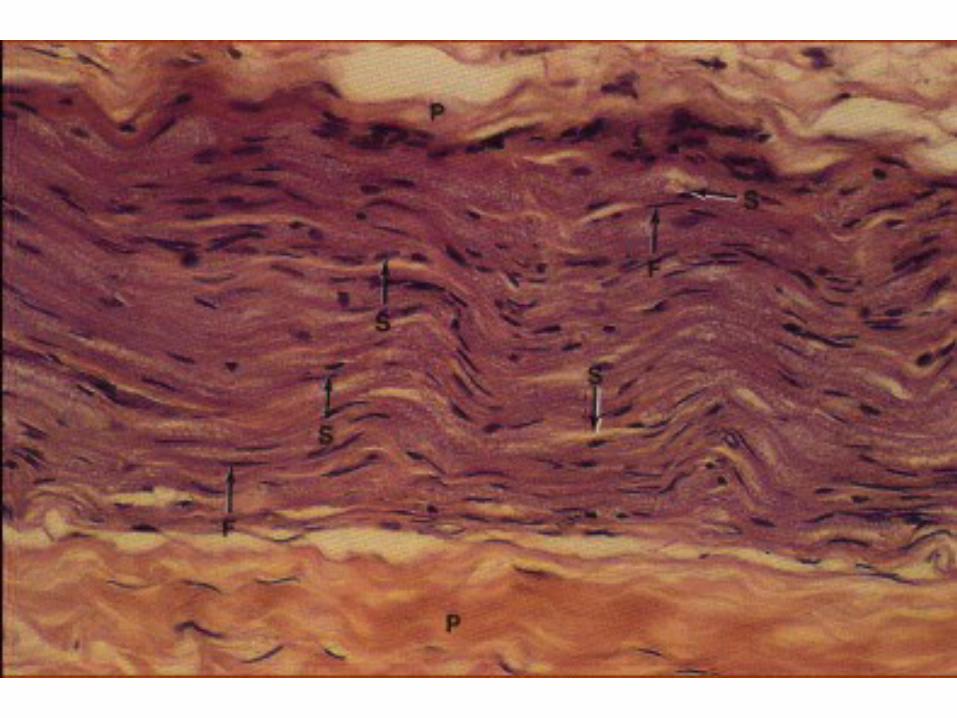
• Nerve fibers:
These are process of neurons and are
collected to form nerve trunk.
In longitudinal section: The nerve fiber
is seen to consist of the central axis cylinder
or axon arising from the neuron. It is
surrounding by layer myelin which consist
of phospholipids which acts as an insulator.

• It is interrupted at places. At these points,
the points are called nodes of Ranvier.
Outside the myelin sheath is a thin cell of
Schwann, which are neurilemma.The cells
of neurilemma are also known as cells of
Schwann, which are neuroectodermal in
origin. These cells are responsible for
laying down the myelin sheath of the
peripheral nerves.

Myelin sheath
Nodes of Ranvier
Axon
internode


施 -万切迹


• Transverse section
• epineurium: nerve truck
• perineurium: Each of fascicles
endoneurium:each nerve fibers.


Peripheral nerve ending

• Exteroceptive receptors:
Free Nerve Endings
Lamellated Corpuscles (of Pacini)
Tactile Corpuscles (of Meissner)
Proprioceptive receptors:
Muscle Spindles

When the terminals of sensory nerves do
not
show any particular specialization of
structure they are called free nerve
endings. Such endings are widely
distributed in the body: connective
tissue,epithelial lining of the skin, cornea,
alimentary canal, and respiratory system.


Lamellated Corpuscles (of Pacini) :
Pacinian corpuscles are circular or oval
structures. These are much larger than tactile
corpuscles. They may be up to 2 mm in length,
and up to 0.5 mm across. They are found in
the subcutaneous tissue of the palm and sole,
in the digits, and in various other situations.


Tactile Corpuscles (of Meissner) :
These are small oval or cylindrical structures
seen in relation to dermal papillae in the hand
and foot, and in some other situations. These
corpuscles are believed to be responsible for
touch.


Muscle Spindles
These are spindle-shaped sensory end organs
located within striated muscle .The spindle is
bounded by a fusiform connective tissue
covering (forming an external capsule)
within which there are a few muscle fibres of
a special kind. These are called intrafusal
fibers.

• Intrafusal fibers contain several nuclei that are located near the middle of the fiber.
nuclear bag fiber
nuclear chain fibers

• Motor end plate
In most neuromuscular junctions the nerve
terminal comes in contact with a specialized
area near the middle of the muscle fiber.
This area is roughly oval or circular, and is
referred to as the sole plate. The sole plate
plus the axon terminal constitute the motor
end plate.


Neurons vary considerably in the size and
shape of their cell bodies (somata) and in
the length and manner of branching of their
processes. The cell body varies in diameter
from about 5 um, in the smallest neurons, to
as much as 120 um in the largest ones.