Autonomic nervous system—arrangement, function, pain,visceral sensebility
Upload
georgina-porterCategory
view
235download
0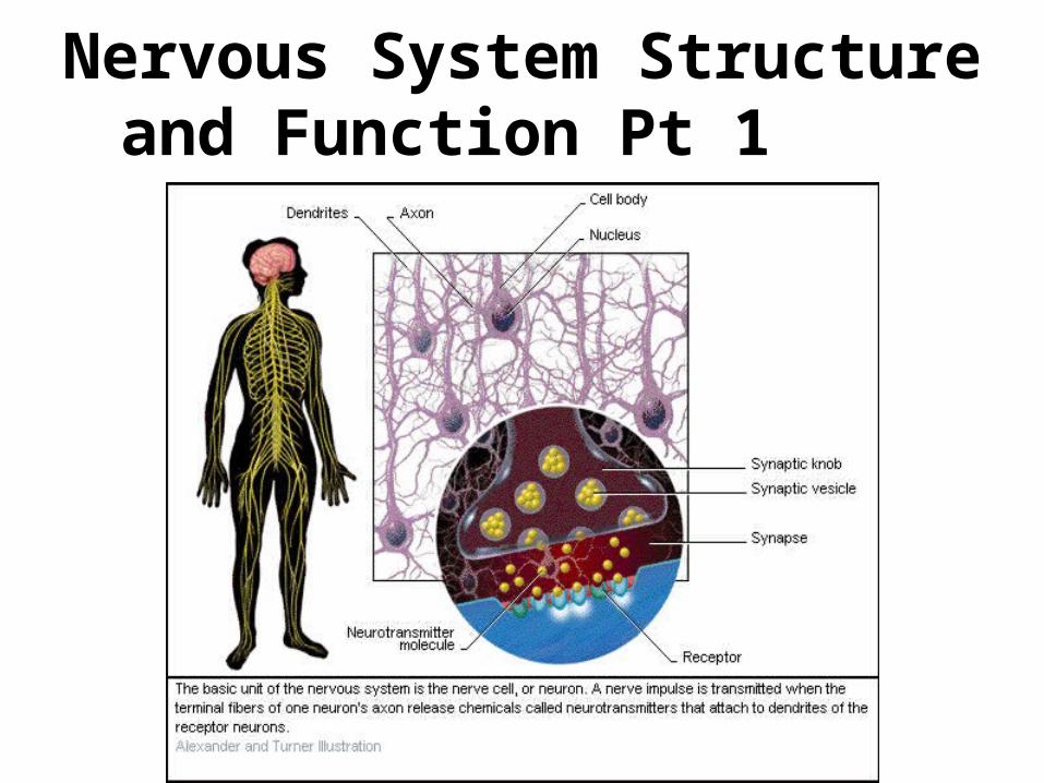
Nervous System Structure and Function Pt 1
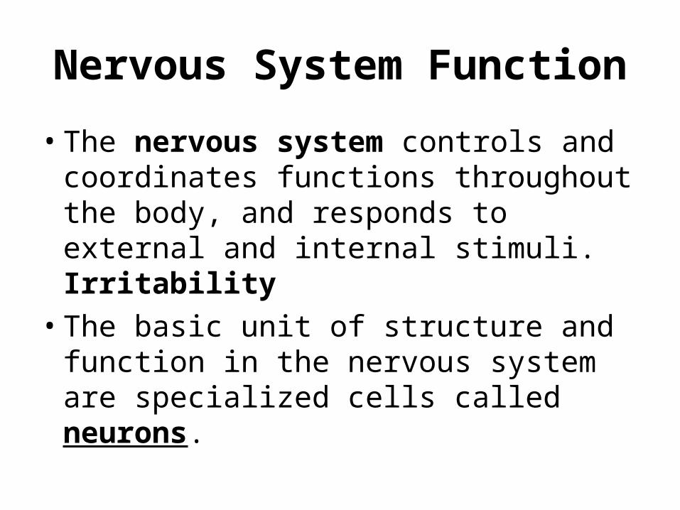
Nervous System Function
• The nervous system controls and coordinates functions throughout the body, and responds to external and internal stimuli. Irritability
• The basic unit of structure and function in the nervous system are specialized cells called neurons.

Neurons
• Neurons are specialized cells that can transmit electrical signals called impulses.
• Impulses are the messages carried by the nervous system.
• Neurons can be classified into three types
–Motor neurons
–Interneurons
–Sensory neurons

Neuron Structure
Axon Terminals
Node
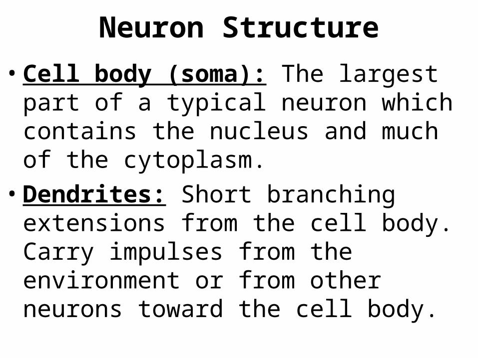
Neuron Structure
• Cell body (soma): The largest part of a typical neuron which contains the nucleus and much of the cytoplasm.
• Dendrites: Short branching extensions from the cell body. Carry impulses from the environment or from other neurons toward the cell body.

Neuron Structure
• Axon: Long fiber that carries impulses away from the cell body.
• Axon Terminals: Small swellings at the ends of axons.
• Myelin Sheath: Insulating membrane surrounding the axon of a neuron. Contains gaps called nodes that speed up the transmission of impulses.
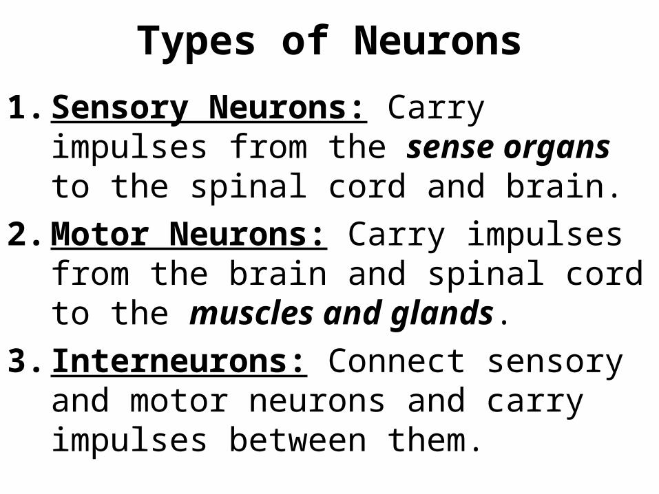
Types of Neurons
1. Sensory Neurons: Carry impulses from the sense organs to the spinal cord and brain.
2. Motor Neurons: Carry impulses from the brain and spinal cord to the muscles and glands.
3. Interneurons: Connect sensory and motor neurons and carry impulses between them.

Sensory and Motor Neurons
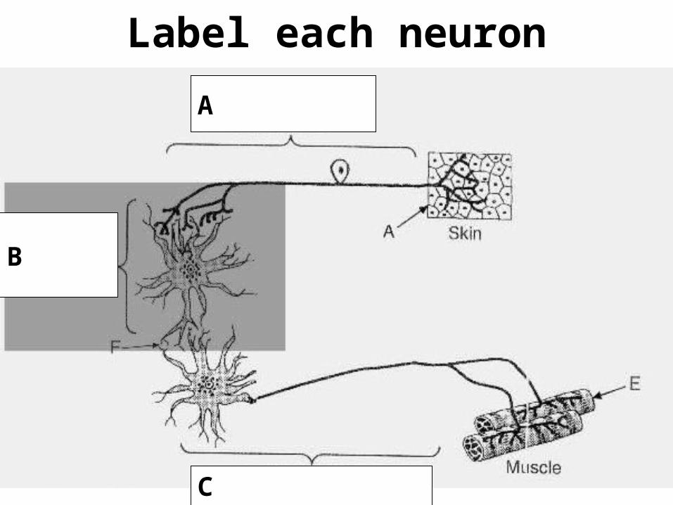
Label each neuron
A
C
B
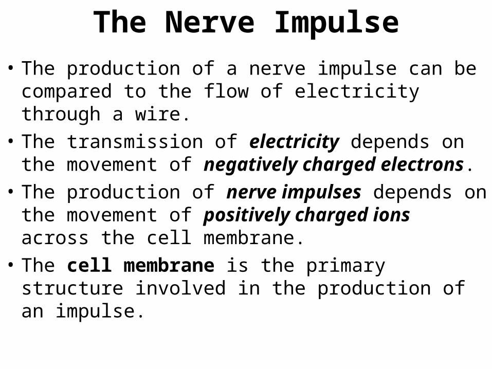
The Nerve Impulse
• The production of a nerve impulse can be compared to the flow of electricity through a wire.
• The transmission of electricity depends on the movement of negatively charged electrons.
• The production of nerve impulses depends on the movement of positively charged ions across the cell membrane.
• The cell membrane is the primary structure involved in the production of an impulse.

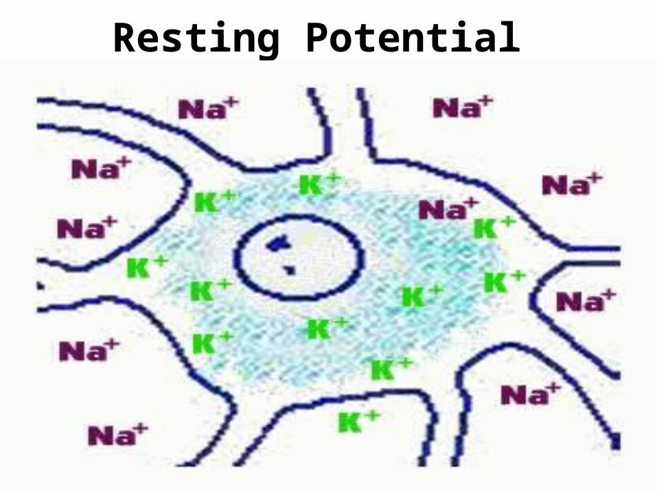
Resting Potential• The distribution of sodium (Na +) and
Potassium (K+) ions inside and outside of a neuron is shown in the following diagram.
• There are more potassium (K +) ions in the cytoplasm of the neuron than in the fluid outside of the cell.
• There are more sodium (Na +) ions in the fluid outside of the cell than inside the neuron.
Resting Potential
Resting Potential
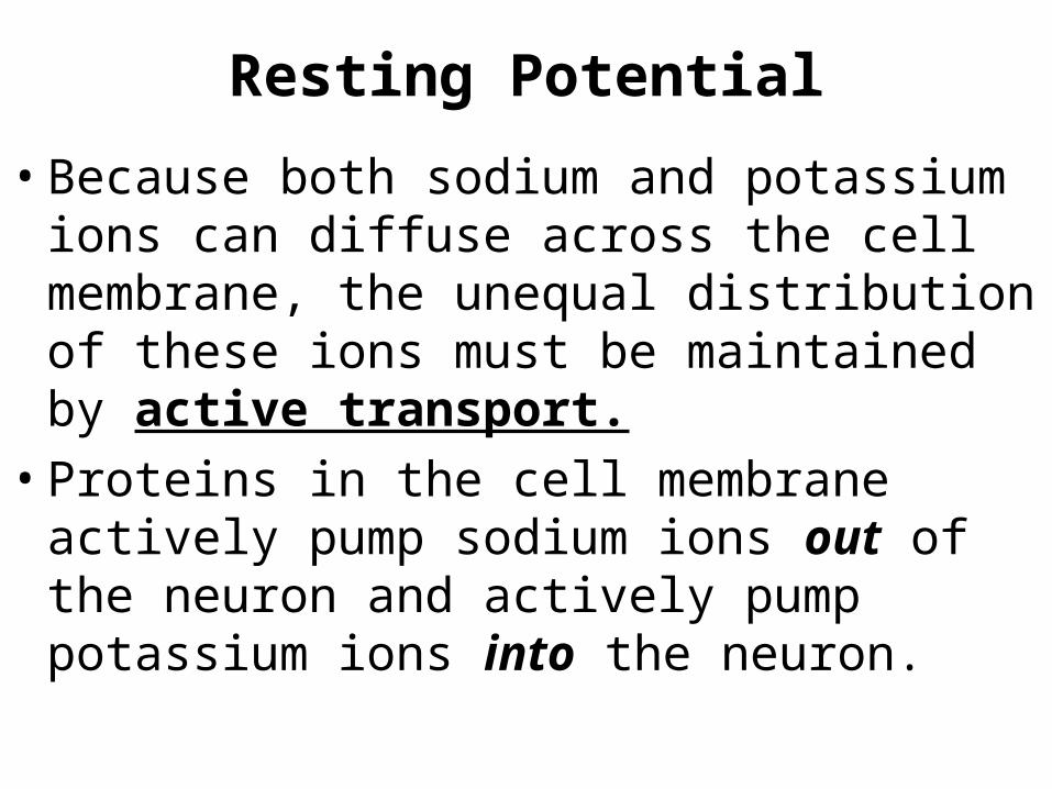
• Because both sodium and potassium ions can diffuse across the cell membrane, the unequal distribution of these ions must be maintained by active transport.
• Proteins in the cell membrane actively pump sodium ions out of the neuron and actively pump potassium ions into the neuron.

Sodium and Potassium Pumps Actively Transport Na+ and K+
Across the Cell Membrane
Resting Potential
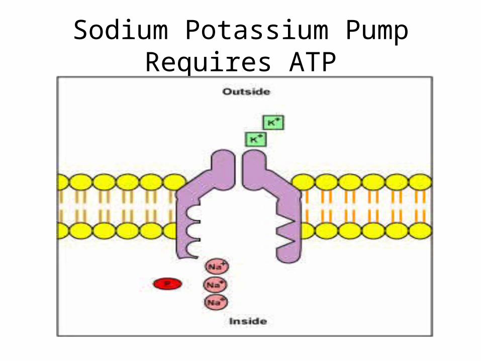
Sodium Potassium Pump Requires ATP
Resting Potential• As a result of active transport (K+ in,
Na+ out) and diffusion (K+ out, Na+ in), a negative charge builds up on the inside of the membrane and a positive charge builds up on the outside of the membrane.
• The difference in electrical charge across the cell membrane of a resting neuron is called is resting potential.
• A neuron has a resting potential of about -70 millivolts (mV)
The Moving Nerve Impulse
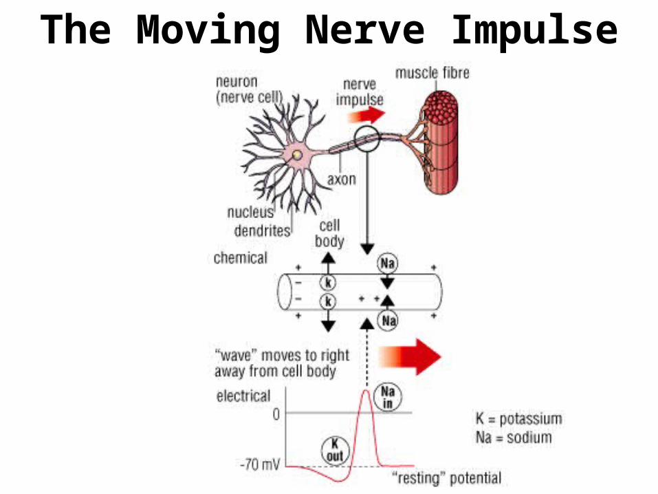
The Moving Nerve Impulse• In most animals, the axons and dendrites
of neurons are clustered into bundles of fibers called nerves.
• A nerve impulse is similar to the ripple caused when a pebble is dropped into a pond.
• The ripple is caused by the up and down movement of water. The impulse is caused by the movement of ions across the cell membrane.
The Moving Nerve Impulse• A nerve impulse begins when a neuron is
stimulated by another neuron or its environment.
• The nerve impulse travels along the axon, away from the cell body and toward the axon terminals.
• The cell membrane contains thousands of protein channels. Generally the sodium channels are closed.
The Moving Nerve Impulse• At the leading edge of an impulse, sodium
channels open allowing sodium ions to flow into the cell.
• This flow of positive ions causes a temporary change in the charges on the cell membrane.
• The inside of the membrane gains a positive charge and the outside of the membrane gains a negative charge.
• This reversal of charges across the membrane along the length of an axon is called an action potential.

Action Potentials
• A neuron has an action potential of about +30 mV.
• As the impulse passes through the axon, potassium channels open allowing K+ ions to flow out of the cell.
• The resting potential is now reestablished with the negative charge inside the membrane and the positive charge outside the membrane.

Action Potential• An action potential is caused by positive ions
moving in and then out of the neuron at a certain spot on the neuron membrane.
• An action potential is initiated by a stimulus above a certain intensity or threshold.
• Not all stimuli initiate an action potential. The stimulus could be a pin prick, light, heat, sound or an electrical disturbance in another part of the neuron.
• Action potential is an all or nothing mechanism, just like a mousetrap or stack of dominoes.

Action Potential: Depolarization
• Depolarization
• A stimulus causes a gate in the Na+ Channel to open. Since there is a high concentration of Na+ outside, Na+ diffuses into the neuron. The electrical potential changes to ~ +40 mV.

Action Potential - Repolarization
• Repolarization
• Depolarization causes the K+ Channel gate to immediately open. K+ diffuses out of the neuron. This reestablishes the initial electrical potential of ~-60 mV.

Action Potential – Refractory Period
• Refractory Period
• During this time (~ 1 msec), the Na+ and K+ Channels cannot be opened by a stimulus.
• The Na+/K+ Pump actively pumps Na+ out of the neuron and K+ into the neuron. This reestablishes the initial ion distribution of the resting neuron.
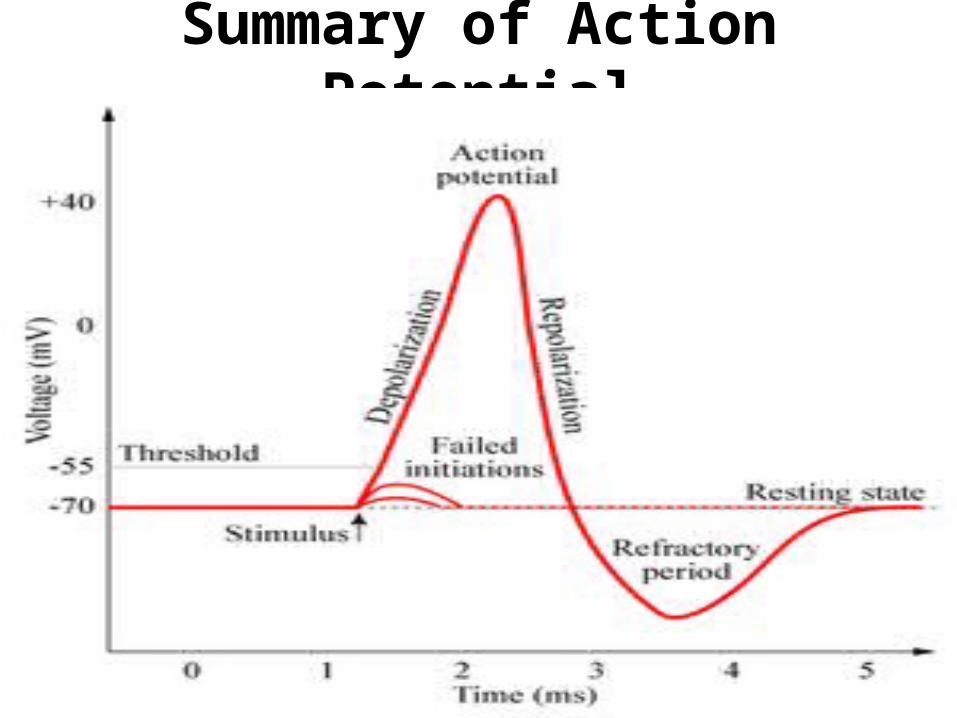
Summary of Action Potential

Action Potential• This single action potential acts as a stimulus to
neighboring proteins and initiates an action potential in another part of the neuron.
• Ultimately a wave of action potentials travels from the dendrites all the way to the axon terminals.
• At the axon terminal, the electrical impulse is converted to a chemical signal that stimulates a neighboring neuron. These chemical signals are called neurotransmitters.
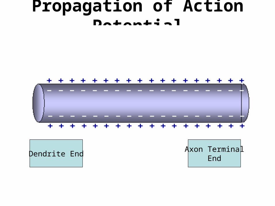
Propagation of Action Potential
Dendrite EndAxon Terminal
End
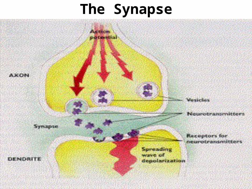
The Synapse• Synapse: Small space where a neuron
can transfer an impulse to another cell.
• It is a small gap that separates the axon terminal from the dendrites of the next neuron.
• The axon terminals contain tiny sacs filled with neurotransmitters.
• Neurotransmitters: Chemicals used by a neuron to transmit an impulse to another cell.

The Synapse• When an action potential arrives at an
axon terminal, the sacs release the neurotransmitters into the synapse.
• The neurotransmitter molecules diffuse across the synapse from one neuron to the next stimulating an impulse or action potential in the neighboring cell.
• Dopamine, seratonin, and acetylcholine are all neurotransmitters.

The Synapse
The Synapse
Cool sites for animations
http://www.blackwellpublishing.com/matthews/nmj.html (synapse and neurotransmitters)
http://www.blackwellpublishing.com/matthews/channel.html
(Action potentials)