Nervous system. Nervous system - organization Somatic NS Receptors IntegrationEffectors 5 senses...
69
Nervous system
-
date post
21-Dec-2015 -
Category
Documents
-
view
228 -
download
2
Transcript of Nervous system. Nervous system - organization Somatic NS Receptors IntegrationEffectors 5 senses...
Nervous system

Nervous system - organizationSomatic NS
Receptors Integration Effectors5 senses Cortex (& assoc.) Skeletal
muscles
Autonomic NSReceptors Integration EffectorsChemo-baro hypothalamus cardiac,Osmo- medulla oblongata smoothReceptors muscles
glands
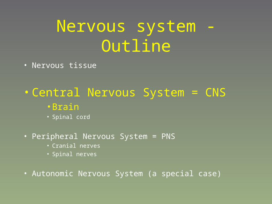
Nervous system - Outline
• Nervous tissue
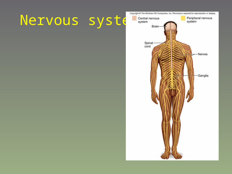
• Central Nervous System = CNS • Brain
• Spinal cord
• Peripheral Nervous System = PNS• Cranial nerves
• Spinal nerves
• Autonomic Nervous System (a special case)

Nervous system - Outline
• Nervous tissue
• Central Nervous System = CNS • Brain • Spinal cord
• Peripheral Nervous System = PNS• Cranial nerves• Spinal nerves
• Autonomic Nervous System (a special case)

Nervous tissue
• Neuron – main cell
• Neuroglia – supporting cells
• Astrocytes
• Microglia
• Oligodendrocytes
• Schwann cells
• Ependymal cells
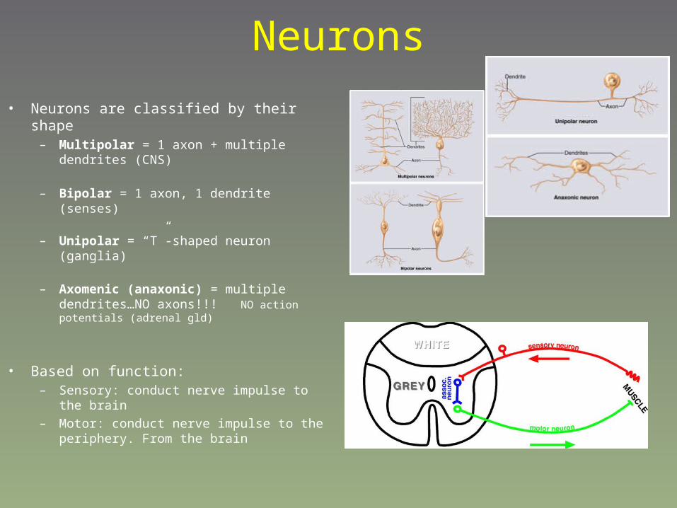
Neurons
• Neurons are classified by their shape– Multipolar = 1 axon + multiple dendrites (CNS)
– Bipolar = 1 axon, 1 dendrite (senses)
– Unipolar = “T”-shaped neuron (ganglia)
– Axomenic (anaxonic) = multiple dendrites…NO axons!!! NO action potentials (adrenal gld)
• Based on function:– Sensory: conduct nerve impulse to the brain
– Motor: conduct nerve impulse to the periphery. From the brain
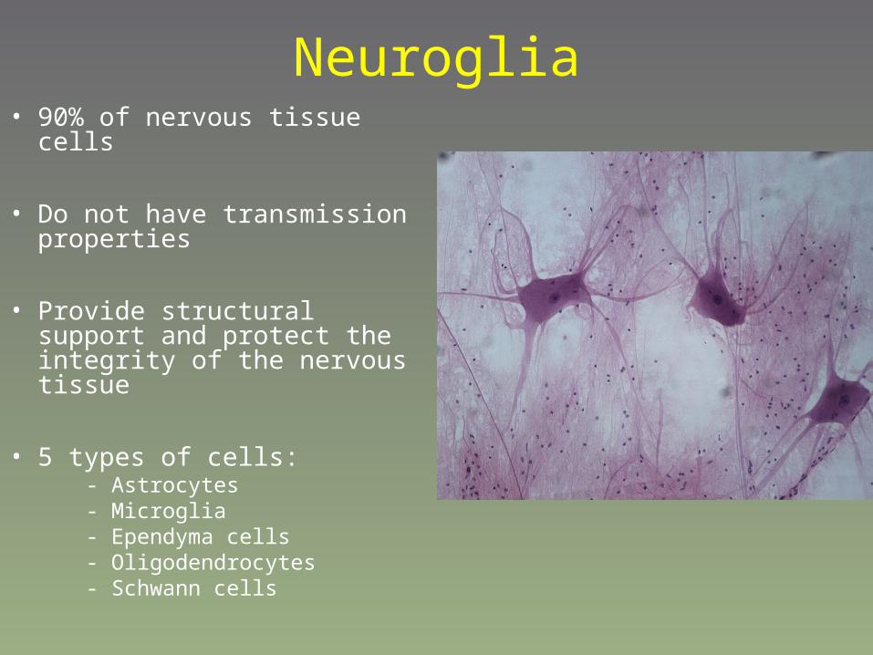
Neuroglia• 90% of nervous tissue cells
• Do not have transmission properties
• Provide structural support and protect the integrity of the nervous tissue
• 5 types of cells: - Astrocytes - Microglia - Ependyma cells - Oligodendrocytes - Schwann cells

Astrocytes
• Provide physical support for neurons• Control movements of nutrients and
wastes to and from neurons• Help recycle and process some
neurotransmitters• Play a role in the Blood-Brain Barrier
(BBB)• Play a role in synapse formation• Maintain constant brain ECF

Blood-brain barrier• Protects the brain from
“external” influences• Inability of some
compounds to cross from the blood into the brain
• Due to tight junctions between endocytes of he capillary wall
• Maybe due to astrocytes• Glucose, gases can pass• Some medications and
other compounds can not pass

Microglia
• Derived from macrophages
• Defense of the brain• Clean up old, sick and
dead cells

Ependymal cells
• Line the ventricles of the brain and form, with the blood vessels, the choroid plexuses
• Secrete the cerebrospinal fluid (CSF)
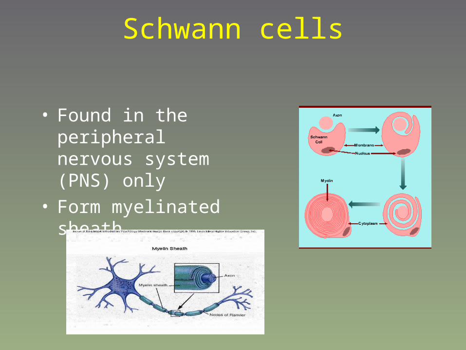
Schwann cells
• Found in the peripheral nervous system (PNS) only
• Form myelinated sheath
Oligodendrocytes
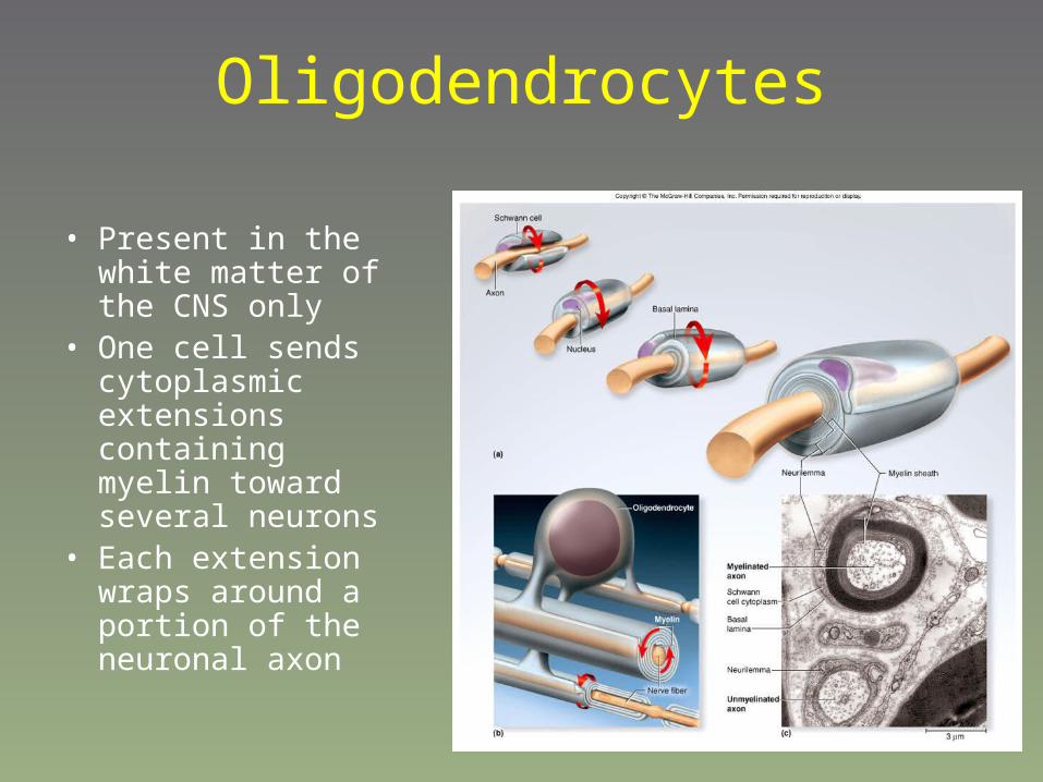
• Present in the white matter of the CNS only
• One cell sends cytoplasmic extensions containing myelin toward several neurons
• Each extension wraps around a portion of the neuronal axon

Myelin• The myelin sheath acts as an insulating layer
– Remember that in the CNS, myelin is formed by oligodendrites, and in the PNS, myelin = Schwann cells
• Myelination (the wrapping of neurons) starts during fetal development, BUT, does not complete until adolescence– Low fat diets for children contraindicated
• Myelin is high in phospholipids

Myelin• Why myelin?
– Allows for a fast response (10X faster than unmyelinated axon)
– Usually reserved for nerve fibers that innervate organs/tissues that are important for speed
• Not all neurons will be myelinated– If every neuron were myelinated, the neural mass would be
far too big (myelin adds mass to the neuron)
– “slow” unmyelinated fibers are fine for areas where you don’t really need an instant response
• Dilating pupils, secreting stomach acid etc.

The classic “myelinated neuron” vs. the “confusing” ensheathed
neuron. Un-myelinated neurons are not truly “bald”, but are
ensheathed in a Schwann cell as shown to the right
Ensheathed or “unmyelinated
neurons”
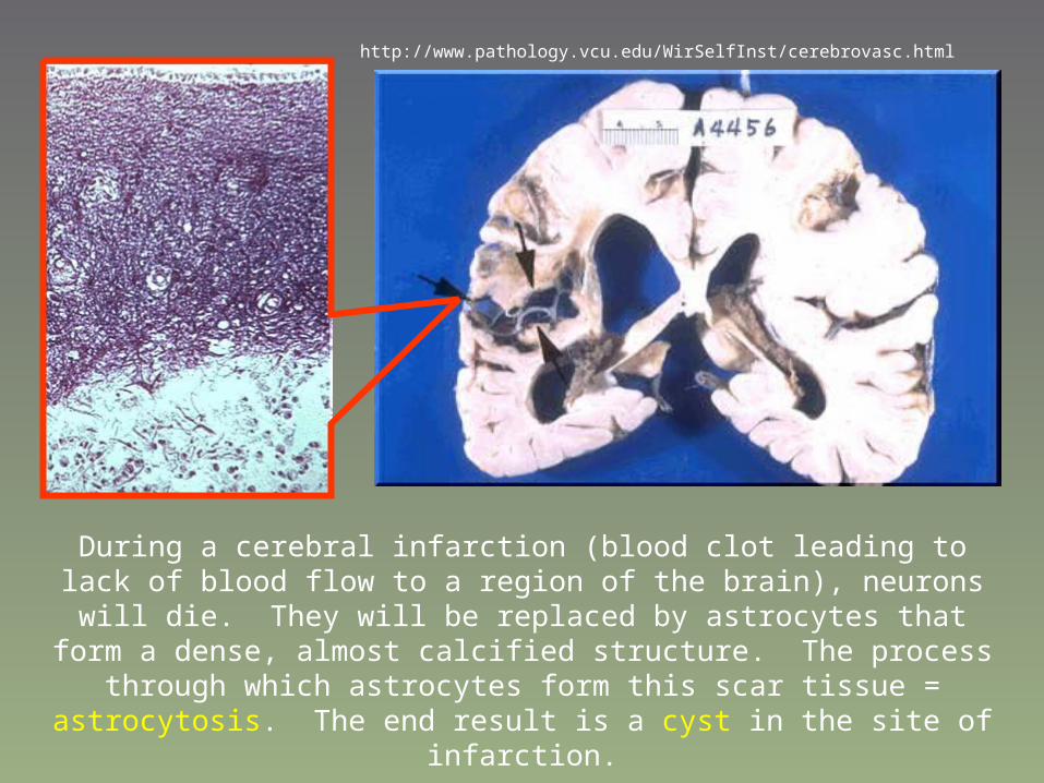
http://www.pathology.vcu.edu/WirSelfInst/cerebrovasc.html
During a cerebral infarction (blood clot leading to lack of blood flow to a region of the brain), neurons will die. They will be replaced by astrocytes that form a
dense, almost calcified structure. The process through which astrocytes form this scar tissue = astrocytosis. The end result is a cyst in the site of infarction.
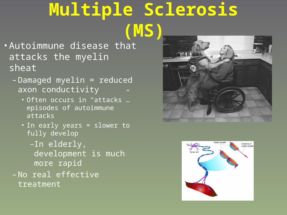
Multiple Sclerosis (MS)• Autoimmune disease that
attacks the myelin sheat– Damaged myelin = reduced
axon conductivity• Often occurs in “attacks”…
episodes of autoimmune attacks
• In early years = slower to fully develop
–In elderly, development is much more rapid
– No real effective treatment
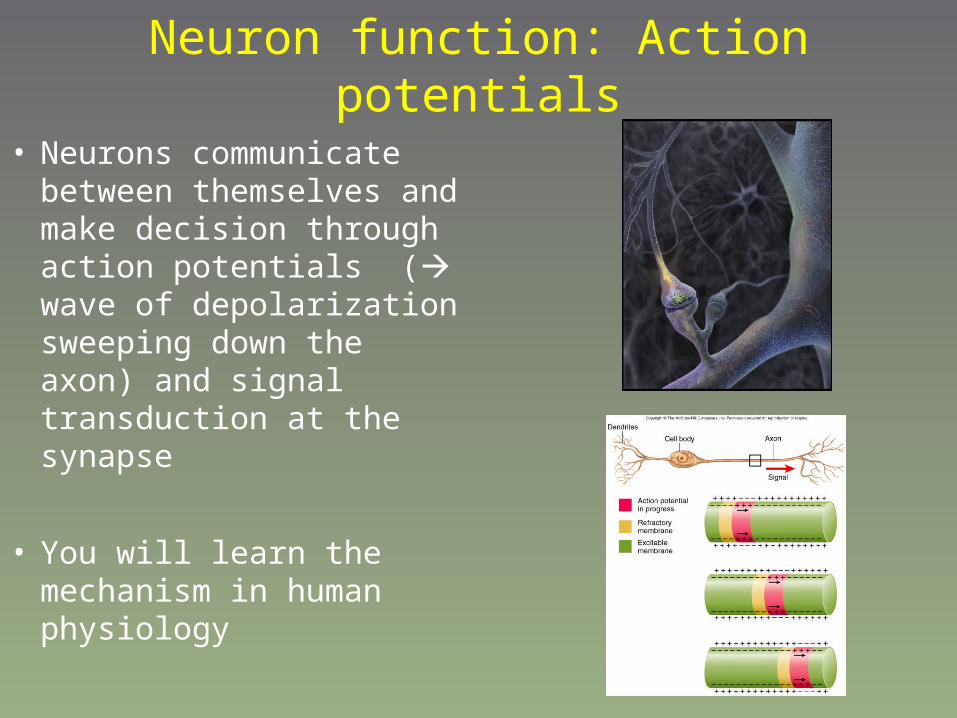
Neuron function: Action potentials
• Neurons communicate between themselves and make decision through action potentials ( wave of depolarization sweeping down the axon) and signal transduction at the synapse
• You will learn the mechanism in human physiology

Patterns of connections between neurons
Nervous system - Outline
• Nervous tissue
• Central Nervous System = CNS • Brain • Spinal cord
• Peripheral Nervous System = PNS• Cranial nerves• Spinal nerves
• Autonomic Nervous System (a special case)
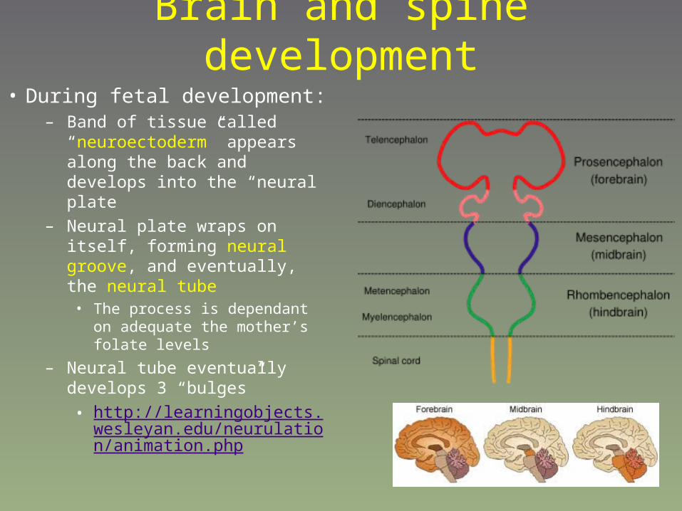
Brain and spine development
• During fetal development:– Band of tissue called
“neuroectoderm” appears along the back and develops into the “neural plate
– Neural plate wraps on itself, forming neural groove, and eventually, the neural tube
• The process is dependant on adequate the mother’s folate levels
– Neural tube eventually develops 3 “bulges”
• http://learningobjects.wesleyan.edu/neurulation/animation.php
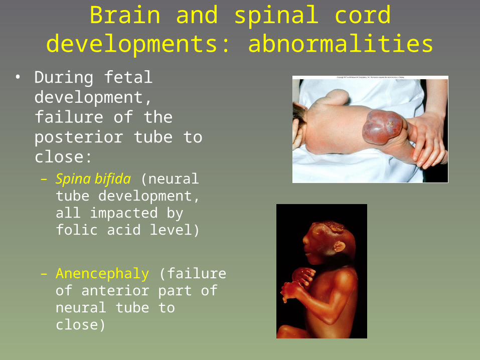
Brain and spinal cord developments: abnormalities
• During fetal development, failure of the posterior tube to close:– Spina bifida (neural tube
development, all impacted by folic acid level)
– Anencephaly (failure of anterior part of neural tube to close)

The Brain

The brain• General considerations
– Meninges
– Cerebrospinal fluid
– Brain matter
• Forebrain– Cerebrum
– Diencephalon
– Limbic system
• Brainstem– Midbrain
– Pons
– Medulla oblongata
– Cerebellum
– Reticular formation

The brain• General considerations
– Meninges– Cerebrospinal fluid– Brain matter
• Forebrain– Cerebrum– Diencephalon– Limbic system
• Brainstem– Midbrain– Pons– Medulla oblongata– Cerebellum
– Reticular formation

Brain - Meninges
• Like in the spinal cord: 3 layers– Dura mater– Arachnoid– Pia mater
• Spaces between:– Cranium-dura mater = no space– Dura mater-arachnoid = subdural
space -> connective tissues, blood sinuses
– Arachnoid-pia mater = subarachnoid space CSF
– Pia mater – brain = no space

The brain• General considerations
– Meninges
– Cerebrospinal fluid– Brain matter
• Forebrain– Cerebrum– Diencephalon– Limbic system
• Brainstem– Midbrain– Pons– Medulla oblongata– Cerebellum
– Reticular formation

Brain – Ventricles and CSF• Ventricles: chambers within the brain
– 4 ventricles:• 2 lateral
• “third ventricle”
• “fourth ventricle”
• Ventricles are connected: -Interventricular foramen connect the 2 lateral ventricles and third ventricle
– Cerebral aqueduct connect third and fourth ventricles
– The lateral apertures allow circulation of the CSF into the subarachnoid space
– The median aperture connect fourth ventricle to the central canal, into the spinal cord

Cerebrospinal fluid - CSF• Three main functions:
– Maintains brain at neutral buoyancy (brain matter is too soft)
– Protects the brain from striking the cranium
– Chemical stability• CSF acts to wash out the
metabolic wastes of the CNS into the blood supply

CSF• Secreted by the ependymal cells
lining the ventricles. With connective tissue and blood vessels, they form the choroid plexus
• Secreted CSF travels through the ventricle and exits into the subarachnoid space.
• CSF is reabsorbed by the arachnoid granulations, located in the arachnoid meninges
The brain• General considerations
– Meninges– Cerebrospinal fluid
– Brain matter
• Forebrain– Cerebrum– Diencephalon– Limbic system
• Brainstem– Midbrain– Pons– Medulla oblongata– Cerebellum
– Reticular formation

Brain – Grey and white matter
• Grey matter : – body of neurons +
unmyelinated axons• Surface cortex
• Deep nuclei (clump of grey matter)
• White matter – within the grey matter,
made of bundles of nerve fibers/tracts relaying information from 1 part of the brain to another

Brain – Grey matter• Integration of sensory input and
output
• Cerebral cortex (surface of the brain)• 6 layers in human form the neocortex: I,
II, III, IV, V, and VI
• In nuclei
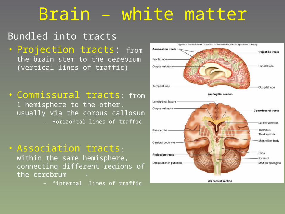
Brain – white matterBundled into tracts• Projection tracts: from the brain
stem to the cerebrum (vertical lines of traffic)
• Commissural tracts: from 1 hemisphere to the other, usually via the corpus callosum
– Horizontal lines of traffic
• Association tracts: within the same hemisphere, connecting different regions of the cerebrum
– “internal” lines of traffic
The brain• General considerations
– Meninges
– Cerebrospinal fluid
– Brain matter
• Forebrain– Cerebrum– Diencephalon
– Limbic system
• Brainstem– Midbrain
– Pons
– Medulla oblongata
– Cerebellum
– Reticular formation
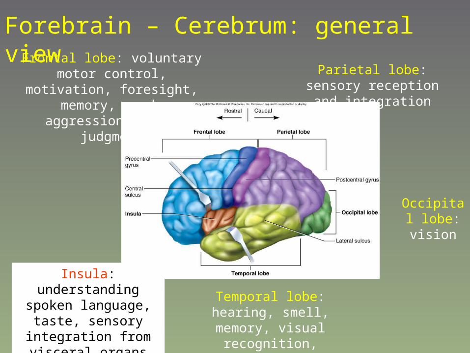
Frontal lobe: voluntary motor control, motivation, foresight,
memory, mood, aggression/social judgment
Parietal lobe: sensory reception and integration
Occipital lobe: vision
Temporal lobe: hearing, smell, memory, visual recognition,
emotions
Insula: understanding spoken language, taste,
sensory integration from visceral organs
Forebrain – Cerebrum: general view

Cerebrum
• General considerations
• The lobes– Frontal lobes
– Parietal lobes
– Temporal lobes
– Occipital lobes
• Basal ganglia

Cerebrum
• General considerations
• The lobes– Frontal lobes
– Parietal lobes
– Temporal lobes
– Occipital lobes
• Basal ganglia

Brain anatomy
• cerebrum– Right and left sides
(hemispheres), separated by “longitudinal fissure”
– They’re not truly separate; deep to the fissure is a connection between both sides = corpus callosum
– Other landmarks:• Central sulcus divides the
frontal lobe of the cerebrum from the parietal lobe
• Most dorsal (caudal), right above the cerebellum = occipital lobe

Cerebrum
• General considerations
• The lobes– Frontal lobes
– Parietal lobes
– Temporal lobes
– Occipital lobes
• Basal ganglia
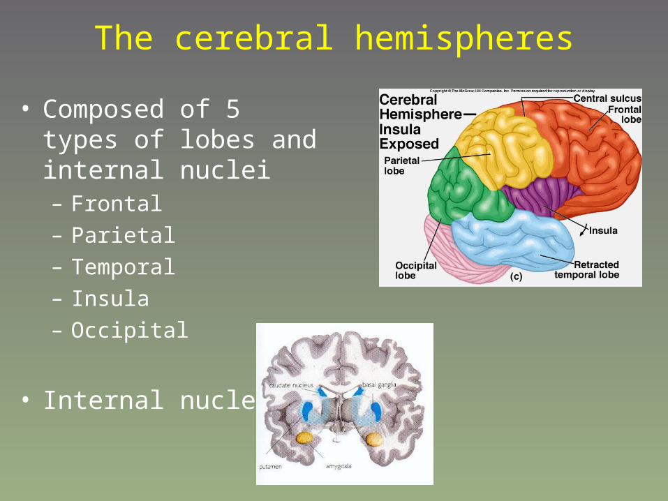
The cerebral hemispheres
• Composed of 5 types of lobes and internal nuclei– Frontal
– Parietal
– Temporal
– Insula
– Occipital
• Internal nuclei:

Frontal lobeMany functions:
- personality
- Problem solving
- motor regions of the brain– In fact, they’re adjacent to the
respective sensory areas, on the pre-central gyrus
– Thus, you can also create a motor humunculus
• Signals from these regions travel to the brain stem
– Decussate in the neck and onward to their target muscle groups
• The basal nuclei are important for coordinating motor function
– Feedback signals of muscle movements (from the various muscle spindle
formations)
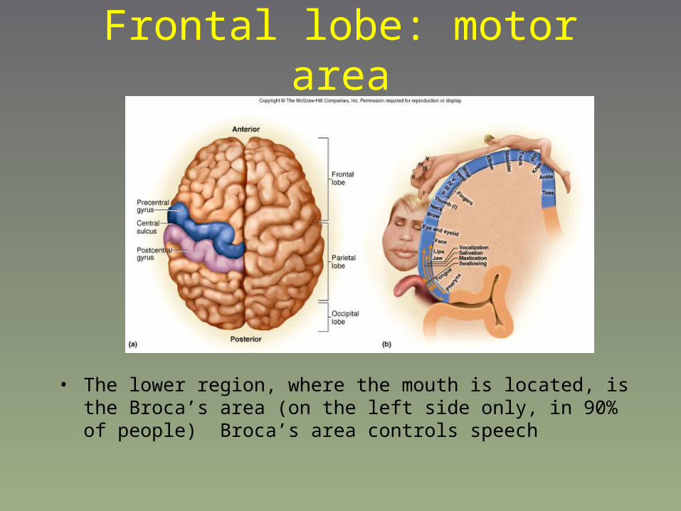
Frontal lobe: motor area
• The lower region, where the mouth is located, is the Broca’s area (on the left side only, in 90% of people) Broca’s area controls speech
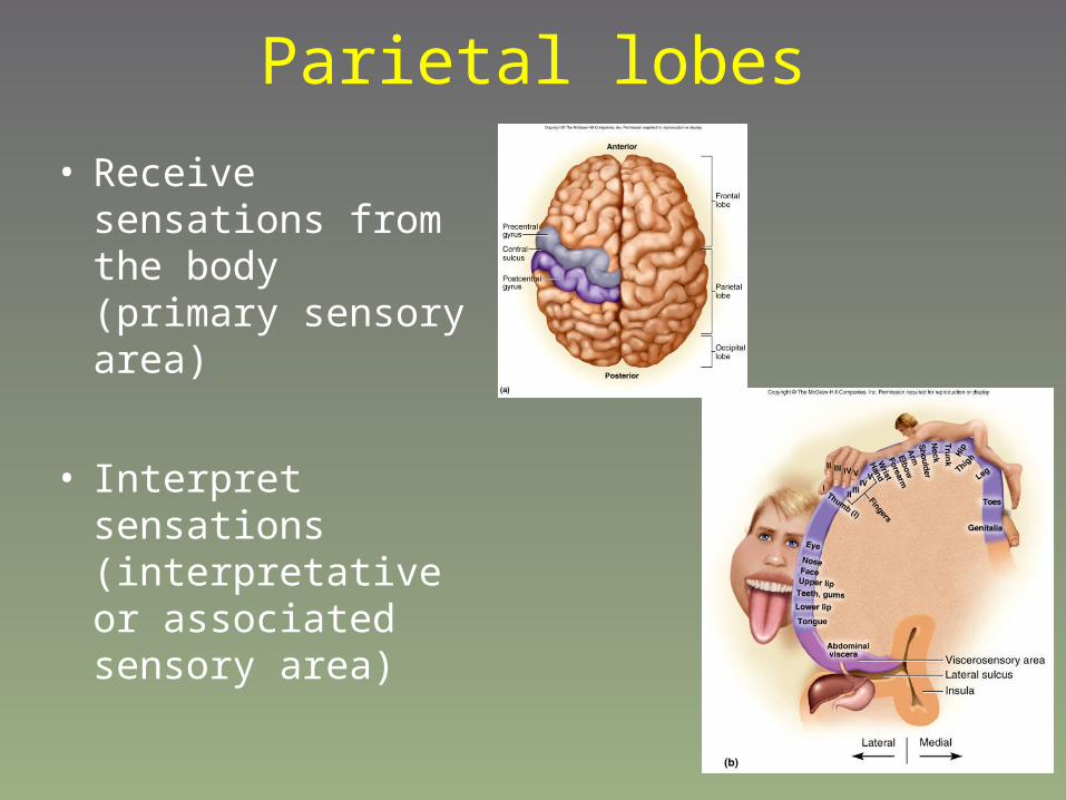
Parietal lobes
• Receive sensations from the body (primary sensory area)
• Interpret sensations (interpretative or associated sensory area)
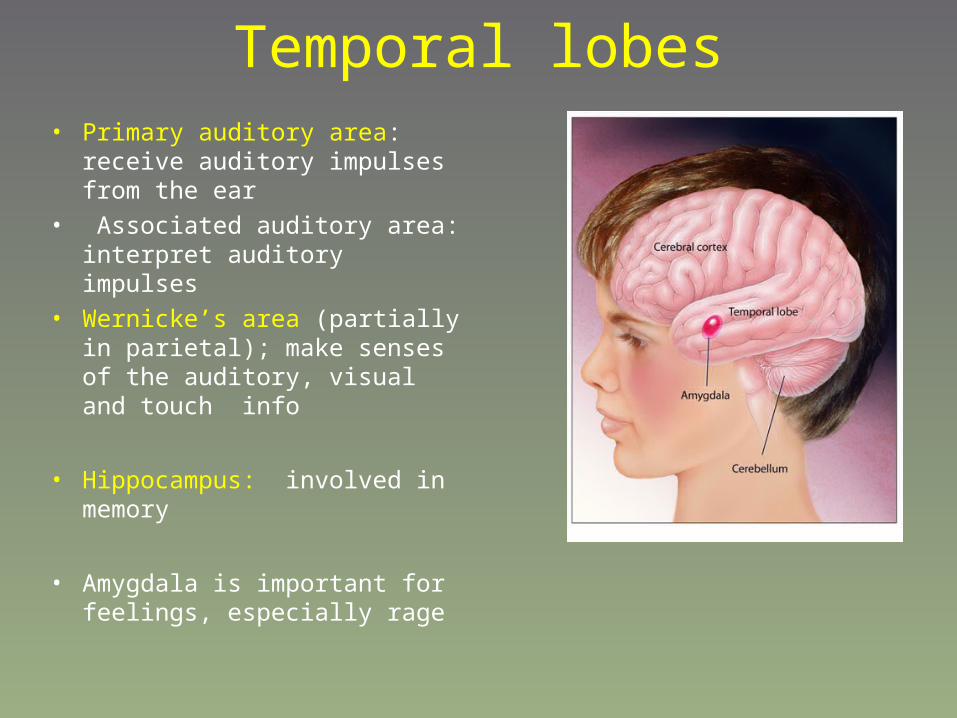
Temporal lobes• Primary auditory area: receive
auditory impulses from the ear
• Associated auditory area: interpret auditory impulses
• Wernicke’s area (partially in parietal); make senses of the auditory, visual and touch info
• Hippocampus: involved in memory
• Amygdala is important for feelings, especially rage
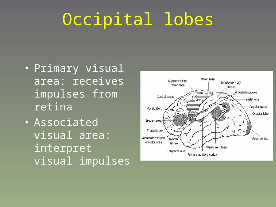
Occipital lobes
• Primary visual area: receives impulses from retina
• Associated visual area: interpret visual impulses
Cerebrum
• General considerations
• The lobes– Frontal lobes
– Parietal lobes
– Temporal lobes
– Occipital lobes
• Basal ganglia

Basal nuclei
• Buried deep into the brain matter
• There are at least 3 “nuclei” or neural control centers
• These nuclei are involved in relaying neural signals from the substantia nigra region of the midbrain (on the brainstem, but a separate region of the brain)
• Also relay signals from the motor centers of the cerebral cortex
• Help control smoothness of movement, muscle tension and posture

The basal nuclei consists of at least 3 regions of the deep cerebrum: Caudate
nucleus, Putamen and Globus palidus (all 3 together are known as the corpus striatum)
The brain• General considerations
– Meninges
– Cerebrospinal fluid
– Brain matter
• Forebrain– Cerebrum
– Diencephalon– Limbic system
• Brainstem– Midbrain
– Pons
– Medulla oblongata
– Cerebellum
– Reticular formation

Diencephalon
• Thalamus• Hypothalamus• Epithalamus

Thalamus• Egg-shaped mass beneath the ventricles
– At the top of the brain stem
• Acts as the “gateway” to the cerebral cortex– Almost all information destined for the
cerebrum will pass through the thalamus
• Provides a feedback loop for motor control
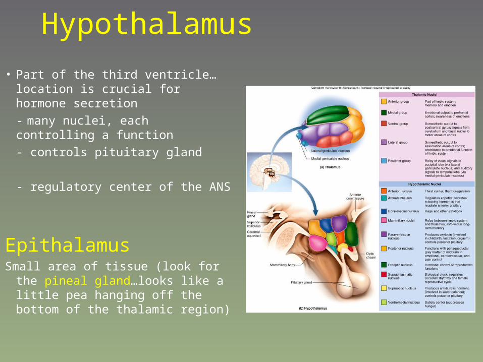
Hypothalamus
• Part of the third ventricle…location is crucial for hormone secretion
- many nuclei, each controlling a function
- controls pituitary gland
- regulatory center of the ANS
EpithalamusSmall area of tissue (look for the pineal
gland…looks like a little pea hanging off the bottom of the thalamic region)
The brain• General considerations
– Meninges– Cerebrospinal fluid– Brain matter
• Forebrain– Cerebrum
– Diencephalon
– Limbic system
• Brainstem– Midbrain– Pons– Medulla oblongata– Cerebellum
– Reticular formation

Limbic systemFor emotion and learning
– The “cingulate gyrus” or fold over the corpus callosum
– Hippocampus
– Amygdala
– Sense of pleasure or reward
– Feeling of fear, sadness, rage

The brain• General considerations
– Meninges
– Cerebrospinal fluid
– Brain matter
• Forebrain– Cerebrum
– Diencephalon
– Limbic system
• Brainstem– Midbrain– Pons
– Medulla oblongata
– Cerebellum
– Reticular formation

Midbrain• cerebral peduncles: nerve fibers connecting
cerebrum to brainstem
• tegmentum” = main mass
• Very red because of high amount of blood vessels
• substantia nigra”• Inhibitory motor control, sends signals
to thalamus and “basal nuclei”
• Degeneration here leads to Parkinson’s disease
• Mammillary bodies
– Superior colliculi: visual reflexes
– Inferior colliculi: auditory reflexes
• Origin for cranial nerves III and IV

The brain• General considerations
– Meninges
– Cerebrospinal fluid
– Brain matter
• Forebrain– Cerebrum
– Diencephalon
– Limbic system
• Brainstem– Midbrain
– Pons– Medulla oblongata
– Cerebellum
– Reticular formation
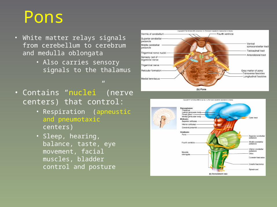
Pons• White matter relays signals from
cerebellum to cerebrum and medulla oblongata
• Also carries sensory signals to the thalamus
• Contains “nuclei” (nerve centers) that control:
• Respiration (apneustic and pneumotaxic centers)
• Sleep, hearing, balance, taste, eye movement, facial muscles, bladder control and posture

The brain• General considerations
– Meninges
– Cerebrospinal fluid
– Brain matter
• Forebrain– Cerebrum
– Diencephalon
– Limbic system
• Brainstem– Midbrain
– Pons
– Medulla oblongata– Cerebellum
– Reticular formation

Medulla oblongata• 2 ridges = “pyramids”
• Corticospinal tracts connection from the brain to the spine
– Site of nuclei (nerve centers) that control:
• heart rate• blood pressure• Respiration• Many other functions:
coughing, sneezing, salivation, swallow/gagging, intestinal secretions, sweating, speech and many head and tongue movements

Medulla oblongata• Most nerve fibers decussate
in the medulla oblongata cross over left side of the brain controls right side of body and vice versa

The brain• General considerations
– Meninges
– Cerebrospinal fluid
– Brain matter
• Forebrain– Cerebrum
– Diencephalon
– Limbic system
• Brainstem– Midbrain
– Pons
– Medulla oblongata
– Cerebellum– Reticular formation

Cerebellum• Largest part of the hindbrain
– 2 hemispheres, connected by “vermis” structure
• Folds (“folia”= gyri)
– Cross-section shows white matter arranged like roots = “arbor vitae” within grey matter “cortex”
• Within the white matter are “nuclei” (nerve centers)
– 4 per hemisphere
– Any input into the cerebellum goes to the cortex, all output comes from the “nuclei”
– Exchanges info with the brain stem to the brainstem

Cerebellum• Major functions:
– Evaluates sensory input and coordinates motor response
– Body equilibrium
– Also involved in spatial reasoning (where you are in traffic)
• Tactile reasoning (close your eyes, try to dial phone or type)
– Time judgement (can you make the light…why has this red light taken so long…)

The brain• General considerations
– Meninges– Cerebrospinal fluid– Brain matter
• Forebrain– Cerebrum– Diencephalon– Limbic system
• Brainstem– Midbrain– Pons– Medulla oblongata– Cerebellum
– Reticular formation
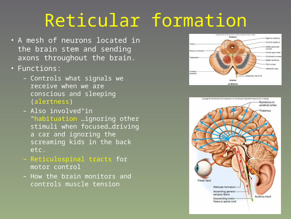
Reticular formation• A mesh of neurons located in the brain
stem and sending axons throughout the brain.
• Functions:– Controls what signals we receive when
we are conscious and sleeping (alertness)
– Also involved in “habituation”…ignoring other stimuli when focused…driving a car and ignoring the screaming kids in the back etc.
– Reticulospinal tracts for motor control
– How the brain monitors and controls muscle tension

Nervous system - Outline
• Nervous tissue
• Central Nervous System = CNS • Brain
• Spinal cord
• Peripheral Nervous System = PNS• Cranial nerves
• Spinal nerves
• Autonomic Nervous System (a special case)