Nervous System Anatomy: Neuron Neuron Soma Dendrites Axon Axolemma Axoplasm Axon Terminal ...
17
Nervous System Anatomy: Neuron • Neuron Soma Dendrites Axon Axolemma Axoplasm Axon Terminal Synaptic End Bulb Axon Hillock Trigger Zone Schwann Cell Myelin Sheath Node of Ranvier
-
Upload
melvyn-jacobs -
Category
Documents
-
view
467 -
download
12
Transcript of Nervous System Anatomy: Neuron Neuron Soma Dendrites Axon Axolemma Axoplasm Axon Terminal ...
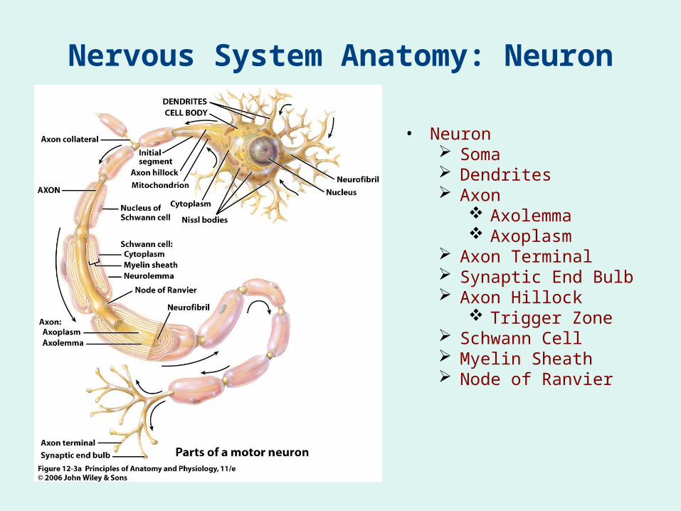
Nervous System Anatomy: Neuron
• Neuron Soma Dendrites Axon
Axolemma Axoplasm
Axon Terminal Synaptic End Bulb Axon Hillock
Trigger Zone Schwann Cell Myelin Sheath Node of Ranvier

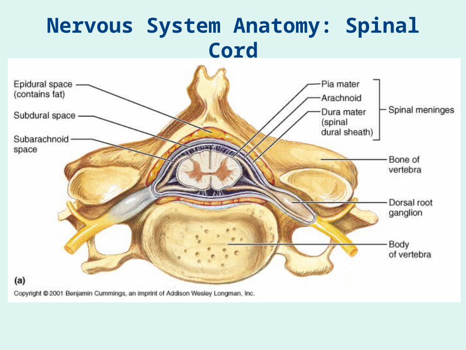
Nervous System Anatomy: Spinal Cord
• Gray Matter• White Matter• Meninges
Dura Mater (Outer) Subdural Space
Arachnoid Mater Subarachnoid
Space Denticulate
Ligaments Pia Mater (Inner)
Nervous System Anatomy: Spinal Cord

Nervous System Anatomy: Spinal Cord
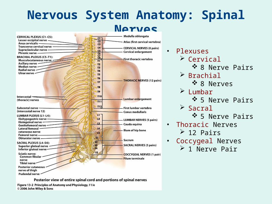
Nervous System Anatomy: Spinal Nerves
• Plexuses Cervical
8 Nerve Pairs Brachial
8 Nerves Lumbar
5 Nerve Pairs Sacral
5 Nerve Pairs• Thoracic Nerves
12 Pairs• Coccygeal Nerves
1 Nerve Pair

Nervous System Anatomy: Spinal Column
• Horns: Gray & White, Anterior & Posterior• Commisures: Gray & White, Anterior & Posterior• Roots: Ventral & Dorsal• Fissures & Sulci: Ventral & Dorsal• Columns: Lateral, Anterior & Posterior• Central Canal

Nervous System Anatomy
• Ventral & Dorsal Ramus
• Rami Communicantes

Nervous System Anatomy

Nervous System Anatomy

Nervous System Anatomy
Cervical Plexus
• Most major nerves occur on both right and left sides
• Most major nerves carry both motor and sensory information, although they may be mostly one or the other
• Phrenic n. Innervates the diaphragm Emerges C3 - C5 Damage to the spinal cord
above this point causes respiratory failure
• Supraclavicular n. Deep to the platysma Innervates skin in the neck,
shoulder, & upper chest

Nervous System Anatomy
Brachial Plexus
• Axillary n. Innervates
deltoid & teres minor
• Suprascapular n. Innervates
supraspinatus & infraspinatus

Nervous System Anatomy
Brachial Plexus
• Radial n. Innervates extensors
of arm & forearm
• Median n. Innervates flexors of
forearm
• Ulnar n. Innervates flexors of
wrist & hand
• Musculocutaneous n. Innervates
coracobrachialis, biceps brachii, & brachialis

Nervous System Anatomy
Lumbar Plexus
• Genitofemoral n. Innervates skin over
genitals & thighs
• Ilioinguinal n. Innervates skin over
genitals & thighs
• Femoral n. Innervates flexors of
thigh & extensors of leg
• Saphenous n. Branch of the Femoral n. Innervates skin over
medial surface of leg
• Obturator n. Innervates adductors of
leg

Nervous System Anatomy
Sacral Plexus
• Pudendal n. Innervates muscles of
the perineum & skin of the genitals
• Sciatic n. Largest nerve in the
body Splits into tibial &
common peroneal nerves
Innervates hamstrings & adductors
• Tibial n. Splits into lateral &
medial plantar nerves Innervates
gastrocnemius, soleus & other lower leg muscles

Nervous System Anatomy
Sacral Plexus
• Common peroneal n. Splits into superficial &
deep peroneal nerves Also known as the
common fibular nerve Innervates tibialis
anterior, peroneus (fibularis) longus & brevis, & other muscles of the lower leg & foot
• Sural n. Formed from branches of
the Tibial & Common Peroneal nn.
Innervates skin of lower leg & foot
• Medial & Lateral Plantar nn.
Innervate muscles of the foot

Nervous System Anatomy
Branches of the Trigeminal Nerve(Cranial Nerve V)
• The trigeminal nerve is Cranial Nerve V
Largest cranial nerve, with three major divisions
Many branches that provide somatic sensory information from the head & face
Also has motor output to muscles of mastication
• Infraorbital n.• Supraorbital n.• Lingual n.• Mental n.
Nervous System Anatomy
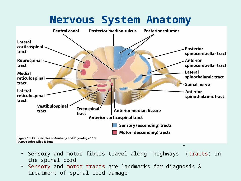
• Sensory and motor fibers travel along “highways” (tracts) in the spinal cord
• Sensory and motor tracts are landmarks for diagnosis & treatment of spinal cord damage