Nervous. Function command unit provides senses sends signal between body and brain Responds to...
31
Nervous
-
Upload
franklin-curtis -
Category
Documents
-
view
217 -
download
0
Transcript of Nervous. Function command unit provides senses sends signal between body and brain Responds to...

Nervous

Functioncommand unit provides sensessends signal between body and brainResponds to internal and external stimuli

NeuronDendrite- impulse
passes here 1st
Cell BodyAxon- covered in
myelin- acts as insulator, “white matter”-end at axon terminal

3 types of neurons
Sensory- carries impulse from body to brain or spinal cord
Motor- carries impulse from brain to muscle or gland cell(target cell)
Interneuron- connects brain to spinal cord

Impulse moves dendrite cell body axon target cell
How does impulse move along axon?
Neurons have a charge(-70 mv) on the cell membrane.This is due to more + ions on the outside of the cell than on the inside.
Neurons are excitable- charge can changeThis is called action potential- ability of
membrane to or charge.

Normal conditions of cell
More
K+inside
More Na+ outsideThis is the resting
potential of a cellThe cell is
Polarized because of the charge difference.
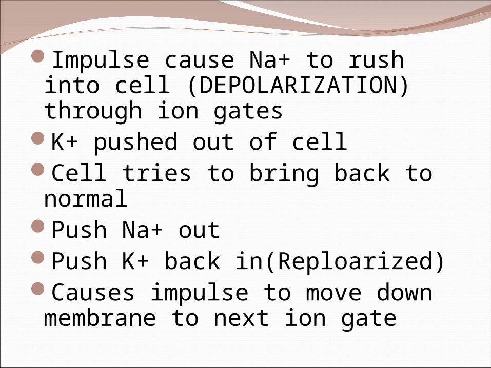
Impulse cause Na+ to rush into cell (DEPOLARIZATION) through ion gates
K+ pushed out of cellCell tries to bring back to normal
Push Na+ outPush K+ back in(Reploarized)Causes impulse to move down membrane to next ion gate

How does impulse travel so quickly?Axon covered with
an insulator –MYELIN- “White matter”
Impulse jumps from node to node
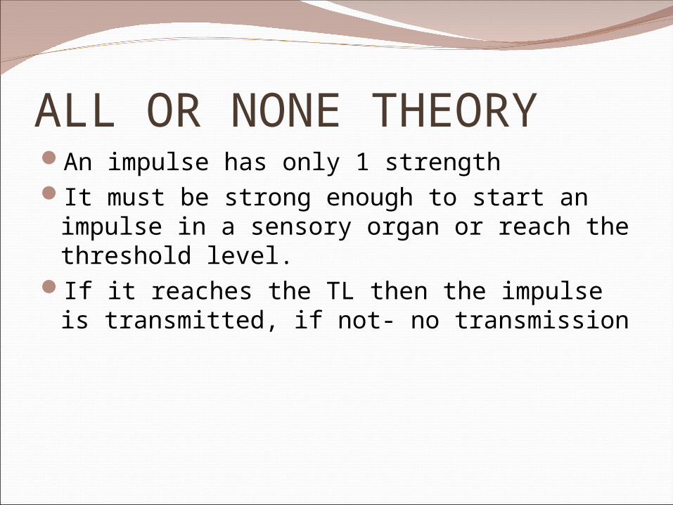
ALL OR NONE THEORYAn impulse has only 1 strengthIt must be strong enough to start an impulse
in a sensory organ or reach the threshold level.
If it reaches the TL then the impulse is transmitted, if not- no transmission

How does impulse jump from axon to next nerve or cell?Need to make connection between neuron
and empty space.1.Vesicles filled with neurotransmitter2.Vesicles fuse with membrane3.Neurotransmitters released into synapse4.Neurotransmitter binds to receptor site on
next neuron or target cell5.Depolarization occurs in cell #26.Ion gates (K+ and Na+) open up7.Impulse transmitted

Synapse

There are 2 parts to your nervous systemCentral Nervous system- Spinal cord + brainPeripheral Nervous System- All nerves
coming from cord

BrainProtected in 3 ways:1.Skull2.Wrapped in connective tissue: Meninges
3 layers- inner-pia matter middle- arachnoid outer-dura matter3. Cerebrospinal fluid- found between pia matter and
arachnoid layers and serves as a shock absorberBrain must have constant supply of oxygen!!!

BRAIN CerebrumBrain StemCerebellum
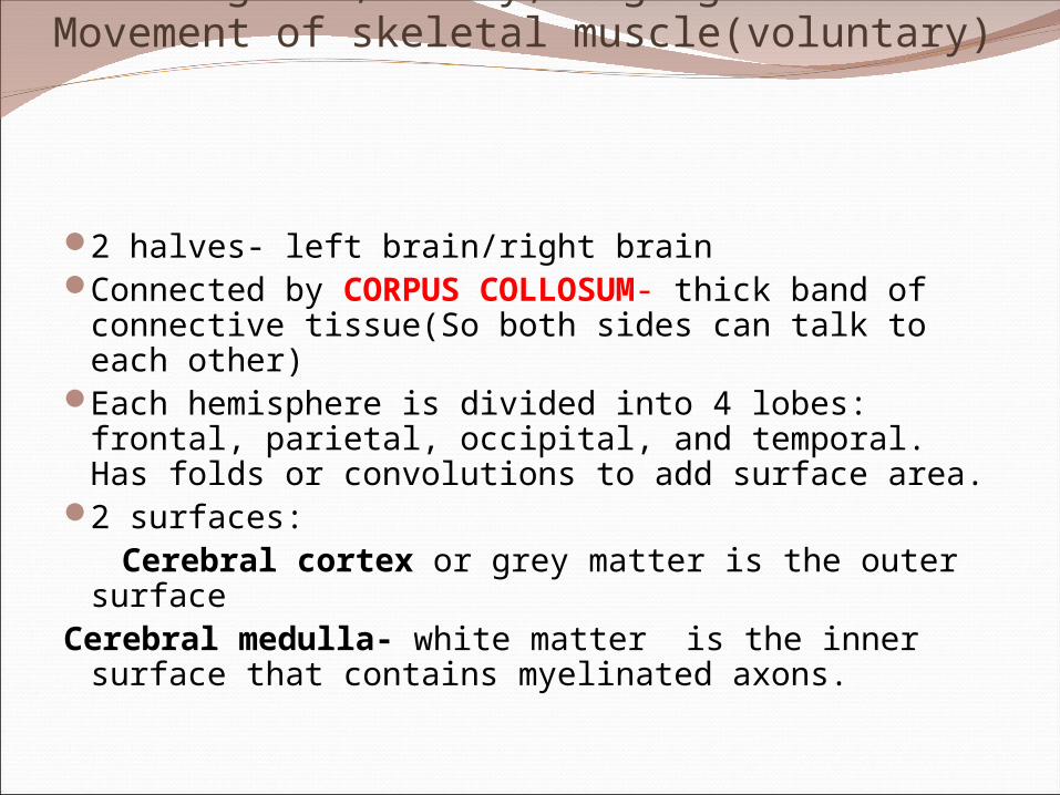
Cerebrum: Site of Intelligence,Memory,LanguageMovement of skeletal muscle(voluntary)
2 halves- left brain/right brainConnected by CORPUS COLLOSUM- thick
band of connective tissue(So both sides can talk to each other)
Each hemisphere is divided into 4 lobes: frontal, parietal, occipital, and temporal. Has folds or convolutions to add surface area.
2 surfaces: Cerebral cortex or grey matter is the outer
surface Cerebral medulla- white matter is the inner
surface that contains myelinated axons.

Brain Stem : 3 parts-connects brain to cord
Medulla Oblongata- breathing/ heart rate/swallowing
Pons -above medulla, links cerebral cortex and cerebellum
Midbrain -involved in hearing and vision

Cerebellum:
BalancePostureCoordination

Two other parts of the brain are found between brainstem and cerebrum
Hypothalmus-control center for hunger, thirst, fatigue, anger, and temperature
Thalmus- switching station for sensory input, passes info to cerebrum

Brain is a source of weak electrical activity that can be detected by an EEG(electroencephalogram)
Sleep is when the cerebral cortex falls to its lowest possible level
Memory is thought to be short and long term

THE SPINAL CORDEmerges from the base of the skullProtected by bone, the vertebral column or
backbone; also the meninges and cerebrospinal fluid.
Links the brain with the peripheral nervous system.
Carries impulses to and from the brain and regulates reflexes.
A reflex is a response to a stimulus.31 pairs of spinal nerves branch out to the body
from the spinal cord.

The spinal cord consists of 2 kinds of nerve tissue:1.Central part is H shaped and consists of gray matter2.Outer part is the white matter and consists of myelinated axons.
Sensory neurons carry impulses from receptors to the spinal cord.
Motor neurons carry impulses from the spinal cord to the effectors (glands/muscles).
Interneurons connect the sensory and motor neurons.
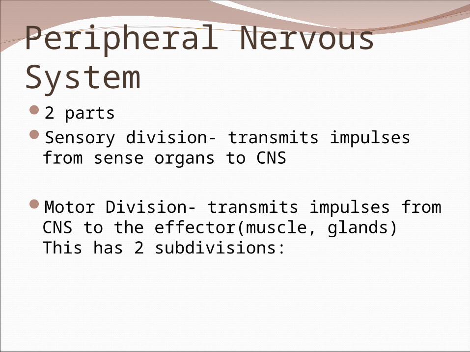
2 partsSensory division- transmits impulses from
sense organs to CNS
Motor Division- transmits impulses from CNS to the effector(muscle, glands) This has 2 subdivisions:
Peripheral Nervous System

Somatic
12 pairs of cranial nerves voluntary 31 pairs of spinal nerves
Also controls reflexes- bypasses brain- signal goes directly to spinal column
sensory nerve spinal column motor nerve target cell
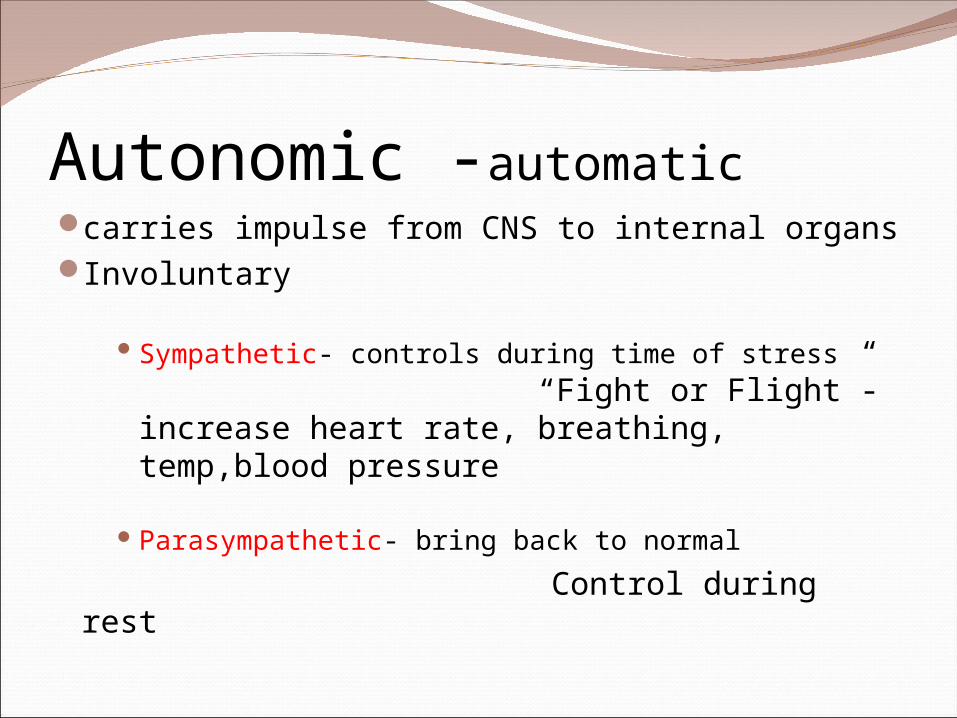
Autonomic -automaticcarries impulse from CNS to internal organsInvoluntary
Sympathetic- controls during time of stress “Fight or Flight”-increase heart rate, breathing, temp,blood pressure
Parasympathetic- bring back to normal
Control during rest
Sensing Chemicals
Chemoreceptors in mouth and noseNose receptors- sense chemicals –impulse
carried on Olfactory Nerve- sent to brainMouth receptors- taste buds on tongue
(10,000)
Sweet, sour, salty, bitter

Eyeconsists of 3 layers:1.outer layer contains the sclera or white of the eye
which consists of tough connective tissue and the cornea which is the transparent covering of the eye. Between the cornea and the sclera is the aqueous humor, a clear fluid.
2.middle layer is the choroid that contains the iris (colored part of the eye) which has an opening called the pupil. Just behind the pupil is the lens, that refracts incoming light. The eyeball itself consists of a large chamber filled with jellylike fluid, the vitreous humor.
3.inner layer is the retina located at the very back of the eye and contains light sensitive cells called photoreceptor cells. This is the place where light energy is converted into electrical impulses.

Sensing light-
These cells contain the pigment rhodopsin which is sensitive to different wavelengths of light. These cells fall into 2 groups;
Rods sense light and dark and the
Cones sense color. It is in these cells where
light is converted into impulses that then travel to the brain via the optic nerve.

Mechanical StimulationHearing- detect vibrations, sound waves
Outer ear consist of auditory canal-collects vibrations
Sound waves hit tympanic membrane (eardrum)Ear drum vibratesVibration passed to 3 bones- malleus, incus,
stapesVibration passed to oval window- causes fluid in
cochlea to vibrateThis causes little hairs in cochlea to bendSends impulse to brain via auditory nerveBrain interprets


Balance- center located in inner ear
Sends info to brainSemicircular canal filled with fluid and hairs
and ear stones(otoliths-CaCO2)Stones lay on hairsHair bends- stimulates nerveSends impulse to brain

Touch- receptors in dermis
Feel temperature, pressure, painReceptors located in specific areas
Light touch- tips of fingers, eyelids, mouthHeavy touch- joints, muscles, palmsFree nerve endings- itch, tickle, hot, cold,
pain
Pain- all over body, except brain Heat- deep in dermis Cold- near surface