Nerve growth factor is expressed and stored in central ...
33
HAL Id: hal-02091213 https://hal-univ-rennes1.archives-ouvertes.fr/hal-02091213 Submitted on 5 Jul 2019 HAL is a multi-disciplinary open access archive for the deposit and dissemination of sci- entific research documents, whether they are pub- lished or not. The documents may come from teaching and research institutions in France or abroad, or from public or private research centers. L’archive ouverte pluridisciplinaire HAL, est destinée au dépôt et à la diffusion de documents scientifiques de niveau recherche, publiés ou non, émanant des établissements d’enseignement et de recherche français ou étrangers, des laboratoires publics ou privés. Nerve growth factor is expressed and stored in central neurons of adult zebrafish Pietro Cacialli, Claudia Gatta, Livia d’Angelo, Adele Leggieri, Antonio Palladino, Paolo de Girolamo, Elisabeth Pellegrini, Carla Lucini To cite this version: Pietro Cacialli, Claudia Gatta, Livia d’Angelo, Adele Leggieri, Antonio Palladino, et al.. Nerve growth factor is expressed and stored in central neurons of adult zebrafish. Journal of Anatomy, Wiley, 2019, 325 (1), pp.167-179. 10.1111/joa.12986. hal-02091213
Transcript of Nerve growth factor is expressed and stored in central ...

HAL Id: hal-02091213https://hal-univ-rennes1.archives-ouvertes.fr/hal-02091213
Submitted on 5 Jul 2019
HAL is a multi-disciplinary open accessarchive for the deposit and dissemination of sci-entific research documents, whether they are pub-lished or not. The documents may come fromteaching and research institutions in France orabroad, or from public or private research centers.
L’archive ouverte pluridisciplinaire HAL, estdestinée au dépôt et à la diffusion de documentsscientifiques de niveau recherche, publiés ou non,émanant des établissements d’enseignement et derecherche français ou étrangers, des laboratoirespublics ou privés.
Nerve growth factor is expressed and stored in centralneurons of adult zebrafish
Pietro Cacialli, Claudia Gatta, Livia d’Angelo, Adele Leggieri, AntonioPalladino, Paolo de Girolamo, Elisabeth Pellegrini, Carla Lucini
To cite this version:Pietro Cacialli, Claudia Gatta, Livia d’Angelo, Adele Leggieri, Antonio Palladino, et al.. Nerve growthfactor is expressed and stored in central neurons of adult zebrafish. Journal of Anatomy, Wiley, 2019,325 (1), pp.167-179. �10.1111/joa.12986�. �hal-02091213�

NERVE GROWTH FACTOR IS EXPRESSED AND STORED IN CENTRAL NEURONS OF 1
ADULT ZEBRAFISH 2
3
4
Pietro Cacialli1, 4, Claudia Gatta1, Livia D’Angelo1,2, Adele Leggieri1, Antonio Palladino3, Paolo de 5
Girolamo1, Elisabeth Pellegrini4, Carla Lucini1 6
7
8
9
Dept. of Veterinary Medicine and Animal Productions, University of Naples Federico II, Naples, 10
Italy; 2. Stazione Zoologica Anton Dohrn, Napoli, Italy; 3. Centro Ricerche Interdipartimentali sui 11
Biomateriali, University of Naples Federico II, Naples, Italy; 4. Univ Rennes, Inserm, EHESP, 12
Irset (Institut de recherche en santé, environnement et travail) - UMR_S 1085, F-35000 Rennes, 13
France 14
1. 15
16
17
18
19
Accep
ted m
anus
cript

ABSTRACT 20
21
Nerve Growth Factor (NGF), a member of the neurotrophin family, was initially described as 22
neuronal survival and growth factor, but successively has emerged as an active mediator in many 23
essential functions in the central nervous system of mammals. NGF is synthesized as a precursor pro-24
NGF and is cleaved intracellularly into mature NGF. However, recent evidence demonstrates proNGF 25
is not a simple inactive precursor, but is also secreted outside the cells and can exert multiple roles. 26
.Despite the vast literature present in mammals, studies devoted to NGF in the brain of other 27
vertebrate models are scarce. Zebrafish is a teleost fish widely known for developmental genetic 28
studies and is well established as model for translational neuroscience research. Genomic 29
organization of zebrafish and mouse NGF are highly similar, and zebrafish NGF protein has been 30
reported in a mature and two precursors forms. To add further knowledge on neurotrophic factors in 31
vertebrate brain models, we decided to determine the NGF mRNA and protein distribution in the 32
adult zebrafish brain and to characterize the phenotype of NGF positive cells. 33
NGF mRNA was visualized by in situ hybridization on whole mount brains. NGF protein distribution 34
was assessed on microtomic sections by using an antiserum against NGF, able to recognize proNGF 35
in adult zebrafish brain as demonstrated also in previous studies. To characterize NGF positive cells, 36
anti-NGF was employed on microtomic slides of aromatase B transgenic zebrafish (where radial 37
glial cells appeared fluorescent) and by means of double immunolabelling against NGF/PCNA 38
(proliferation marker) and NGF/MAP2 (neuronal marker). 39
NGF mRNA and protein were widely distributed in the brain of adult zebrafish and their pattern of 40
distribution of positive perikaryal was overlapping, both in males and females, with few slight 41
differences. Specifically, the immunoreactivity to the protein was observed in fibers over the entire 42
encephalon. MAP2 immunoreactivity was present in the majority of NGF positive cells, throughout 43
the zebrafish brain. PCNA and aromatase B cells were not positive to NGF, but they were closely 44
Accep
ted m
anus
cript

intermingled with NGF cells. In conclusion, our study demonstrated that mature neurons in the 45
zebrafish brain express NGF mRNA and store proNGF. 46
47
Key words: fish; neurotrophins; brain; encephalon; zebrafish neurons; 48
49
Accep
ted m
anus
cript
INTRODUCTION 50
51
Nerve Growth Factor (NGF) is the first identified factor belonging, together with Brain Derived 52
Neurotrophic Factor (BDNF), Neurotrophin (NT) 3 and NT 4/5, the neurotrophin family. As all 53
members of the family, NGF is synthesized as a precursor form (pro-NGF) and either secreted outside 54
the cells, as pro- and mature NGF, or cleaved intracellularly to mature NGF. Active forms of pro- 55
and mature NGF are homodimers. To date, three types of NGF receptors are known: TrkA, p75 and 56
sortilin. TrkA, the high affinity receptor, is a member of receptor tyrosine kinases family which 57
includes receptor TrkB (for BDNF and NT 4/5) and TrkC (for NT 3). p75NTR is a member of the 58
tumor necrosis factor receptor (TNFR) superfamily and can transduce signals of all neurotrophins. 59
Sortilin is a member of the family of Vps10p-domain transmembrane receptors, and was earlier 60
characterized as a receptor for neurotensin. While TrkA mediates trophic signalling of the mature 61
form of NGF, p75NTR bifunctionally mediates: (a) a signal to neuronal survival, specially when 62
binding to mature NGF and acting together with TrkA; or (b) the induction of neuronal death when 63
forming a receptor complex with sortilin and mediating proNGF signal (for a review 64
see Niewiadomska et al., 2011). 65
NGF, initially described as neuronal survival and growth factor, encompasses roles regarding the 66
density of innervation, synthesis of neurotransmitters and neuropeptides and cell body size, axonal 67
sprouting and dendritic arborization (for a review see Minnone et al 2017). 68
The brain of fish possesses, beyond structural organization similar to all vertebrates, extremely high 69
adult neurogenesis and astonishing regenerative properties. These peculiarities make the fish brain 70
suitable for discovering regenerative mechanisms probably suppressed in mammals during evolutive 71
adaptation (Panula et al 2010; Cacialli et al 2018b). Among teleost fish, zebrafish (Danio rerio) is 72
widely used as model species for developmental genetic studies and functional mechanisms of 73
numerous genes responsible of human diseases (D’Angelo et al., 2016). In addition, zebrafish brain 74
has been largely utilized in numerous studies devoted to adult neurogenesis (Pellegrini et al 2007; 75
Accep
ted m
anus
cript

Diotel et al 2013; Coumailleau et al 2015; Than-Trong and Bally-Cuif 2015; Anand et al 2017) and 76
regenerative ability after injury (Cosacak et al 2015; Alunni and Bally-Cuif 2016; Cacialli et al 2018 77
a,b). 78
In fish species, homologs of mammalian neurotrophins have been identified, and an additional 79
member, named NT6/7, probably originated by a duplication of the ray-finned fish NGF (Dethleffsen 80
et al 2003). NT6/7 has been identified also in zebrafish (Nilsson et al., 1998). Genomic organization 81
of zebrafish and mouse NGF are highly similar, and zebrafish NGF protein has been reported in a 82
mature form of 194 aminoacid. (Dethleffsen et al 2003). In addition, two proNGF isoforms have been 83
described in zebrafish: isoform 1 (NP_001338647.1) and isoform 2 (NP_954680.2), respectively of 84
224 and 261 a.a.. (Dethleffsen et al 2003). 85
The distribution pattern of neurotrophins and their Trk receptors was described in the brain of adult 86
fish (Dalton et al 2009a,b, D’Angelo et al 2012, D’Angelo et al 2014a, D’Angelo et al 2014b, Cacialli 87
et al 2016, D’Angelo et al 2016, Gatta et al 2016). Furthermore, BDNF appeared involved in repairing 88
mechanisms of adult zebrafish after traumatic brain injury (Cacialli et al 2018 a,b; Lucini et al 2018). 89
In the present study, we evaluated NGF mRNA and protein distributionin the brain of zebrafish and 90
identified neurons as the NGF source cells. Thanks to the conserved adult neurogenesis in fish brain 91
and the potential involvement of neurotrophin in the regenerative ability, the identification of NGF 92
neurons in the adult brain of zebrafish could represent a useful tool to evaluate its involvement in the 93
regenerative process after injury or chemical/genetic induced degeneration. 94
Accep
ted m
anus
cript

MATERIALS AND METHODS 95
Animals and brain dissection 96
Animals used in this study were housed in zebrafish facilities (INRA LPGP, BIOSIT, Rennes, France, 97
agreement number: B 35-238-6) under standard conditions of photoperiod (14/10) and temperature 98
(28°C). This project was approved by the local animal care and Westerfield ethics committee (Comité 99
Rennais d'Ethique en matière d'Expérimentation Animale, Rennes, France), under the number EEA 100
B-35-040. Zebrafish did not receive medical treatment prior or during the experience. No deaths 101
occurred in the facilities before the euthanasia of animals used for in situ hybridization and 102
immunohistochemistry experiments. Fish were suppressed with an overdose of tricaine 103
methanesulfonate (MS-222). 104
105
In situ hybridization (ISH) 106
Oligonucleotide primers used to amplify and clone cDNA for the production of NGF ISH probes are: 107
forward 5’-ACATGTACCATGAGGAGCAC-3’; reverse 5’-GTCGCTGGTGTGTGGAAAAT-3’ 108
(708 bp; NM_001351718.1). For preparation of NGF digoxigenin (DIG) labeled antisense riboprobe, 109
the vector pCRII-TOPO containing NGF was linearized by BamHI digestion and digoxigenin-labeled 110
riboprobe was prepared using in vitro transcription with T7 RNA polymerase. For sense riboprobe 111
the vector containing NGF was linearized by Not1 and digoxigenin-labeled riboprobe was prepared 112
using in vitro transcription with SP6 RNA polymerase. 113
ISH was performed on whole mount brains as previously described by Adolf et al. (2006); Diotel et 114
al. (2015). Briefly, six zebrafish brains (males and females) were excised and fixed in 115
paraformaldehyde (PFA 4%) dissolved in phosphate-buffered saline (PBS), for 24 hours (h) at 4°C. 116
Then, the brains were dehydrated in a methanol/PBS concentration (25%; 50%; 75%; 100%) and 117
stored at -20°C. After rehydrating through methanol/PBS gradient series and washed in PBS, the 118
brains were incubated for 40 minutes (min) in PBS containing proteinase K (10 µg/ml) at room 119
temperature (RT). After post-fixation in 4% PFA for 20 min and washes in PBS, brains were then 120
Accep
ted m
anus
cript

prehybridized for 1 h and incubate overnight at 65°C in the hybridization buffer (pH 6) containing 121
the DIG-labeled probe. Then, brains were washed in SCC 2x/formamide 50% and SSC 0.2x, and pre-122
incubated with blocking buffer for 3 h and then overnight with antidigoxigenin-AP, Fab fragments 123
(1:5,000; Roche, NJ; Cat# 11093274910, RRID: AB_514497) at 4°C. The next day the brain sections 124
were washed with PBS before staining with NBT/BCIP buffer (pH 9.5). 125
Whole mount stained brains were embedded in agar 2% and photographed with a Digital camera 126
equipped on Zeiss Stemi. Then, the embedded whole mount stained brains were transversally 127
sectioned with a razor blade or vibratome and the sections were mounted on the slide. 128
The specificity of the ISH labelling was demonstrated by using sense riboprobe, that showed absence 129
of any staining. 130
131
Immunohistochemistry (IHC) 132
Imunohistochemical procedures were performed following detailed suggestions reported by de 133
Girolamo and Lucini (2011). Six adult zebrafish brains (male and female) were fixed in Bouin’s 134
solution for 24 h and processed for paraffin embedding. Transverse microtome sections were mounted 135
on poly-lysine slides. Sections were deparaffinised in xylene, rehydrated through graded ethanol, 136
treated with 3% H2O2 for 30 min and rinsed in PBS (pH 7.4) followed by antigen retrieval in sodium 137
citrate buffer (pH 6; 80 °C) for 30 min. 138
Single immunohistochemistry 139
After rinsing 2 times in 0.2% Triton PBS (PBT), non-specific binding was blocked by treating 140
sections with 1/5 normal goat serum (Vector, Burlingame, CA, USA, cod S-1000-20) for 30 min at 141
RT. Then, sections were incubated over night at RT in a humidified chamber with rabbit antibody 142
against NGF (Santa Cruz Biotechnology, CA, USA, cod sc-549. It recognizes N-terminus of the 143
mature chain of NGF of human origin) diluted 1/100 or 1/300, respectively depending on the 144
secondary antibody specified at following point a) or b). The next day, the sections were washed 145
several times in PBT and alternatively incubated with a) Alexa Fluor® goat anti-rabbit 488 (1:200; 146
Accep
ted m
anus
cript

Invitrogen Molecular probes, Eugene, OR, REF: A-11037; RRID: AB_10561549) for 2 h at RT in a 147
dark and humidified chamber or b) EnVision-horseradish anti-peroxidase (HRP)-system (Dako, 148
Santa Barbara, CA, cod. K4002). This system is based on a HRP labeled polymer conjugated with 149
goat anti-rabbit IgG. 150
Sections treated with Alexa Fluor® goat anti-rabbit 488, after three washes in PBT, were mounted 151
with the medium Vectashield (Vector) containing 4,6-diamino-2-phenylindole (DAPI), to visualize 152
cell nuclei. Sections treated with EnVision-HRP-system were immersed in a fresh solution of 10 μg 153
of 3,3′-diaminobenzidine tetrahydrochloride (Sigma–Aldrich Corporation, St. Louis, MO, USA, cod. 154
D5905) in 15 ml of a 0.5 mol, Tris buffer, pH 7.6, containing 1.5 ml of 0.03% H2O2. Then, sections 155
were dehydrated and mounted. 156
For immunohistochemistry on cyp19a1bGFP transgenic zebrafish (glial cell marker, Pellegrini et al., 157
2007; Tong et al., 2008), NGF antibody was detected with Alexa Fluor® goat anti-rabbit 594 (1:200; 158
Invitrogen Molecular probes, Eugene, OR, USA, REF: A-11037; RRID: AB_10561549). 159
The specificity of IHC, assessed by substitution of NGF antiserum, secondary antibody fluorescent 160
dye -conjugated or the EnVision with PBS or normal serum, achieved no specific immunostaining. 161
Moreover, the incubation of NGF antiserum preincubated with its homologous antigen showed no 162
immunoreactivity, and NGF antiserum preincubated with its heterologous antigens did not modify 163
the normal pattern of immunostaining. 164
165
Double immunolabelling 166
Double immunocytochemical staining NGF/PCNA and NGF/MAP2 was performed as follows: 167
dewaxed and rehydrated consecutive sections were rinsed in PBS, and incubated for 48 h at RT. 168
PCNA (proliferative cell nuclear antigen), antibody at diluted 1:100, was used to detect proliferative 169
cells (Clone PC10; Dako, Glostrup, Denmark; REF: M0879; RRID: AB_2160651). This antibody is 170
a marker of proliferating cells in vertebrate species, including zebrafish (Pellegrini et al., 2007; Marz 171
et al., 2011; Cacialli et al., 2016; 2018). MAP2 (Microtube-Associated Protein2) antibody, diluted 172
Accep
ted m
anus
cript

1:100, was used to detect neurons, (sc-74422 MAP-2 (A-8) Santa Cruz Biotechnology, Santa Cruz, 173
CA, USA). After rinsing in PBS, the sections were incubated for 2 h at RT with mixture of the 174
secondary antibodies directed against rabbit and mouse IgG. a) Alexa fluor® goat anti-mouse 594 175
(1:200; Invitrogen Molecular probes, Eugene, OR, REF: A-11005; RRID: AB_10561507; b) Alexa 176
Fluor® goat anti-rabbit 594 (1:200; Invitrogen Molecular probes, Eugene, OR, USA, REF: A-11037; 177
RRID: AB_10561549). Tissue sections were washed in PBS-Triton 0,2%, and slides were mounted 178
with the Vectashield medium containing DAPI for nuclei counterstaining (Vector Laboratories, 179
Burlingame, CA). Controls for double immunolabelling were performed by incubating the sections 180
with one of the two primary antisera and with the mismatched secondary antibodies. 181
182
Microscopy 183
The stained sections were photographed using a Nikon Eclipse 90i microscope, an epifluorescence 184
microscope Olympus equipped with a DP71 digital camera and Leica DM6B, SN: 449492. 185
The digital raw images were optimized for image resolution, contrast, evenness of illumination, and 186
background by using Adobe Photoshop CS5 (Adobe Systems, San Jose, CA, USA). 187
188
189
Accep
ted m
anus
cript

RESULTS 190
191
NGF mRNA (Fig 1A) and protein were widely distributed in the brain of adult zebrafish(Tab. 1). The 192
pattern of distribution of positive perikarya was overlapping, both in males and females, with few 193
slight differences (Table 1). Specifically, the immunoreactivity to the protein was observed in fibers 194
over the entire encephalon. Thus, regions characterized only by presence of fibers, such as the 195
glomerular layer of olphactory bulbs and deep and white zone of the optic tect, showed positivity to 196
the protein. Based on these general considerations, for sake of the simplicity, the term NGF in place 197
of NGF mRNA and NGF protein was used in the following description of results. The anatomical 198
terminology follows “Neuroanatomy of the zebrafish brain” by Wulliman et al. (1996). 199
Telencephalon 200
The olfactory bulbs showed moderate quantity of NGF (Fig 1C, D). The cells of both external and 201
internal cellular layer and fibers of the glomerular layer resulted positive. 202
In the whole telencephalon, more intense NGF positivity was seen in the ventral telencephalon (Fig 203
1C, D) and in the posterior zone dorsal telencephalic area (Fig 1B, C). Positive cells were distributed 204
in the medial (Fig 3B, B1), dorsal (Fig 8B, D), lateral (Fig 2A1, 3C, C1, 10 B, D), central (Fig 11B, 205
D) and posterior part of dorsal part of telencephalic area. In the ventral telencephalic area, small round 206
cells were seen in the dorsal and ventral part. 207
Diencephalon 208
Numerous intensely stained cells were seen in the anterior parvocellular preoptic nucleus (Fig 1C, 209
2B1), and in the posterior parvocellular preoptic nucleus (Fig 9 B, D). Weak signal was detected in 210
few cells of magnocellular preoptic nucleus. In dorsal and ventral habenular nucleus few positive 211
cells were detected. High density of NGF positive cells were observed in the ventro-medial and 212
ventro-lateral thalamic nuclei (Fig 9B, D). Few and weak positive cells were seen in the central 213
posterior thalamic nucleus. Numerous positive cells were detected in the posterior tuberal nucleus. In 214
the lateral and medial preglomerular nuclei, positive cells and some fibers were detected. 215
Accep
ted m
anus
cript
In the hypothalamus, the ventral zone of periventricular hypothalamus showed intense positivity in 216
the whole mount brain (Fig 1E) and numerous NGF positive cells were seen in histological sections 217
(Fig 2C1, 5B-C1). In the dorsal (Fig 1E) and caudal zone of periventricular hypothalamus moderate 218
positivity was seen. Large NGF positive cells belonging to the diffuse nucleus of the inferior lobe 219
were seen (Fig 6 A–B1). Few NGF positive cells were also present in the mammillary body and in 220
the nucleus of the medial longitudinal fascicle 221
Mesencephalon 222
NGF positive fibers were present in the longitudinal tori and the optic tect, particularly in the deep 223
white zone and superficial white zone. Numerous small NGF positive cells appeared scattered in the 224
periventricular grey zone (Fig 4, B1, B2). 225
In the tegmentum, NGF was observed in cells of central nucleus of semi-circular torus and superior 226
reticular formation. 227
Rhombencephalon 228
The cerebellar body was intensely reactive (Fig 1F). NGF positivity was observed in numerous large 229
cells of the Purkinje layer and in few cells of the molecular layer of valvula and body (Fig. 7A – C1). 230
Few weak stained cells were observed in the granular eminence. 231
In the medulla oblongata, few large cells containing NGF and belonging to the inferior reticular 232
formation were seen. 233
234
Characterization of NGF containing cells 235
In order to identify the nature of NGF positive-cells in the brain of adult zebrafish, we carried out 236
immunohistochemical staining against NGF on slides of aromatase B transgenic fish (cyp19a1b 237
GFP), where radial glial cells appeared green fluorescent. Also, we performed double labelling using 238
antibodies against NGF/PCNA (proliferation marker) and NGF/MAP2 (neuronal marker). Aromatase 239
positive cells, distributed along ventricles, were closely intermingled with NGF positive cells along 240
telencephalic (Fig 8 B -D) and diencephalic (Fig 9B–D) ventricles. PCNA-positive cells were 241
Accep
ted m
anus
cript

positioned along the ventricular lining of the brain, and NGF was detected in the cytoplasm of cells 242
very close to PCNA labelled cells (Fig 10B-D). MAP2 immunoreactivity was present in the majority 243
of NGF positive cells, throughout the zebrafish brain (Fig 11B-D). 244
245
246
247
248
249
Accep
ted m
anus
cript
DISCUSSION 250
This study documents the neuroanatomical distribution of NGF mRNA and protein in zebrafish. 251
The serum raised against NGF was previously characterized (Gatta et al 2016), where brain 252
homogenates of adult zebrafish showed only a band of 25 kDa, corresponding to the molecular weight 253
of proNGF isoform 1 (NP_001338647.1) (Gatta et al 2016). Accordingly, the presence of proNGF 254
was also reported in the brain of the teleost N. furzeri, by employing the same antiserum(D’Angelo 255
et al 2014). On the other hand, this antiserum detected the mature form of NGF in different organs 256
of fish, such as gut (Lucini et al 2003) and kidney (Arcamone et al., 2005). Taking into consideration 257
that the employed NGF antiserum is able to detect both pro- and mature NGF in fish tissues, our 258
results suggest that that only the proNGF form is present in the brain of zebrafish. Remarkably, in 259
mammalian brain, precursor and intermediate forms of NGF are expressed (Fahnestock et al 2001, 260
2004), and has been demonstrated to be actually the predominant form of NGF in central nervous 261
system (CNS) (Fahnestock et al., 2001), whereas mature NGF appears to be lacking. 262
The co-presence of pro-NGF and the neuronal marker MAP2 immunoreactivity throughout the brain 263
of adult zebrafish demonstrates that pro-NGF containing cells are neurons. This result was further 264
confirmed by the absence of the glial marker aromatase B and proliferative marker PCNA. 265
Consistently with our observations, in the teleost fish N. furzeri, NGF was morphologically detected 266
in neurons widespread throughout all brain regions (D’Angelo et al., 2014). Only some glial cells 267
lining the mesencephalic and rhomboencephalic ventricles of N. furzeri seemed to express NGF. At 268
opposite, in goldfish NGF was almost totally localized in radial glia cells lining the ventricles 269
(Benowitz and Shashoua, 1979). Species specific characteristics could explain the different results 270
achieved in zebrafish, N. furzeri and goldfish. In agreement with our results, in the rat brain, NGF 271
production was reported in neurons, predominantly localized in GABAergic neurons of the cortex, 272
hippocampus, striatum and basal forebrain (Lauterborn et al 1993, 1995; Pascual et al 1998; Bizon et 273
al 1999; Sofroniew et al 2001; Biane et al 2014). NGF was also reported in neuronal populations of 274
adult monkey brain (Hayashi et al 1993; Zhang et al 2007). However, oligodendroglial progenitors 275
Accep
ted m
anus
cript

derived from human embryonic stem cells are a source of NGF (Zhang et al 2006; Althaus et al 2000). 276
Remarkably in adult zebrafish, another member of neurotrophin family, BDNF, was also expressed 277
in neuronal populations of the whole brain (Cacialli et al 2016) and of telencephalon after injury 278
(Cacialli et al 2018). 279
NGF expression was comparable between zebrafish and N. furzeri (D’Angelo et al 2014), despite 280
some slight differences in the neuroanatomy, whereas substantial differences in NGF cell localization 281
and distribution were seen between goldfish and zebrafish as previously described. In rat brain, NGF 282
levels resulted consistent in all regions with the highest presence in cortical areas (Hoener et al., 1996; 283
Sakamoto et al., 1998). Specifically, the pattern of distribution of both NGF mRNA (Shelton and 284
Reichardt, 1986) and protein (Nishio et al., 1992) was described throughout the rat brain, with the 285
highest intensity in the neocortex, the hippocampal pyramidal layer and striatum (Gall and Isackson, 286
1989, Rylett and Williams, 1994). Notably, observations in adult zebrafish brain do not support the 287
fact that the forebrain is the NGF prelavent containing region. 288
In our study, both NGF mRNA and protein were detected in the perikaryon, and only proNGF protein 289
was distributed along neuronal prolongations. Although the cellular co-presence of NGF mRNA and 290
protein was not investigated, the overlapping pattern of distribution of NGF mRNA and protein 291
throughout the zebrafish brain suggests that NGF expression and translation take place in mature 292
neurons. The presence of NGF protein along neuronal prolongments could be retrogradely 293
transported, according to the classical view on neurotrophins considered as target-derived 294
retrogradely transported substances, but also anterogradely transported, accordingly to a vast 295
literature. The idea that NGF may be produced locally was suggested since two decades ago 296
(Lauterborn et al 1991, Conner and Varon 1992) and activity-dependent release of NGF and its effect 297
on synaptic plasticity was postulated (Blöchl and Thoenen, 1995, 1996; Wu et al., 2004). Finally, 298
Guo and collaborators (2012) demonstrated, by immunohistochemical, ELISA and 299
electrophysiological analyses, anterograde delivery of NGF in hippocamposeptal system of mice. 300
Accep
ted m
anus
cript

ProNGF in the CNS is released in the extracellular space (Bruno and Cuello 2006) and induces 301
activation of the apoptotic machinery with subsequent death of different neuronal populations, mostly 302
after injury and neurodegenerative disorders (for a review see Costa et al 2018). 303
The present results demonstrate that also in zebrafish brain NGF is synthesized in perykaria, however 304
future studies are necessary to test whether proNGF is anterogradely transported and released in the 305
extracellular space, at terminal ending of NGF positive fibers. 306
In conclusion, our study demonstrated that mature neurons of the zebrafish brain express NGF mRNA 307
and store proNGF. Experimental studies reported proNGF as inhibition factor of the proliferation 308
of neural stem cells isolated from postnatal mouse hippocampus, causing cell cycle arrest in the 309
G0/G1 phase (Guo et al 2013). Thus, it is tempting to speculate that proNGF in the zebrafish brain, 310
where cell proliferation is considerably high and persist along the entire lifespan, could represent a 311
key negative regulator factor of this process. 312
313
Author’s contribution 314
PC conceived and planned the experimentation, acquired and analyzed data; CG, AL, AP acquired 315
and analyzed data; LDA, PdG, EP critically revised the manuscript; CL analyzed data and wrote the 316
paper. 317
318
Accep
ted m
anus
cript
REFERNCES 319
320
Adolf B, Chapouton P, Lam CS, et al. (2006) Conserved and acquired features of adult neurogenesis 321
in the zebrafish telencephalon. Developmental Biology, 295, 278-293. 322
Althaus HH, Richter-Landsberg C (2000) Glial cells as targets and producers of neurotrophins. 323
International Review of Cytology - a Survey of Cell Biology, Vol. 197, 197, 203-277. 324
Alunni A, Bally-Cuif L (2016) A comparative view of regenerative neurogenesis in vertebrates. 325
Development, 143, 741-753. 326
Anand SK, Mondal AC (2017) Cellular and molecular attributes of neural stem cell niches in adult 327
zebrafish brain. Developmental Neurobiology, 77, 1188-1205. 328
Arcamone N, Lucini C, Borzacchiello G, Castaldo L, Gargiulo G, De Girolamo P (2005) Distribution 329
of NGF and NT-3-like protein immunoreactivity in the teleost kidney. Microscopy Research 330
and Technique, 66, 17-24. 331
Biane J, Conner JM, Tuszynski MH (2014) Nerve growth factor is primarily produced by GABAergic 332
neurons of the adult rat cortex. Frontiers in Cellular Neuroscience, 8. 333
Bizon JL, Lauterborn JC, Gall CM (1999) Subpopulations of striatal interneurons can be 334
distinguished on the basis of neurotrophic factor expression. Journal of Comparative 335
Neurology, 408, 283-298. 336
Blochl A, Thoenen H (1995) Characterization of Nerve Growth -Factor (NGF) release from 337
hippocampal-neurons- evidence for a constitutive and unconventional sodium-dependent 338
regulated pathway. European Journal of Neuroscience, 7, 1220-1228. 339
Blochl A, Thoenen H (1996) Localization of cellular storage compartments and sites of constitutive 340
and activity-dependent release of nerve growth factor (NGF) in primary cultures of 341
hippocampal neurons. Molecular and Cellular Neuroscience, 7, 173-190. 342
Bruno MA, Cuello AC (2006) Activity-dependent release of precursor nerve growth factor, 343
conversion to mature nerve growth factor, and its degradation by a protease cascade. 344
Proceedings of the National Academy of Sciences of the United States of America, 103, 6735-345
6740. 346
Cacialli P, D'Angelo L, Kah O, et al. (2018a) Neuronal expression of brain derived neurotrophic 347
factor in the injured telencephalon of adult zebrafish. Journal of Comparative Neurology, 526, 348
569-582. 349
Cacialli P, Gueguen MM, Coumailleau P, et al. (2016) BDNF Expression in Larval and Adult 350
Zebrafish Brain: Distribution and Cell Identification. Plos One, 11. 351
Cacialli P, Palladino A, Lucini C (2018b) Role of brain-derived neurotrophic factor during the 352
Accep
ted m
anus
cript
regenerative response after traumatic brain injury in adult zebrafish. Neural Regeneration 353
Research, 13, 941-944. 354
Conner JM, Varon S (1992) Distribution of nerve Growth factor-like immunoreactive neurons in the 355
adult-rat brain following colchicine treatment. Journal of Comparative Neurology, 326, 347-356
362. 357
Cosacak MI, Papadimitriou C, Kizil C (2015) Regeneration, Plasticity, and Induced Molecular 358
Programs in Adult Zebrafish Brain. Biomed Research International. 359
Costa CJ, Willis DE (2018) To the end of the line: Axonal mRNA transport and local translation in 360
health and neurodegenerative disease. Developmental Neurobiology, 78, 209-220. 361
Coumailleau P, Pellegrini E, Adrio F, et al. (2015) Aromatase, estrogen receptors and brain 362
development in fish and amphibians. Biochimica Et Biophysica Acta-Gene Regulatory 363
Mechanisms, 1849, 152-162. 364
D'Angelo L, Avallone L, Cellerino A, et al. (2016a) Neurotrophin-4 in the brain of adult 365
Nothobranchius furzeri. Annals of Anatomy-Anatomischer Anzeiger, 207, 47-54. 366
D'Angelo L, Castaldo L, Cellerino A, de Girolamo P, Lucini C (2014a) Nerve growth factor in the 367
adult brain of a teleostean model for aging research: Nothobranchius furzeri. Annals of 368
Anatomy-Anatomischer Anzeiger, 196, 183-191. 369
D'Angelo L, de Girolamo P, Cellerino A, Tozzini ET, Castaldo L, Lucini C (2012) Neurotrophin Trk 370
receptors in the brain of a teleost fish, Nothobranchius furzeri. Microscopy Research and 371
Technique, 75, 81-88. 372
D'Angelo L, De Girolamo P, Lucini C, et al. (2014b) Brain-Derived Neurotrophic Factor: mRNA 373
Expression and Protein Distribution in the Brain of the Teleost Nothobranchius furzeri. 374
Journal of Comparative Neurology, 522, 1004-1030. 375
D'Angelo L, Lossi L, Merighi A, de Girolamo P (2016b) Anatomical features for the adequate choice 376
of experimental animal models in biomedicine: I. Fishes. Annals of Anatomy-Anatomischer 377
Anzeiger, 205, 75-84. 378
Dalton VS, Borich SM, Murphy P, Roberts BL (2009a) Brain-Derived Neurotrophic Factor mRNA 379
Expression in the Brain of the Teleost Fish, Anguilla anguilla, the European Eel. Brain 380
Behavior and Evolution, 73, 43-58. 381
Dalton VS, Roberts BL, Borich SM (2009b) Brain derived neurotrophic factor and trk B mRNA 382
expression in the brain of a brain stem-spinal cord regenerating model, the European eel, after 383
spinal cord injury. Neuroscience Letters, 461, 275-279. 384
de Girolamo P, Lucini C (2011) Neuropeptide localization in nonmammalian vertebrates. Methods in 385
molecular biology (Clifton, N.J.), 789, 37-56. 386
Accep
ted m
anus
cript

Dethleffsen K, Heinrich G, Lauth M, Knapik EW, Meyer M (2003) Insert-containing neurotrophins 387
in teleost fish and their relationship to nerve growth factor. Molecular and Cellular 388
Neuroscience, 24, 380-394. 389
Diotel N, Vaillant C, Gabbero C, et al. (2013) Effects of estradiol in adult neurogenesis and brain 390
repair in zebrafish. Hormones and Behavior, 63, 193-207. 391
Diotel N, Viales RR, Armant O, et al. (2015) Comprehensive Expression Map of Transcription 392
Regulators in the Adult Zebrafish Telencephalon Reveals Distinct Neurogenic Niches. 393
Journal of Comparative Neurology, 523, 1202-1221. 394
Fahnestock M, Michalski B, Xu B, Coughlin MD (2001) The precursor pro-nerve growth factor is 395
the predominant form of nerve growth factor in brain and is increased in Alzheimer's disease. 396
Molecular and Cellular Neuroscience, 18, 210-220. 397
Fahnestock M, Yu G, Michalski B, et al. (2004) The nerve growth factor precursor proNGF exhibits 398
neurotrophic activity but is less active than mature nerve growth factor. Journal of 399
Neurochemistry, 89, 581-592. 400
Gall CM, Isackson PJ (1989) Limbic seizures increase neuronal production of messenger-RNA for 401
nerve growth-factor. Science, 245, 758-761. 402
Gatta C, Altamura G, Avallone L, et al. (2016) Neurotrophins and Their Trk-Receptors in the 403
Cerebellum of Zebrafish. Journal of Morphology, 277, 725-736. 404
Guo L, Yeh ML, Carlson VCC, Johnson-Venkatesh EM, Yeh HH (2012) Nerve Growth Factor in the 405
Hippocamposeptal System: Evidence for Activity-Dependent Anterograde Delivery and 406
Modulation of Synaptic Activity. Journal of Neuroscience, 32, 7701-7710. 407
Guo JJ, Wang JN, Liang CR, Yan J, Wang Yr, Liu GX, Jiang ZZ, Zhang LY, Wang XB, Wang YJ, 408
Zhou XF, Liao H (2013) proNGF inhibits proliferation and oligodendrogenesis of postnatal 409
hippocampal neural stem/progenitor cells through p75NTR in vitro. Stem Cell Research, 11, 410
874-887. 411
Hayashi M, Yamashita A, Shimizu K, Sogawa K, Fujii Y (1993) Expression of the gene for nerve 412
growth-factor (NGF) in the monkey central nervous system. Brain Research, 618, 142-148. 413
Hoener MC, Hewitt E, Conner JM, Costello JW, Varon S (1996) Nerve growth factor (NGF) content 414
in adult rat brain tissues is several-fold higher than generally reported and is largely associated 415
with sedimentable fractions. Brain Research, 728, 47-56. 416
Lauterborn JC, Bizon JL, Tran TMD, Gall CM (1995) NGF messenger-RNA is expressed by 417
gabaergic but not cholinergic neurons in rat basal forebrain. Journal of Comparative 418
Neurology, 360, 454-462. 419
Lauterborn JC, Isackson PJ, Gall CM (1991) NERVE GROWTH-FACTOR MESSENGER RNA-420
Accep
ted m
anus
cript

CONTAINING CELLS ARE DISTRIBUTED WITHIN REGIONS OF CHOLINERGIC 421
NEURONS IN THE RAT BASAL FOREBRAIN. Journal of Comparative Neurology, 306, 422
439-446. 423
Lauterborn JC, Tran TMD, Isackson PJ, Gall CM (1993) NERVE GROWTH-FACTOR 424
MESSENGER-RNA IS EXPRESSED BY GABAERGIC NEURONS IN RAT 425
HIPPOCAMPUS. Neuroreport, 5, 273-276. 426
Lucini C, D'Angelo L, Cacialli P, Palladino A, de Girolamo P (2018) BDNF, Brain, and Regeneration: 427
Insights from Zebrafish. International Journal of Molecular Sciences, 19. 428
Lucini C, Maruccio L, Arcamone N, Lamanna C, Castaldo L (2003) Neurotrophin-like 429
immunoreactivity in the gut of teleost species. Neuroscience Letters, 345, 33-36. 430
Marz M, Schmidt R, Rastegar S, Strahle U (2011) Regenerative Response Following Stab Injury in 431
the Adult Zebrafish Telencephalon. Developmental Dynamics, 240, 2221-2231. 432
Minnone G, De Benedetti F, Bracci-Laudiero L (2017) NGF and Its Receptors in the Regulation of 433
Inflammatory Response. International Journal of Molecular Sciences, 18. 434
Niewiadomska G, Mietelska-Porowska A, Mazurkiewicz M (2011) The cholinergic system, nerve 435
growth factor and the cytoskeleton. Behavioural Brain Research, 221, 515-526. 436
Nilsson AS, Fainzilber M, Falck P, Ibanez CF (1998) Neurotrophin-7: a novel member of the 437
neurotrophin family from the zebrafish. Febs Letters, 424, 285-290. 438
Nishio T, Akiguchi I, Furukawa S (1992) Detailed distribution of nerve growth-factor in rat-brain 439
determined by a highly sensitive enzyme-immunoassay. Experimental Neurology, 116, 76-440
84. 441
Panula P, Chen YC, Priyadarshini M, et al. (2010) The comparative neuroanatomy and 442
neurochemistry of zebrafish CNS systems of relevance to human neuropsychiatric diseases. 443
Neurobiology of Disease, 40, 46-57. 444
Pascual M, Rocamora N, Acsady L, Freund TF, Soriano E (1998) Expression of nerve growth factor 445
and neurotrophin-3 mRNAs in hippocampal interneurons: Morphological characterization, 446
levels of expression, and colocalization of nerve growth factor and neurotrophin-3. Journal 447
of Comparative Neurology, 395, 73-90. 448
Pellegrini E, Mouriec K, Anglade I, et al. (2007) Identification of aromatase-positive radial glial cells 449
as progenitor cells in the ventricular layer of the forebrain in zebrafish. Journal of 450
Comparative Neurology, 501, 150-167. 451
Rylett RJ, Williams LR (1994) ROLE OF NEUROTROPHINS IN CHOLINERGIC-NEURON 452
FUNCTION IN THE ADULT AND AGED CNS. Trends in Neurosciences, 17, 486-490. 453
Sakamoto H, Kuzuya H, Tamaru M, et al. (1998) Developmental changes in the NGF content in the 454
Accep
ted m
anus
cript

brain of young, growing, low-birth-weight rats. Neurochemical Research, 23, 115-120. 455
Shelton DL, Reichardt LF (1986) Studies on the expression of the beta-nerve growth-factor (NGF) 456
gene in the central nervous system level and regional distribution of NGF messenger RNA 457
suggest that NGF functions as a trophic factor for several distinct populations of neurons. 458
Proceedings of the National Academy of Sciences of the United States of America, 83, 2714-459
2718. 460
Sofroniew MV, Howe CL, Mobley WC (2001) Nerve growth factor signaling, neuroprotection, and 461
neural repair. Annual Review of Neuroscience, 24, 1217-1281. 462
Than-Trong E, Bally-Cuif L (2015) Radial glia and neural progenitors in the adult zebrafish central 463
nervous system. Glia, 63, 1406-1428. 464
Wu YJ, Kruttgen A, Moller JC, et al. (2004) Nerve growth factor, brain-derived neurotrophic factor, 465
and neurotrophin-3 are sorted to dense-core vesicles and released via the regulated pathway 466
in primary rat cortical neurons. Journal of Neuroscience Research, 75, 825-834. 467
Wulliman MF, Rupp B, Reichert H (1996) Neuroanatomy of the Zebrafish Brain. A topological atlas. 468
Birkhauser Basel, Birkhauser Verlag. 469
Zhang HT, Li LY, Zou XL, et al. (2007) Immunohistochemical distribution of NGF, BDNF, NT-3, 470
and NT-4 in adult rhesus monkey brains. Journal of Histochemistry & Cytochemistry, 55, 1-471
19. 472
Zhang YH, Vasko MR, Nicol GD (2006) Intracellular sphingosine 1-phosphate mediates the 473
increased excitability produced by nerve growth factor in rat sensory neurons. Journal of 474
Physiology-London, 575, 101-113. 475
476
477
Accep
ted m
anus
cript

LEGENDS 478
479
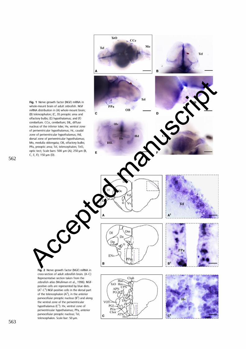
Fig.1 NGF mRNA in whole mount-brain of adult zebrafish. 480
NGF mRNA distribution in whole mount brain (A); telencephalon (B); preoptic area and olfactory 481
bulbs (C, D); hypothalamus (E), cerebellum (F). CCe: cerebellum; DIL: diffuse nucleus of the inferior 482
lobe; Hv: ventral zone of periventricular hypothalamus; Hc: caudal zone of periventricular 483
hypothalamus; Hd: dorsal zone of periventricular hypothalamus; Mo: medulla oblongata; OB: 484
olfactory bulbs; PPa: preoptic area; Tel: telencephalon; TeO: optic tect; Scale bar: (A) 500 μm; (B, 485
C, E, F) 250 μm; D: 150 μm. 486
487
Fig.2 NGF mRNA in cross-section of adult zebrafish brain. 488
(A – C) Representative section taken from the zebrafish atlas (Wullimann et al., 1996). NGF positive 489
cells are represented by blue dots. (A1 – C1) NGF positive cells in the dorsal part of telencephalon 490
(A1), in the anterior parvocellular preoptic nucleus (B1) and along the ventral zone of periventricular 491
hypothalamus (C1). Hv: ventral zone of periventricular hypothalamus; PPa: anterior parvocellular 492
preoptic nucleus; Tel: telencephalon. Scale bar: 50 μm. 493
494
Fig.3 NGF protein in the telencephalon. 495
(A) Representative section taken from the zebrafish atlas (Wullimann et al., 1996). NGF positive cells 496
are represented by green dots. (B - C) Cells positive to NGF in the medial (B) and lateral (C) part of 497
dorsal telencephalic area. (B1, C1) NGF positive cells co-marked with DAPI. Dl: lateral zone of 498
dorsal telencephalic area; Dm: medial zone of dorsal telencephalic area. Scale bar: (C, C’) 100 μm; 499
(B, B’)50 μm. 500
501
Fig.4 NGF protein in the forebrain and midbrain. 502
Accep
ted m
anus
cript

(A, B) Representative sections taken from the zebrafish atlas (Wullimann et al., 1996). NGF positive 503
cells are represented by green dots. Cells positive to NGF in the posterior zone of dorsal telencephalic 504
area (A1) and in periventricular gray zone of optic tect (B1). (A2, B2) NGF positive cells co-marked 505
with DAPI. TelV: telencephalic ventricle; Scale bar: 50 μm. 506
507
Fig.5 NGF protein in the hypothalamus. 508
(A) Representative section taken from the zebrafish atlas (Wullimann et al., 1996). NGF positive cells 509
are represented by green dots. (B – C) NGF protein (green) along the ventral hypothalamus at low 510
(B) and high (C) magnification. (C1) NGF positive cells co-marked with DAPI. Hv: ventral zone of 511
periventricular hypothalamus. Scale bar: (B) 100 μm; (C, C1) 50 μm. 512
513
Fig.6 NGF protein in the hypothalamus. 514
NGF positive cells in the diffuse nucleus of inferior lobe (DIL) at low (A) and high (A1, B) 515
magnification. In B1 NGF positive cells co-marked with DAPI. Scale bar: (A) 120 μm; (A1) 30 μm; 516
(B, B1) 20 μm. 517
518
Fig.7 NGF protein in the cerebellum. 519
(A) Representative section taken from the zebrafish atlas (Wullimann et al., 1996). NGF positive cells 520
are represented by green dots. (B, C) NGF positive cells in the Purkinje layer of cerebellum. In C’ 521
NGF positive cells co-marked with DAPI. CCe: cerebellar body; Scale bar: 50 μm. 522
523
Fig.8 NGF positive cells are close to aromatase B cells along telencephalic ventricle. 524
(A) Representative section of dorsal and ventral telencephalon taken from the zebrafish atlas 525
(Wullimann et al., 1996). NGF positive cells are represented by red dots and Aromatase B is 526
represented by black dots with thin lines indicating radial glia cytoplasmic processes. (B – F) Cross 527
sections of dorsal telencephalic area. Double staining for NGF (red) (B), Aromatase-B (green) (C) 528
Accep
ted m
anus
cript
and merge with DAPI (D). High magnification of NGF and aromatase B cells, closely intermingled 529
(E, F). Scale bar: (B – D) 40 μm; (E) 20 μm; (F) 10 μm. 530
531
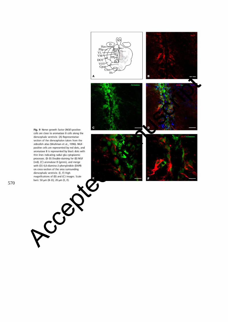
Fig.9 NGF positive cells are close to aromatase B cells along diencephalic ventricle. 532
(A) Representative section of diencephalon taken from the zebrafish atlas (Wullimann et al., 1996). 533
NGF positive cells are represented by red dots and Aromatase B is represented by black dots with 534
thin lines indicating radial glia cytoplasmic processes. (B – D) Double staining for NGF (red) (B), 535
Aromatase-B (green) (C) and merge with DAPI (D) on cross-section of area surrounding diencephalic 536
ventricle. (E -F) high magnifications of B and C images. Scale bar: (B – D) 50 μm; (E _F) 20 μm. 537
538
Fig.10 NGF positive cells are intermingled with PCNA positive cells. 539
(A) Representative section of dorsal and ventral telencephalon taken from the zebrafish atlas 540
(Wullimann et al., 1996) NGF positive cells are represented by green dots and PCNA positive cells 541
by red dots. (B – D) Double staining for NGF (green) (B), PCNA(red) (C), merge with DAPI and 542
high-magnification of a zoom area (D) on cross-sections through the telencephalon. Scale bar: 50 μm, 543
and particular of the region in (D) 30 μm. 544
545
Fig.11 NGF immunoreactivity is colocalized with MAP2 in cells of central zone of dorsal 546
telencephalic area. 547
Representative section dorsal and ventral telencephalon taken from the zebrafish atlas (Wullimann et 548
al., 1996). NGF positive cells are represented by red dots and MAP2 positive cells by green dots. (B 549
– D) Double staining for NGF (red) (B), MAP2 (green) (C) and merge (D) on cross-sections through 550
the telencephalon. Scale bar: 20 μm 551
552
Supplementary material 553
Accep
ted m
anus
cript

Negative controls performed by NGF antiserum preincubated with its homologous antigen did not 554
show any reactivity. Scale bar: (A) 100 μm; (B) 50 μm 555
556
Accep
ted m
anus
cript

Table 1. Distribution of NGF mRNA and protein in the brain of adult zebrafish. The scheme was done following 557 qualitative and not quantitative criteria. 558 559
Brain region NGF mRNA NGF protein
Olfactory bulbs
Glomerular layer ++
External cellular layer + +
Internal cellular layer + +
Dorsal telencephalic area
Medial zone of dorsal telencephalic area + ++
Dorsal zone of dorsal telencephalic area + +
Lateral zone of dorsal telencephalic area + +
Central zone of dorsal telencephalic area ++ ++
Posterior zone of dorsal telencephalic area ++ +
Ventral telencephalic area
Ventral-dorsal part + ++
Ventral-central part + ++
Preoptic area
Magnocellular preoptic nucleus + +
Parvocellular preoptic nucleus, anterior part ++ ++
Parvocellular preoptic nucleus, posterior part + ++
Epithalamus
Dorsal habenular nucleus + +
Ventral habenular nucleus + +
Dorsal thalamus
Central posterior thalamic nucleus + ++
Ventral thalamus
Ventromedial thalamic nucleus + ++
Ventrolateral thalamic nucleus + +
Posterior Tuberculum
Posterior tuberal nucleus ++ ++
Lateral preglomerular nucleus + +
Medial preglomerular nucleus + ++
Hypothalamus
Diffuse nucleus of the inferior lobe + ++
Ventral zone of periventricular hypothalamus ++ ++
Dorsal zone of periventricular hypothalamus + +
Caudal zone of periventricular hypothalamus + +
Mammillary body + +
Synencephalon
Nucleus of the medial longitudinal fascicle + +
Optic Tectum
Periventricular grey zone of optic tectum ++ ++
Deep white zone ++
Central zone + +
Superficial white grey zone + ++
Longitudinal torus + +
Torus semicircularis
Central nucleus of semicircular torus + +
Tegmentum
Superior reticular formation + ++
Cerebellum
Molecular layer + +
Purkinje cell layer ++ ++
Medulla oblongata
Inferior reticular formation + +
560 561
Accep
ted m
anus
cript
562
563
Accep
ted m
anus
cript

564
Accep
ted m
anus
cript

565
566 Accep
ted m
anus
cript

567
568
Accep
ted m
anus
cript

569
Accep
ted m
anus
cript
570
Accep
ted m
anus
cript

571
Accep
ted m
anus
cript