Nephrolithiasis Adnan Alsaka M.D. Nephrology Fellow.
69
Nephrolithiasis Adnan Alsaka M.D. Nephrology Fellow
-
Upload
nelson-randolph-lynch -
Category
Documents
-
view
224 -
download
2
Transcript of Nephrolithiasis Adnan Alsaka M.D. Nephrology Fellow.

Nephrolithiasis
Adnan Alsaka M.D.
Nephrology Fellow

Nephrolithiasis is estimated to produce medical costs of $2.1 billion per year in the US
Incidence is 0.5% Prevalence is 5.2%

More common in Asians and whites than in Native Americans, Africans, African Americans
male-to-female ratio of 3:1 Stones due to infection (struvite) are
more common in women
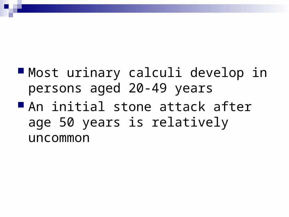
Most urinary calculi develop in persons aged 20-49 years
An initial stone attack after age 50 years is relatively uncommon

Pathophysiology

1- supersaturation of the urine by stone-forming constituents
Crystals or foreign bodies can act as nidi, upon which ions from the supersaturated urine form microscopic crystalline structures

Randall plaque
is deposition of stone material on a renal papillary calcium phosphate nidus
Calcium phosphate precipitates in the basement membrane of the thin loops of Henle, erodes into the interstitium, and then accumulates in the subepithelial space of the renal papilla
The subepithelial deposits, eventually erode through the papillary urothelium

Causes of Nephrolithiasis

Hypercalciuria
most common metabolic abnormality can be subdivided into absorptive,
resorptive, and renal-leak categories

Absorptive Hypercalciuria
related to increased intestinal absorption of calcium (associated with excess dietary calcium and/or overactive calcium absorption mechanisms)
the treatment may include modest dietary calcium restriction, thiazide diuretics, oral calcium binders

Resorptive hypercalciuria
related to excess resorption of calcium from bone (i.e., hyperparathyroidism)
Treatment requires parathyroidectomy

Renal-leak hypercalciuria
less common than absorptive hypercalciuria related to an inability of the renal tubules to
properly reclaim calcium in the glomerular filtrate
Usually associated with secondary hyperparathyroidism and is best managed with thiazide diuretics
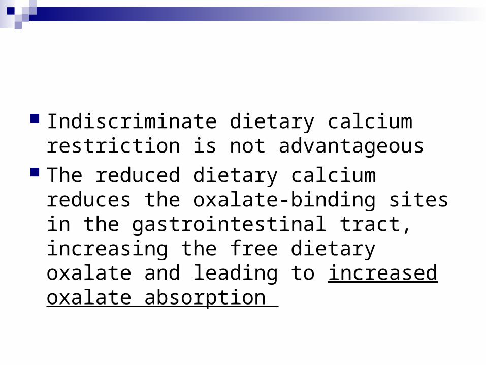
Indiscriminate dietary calcium restriction is not advantageous
The reduced dietary calcium reduces the oxalate-binding sites in the gastrointestinal tract, increasing the free dietary oxalate and leading to increased oxalate absorption
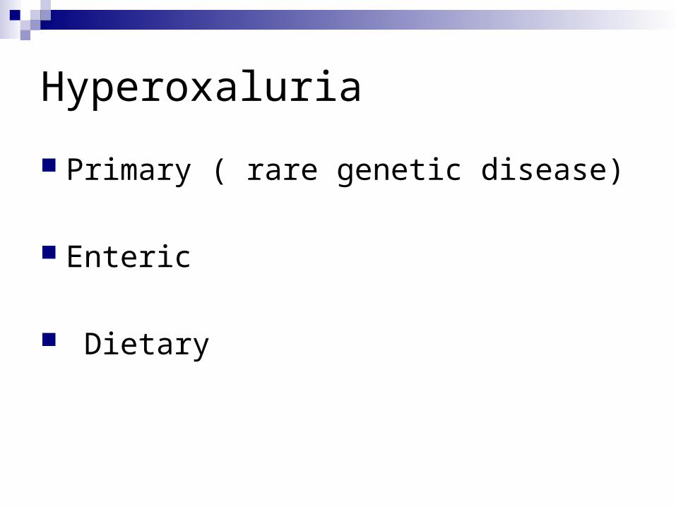
Hyperoxaluria
Primary ( rare genetic disease)
Enteric
Dietary

Primary hyperoxaluria
Type I mutation of AGXT gene on chromosome 2 that
codes for alanine glyoxylate aminotransferase Type II mutation of GRHPR gene on chromosome 9 that
codes for glyoxylate reductase and hydroxypyruvate reductase.

Enteric
Due to malabsorption Associated with chronic diarrhea or short
bowel syndrome Normally, calcium binds to intestinal
oxalate reducing its absorption

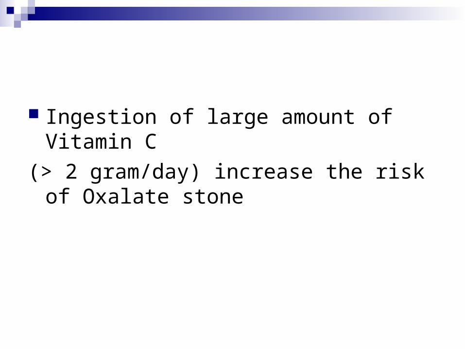
Ingestion of large amount of Vitamin C
(> 2 gram/day) increase the risk of Oxalate stone

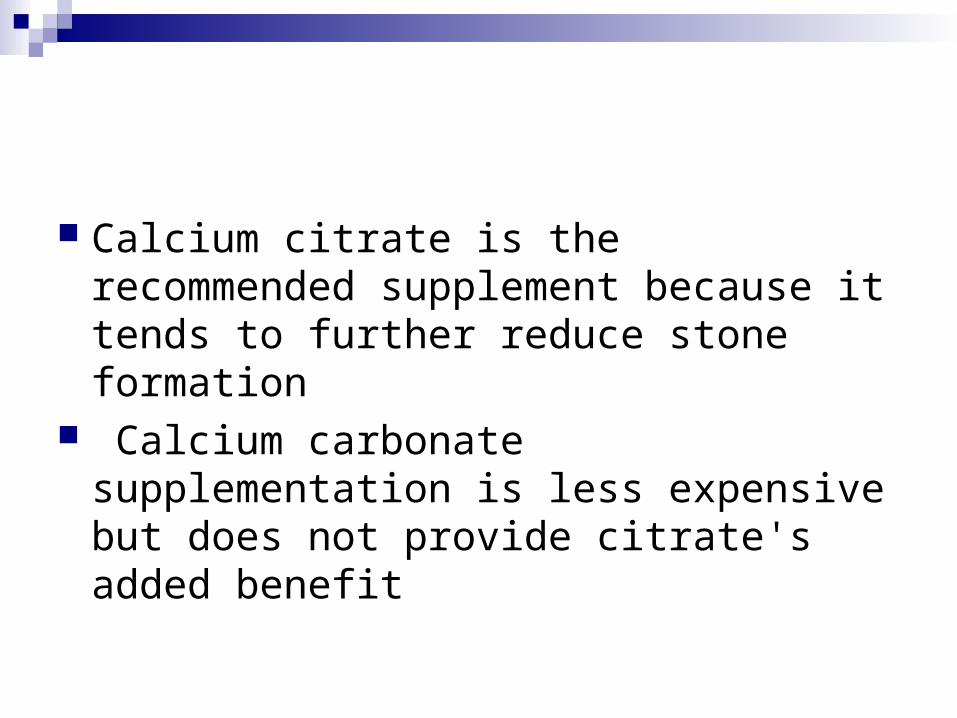
Calcium citrate is the recommended supplement because it tends to further reduce stone formation
Calcium carbonate supplementation is less expensive but does not provide citrate's added benefit
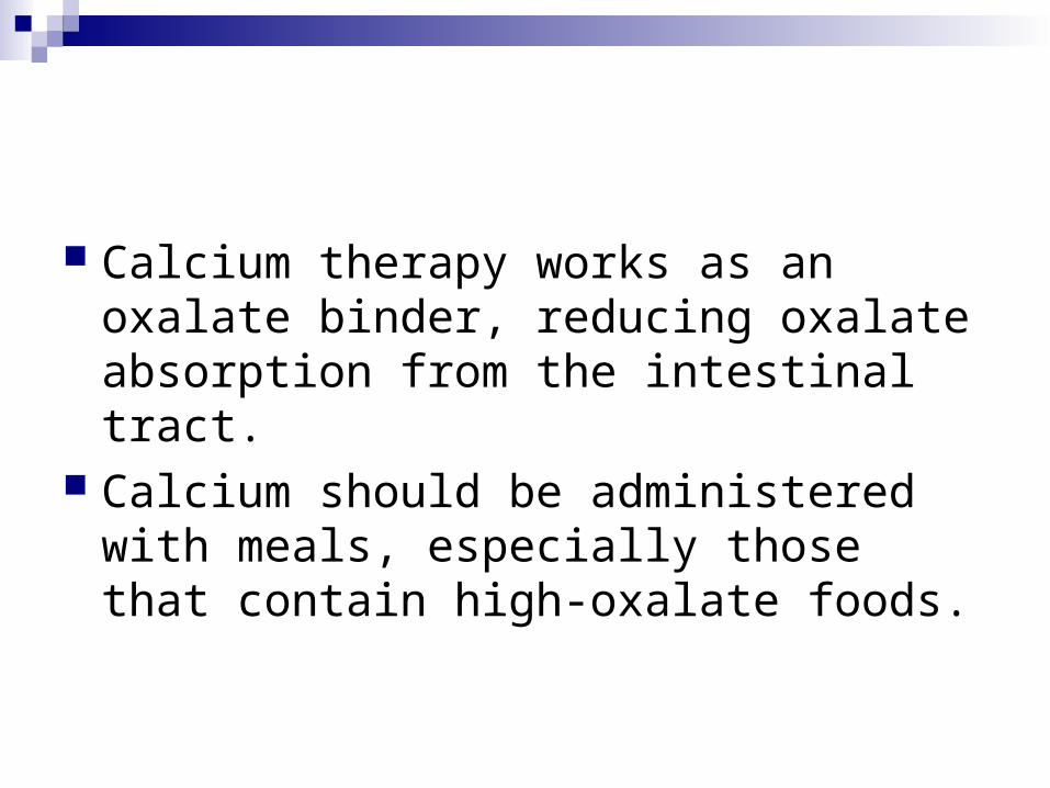
Calcium therapy works as an oxalate binder, reducing oxalate absorption from the intestinal tract.
Calcium should be administered with meals, especially those that contain high-oxalate foods.
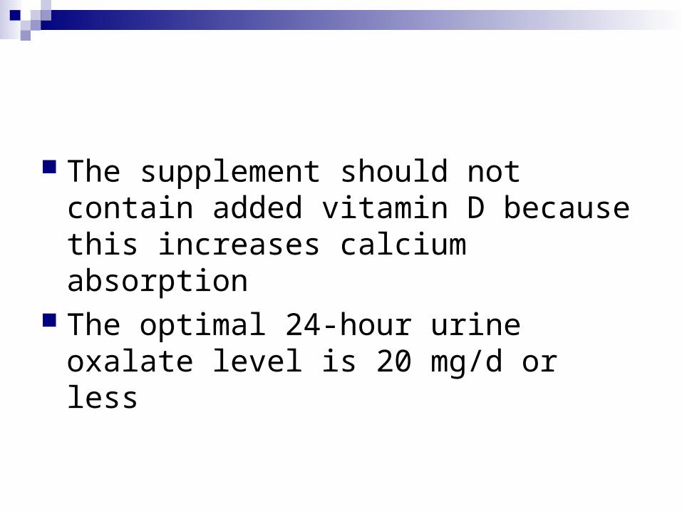
The supplement should not contain added vitamin D because this increases calcium absorption
The optimal 24-hour urine oxalate level is 20 mg/d or less
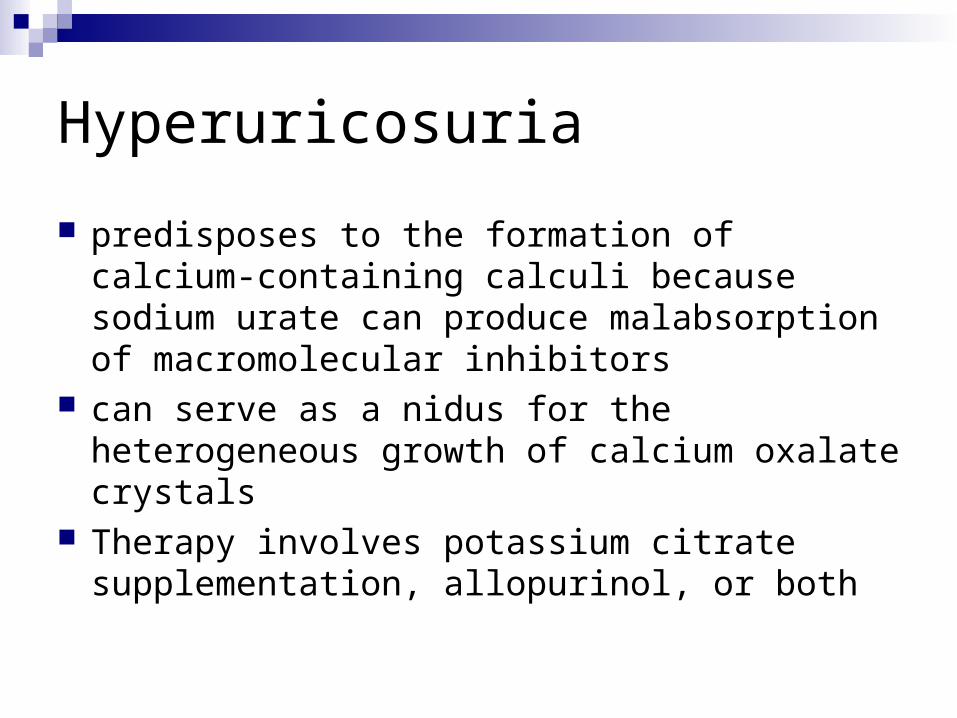
Hyperuricosuria
predisposes to the formation of calcium-containing calculi because sodium urate can produce malabsorption of macromolecular inhibitors
can serve as a nidus for the heterogeneous growth of calcium oxalate crystals
Therapy involves potassium citrate supplementation, allopurinol, or both
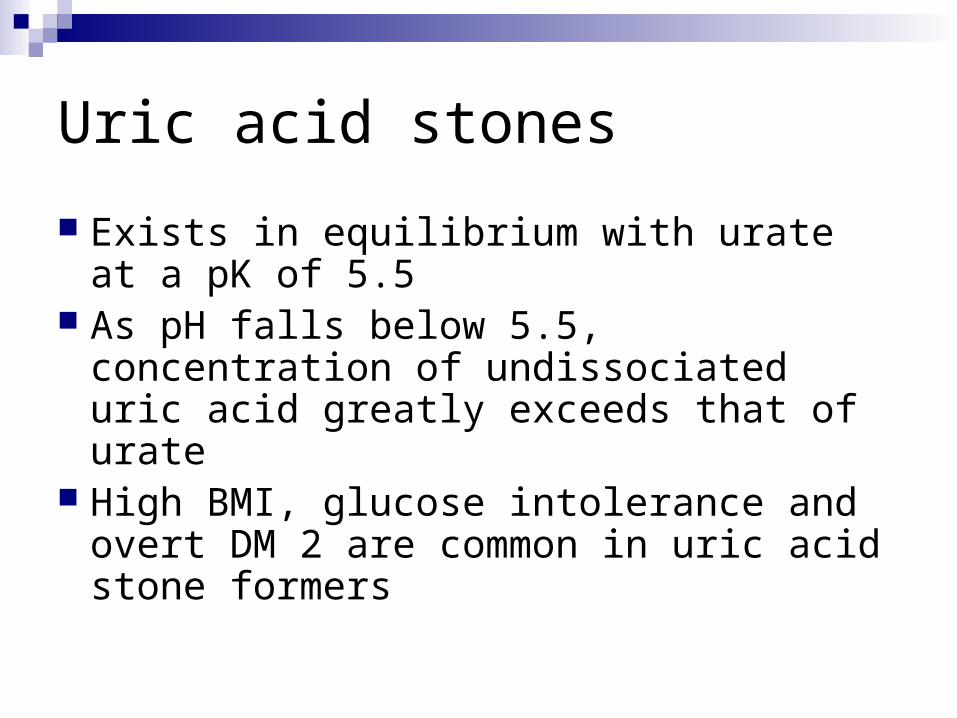
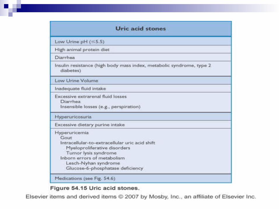
Uric acid stones
Exists in equilibrium with urate at a pK of 5.5
As pH falls below 5.5, concentration of undissociated uric acid greatly exceeds that of urate
High BMI, glucose intolerance and overt DM 2 are common in uric acid stone formers
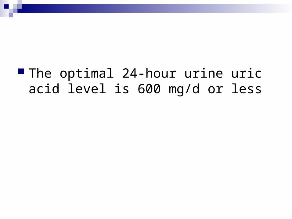
The optimal 24-hour urine uric acid level is 600 mg/d or less
Sodium and phosphorus
Elevated urinary sodium levels are almost always associated with dietary indiscretions
Decreasing the oral sodium (<2.5 gm/day) intake can decrease calcium excretion by increasing proximal calcium absorption

Hyperphophaturia
Renal phosphate leak: high urinary phosphate levels, low serum phosphate levels, high serum 1,25 vitamin D-3 (calcitriol) levels, and hypercalciuria.
This type of hypercalciuria is uncommon and does not respond well to standard therapies

Phosphate supplements are used to correct the low serum phosphate level, which then decreases the inappropriate activation of vitamin D originally caused by the hypophosphatemia
Citrate and magnesium
are important chemical inhibitors of stone formation
Hypocitraturia is one of the most common metabolic defects that predispose to stone formation
24-hour urine citrate levels of 320 mg/d is the normal threshold

Magnesium is a more recently recognized inhibitor of stone formation, and the clinical role of magnesium replacement therapy is less well defined than that of citrate

High Protein diet
The institution of a high protein diet (2 g/kg per day) adversely effects the metabolic parameters determining the risk of calcium stone formation
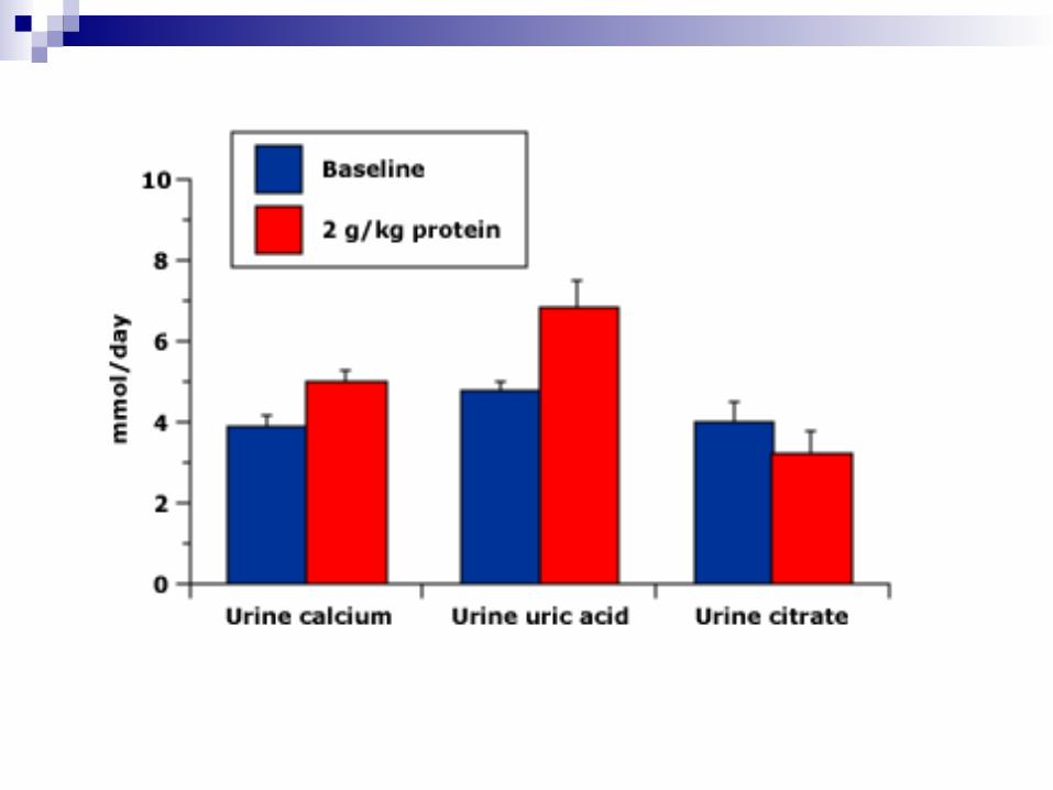

Protein restriction reduce Ca oxalate formation by:
less acidic urine reduces the formation of UA stones
less acidic urine favors the trivalent form of the citrate anion, which is less able to bind the Na/citrate cotransporter in PT
Reduction of daily acid load reduces bone buffering (calcium resorption) to reduce ca excretion

Struvite Stones
form in chronic upper urinary tract infection due to a urease-producing organism
are composed of magnesium ammonium phosphate (struvite) and calcium carbonate-apatite
Normal urine is undersaturated with ammonium phosphate, and struvite stone occurs only when ammonia production is increased and the urine pH is elevated to decrease the solubility of phosphate

Struvite Stones
may grow rapidly over a period of weeks to months can develop into a staghorn calculus involving the entire renal collecting system


Cystine stones
only develop in patients with cystinuria (an autosomal recessive disorder)
due to the poor solubility of cystine in the urine
Autosomal recessive or dominant Increased excretion of COAL (cystine,
ornithine, arginine, lysine)

Cystine superstauration occurs at cystine concentration > 250 mg/L
its solubility will gradually increase as pH increases from 6.5 to 7.5
The hallmark of treatment is water, water and more water.


Cystine crystals

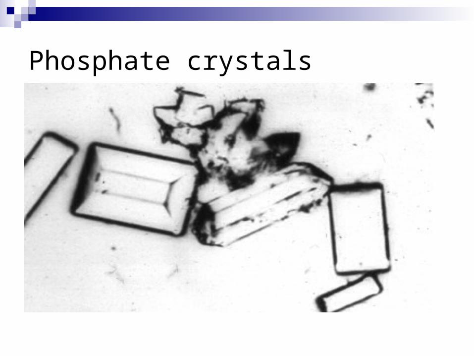
Phosphate crystals

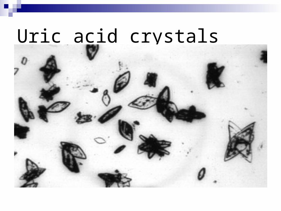
Uric acid crystals

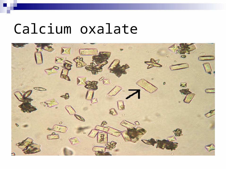
Calcium oxalate
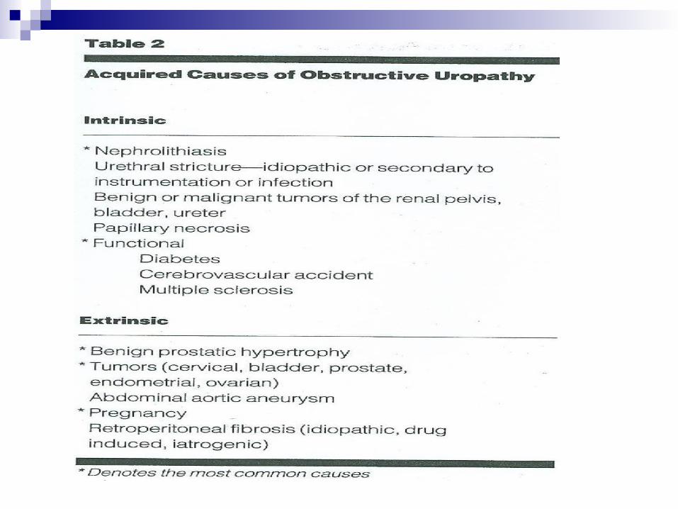
Obstructive Uropathy
refers to obstruction of the urinary tract at any point from the renal pelvis to the distal urethra
The acute or chronic loss of kidney function resulting from obstruction is termed obstructive nephropathy

The likelihood of functional impairment
depends on The duration of the obstruction
Whether it is partial or complete
Whether it involves one or both functioning kidneys
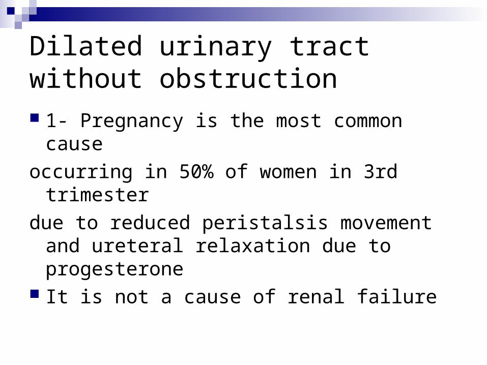
Dilated urinary tract without obstruction 1- Pregnancy is the most common cause
occurring in 50% of women in 3rd trimester
due to reduced peristalsis movement and ureteral relaxation due to progesterone
It is not a cause of renal failure
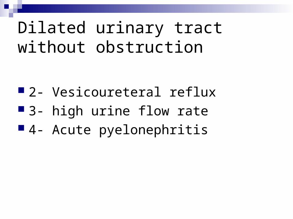
Dilated urinary tract without obstruction
2- Vesicoureteral reflux 3- high urine flow rate 4- Acute pyelonephritis

Effects on glomerular filtration

The first 2-3 hours: the release of Prostaglandin from Macula
densa in response to distal tubular flow will lead to vasodilation
GFR is maintained because the increase in tubular pressure is offset by increase in tubular blood flow
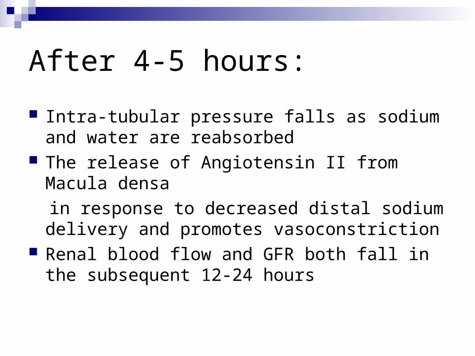
After 4-5 hours:
Intra-tubular pressure falls as sodium and water are reabsorbed
The release of Angiotensin II from Macula densa
in response to decreased distal sodium delivery and promotes vasoconstriction
Renal blood flow and GFR both fall in the subsequent 12-24 hours

Tubular functions during obstruction Early on, urine indices are suggestive of a
pre-renal insult due to enhanced absorption of sodium and water in response to decreased distal delivery
With more prolonged obstruction, FENA >1 as sodium reabsorption falls
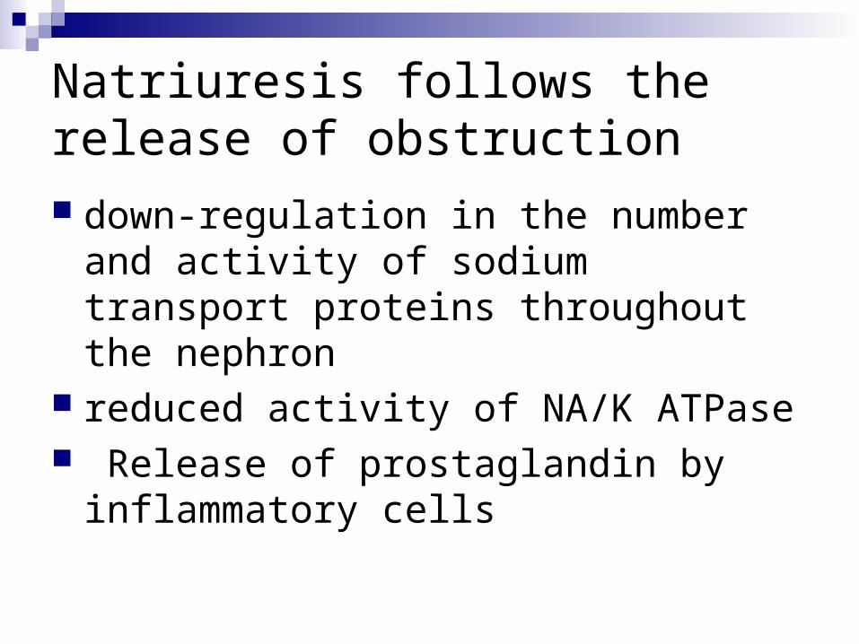
Natriuresis follows the release of obstruction down-regulation in the number and activity
of sodium transport proteins throughout the nephron
reduced activity of NA/K ATPase Release of prostaglandin by inflammatory
cells
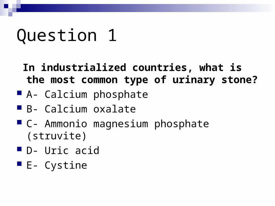
Question 1
In industrialized countries, what is the most common type of urinary stone?
A- Calcium phosphate B- Calcium oxalate C- Ammonio magnesium phosphate (struvite) D- Uric acid E- Cystine

Question 1
In industrialized countries, what is the most common type of urinary stone?
A- Calcium phosphate B- Calcium oxalate C- Ammonio magnesium phosphate (struvite) D- Uric acid E- Cystine
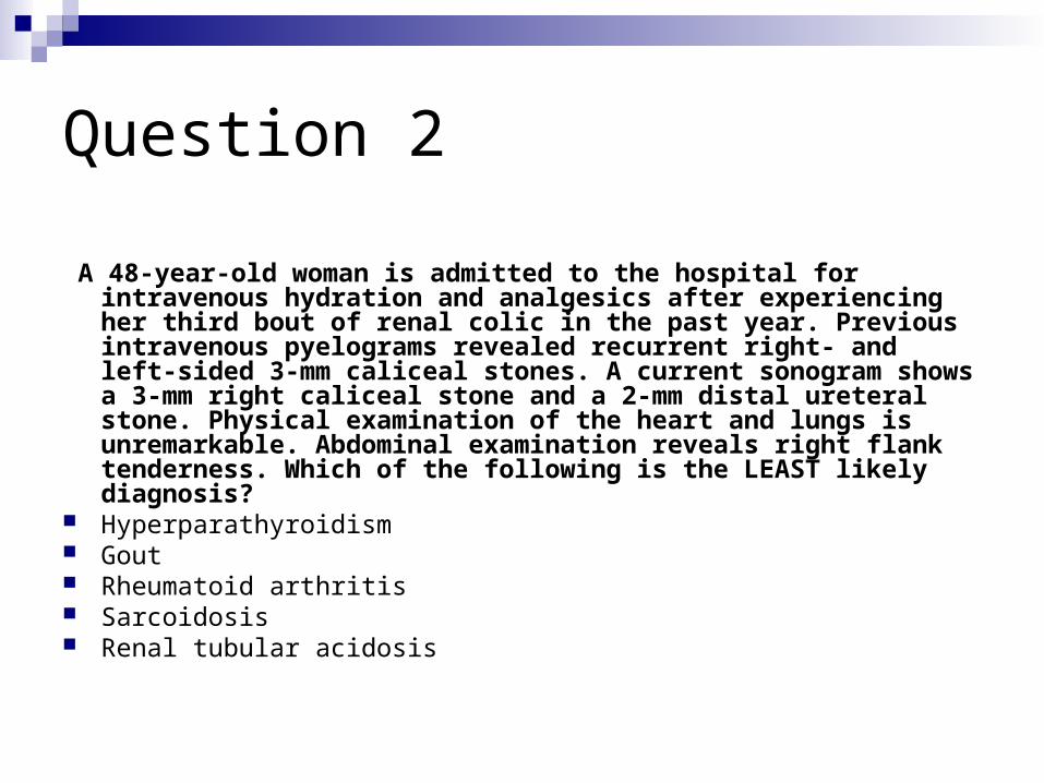
Question 2
A 48-year-old woman is admitted to the hospital for intravenous
hydration and analgesics after experiencing her third bout of renal colic in the past year. Previous intravenous pyelograms revealed recurrent right- and left-sided 3-mm caliceal stones. A current sonogram shows a 3-mm right caliceal stone and a 2-mm distal ureteral stone. Physical examination of the heart and lungs is unremarkable. Abdominal examination reveals right flank tenderness. Which of the following is the LEAST likely diagnosis?
Hyperparathyroidism Gout Rheumatoid arthritis Sarcoidosis Renal tubular acidosis

Question 2
3. A 48-year-old woman is admitted to the hospital for intravenous hydration and analgesics after experiencing her third bout of renal colic in the past year. Previous intravenous pyelograms revealed recurrent right- and left-sided 3-mm caliceal stones. A current sonogram shows a 3-mm right caliceal stone and a 2-mm distal ureteral stone. Physical examination of the heart and lungs is unremarkable. Abdominal examination reveals right flank tenderness. Which of the following is the LEAST likely diagnosis?
Hyperparathyroidism Gout Rheumatoid arthritis Sarcoidosis Renal tubular acidosis
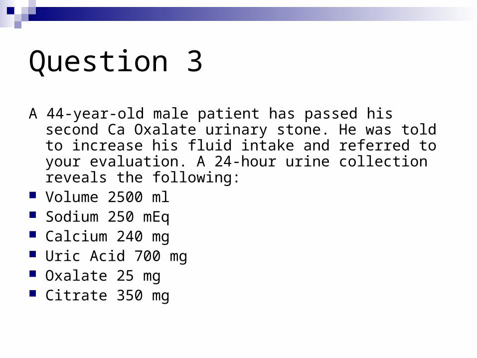
Question 3
A 44-year-old male patient has passed his second Ca Oxalate urinary stone. He was told to increase his fluid intake and referred to your evaluation. A 24-hour urine collection reveals the following:
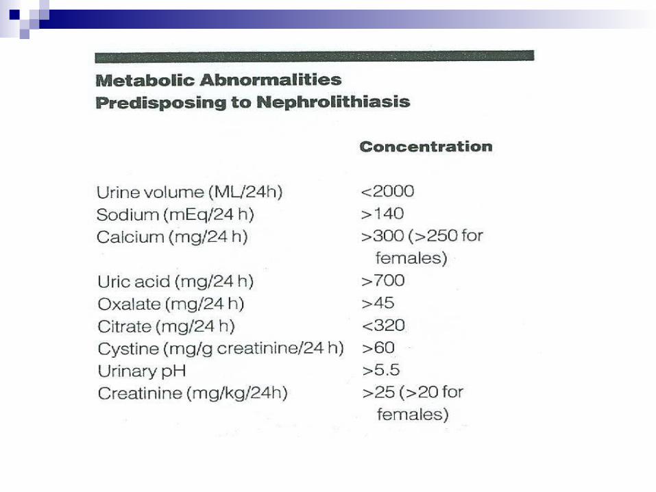
Volume 2500 ml Sodium 250 mEq Calcium 240 mg Uric Acid 700 mg Oxalate 25 mg Citrate 350 mg
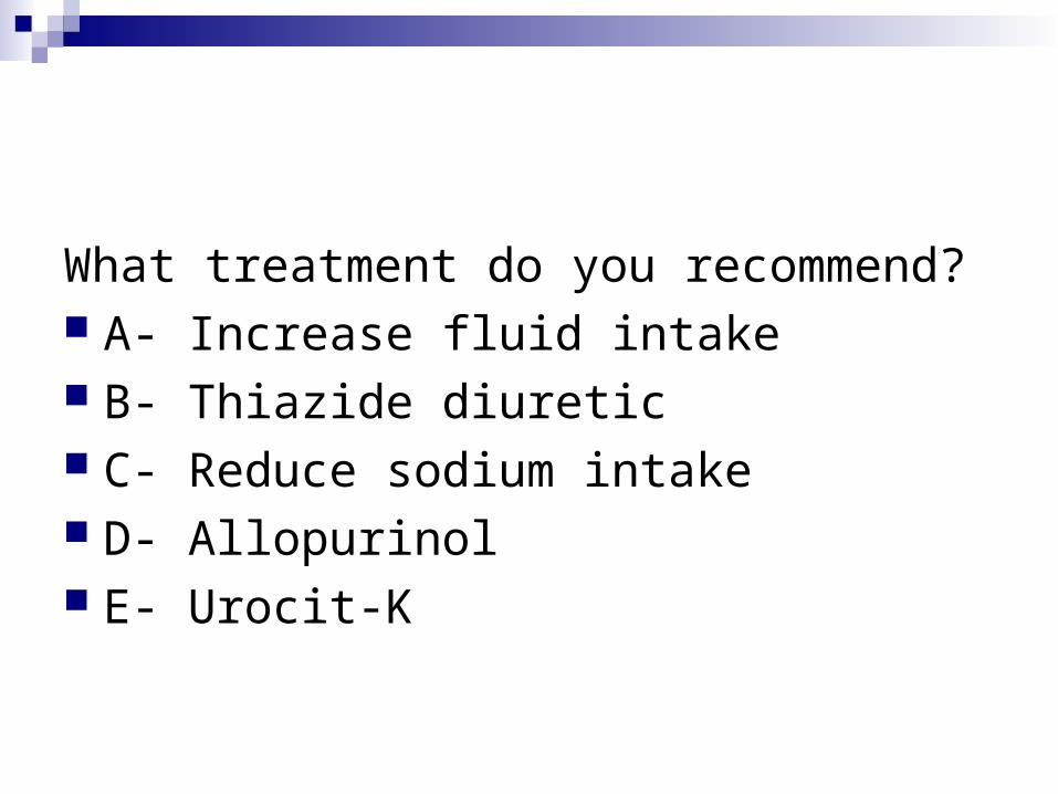
What treatment do you recommend? A- Increase fluid intake B- Thiazide diuretic C- Reduce sodium intake D- Allopurinol E- Urocit-K

Thank you