
Neonatal Cranial Ultrasound
28
By: Dr. Nour-Eldin Mohammed Reference: Gerda van Wezel-Meijler
Transcript of Neonatal Cranial Ultrasound

By:Dr. Nour-Eldin Mohammed
Reference: Gerda van Wezel-Meijler

Safe Bedside- compatible Reliable Early imaging Serial imaging:
Brain maturationEvolution of lesions
Inexpensive Suitable for screening


Exclude/demonstrate cerebral pathology Assess timing of injury Assess neurological prognosis Help make decisions on continuation of
neonatal intensive care Optimise treatment and support

Transucers : 5–7.5–10 MHz Appropriately sized Standard examination: use 7.5–8 MHz Tiny infant and/or superficial structures: use
additional higher frequency (10 MHz) Large infant, thick hair, and/or deep
structures: use additional lower frequency (5 MHz)


Anterior FontanelThe Standard view window
Posterior FontanelSupplementary view window
Mastoid FontanelSupplementary view window
TemporalSupplementary view window

Coronal Views (at least 6 standard planes)


Sagittal Views (at least 5 standard planes)


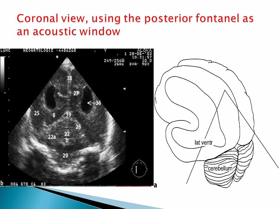


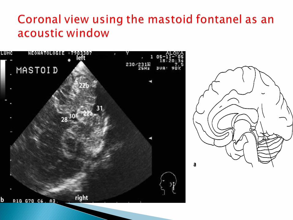

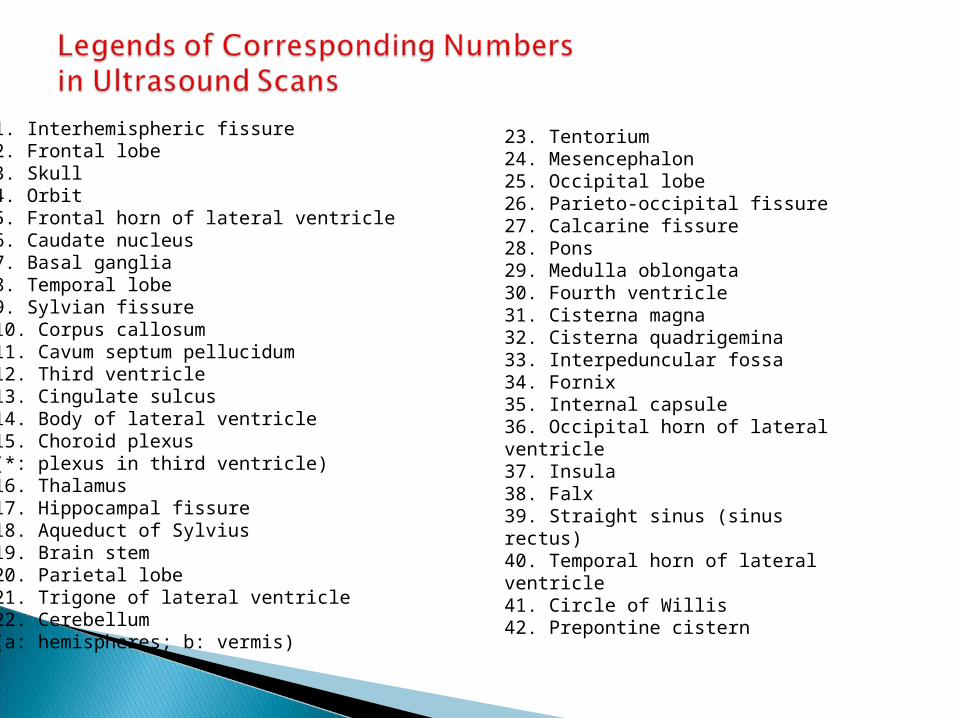
23. Tentorium24. Mesencephalon25. Occipital lobe26. Parieto-occipital fissure27. Calcarine fissure28. Pons29. Medulla oblongata30. Fourth ventricle31. Cisterna magna32. Cisterna quadrigemina33. Interpeduncular fossa34. Fornix35. Internal capsule36. Occipital horn of lateralventricle37. Insula38. Falx39. Straight sinus (sinus rectus)40. Temporal horn of lateralventricle41. Circle of Willis42. Prepontine cistern
1. Interhemispheric fissure2. Frontal lobe3. Skull4. Orbit5. Frontal horn of lateral ventricle6. Caudate nucleus7. Basal ganglia8. Temporal lobe9. Sylvian fissure10. Corpus callosum11. Cavum septum pellucidum12. Third ventricle13. Cingulate sulcus14. Body of lateral ventricle15. Choroid plexus(*: plexus in third ventricle)16. Thalamus17. Hippocampal fissure18. Aqueduct of Sylvius19. Brain stem20. Parietal lobe21. Trigone of lateral ventricle22. Cerebellum(a: hemispheres; b: vermis)

Thank you


