NEONATAL ASPHYXIA Prof. Maria Stamatin MD,PhD CUZA – VODA Clinical Hospital of Obstetrics &...
40
NEONATAL ASPHYXIA Prof. Maria Stamatin MD,PhD CUZA – VODA Clinical Hospital of Obstetrics & Gynaecology Iasi, NICU
-
Upload
clifton-warren -
Category
Documents
-
view
225 -
download
1
Transcript of NEONATAL ASPHYXIA Prof. Maria Stamatin MD,PhD CUZA – VODA Clinical Hospital of Obstetrics &...
NEONATAL ASPHYXIA
Prof. Maria Stamatin MD,PhD CUZA – VODA Clinical Hospital of Obstetrics & Gynaecology Iasi, NICU

NEONATAL ASPHYXIA Neonatal asphyxia is the result of a problem that occurs
during: Fetal life Labor or Delivery and leads to delayed onset of first respiration (at the end of first
two minutes after birth). If lung expansion does not occur in the minutes following birth and the infant is unable to establish ventilation and pulmonary perfusion, a progressive cycle of worsening hypoxemia, hipercapnia and metabolic acidosis evolves. So, we may define asphyxia by the following parameters:
1. pH from umbilical artery less than 7;2. Apgar score less than 3 at one minute and 5 at ten minutes;3. Signs of hypoxic-ischemic encephalopathy like hyper- or
hypotonia;4. Dysfunctions of other organs (cardiovascular system, liver,
bowel)

NEONATAL ASPHYXIA
The incidence of perinatal asphyxia is usually related with gestational age and birth weight: 6‰ at term newborn and much higher at premature babies under 36 weeks of gestation. In 50-91% of cases, asphyxia takes place ante- and intrapartum, generally by hypoxic traumatism and only in 9% of cases postpartum, by ventilation disturbances, malformations, meconium aspiration syndrome, etc.

NEONATAL ASPHYXIA Common causes and risk factors of this major emergency are: I. Maternal diseases: - Diabetes mellitus and/or gestational diabetes; - Arterial hypertension and pregnancy toxemia; - Pulmonary and cardiac diseases; - Maternal infections; - Anemia; - Drugs, maternal sedation; - Alcoholism;
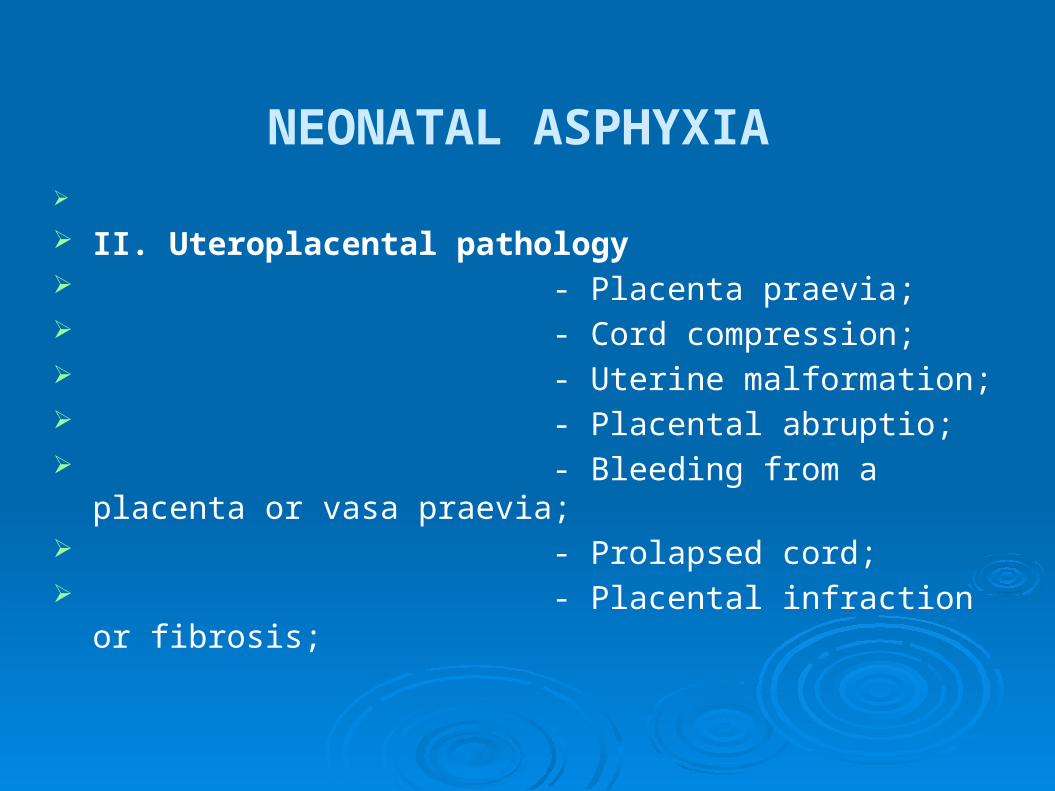
NEONATAL ASPHYXIA II. Uteroplacental pathology - Placenta praevia; - Cord compression; - Uterine malformation; - Placental abruptio; - Bleeding from a placenta or vasa
praevia; - Prolapsed cord; - Placental infraction or fibrosis;

NEONATAL ASPHYXIA
III. Fetal pathology - Congenital and genetics anomalies; - Prematurity; - Intrauterine growth retardation; - Postmaturity; - Multiple pregnancy; - Hemolytic anemia by fetal
isoimunization; - Fetal infections; - Hydramnios;
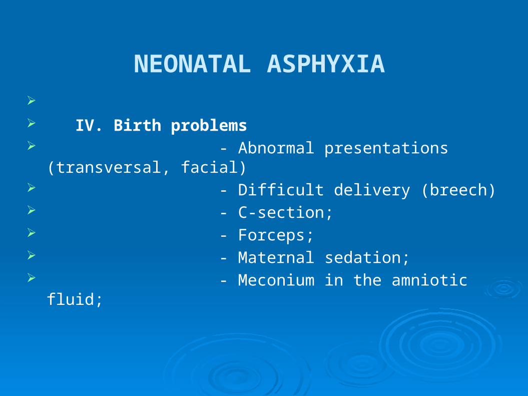
NEONATAL ASPHYXIA IV. Birth problems - Abnormal presentations (transversal, facial) - Difficult delivery (breech) - C-section; - Forceps; - Maternal sedation; - Meconium in the amniotic fluid;
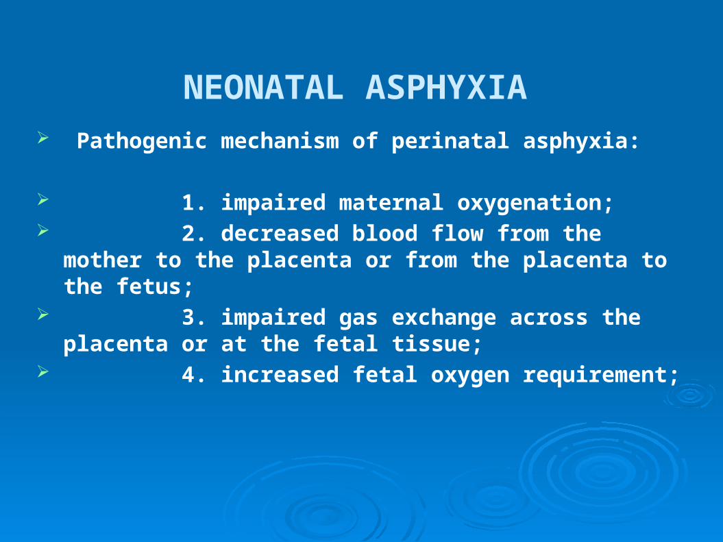
NEONATAL ASPHYXIA Pathogenic mechanism of perinatal asphyxia:
1. impaired maternal oxygenation; 2. decreased blood flow from the mother to
the placenta or from the placenta to the fetus; 3. impaired gas exchange across the
placenta or at the fetal tissue; 4. increased fetal oxygen requirement;

NEONATAL ASPHYXIA For example, maternal diabetes can cause perinatal
asphyxia by worsening oxygen transport from the mother to the placenta, with circulatory uteroplacental deficiency. These babies present risk for prematurity, HMD, macrosomia, obstetrical traumatism, congenital malformations.
Maternal hypertension and pregnancy toxemia can also produce deficiency of uteroplacental circulation by fibroid degeneration of chorial vilosities.
Pulmonary and cardiac chronical diseaes impaires the oxygen transport to the fetus (hypoxic hypoxia)
Drugs administered to mother, can produce respiratory depression to the newborn.

NEONATAL ASPHYXIA
Uteroplacental risck factors can produce perinatal asphyxia by changing uteroplacental blood flow and decreasing fetal 02.
Fetal risck factors, also impaires 02 transport to fetus and increase fetal 02 requirements.
Any pathology linked with delivery can lead to asphyxia by decreasing neonatal oxygenation.

NEONATAL ASPHYXIA Pathophysiology Asphyxia means hypoxia with hypoxemia, hypercarbia
with or without metabolic acidosis. These changes cause shunting of blood to the brain, heart and adrenals and away from the lungs, gut, liver, kidneys, spleen, bone, skeletal muscle and skin ("diving reflex").
In mild hypoxia there is: - A decreased heart rate - Slight increase in blood pressure to maintain cerebral
perfusion; - Increased central venous pressure; - Little changes in cardiac output;

NEONATAL ASPHYXIA
As asphyxia progresses, with severe hypoxia and acidosis, there is a:
- Decreased heart rate; - Decreased cardiac output; An initial increased then falling blood pressure as
oxidative phosphorylation fails and energy reserves become depleted;
Although the degree of neurological perturbance depends of the severity of asphyxia, it isn't always a strict correlation.

NEONATAL ASPHYXIAPerinatal Asphyxia
HYPERCAPNEEA HYPOXIA
ACIDOSIS
Cerebral
HYPOXEMIAHEMORRHAGE
SEIZURES
HYPOXIC ISCHEMIC ENCEPHALOPATHY
↓ ATP ↓ enzymatic activity
↑K Increase extracell
Acidosis
INCREASE CEREBRAL BLOOD FLOW
Myocardial dysfunction
Cardiac failure
Decrease surfactant synthesis
HMD
Arterial VASOCONSTRICTION
Peripheral bed
Mesenteric bed NEC
Renal bed ATN
Pulmonary bed

NEONATAL ASPHYXIA Clinical forms: Clinical forms of perinatal asphyxia are correlated with
severity and the duration of hypoxia and with association of hypoxia with hypercarbia and metabolic acidosis. A classic classification of asphyxia was:
A. Mild asphyxia (Apgar score 6—7) which requires only tactile stimulation for initiating breathing;
B. Medium asphyxia (Apgar score 4-5) which requires bag and mask ventilation with 100% O2, to initiate breathing;
C. Severe asphyxia (Apgar score 0-3) which requires bag and mask ventilation and medication;
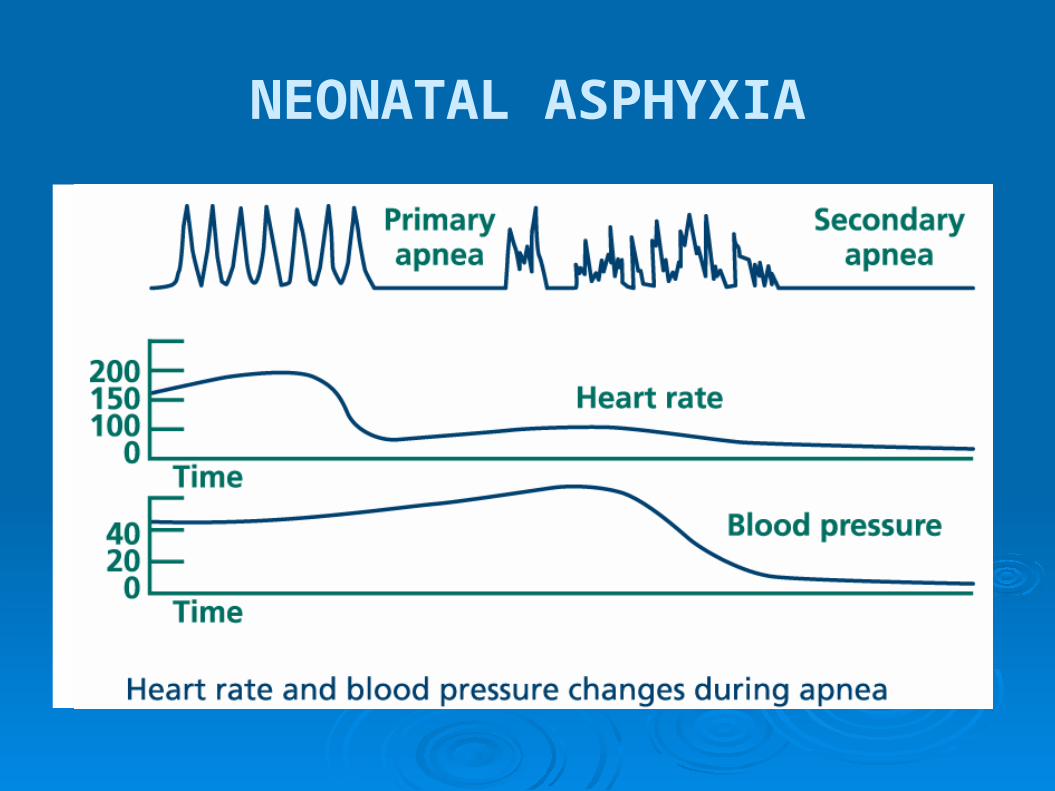
NEONATAL ASPHYXIA The disturbances caused by hypoxia, hypercarbia and acidosis,
intrauterine or during or post delivery, present a well defined order: 1. Primary apnea - the absence of 02 at fetus or newborn is followed by
quickly and irregular breathings, then gasping and cessation of breathings for approx. one minute, with bradycardia. The newborn is cyanotic, with spontaneous movement of lips and eyelids, with normal blood pressure. A mild stimulation with nasogastric tube or tactile stimulation can induce the initiation of breathings. In classic literature, this form of asphyxia was known as "blue asphyxia".
2. Secondary apnea if hypoxia continues, the respiration becomes weaker and slower, the baby presents a last gasp and passes into secondary apnea. During this period heart rate and arterial blood pressure decreased until zero, the newborn is pale and doesn't answer at any stimulation. Bag and mask ventilation with 100% 02 must be initiated to increase tissue perfusion. In classic literature this form of asphyxia was known as "white asphyxia".
NEONATAL ASPHYXIA
NEONATAL ASPHYXIA There is no method to differentiate primary from secondary apnea, at
birth, therefore, any asphyxiated newborn must be considered in secondary apnea and resuscitation must be initiated at once, because the duration of secondary apnea is strictly correlated with the severity of hypoxic-ischemic cerebral injury. Asphyxia can be assessed prenatally, intrapartum and postpartum.
Perinatal assessment A. Antepartum tests -generally rely on biophysical studies, which
require a certain degree of fetal neurophysiologic maturity. 1.Fetal movement-Fetuses normally have a sleep-wake cycle, and
mothers generally perceive a diurnal variation in fetal activity. Active periods average 30 to 40 minutes. Periods of inactivity greater than 1 hour are unusual in a healthy fetus and should alert the physician to the possibility of fetal compromise like asphyxia insult.
2.The nonstress test (NTS) - is based on principle that fetal activity results in a reflex acceleration in fetal heart rate. The required fetal maturity is typically reached by about 32 weeks of gestation. Absence of these accelerations in a fetus who previously demonstrate them may indicate that hypoxia has sufficiently depressed the central nervous system to inactivate the cardiac reflex.

NEONATAL ASPHYXIA The test is performed by monitoring fetal heart
rate either through a Doppler ultrasound device or through skin surface electrodes on maternal abdomen. Uterine activity is simultaneously reordered through a tocodynamometer or by palpation by trained personnel. The test result may be reactive, nonreactive, or inadequate.
The criteria for a reactive test are the following: 1.normal heart rate between 120-160 bpm 2.normal beat to beat variability (5 bpm) 3.two accelerations of least 15 beats per minute lasting
for non-less than 15 seconds each within a 20 - minute period. A nonreactive test fails to meet the three criteria. If an adequate fetal heart tracing cannot be obtained for any reason, the test is considered inadequate.

3.The contraction stress test (CST)-is based on idea that uterine contractions can compromise unhealthy fetus. The pressure generated during contractions can briefly reduce or eliminate perfusion of the intervillous space. A healthy fetoplacental unit has sufficient reserve to tolerate this short reduction in 02 supply.
Under hypoxic condition, the fetal heart rate slows in a characteristic way relative to the contraction. Fetal heart rate begins to decelerate 15 to 30 seconds after onset of the contraction and does not return to baseline until after the contraction ends. This heart- rate pattern is known as a late deceleration because of its relationship to the uterine contraction.
A CST is considered completed if uterine contractions have spontaneously occurred within 30 min., lasted 40 to 60 seconds each, and occurred at a frequency of three within a 10 minutes interval. If no spontaneous contractions occur, they can be induced. With intravenous oxytocin, in case the test is called an oxytocin challenge test.
A CST is positive if late decelerations are consistently seen in association with contractions .
CST is negative if at least three contractions at least 40 seconds each occur within a 10-min. period without associated late deceleration.
If a positive CST follows a nonreactive NTS the risk of stillbirth are 88 per 1000 and the risk of neonatal mortality is also the same. Statistically, about, one-third of patient with a positive CST will require C-section for persistent late deceleration in labor.
NEONATAL ASPHYXIA 4.The biophysical profile combine a NST with others
parameters:I. - Amniotic fluid volume;II. - Fetal breathing movements;III.- Fetal activity;IV. - Fetal musculoskeletal tone;
The presence or absence of these parameters assigns a score of 0 to 2.
If score is between 8 to 10 must be repeated weekly, if it is between 4 to 6 must be repeated later on same day. Very low score, 0 to 2 generally prompt delivery.

NEONATAL ASPHYXIA 5.Doppler study-of fetal umbilical artery blood flow velocity is
considered an investigation tool but may provide indirect evidence of placental function.
6.Ultrasonic investigation: - Gestational age estimation-for measurement made at 14 to 20
weeks of gestation the variation is 2-3 days; at 29 to 40 w.g., the variation is +/- 21 days
- Fetal size and growth- rate abnormalities - Amniotic fluid volume-oligohydramnios is associated with: Placental insufficiency Cord compression fetal distress; meconium aspiration syndrome; urinary tract obstructions; pulmonary hypoplasia, with lethal prognostic;

NEONATAL ASPHYXIA Polyhydramnios, more than 2000 ml amniotic fluid, is associated
with:
maternal diabetes intestinal atresia duodenal atresia esofagian obstruction; Ultrasound examination also may find structural anomalies such
as: hydrocephalus, cardiac and renal anomalies.

NEONATAL ASPHYXIA
B. Intrapartum assessment-by amniocentesis or percutaneous umbilical blood sampling may determinate:
Hb and Ht from cord blood; pH and blood gases; Ig-M fetal determination; Karyotype abnormalities; Lecithin-sphingomyelin ratio;

NEONATAL ASPHYXIA C.Postpartum assessment: Blood gases determinations- normal values at 10 minutes after birth,
are:-ph=7,21; PaO2=50 mmHg; PaC02=40 mmHg; Transcutaneous blood gases monitorization; Monitorization of Hb saturation-which must be maintained at least
92%(89% for premature) and Hb and Ht monitorization; Monitorization of blood pressure. Normal values are between 60-90
mmHg for term N.B. and between 40-80 mmHg for premature, with MAP more than 30 mmHg, independent of gestational age;
Determination of blood glucose; values less than 40 mg%, can impair cerebral lesions;
Calcium level determination; less than 7 mg% means hypocalcemia; Ionogram determination; Determination of urea, creatinine and unproteic nitrogen (BUN>15 mg
% and creatinine>l,50 mg.% indicates asphyxic renal injuries); Radiological exams, EKG, EEG and ultrasound transfontanelar
examination; IRM and CT scan.

NEONATAL ASPHYXIA Positive diagnosis : Maternal anamnesis-perinatal risk factors; Perinatal assessment; Clinical forms of asphyxia, with primary and secondary
apnea, neurological cardiac and renal perturbances, Apgar score <3 at 5 and 10 minutes.
Paraclinic exams-the most important change which determine the diagnosis of asphyxia is pH from cord less than 7.

NEONATAL ASPHYXIA
Differential diagnosis: Effect of maternal drugs or anesthesia; Acute blood loss; Acute intracranial bleeding; CNS malformation; Neuromuscular disease; Cardiopulmonary disease; Mechanical impediments to ventilation ( airway obstruction,
pneumothorax, hydrops, pleural effusion, ascites, diaphragmatic hernia);
Neonatal infections;t The differential diagnosis is often difficult because these problems
may be cause of asphyxia or coincident with it.

NEONATAL ASPHYXIA
TREATMENT . Prophylactic Correct follow-up of risk pregnancies; Avoid traumatic delivery (mechanic and hypoxic) Precocious diagnosis of acute or chronic fetal
disturbances and prompt intervention; Prompt resuscitation in delivery room; Management of perinatal asphyxia includes:1. Respiratory therapy;2. Circulatory therapy;3. Correction of metabolic and acid- base disturbances;4. Correction of postasphyxic complications (nervous, cardiac,
renal);5. Treatment of seizures;
NEONATAL ASPHYXIA
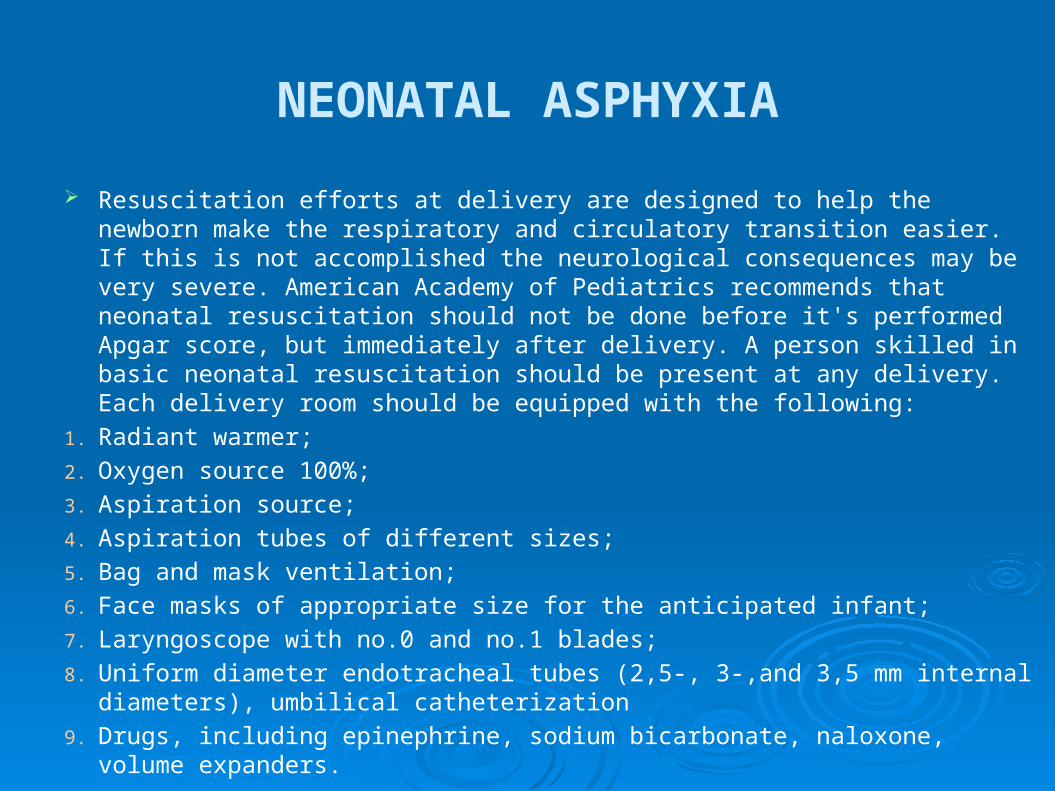
Resuscitation efforts at delivery are designed to help the newborn make the respiratory and circulatory transition easier. If this is not accomplished the neurological consequences may be very severe. American Academy of Pediatrics recommends that neonatal resuscitation should not be done before it's performed Apgar score, but immediately after delivery. A person skilled in basic neonatal resuscitation should be present at any delivery. Each delivery room should be equipped with the following:
1. Radiant warmer;2. Oxygen source 100%;3. Aspiration source;4. Aspiration tubes of different sizes;5. Bag and mask ventilation;6. Face masks of appropriate size for the anticipated infant;7. Laryngoscope with no.0 and no.1 blades;8. Uniform diameter endotracheal tubes (2,5-, 3-,and 3,5 mm internal
diameters), umbilical catheterization9. Drugs, including epinephrine, sodium bicarbonate, naloxone, volume
expanders.

NEONATAL ASPHYXIA
Immediately after birth the following parameters will be evaluated:
1. RESPIRATION;2. HEART RATE;3. OXYGEN SATURATION;• The major steps to a successful resuscitation are
known as ABC of resuscitation:1. AIRWAY CLEARANCE2. BREATHING SUPPORT;3. CIRCULATORY SUPPORT;4. DRUGS;
NEONATAL ASPHYXIA
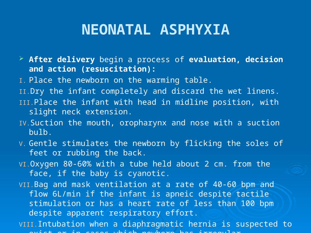
After delivery begin a process of evaluation, decision and action (resuscitation):
I. Place the newborn on the warming table.II. Dry the infant completely and discard the wet linens.III. Place the infant with head in midline position, with slight neck
extension.IV. Suction the mouth, oropharynx and nose with a suction bulb.V. Gentle stimulates the newborn by flicking the soles of feet or
rubbing the back.VI. Oxygen 80-60% with a tube held about 2 cm. from the face, if the
baby is cyanotic.VII.Bag and mask ventilation at a rate of 40-60 bpm and flow 6L/min if
the infant is apneic despite tactile stimulation or has a heart rate of less than 100 bpm despite apparent respiratory effort.
VIII.Intubation when a diaphragmatic hernia is suspected to exist or in cases which newborn has irregular breathings.

NEONATAL ASPHYXIA
IX. Cardiac massage should be instituted if after intubation and 15 to 30 seconds of ventilation with 100% 02, the heart rate remains below 60 bpm, or 80 and not increasing. The best technique is to stand at the foot of infant and place both thumbs at the junction of the middle and lower thirds of the sternum, with the fingers wrapped around and supporting the back. Compress the stern 1 to 2 cm in a ratio of three compression for one breath (3/1).
X. If despite adequate ventilation with 100% 02 and chest compression, a heart rate of more than 80 bpm has not been achieved by 1 to 2 min. after delivery, or the iniţial heart rate is 0, medications such as chronotropic and inotropic agents should be given to support myocardium, correct acidosis and ensure adequate fluid status.
NEONATAL ASPHYXIA
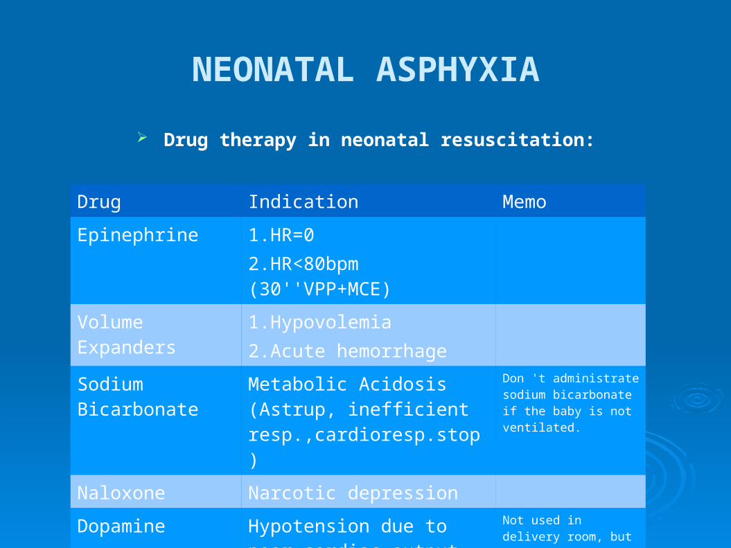
Drug therapy in neonatal resuscitation:
Drug Indication Memo
Epinephrine 1.HR=02.HR<80bpm (30''VPP+MCE)
Volume Expanders 1.Hypovolemia2.Acute hemorrhage
Sodium Bicarbonate
Metabolic Acidosis (Astrup, inefficient resp.,cardioresp.stop)
Don 't administrate sodium bicarbonate if the baby is not ventilated.
Naloxone Narcotic depression
Dopamine Hypotension due to poor cardiac output;
Not used in delivery room, but in NICU in slow perfusion.
NEONATAL ASPHYXIA
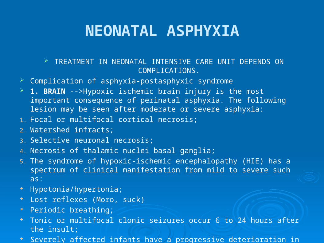
TREATMENT IN NEONATAL INTENSIVE CARE UNIT DEPENDS ON COMPLICATIONS.
Complication of asphyxia-postasphyxic syndrome 1. BRAIN -->Hypoxic ischemic brain injury is the most important
consequence of perinatal asphyxia. The following lesion may be seen after moderate or severe asphyxia:
1. Focal or multifocal cortical necrosis;2. Watershed infracts;3. Selective neuronal necrosis;4. Necrosis of thalamic nuclei basal ganglia;5. The syndrome of hypoxic-ischemic encephalopathy (HIE) has a spectrum
of clinical manifestation from mild to severe such as: Hypotonia/hypertonia; Lost reflexes (Moro, suck) Periodic breathing; Tonic or multifocal clonic seizures occur 6 to 24 hours after the insult; Severely affected infants have a progressive deterioration in CNS function,
with prolonged apnea and coma.
NEONATAL ASPHYXIA
2. CARDIOVASCULAR SYSTEM Infants with perinatal asphyxia may have transient myocardial
ischemia. They develop respiratory distress and cyanosis shortly after birth. They will have signs of congestive heart failure such as:
Tachypnea; Tahycardia; Enlarged liver; Gallop rhythm;• In its severe form, cardiac dilatation and tricuspid
incompetence may accompany congestive heart failure.
NEONATAL ASPHYXIA
3. LUNGS Increased pulmonary vascular resistance; Pulmonary hemorrhage; Pulmonary edema secondary to cardiac failure; Inhibition of surfactant synthesis due to persistent acidemia with
secondary hvaline membrane disease (HMD); Meconium aspiration syndrome;

NEONATAL ASPHYXIA
4. KIDNEY-Whenever a neonate develops severe asphyxia, kidney damage may result. After birth asphyxia, proteinuria is common and myoglobinuria leading to acute tubular necrosis with renal failure can also occur.
5. LIVER-The liver also may be damaged by asphyxic insult with centers of necrosis, clotting factor deficiency not reversed by vitamin K and perturbances of enzymatic process.
6. BLOOD: Polycythemia; Anemia; Disseminated intravascular coagulation (DIC); 7. GASTROINTESTINAL EFFECTS-The asphyxiated infants are at
the risk for bowel ischemia and necrotizing enterocolitis (NEC). 8. TERMOREGULATION-Hypoxia inhibit termoregulation making
very difficult transition to extrauterine life → hypotermia.

NEONATAL ASPHYXIA
Treatment in N.I.C. U. Careful monitoring of: 1. CARDIO-RESPIRATORY FUNCTION: Blood pressure; Heart rate; Respiratory rate; 2. DIURESIS: >1-2 ml/kg/h. Apparition of diuresis
is a sign of good prognosis. 3. HEMOGLOBIN SATURATION. 4. BLOOD GASES.

NEONATAL ASPHYXIA
General management:1. thermal comfort;2. minimal maneuvers;3. careful administration of fluids;4. oxygenotherapy if necessary;5. correction of acidosis perturbances by bicarbonate adm.- 2mEq/kgw ;6. correction of hypoglycemia if glucose blood level is less than 40 mg/dl.7. correction of hypocalcemia if calcium blood level is under 8mg%;8. protection with antibiotics, usually with large spectrum likes ampicillin and gentamicin;9. parenteral nutrition in order to prevent NEC.

NEONATAL ASPHYXIA
Management of complications The most frequent complication of perinatal asphyxia is cerebral
edema, then seizures and cerebral hemorrhage. For cerebral edema it is indicated to reduce the amount of fluid
perday, that is between 50-60 ml/kg/w/d, to induce alkalosis by hyperventilation or by bicarbonate administration.
Hemorrhage and cerebral infracts represent the most severe consequence of perinatal asphyxia. For prevention it has been tried the administration of Phenobarbital in a unique dose of 40mg/kg/w.
Some authors recommend the administration of antioxidant agents such as: ascorbic acid, allopurinol, and vitamin E.
Treatment of seizures is done with phenobarbital-20-30 mg/w/dose and maintenance dose of 2,5 mg/w/d.For renal effects of perinatal asphyxia fluid therapy and administration of a single dose of furosemide-lmg/kg/dose are usually enough.
Treatment of cardiac sequel-fluid restriction, oxygen administration, correction of acidosis and sometimes administration of cardiotonic agents: dopamine. In congestive heart failure, digoxin can be used.
NEONATAL ASPHYXIA
Outcome and prognosis: Neonatal asphyxia is associated with increased neonatal
mortality, according with gestational age. The long-term prognosis depends on the severity and the length of hypoxic insult and the precocity of resuscitation. Approximately 25% of asphyxiated newborns will die in the first hours or days after birth. Among the survivors, even those with seizures can present a good evolution. The survivors with prolonged asphyxia can present neurological sequel in about 25%-45% and much more at premature babies. Neurological sequel can be:
1. Cerebral palsy;2. Severe mental retard;3. Blindness;4. Hearing perturbances;5. Recurrent seizures;
6. Accommodation perturbances in childhood;• This sequel can appear after a period of 2-3 years, so is very
important to foliow-up this babies.