Negative effects of Kudoa islandica n. sp. (Myxosporea: Kudoidae) on aquaculture and wild fisheries...
12
1 3 Negative effects of Kudoa islandica n. sp. (Myxosporea: Kudoidae) 4 on aquaculture and wild fisheries in Iceland 5 6 7 Árni Kristmundsson a,⇑ Q1 , Mark Andrew Freeman b 8 a Institute for Experimental Pathology at Keldur, University of Iceland, Keldnavegur 1-3, IS-112, Reykjavik, Iceland 9 b Institute of Ocean and Earth Sciences, University of Malaya, Kuala Lumpur, Malaysia 10 11 12 14 article info 15 Article history: 16 Received 14 February 2014 17 Revised 5 June 2014 18 Accepted 5 June 2014 19 Available online xxxx 20 Keywords: 21 Kudoa 22 Myoliquefaction 23 Muscle 24 Soft flesh syndrome Cyclopterus lumpus 25 Anarhichas spp. 26 27 abstract 28 In the early 2000s, experimental rearing of spotted wolffish, Anarhichas minor, was started in Iceland. 29 Health surveillance, carried out at regular intervals during the rearing period, revealed persistent and 30 highly prevalent Kudoa infections of fish muscles which caused great financial losses due to post mortem 31 myoliquefaction. In addition, during the traditional process of drying and smoking wild Atlantic lumpfish, 32 Cyclopterus lumpus, the muscles from some fish almost completely disappear and the fish have to be dis- 33 carded. 34 To describe the etiological agent responsible for these conditions, spotted wolffish, Atlantic wolffish 35 Anarhichas lupus, northern wolffish Anarhichas denticulatus and Atlantic lumpfish were caught off the 36 Icelandic coast and examined for the presence of Kudoa. We describe a novel myxosporean, Kudoa islan- 37 dica n. sp., using morphological and molecular data, and show with histopathology that it causes exten- 38 sive myoliquefaction in three different wild fish hosts, which all are commercially valuable species in 39 Iceland. Although some spore dimensions varied significantly between fish species, the molecular anal- 40 yses showed that the same parasite was responsible for infection in all fish. The northern wolffish was 41 not found to be infected. Although robustly placed in the Kudoa clade in phylogenetic analyses, K. islan- 42 dica was phylogenetically distinct from other kudoids. 43 A single myxosporean, K. islandica, is responsible for the infections in the somatic muscles of lumpfish 44 and wolffish, causing extensive post mortem myoliquefaction. This myxosporean is likely to infect other 45 fish species and it is important to study its life cycle in order to evaluate any threat to salmonid culture 46 via the use of lumpfish as a biocontrol for sea lice. 47 Ó 2014 The Authors. Published by Elsevier Ltd. This is an open access article under the CC BY-NC-ND license 48 (http://creativecommons.org/licenses/by-nc-nd/3.0/). 49 50 51 1. Introduction 52 Myxosporeans are a diverse group of parasites commonly found 53 infecting fish. Although the life cycles of many species are not 54 described, known life cycles require an alternate host which typi- 55 cally is an annelid worm. To date 95 nominal species of the 56 myxosporean genus Kudoa (Kudoidae) have been described, pres- 57 ently all with unknown life cycles. Most commonly kudoids are 58 histozoic in skeletal muscles of fish (Moran et al., 1999a; Lom 59 and Dykova, 2006; Eiras et al., 2014) but have also been found to 60 infect other organs such as brain, heart, gills, kidney, gall bladder, 61 ovary and intestines (Egusa, 1986; Sandeep et al., 1986; Sarkar 62 and Ghosh, 1991; Yurakhno, 1991; Blaylock et al., 2004; 63 Yurakhno et al., 2007; Mansour et al., 2013). Furthermore, at least 64 one Kudoa sp. has been found infecting the musculature of a non- 65 piscine host, the giant octopus Paroctopus dofleini (Yokoyama and 66 Masuda, 2001). Although most Kudoa species are described from 67 a single host, several have been identified in numerous fish species 68 from different families, such as Kudoa thyrsites (with at least 38 dif- 69 ferent hosts), Kudoa nova (20 hosts) and Kudoa iwatai (19 hosts) 70 (Burger and Adlard, 2011). While kudoids are generally not consid- 71 ered highly pathogenic to the host, some species have caused sig- 72 nificant detrimental effects on both commercial fisheries and 73 aquaculture, due to the unsightly muscle cysts they form and more 74 importantly the post mortem myoliquefaction some species cause, 75 commonly known as ‘‘soft flesh’’, which greatly reduces the market 76 value of the fillets (Langdon, 1991; Moran et al., 1999a; 77 Alvarez-Pellitero and Sitjà-Bobadilla, 1993) Q3 . In addition, cysts of 78 Kudoa septempunctata from farmed fish in Japan and Korea have 79 been strongly linked with numerous cases of food poisoning in 80 humans (Matsukane et al. 2010; Kawai et al. 2012; Iwashita http://dx.doi.org/10.1016/j.ijppaw.2014.06.001 2213-2244/Ó 2014 The Authors. Published by Elsevier Ltd. This is an open access article under the CC BY-NC-ND license (http://creativecommons.org/licenses/by-nc-nd/3.0/). ⇑ Corresponding author. Tel.: +354 5855100, mobile: +354 8664404. E-mail addresses: [email protected] (Á. Kristmundsson), [email protected] (M.A. Freeman). International Journal for Parasitology: Parasites and Wildlife xxx (2014) xxx–xxx Contents lists available at ScienceDirect International Journal for Parasitology: Parasites and Wildlife journal homepage: www.elsevier.com/locate/ijppaw IJPPAW 69 No. of Pages 12, Model 5G 20 June 2014 Please cite this article in press as: Kristmundsson, Á., Freeman, M.A. Negative effects of Kudoa islandica n. sp. (Myxosporea: Kudoidae) on aquaculture and wild fisheries in Iceland. International Journal for Parasitology: Parasites and Wildlife (2014), http://dx.doi.org/10.1016/j.ijppaw.2014.06.001
-
Upload
mark-andrew -
Category
Documents
-
view
214 -
download
0
Transcript of Negative effects of Kudoa islandica n. sp. (Myxosporea: Kudoidae) on aquaculture and wild fisheries...

1
3
4
5
6
7 Q1
89
101112
1 4
1516171819
20212223242526
2 7
50
51
52
53
54
55
56
57
58
59
60
61
62
63
International Journal for Parasitology: Parasites and Wildlife xxx (2014) xxx–xxx
IJPPAW 69 No. of Pages 12, Model 5G
20 June 2014
Contents lists available at ScienceDirect
International Journal for Parasitology:Parasites and Wildlife
journal homepage: www.elsevier .com/locate / i jppaw
Negative effects of Kudoa islandica n. sp. (Myxosporea: Kudoidae)on aquaculture and wild fisheries in Iceland
http://dx.doi.org/10.1016/j.ijppaw.2014.06.0012213-2244/� 2014 The Authors. Published by Elsevier Ltd.This is an open access article under the CC BY-NC-ND license (http://creativecommons.org/licenses/by-nc-nd/3.0/).
⇑ Corresponding author. Tel.: +354 5855100, mobile: +354 8664404.E-mail addresses: [email protected] (Á. Kristmundsson), [email protected]
(M.A. Freeman).
Please cite this article in press as: Kristmundsson, Á., Freeman, M.A. Negative effects of Kudoa islandica n. sp. (Myxosporea: Kudoidae) on aquacultuwild fisheries in Iceland. International Journal for Parasitology: Parasites and Wildlife (2014), http://dx.doi.org/10.1016/j.ijppaw.2014.06.001
Árni Kristmundsson a,⇑, Mark Andrew Freeman b
a Institute for Experimental Pathology at Keldur, University of Iceland, Keldnavegur 1-3, IS-112, Reykjavik, Icelandb Institute of Ocean and Earth Sciences, University of Malaya, Kuala Lumpur, Malaysia
a r t i c l e i n f o a b s t r a c t
282930313233343536373839
Article history:Received 14 February 2014Revised 5 June 2014Accepted 5 June 2014Available online xxxx
Keywords:KudoaMyoliquefactionMuscleSoft flesh syndrome Cyclopterus lumpusAnarhichas spp.
404142434445464748
In the early 2000s, experimental rearing of spotted wolffish, Anarhichas minor, was started in Iceland.Health surveillance, carried out at regular intervals during the rearing period, revealed persistent andhighly prevalent Kudoa infections of fish muscles which caused great financial losses due to post mortemmyoliquefaction. In addition, during the traditional process of drying and smoking wild Atlantic lumpfish,Cyclopterus lumpus, the muscles from some fish almost completely disappear and the fish have to be dis-carded.
To describe the etiological agent responsible for these conditions, spotted wolffish, Atlantic wolffishAnarhichas lupus, northern wolffish Anarhichas denticulatus and Atlantic lumpfish were caught off theIcelandic coast and examined for the presence of Kudoa. We describe a novel myxosporean, Kudoa islan-dica n. sp., using morphological and molecular data, and show with histopathology that it causes exten-sive myoliquefaction in three different wild fish hosts, which all are commercially valuable species inIceland. Although some spore dimensions varied significantly between fish species, the molecular anal-yses showed that the same parasite was responsible for infection in all fish. The northern wolffish wasnot found to be infected. Although robustly placed in the Kudoa clade in phylogenetic analyses, K. islan-dica was phylogenetically distinct from other kudoids.
A single myxosporean, K. islandica, is responsible for the infections in the somatic muscles of lumpfishand wolffish, causing extensive post mortem myoliquefaction. This myxosporean is likely to infect otherfish species and it is important to study its life cycle in order to evaluate any threat to salmonid culturevia the use of lumpfish as a biocontrol for sea lice.� 2014 The Authors. Published by Elsevier Ltd. This is an open access article under the CC BY-NC-ND license
(http://creativecommons.org/licenses/by-nc-nd/3.0/).
49
64
65
66
67
68
69
70
71
72
73
74
75
76
77Q3
1. Introduction
Myxosporeans are a diverse group of parasites commonly foundinfecting fish. Although the life cycles of many species are notdescribed, known life cycles require an alternate host which typi-cally is an annelid worm. To date 95 nominal species of themyxosporean genus Kudoa (Kudoidae) have been described, pres-ently all with unknown life cycles. Most commonly kudoids arehistozoic in skeletal muscles of fish (Moran et al., 1999a; Lomand Dykova, 2006; Eiras et al., 2014) but have also been found toinfect other organs such as brain, heart, gills, kidney, gall bladder,ovary and intestines (Egusa, 1986; Sandeep et al., 1986; Sarkarand Ghosh, 1991; Yurakhno, 1991; Blaylock et al., 2004;Yurakhno et al., 2007; Mansour et al., 2013). Furthermore, at least
78
79
80
one Kudoa sp. has been found infecting the musculature of a non-piscine host, the giant octopus Paroctopus dofleini (Yokoyama andMasuda, 2001). Although most Kudoa species are described froma single host, several have been identified in numerous fish speciesfrom different families, such as Kudoa thyrsites (with at least 38 dif-ferent hosts), Kudoa nova (20 hosts) and Kudoa iwatai (19 hosts)(Burger and Adlard, 2011). While kudoids are generally not consid-ered highly pathogenic to the host, some species have caused sig-nificant detrimental effects on both commercial fisheries andaquaculture, due to the unsightly muscle cysts they form and moreimportantly the post mortem myoliquefaction some species cause,commonly known as ‘‘soft flesh’’, which greatly reduces the marketvalue of the fillets (Langdon, 1991; Moran et al., 1999a;Alvarez-Pellitero and Sitjà-Bobadilla, 1993). In addition, cysts ofKudoa septempunctata from farmed fish in Japan and Korea havebeen strongly linked with numerous cases of food poisoning inhumans (Matsukane et al. 2010; Kawai et al. 2012; Iwashita
re and

81
82
83
84
85
86
87
88
89
90
91
92
93
94
95
96
97
98
99
100
101
102
103
104
105
106
107
108
109
110
111
112
113
114
115
116
117
118
119
120
121
122
123
124
125
126
127
128
129
130
131
132
133
134
135
136
137
138
139
140
141
142
143
144
145
146
147
148
149
150
151
152
153
154
155
156
157
158
159
160
161
162
163
164
165
166
167
168
169
170
171
172
173
174
175
176
177
178
179
180
181
182
183
184
185
186
187
188
189
190
191
192
193
194
195
196
197
198
199
200
201
202
203
2 Á. Kristmundsson, M.A. Freeman / International Journal for Parasitology: Parasites and Wildlife xxx (2014) xxx–xxx
IJPPAW 69 No. of Pages 12, Model 5G
20 June 2014
et al., 2013). Therefore, the production of Kudoa-free fish productsfrom aquaculture ventures has become a priority.
Atlantic wolffish Anarhichas lupus L. 1758, spotted wolffish Ana-rhichas minor Olafsen, 1772 and Atlantic lumpfish Cyclopterus lum-pus L. 1758 are all commercially important species in Iceland, themean annual catch over the last 10 years being around 14,000 tn,2500 tn and 5500 tn, respectively (Anonymous, 2012). The wolffishspecies are considered desirable products because of their rich andtasty fillet but also their unusual and popular skin, which is usedfor making designer wear. The northern wolffish Anarhichas denti-culatus is common in sea water around Iceland, especially off theNW coast, but is not considered a commercially valuable speciesdue to the unusual texture of its flesh. Lumpfish are targeted dur-ing a coastal spring fisheries as their valuable eggs are harvestedfor use as a caviar substitute. Traditionally, female lumpfish aredried and the fillets of male fish are smoked, and processed forhuman consumption. Recently, new markets have opened forlumpfish muscle and presently most of it is exported to Asia.
Knowledge of Kudoa infections in wild Icelandic fish are veryscarce. The only report of such infections are in a local fisheriesmagazine from 1986, where infections in lumpfish muscle arementioned (Thorsteinsson, 1986) and a report on the healthinessof seafood from 1999 where Kudoa sp. is briefly mentioned causingspoilage of fillets of lumpfish and Atlantic wolffish (Bjarnason et al.,1999). No data existed on Kudoa in the spotted wolffish until 2003when an experimental rearing of this fish species was carried outin land based tanks in Iceland. The major problems experiencedduring culture and harvest were persistent Kudoa infections inmuscles of the spotted wolffish. No data exists on Kudoa infectionsin northern wolffish.
The aim of the present study was to identify and describe thekudoid myxosporean responsible for the problems experiencedduring the rearing of the spotted wolffish and to analyse the detri-mental effect of the parasite on the farmed fish. In addition, toidentify the Kudoa causing spoilage of fillets of lumpfish andAtlantic wolffish. Although of no commercial value, the northernwolffish is closely related to the other two wolffish species sowas included in the study.
2. Materials and methods
2.1. Fish studied
The experimental rearing of spotted wolffish in Iceland was car-ried out in shallow raceways in a land-based facility. The brood-stock consisted of wild fish which were sampled in June 2002. Inthe beginning, while adopting to dry food pellets, they were fedon homogenised fish pulp (mostly herring and capelin). The firstgeneration of farmed juvenile fish hatched in February 2003. Theywere kept separate from the broodfish for the entire rearing periodand fed on commercially formulated feed. The intake seawater forthe farm, which was taken at 30 m depth from a nearby fjord, wasunfiltered in the case of the broodfish but filtered through a sandfilter for the farmed juveniles.
At regular intervals, fish were sent to the Fish Disease Labora-tory at the Institute for Experimental Pathology at Keldur for thor-ough examination with regards to infectious diseases. Broodfish(length 60–80 cm – corresponding to age 5–12 year according toGunnarsson (2010)) were examined in November 2002 (2 fish),March 2003 (2 fish), November 2003 (4 fish) and January 2004 (4fish). Examination of the 1st generation of the farmed fish was per-formed twice in 2004; January (2 fish; age = 11 months - length18–20 cm) and April (16 fish; age 14 months – 22–28 cm) andthree times in 2005; April (4 fish; age 26 months – 34–35 cm),May (4 fish; age 27 months – 34–36 cm) and June (4 fish, age28 months – 37–41 cm).
Please cite this article in press as: Kristmundsson, Á., Freeman, M.A. Negative ewild fisheries in Iceland. International Journal for Parasitology: Parasites and W
In February 2013, wild spotted wolffish (length 65–103 cm),Atlantic wolffish (62–89 cm) and Atlantic lumpfish (41–48 cm), 5fish of each species, were caught in Bay Faxaflói off the west coastof Iceland and examined for the presence of Kudoa spp. In addition,in July 2013, ten northern wolffish (length, 65–105 cm), werecaught off the NW coast of Iceland and examined for Kudoainfections.
2.2. Examination of fresh fish and sampling methods
Skeletal muscles of all fish, farmed and wild, were examined forthe presence of Kudoa sp. Three slices of muscles, approximately4 cm wide and 8 cm long, from the anterior-, mid- and posteriorparts of the fish fillets, were thoroughly searched under a stereomi-croscope for the presence of plasmodia/pseudocysts. If detected,they were removed from the fillet and placed on a microscope slideand examined at 400� magnification, to confirm the presence ofKudoa spores. If no plasmodia were detected, the muscle sliceswere homogenised in PBS using a pestle and mortar. The homoge-nised muscle was sieved (500 lm) and the fluid centrifuged at1500 g for 5 min and the pellet microscopically screened for thepresence of Kudoa spores. Infection intensity was determined asfollows: very light = no pseudocysts seen but spores detected fromcentrifuged muscle homogenate; light = 1–20 pseudocystsdetected in any of the muscle slices; moderate = pseudocystsdetected in all muscle slices, total number 20–50; severe = pseud-ocysts detected in all muscle slices, total number >50.
The size of 10 pseudocysts from each fish species (except thewild spotted wolfish) were measured and photographed using astereomicroscope. Morphological characteristics of 50 fresh sporesfrom each fish species (except for spore length: n = 30 and length ofpolar filaments: n = 10) were measured following the recommen-dations of Lom and Arthur (1989) and Adlard et al. (2005).
Pieces of fresh trunk muscle from all examined fish were fixedin 10% buffered formalin at approximately 24 h p.m. In addition,moderately or heavily infected muscle samples from two fish ofall species (wild Atlantic wolffish and lumpfish, farmed spottedwolffish), were kept at 4 �C until fixed for histopathological exam-ination, at 48 h p.m. To examine whether freezing affected the p.m.proteolysis of muscles, portions of muscles from one heavilyinfected wild Atlantic lumpfish were frozen at 48 h p.m. thenthawed and stored at 4 �C for 24 h and 120 h before being fixedin 10% buffered formalin.
Air dried smears from infected muscles were fixed in methanol,stained with Giemsa and mounted and used as reference material.
2.3. Histology
Formalin fixed muscle samples were embedded in paraffin wax,sectioned (4 lm) and stained with Giemsa and Haematoxylin andEosin (HE). The parasite and associated histological changes wereexamined using a compound microscope.
2.4. Scanning electron microscopy
For SEM examination, whole plasmodia were removed frominfected muscle from Atlantic wolffish and placed in a tube con-taining PBS and gently disrupted to liberate the spores. Tubes werethen centrifuged at 1500 g for 5 min, the supernatant removed andthe pellet fixed in 2.5% glutaraldehyde for 4 h. After fixation, thespores were prepared for SEM and viewed following the methodsdescribed by Kristmundsson and Freeman (2013). In brief, washedspores were syringed onto a polycarbonate membrane, fixed with1% osmium tetroxide and dehydrated through an ethanol series.Membranes were dried, mounted on stubs, sputter-coated withgold and viewed.
ffects of Kudoa islandica n. sp. (Myxosporea: Kudoidae) on aquaculture andildlife (2014), http://dx.doi.org/10.1016/j.ijppaw.2014.06.001
204
205
206
207
208
209
210
211
212
213
214
215
216
217
218
219
220
221
222
223
224
225
226
227
228
229
230
231
232
233
234
235
236
237
238
239
240
241
242
243
244
245
246
247
248
249
250
251
252
253
254
255
256
257
258
259
260
261
262
263
264
265
266
267
268
269
270
271
272
273
274
275
276
277
278
279
280
281
282
283
284
285
286
287
288
289
290
291
292
293
294
295
296
297
298
299
300
301
302
303
304
305
306
307
308
309
310
311
312
313
314
315
316
317
318
319
320
321
322
323
324
325
326
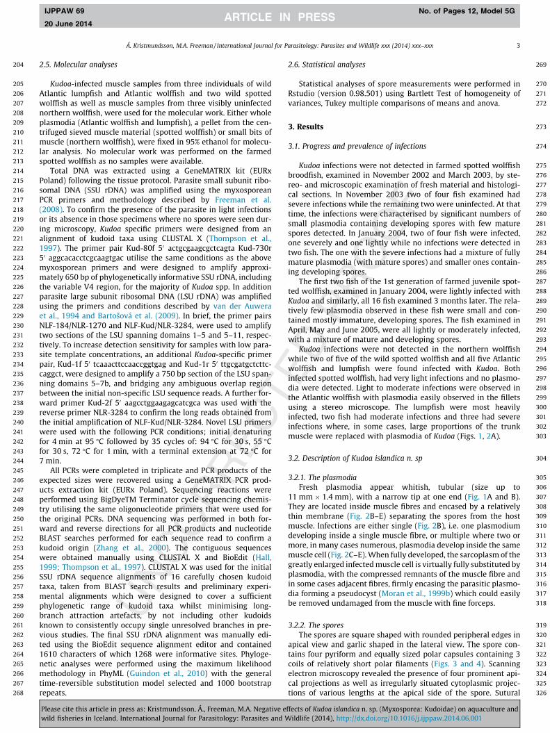
Á. Kristmundsson, M.A. Freeman / International Journal for Parasitology: Parasites and Wildlife xxx (2014) xxx–xxx 3
IJPPAW 69 No. of Pages 12, Model 5G
20 June 2014
2.5. Molecular analyses
Kudoa-infected muscle samples from three individuals of wildAtlantic lumpfish and Atlantic wolffish and two wild spottedwolffish as well as muscle samples from three visibly uninfectednorthern wolffish, were used for the molecular work. Either wholeplasmodia (Atlantic wolffish and lumpfish), a pellet from the cen-trifuged sieved muscle material (spotted wolffish) or small bits ofmuscle (northern wolffish), were fixed in 95% ethanol for molecu-lar analysis. No molecular work was performed on the farmedspotted wolffish as no samples were available.
Total DNA was extracted using a GeneMATRIX kit (EURxPoland) following the tissue protocol. Parasite small subunit ribo-somal DNA (SSU rDNA) was amplified using the myxosporeanPCR primers and methodology described by Freeman et al.(2008). To confirm the presence of the parasite in light infectionsor its absence in those specimens where no spores were seen dur-ing microscopy, Kudoa specific primers were designed from analignment of kudoid taxa using CLUSTAL X (Thompson et al.,1997). The primer pair Kud-80f 50 actgcgaagcgctcagta Kud-730r50 aggcacacctcgcaagtgac utilise the same conditions as the abovemyxosporean primers and were designed to amplify approxi-mately 650 bp of phylogenetically informative SSU rDNA, includingthe variable V4 region, for the majority of Kudoa spp. In additionparasite large subunit ribosomal DNA (LSU rDNA) was amplifiedusing the primers and conditions described by van der Auweraet al., 1994 and Bartošová et al. (2009). In brief, the primer pairsNLF-184/NLR-1270 and NLF-Kud/NLR-3284, were used to amplifytwo sections of the LSU spanning domains 1–5 and 5–11, respec-tively. To increase detection sensitivity for samples with low para-site template concentrations, an additional Kudoa-specific primerpair, Kud-1f 50 tcaaacttccaaccggtgag and Kud-1r 50 ttgcgatgctcttc-caggct, were designed to amplify a 750 bp section of the LSU span-ning domains 5–7b, and bridging any ambiguous overlap regionbetween the initial non-specific LSU sequence reads. A further for-ward primer Kud-2f 50 aagcctggaagagcatcgca was used with thereverse primer NLR-3284 to confirm the long reads obtained fromthe initial amplification of NLF-Kud/NLR-3284. Novel LSU primerswere used with the following PCR conditions; initial denaturingfor 4 min at 95 �C followed by 35 cycles of: 94 �C for 30 s, 55 �Cfor 30 s, 72 �C for 1 min, with a terminal extension at 72 �C for7 min.
All PCRs were completed in triplicate and PCR products of theexpected sizes were recovered using a GeneMATRIX PCR prod-ucts extraction kit (EURx Poland). Sequencing reactions wereperformed using BigDyeTM Terminator cycle sequencing chemis-try utilising the same oligonucleotide primers that were used forthe original PCRs. DNA sequencing was performed in both for-ward and reverse directions for all PCR products and nucleotideBLAST searches performed for each sequence read to confirm akudoid origin (Zhang et al., 2000). The contiguous sequenceswere obtained manually using CLUSTAL X and BioEdit (Hall,1999; Thompson et al., 1997). CLUSTAL X was used for the initialSSU rDNA sequence alignments of 16 carefully chosen kudoidtaxa, taken from BLAST search results and preliminary experi-mental alignments which were designed to cover a sufficientphylogenetic range of kudoid taxa whilst minimising long-branch attraction artefacts, by not including other kudoidsknown to consistently occupy single unresolved branches in pre-vious studies. The final SSU rDNA alignment was manually edi-ted using the BioEdit sequence alignment editor and contained1610 characters of which 1268 were informative sites. Phyloge-netic analyses were performed using the maximum likelihoodmethodology in PhyML (Guindon et al., 2010) with the generaltime-reversible substitution model selected and 1000 bootstraprepeats.
Please cite this article in press as: Kristmundsson, Á., Freeman, M.A. Negative ewild fisheries in Iceland. International Journal for Parasitology: Parasites and W
2.6. Statistical analyses
Statistical analyses of spore measurements were performed inRstudio (version 0.98.501) using Bartlett Test of homogeneity ofvariances, Tukey multiple comparisons of means and anova.
3. Results
3.1. Progress and prevalence of infections
Kudoa infections were not detected in farmed spotted wolffishbroodfish, examined in November 2002 and March 2003, by ste-reo- and microscopic examination of fresh material and histologi-cal sections. In November 2003 two of four fish examined hadsevere infections while the remaining two were uninfected. At thattime, the infections were characterised by significant numbers ofsmall plasmodia containing developing spores with few maturespores detected. In January 2004, two of four fish were infected,one severely and one lightly while no infections were detected intwo fish. The one with the severe infections had a mixture of fullymature plasmodia (with mature spores) and smaller ones contain-ing developing spores.
The first two fish of the 1st generation of farmed juvenile spot-ted wolffish, examined in January 2004, were lightly infected withKudoa and similarly, all 16 fish examined 3 months later. The rela-tively few plasmodia observed in these fish were small and con-tained mostly immature, developing spores. The fish examined inApril, May and June 2005, were all lightly or moderately infected,with a mixture of mature and developing spores.
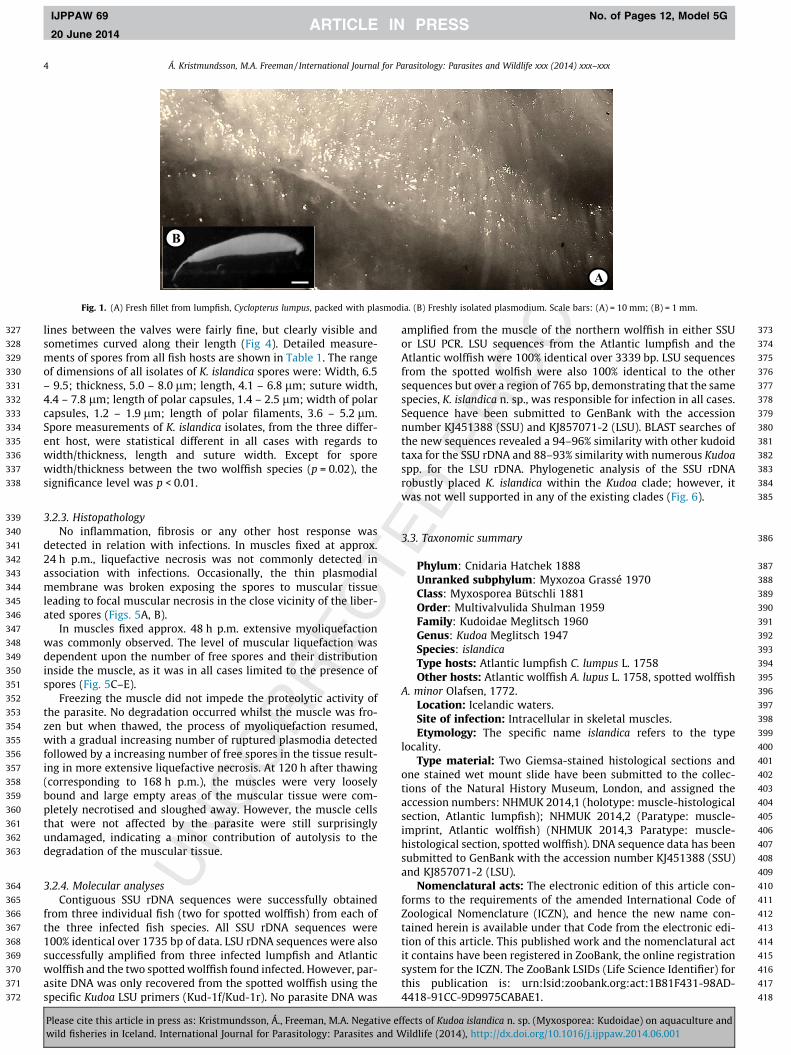
Kudoa infections were not detected in the northern wolffishwhile two of five of the wild spotted wolffish and all five Atlanticwolffish and lumpfish were found infected with Kudoa. Bothinfected spotted wolffish, had very light infections and no plasmo-dia were detected. Light to moderate infections were observed inthe Atlantic wolffish with plasmodia easily observed in the filletsusing a stereo microscope. The lumpfish were most heavilyinfected, two fish had moderate infections and three had severeinfections where, in some cases, large proportions of the trunkmuscle were replaced with plasmodia of Kudoa (Figs. 1, 2A).
3.2. Description of Kudoa islandica n. sp
3.2.1. The plasmodiaFresh plasmodia appear whitish, tubular (size up to
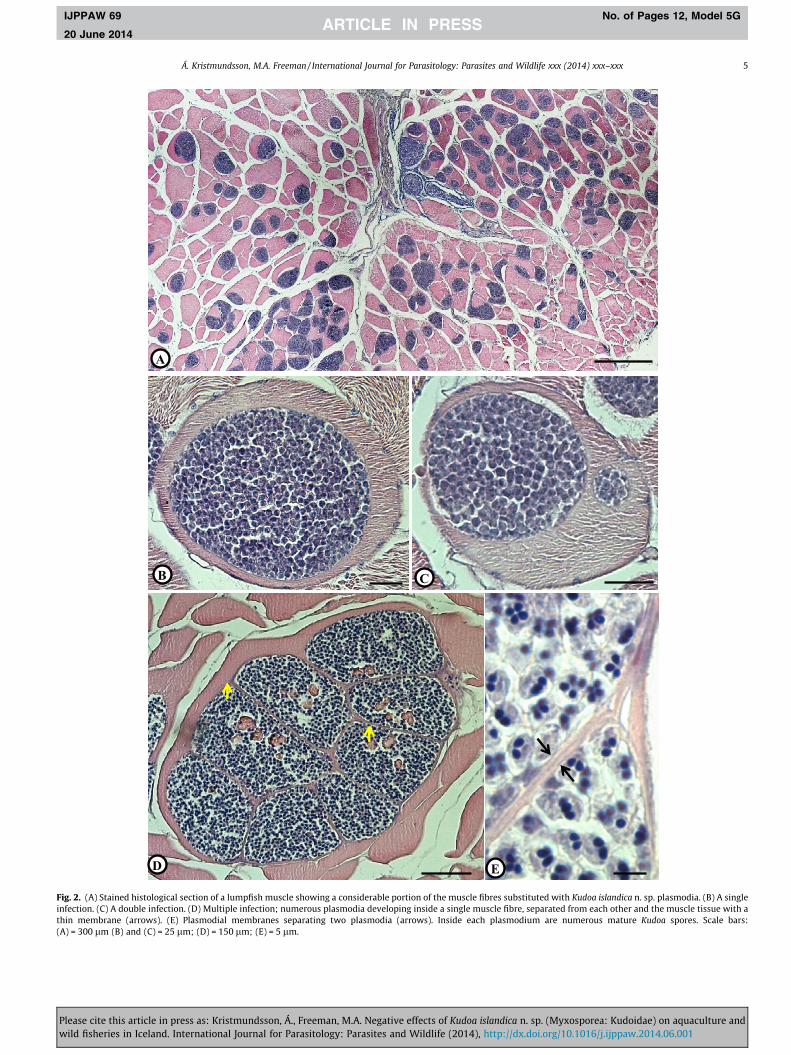
11 mm � 1.4 mm), with a narrow tip at one end (Fig. 1A and B).They are located inside muscle fibres and encased by a relativelythin membrane (Fig. 2B–E) separating the spores from the hostmuscle. Infections are either single (Fig. 2B), i.e. one plasmodiumdeveloping inside a single muscle fibre, or multiple where two ormore, in many cases numerous, plasmodia develop inside the samemuscle cell (Fig. 2C–E). When fully developed, the sarcoplasm of thegreatly enlarged infected muscle cell is virtually fully substituted byplasmodia, with the compressed remnants of the muscle fibre andin some cases adjacent fibres, firmly encasing the parasitic plasmo-dia forming a pseudocyst (Moran et al., 1999b) which could easilybe removed undamaged from the muscle with fine forceps.
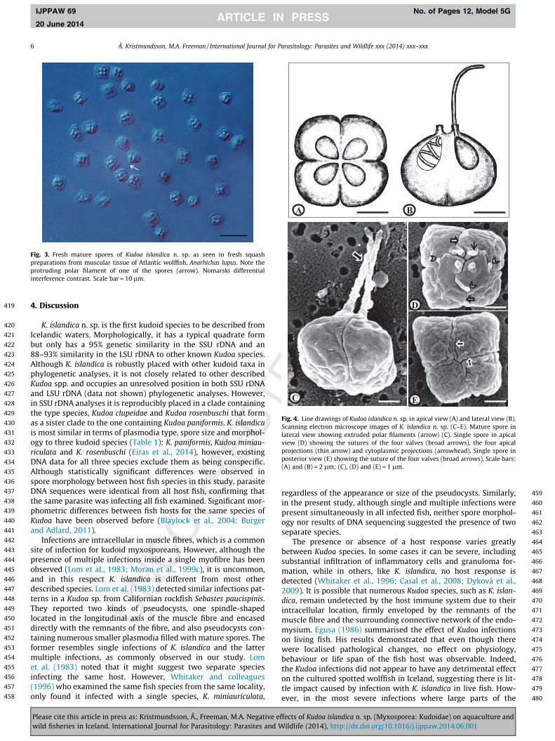
3.2.2. The sporesThe spores are square shaped with rounded peripheral edges in
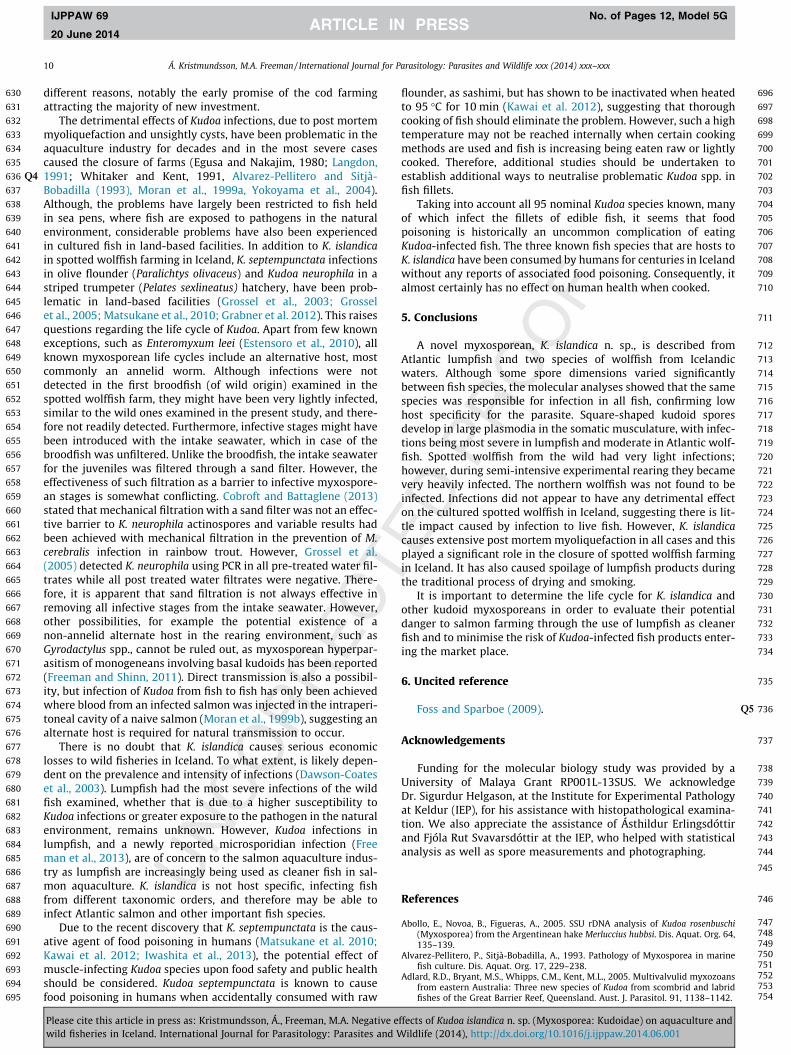
apical view and garlic shaped in the lateral view. The spore con-tains four pyriform and equally sized polar capsules containing 3coils of relatively short polar filaments (Figs. 3 and 4). Scanningelectron microscopy revealed the presence of four prominent api-cal projections as well as irregularly situated cytoplasmic projec-tions of various lengths at the apical side of the spore. Sutural
ffects of Kudoa islandica n. sp. (Myxosporea: Kudoidae) on aquaculture andildlife (2014), http://dx.doi.org/10.1016/j.ijppaw.2014.06.001
327
328
329
330
331
332
333
334
335
336
337
338
339
340
341
342
343
344
345
346
347
348
349
350
351
352
353
354
355
356
357
358
359
360
361
362
363
364
365
366
367
368
369
370
371
372
373
374
375
376
377
378
379
380
381
382
383
384
B
A
Fig. 1. (A) Fresh fillet from lumpfish, Cyclopterus lumpus, packed with plasmodia. (B) Freshly isolated plasmodium. Scale bars: (A) = 10 mm; (B) = 1 mm.
4 Á. Kristmundsson, M.A. Freeman / International Journal for Parasitology: Parasites and Wildlife xxx (2014) xxx–xxx
IJPPAW 69 No. of Pages 12, Model 5G
20 June 2014
lines between the valves were fairly fine, but clearly visible andsometimes curved along their length (Fig 4). Detailed measure-ments of spores from all fish hosts are shown in Table 1. The rangeof dimensions of all isolates of K. islandica spores were: Width, 6.5– 9.5; thickness, 5.0 – 8.0 lm; length, 4.1 – 6.8 lm; suture width,4.4 – 7.8 lm; length of polar capsules, 1.4 – 2.5 lm; width of polarcapsules, 1.2 – 1.9 lm; length of polar filaments, 3.6 – 5.2 lm.Spore measurements of K. islandica isolates, from the three differ-ent host, were statistical different in all cases with regards towidth/thickness, length and suture width. Except for sporewidth/thickness between the two wolffish species (p = 0.02), thesignificance level was p < 0.01.
385
386
387
388
389
390
391
392
393
394
395
396
397
398
399
400
401
402
403
404
405
406
407
408
3.2.3. HistopathologyNo inflammation, fibrosis or any other host response was
detected in relation with infections. In muscles fixed at approx.24 h p.m., liquefactive necrosis was not commonly detected inassociation with infections. Occasionally, the thin plasmodialmembrane was broken exposing the spores to muscular tissueleading to focal muscular necrosis in the close vicinity of the liber-ated spores (Figs. 5A, B).
In muscles fixed approx. 48 h p.m. extensive myoliquefactionwas commonly observed. The level of muscular liquefaction wasdependent upon the number of free spores and their distributioninside the muscle, as it was in all cases limited to the presence ofspores (Fig. 5C–E).
Freezing the muscle did not impede the proteolytic activity ofthe parasite. No degradation occurred whilst the muscle was fro-zen but when thawed, the process of myoliquefaction resumed,with a gradual increasing number of ruptured plasmodia detectedfollowed by a increasing number of free spores in the tissue result-ing in more extensive liquefactive necrosis. At 120 h after thawing(corresponding to 168 h p.m.), the muscles were very looselybound and large empty areas of the muscular tissue were com-pletely necrotised and sloughed away. However, the muscle cellsthat were not affected by the parasite were still surprisinglyundamaged, indicating a minor contribution of autolysis to thedegradation of the muscular tissue.
409
410
411
412
413
414
415
416
417
418
3.2.4. Molecular analysesContiguous SSU rDNA sequences were successfully obtained
from three individual fish (two for spotted wolffish) from each ofthe three infected fish species. All SSU rDNA sequences were100% identical over 1735 bp of data. LSU rDNA sequences were alsosuccessfully amplified from three infected lumpfish and Atlanticwolffish and the two spotted wolffish found infected. However, par-asite DNA was only recovered from the spotted wolffish using thespecific Kudoa LSU primers (Kud-1f/Kud-1r). No parasite DNA was
Please cite this article in press as: Kristmundsson, Á., Freeman, M.A. Negative ewild fisheries in Iceland. International Journal for Parasitology: Parasites and W
amplified from the muscle of the northern wolffish in either SSUor LSU PCR. LSU sequences from the Atlantic lumpfish and theAtlantic wolffish were 100% identical over 3339 bp. LSU sequencesfrom the spotted wolfish were also 100% identical to the othersequences but over a region of 765 bp, demonstrating that the samespecies, K. islandica n. sp., was responsible for infection in all cases.Sequence have been submitted to GenBank with the accessionnumber KJ451388 (SSU) and KJ857071-2 (LSU). BLAST searches ofthe new sequences revealed a 94–96% similarity with other kudoidtaxa for the SSU rDNA and 88–93% similarity with numerous Kudoaspp. for the LSU rDNA. Phylogenetic analysis of the SSU rDNArobustly placed K. islandica within the Kudoa clade; however, itwas not well supported in any of the existing clades (Fig. 6).
3.3. Taxonomic summary
Phylum: Cnidaria Hatchek 1888Unranked subphylum: Myxozoa Grassé 1970Class: Myxosporea Bütschli 1881Order: Multivalvulida Shulman 1959Family: Kudoidae Meglitsch 1960Genus: Kudoa Meglitsch 1947Species: islandicaType hosts: Atlantic lumpfish C. lumpus L. 1758Other hosts: Atlantic wolffish A. lupus L. 1758, spotted wolffish
A. minor Olafsen, 1772.Location: Icelandic waters.Site of infection: Intracellular in skeletal muscles.Etymology: The specific name islandica refers to the type
locality.Type material: Two Giemsa-stained histological sections and
one stained wet mount slide have been submitted to the collec-tions of the Natural History Museum, London, and assigned theaccession numbers: NHMUK 2014,1 (holotype: muscle-histologicalsection, Atlantic lumpfish); NHMUK 2014,2 (Paratype: muscle-imprint, Atlantic wolffish) (NHMUK 2014,3 Paratype: muscle-histological section, spotted wolffish). DNA sequence data has beensubmitted to GenBank with the accession number KJ451388 (SSU)and KJ857071-2 (LSU).
Nomenclatural acts: The electronic edition of this article con-forms to the requirements of the amended International Code ofZoological Nomenclature (ICZN), and hence the new name con-tained herein is available under that Code from the electronic edi-tion of this article. This published work and the nomenclatural actit contains have been registered in ZooBank, the online registrationsystem for the ICZN. The ZooBank LSIDs (Life Science Identifier) forthis publication is: urn:lsid:zoobank.org:act:1B81F431-98AD-4418-91CC-9D9975CABAE1.
ffects of Kudoa islandica n. sp. (Myxosporea: Kudoidae) on aquaculture andildlife (2014), http://dx.doi.org/10.1016/j.ijppaw.2014.06.001
B C
D E
A
Fig. 2. (A) Stained histological section of a lumpfish muscle showing a considerable portion of the muscle fibres substituted with Kudoa islandica n. sp. plasmodia. (B) A singleinfection. (C) A double infection. (D) Multiple infection; numerous plasmodia developing inside a single muscle fibre, separated from each other and the muscle tissue with athin membrane (arrows). (E) Plasmodial membranes separating two plasmodia (arrows). Inside each plasmodium are numerous mature Kudoa spores. Scale bars:(A) = 300 lm (B) and (C) = 25 lm; (D) = 150 lm; (E) = 5 lm.
Á. Kristmundsson, M.A. Freeman / International Journal for Parasitology: Parasites and Wildlife xxx (2014) xxx–xxx 5
IJPPAW 69 No. of Pages 12, Model 5G
20 June 2014
Please cite this article in press as: Kristmundsson, Á., Freeman, M.A. Negative effects of Kudoa islandica n. sp. (Myxosporea: Kudoidae) on aquaculture andwild fisheries in Iceland. International Journal for Parasitology: Parasites and Wildlife (2014), http://dx.doi.org/10.1016/j.ijppaw.2014.06.001
419
420
421
422
423
424
425
426
427
428
429
430
431
432
433
434
435
436
437
438
439
440
441
442
443
444
445
446
447
448
449
450
451
452
453
454
455
456
457
458
459
460
461
462
463
464
465
466
467
468
469
470
471
472
473
474
475
476
477
478
479
480
Fig. 3. Fresh mature spores of Kudoa islandica n. sp. as seen in fresh squashpreparations from muscular tissue of Atlantic wolffish, Anarhichas lupus. Note theprotruding polar filament of one of the spores (arrow). Nomarski differentialinterference contrast. Scale bar = 10 lm.
A B
C
D
E
Fig. 4. Line drawings of Kudoa islandica n. sp. in apical view (A) and lateral view (B).Scanning electron microscope images of K. islandica n. sp. (C–E). Mature spore inlateral view showing extruded polar filaments (arrow) (C). Single spore in apicalview (D) showing the sutures of the four valves (broad arrows), the four apicalprojections (thin arrow) and cytoplasmic projections (arrowhead). Single spore inposterior view (E) showing the suture of the four valves (broad arrows), Scale bars:(A) and (B) = 2 lm; (C), (D) and (E) = 1 lm.
6 Á. Kristmundsson, M.A. Freeman / International Journal for Parasitology: Parasites and Wildlife xxx (2014) xxx–xxx
IJPPAW 69 No. of Pages 12, Model 5G
20 June 2014
4. Discussion
K. islandica n. sp. is the first kudoid species to be described fromIcelandic waters. Morphologically, it has a typical quadrate formbut only has a 95% genetic similarity in the SSU rDNA and an88–93% similarity in the LSU rDNA to other known Kudoa species.Although K. islandica is robustly placed with other kudoid taxa inphylogenetic analyses, it is not closely related to other describedKudoa spp. and occupies an unresolved position in both SSU rDNAand LSU rDNA (data not shown) phylogenetic analyses. However,in SSU rDNA analyses it is reproducibly placed in a clade containingthe type species, Kudoa clupeidae and Kudoa rosenbuschi that formas a sister clade to the one containing Kudoa paniformis. K. islandicais most similar in terms of plasmodia type, spore size and morphol-ogy to three kudoid species (Table 1): K. paniformis, Kudoa miniau-riculata and K. rosenbuschi (Eiras et al., 2014), however, existingDNA data for all three species exclude them as being conspecific.Although statistically significant differences were observed inspore morphology between host fish species in this study, parasiteDNA sequences were identical from all host fish, confirming thatthe same parasite was infecting all fish examined. Significant mor-phometric differences between fish hosts for the same species ofKudoa have been observed before (Blaylock et al., 2004; Burgerand Adlard, 2011).
Infections are intracellular in muscle fibres, which is a commonsite of infection for kudoid myxosporeans. However, although thepresence of multiple infections inside a single myofibre has beenobserved (Lom et al., 1983; Moran et al., 1999c), it is uncommon,and in this respect K. islandica is different from most otherdescribed species. Lom et al. (1983) detected similar infections pat-terns in a Kudoa sp. from Californian rockfish Sebastes paucispinis.They reported two kinds of pseudocysts, one spindle-shapedlocated in the longitudinal axis of the muscle fibre and encaseddirectly with the remnants of the fibre, and also pseudocysts con-taining numerous smaller plasmodia filled with mature spores. Theformer resembles single infections of K. islandica and the lattermultiple infections, as commonly observed in our study. Lomet al. (1983) noted that it might suggest two separate speciesinfecting the same host. However, Whitaker and colleagues(1996) who examined the same fish species from the same locality,only found it infected with a single species, K. miniauriculata,
Please cite this article in press as: Kristmundsson, Á., Freeman, M.A. Negative ewild fisheries in Iceland. International Journal for Parasitology: Parasites and W
regardless of the appearance or size of the pseudocysts. Similarly,in the present study, although single and multiple infections werepresent simultaneously in all infected fish, neither spore morphol-ogy nor results of DNA sequencing suggested the presence of twoseparate species.
The presence or absence of a host response varies greatlybetween Kudoa species. In some cases it can be severe, includingsubstantial infiltration of inflammatory cells and granuloma for-mation, while in others, like K. islandica, no host response isdetected (Whitaker et al., 1996; Casal et al., 2008; Dyková et al.,2009). It is possible that numerous Kudoa species, such as K. islan-dica, remain undetected by the host immune system due to theirintracellular location, firmly enveloped by the remnants of themuscle fibre and the surrounding connective network of the endo-mysium. Egusa (1986) summarised the effect of Kudoa infectionson living fish. His results demonstrated that even though therewere localised pathological changes, no effect on physiology,behaviour or life span of the fish host was observable. Indeed,the Kudoa infections did not appear to have any detrimental effecton the cultured spotted wolffish in Iceland, suggesting there is lit-tle impact caused by infection with K. islandica in live fish. How-ever, in the most severe infections where large parts of the
ffects of Kudoa islandica n. sp. (Myxosporea: Kudoidae) on aquaculture andildlife (2014), http://dx.doi.org/10.1016/j.ijppaw.2014.06.001

481
482
483
484
485
486
487
488
489
490
491
492
493
494
495
496
497
498
499
500
501
502
503
504
505
506
507
508
509
510
511
512
513
514
515
516
517
518
519
520
521
522
523
524
525
526
527
528
529
530
531
532
533
534
535
536
537
538
539
540
541
542
543
544
545
546Tabl
e1
Rang
ean
dm
ean
valu
e(w
ithi
npa
rent
hesi
s)of
mor
phol
ogic
alm
easu
rem
ents
ofK
udoa
isla
ndic
an.
sp.s
pore
sfr
omA
tlan
tic
lum
pfish
,Cyc
lopt
erus
lum
pus,
Atl
anti
cw
olffi
sh,A
narh
icha
slu
pus,
and
spot
ted
wol
ffish
,A.
min
oran
dot
her
mor
phol
ogic
ally
orge
neti
cally
sim
ilar
kudo
ids
infe
ctin
gm
uscu
lar
tiss
ueof
fish
.Abb
revi
atio
n:n.
a.=
noda
taav
aila
ble,
PC=
pola
rca
psul
es.
Wid
thLe
ngt
hTh
ickn
ess
Sutu
rew
idth
PCle
ngt
hPC
wid
thPo
lar
fila
men
tsle
ngt
hPl
asm
odia
Mor
phol
ogy
size
Fish
hos
tLo
cali
tyR
efer
ence
s
K.i
slan
dica
6.5–
8.6
(7.4
)4.
1–5.
1(4
.8)
5.0–
6.5
(5.6
)4.
4–5.
5(4
.9)
1.4–
1.9
(1.7
)1.
2–1.
8(1
.5)
3.6–
5.0
(4.3
)Tu
bula
r6
11�
1.4
mm
Cycl
opte
rus
lum
pus
Icel
and
Pres
ent
stu
dyK
.isl
andi
ca8.
6–9.
4(8
.9)
5.1–
6.2
(5.9
)6.
2–7.
9(7
.2)
5.5–
7.5
(6.6
)1.
4–2.
5(2
.2)
1.2–
1.9
(1.5
)3.
8–5.
1(4
.4)
Ana
rhic
has
lupu
sK
.isl
andi
ca8.
6–9.
5(9
.0)
5.5–
6.8
(6.2
)6.
5–8.
0(7
.4)
5.8–
7.8
(7.0
)1.
7–2.
4(2
.1)
1.5–
1.9
(1.7
)3.
9–5.
2(4
.5)
Ana
rhic
has
min
orK
.pan
ifor
mis
5.0–
6.5
(5.9
)4.
5–6.
0(5
.0)
6.0–
7.0
(6.6
)n
.a.
1.9–
2.4
(2.0
)1.
4–1.
9(1
.6)
n.a
.Tu
bula
rn
.a.
Mer
lucc
ius
prod
uctu
sC
anad
aK
abat
aan
dW
hit
aker
(198
1)
K.m
inia
uric
ulat
a7.
0–8.
5(7
.9)
5.0–
5.9
(5.4
)n
.a.(
7.9)
n.a
.1.
8–2.
3(2
.2)
n.a
.n
.a.
Tubu
lar
620�
2m
mSe
bast
espa
ucis
pini
sU
SAW
hit
aker
etal
.(19
96)
K.r
osen
busc
hin
.a.(
7.0)
n.a
.n
.a.(
7.0)
n.a
.n
.a.(
2.5)
n.a
.n
.a.
Tubu
lar
0.5–
1.2�
2.1�
10.5
mm
Mer
lucc
ius
spp.
Arg
enti
na
Abo
llo
etal
.(20
05)
and
Eira
set
al.(
2014
)K
.clu
peid
ae6.
3–7.
5(6
.4)
4.0–
5.3
(5.1
)n
.a.
n.a
.1.
5–2.
6(2
.0)�
(1.0
)n
.a.
Tubu
lar
Len
gth
5m
mCl
upea
hare
ngus
USA
Eira
set
al.(
2014
)
Á. Kristmundsson, M.A. Freeman / International Journal for Parasitology: Parasites and Wildlife xxx (2014) xxx–xxx 7
IJPPAW 69 No. of Pages 12, Model 5G
20 June 2014
Please cite this article in press as: Kristmundsson, Á., Freeman, M.A. Negative effwild fisheries in Iceland. International Journal for Parasitology: Parasites and W
muscle are substituted by Kudoa pseudocysts, as observed in wildlumpfish in the present study, it is hard to imagine that suchinfections do not affect the functionality of the muscle to someextent.
K. islandica is similar to other Kudoa species, such as K. iwatai,K. nova and K. thyrsites (Burger and Adlard, 2011), as it is nonspe-cific with regards to host fish, infecting unrelated fish such aswolffishes (Order: Perciformes, Family: Anarhichadidae) andlumpfish (Order: Scorpaeniformes, Family: Cyclopteridae). Thissuggests that habitat or diet could play a role in K. islandica infec-tions dynamics or transmission cycles. Atlantic and spotted wolf-fish are benthic species which prefer a sandy or muddy bottom.The habitat of lumpfish depends on age and season; mature fishmove inshore in the spring and early summer to spawn in shal-lower coastal waters but return to deeper waters offshore in latesummer and early autumn, while the juveniles spend their firstyear of life among weed clumps in bays and fjords. A considerableportion of the diet of all these three fish species is composed ofbenthic animals like echinoderms, crustaceans, ctenophores, mol-luscs, polychaetes and smaller fish (Moring, 1989; Kristinssonet al., 1997; Kristjánsson et al., 1997; Jónsson and Pálsson,2013). The northern wolffish is, however, considered more pela-gic and consequently its diet contains fewer benthic species(Jónsson and Pálsson, 2013). As all known marine myxosporeanlife cycles include benthic polychaetes as alternate hosts, thiscould explain the lack of K. islandica infections in the northernwolffish. The lack of host specificity of K. islandica, and the factthat numerous other fish species utilise a similar habitat as thethree known hosts; it would seem likely that other fish speciesare susceptible to infection by this parasite.
The most serious impact of K. islandica is the spoilage of fishfillets as it causes extensive post mortem myoliquefactive necro-sis. The effect and progress of this is similar in all three fish spe-cies examined, shortly after fish death the thin plasmodialmembrane degenerates exposing the spores to the muscular tis-sue enveloping the parasites. The presence of the spores causesthis muscular envelope to necrotise with a subsequent liberationof numerous spores into the surrounding muscular tissue. Thepresence of large white pseudocysts also reduces the marketabil-ity of the product.
Myoliquefaction and the presence of unsightly cysts in fish fil-lets due to Kudoa infections has been known to cause significantdamage to products of commercially valuable fish species for dec-ades (Moran et al., 1999a). Although poorly studied, the cause ofpost mortem myoliquefaction has long been thought to be due toproteolytic enzymes released by the parasite (Moran et al.,1999a). Funk et al. (2008) characterised a protease from K. thyr-sites responsible for myoliquefaction in Atlantic salmon, Salmosalar, and found it to be pH dependant. Following death of theAtlantic salmon, the pH decreases due to anaerobic glycolysis,which generates lactic acid. This decrease apparently triggers aprocessing of the enzyme, which is stable in live fish, resultingin post mortem muscle proteolysis. Therefore, reducing theamount of glycogen in the muscle fish, prior to harvesting, wasconsidered a possible way of reducing the post mortem effect ofK. thyrsites (Funk et al., 2008). However, due to potential interspe-cies differences of both fish and Kudoa species, the results fromthat study cannot necessarily be extrapolated to all Kudoa species,and also may not be easy to implement.
Moran et al. (1999a) published a review of the known speciesfrom the genus Kudoa. At that time 44 species had been identified,27 of which infect somatic muscles of various fish species andnine of those were known to cause post mortem myoliquefactionwith numerous others developing into large visual and unsightlycysts affecting the value of the fillets. Since this report, a further51 Kudoa species have been described, as well as new hosts for
ects of Kudoa islandica n. sp. (Myxosporea: Kudoidae) on aquaculture andildlife (2014), http://dx.doi.org/10.1016/j.ijppaw.2014.06.001

547
548
549
550
551
552
553
554
555
556
557
558
C
D
D
**
AC
AC
*
MF
MF
MF
MFC
E
A B
Fig. 5. (A) and (B) Muscle section from lumpfish, Cyclopterus lumpus, fixed 24 h post mortem. (A) Ruptured Kudoa plasmodia with subsequent liberation of mature sporescausing focal necrosis of the muscle fibre enveloping the plasmodium. (B) Higher magnification showing liberated spores (arrows) and a focal necrosis in the vicinity of thespores (asterisk). (C) Muscle section of an uninfected fish at approx. 48 h p.m. (D) Section of muscle of a heavily infected fish at approximately 48 h p.m. showing extensivemyoliquefaction (asterisk). (E) A close up of the affected area showing numerous Kudoa spores (arrowhead) and the associated liquefactive necrosis. Scale bars: (A) = 50 lm,(B) = 10 lm, (C) and (D) = 200 lm, (E) = 10 lm. Abbreviations: MF = Muscle fibres, AC = Adipocytes.
8 Á. Kristmundsson, M.A. Freeman / International Journal for Parasitology: Parasites and Wildlife xxx (2014) xxx–xxx
IJPPAW 69 No. of Pages 12, Model 5G
20 June 2014
formerly described species, some of which (Kudoa camarguensis,Kudoa megacapsula and Kudoa aequidens) considerably reduce thecommercial value of the fish species they infect (Pampoulie et al.,2008; Yokoyama and Itoh, 2005; Casal et al., 2008).
Infections with K. islandica appear to be prevalent in wild lump-fish and in at least two of the three wolffish species present in
Please cite this article in press as: Kristmundsson, Á., Freeman, M.A. Negative ewild fisheries in Iceland. International Journal for Parasitology: Parasites and W
Icelandic waters. Despite a lack of scientific publications on Kudoain Icelandic fish, the knowledge of soft flesh fish has been knownfor decades among fishermen and people processing fish. In Ice-land, female lumpfish are traditionally dried for human consump-tion after roe harvesting. It has been noticed over the years thatduring this process some fish wither and after drying contain
ffects of Kudoa islandica n. sp. (Myxosporea: Kudoidae) on aquaculture andildlife (2014), http://dx.doi.org/10.1016/j.ijppaw.2014.06.001

559
560
561
562
563
564
565
566
567
568
569
570
571
572
573
574
575
576
577
578
579
580
581
582
583
584
585
586
587
588
589
590
591
592
593
594
595
596
597
598
599
600
601
602
603
604
605
606
607
608
609
610
611
612
613
614
615
616
617
618
619
620
621
622
623
624
625
626
627
628
629
0.01
Unicapsula sp. AY302725 Kudoa amamiensis AF034638
Kudoa hypoepicardialis AY302722
Kudoa shiomitsui AB183718
998
Kudoa paniformis AF034640
Kudoa sp. FJ790311 711
Kudoa islandica n. sp. Kudoa clupeidae AY197771
Kudoa rosenbuschi AY623795 937
Kudoa ovivora AY152750
Kudoa megacapsula AB263074
Kudoa thyrsites AY152747 999
Kudoa septempunctata AB553293
Kudoa thalassomi AY302738
Kudoa yasunagai AY302741
Kudoa neurophila AY172511 774
952
507
818
ns
ns
ns
ns
ns
Fig. 6. SSU rDNA maximum likelihood phylogenetic tree of 16 Kudoa spp. Kudoa islandica is robustly and consistently placed with other Kudoa taxa in all analyses, but is notwell supported in the clade it is placed in. Numbers at the nodes represent bootstrap support from 1000 samplings, nodes with a support of <50 are considered not supported(ns).
Á. Kristmundsson, M.A. Freeman / International Journal for Parasitology: Parasites and Wildlife xxx (2014) xxx–xxx 9
IJPPAW 69 No. of Pages 12, Model 5G
20 June 2014
numerous white spots and have little muscle compared to otherfish. Similarly, male lumpfish fillets are smoked and used as breadtoppings. Prior to smoking, the fillets are soaked in brine to addtaste to the product and also to act as a preservative. It has beenobserved by the processors that fillets having white cysts do notretain the brine and consequently decay. Furthermore, these filletsare known to be undesirable to consume fresh as they disintegrateduring cooking, as has been reported with K. thyrsites (Langdon,1991). These historical findings are supported by a short reportfrom 1986 where protozoan infections in lumpfish muscle arementioned as a cause for soft flesh (Thorsteinsson, 1986). Further-more, infections with an unidentified Kudoa sp., causing spoilage oflumpfish flesh, are mentioned in another report from 1999(Bjarnason et al., 1999). Similar to the lumpfish, muscular infec-tions causing flesh spoilage have also been known in Atlantic wolf-fish from Iceland for a long time. Infections in Atlantic wolffishwith an unidentified Kudoa are mentioned in the report from1999 (Bjarnason et al., 1999). Indeed, among fisherman, soft fleshwolffish have for decades been called ‘‘hárasteinbítur’’ in Icelandic,meaning ‘‘hairy wolffish’’, referring to the long white pseudocystspresent in the fillets (Bjarnason et al., 1999). Unlike lumpfish andthe Atlantic wolffish, Kudoa infections were unknown from thespotted wolffish until they became a major problem in an experi-mental farming of this species. Possibly, Kudoa infections in thewild spotted wolffish are not as prevalent or as severe as in theAtlantic wolffish and consequently not noticed. However, undersemi-intensive rearing conditions there is little doubt that thespotted wolffish is extremely sensitive to Kudoa infections. Nostudies have been made on Kudoa infections in the northern wolf-fish which may be due to the fact that it is not considered suitablefor human consumption. It is commonly called the ‘‘jelly cat’’, dueto its jelly-like muscle, and in most cases is thrown back into thesea when caught. Interestingly, these wolffish that are known tohave gelatinous musculature were not found to be infected withKudoa in this study and the reason for their unusual muscle consis-tency remains unknown.
Please cite this article in press as: Kristmundsson, Á., Freeman, M.A. Negative ewild fisheries in Iceland. International Journal for Parasitology: Parasites and W
Experimental rearing of spotted wolffish started in Norway inthe early 1990s and the first reproducing broodstock was estab-lished in 1993. During the following years, this fish species alsoreceived attention in Canada and Chile and latterly in Iceland(Falk-Petersen et al., 1999; Foss et al., 2004). Among farmers andscientists, the spotted wolffish was considered one of the mostpromising candidates for cold water aquaculture due to its fastgrowth, its unique farming-friendly behaviour and its robustnesswith regards to infectious diseases (Falk-Petersen et al., 1999;Espelid, 2002; Foss et al., 2004). The greatest mortality observedin the Norwegian culture of spotted wolffish was due to atypicalAeromonas salmonicida infections under stressful conditions, butalso to a smaller extent, parasitic infections such as Ichthyobodosp., Trichodina sp. Pleistophora sp. and Gyrodactylus spp. (Espelid,2002; Foss et al., 2004). In Iceland, there were few pathogens caus-ing problems, other than infections with atypical A. salmonicidaand Gyrodactylus anarhichatis (Kristmundsson et al., 2006). Themain difference was that Kudoa infections became a serious prob-lem in Iceland but were never reported in other countries farmingspotted wolffish. This could be due to the lack of an obligate alter-nate host, or possibly due to the large number of infected lumpfishfound around the Icelandic coast that could act as a reservoir forinfection.
As demonstrated in the present paper, Kudoa infections gradu-ally increased in the broodstock and similarly in the 1st generationof the farmed juvenile fish. The infections did not appear to affectthe fish health and the extent of the problem did not becomeapparent until the fish were slaughtered. At that time, significantparts of the fish were extensively infected. Post mortem myolique-faction became evident and many of the fillets proved unfit forhuman consumption. In 2008, the farming ceased and all fish wereslaughtered. There may have been multiple reasons for the termi-nation of wolffish culture in Iceland; however, the negative impactof K. islandica certainly played a significant part. Wolffish aquacul-ture in Norway has also largely ceased; however, this was due to
ffects of Kudoa islandica n. sp. (Myxosporea: Kudoidae) on aquaculture andildlife (2014), http://dx.doi.org/10.1016/j.ijppaw.2014.06.001
630
631
632
633
634
635
636 Q4
637
638
639
640
641
642
643
644
645
646
647
648
649
650
651
652
653
654
655
656
657
658
659
660
661
662
663
664
665
666
667
668
669
670
671
672
673
674
675
676
677
678
679
680
681
682
683
684
685
686
687
688
689
690
691
692
693
694
695
696
697
698
699
700
701
702
703
704
705
706
707
708
709
710
711
712
713
714
715
716
717
718
719
720
721
722
723
724
725
726
727
728
729
730
731
732
733
734
735
736Q5
737
738
739
740
741
742
743
744
745
746
747748749750751752753754
10 Á. Kristmundsson, M.A. Freeman / International Journal for Parasitology: Parasites and Wildlife xxx (2014) xxx–xxx
IJPPAW 69 No. of Pages 12, Model 5G
20 June 2014
different reasons, notably the early promise of the cod farmingattracting the majority of new investment.
The detrimental effects of Kudoa infections, due to post mortemmyoliquefaction and unsightly cysts, have been problematic in theaquaculture industry for decades and in the most severe casescaused the closure of farms (Egusa and Nakajim, 1980; Langdon,1991; Whitaker and Kent, 1991, Alvarez-Pellitero and Sitjà-Bobadilla (1993), Moran et al., 1999a, Yokoyama et al., 2004).Although, the problems have largely been restricted to fish heldin sea pens, where fish are exposed to pathogens in the naturalenvironment, considerable problems have also been experiencedin cultured fish in land-based facilities. In addition to K. islandicain spotted wolffish farming in Iceland, K. septempunctata infectionsin olive flounder (Paralichtys olivaceus) and Kudoa neurophila in astriped trumpeter (Pelates sexlineatus) hatchery, have been prob-lematic in land-based facilities (Grossel et al., 2003; Grosselet al., 2005; Matsukane et al., 2010; Grabner et al. 2012). This raisesquestions regarding the life cycle of Kudoa. Apart from few knownexceptions, such as Enteromyxum leei (Estensoro et al., 2010), allknown myxosporean life cycles include an alternative host, mostcommonly an annelid worm. Although infections were notdetected in the first broodfish (of wild origin) examined in thespotted wolffish farm, they might have been very lightly infected,similar to the wild ones examined in the present study, and there-fore not readily detected. Furthermore, infective stages might havebeen introduced with the intake seawater, which in case of thebroodfish was unfiltered. Unlike the broodfish, the intake seawaterfor the juveniles was filtered through a sand filter. However, theeffectiveness of such filtration as a barrier to infective myxospore-an stages is somewhat conflicting. Cobroft and Battaglene (2013)stated that mechanical filtration with a sand filter was not an effec-tive barrier to K. neurophila actinospores and variable results hadbeen achieved with mechanical filtration in the prevention of M.cerebralis infection in rainbow trout. However, Grossel et al.(2005) detected K. neurophila using PCR in all pre-treated water fil-trates while all post treated water filtrates were negative. There-fore, it is apparent that sand filtration is not always effective inremoving all infective stages from the intake seawater. However,other possibilities, for example the potential existence of anon-annelid alternate host in the rearing environment, such asGyrodactylus spp., cannot be ruled out, as myxosporean hyperpar-asitism of monogeneans involving basal kudoids has been reported(Freeman and Shinn, 2011). Direct transmission is also a possibil-ity, but infection of Kudoa from fish to fish has only been achievedwhere blood from an infected salmon was injected in the intraperi-toneal cavity of a naive salmon (Moran et al., 1999b), suggesting analternate host is required for natural transmission to occur.
There is no doubt that K. islandica causes serious economiclosses to wild fisheries in Iceland. To what extent, is likely depen-dent on the prevalence and intensity of infections (Dawson-Coateset al., 2003). Lumpfish had the most severe infections of the wildfish examined, whether that is due to a higher susceptibility toKudoa infections or greater exposure to the pathogen in the naturalenvironment, remains unknown. However, Kudoa infections inlumpfish, and a newly reported microsporidian infection (Freeman et al., 2013), are of concern to the salmon aquaculture indus-try as lumpfish are increasingly being used as cleaner fish in sal-mon aquaculture. K. islandica is not host specific, infecting fishfrom different taxonomic orders, and therefore may be able toinfect Atlantic salmon and other important fish species.
Due to the recent discovery that K. septempunctata is the caus-ative agent of food poisoning in humans (Matsukane et al. 2010;Kawai et al. 2012; Iwashita et al., 2013), the potential effect ofmuscle-infecting Kudoa species upon food safety and public healthshould be considered. Kudoa septempunctata is known to causefood poisoning in humans when accidentally consumed with raw
Please cite this article in press as: Kristmundsson, Á., Freeman, M.A. Negative ewild fisheries in Iceland. International Journal for Parasitology: Parasites and W
flounder, as sashimi, but has shown to be inactivated when heatedto 95 �C for 10 min (Kawai et al. 2012), suggesting that thoroughcooking of fish should eliminate the problem. However, such a hightemperature may not be reached internally when certain cookingmethods are used and fish is increasing being eaten raw or lightlycooked. Therefore, additional studies should be undertaken toestablish additional ways to neutralise problematic Kudoa spp. infish fillets.
Taking into account all 95 nominal Kudoa species known, manyof which infect the fillets of edible fish, it seems that foodpoisoning is historically an uncommon complication of eatingKudoa-infected fish. The three known fish species that are hosts toK. islandica have been consumed by humans for centuries in Icelandwithout any reports of associated food poisoning. Consequently, italmost certainly has no effect on human health when cooked.
5. Conclusions
A novel myxosporean, K. islandica n. sp., is described fromAtlantic lumpfish and two species of wolffish from Icelandicwaters. Although some spore dimensions varied significantlybetween fish species, the molecular analyses showed that the samespecies was responsible for infection in all fish, confirming lowhost specificity for the parasite. Square-shaped kudoid sporesdevelop in large plasmodia in the somatic musculature, with infec-tions being most severe in lumpfish and moderate in Atlantic wolf-fish. Spotted wolffish from the wild had very light infections;however, during semi-intensive experimental rearing they becamevery heavily infected. The northern wolffish was not found to beinfected. Infections did not appear to have any detrimental effecton the cultured spotted wolffish in Iceland, suggesting there is lit-tle impact caused by infection to live fish. However, K. islandicacauses extensive post mortem myoliquefaction in all cases and thisplayed a significant role in the closure of spotted wolffish farmingin Iceland. It has also caused spoilage of lumpfish products duringthe traditional process of drying and smoking.
It is important to determine the life cycle for K. islandica andother kudoid myxosporeans in order to evaluate their potentialdanger to salmon farming through the use of lumpfish as cleanerfish and to minimise the risk of Kudoa-infected fish products enter-ing the market place.
6. Uncited reference
Foss and Sparboe (2009).
Acknowledgements
Funding for the molecular biology study was provided by aUniversity of Malaya Grant RP001L-13SUS. We acknowledgeDr. Sigurdur Helgason, at the Institute for Experimental Pathologyat Keldur (IEP), for his assistance with histopathological examina-tion. We also appreciate the assistance of Ásthildur Erlingsdóttirand Fjóla Rut Svavarsdóttir at the IEP, who helped with statisticalanalysis as well as spore measurements and photographing.
References
Abollo, E., Novoa, B., Figueras, A., 2005. SSU rDNA analysis of Kudoa rosenbuschi(Myxosporea) from the Argentinean hake Merluccius hubbsi. Dis. Aquat. Org. 64,135–139.
Alvarez-Pellitero, P., Sitjà-Bobadilla, A., 1993. Pathology of Myxosporea in marinefish culture. Dis. Aquat. Org. 17, 229–238.
Adlard, R.D., Bryant, M.S., Whipps, C.M., Kent, M.L., 2005. Multivalvulid myxozoansfrom eastern Australia: Three new species of Kudoa from scombrid and labridfishes of the Great Barrier Reef, Queensland. Aust. J. Parasitol. 91, 1138–1142.
ffects of Kudoa islandica n. sp. (Myxosporea: Kudoidae) on aquaculture andildlife (2014), http://dx.doi.org/10.1016/j.ijppaw.2014.06.001
755756757758759760761762763764765766767768769770771772773774775776777778779780781782783784785786787788789790791792793794795796797798799800801802803804805806807808809810811812813814815816817818819820821822823824825826827828829830831832833834835836837838839
840841842843844845846847848849850851852853854855856857858859860861862863864865866867868869870871872873874875876877878879880881882883884885886887888889890891892893894895896897898899900901902903904905906907908909910911912913914915916917918919920921922923924
Á. Kristmundsson, M.A. Freeman / International Journal for Parasitology: Parasites and Wildlife xxx (2014) xxx–xxx 11
IJPPAW 69 No. of Pages 12, Model 5G
20 June 2014
Anonymous, 2012. State of stocks 2011/2012 Prospects 2012/2013. Reykjavik:Marine Research Institute. Available from: <http://www.hafro.is/Astand/2012/Astandsskyrsla_hafrannsoknastofnunarinnar_2012_lokaprentun.pdf> (lastaccessed on the 15th of May, 2014).
Bartošová, P., Fiala, I., Hypša, V., 2009. Concatenated SSU and LSU rDNA data confirmthe main evolutionary trends within myxosporeans (Myxozoa: Myxosporea)and provide an effective tool for their molecular phylogenetics. Mol.Phylogenet. Evol. 53, 81–93.
Bjarnason, B., 1999. Hollusta sjávarfangs [The healthiness of seafood].Rannsóknastofnun fiskiðnaðarins, Report no. 15, October 1999, p. 7 (InIcelandic).
Blaylock, R.B., Bullard, S.A., Whipps, C.M., 2004. Kudoa hypoepicardialis n. sp.(Myxozoa: Kudoidae) and associated lesions from the heart of seven perciformfishes in the northern Gulf of Mexico. J. Parasitol. 90, 584–593.
Burger, M.A.A., Adlard, R.D., 2011. Low host specificity in the Kudoidae(Myxosporea: Multivalvulida) including seventeen new host records fromKudoa thalasomi. Folia Parasitol. 58, 1–16.
Casal, G., Matos, E., Matos, P., Azevedo, C., 2008. Ultrastructural description of a newMyxosporean parasite Kudoa aeqidens sp. n. (Myxozoa, Myxosporea), found inthe sub-opercular musculature of Aequidens plagiozonatus (Teleostei) from theAmazon River. Acta Protozool. 47, 135–141.
Cobroft, J.M., Battaglene, S.C., 2013. Ultraviolet irradiation is an effective alternativeto ozonation as a sea water treatment to prevent Kudoa neurophila (Myxozoa:Myxosporea) infection of striped trumpeter, Latris lineata (Forster). J. Fish Biol.36, 57–65.
Dawson-Coates, J.A., Chase, J.C., Funk, V., Booy, M.H., Haines, L.R., Falkenberg, C.L.,Whitaker, D.J., Olafsson, R.W., Pearson, T.W., 2003. The relationship betweenflesh quality and numbers of Kudoa thyrsites plasmodia and spores in farmedAtlantic salmon, Salmo salar L.. J. Fish Dis. 26, 451–459.
Dyková, I., de Buron, I., Fiala, I., Roumillat, W.A., 2009. Kudoa inornata sp. n.(Myxosporea: Multivalvulida) from the skeletal muscles of Cynoscion nebulosus(Teleostei: Sciaenidae). Folia Parasitol. 56, 91–98.
Egusa, S., 1986. The order Multivalvulida Shulman, 1959 (Myxozoa; Myxosporea): areview. Fish Pathol. 21, 261–274.
Eiras, J.C., Saraiva, A., Cruz, C., 2014. Synopsis of the species of Kudoa Meglitsch,1947 (Myxozoa: Myxosporea: Multivalvulida). Syst. Parasitol. 87, 153–180.
Espelid, S., 2002. Overvaakning av Helsetilstanden og HelserelatertKunnskapsutveksling i Oppdrett av Flekksteinbit [Health surveillance anddiscussions on general health status of farmed spotted wolffish], pp. 5–7.Fiskeriforskning, Tromsø. Report no. 16/. (In Norwegian).
Estensoro, I., Redondo, M.J., Alvarez-Pellitero, P., Sitjà-Bobadilla, A., 2010. Novelhorizontal transmission route for Enteromyxum leei (Myxozoa) by analintubation of gilthead sea bream Sparus aurata. Dis. Aquat. Org. 92, 51–58.
Falk-Petersen, I.-B., Hansen, T.K., Fieler, R., Sunde, I.M., 1999. Cultivation of thespotted wolffish Anarhichas minor (Olafssen) a new candidate for cold-waterfish farming. Aqua Res. 30, 711–718.
Foss, A., Imsland, A.K., Falk-Petersen, I.-B., Öiestad, V., 2004. A review of the culturepotential of spotted wolffish Anarhichas minor Olafsen. Rev. Fish Biol. 14, 277–294.
Foss, A., Sparboe, L.O., 2009. Spotted wolffish culture. Global Aquaculture AdvocateMay/June 2009, 41-43. Available from: <http://pdf.gaalliance.org/pdf/GAA-Foss-May09.pdf> (last accessed on the 15th of May).
Freeman, M.A., Yokoyama, H., Ogawa, K., 2008. Description and phylogeny ofCeratomyxa anko sp. n. and Zschokkella lophii sp. n. from the Japanese anglerfish,Lophius litulon (Jordan). J. Fish Dis. 31, 921–930.
Freeman, M.A., Shinn, A.P., 2011. Myxosporean hyperparasitism of gillmonogeneans are basal to the Multivalvulida. Parasites Vectors 4, 220.
Freeman, M.A., Kasper, J.M., Kristmundsson, A., 2013. Nucleospora cyclopteri n. sp.,an intranuclear microsporidian infecting wild lumpfish, Cyclopterus lumpus L., inIcelandic waters. Parasites Vectors 6, 49.
Funk, V.A., Olafson, R.W., Raap, M., Smith, D., Aitken, L., Haddow, J.D., Wang, D.,Dawson-Coates, J.A., Burke, R.D., Miller, K.M., 2008. Identification,characterization and deduced amino acid sequence of the dominant proteasesfrom Kudoa panformis and K. thyrsites: A unique cytoplasmic cysteine protease.Comp. Biochem. Physiol. 149, 477–489.
Grabner, D.S., Yokoyama, H., Shirakashi, S., Kinami, R., 2012. Diagnostic PCR assaysto detect and differentiate Kudoa septempunctata, K. thyrsites and K. lateolabracis(Myxozoa, Multivalvulida) in muscle tissue of olive flounder (Paralichthysolivaceus). Aquaculture 338, 36–40.
Grossel, G.W., Dykova, I., Handlinger, J., Munday, B.L., 2003. Pentacapsula neurophilasp.n. (Multivalvulida) from the central nervous system of striped trumpeter,Latris lineata (Forster). J. Fish Dis. 26, 315–320.
Grossel, G., Handlinger, J., Battaglene, S., Munday, B., 2005. Diagnostic polymerasechain reaction assay to detect Kudoa neurophila (Myxozoa; Multivalvulida) in amarine finfish hatchery. Dis. Aquat. Org. 64, 141–149.
Guindon, S., Dufayard, J.F., Lefort, V., Anisimov, M., Hordijk, W., Gascuel, O., 2010.New algorithms and methods to estimate maximum-likelihood phylogenies:assessing the performance of PhyML 3.0. Syst. Biol. 59, 307–321.
Gunnarsson, Á., 2010. Vöxtur, kynþroski og frjósemi hlyra við Ísland [Growth,maturity and fecundity of spotted wolffish in Icelandic waters]. Ægir 103, 23–25(In Icelandic).
Hall, T.A., 1999. BioEdit: a user-friendly biological sequence alignment editor andanalysis program for Windows 95/98/ NT. Nucleic Acids Symp. Ser. 41, 95–98.
Iwashita, Y., Kamijo, Y., Nakahashi, S., Shindo, A., Yokoyama, K., Yamamoto, A.,Omori, Y., Ishikura, K., Fujioka, M., Hatada, T., Takeda, T., Maruyama, K., Imai, H.,
Please cite this article in press as: Kristmundsson, Á., Freeman, M.A. Negative ewild fisheries in Iceland. International Journal for Parasitology: Parasites and W
2013. Food poisoning associated with Kudoa septempunctata. J. Emerg. Med. 44,943–945.
Jónsson, G., Pálsson, J. 2013. Íslenskir fiskar [Icelandic fishes]. Forlagið, Reykjavik, p.493 (In Icelandic).
Kabata, Z., Whitaker, D.J., 1981. Two species of Kudoa (Myxosporea: Multivalvulida)parasitic in the flesh of Merluccius productus (Ayres, 1855) (Pisces: Teleostei) inthe Canadian Pacific. Can. J. Zool. 59, 2085–2091.
Kawai, T., Sekizuka, T., Yahata, Y., Kuroda, M., Kumeda, Y., Iijima, Y., Kamata, Y.,Sugita-Konishi, Y., Ohnishi, T., 2012. Identification of Kudoa septempunctata asthe causative agent of novel food poisoning outbreaks in Japan by consumptionof Paralichthys olivaceus in raw fish. Clin. Infect. Dis. 54, 1046–1052.
Kristinsson, K., 1997. Fæða steinbíts (Anarichas lupus) og hlyra (A. minor) við Ísland[The diet of Atlantic wolffish (Anarhichas lupus) and spotted wolffish (A. minor)in Icelandic waters] in: Fjölstofnarannsóknir 1992–1995, Marine ResearchInstitute, Reykjavik, Fjölrit 57, 79–88. (In Icelandic).
Kristjánsson, B.K., 1997. Fæða hrognkelsaseiða (Cyclopterus lumpus L.) í fljótandiþangi og fjöru [The diet of juvenile lumpfish (Cyclopterus lumpus L.) í floatingclumps and beaches] in: Fjölstofnarannsóknir 1992–1995, Marine ResearchInstitute, Reykjavik, Fjölrit 57, 149–156. (In Icelandic).
Kristmundsson, A., Bambir, S.H., Helgason, S., 2006. Gyrodactylus anarhichatis Mo &Lile (Monogenea: Gyrodactylidae) infection of farmed spotted wolf-fish,Anarhichas minor Olafsen, in Iceland. J. Fish Dis. 29, 965–970.
Kristmundsson, A., Freeman, M.A., 2013. Sphaeromyxids form part of a diversegroup of myxosporeans infecting the hepatic biliary systems of a wide range ofhost organisms. Parasites Vectors 6, 51.
Langdon, J.S., 1991. Myoliquefaction post-mortem (‘milky flesh’) due to Kudoathyrsites (Gilchrist) (Myxosporea: Multivalulida) in mahi mahi, Coryphaenahippurus L.. J. Fish Dis. 14, 45–54.
Lom, J., Dyková, I., Lhotáková, S., 1983. Kudoa lunata n. sp. (Myxozoa, Myxosporea)and notes on the nature of muscular ‘‘cysts’’ of the genus Kudoa. Arch.Protistenk. 127, 387–397.
Lom, J., Arthur, J.R., 1989. A guideline for the preparation of species description onMyxosporea. J. Fish Dis. 12, 151–156.
Lom, J., Dykova, I., 2006. Myxozoan genera: definition and notes on taxonomy, life-cycle terminology and pathogenic species. Folia Parsitol. 53, 1–36.
Mansour, L., Thabet, A., Chourabi, K., Harrath, A.H., Gtari, M., Omar, S.Y., Hassine,O.K.B., 2013. Kudoa azeve n. sp. (Myxozoa, Multivavulida) from the oocytes ofthe Atlantic horse mackerel Trachurus trachurus (Perciformes, Carangidae) inTunisian coast. Parasitol. Res. 112, 1737–1747.
Matsukane, Y., Sato, H., Tanaka, S., Kamata, Y., Sugita-Konishi, Y., 2010. Kudoaseptempunctata n. sp. (Myxosporea: Multivalvulida) from an aquacultured oliveflounder (Paralichthys olivaceus) imported from Korea. Parasitol. Res. 107, 865–872.
Moran, J.D.W., Whitaker, D.J., Kent, M.L., 1999a. A review of the myxosporean genusKudoa Meglitsch, 1947, and its impact on the international aquaculture industryand commercial fishery. Aquaculture 172, 163–196.
Moran, J.D.W., Whitaker, D.J., Kent, M.L., 1999b. Natural and laboratorytransmission of the marine myxosporean parasite Kudoa thyrsites (Gilchrist,1924) to Atlantic salmon. J. Aquat. Anim. Health 11, 110–115.
Moran, J.D.W., Margolis, L., Webster, J.M., Kent, M.L., 1999c. Development of Kudoathyrsites (Myxozoa: Myxosporea) in netpen-reared Atlantic salmon determinedby light microscopy and a polymerase chain reaction. Dis. Aquat. Org. 37, 185–193.
Moring, J.R., 1989. Food habits and algal associations of juvenile lumpfish,Cyclopterus lumpus L., in intertidal waters. Fish. Bull. (Washington D C) 87,233–237.
Pampoulie, C., Marques, A., Rosecchi, E., Crivelli, A.J., Bouchereau, J.L., 2008. A newmyxosporean parasite, Kudoa camarguensis n. sp., recorded on two goby species(Teleostei: Pisces) in the Rhone Delta (Mediterranean Sea, France). J. Euk.Microbiol. 46, 304–310.
Sandeep, B.V., Kalavati, C., Narasimhamurti, C.C., 1986. Kudoa atropi sp. n.(Myxosporea: Multivalulida) a myxosporidian parasite from the gills ofAtropus atropus. Vestn. Cesk. Spol. Zool. 50, 132–135.
Sarkar, N.K., Ghosh, S., 1991. Two new coelozoic Myxosporidia (Myxazoa:Myxosporea) from estuarine teleost fishes (Mugilidae) of West Bengal, India.Proc. Zool. Soc. Calcutta 44, 131–135.
Thompson, J.D., Gibson, T.J., Plewniak, F., Jeanmougin, F., Higgins, D.G., 1997. TheCLUSTAL X windows interface: flexible strategies for multiple sequencealignment aided by quality analysis tools. Nucl. Acids Res. 24,4876–4882.
Thorsteinsson, V., 1986. Athuganir á ástandi hrognkelsastofna [A survey on thestatus of lumpfish stocks]. Sjómannablaðið Víkingur 48, 22–23 (In Icelandic).
Van der Auwera, G., Chapelle, S., De Wachter, R., 1994. Structure of the largeribosomal subunit RNA of Phytophthora megasperma, and phylogeny of theoomycetes. FEBS Lett. 338, 133–136.
Whitaker, D.J., Kent, M.L., Sakanari, J.A., 1996. Kudoa miniauriculata n. sp. (Myxozoa,Myxosporea) from the musculature of bocaccio (Sebastes paucispinis) formCalifornia. J. Parasitol. 82, 312–315.
Yokoyama, H., Masuda, K., 2001. Kudoa sp. (Myxozoa) causing post-mortemmyoliquefaction of North Pacific octopus Paroctopus dofleini (Cephalopoda:Octopodidae). Bull. Eur. Ass. Fish Pathol. 21, 266–268.
Yokoyama, H., Whipps, C.M., Kent, M.L., Mizuno, K., Kawakami, H., 2004. Kudoathyrsites from Japanese flounder and Kudoa lateolabracis n. sp from Chinese seabass: causative myxozoans of post-mortem myoliquefaction. Fish Pathol. 39,79–85.
ffects of Kudoa islandica n. sp. (Myxosporea: Kudoidae) on aquaculture andildlife (2014), http://dx.doi.org/10.1016/j.ijppaw.2014.06.001
925926927928929930
931932933934935936
12 Á. Kristmundsson, M.A. Freeman / International Journal for Parasitology: Parasites and Wildlife xxx (2014) xxx–xxx
IJPPAW 69 No. of Pages 12, Model 5G
20 June 2014
Yokoyama, H., Itoh, N., 2005. Two multivalvulid myxozoans causing postmortemmyoliquefaction: Kudoa megacapsula n. sp. from red barracuda (Sphyraenapinguis) and Kudoa thyrsites from splendid alfonso (Beryx splendens). J. Parasitol.91, 1132–1137.
Yurakhno, V.M., 1991. New species of Myxosporidia from fishes of the Black Sea.Parasitologiya 25, 104–109 (in Russian).
937
Please cite this article in press as: Kristmundsson, Á., Freeman, M.A. Negative ewild fisheries in Iceland. International Journal for Parasitology: Parasites and W
Yurakhno, V.M., Ovcharenko, A.S., Holzer, A.S., Sarabeev, V.L., Balbuena, J.A., 2007.Kudoa unicapsula n. sp. (Myxosporea: Kudoidae) a parasite of the Mediterraneanmullets Liza ramada and L. aurata (Teleostei: Mugilidae). Parasitol. Res. 101,1671–1680.
Zhang, Z., Schwartz, S., Wagner, L., Miller, W., 2000. A greedy algorithm for aligningDNA sequences. J. Comput. Biol. 7, 203–214.
ffects of Kudoa islandica n. sp. (Myxosporea: Kudoidae) on aquaculture andildlife (2014), http://dx.doi.org/10.1016/j.ijppaw.2014.06.001