NCCN Breast Guidelines
84
7/27/2019 NCCN Breast Guidelines http://slidepdf.com/reader/full/nccn-breast-guidelines 1/84 Breast Cancer Treatment Guidelines for Patients Version VII/ August 2005
-
Upload
susana-ramos -
Category
Documents
-
view
245 -
download
0
Transcript of NCCN Breast Guidelines
7/27/2019 NCCN Breast Guidelines
http://slidepdf.com/reader/full/nccn-breast-guidelines 1/84
Breast CancerTreatment Guidelines for Patients
Version VII/ August 2005

7/27/2019 NCCN Breast Guidelines
http://slidepdf.com/reader/full/nccn-breast-guidelines 2/84
7/27/2019 NCCN Breast Guidelines
http://slidepdf.com/reader/full/nccn-breast-guidelines 3/84
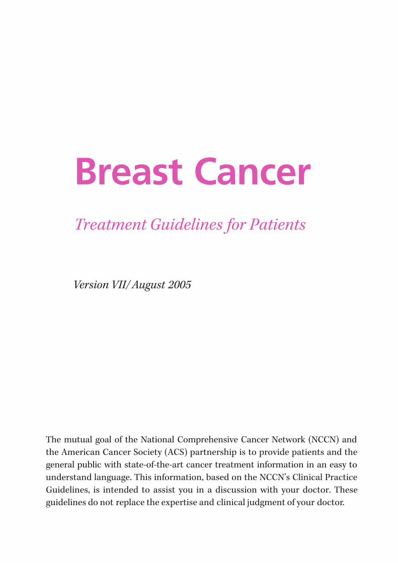
The mutual goal of the National Comprehensive Cancer Network (NCCN) and
the American Cancer Society (ACS) partnership is to provide patients and the
general public with state-of-the-art cancer treatment information in an easy to
understand language. This information, based on the NCCN’s Clinical Practice
Guidelines, is intended to assist you in a discussion with your doctor. These
guidelines do not replace the expertise and clinical judgment of your doctor.
Breast CancerTreatment Guidelines for Patients
Version VII/ August 2005
7/27/2019 NCCN Breast Guidelines
http://slidepdf.com/reader/full/nccn-breast-guidelines 4/84
NCCN Clinical Practice Guidelines, the professional versions, were developed by a diverse panel of experts. The guidelines are a statement of consensus of its
authors regarding the scientific evidence and their views of currently accepted
approaches to treatment. The NCCN guidelines are updated as new significant
data become available. The Patient Information version will be updated accord-
ingly and will be available on-line through the NCCN and the American Cancer
Society web sites. To ensure you have the most recent version, you may contact
the American Cancer Society at 1-800-ACS-2345 or the NCCN at 1-888-909-
NCCN.
©2005 by the American Cancer Society (ACS) and the National Comprehensive
Cancer Network. All rights reserved. The information herein may not be
reprinted in any form for commercial purposes without the expressed written
permission of the ACS. Single copies of each page may be reproduced for per-
sonal and non-commercial uses by the reader.
7/27/2019 NCCN Breast Guidelines
http://slidepdf.com/reader/full/nccn-breast-guidelines 5/84
Contents
Introduction . . . . . . . . . . . . . . . . . . . . . . . . . . . . . . . . . . . . . . . . . . . . . . . . . . . . . . . . . . . . . . . . .5Making Decisions About Breast Cancer Treatment . . . . . . . . . . . . . . . . . . . . . . . . . . .5
Inside Breast Tissue . . . . . . . . . . . . . . . . . . . . . . . . . . . . . . . . . . . . . . . . . . . . . . . . . . . . . . . . . .6
Types of Breast Cancer . . . . . . . . . . . . . . . . . . . . . . . . . . . . . . . . . . . . . . . . . . . . . . . . . . . . . . .6
Benign Breast Lumps . . . . . . . . . . . . . . . . . . . . . . . . . . . . . . . . . . . . . . . . . . . . . . . . . . . . . . . .8
Breast Cancer Work-Up . . . . . . . . . . . . . . . . . . . . . . . . . . . . . . . . . . . . . . . . . . . . . . . . . . . . .8
Breast Cancer Stages . . . . . . . . . . . . . . . . . . . . . . . . . . . . . . . . . . . . . . . . . . . . . . . . . . . . . . .13
Breast Cancer Treatment . . . . . . . . . . . . . . . . . . . . . . . . . . . . . . . . . . . . . . . . . . . . . . . . . . .16Choosing Between Lumpectomy and Mastectomy . . . . . . . . . . . . . . . . . . . . . . . . . . .25
Reconstructive Surgery . . . . . . . . . . . . . . . . . . . . . . . . . . . . . . . . . . . . . . . . . . . . . . . . . . . . .26
Treatment of Pain and Other Symptoms . . . . . . . . . . . . . . . . . . . . . . . . . . . . . . . . . . . . .26
Complementary and Alternative Therapies . . . . . . . . . . . . . . . . . . . . . . . . . . . . . . . . . .27
Other Things to Consider During and After Treatment . . . . . . . . . . . . . . . . . . . . . .27
Clinical Trials . . . . . . . . . . . . . . . . . . . . . . . . . . . . . . . . . . . . . . . . . . . . . . . . . . . . . . . . . . . . . . .29
Work-Up (Evaluation) and Treatment Guidelines . . . . . . . . . . . . . . . . . .31
Decision Trees
Stage 0 Lobular Carcinoma In Situ . . . . . . . . . . . . . . . . . . . . . . . . . . . . . . . . . . . . . . .32
Stage 0 Ductal Carcinoma In Situ . . . . . . . . . . . . . . . . . . . . . . . . . . . . . . . . . . . . . . . .34
Stage I, II, and Some Stage III Breast Cancers . . . . . . . . . . . . . . . . . . . . . . . . . . . .38
Axillary Lymph Node Surgery . . . . . . . . . . . . . . . . . . . . . . . . . . . . . . . . . . . . . . . . . . .44
Additional Treatment (Adjuvant Therapy) After Surgery . . . . . . . . . . . . . . . . .46
Adjuvant Hormone Treatment . . . . . . . . . . . . . . . . . . . . . . . . . . . . . . . . . . . . . . . . . .50
Additional Treatment (Adjuvant Therapy) After Surgery for
Non-Hormone Responsive Tumors . . . . . . . . . . . . . . . . . . . . . . . . . . . . . . . . . . . . . .52
Treatment for Large Stage II Breast Cancers or Stage IIIA . . . . . . . . . . . . . . . .56
Stage III Locally Advanced Breast Cancers . . . . . . . . . . . . . . . . . . . . . . . . . . . . . . .60
Follow-up and Treatment of Stage IV Disease or Recurrence . . . . . . . . . . . . .64
Glossary . . . . . . . . . . . . . . . . . . . . . . . . . . . . . . . . . . . . . . . . . . . . . . . . . . . . . . . . . . . . . . . . . . . .73

7/27/2019 NCCN Breast Guidelines
http://slidepdf.com/reader/full/nccn-breast-guidelines 6/84
Arthur G. James Cancer Hospital and Richard J. SoloveResearch Institute at The Ohio State University
City of Hope Cancer Center
Dana-Farber/Partners CancerCare
Duke Comprehensive Cancer Center
Fox Chase Cancer Center
Fred Hutchinson Cancer Research Center/ Seattle Cancer Care Alliance
H. Lee Moffitt Cancer Center & Research Instituteat the University of South Florida
Huntsman Cancer Institute at the University of Utah
Memorial Sloan-Kettering Cancer Center
Robert H. Lurie Comprehensive Cancer Centerof Northwestern University
Roswell Park Cancer Institute
The Sidney Kimmel Comprehensive Cancer Centerat Johns Hopkins
St. Jude Children’s Research Hospital/ University of Tennessee Cancer Institute
Stanford Hospital and Clinics
UCSF Comprehensive Cancer Center
University of Alabama at BirminghamComprehensive Cancer Center
University of Michigan Comprehensive Cancer Center
The University of Texas M.D. Anderson Cancer Center
UNMC/Eppley Cancer Center at The Nebraska Medical Center
Member Institutions

7/27/2019 NCCN Breast Guidelines
http://slidepdf.com/reader/full/nccn-breast-guidelines 7/84
Introduction
With this booklet, women have access to
information on the way breast cancer is
treated at the nation’s leading cancer centers.
Originally developed for cancer specialists by
the National Comprehensive Cancer Network
(NCCN), these treatment guidelines have
now been translated for the public by the
American Cancer Society (ACS).
Since 1995, doctors have looked to the
NCCN for guidance on the highest quality,
most effective advice on treating cancer. For
more than 85 years, the public has relied on
the American Cancer Society for informationabout cancer. The Society’s books and bro-
chures provide comprehensive, current, and
understandable information to hundreds of
thousands of patients, their families, and
friends. This collaboration between the NCCN
and ACS provides an authoritative and
understandable source of cancer treatment
information for the public.
These patient guidelines will help you bet-ter understand your cancer treatment and
your doctor’s counsel. We urge you to discuss
them with your doctor. To make the best pos-
sible use of this information, you might begin
by asking your doctor the following questions:
• How large is my cancer? Do I have
more than one tumor in the breast?
• What is my cancer’s grade (how
abnormal the cells appear) and histology
(type and arrangement of tumor cells),
as seen under a microscope?
• Do I have any lymph nodes with
cancer (positive lymph nodes)? If yes,
how many?
• What is the stage of my cancer?
• Does my cancer contain hormone
receptors?
• Is breast-conserving treatment an
option for me?
• In addition to surgery, what other
treatments do you recommend?
Radiation? Chemotherapy? HormonalTherapy?
• What are their side effects?
• Are there any clinical trials that I
should consider?
Making Decisions About
Breast Cancer TreatmentOn the pages following the general informa-
tion about breast cancer, you’ll find flow charts
that doctors call decision trees. The charts
represent different stages of breast cancer.
Each one shows you step-by-step how you
and your doctor can arrive at the choices you
need to make about your treatment.
Here you will find background informa-tion on breast cancer with explanations of
cancer stage, work-up, and treatment — all
categories used in the flow charts. We’ve also
provided a glossary at the end of the booklet.
Words in the glossary will appear in italics.
Although breast cancer is a very serious
disease, it can be treated, and it should be
treated by a team of health care professionals
with experience in treating women with breastcancer. This team may include a surgeon,
radiation oncologist, medical oncologist,
radiologist, pathologist, oncology nurse, social
worker, and others. But not all women with
breast cancer receive the same treatment.
Doctors must consider a woman’s specific
7/27/2019 NCCN Breast Guidelines
http://slidepdf.com/reader/full/nccn-breast-guidelines 8/84
medical situation. This booklet can help you
and your doctor decide which choices best
meet your medical and personal needs.
Breast cancer can occur in men. However,
since the incidence is very low, this booklet is
focused on female breast cancer. To learn more
about breast cancer in men, speak with yourdoctor and contact the American Cancer
Society at 1-800-ACS-2345 or www.cancer.org.
Inside Breast Tissue
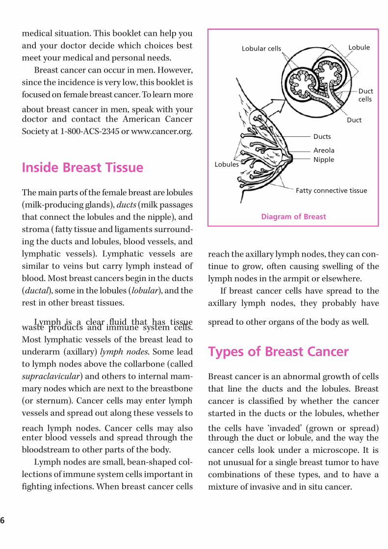
The main parts of the female breast are lobules
(milk-producing glands), ducts (milk passages
that connect the lobules and the nipple), and
stroma ( fatty tissue and ligaments surround-
ing the ducts and lobules, blood vessels, and
lymphatic vessels). Lymphatic vessels are
similar to veins but carry lymph instead of
blood. Most breast cancers begin in the ducts
(ductal ), some in the lobules (lobular ), and the
rest in other breast tissues.
Lymph is a clear fluid that has tissue waste products and immune system cells.
Most lymphatic vessels of the breast lead to
underarm (axillary) lymph nodes . Some lead
to lymph nodes above the collarbone (called
supraclavicular ) and others to internal mam-
mary nodes which are next to the breastbone
(or sternum). Cancer cells may enter lymph
vessels and spread out along these vessels to
reach lymph nodes. Cancer cells may alsoenter blood vessels and spread through the
bloodstream to other parts of the body.
Lymph nodes are small, bean-shaped col-
lections of immune system cells important in
fighting infections. When breast cancer cells
reach the axillary lymph nodes, they can con-
tinue to grow, often causing swelling of the
lymph nodes in the armpit or elsewhere.
If breast cancer cells have spread to the
axillary lymph nodes, they probably have
spread to other organs of the body as well.
Types of Breast Cancer
Breast cancer is an abnormal growth of cells
that line the ducts and the lobules. Breast
cancer is classified by whether the cancer
started in the ducts or the lobules, whether
the cells have ‘invaded’ (grown or spread)through the duct or lobule, and the way the
cancer cells look under a microscope. It is
not unusual for a single breast tumor to have
combinations of these types, and to have a
mixture of invasive and in situ cancer.
6
Lobular cells
Lobules
Lobule
Ductcells
Duct
Ducts
Nipple
Areola
Fatty connective tissue
Diagram of Breast

7/27/2019 NCCN Breast Guidelines
http://slidepdf.com/reader/full/nccn-breast-guidelines 9/84
Carcinoma In SituIn situ means that the cancer stays confined
to ducts or lobules and has not spread into
surrounding fatty tissues in the breast or to
other organs in the body. There are 2 types of
breast carcinoma in situ:
• Lobular carcinoma in situ (LCIS): Also called lobular neoplasia. It begins
in the lobules, but does not grow
through the lobule walls. Breast cancer
specialists do not think that LCIS,
itself, becomes an invasive cancer, but
women with this condition do run a
higher risk of developing an invasive
cancer in either breast.
• Ductal carcinoma in situ (DCIS):
The most common type of noninvasive
breast cancer. Cancer cells inside the
ducts do not spread through the walls
of the ducts into the fatty tissue of the
breast. DCIS is treated with surgery
and sometimes radiation, which are
usually curative. Having untreated DCIS
greatly increases the risk of invasivebreast cancer.
Infiltrating (or invasive) DuctalCarcinoma (IDC)The cancer starts in a milk passage, or duct,
of the breast, but then the cancer cells break
through the wall of the duct and spread into
the breast’s fatty tissue. They can then spread
into lymphatic channels or blood vessels of the breast and to other parts of the body.
About 80 % of all breast cancers are infiltrating
or invasive ductal carcinoma.
Infiltrating (or invasive) LobularCarcinoma (ILC)This type of cancer starts in the milk-producing
glands. Like IDC, this cancer can spread
beyond the breast to other parts of the body.
About 10% to 15% of invasive breast cancers
are invasive lobular carcinomas.
Medullary CarcinomaThis special type of infiltrating ductal cancer
has a relatively well-defined, distinct bound-
ary between tumor tissue and normal breast
tissue. It also has a number of other special
features, including the large size of the cancer
cells and the presence of immune system
cells at the edges of the tumor. It accounts for
about 5% of all breast cancers. It is difficult to
distinguish medullary breast cancer from the
more common infiltrating ductal breast
cancer. NCCN recommends that medullary
breast cancers be treated as if they were the
usual form of infiltrating ductal cancer.
Colloid CarcinomaThis rare type of invasive ductal breast cancer,
also called mucinous carcinoma, is formed by
mucus-producing cancer cells. Colloid carci-
noma has a slightly better prognosis and a
slightly lower chance of metastasis than inva-
sive lobular or invasive ductal cancers of the
same size.
Tubular CarcinomaTubular carcinoma is a special type of infil-
trating ductal breast carcinoma. About 2% of
all breast cancers are tubular carcinomas.
Women with this type of breast cancer have a
better outlook because the cancer is less likely

7/27/2019 NCCN Breast Guidelines
http://slidepdf.com/reader/full/nccn-breast-guidelines 10/84
to spread outside the breast than invasive
lobular or invasive ductal cancers of the same
size.
Inflammatory Breast CancerThis uncommon type of invasive breast
cancer accounts for about 1% to 3% of allbreast cancers. The skin of the affected breast
is red, feels warm, and has the appearance of
an orange peel. Doctors now know that these
changes are not caused by inflammation, but
by cancer cells invading the skin and block-
ing lymph vessels.
Inflammatory breast cancer has a higher
chance of spreading and a worse prognosis
than typical invasive ductal or lobular cancers.
Inflammatory breast cancer is always staged
as stage IIIB unless it has already spread to
other organs at the time of diagnosis which
would then make it stage IV. (See discussion
of stage below.)
Benign Breast LumpsMost breast lumps are benign (not cancerous).
Fibrocystic changes usually cause most of these
lumps. Fibrosis refers to excessive formation
of scar-like connective tissue; cysts are fluid-
filled sacs. Women with fibrocystic changes
often experience breast swelling and pain.
The breasts may feel lumpy and the nipple may
discharge a clear or slightly cloudy liquid.Benign breast lumps such as fibroadenomas
or papillomas are quite common. They cannot
spread outside of the breast to other organs.
Some breast lumps need to have a biopsy
(see below). Women who have some types of
benign conditions have a higher risk of devel-
oping a future invasive breast cancer. Talk to
your doctor about whether he or she feels it
necessary to remove these lumps. This booklet
only refers to treating breast cancer, not benign
breast conditions.
Breast Cancer Work-Up
Evaluating a Breast Lump orMammogram Finding
An evaluation of a breast lump or mammo-
gram includes a thorough medical history, a
physical examination, and breast imaging
(such as x-rays) including a mammogram. A
biopsy is needed for a suspicious or irregular
finding, though often, these suspicious areas
prove to be benign (not cancer). If cancer is
found, other imaging and laboratory tests are
needed. Exactly which tests are helpful
depends on the type of cancer, and if and
where it has spread. This section provides a
summary of the steps, tests, and types of biopsy that may be suggested.
Doctor Visit and Examination A woman’s first step in having a new breast
lump, symptom, or a change on a mammogram
evaluated is to meet with her doctor. The
doctor will take a medical history including
asking a series of questions about symptoms
and factors that may be related to breastcancer risk (such as family history of cancer).
The physical examination should include a
general examination of the woman’s body as
well as careful examination of her breasts.
The doctor will examine:
8
7/27/2019 NCCN Breast Guidelines
http://slidepdf.com/reader/full/nccn-breast-guidelines 11/84
• The breast, including its texture, size,
relationship to skin and chest muscles,
and the presence of lumps or masses
• The nipples or skin of the breast
• Lymph nodes under the armpit or
above the collarbone
• Other organs to check for obvious spreadof breast cancer and to help evaluate the
general condition of the woman’s health.
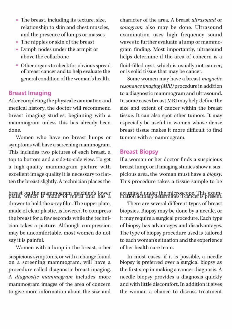
Breast Imaging After completing the physical examination and
medical history, the doctor will recommend
breast imaging studies, beginning with a
mammogram unless this has already been
done.
Women who have no breast lumps or
symptoms will have a screening mammogram.
This includes two pictures of each breast, a
top to bottom and a side-to-side view. To get
a high-quality mammogram picture with
excellent image quality it is necessary to flat-
ten the breast slightly. A technician places the
breast on the mammogram machine’s lower plate, which is made of metal and has a
drawer to hold the x-ray film. The upper plate,
made of clear plastic, is lowered to compress
the breast for a few seconds while the techni-
cian takes a picture. Although compression
may be uncomfortable, most women do not
say it is painful.
Women with a lump in the breast, other
suspicious symptoms, or with a change foundon a screening mammogram, will have a
procedure called diagnostic breast imaging.
A diagnostic mammogram includes more
mammogram images of the area of concern
to give more information about the size and
character of the area. A breast ultrasound or
sonogram also may be done. Ultrasound
examination uses high frequency sound
waves to further evaluate a lump or mammo-
gram finding. Most importantly, ultrasound
helps determine if the area of concern is a
fluid-filled cyst, which is usually not cancer,or is solid tissue that may be cancer.
Some women may have a breast magnetic
resonance imaging (MRI) procedure in addition
to a diagnostic mammogram and ultrasound.
In some cases breast MRI may help define the
size and extent of cancer within the breast
tissue. It can also spot other tumors. It may
especially be useful in women whose dense
breast tissue makes it more difficult to find
tumors with a mammogram.
Breast BiopsyIf a woman or her doctor finds a suspicious
breast lump, or if imaging studies show a sus-
picious area, the woman must have a biopsy .
This procedure takes a tissue sample to be
examined under the microscope. This exam-ination actually determines if cancer is present.
There are several different types of breast
biopsies. Biopsy may be done by a needle, or
it may require a surgical procedure. Each type
of biopsy has advantages and disadvantages.
The type of biopsy procedure used is tailored
to each woman’s situation and the experience
of her health care team.
In most cases, if it is possible, a needlebiopsy is preferred over a surgical biopsy as
the first step in making a cancer diagnosis. A
needle biopsy provides a diagnosis quickly
and with little discomfort. In addition it gives
the woman a chance to discuss treatment
7/27/2019 NCCN Breast Guidelines
http://slidepdf.com/reader/full/nccn-breast-guidelines 12/84
options with her doctor before any surgery is
done. There is no danger that needle biopsy
itself will spread the breast cancer. In some
patients, a surgical biopsy may still be needed
to remove all or part of a lump for micro-
scopic examination after a needle biopsy has
been performed, or it may be necessary to doa surgical biopsy instead of needle biopsy.
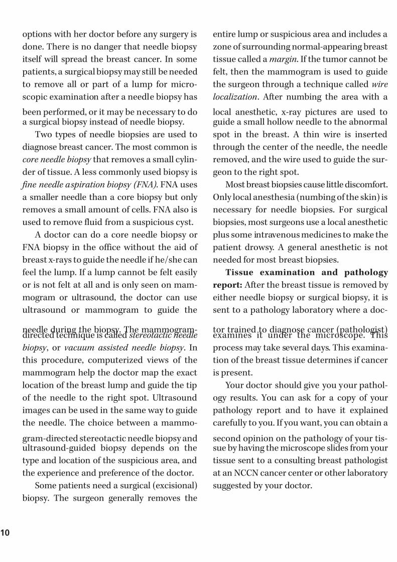
Two types of needle biopsies are used to
diagnose breast cancer. The most common is
core needle biopsy that removes a small cylin-
der of tissue. A less commonly used biopsy is
fine needle aspiration biopsy (FNA). FNA uses
a smaller needle than a core biopsy but only
removes a small amount of cells. FNA also is
used to remove fluid from a suspicious cyst.
A doctor can do a core needle biopsy or
FNA biopsy in the office without the aid of
breast x-rays to guide the needle if he/she can
feel the lump. If a lump cannot be felt easily
or is not felt at all and is only seen on mam-
mogram or ultrasound, the doctor can use
ultrasound or mammogram to guide the
needle during the biopsy. The mammogram-directed technique is called stereotactic needle
biopsy , or vacuum assisted needle biopsy . In
this procedure, computerized views of the
mammogram help the doctor map the exact
location of the breast lump and guide the tip
of the needle to the right spot. Ultrasound
images can be used in the same way to guide
the needle. The choice between a mammo-
gram-directed stereotactic needle biopsy andultrasound-guided biopsy depends on the
type and location of the suspicious area, and
the experience and preference of the doctor.
Some patients need a surgical (excisional)
biopsy. The surgeon generally removes the
entire lump or suspicious area and includes a
zone of surrounding normal-appearing breast
tissue called a margin. If the tumor cannot be
felt, then the mammogram is used to guide
the surgeon through a technique called wire
localization. After numbing the area with a
local anesthetic, x-ray pictures are used toguide a small hollow needle to the abnormal
spot in the breast. A thin wire is inserted
through the center of the needle, the needle
removed, and the wire used to guide the sur-
geon to the right spot.
Most breast biopsies cause little discomfort.
Only local anesthesia (numbing of the skin) is
necessary for needle biopsies. For surgical
biopsies, most surgeons use a local anesthetic
plus some intravenous medicines to make the
patient drowsy. A general anesthetic is not
needed for most breast biopsies.
Tissue examination and pathology
report: After the breast tissue is removed by
either needle biopsy or surgical biopsy, it is
sent to a pathology laboratory where a doc-
tor trained to diagnose cancer (pathologist)examines it under the microscope. This
process may take several days. This examina-
tion of the breast tissue determines if cancer
is present.
Your doctor should give you your pathol-
ogy results. You can ask for a copy of your
pathology report and to have it explained
carefully to you. If you want, you can obtain a
second opinion on the pathology of your tis-sue by having the microscope slides from your
tissue sent to a consulting breast pathologist
at an NCCN cancer center or other laboratory
suggested by your doctor.
10
7/27/2019 NCCN Breast Guidelines
http://slidepdf.com/reader/full/nccn-breast-guidelines 13/84
Other Tests After Cancer Has BeenDiagnosedIf the breast biopsy results show that cancer
is present, the doctor may order other tests
to find out if the cancer has spread and to
help determine treatment. For most women
with breast cancer, extensive testing providesno benefit and is not necessary. Unfortu-
nately, there is no test that can completely
reassure you that the cancer has not spread.
The NCCN Guidelines describe which tests
are needed based on the extent (spread) of
the cancer, and on the results of the history
and physical examination. Tests that may be
done include:
Chest x-ray: All women with breast cancer
should have a chest x-ray before surgery and
to make sure that the breast cancer has not
spread to the lungs.
Bone scan: This may provide information
about spread of breast cancer to the bones.
However, many changes that show up on a
bone scan are not cancer. Unless there are
symptoms of spread to the bone including new pains or changes on blood tests, a bone
scan is not recommended except in patients
with advanced cancer. To scan bones, a small
dose of a radioactive substance is injected
into your vein. The radioactive substance col-
lects in areas of abnormal bone. These areas
can be seen on the bone scan image. Other
than the needle stick, a bone scan is painless.
Computerized tomography (CT) scans:CT scans are done when symptoms or other
findings suggest the cancer has spread to
other organs. For most women with an early
stage breast cancer, a CT scan is not needed.
But if the cancer appears more advanced, a
CT of the abdomen and/or chest may be
done to see if the cancer has spread. CT scans
take multiple x-rays of the same part of the
body from different angles to provide detailed
pictures of internal organs. Except for the
injection of intravenous dye, necessary for
most patients, this is a painless procedure.
Magnetic resonance imaging (MRI):MRI scans use radio waves and magnets to
produce detailed images of internal organs
without any x-rays. MRI is useful in looking at
the brain and spinal cord, and in examining
any specific area in the bone. A special MRI
procedure called a breast MRI with dedicated
breast coil can also be used to look for
tumors in the breast. Routine MRI for all
patients with breast cancer is not helpful and
is not needed.
Positron emission tomography (PET):
PET scans use a form of sugar (glucose) that
contains a radioactive atom. A small amount
of the radioactive material is injected into
your arm. Then you are put into the PET
machine where a special camera can detect
the radioactivity. Because of their high rate of metabolism, cancer cells of the body absorb
large amounts of the radioactive sugar. PET
scans can be used instead of several different
x-rays because it scans your whole body. Newer
devices combine PET scans and CT scans.
Blood tests: Some blood tests are needed
to plan surgery, to screen for evidence of
cancer spread, and to plan treatment after
surgery. These blood tests include:• Complete blood count (CBC). This
determines whether the blood has the
correct type and number of blood cells.
Abnormal test results could reveal other
health problems including anemia, and
could suggest the cancer has spread to
7/27/2019 NCCN Breast Guidelines
http://slidepdf.com/reader/full/nccn-breast-guidelines 14/84
the bone marrow. Also, if you receive
chemotherapy, doctors repeat this test
because chemotherapy often affects the
blood-forming cells of the bone marrow.
• Blood chemical and enzyme tests: These
tests are done in patients with invasive
breast cancer (not needed with in situcancer). They can sometimes tell if the
cancer has spread to the bone or liver.
If these test results are not normal, your
doctor will order imaging tests such as
bone scans or CT scans.
Tumor tests (estrogen receptor, prog-
esterone receptor, HER-2/neu): Testing the
tumor itself for certain substances calledreceptors helps determine the chances the
cancer will spread, and helps the doctor
determine the best treatment. The pathology
laboratory tests the cancer tissue that is
removed, either from the first biopsy or the
final surgery.
Testing for hormone receptors helps deter-
mine the best treatment. Two hormones in
women – estrogen and progesterone – stimu-
late the growth of normal breast cells and
play a role in many breast cancers. Cancer
cells respond to these hormones through the
estrogen receptors (ER) and progesterone
receptors (PR). ER and PR are the cell’s “wel-
come mat” for these hormones circulating in
the blood. If a cancer does not have these
receptors, it is referred to as hormone receptor
negative (estrogen-receptor negative and/or
progesterone-receptor negative). If the cancer
has these receptors, it is referred to as hormone-
receptor positive (estrogen-receptor positive
and/or progesterone-receptor positive) or just
ER-positive or PR-positive.
These hormone receptors are important
because cancer cells that are ER- or PR-positive
will stop growing if the woman takes drugs
that block the effect of estrogen and proges-
terone or block her own hormone production.
These drugs lower the chance that the cancer
will come back (recur) in other body organs,and improve the chances of long-term survival.
Most women whose breast cancer is ER- or
PR- positive will take these drugs as part of
their treatment. However, these hormone-
active drugs are not effective if the cancer
does not contain these receptors.
All invasive breast cancers (not necessary
with in situ cancer) should be tested for hor-
mone receptors at the time of the breast
biopsy or surgery. Each woman should ask her
doctor for these test results, and if hormone-
like drugs or blocking her own hormones
should be part of the treatment.
About one third of breast cancers have too
much of a growth-promoting protein called
HER2/neu and too many copies (more than 2)
of the gene that instructs the cells to producethat protein. In other cases, a normal number
of HER2/neu genes are present, but they are
too active in instructing the cells to produce
HER2/neu protein.
These cancers tend to grow and spread
more aggressively than other breast cancers.
They can be treated with a drug called
trastuzumab that prevents the HER2/neu
protein from stimulating breast cancer cellgrowth. Studies also suggest that chemother-
apy combinations containing anthracycline
drugs (such as doxorubicin or epirubicin) treat
breast cancers with too much HER2/neu
more effectively than combinations that do
not include these drugs.
12
7/27/2019 NCCN Breast Guidelines
http://slidepdf.com/reader/full/nccn-breast-guidelines 15/84
Grades of breast cancer: A pathologist
looks at the tissue sample under a microscope
and then assigns a grade to it. The grade
helps predict the patient’s prognosis because
cancers that closely resemble normal breast
tissue tend to grow and spread more slowly.
In general, a lower grade number indicates acancer that is slightly less likely to spread,
and a higher number indicates a cancer that
is slightly more likely to spread.
Grade is based on the arrangement of the
cells in relation to each other: whether they
form tubules; how closely they resemble nor-
mal breast cells (nuclear grade); and how
many of the cancer cells are in the process of
dividing (mitotic count). The best way for
doctors to report grade is called the Nottingham
Modification of the Bloom Scarf Richardson
Score. In this, the pathology doctor assigns a
score from 1 to 3 to each of 3 different char-
acteristics of the cancer. This score for each
characteristic, the total score, and the final
grade (Grade I, Grade II, and Grade III) should
be recorded on the pathology report. Thissystem of grading is used for invasive cancers
but not for in situ cancers.
Ductal carcinoma in situ (DCIS) is graded
by examining the central portion of the
cancer cells. It is given a nuclear grade, which
describes how abnormal the cancer cells
appear. The presence or absence of necrosis
(areas of dead or degenerating cancer cells) is
also noted.
Breast Cancer Stages
Cancers are classified by stage. Staging a
cancer is the process of finding out how far
the cancer has progressed when it is diagnosed.
Doctors determine the stage of a cancer by
gathering information from examinations and
tests on the tumor, lymph nodes, and distant
organs.
A breast cancer’s stage is one of the most
important factors that may predict prognosis
(outlook for cure versus the chance of cancer
coming back or spreading to other organs). A
cancer’s stage therefore is an important fac-
tor in choosing the best treatment.Each woman’s outlook with breast cancer
differs, depending on the cancer’s stage and
other cancer factors such as hormone receptors,
her general state of health, and her treatment.
You should feel you can talk frankly with
your doctors about your cancer stage and
prognosis, and how they affect treatment
options.
System to Define Cancer StageThe system most often used to describe the
growth and spread of breast cancer is the
TNM staging system, also known as the
American Joint Committee on Cancer (AJCC)
system. This was most recently updated in
2002, and published as the 6th Edition of the
Cancer Staging Manual . In TNM staging,
information about the tumor, nearby lymph
nodes, and distant organ metastases is com-
bined and a stage is assigned to specific TNM
groupings. The TNM stage groupings are
described using Roman numerals from I to
IV. The clinical stage is determined by what
7/27/2019 NCCN Breast Guidelines
http://slidepdf.com/reader/full/nccn-breast-guidelines 16/84
the doctor learns from the physical examina-
tion and tests. The pathologic stage includes
the findings of the pathologist after surgery.
Most of the time, pathologic stage is the most
important stage because usually the cancer
isn’t known to have spread to lymph nodes
until the pathologist examines them underthe microscope.
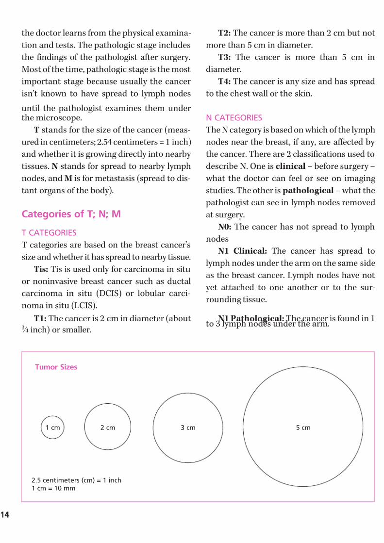
T stands for the size of the cancer (meas-
ured in centimeters; 2.54 centimeters = 1 inch)
and whether it is growing directly into nearby
tissues. N stands for spread to nearby lymph
nodes, and M is for metastasis (spread to dis-
tant organs of the body).
Categories of T; N; M
T CATEGORIES
T categories are based on the breast cancer’s
size and whether it has spread to nearby tissue.
Tis: Tis is used only for carcinoma in situ
or noninvasive breast cancer such as ductal
carcinoma in situ (DCIS) or lobular carci-
noma in situ (LCIS).
T1: The cancer is 2 cm in diameter (about3 ⁄ 4 inch) or smaller.
T2: The cancer is more than 2 cm but not
more than 5 cm in diameter.
T3: The cancer is more than 5 cm in
diameter.
T4: The cancer is any size and has spread
to the chest wall or the skin.
N CATEGORIES
The N category is based on which of the lymph
nodes near the breast, if any, are affected by
the cancer. There are 2 classifications used to
describe N. One is clinical – before surgery –
what the doctor can feel or see on imaging
studies. The other is pathological – what the
pathologist can see in lymph nodes removed
at surgery.
N0: The cancer has not spread to lymph
nodes
N1 Clinical: The cancer has spread to
lymph nodes under the arm on the same side
as the breast cancer. Lymph nodes have not
yet attached to one another or to the sur-
rounding tissue.
N1 Pathological: The cancer is found in 1to 3 lymph nodes under the arm.
14
5 cm
2.5 centimeters (cm) = 1 inch1 cm = 10 mm
Tumor Sizes
1 cm 2 cm 3 cm

7/27/2019 NCCN Breast Guidelines
http://slidepdf.com/reader/full/nccn-breast-guidelines 17/84
N2 Clinical: The cancer has spread to
lymph nodes under the arm on the same side
as the breast cancer and are attached to one
another or to the surrounding tissue or
enlarged. Or the cancer can be seen to have
spread to the internal mammary lymph
nodes (next to the sternum), but not to thelymph nodes under the arm.
N2 Pathological: The cancer has spread
to 4 to 9 lymph nodes under the arm.
N3 Clinical: The cancer has spread to
lymph nodes above or just below the collar-
bone on the same side as the cancer, and may
or may not have spread to lymph nodes
under the arm. Or the cancer has spread to
internal mammary lymph nodes and lymph
nodes under the arm, both on the same side
as the cancer.
N3 Pathological: The cancer has spread
to 10 or more lymph nodes under the arm, or
also involves lymph nodes in other areas
around the breast
M CATEGORIES
The M category depends on whether thecancer has spread to any distant tissues and
organs.
M0: No distant cancer spread.
M1: Cancer has spread to distant organs.
Stage Grouping for Breast CancerOnce the T, N, and M categories have been
assigned, this information is combined to
assign an overall stage of 0, I, II, III, or IV.
Breast Cancer Stages
Overall Stage T category N category M category
Stage 0 Tis N0 M0
Stage I T1 N0 M0
Stage IIA T0 N1 M0T1 N1 M0
T2 N0 M0
Stage IIB T2 N1 M0T3 N0 M0
Stage IIIA T0 N2 M0T1 N2 M0
T2 N2 M0T3 N1 M0
T3 N2 M0
Stage IIIB T4 Any N M0
Stage IIIC Any T N3 M0
Stage IV Any T Any N M1
7/27/2019 NCCN Breast Guidelines
http://slidepdf.com/reader/full/nccn-breast-guidelines 18/84
Breast Cancer Treatment
Most women with breast cancer will have
surgery. The 2 common types of surgery are
breast-conserving surgery and mastectomy.
Breast-Conserving Surgery Lumpectomy removes only the breast lump
and a rim of normal surrounding breast tis-
sue. If cancer cells are present at the outside
edge of the biopsy (the margin), more surgery
is usually needed to remove any remaining
cancer. Most often this additional surgery is a
repeat lumpectomy, but sometimes it requires
removal of the entire breast (mastectomy).In almost all cases of invasive breast
cancer, radiation therapy is given after
lumpectomy. Doctors call this combination (of
lumpectomy and radiation) breast-conserving
therapy. It’s an option for most, but not all,
women with breast cancer. Those who may
not have lumpectomy, or breast-conserving
therapy include:
• Women who have already had radiationtherapy to the affected breast or chest
• Women with 2 or more areas of cancer,
in the same breast, too far apart to be
removed in one incision
• Women whose lumpectomy, including
any possible repeat lumpectomy when
needed, cannot completely remove
their cancers
• Women with active connective tissue
diseases involving the skin (especially
scleroderma or lupus) that make body
tissues especially sensitive to the side
effects of radiation
• Pregnant women who would require
radiation while still pregnant
• Women whose tumor is larger than 5
centimeters (2 inches) and can’t be
shrunk by treatment before surgery
Radiation therapy as a part of breast-
conserving therapy can sometimes be omitted.
Women who may consider lumpectomy without radiation therapy have all of the
following:
• Age 70 years or older
• A tumor 2 cm or less that has been
completely removed
• A tumor that contains hormone
receptors
• No lymph node involvement
• Received treatment with hormonetherapy (see page 18)
MastectomyMastectomy is removal of the entire breast,
including the nipple. Mastectomy is needed
for some cases, and some women choose
mastectomy (see discussion below of choosing
between lumpectomy and mastectomy).
Different words are used to describe the
mastectomy type depending on the extent of
the surgery in the armpit and the muscles
under the breast. In a simple (total) mastectomy
procedure surgeons remove the entire breast
but do not remove any lymph nodes from
under the arm or muscle tissue from beneath
the breast. In a modified radical mastectomy ,
surgeons remove the entire breast and some
axillary (underarm) lymph nodes. In a radical
mastectomy , all the muscle under the breast
is also removed. Radical mastectomy used to
be the standard mastectomy, but it is not
more effective and is only rarely done today.
16

7/27/2019 NCCN Breast Guidelines
http://slidepdf.com/reader/full/nccn-breast-guidelines 19/84
Lymph Node SurgeryIn the treatment of invasive cancer, whether
a woman has a mastectomy or a lumpectomy
for invasive cancer, she and her doctor usually
need to know if the cancer has spread to the
lymph nodes. If the lymph nodes are affected,
that increases the likelihood that cancer cellshave spread through the bloodstream to other
parts of the body.
Doctors once believed that removing as
many lymph nodes as possible would reduce
the risk of developing spread of breast cancer
and improve a woman’s chances for long-term
survival. We now know that removing the
lymph nodes probably does not improve the
chance for long-term survival. However,
knowing whether the lymph nodes are
involved is important in selecting the best
drug treatments to prevent cancer recurrence.
The only way to accurately determine if
lymph nodes are involved is surgery to
remove lymph nodes. This means removing
some or all of the lymph nodes in the armpit.
There are 10 to 20 lymph nodes in the armpit.In the standard operation called an axillary
lymph node dissection, all these lymph nodes
are removed. This is often necessary. However,
in many cases, lymph node testing may be done
with more limited surgery that only removes
a few lymph nodes with fewer side effects.
This is called sentinel lymph node biopsy .
For some women with invasive cancer,
removing the underarm lymph nodes can beconsidered optional. This includes:
• Women with tumors so small and with
such a favorable outlook that lymph
node spread is unlikely
• Instances where it would not affect
whether adjuvant treatment is given
• Elderly women
• Women with other serious medical
conditions.
Lymph node surgery is not necessary with
pure ductal carcinoma in situ or pure lobular
carcinoma in situ. However, a sentinel nodebiopsy (see below) may be performed if the
woman is having surgery (such as mastectomy)
that would make it impossible to do the sen-
tinel node procedure if invasive cancer were
found in the tissue removed at surgery.
The surgical technique used to remove
lymph nodes from under the armpit depends
on the personal circumstances of the patient.
If there are enlarged lymph nodes with appar-ent spread of the cancer, or the lymph nodes
are otherwise found to be involved with
cancer, then complete axillary lymph node
dissection is necessary. However, in many
cases the lymph nodes are not enlarged and
otherwise are not likely to contain cancer. In
such cases, the more limited sentinel lymph
node biopsy procedure can be performed.
In the sentinel lymph node biopsy procedure
the surgeon finds and removes the ‘sentinel
node’ – the first few lymph nodes into which
a tumor drains. These are the lymph nodes
most likely to contain cancer cells. To find
these so-called “sentinel lymph nodes,” the
surgeon injects a radioactive substance and/
or a blue dye under the nipple or into the area
around the tumor. Lymphatic vessels carry
these substances into the sentinel lymph
nodes and provide the doctor with a “lymph
node map.” The doctor can either see the blue
dye or detect the radioactivity with a Geiger
counter. He or she then removes the nodes
for examination by the pathologist and the
incision is closed.

7/27/2019 NCCN Breast Guidelines
http://slidepdf.com/reader/full/nccn-breast-guidelines 20/84
If the sentinel node contains cancer, the
surgeon removes more lymph nodes in the
armpit (axillary dissection). This may be done
at the same time or several days after the
original sentinel node biopsy. If the sentinel
node is cancer-free, the patient will not need
more lymph node surgery and can avoid theside effects of full lymph node surgery.
This limited sampling of lymph nodes is
not appropriate for some women. A sentinel
lymph node biopsy should be considered only
if there is a team with experience with this
technique. In addition, it is only done if there
is a single tumor less than 5 cm in the breast,
and the lymph nodes feel normal on physical
examination. Previously, it was not advised
in cases where chemotherapy or hormonal
therapy was given before surgery. However,
sentinel lymph node biopsy may now be used
in some of these cases.
SIDE EFFECTS OF LYMPH NODE SURGERY
Side effects of lymph node surgery can be
bothersome to many women. The side effects
can occur with either the full axillary lymph
node dissection or sentinel lymph node
biopsy. However, the side effects are much less
common, and less severe with the sentinellymph node procedure.
Side effects of lymph node surgery
include:
• Temporary or permanent numbness in
the skin on the inside of the upper arm
• Temporary limitation of arm and
shoulder movements
• Swelling of the breast and arm called
lymphedema
Lymphedema is the most significant of
these side effects. If it develops it may be
permanent. In most women who develop
lymphedema it is bothersome but not dis-
abling. No one can predict which patients
will develop this condition or when it will
develop. Lymphedema can develop just after
surgery, or even months or years later.Lymphedema occurs in about 10% of women
who have axillary lymph node dissection and
about 1% of women who have sentinel lymph
node biopsy.
With care, patients can take steps to help
avoid lymphedema or at least keep it under
control. Talk to your doctor for more details.
Some of the steps to take to help avoid
lymphedema include:
• Avoid having blood drawn from or IV’s
inserted into the arm on the side of the
lymph node surgery.
18
Normal Lymph Drainage
Lymphnodes
Lymphvessels
Internalmammarylymph node
Axillarylymph nodes

7/27/2019 NCCN Breast Guidelines
http://slidepdf.com/reader/full/nccn-breast-guidelines 21/84
• Do not allow a blood pressure cuff to
be placed on that arm. If you are in the
hospital, tell all health care workers
about your arm.
• If your arm or hand feels tight or
swollen, don’t ignore it. Tell your
doctor immediately.• If needed, wear a well-fitted
compression sleeve.
• Wear gloves when gardening or doing
other things that are likely to lead
to cuts.
For more information on lymphedema, call
the American Cancer Society at 1-800-ACS-2345
and ask for Lymphedema: What Every WomanWith Breast Cancer Should Know .
Radiation TherapyRadiation is used to destroy cancer cells left
behind in the breast, chest wall, or lymph nodes
after surgery. Radiation treatments are usually
given 5 days a week for 6 to 7 weeks. Radiation
is used in most cases of breast conserving
therapy. Radiation may also be needed after
mastectomy in cases with either a cancer larger
than 5 cm in size, or with positive lymph nodes.
Radiation therapy uses a beam of high-
energy rays (or particles) to destroy cancer
cells or slow their rate of growth. This type of
therapy can be given in several ways.
• External beam radiation. The most
common way, delivers a carefully
focused beam of radiation from a
machine outside the body. This is
known as external beam radiation. This
is the usual treatment after lumpectomy.
• Internal radiation, brachytherapy, or
interstitial radiation. Another method
of delivering radiation is to place tiny
pellets that contain radioactive materi-
als in or near the tumor. This method
is also called internal radiation,
brachytherapy , or interstitial radiation.These may be placed in the tumor site
to “boost” the radiation dose.
If the procedure done was a lumpectomy,
the entire breast receives radiation, sometimes
along with lymph nodes above and below the
clavicle and near the sternum. Giving an extra
boost of radiation to the area in the breast
where the cancer was removed may better
prevent it from coming back in that area.
If the surgery was mastectomy, radiation
is given to the entire area of the skin and
muscle where the mastectomy was per-
formed. This radiation is needed only if the
tumor was over 5 cm in size, or there were
positive lymph nodes.
With mastectomy and lumpectomy, radi-
ation may also be given to the area of lymphnodes in the armpit, and the shoulder. This is
needed if there are positive lymph nodes, and
especially if there are 4 or more involved
lymph nodes.
Side effects most likely to occur include
swelling and heaviness in the breast, sunburn-
like skin changes in the treated area, and
fatigue. Changes to the breast tissue and skin
usually go away in 6 to 12 months. In some women, the breast becomes smaller and firmer
after radiation therapy. If the lymph nodes
under the arm are treated with radiation,
lymphedema can also occur.

7/27/2019 NCCN Breast Guidelines
http://slidepdf.com/reader/full/nccn-breast-guidelines 22/84
A new technique to give radiation over a
much shorter period of time (5 days total) and
to only the part of the breast with the cancer
is currently being studied. This is called partial
breast radiation. It is hoped that partial breast
radiation will prove to be equal to the current
standard whole breast radiation. However, partial breast irradiation is still experimental.
Women are encouraged to participate in the
major national clinical trial of partial breast
radiation that started in 2005 and should ask
their doctor about this.
Systemic TreatmentTo reach cancer cells that may have spread
beyond the breast and nearby tissues, doctors
use drugs that can be given as pills or by injec-
tion. This type of treatment is called systemic
treatment. Examples of systemic treatment
include chemotherapy and hormone therapy.
Almost all women who have breast cancers
that contain estrogen or progesterone recep-
tors will receive one of the anti-estrogen
hormone drugs. The main question they haveto consider is whether to add chemotherapy
to hormonal therapy. For women with breast
cancers that do not contain estrogen or prog-
esterone receptors, anti-estrogen hormone
drugs are not effective and their only choice
is whether or not to receive chemotherapy.
Systemic treatment given to patients who
have no evidence of spread of cancer but who
may develop spread in the future is calledadjuvant therapy . The goal of adjuvant treat-
ment is to kill undetected cells that have
traveled from the breast. Even in the early
stages of the disease, cancer cells can break
away from the primary breast tumor and
spread through the bloodstream. These cells
usually don’t cause symptoms that can be
felt, they don’t show up on an x-ray, and they
can’t be felt during a physical examination.
But if they are allowed to grow, they can
establish new tumors in other places in the
body.
In most cases, systemic treatment is givensoon after surgery using the results of the
surgery to determine the best choice treat-
ment. In some cases, the systemic therapy is
given to patients before surgery. This is called
neoadjuvant treatment . Oncologists give
patients neoadjuvant treatment to try to
shrink the tumor enough to make surgical
removal possible. This may allow women
who would otherwise need mastectomy to
have breast-conserving surgery.
For women who have spread of breast
cancer to other organs in the body (metastases ),
systemic treatment is the main treatment. This
treatment may be chemotherapy, hormonal
therapy, or a combination of both.
ChemotherapyUsually these cancer-fighting drugs are given
intravenously (injected into a vein) or as a pill
by mouth. Either way, the drugs travel through
the bloodstream to the entire body. Doctors
who prescribe these drugs (medical oncolo-
gists) sometimes use only a single drug and
other times use a combination of drugs.
In most cases, chemotherapy is most
effective when combinations of more thanone chemotherapy drug are used together.
Clinical research studies over the last 30
years have determined which combinations
of chemotherapy drugs are most effective.
However, the “best” combination may not
have yet been discovered, so there continue
20
7/27/2019 NCCN Breast Guidelines
http://slidepdf.com/reader/full/nccn-breast-guidelines 23/84
to be clinical research studies comparing one
of today’s most effective treatments against
something that may be better.
Chemotherapy Drugs Commonly Usedto Treat Breast Cancer
Brand Name Generic Name
Adriamycin Doxorubicin
Cytoxan cyclophosphamide
Ellence Epirubicin
Navelbine Vinorelbine
Taxol Paclitaxel
Taxotere Docetaxel
Xeloda capecitabine
Gemzar gemcitabine
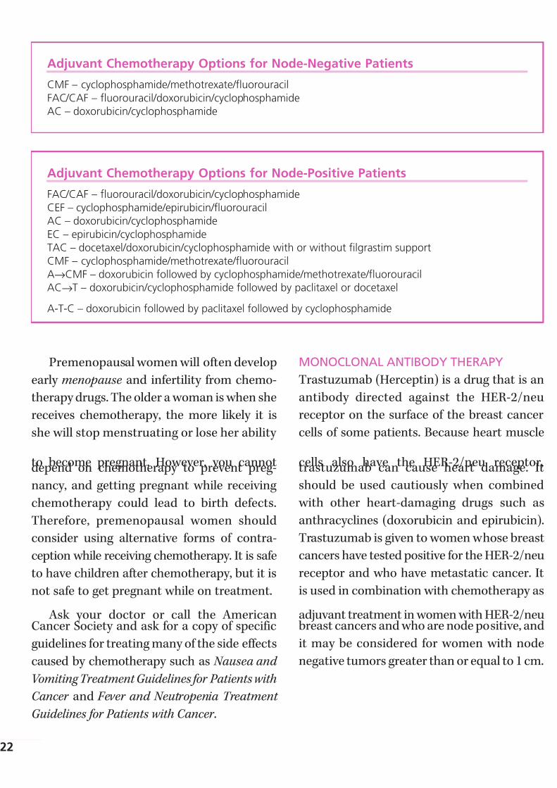
On the next two pages are listed the com-
monly used combinations of chemotherapy
drugs for women who have negative lymph
nodes and those who have positive lymph
nodes who have no evidence of spread, and the
common chemotherapy options for women
who have recurrent or metastatic breast cancer.
Doctors give chemotherapy in cycles, with
each period of treatment followed by a rest
period. The chemotherapy is given on the first
day of each cycle, and then the body is given
time to recover from the effects of chemo-
therapy. The chemotherapy drugs are thenrepeated to start the next cycle. The time
between giving the chemotherapy drugs is
generally every 2 weeks or every 3 weeks.
Some drugs are given more often. These cycles
generally last for a total time of 3 to 6 months
depending on the drugs used.
The side effects of chemotherapy depend
on the type of drugs used, the amount taken,
and the length of treatment.
• Doxorubicin and epirubicin may cause
heart damage but this is uncommon in
people who do not have preexisting
heart disease. If you know you haveheart disease or there is concern you
might have heart disease, your doctor
may suggest special heart tests before
you use these drugs and may suggest
other chemotherapy drugs if your
heart function is impaired.
• Temporary side effects might include
loss of appetite, nausea and vomiting,
mouth sores, hair loss, and changes in
the menstrual cycle.
• Chemotherapy can damage the blood-
producing cells of the bone marrow. A
drop in white blood cells can raise a
patient’s risk of infection; a shortage of
blood platelets can cause bleeding or
bruising after minor cuts or injuries;
and a decline in red blood cells canlead to fatigue.
There are treatments for these side effects.
There are excellent drugs that prevent or at
least reduce nausea and vomiting. A group of
drugs called growth factors that stimulate
the production of white blood cells or red blood
cells can help bone marrow recover after
chemotherapy and prevent problems resulting
from low blood counts. Although these drugs
are often not necessary, doctors have been
using them to allow them to give the chemo-
therapy more often. Talk with your doctor
about which treatment will be right for you.
7/27/2019 NCCN Breast Guidelines
http://slidepdf.com/reader/full/nccn-breast-guidelines 24/84
Premenopausal women will often develop
early menopause and infertility from chemo-
therapy drugs. The older a woman is when she
receives chemotherapy, the more likely it is
she will stop menstruating or lose her ability
to become pregnant. However, you cannotdepend on chemotherapy to prevent preg-
nancy, and getting pregnant while receiving
chemotherapy could lead to birth defects.
Therefore, premenopausal women should
consider using alternative forms of contra-
ception while receiving chemotherapy. It is safe
to have children after chemotherapy, but it is
not safe to get pregnant while on treatment.
Ask your doctor or call the AmericanCancer Society and ask for a copy of specific
guidelines for treating many of the side effects
caused by chemotherapy such as Nausea and
Vomiting Treatment Guidelines for Patients with
Cancer and Fever and Neutropenia Treatment
Guidelines for Patients with Cancer .
MONOCLONAL ANTIBODY THERAPY
Trastuzumab (Herceptin) is a drug that is an
antibody directed against the HER-2/neu
receptor on the surface of the breast cancer
cells of some patients. Because heart muscle
cells also have the HER-2/neu receptor,trastuzumab can cause heart damage. It
should be used cautiously when combined
with other heart-damaging drugs such as
anthracyclines (doxorubicin and epirubicin).
Trastuzumab is given to women whose breast
cancers have tested positive for the HER-2/neu
receptor and who have metastatic cancer. It
is used in combination with chemotherapy as
adjuvant treatment in women with HER-2/neubreast cancers and who are node positive, and
it may be considered for women with node
negative tumors greater than or equal to 1 cm.
22
Adjuvant Chemotherapy Options for Node-Negative Patients
CMF – cyclophosphamide/methotrexate/fluorouracil
FAC/CAF – fluorouracil/doxorubicin/cyclophosphamideAC – doxorubicin/cyclophosphamide
Adjuvant Chemotherapy Options for Node-Positive Patients
FAC/CAF – fluorouracil/doxorubicin/cyclophosphamide
CEF – cyclophosphamide/epirubicin/fluorouracilAC – doxorubicin/cyclophosphamide
EC – epirubicin/cyclophosphamideTAC – docetaxel/doxorubicin/cyclophosphamide with or without filgrastim support
CMF – cyclophosphamide/methotrexate/fluorouracilA→CMF – doxorubicin followed by cyclophosphamide/methotrexate/fluorouracilAC→T – doxorubicin/cyclophosphamide followed by paclitaxel or docetaxel
A-T-C – doxorubicin followed by paclitaxel followed by cyclophosphamide
7/27/2019 NCCN Breast Guidelines
http://slidepdf.com/reader/full/nccn-breast-guidelines 25/84
HORMONE TREATMENT
Estrogen, a hormone produced mostly by the
ovaries, but also from hormones produced by
the adrenal glands, and fat tissue in a woman’s body, causes some breast cancers to
grow. Doctors use several approaches to
block the effect of estrogen or to lower estro-
gen levels. These approaches can be divided
into two main groups:
• Drugs that block the effect of estrogen
on cancer cells, called anti-estrogens.
These medicines have no effect on
estrogen levels; instead, they preventestrogen from causing the breast cancer
cells to grow.
• Drugs that lower the production of
estrogen in the body.
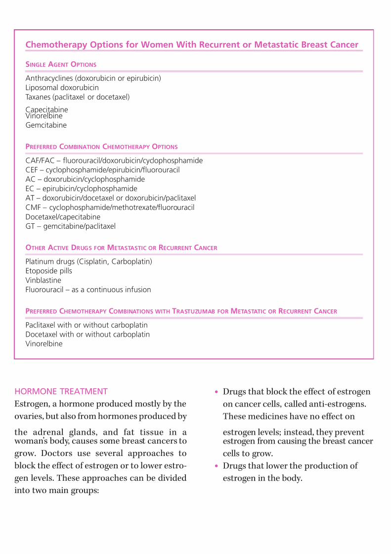
Chemotherapy Options for Women With Recurrent or Metastatic Breast Cancer
SINGLE AGENT OPTIONS
Anthracyclines (doxorubicin or epirubicin)Liposomal doxorubicin
Taxanes (paclitaxel or docetaxel)
CapecitabineVinorelbineGemcitabine
PREFERRED COMBINATION CHEMOTHERAPY OPTIONS
CAF/FAC – fluorouracil/doxorubicin/cyclophosphamideCEF – cyclophosphamide/epirubicin/fluorouracilAC – doxorubicin/cyclophosphamide
EC – epirubicin/cyclophosphamideAT – doxorubicin/docetaxel or doxorubicin/paclitaxel
CMF – cyclophosphamide/methotrexate/fluorouracil
Docetaxel/capecitabineGT – gemcitabine/paclitaxel
OTHER ACTIVE DRUGS FOR METASTASTIC OR RECURRENT CANCER
Platinum drugs (Cisplatin, Carboplatin)Etoposide pills
VinblastineFluorouracil – as a continuous infusion
PREFERRED CHEMOTHERAPY COMBINATIONS WITH TRASTUZUMAB FOR METASTATIC OR RECURRENT CANCER
Paclitaxel with or without carboplatinDocetaxel with or without carboplatinVinorelbine
7/27/2019 NCCN Breast Guidelines
http://slidepdf.com/reader/full/nccn-breast-guidelines 26/84
These treatments are used in two situations:
• Women who have hormone receptor-
positive breast cancers that appear to
have been completely removed by sur-
gery. Here hormone therapy is used as
adjuvant therapy to kill any remaining
breast cancer cells. Hormone therapy may be used alone or in combination
with chemotherapy.
• Women whose cancer remains after
surgery or in whom the cancer comes
back months or years after surgery.
Hormone drugs are only effective in
women whose cancer have the estrogen or
progesterone receptor. Every breast cancer istested for this protein, and you should ask
your doctor the result of this test on your
cancer. If the cancer is negative for both
these receptors, then the hormone drugs are
of no benefit.
Anti-Estrogen Drugs: Tamoxifen is the
antiestrogen drug used most often. Taking
tamoxifen as adjuvant therapy after surgery,
usually for five years, reduces the chances of
hormone receptor positive breast cancers
coming back. Tamoxifen is also used to treat
metastatic breast cancer.
In many women tamoxifen causes many
symptoms of menopause including hot
flashes, vaginal discharge, and mood swings.
Tamoxifen has two rare but more serious side
effects. These are a slight increased risk of
developing cancer of the lining of the uterus
(endometrial cancer) and uterine sarcoma,
and a slightly higher chance of developing
blood clots. For most women with breast
cancer, the benefits of taking the drug far
outweigh these risks.
Toremifene is another antiestrogen closely
related to tamoxifen. It may be an option for
postmenopausal women with metastatic
breast cancer.
Fulvestrant is a newer drug that reduces
the number of estrogen receptors. It is often
effective even if the breast cancer is no longerresponding to tamoxifen. Hot flashes, mild
nausea, and fatigue are the major side effects.
Drugs that Lower Estrogen Levels –
Aromatase inhibitors: Three drugs that stop
estrogen production in postmenopausal
women have been approved for treating breast
cancer. These drugs are anastrozole, letrozole,
and exemestane. They work by blocking an
enzyme that makes small amounts of estrogen
in postmenopausal women. They cannot stop
the ovaries of premenopausal women from
making estrogen. For this reason they are only
effective in postmenopausal women. For pre-
menopausal women, tamoxifen remains the
best drug to use.
The aromatase inhibitors have been com-
pared with tamoxifen as adjuvant hormonetherapy. They have fewer side effects than
tamoxifen because they don’t cause cancer of
the uterus and very rarely cause blood clots.
They can, however, cause osteoporosis and
bone fractures because they remove all
estrogen from a postmenopausal woman.
They also cause side effects of hot flashes and
sometimes joint pain.
They are a least as effective as tamoxifenin preventing breast cancer from coming
back in postmenopausal women. Based on
recent studies, many doctors recommend
aromatase inhibitors as the first choice for
breast cancer adjuvant hormonal therapy,
except in women who have some medical
reason to avoid these drugs.24

7/27/2019 NCCN Breast Guidelines
http://slidepdf.com/reader/full/nccn-breast-guidelines 27/84
Ovarian ablation: The ovaries are a
source of most estrogen in premenopausal
women. Destroying the ability of the ovaries to
produce estrogen may be an effective hormone
therapy to treat premenopausal women with
cancers that are positive for the estrogen or
progesterone receptors. Destruction of theovary production of estrogen can be done in
a number of ways:
• The ovaries can be removed by surgery.
• Radiation therapy can be given to the
ovaries.
• Drugs called luteinizing hormone-
releasing hormone agonists block
estrogen production by the ovaries.
The usual LHRH agonists are goserelin
and leuprolide.
Other hormonal therapies for breast
cancer: Other hormone treatments include
megestrol acetate (a progesterone-like drug),
fluoxymesterone (a male hormone like testos-
terone), and ethinylestradiol (an estrogen drug
that is effective if it is given in high doses).
Hormones Used to Treat Breast Cancer
Brand Name Generic Name
Arimidex Anastrozole
Aromasin Exemestane
Fareston Toremifene
Faslodex Fulvestrant
Femara Letrozole
Lupron Luprolide
Megace Megesterol
Nolvadex Tamoxifen
Zoladex Goserelin
BISPHOSPHONATES
These drugs are used to strengthen bones
that have been weakened by invading breast
cancer cells. The most commonly used drugs
are pamidronate and zoledronate. These drugs
are not used unless the cancer has spread to
the bone.
Choosing BetweenLumpectomy andMastectomy
The advantage of lumpectomy is that it saves
the appearance of the breast. A disadvantage
is the need for several weeks of radiation
therapy after surgery. However, some women
who have a mastectomy will still need radia-
tion therapy. Women who choose lumpectomy
and radiation can expect the same chance of
survival as those who choose mastectomy.
Although most women and their doctors
prefer lumpectomy and radiation therapy, your
choice will depend on a number of factors,such as:
• How you feel about losing your breast
• Whether you want to devote the addi-
tional time and travel for radiation
therapy
• Whether you would want to have more
surgery to reconstruct your breast after
having a mastectomy
• Your preference for mastectomy as a way to “take it all out as quickly as possible”
In determining the preference for lumpec-
tomy or mastectomy, be sure to get all the
facts. Though you may have a gut feeling for
mastectomy to “take it all out as quickly as
7/27/2019 NCCN Breast Guidelines
http://slidepdf.com/reader/full/nccn-breast-guidelines 28/84
possible”, the fact is that doing so does not
provide any better chance of long term survival
or a better outcome from treatment in most
cases. Large research studies with thousands
of women participating, and over 20 years of
information show that when lumpectomy
can be performed, that mastectomy does not provide any better chance of survival from
breast cancer than lumpectomy. It is because
of these facts that most women do not have
the breast removed.
A lumpectomy and radiation therapy are
not an appropriate option in some situations.
These include:
• Previous radiation to the breast or chest
• Being pregnant
• Presence of breast cancer in several
areas of the breast
• Widespread, suspicious areas of calcium
in the breast
• Tumor at the margin of the lumpectomy
that cannot be treated with repeat
breast-conserving surgery.
• The patient has active connective tissuedisease such as scleroderma or lupus
• The tumor is larger than 5 cm and is
not shrunk in size with chemotherapy
or hormonal therapy before surgery.
Reconstructive Surgery
If a woman has a mastectomy, she may wantto consider having the breast rebuilt; this is
called reconstruction. This is additional sur-
gery to create the appearance of a breast
after mastectomy. For most women, the
breast can be reconstructed at the same time
the mastectomy is done (immediate breast
reconstruction) or at a later date (delayed
reconstruction). Surgeons may use saline-filled
implants or tissue from other parts of your
body.
How do a woman and her doctor decideon the type of reconstruction and when she
should have the procedure? The answer
depends on the woman’s personal preferences,
the size and shape of her breasts, the size and
shape of her body, her level of physical exercise
and details of her medical situation, such as
how much skin is removed, and if she needs
chemotherapy or radiation.
If you are thinking about breast recon-
struction, please discuss this with your doctor
when you are planning your treatment.
Treatment of Pain andOther Symptoms
Most of this booklet discusses ways to removeor destroy breast cancer cells or to slow their
growth. But helping you feel as well as you
can and continuing to do the things you enjoy
doing is an important goal. Don’t hesitate to
discuss your symptoms or how you feel with
your cancer care team. There are effective and
safe ways to treat pain, other symptoms of
breast cancer, and most of the side effects
caused by breast cancer treatment. If youdon’t tell your health care team, they may
have no way of knowing about your problems.
26
7/27/2019 NCCN Breast Guidelines
http://slidepdf.com/reader/full/nccn-breast-guidelines 29/84
Complementary andAlternative Therapies
Complementary and alternative medicines
are a group of different types of health care
practices, systems, and products that are not
part of your usual medical treatment. They
may include Chinese herbs, special supple-
ments, acupuncture, massage, and a host of
other types of treatment. You may hear about
different treatments from your family and
friends. People will offer all sorts of things,
such as vitamins, herbs, stress reduction, and
more as a treatment for your cancer or to help
you feel better. Some of these treatments areharmless in certain situations, while others
have been shown to cause harm. Most of them
are of unproven benefit.
The American Cancer Society defines
complementary medicine or methods as those
that are used in addition to your regular
medical care. If these treatments are carefully
managed, they may add to your comfort and
well-being. Alternative medicines are definedas those that are used instead of your regular
medical care. Some of them have been proven
harmful, but are still promoted as “cures.” If
you choose to use these alternatives, they may
reduce your chance of fighting your cancer by
delaying or replacing regular cancer treatment.
There is a great deal of interest today in
complementary and alternative treatments
for cancer. Many are being studied to find out
if they are truly helpful to people with cancer.
Before changing your treatment or adding
any of these methods, it is best to discuss this
openly with your doctor or nurse. Some
methods can be safely used along with stan-
dard medical treatment. Others, however, can
interfere with standard treatment or cause
serious side effects. That is why it’s important
to talk with your doctor. More information
about complementary and alternative meth-
ods of cancer treatment is available through
the American Cancer Society’s toll-free num-
ber at 1-800-ACS-2345 or on our Web site at www.cancer.org.
Other Things to ConsiderDuring and After Treatment
During and after your treatment for breast
cancer you may be able to speed up your recov-
ery and improve your quality of life by taking
an active role. Learn about the benefits and
risks of each of your treatment options, and ask
questions of your cancer care team if there is
anything you do not understand. Learn about
and look out for side effects of treatment, and
report these right away to your cancer care
team so they can take steps to ease them.
Remember that your body is as unique as your personality and your fingerprints.
Although understanding your cancer’s stage
and learning about your treatment options
can help predict what health problems you
may face, no one can say for sure how you
will respond to cancer or its treatment.
You may have special strengths such as a
history of excellent nutrition and physical
activity, a strong family support system, or adeep faith, and these strengths may make a
difference in how you respond to cancer.
There are also experienced professionals in
mental health services, social work services,
and pastoral services who may assist you in
coping with your illness.

7/27/2019 NCCN Breast Guidelines
http://slidepdf.com/reader/full/nccn-breast-guidelines 30/84
You can also help in your own recovery
from cancer by making healthy lifestyle
choices. If you use tobacco, stop now. Quitting
will improve your overall health and the full
return of the sense of smell may help you
enjoy a healthy diet during recovery. If you use
alcohol, limit how much you drink. Have nomore than 1 drink per day. Good nutrition can
help you get better after treatment. Eat a
nutritious and balanced diet, with plenty of
fruits, vegetables, and whole grain foods.
If you are being treated for cancer, be
aware of the battle that is going on in your
body. Radiation therapy and chemotherapy
add to the fatigue caused by the disease itself.
To help you with the fatigue, plan your daily
activities around when you feel your best. Get
plenty of sleep at night. And ask your cancer
care team about a daily exercise program to
help you feel better.
A woman’s choice of treatment will likely
be influenced by her age, the image she has of
herself and her body, her hopes and fears,
and her stage in life. For example, many women select breast-conserving surgery with
radiation therapy over a mastectomy for body
image reasons. On the other hand, some
women who choose mastectomy may want
the affected area removed, regardless of the
effect on their body image, and others may be
more concerned about the side effects of
radiation therapy than body image.
Other issues that concern women includeloss of hair from chemotherapy and skin
changes of the breast from radiation therapy.
Women on chemotherapy tend to gain weight
and it is important to continue to eat a healthy
diet and exercise as much as your energy level
will permit. In addition to these body changes,
women may also be concerned about the out-
come of their treatment. These are all factors
that affect how a woman will make decisions
about her treatment, how she views herself,
and how she feels about her treatment.
Concerns about sexuality are often very worrisome to a woman with breast cancer.
Some treatments for breast cancer can
change a woman’s hormone levels and may
have a negative impact on sexual interest
and/or response. A diagnosis of breast cancer
when a woman is in her 20s or 30s is espe-
cially difficult because choosing a partner
and childbearing are often very important
during this period. Relationship issues are
also important because the diagnosis can be
very distressing for the partner, as well as the
patient. Partners are usually concerned about
how to express their love physically and emo-
tionally during and after treatment.
Suggestions that may help a woman adjust
to changes in her body image include looking
at and touching her body; seeking the supportof others, preferably before surgery; involving
her partner as soon as possible after surgery;
and openly talking about the feelings, needs,
and wants created by her changed image.
A cancer diagnosis and its treatment is a
major life challenge, with an impact on you
and everyone who cares for you. Before you
get to the point where you feel overwhelmed,
consider attending a meeting of a local sup- port group. If you need assistance in other
ways, contact your hospital’s social service
department or the ACS who can help you
find resources in your area.
28
7/27/2019 NCCN Breast Guidelines
http://slidepdf.com/reader/full/nccn-breast-guidelines 31/84
Clinical Trials
The Purpose of Clinical TrialsStudies of promising new or experimental
treatments or in patients are known as clinical
trials. Researchers conduct studies of new
treatments to answer the following questions:
• Is the treatment helpful?
• How does this new type of treatment
work?
• Does it work better than other treat-
ments already available?
• What side effects does the treatment
cause?
• Are the side effects greater or less thanthe standard treatment?
• Do the benefits outweigh the side effects?
• In which patients is the treatment
most likely to be helpful?
Types of Clinical Trials A new treatment is normally studied in three
phases of clinical trials before it is eligible for
approval by the FDA (Food and Drug Administration).
PHASE I CLINICAL TRIALS
The purpose of a phase I study is to find the
best way to give a new treatment and how
much of it can be given safely. Doctors watch
patients carefully for any harmful side effects.
The treatment has been well tested in labora-
tory and animal studies, but the side effects
in patients are not completely known.
Although doctors are hoping to help patients,
the main purpose of a phase I study is to test
the safety of the drug.
PHASE II CLINICAL TRIALS
These are designed to see if the drug works.
Patients are usually given the highest dose
that doesn’t cause severe side effects (deter-
mined from the phase I study) and closely
observed for an effect on the cancer. The doc-
tors will also look for side effects.
PHASE III CLINICAL TRIALS
Phase III studies involve large numbers of
patients. Some phase III clinical trials may
enroll thousands of patients. One group (the
control group) will receive the standard
(most accepted) treatment. The other group
will receive the new treatment. All patients in
phase III studies are closely watched. The
study will be stopped if the side effects of the
new treatment are too severe or if one group
has had much better results than the others.
If you are in a clinical trial, you will have a
team of experts looking at you and monitoring
your progress very carefully. The study is espe-
cially designed to pay close attention to you.
However, there are some risks. No oneinvolved in the study knows in advance
whether the treatment will work or exactly
what side effects will occur. That is what the
study is designed to discover. While most side
effects will disappear in time, some can be
permanent or even life threatening. Keep in
mind, though, that even standard treatments
have side effects. Depending on many factors,
you may decide to enroll in a clinical trial.
7/27/2019 NCCN Breast Guidelines
http://slidepdf.com/reader/full/nccn-breast-guidelines 32/84
Deciding to Enter a Clinical TrialEnrollment in any clinical trial is completely
up to you. Your doctors and nurses will
explain the risks and possible benefits of the
study to you in detail and will give you a form
to read and sign indicating your understand-
ing of the study and your desire to take part.Even after signing the form and after the clin-
ical trial begins, you are free to leave the
study at any time, for any reason. Taking part
in the study will not prevent you from getting
other medical care you may need.
To find out more about clinical trials, ask
your cancer care team. Among the questions
you should ask are:
• What is the purpose of the study?
• What kinds of tests and treatments
does the study involve?
• What does this treatment do?
• What is likely to happen in my case
with, or without, this new research
treatment?
• What are my other choices and their
advantages and disadvantages?
• How could the study affect my daily life?
• What side effects can I expect from the
study? Can the side effects be controlled?
• Will I have to be hospitalized? If so,
how often and for how long?
• Will the study cost me anything? Will
any of the treatment be free?• If I am harmed as a result of the
research, what treatment would I be
entitled to?
• What type of long-term follow-up care
is part of the study?
• Has the treatment been used to treat
other types of cancers?
The ACS offers a clinical trials matching service that will help you find a clinical trial
that is right for you. Simply go to our Web site
(www.cancer.org ) or call us at 1-800-ACS-2345.
You also can get a list of current National
Cancer Institute sponsored clinical trials by
calling the NCI Cancer Information Service
toll free at 1-800-4-CANCER or visiting the
NCI clinical trials Web site (www.cancer.gov/
clinical_trials/).
30
7/27/2019 NCCN Breast Guidelines
http://slidepdf.com/reader/full/nccn-breast-guidelines 33/84
Work-Up (Evaluation) andTreatment Guidelines
Decision TreesThe decision trees on the following pages represent different stages of breast
cancer. Each one shows you step-by-step how you and your doctor can arrive at
the choices you need to make about your treatment.
Keep in mind, this information is not meant to be used without the expertise of
your own doctor who is familiar with your situation, medical history, and per-
sonal preferences.
Participating in a clinical trial is an option for women at any stage of breast
cancer. Taking part in a study does not prevent you from getting other medical
care you may need.
The NCCN guidelines are updated as new significant data become available. To
ensure you have the most recent version, consult the Web sites of the ACS
( www.cancer.org ) or NCCN (www.nccn.org ). You may also call the NCCN at 1-
888-909-NCCN or the ACS at 1-800-ACS-2345 for the most recent information on
these guidelines or on cancer in general.

7/27/2019 NCCN Breast Guidelines
http://slidepdf.com/reader/full/nccn-breast-guidelines 34/84
Stage 0 Lobular Carcinoma In SituThe work-up for lobular carcinoma in situ
(LCIS) includes a complete medical history
and physical examination. A diagnostic
mammogram of both breasts is done to see if
there are any other abnormal areas in either
breast. Pathology review (another pathologist
to look at the biopsy sample) is suggested by
NCCN to be certain you have LCIS and not an
invasive cancer or another condition.
LCIS is usually not treated with surgery
other than the initial biopsy procedure.
Observation (careful follow-up without mas-
tectomy) is the preferred option for most
women who are diagnosed with LCIS
because LCIS is not an invasive cancer, nor
does it become one. But women with LCIS
have an increased risk of developing invasive
breast cancer in either breast. Ways to reduce
the risk of breast cancer have become an
important option.
There is evidence that tamoxifen, an anti-
estrogen drug that has been used as hormone
therapy for breast cancer, can also lower the
risk of developing an invasive breast cancer
in women diagnosed with LCIS. When used
in this way, tamoxifen is taken daily by mouth
for 5 years.
32
Treatment Guidelines for Patients
Stage
Stage 0
Lobular carcinoma
in situ (LCIS)
Work-Up (Evaluation) Treatment
• Medical history and physical exam
• Diagnostic mammogram (both breasts)
• Pathology review of biopsy sample
Observation
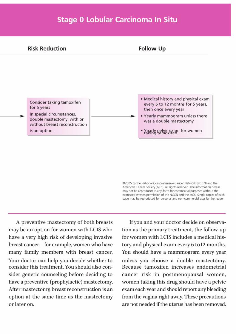
7/27/2019 NCCN Breast Guidelines
http://slidepdf.com/reader/full/nccn-breast-guidelines 35/84
A preventive mastectomy of both breasts
may be an option for women with LCIS who
have a very high risk of developing invasive
breast cancer – for example, women who have
many family members with breast cancer.
Your doctor can help you decide whether toconsider this treatment. You should also con-
sider genetic counseling before deciding to
have a preventive (prophylactic) mastectomy.
After mastectomy, breast reconstruction is an
option at the same time as the mastectomy
or later on.
If you and your doctor decide on observa-
tion as the primary treatment, the follow-up
for women with LCIS includes a medical his-
tory and physical exam every 6 to12 months.
You should have a mammogram every year
unless you choose a double mastectomy.Because tamoxifen increases endometrial
cancer risk in postmenopausal women,
women taking this drug should have a pelvic
exam each year and should report any bleeding
from the vagina right away. These precautions
are not needed if the uterus has been removed.
©2005 by the National Comprehensive Cancer Network (NCCN) and the
American Cancer Society (ACS). All rights reserved. The information herein
may not be reproduced in any form for commercial purposes without the
expressed written permission of the NCCN and the ACS. Single copies of eachpage may be reproduced for personal and non-commercial uses by the reader.
Stage 0 Lobular Carcinoma In Situ
Consider taking tamoxifen
for 5 years
In special circumstances,
double mastectomy, with or
without breast reconstruction
is an option.
• Medical history and physical exam
every 6 to 12 months for 5 years,
then once every year
• Yearly mammogram unless there
was a double mastectomy
• Yearly pelvic exam for womentaking tamoxifen
Risk Reduction Follow-Up
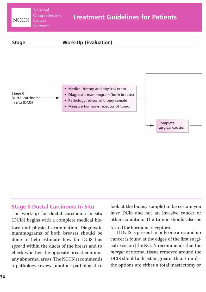
7/27/2019 NCCN Breast Guidelines
http://slidepdf.com/reader/full/nccn-breast-guidelines 36/84
Stage 0 Ductal Carcinoma In SituThe work-up for ductal carcinoma in situ
(DCIS) begins with a complete medical his-
tory and physical examination. Diagnosticmammograms of both breasts should be
done to help estimate how far DCIS has
spread within the ducts of the breast and to
check whether the opposite breast contains
any abnormal areas. The NCCN recommends
a pathology review (another pathologist to
look at the biopsy sample) to be certain you
have DCIS and not an invasive cancer or
other condition. The tumor should also be
tested for hormone receptors.If DCIS is present in only one area and no
cancer is found at the edges of the first surgi-
cal excision (the NCCN recommends that the
margin of normal tissue removed around the
DCIS should at least be greater than 1 mm) –
the options are either a total mastectomy or
34
Treatment Guidelines for Patients
Stage
Stage 0
Ductal carcinoma
in situ (DCIS)
Work-Up (Evaluation)
• Medical history and physical exam
• Diagnostic mammogram (both breasts)
• Pathology review of biopsy sample
• Measure hormone receptor of tumor
Complete
surgical excision
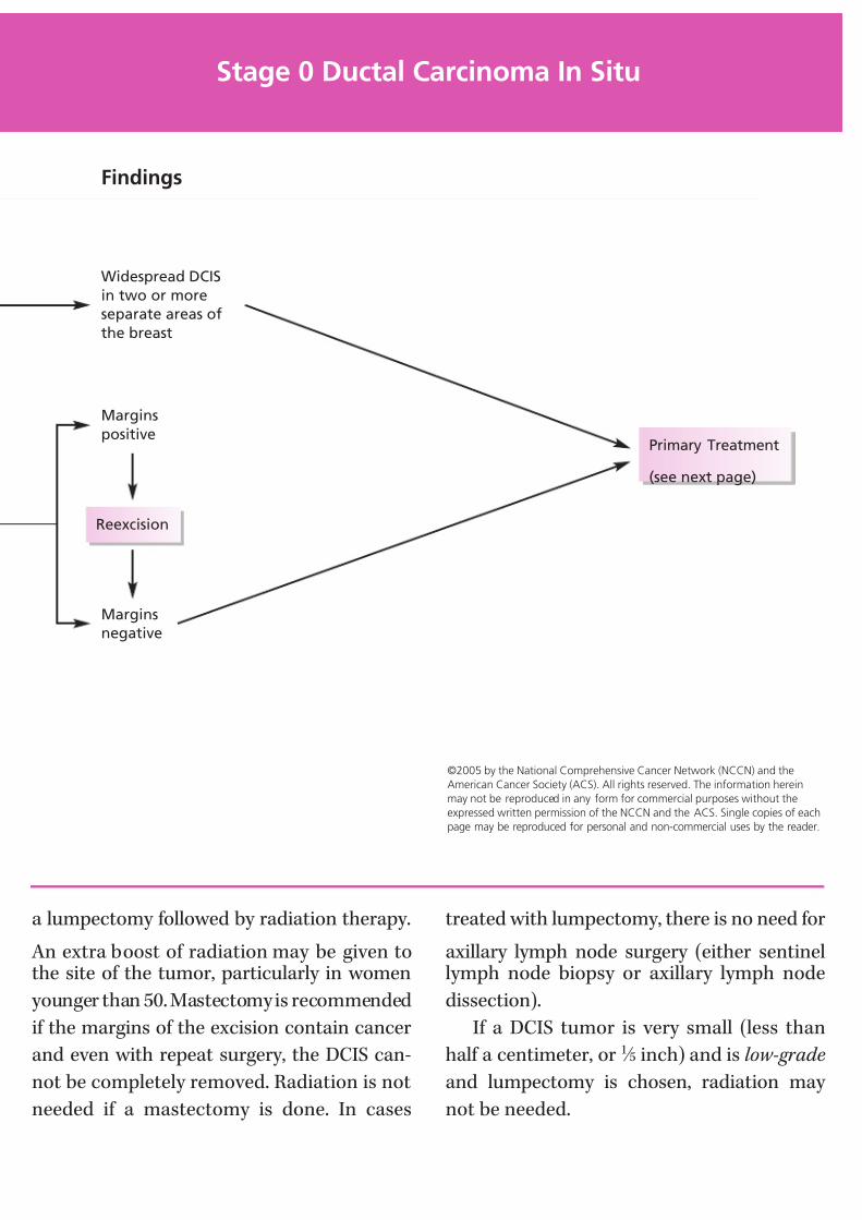
7/27/2019 NCCN Breast Guidelines
http://slidepdf.com/reader/full/nccn-breast-guidelines 37/84
a lumpectomy followed by radiation therapy.
An extra boost of radiation may be given tothe site of the tumor, particularly in women
younger than 50. Mastectomy is recommended
if the margins of the excision contain cancer
and even with repeat surgery, the DCIS can-
not be completely removed. Radiation is not
needed if a mastectomy is done. In cases
treated with lumpectomy, there is no need for
axillary lymph node surgery (either sentinellymph node biopsy or axillary lymph node
dissection).
If a DCIS tumor is very small (less than
half a centimeter, or 1 ⁄ 5 inch) and is low-grade
and lumpectomy is chosen, radiation may
not be needed.
©2005 by the National Comprehensive Cancer Network (NCCN) and the
American Cancer Society (ACS). All rights reserved. The information herein
may not be reproduced in any form for commercial purposes without the
expressed written permission of the NCCN and the ACS. Single copies of each
page may be reproduced for personal and non-commercial uses by the reader.
Stage 0 Ductal Carcinoma In Situ
Findings
Widespread DCIS
in two or more
separate areas of
the breast
Margins
positive
Reexcision
Margins
negative
Primary Treatment
(see next page)
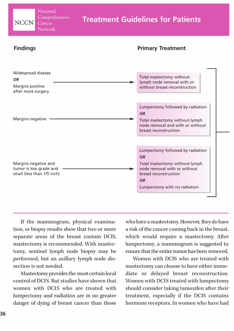
7/27/2019 NCCN Breast Guidelines
http://slidepdf.com/reader/full/nccn-breast-guidelines 38/84
If the mammogram, physical examina-
tion, or biopsy results show that two or more
separate areas of the breast contain DCIS,
mastectomy is recommended. With mastec-
tomy, sentinel lymph node biopsy may be
performed, but an axillary lymph node dis-
section is not needed.
Mastectomy provides the most certain local
control of DCIS. But studies have shown that
women with DCIS who are treated with
lumpectomy and radiation are in no greater
danger of dying of breast cancer than those
who have a mastectomy. However, they do have
a risk of the cancer coming back in the breast,
which would require a mastectomy. After
lumpectomy, a mammogram is suggested to
ensure that the entire tumor has been removed.
Women with DCIS who are treated with
mastectomy can choose to have either imme-
diate or delayed breast reconstruction.
Women with DCIS treated with lumpectomy
should consider taking tamoxifen after their
treatment, especially if the DCIS contains
hormone receptors. In women who have had
36
Treatment Guidelines for Patients
Findings
Widespread disease
OR
Margins positive
after more surgery
Primary Treatment
Total mastectomy without
lymph node removal with or
without breast reconstruction
Margins negative
Lumpectomy followed by radiation
OR
Total mastectomy without lymph
node removal and with or without
breast reconstruction
Margins negative and
tumor is low grade and
small (less than 1/5 inch)
Lumpectomy followed by radiation
OR
Total mastectomy without lymph
node removal with or without
breast reconstruction
OR
Lumpectomy with no radiation
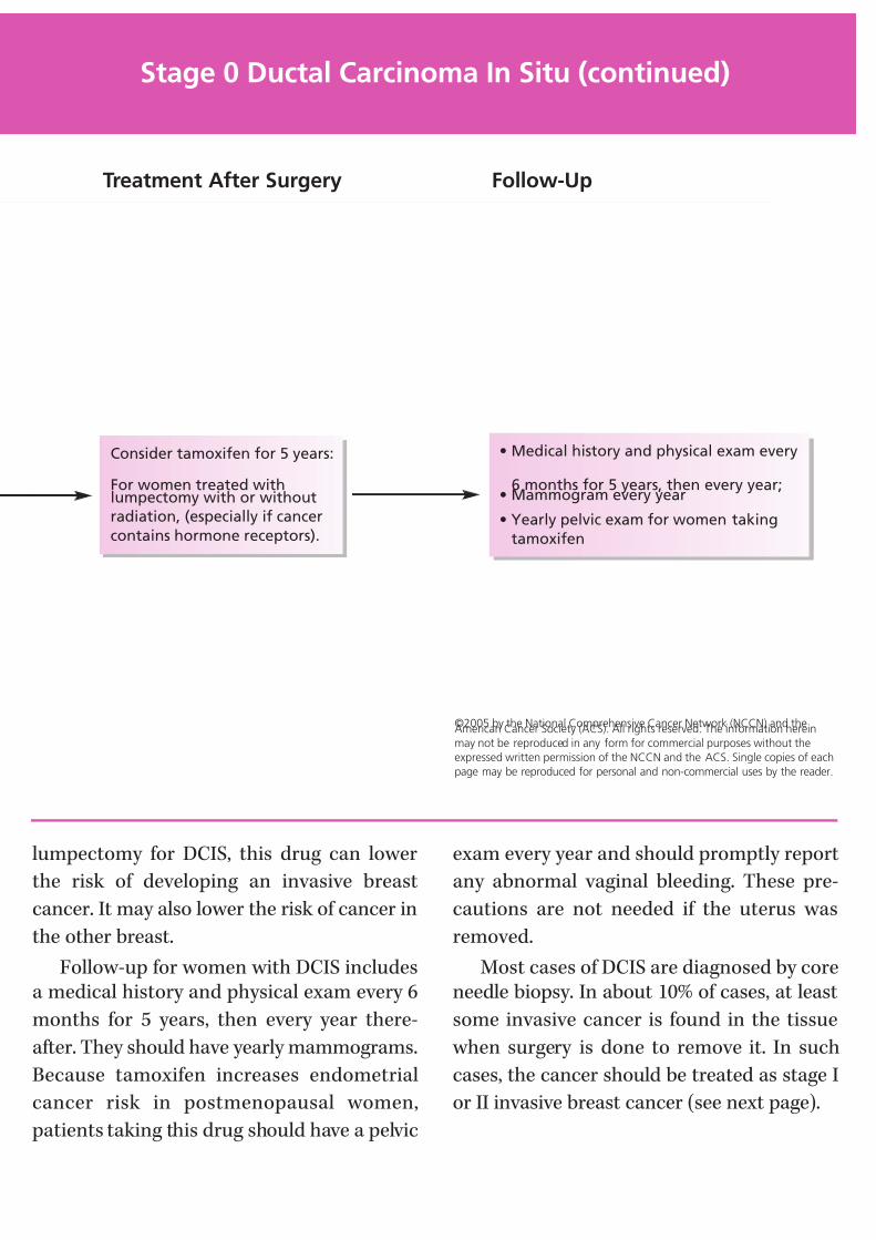
7/27/2019 NCCN Breast Guidelines
http://slidepdf.com/reader/full/nccn-breast-guidelines 39/84
lumpectomy for DCIS, this drug can lower
the risk of developing an invasive breast
cancer. It may also lower the risk of cancer in
the other breast.
Follow-up for women with DCIS includesa medical history and physical exam every 6
months for 5 years, then every year there-
after. They should have yearly mammograms.
Because tamoxifen increases endometrial
cancer risk in postmenopausal women,
patients taking this drug should have a pelvic
exam every year and should promptly report
any abnormal vaginal bleeding. These pre-
cautions are not needed if the uterus was
removed.
Most cases of DCIS are diagnosed by coreneedle biopsy. In about 10% of cases, at least
some invasive cancer is found in the tissue
when surgery is done to remove it. In such
cases, the cancer should be treated as stage I
or II invasive breast cancer (see next page).
©2005 by the National Comprehensive Cancer Network (NCCN) and theAmerican Cancer Society (ACS). All rights reserved. The information herein
may not be reproduced in any form for commercial purposes without the
expressed written permission of the NCCN and the ACS. Single copies of each
page may be reproduced for personal and non-commercial uses by the reader.
Stage 0 Ductal Carcinoma In Situ (continued)
Treatment After Surgery Follow-Up
Consider tamoxifen for 5 years:
For women treated withlumpectomy with or without
radiation, (especially if cancer
contains hormone receptors).
• Medical history and physical exam every
6 months for 5 years, then every year;• Mammogram every year
• Yearly pelvic exam for women taking
tamoxifen
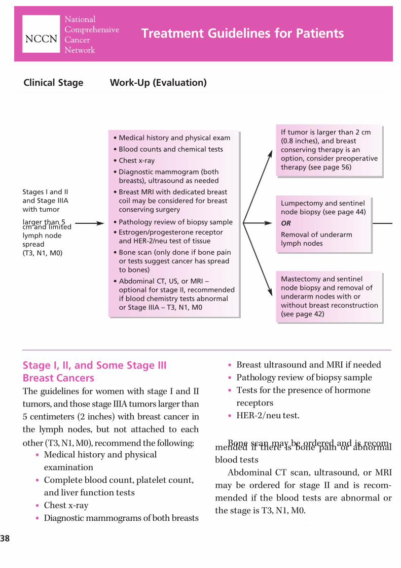
7/27/2019 NCCN Breast Guidelines
http://slidepdf.com/reader/full/nccn-breast-guidelines 40/84
Stage I, II, and Some Stage IIIBreast CancersThe guidelines for women with stage I and II
tumors, and those stage IIIA tumors larger than
5 centimeters (2 inches) with breast cancer in
the lymph nodes, but not attached to each
other (T3, N1, M0), recommend the following:• Medical history and physical
examination
• Complete blood count, platelet count,
and liver function tests
• Chest x-ray
• Diagnostic mammograms of both breasts
• Breast ultrasound and MRI if needed
• Pathology review of biopsy sample
• Tests for the presence of hormone
receptors
• HER-2/neu test.
Bone scan may be ordered and is recom-mended if there is bone pain or abnormal
blood tests
Abdominal CT scan, ultrasound, or MRI
may be ordered for stage II and is recom-
mended if the blood tests are abnormal or
the stage is T3, N1, M0.
38
Treatment Guidelines for Patients
Clinical Stage Work-Up (Evaluation)
Stages I and II
and Stage IIIA
with tumor
larger than 5cm and limited
lymph node
spread
(T3, N1, M0)
• Medical history and physical exam
• Blood counts and chemical tests
• Chest x-ray
• Diagnostic mammogram (both
breasts), ultrasound as needed
• Breast MRI with dedicated breast
coil may be considered for breast
conserving surgery
• Pathology review of biopsy sample
• Estrogen/progesterone receptor
and HER-2/neu test of tissue
• Bone scan (only done if bone pain
or tests suggest cancer has spread
to bones)
• Abdominal CT, US, or MRI –
optional for stage II, recommended
if blood chemistry tests abnormal
or Stage IIIA – T3, N1, M0
If tumor is larger than 2 cm
(0.8 inches), and breast
conserving therapy is an
option, consider preoperative
therapy (see page 56)
Mastectomy and sentinel
node biopsy and removal of
underarm nodes with or
without breast reconstruction
(see page 42)
Lumpectomy and sentinel
node biopsy (see page 44)
OR
Removal of underarm
lymph nodes
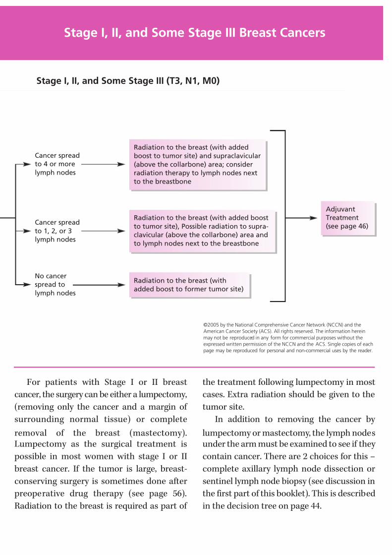
7/27/2019 NCCN Breast Guidelines
http://slidepdf.com/reader/full/nccn-breast-guidelines 41/84
For patients with Stage I or II breast
cancer, the surgery can be either a lumpectomy,
(removing only the cancer and a margin of
surrounding normal tissue) or complete
removal of the breast (mastectomy).Lumpectomy as the surgical treatment is
possible in most women with stage I or II
breast cancer. If the tumor is large, breast-
conserving surgery is sometimes done after
preoperative drug therapy (see page 56).
Radiation to the breast is required as part of
the treatment following lumpectomy in most
cases. Extra radiation should be given to the
tumor site.
In addition to removing the cancer by
lumpectomy or mastectomy, the lymph nodesunder the arm must be examined to see if they
contain cancer. There are 2 choices for this –
complete axillary lymph node dissection or
sentinel lymph node biopsy (see discussion in
the first part of this booklet). This is described
in the decision tree on page 44.
Stage I, II, and Some Stage III Breast Cancers
Stage I, II, and Some Stage III (T3, N1, M0)
Radiation to the breast (with added
boost to tumor site) and supraclavicular
(above the collarbone) area; consider
radiation therapy to lymph nodes next
to the breastbone
Radiation to the breast (with added boost
to tumor site), Possible radiation to supra-
clavicular (above the collarbone) area and
to lymph nodes next to the breastbone
Radiation to the breast (with
added boost to former tumor site)
Adjuvant
Treatment
(see page 46)
Cancer spread
to 4 or more
lymph nodes
Cancer spread
to 1, 2, or 3
lymph nodes
No cancer
spread to
lymph nodes
©2005 by the National Comprehensive Cancer Network (NCCN) and the
American Cancer Society (ACS). All rights reserved. The information herein
may not be reproduced in any form for commercial purposes without the
expressed written permission of the NCCN and the ACS. Single copies of each
page may be reproduced for personal and non-commercial uses by the reader.

7/27/2019 NCCN Breast Guidelines
http://slidepdf.com/reader/full/nccn-breast-guidelines 42/84

7/27/2019 NCCN Breast Guidelines
http://slidepdf.com/reader/full/nccn-breast-guidelines 43/84
• Incomplete removal of the cancerduring lumpectomy or re-excision
• Connective tissue diseases involving the
skin that make tissues overly sensitive
to the side effects of radiation
• If treatment would require radiation
during pregnancy
• The tumor is too large. In that case, a woman and her doctor might consider
chemotherapy before surgery (page 56)
to shrink the tumor so that a lumpec-
tomy is possible.
NOTES
Stage I, II, and Some Stage III Breast Cancers (continued)
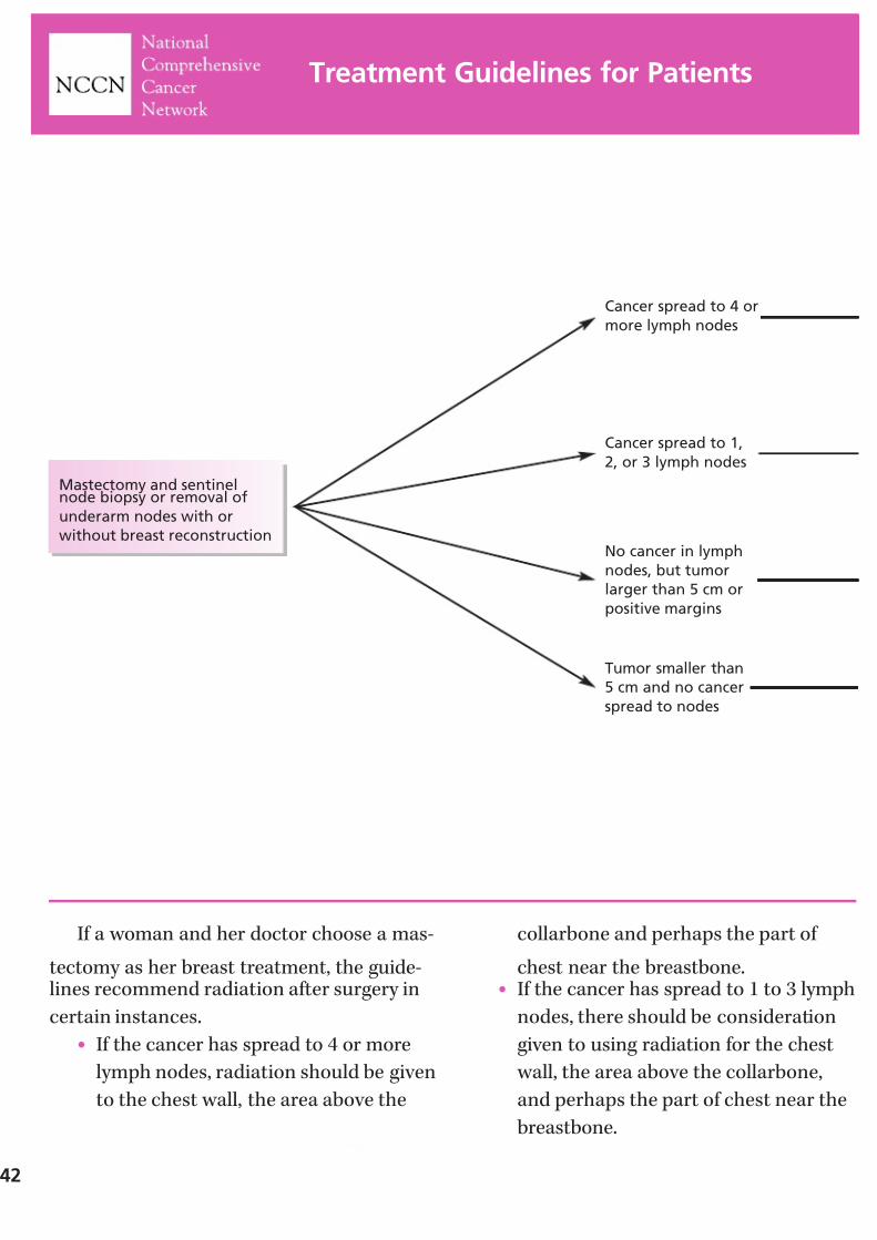
7/27/2019 NCCN Breast Guidelines
http://slidepdf.com/reader/full/nccn-breast-guidelines 44/84
If a woman and her doctor choose a mas-
tectomy as her breast treatment, the guide-lines recommend radiation after surgery in
certain instances.
• If the cancer has spread to 4 or more
lymph nodes, radiation should be given
to the chest wall, the area above the
collarbone and perhaps the part of
chest near the breastbone.• If the cancer has spread to 1 to 3 lymph
nodes, there should be consideration
given to using radiation for the chest
wall, the area above the collarbone,
and perhaps the part of chest near the
breastbone.
42
Treatment Guidelines for Patients
Mastectomy and sentinelnode biopsy or removal of
underarm nodes with or
without breast reconstruction
Cancer spread to 4 or
more lymph nodes
Cancer spread to 1,
2, or 3 lymph nodes
No cancer in lymph
nodes, but tumor
larger than 5 cm or
positive margins
Tumor smaller than
5 cm and no cancer
spread to nodes
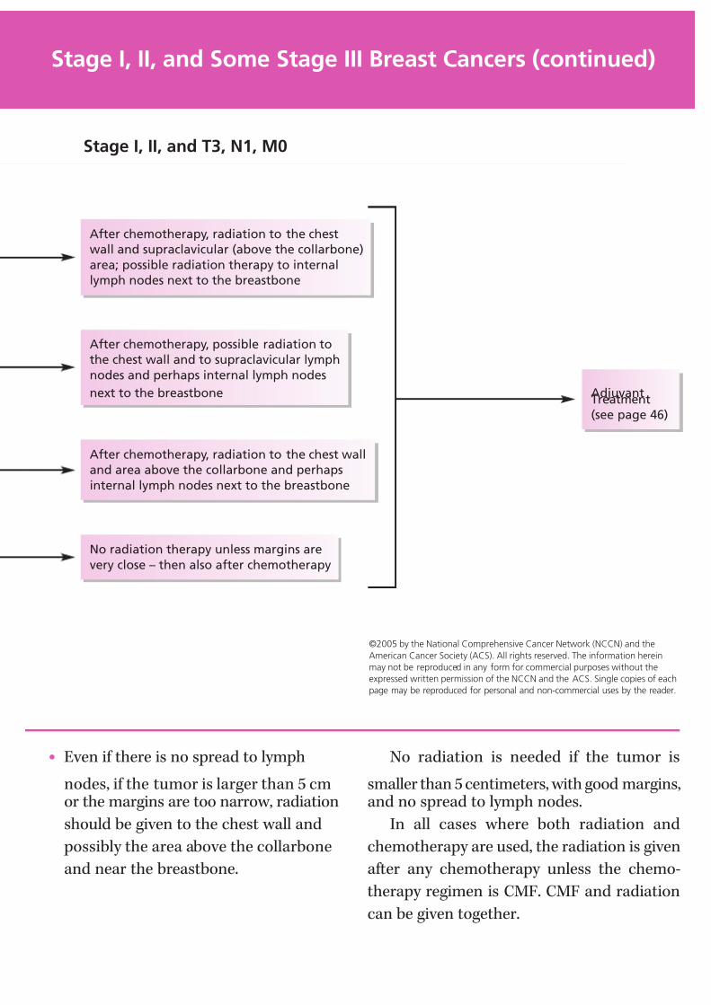
7/27/2019 NCCN Breast Guidelines
http://slidepdf.com/reader/full/nccn-breast-guidelines 45/84
• Even if there is no spread to lymph
nodes, if the tumor is larger than 5 cmor the margins are too narrow, radiation
should be given to the chest wall and
possibly the area above the collarbone
and near the breastbone.
No radiation is needed if the tumor is
smaller than 5 centimeters, with good margins,and no spread to lymph nodes.
In all cases where both radiation and
chemotherapy are used, the radiation is given
after any chemotherapy unless the chemo-
therapy regimen is CMF. CMF and radiation
can be given together.
©2005 by the National Comprehensive Cancer Network (NCCN) and the
American Cancer Society (ACS). All rights reserved. The information herein
may not be reproduced in any form for commercial purposes without the
expressed written permission of the NCCN and the ACS. Single copies of each
page may be reproduced for personal and non-commercial uses by the reader.
Stage I, II, and Some Stage III Breast Cancers (continued)
After chemotherapy, radiation to the chest
wall and supraclavicular (above the collarbone)
area; possible radiation therapy to internal
lymph nodes next to the breastbone
After chemotherapy, possible radiation to
the chest wall and to supraclavicular lymph
nodes and perhaps internal lymph nodes
next to the breastbone
After chemotherapy, radiation to the chest wall
and area above the collarbone and perhaps
internal lymph nodes next to the breastbone
No radiation therapy unless margins are
very close – then also after chemotherapy
AdjuvantTreatment
(see page 46)
Stage I, II, and T3, N1, M0
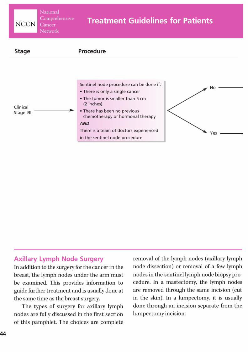
7/27/2019 NCCN Breast Guidelines
http://slidepdf.com/reader/full/nccn-breast-guidelines 46/84
Axillary Lymph Node SurgeryIn addition to the surgery for the cancer in the
breast, the lymph nodes under the arm must
be examined. This provides information to
guide further treatment and is usually done at
the same time as the breast surgery.
The types of surgery for axillary lymph
nodes are fully discussed in the first section
of this pamphlet. The choices are complete
removal of the lymph nodes (axillary lymph
node dissection) or removal of a few lymph
nodes in the sentinel lymph node biopsy pro-
cedure. In a mastectomy, the lymph nodes
are removed through the same incision (cut
in the skin). In a lumpectomy, it is usually
done through an incision separate from the
lumpectomy incision.
44
Treatment Guidelines for Patients
Stage Procedure
Clinical
Stage I/II
Sentinel node procedure can be done if:
• There is only a single cancer
• The tumor is smaller than 5 cm
(2 inches)
• There has been no previous
chemotherapy or hormonal therapy
AND
There is a team of doctors experienced
in the sentinel node procedure
No
Yes
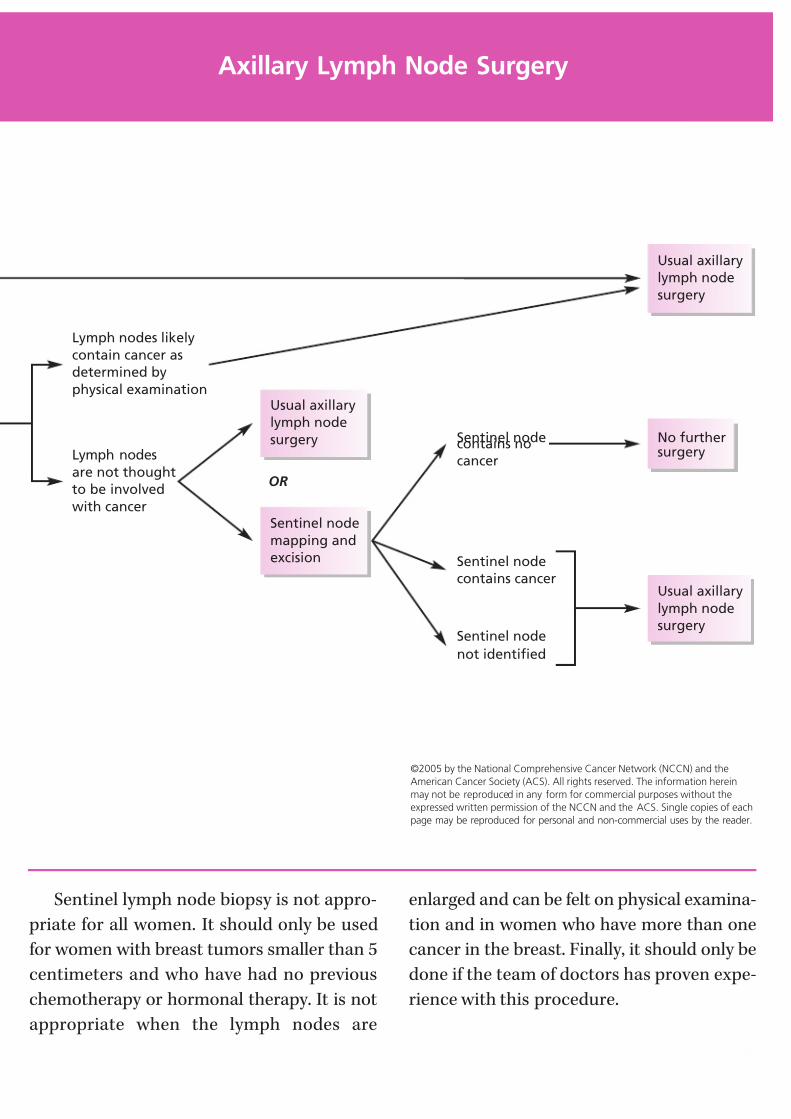
7/27/2019 NCCN Breast Guidelines
http://slidepdf.com/reader/full/nccn-breast-guidelines 47/84
Sentinel lymph node biopsy is not appro-
priate for all women. It should only be used
for women with breast tumors smaller than 5
centimeters and who have had no previous
chemotherapy or hormonal therapy. It is not
appropriate when the lymph nodes are
enlarged and can be felt on physical examina-
tion and in women who have more than one
cancer in the breast. Finally, it should only be
done if the team of doctors has proven expe-
rience with this procedure.
©2005 by the National Comprehensive Cancer Network (NCCN) and the
American Cancer Society (ACS). All rights reserved. The information herein
may not be reproduced in any form for commercial purposes without the
expressed written permission of the NCCN and the ACS. Single copies of each
page may be reproduced for personal and non-commercial uses by the reader.
Axillary Lymph Node Surgery
Lymph nodes likely
contain cancer as
determined by
physical examination
Lymph nodes
are not thought
to be involved
with cancerSentinel node
mapping and
excision
Usual axillary
lymph node
surgery
OR
Sentinel nodecontains no
cancer
Sentinel node
contains cancer
Sentinel node
not identified
No furthersurgery
Usual axillary
lymph node
surgery
Usual axillary
lymph node
surgery
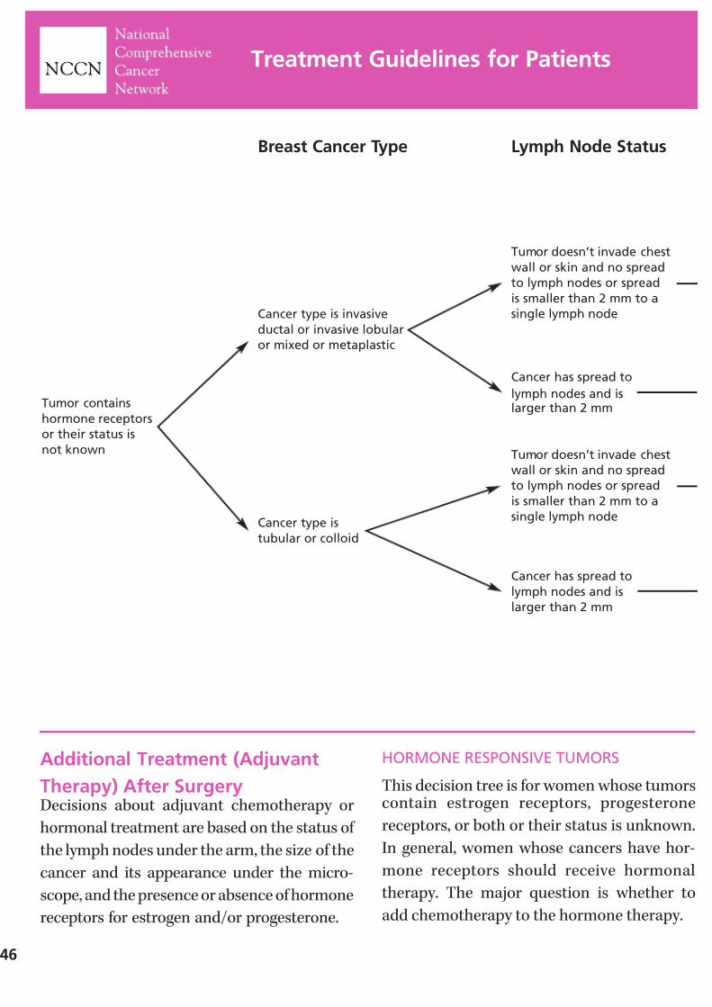
7/27/2019 NCCN Breast Guidelines
http://slidepdf.com/reader/full/nccn-breast-guidelines 48/84
46
Additional Treatment (Adjuvant
Therapy) After SurgeryDecisions about adjuvant chemotherapy or
hormonal treatment are based on the status of
the lymph nodes under the arm, the size of the
cancer and its appearance under the micro-
scope, and the presence or absence of hormone
receptors for estrogen and/or progesterone.
HORMONE RESPONSIVE TUMORS
This decision tree is for women whose tumorscontain estrogen receptors, progesterone
receptors, or both or their status is unknown.
In general, women whose cancers have hor-
mone receptors should receive hormonal
therapy. The major question is whether to
add chemotherapy to the hormone therapy.
Treatment Guidelines for Patients
Tumor contains
hormone receptors
or their status is
not known
Breast Cancer Type Lymph Node Status
Cancer type is
tubular or colloid
Cancer type is invasive
ductal or invasive lobular
or mixed or metaplastic
Tumor doesn’t invade chest
wall or skin and no spread
to lymph nodes or spread
is smaller than 2 mm to a
single lymph node
Tumor doesn’t invade chest
wall or skin and no spread
to lymph nodes or spread
is smaller than 2 mm to a
single lymph node
Cancer has spread to
lymph nodes and islarger than 2 mm
Cancer has spread to
lymph nodes and is
larger than 2 mm
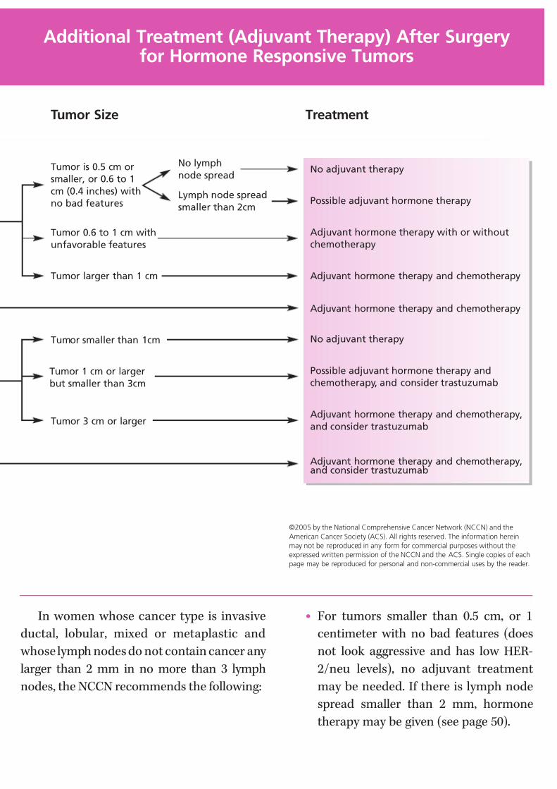
7/27/2019 NCCN Breast Guidelines
http://slidepdf.com/reader/full/nccn-breast-guidelines 49/84
In women whose cancer type is invasive
ductal, lobular, mixed or metaplastic and
whose lymph nodes do not contain cancer any
larger than 2 mm in no more than 3 lymph
nodes, the NCCN recommends the following:
• For tumors smaller than 0.5 cm, or 1
centimeter with no bad features (does
not look aggressive and has low HER-
2/neu levels), no adjuvant treatment
may be needed. If there is lymph node
spread smaller than 2 mm, hormone
therapy may be given (see page 50).
©2005 by the National Comprehensive Cancer Network (NCCN) and the
American Cancer Society (ACS). All rights reserved. The information herein
may not be reproduced in any form for commercial purposes without the
expressed written permission of the NCCN and the ACS. Single copies of each
page may be reproduced for personal and non-commercial uses by the reader.
Additional Treatment (Adjuvant Therapy) After Surgeryfor Hormone Responsive Tumors
No adjuvant therapy
Possible adjuvant hormone therapy
Adjuvant hormone therapy with or without
chemotherapy
Adjuvant hormone therapy and chemotherapy
Adjuvant hormone therapy and chemotherapy
No adjuvant therapy
Possible adjuvant hormone therapy and
chemotherapy, and consider trastuzumab
Adjuvant hormone therapy and chemotherapy,
and consider trastuzumab
Adjuvant hormone therapy and chemotherapy,and consider trastuzumab
Treatment
Tumor smaller than 1cm
Tumor is 0.5 cm or
smaller, or 0.6 to 1
cm (0.4 inches) with
no bad features
Tumor 0.6 to 1 cm with
unfavorable features
Tumor larger than 1 cm
Tumor 1 cm or larger
but smaller than 3cm
Tumor 3 cm or larger
Tumor Size
No lymph
node spread
Lymph node spread
smaller than 2cm

7/27/2019 NCCN Breast Guidelines
http://slidepdf.com/reader/full/nccn-breast-guidelines 50/84
• When the tumor measures 0.6 to 1centimeter and has one or more
unfavorable features (looks aggressive
or has high HER-2/neu levels), hormone
therapy should be given with or without
chemotherapy. Note: Whenever chemo-
therapy is given, it should precede
hormonal therapy.
• If the tumor has grown larger than 1centimeter, hormone therapy and
chemotherapy are recommended.
In women whose cancer type is invasive
ductal, lobular, mixed, or metaplastic and there
is spread to lymph nodes, both hormonal treat-
ment and chemotherapy are recommended.
48
Treatment Guidelines for Patients
NOTES

7/27/2019 NCCN Breast Guidelines
http://slidepdf.com/reader/full/nccn-breast-guidelines 51/84
In women with tubular or colloid breastcancer, and whose lymph nodes do not con-
tain any cancer or a single lymph node has a
metastasis 2 mm in size or less, the NCCN
recommends the following:
• For tumors smaller than 1 cm, no
treatment is needed after surgery.
• If the tumor is between 1 and 3 cm in
size, hormonal treatment and chemo-
therapy may be recommended.
• Hormonal treatment and chemotherapy are recommended if it is larger than 3
cm or has spread to lymph nodes and
the spread is larger than 2 mm.
• Trastuzumab should be added in women
whose breast cancer is HER-2/neu
positive and whose lymph nodes are
positive for breast cancer. It should be
considered in women with lymph node
negative breast cancer whose tumor is1 cm or larger.
Additional Treatment (Adjuvant Therapy) After Surgeryfor Hormone Responsive Tumors (continued)
NOTES
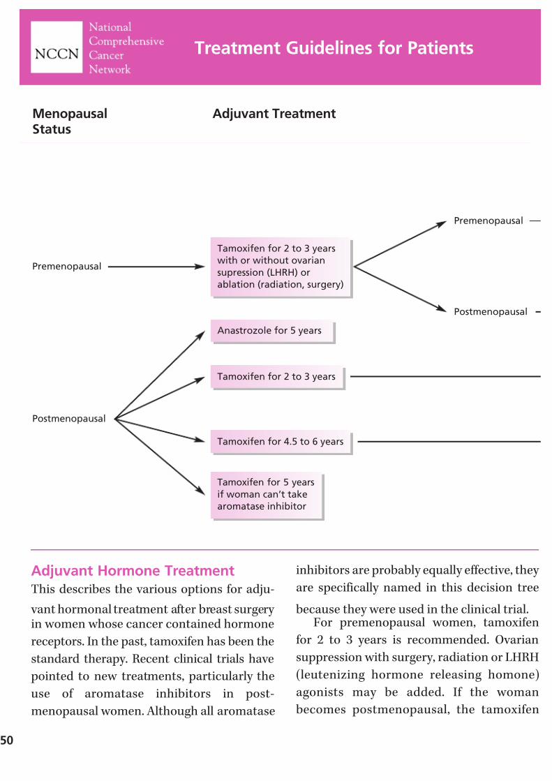
7/27/2019 NCCN Breast Guidelines
http://slidepdf.com/reader/full/nccn-breast-guidelines 52/84
50
Adjuvant Hormone TreatmentThis describes the various options for adju-
vant hormonal treatment after breast surgery in women whose cancer contained hormone
receptors. In the past, tamoxifen has been the
standard therapy. Recent clinical trials have
pointed to new treatments, particularly the
use of aromatase inhibitors in post-
menopausal women. Although all aromatase
inhibitors are probably equally effective, they
are specifically named in this decision tree
because they were used in the clinical trial.For premenopausal women, tamoxifen
for 2 to 3 years is recommended. Ovarian
suppression with surgery, radiation or LHRH
(leutenizing hormone releasing homone)
agonists may be added. If the woman
becomes postmenopausal, the tamoxifen
Treatment Guidelines for Patients
Premenopausal
Postmenopausal
Tamoxifen for 2 to 3 years
with or without ovarian
supression (LHRH) or
ablation (radiation, surgery)
Anastrozole for 5 years
Tamoxifen for 2 to 3 years
Tamoxifen for 4.5 to 6 years
Tamoxifen for 5 years
if woman can’t take
aromatase inhibitor
Premenopausal
Postmenopausal
MenopausalStatus
Adjuvant Treatment

7/27/2019 NCCN Breast Guidelines
http://slidepdf.com/reader/full/nccn-breast-guidelines 53/84
should be continued for a total of 5 years and
followed by 5 years of letrozole. Another
option, which is less favored, would be stop- ping the tamoxifen and taking exemestane
for the remaining 2 to 3 years. If the woman
remains premenopausal, the tamoxifen
should be continued for a total of 5 years. If
she then becomes postmenopausal, letrozole
for 5 years should be added.
For postmenopausal women, one choice is
anastrozole for 5 years. A second option is to
take tamoxifen for 2 to 3 years and complete 5 years of treatment with either exemestane of
anastrozole. A third choice is to take tamoxifen
for 4.5 to 6 years and then take exemestane for
5 years. If a woman can’t take an aromatase
inhibitor, then tamoxifen for 5 years is an
acceptable option.
©2005 by the National Comprehensive Cancer Network (NCCN) and the
American Cancer Society (ACS). All rights reserved. The information herein
may not be reproduced in any form for commercial purposes without the
expressed written permission of the NCCN and the ACS. Single copies of each
page may be reproduced for personal and non-commercial uses by the reader.
Adjuvant Hormone Treatment
Tamoxifen for full 5 years
Tamoxifen for full 5 years
Letrozole for 5 years
Letrozole for 5 years
Letrozole for 5 years
No further
hormone therapy
Exemestane or anastrozole
to complete 5 year adjuvant
therapy
Exemestane or anastrozole
to complete 5 year adjuvant
therapy
Still premenopausal
Now postmenopausal

7/27/2019 NCCN Breast Guidelines
http://slidepdf.com/reader/full/nccn-breast-guidelines 54/84
Additional Treatment (AdjuvantTherapy) After Surgery for Non-Hormone Responsive Tumors
This decision tree is for women whose tumorsdo not contain either estrogen receptors or
progesterone receptors.
In women whose cancer type is invasive duc-
tal, lobular, mixed, or metaplastic and lymph
nodes do not contain breast cancer or a single
lymph node has a metastasis 2 mm in size or
less, the NCCN recommends the following:
• For tumors smaller than 0.5 cm no adju-
vant treatment is recommended. If thereis lymph node spread smaller than 2 mm,
chemotherapy might be recommended.
• When the tumor measures 0.6 to 1
centimeter and has one or more
unfavorable features, chemotherapy
might be recommended.
Treatment Guidelines for Patients
52
Tumor does
not contain
hormone
receptors
Stage Lymph Node Status
Cancer type is
tubular or colloid
Cancer type is invasive
ductal or invasive
lobular or mixed or
metaplastic
Tumor doesn’t invade chest wall
or skin and no spread to lymph
nodes or spread is smaller than
2 mm to a single lymph node
Tumor doesn’t invade chest wall
or skin and no spread to lymph
nodes or spread is smaller than
2 mm to a single lymph node
Cancer has spread to lymphnodes and is larger than 2 mm
Cancer has spread to lymph
nodes and is larger than 2 mm
BreastCancer Type
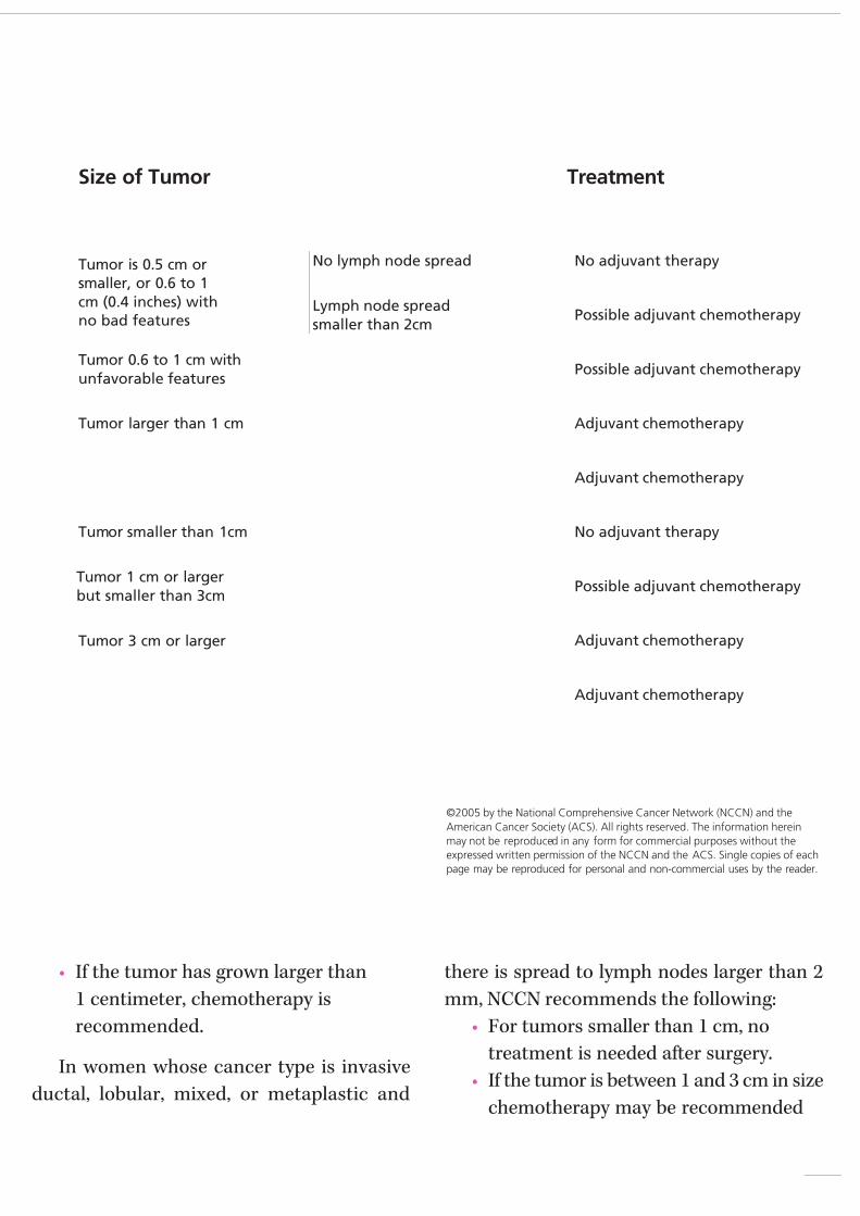
7/27/2019 NCCN Breast Guidelines
http://slidepdf.com/reader/full/nccn-breast-guidelines 55/84
• If the tumor has grown larger than
1 centimeter, chemotherapy is
recommended.
In women whose cancer type is invasive
ductal, lobular, mixed, or metaplastic and
there is spread to lymph nodes larger than 2
mm, NCCN recommends the following:
• For tumors smaller than 1 cm, no
treatment is needed after surgery.
• If the tumor is between 1 and 3 cm in size
chemotherapy may be recommended
Additional Treatment (Adjuvant Therapy) After Surgeryfor Non-Hormone Responsive Tumors
©2005 by the National Comprehensive Cancer Network (NCCN) and the
American Cancer Society (ACS). All rights reserved. The information herein
may not be reproduced in any form for commercial purposes without the
expressed written permission of the NCCN and the ACS. Single copies of each
page may be reproduced for personal and non-commercial uses by the reader.
No adjuvant therapy
Possible adjuvant chemotherapy
Possible adjuvant chemotherapy
Adjuvant chemotherapy
Adjuvant chemotherapy
No adjuvant therapy
Possible adjuvant chemotherapy
Adjuvant chemotherapy
Adjuvant chemotherapy
TreatmentSize of Tumor
Tumor smaller than 1cm
Tumor is 0.5 cm or
smaller, or 0.6 to 1
cm (0.4 inches) with
no bad features
Tumor 0.6 to 1 cm with
unfavorable features
Tumor larger than 1 cm
Tumor 1 cm or larger
but smaller than 3cm
Tumor 3 cm or larger
No lymph node spread
Lymph node spread
smaller than 2cm

7/27/2019 NCCN Breast Guidelines
http://slidepdf.com/reader/full/nccn-breast-guidelines 56/84

7/27/2019 NCCN Breast Guidelines
http://slidepdf.com/reader/full/nccn-breast-guidelines 57/84
any cancer or a single lymph node has ametastasis 2 mm in size or less, the NCCN
recommends the following:
• For tumors smaller than 1 cm, no
treatment is needed after surgery.
• If the tumor is between 1 and 3 cm in sizechemotherapy may be recommended
• Chemotherapy is recommended if it is
larger than 3 cm or has spread to lymph
nodes and the spread is larger than 2 mm.
Additional Treatment (Adjuvant Therapy) After Surgeryfor Non-Hormone Responsive Tumors (continued)
NOTES

7/27/2019 NCCN Breast Guidelines
http://slidepdf.com/reader/full/nccn-breast-guidelines 58/84
Treatment for Large Stage II BreastCancers or Stage IIIAPreoperative chemotherapy is an option that
allows some women with large tumors to have
breast-conserving treatment if they want it.
Breast-conserving treatment is usually not rec-
ommended for women with large tumors. But
chemotherapy may shrink the tumor enough
to permit a lumpectomy that completely
removes the main tumor and still keeps the
size and shape of the breast acceptable.
The work-up recommended before starting
preoperative chemotherapy includes:
• Medical history and physical
examination
• Blood counts and chemical tests• Chest x-ray, diagnostic mammography
of both breasts
• Breast ultrasound and MRI if needed
• Pathology review of biopsy sample
• Hormone receptor tests
• HER-2/neu test
56
Treatment Guidelines for Patients
Clinical Stage Work-Up
The cancer is larger than 2
cm and doesn’t invadechest wall or skin. Lymph
nodes can be enlarged but
are movable. Diagnosed by
needle biopsy not excision.
Breast conserving surgery
not possible because too
large a tumor in the breast.
[2 cm = 0.8 inch]
Wants to preserve
breast (Consider
needle biopsy of
enlarged lymph
nodes or sentinel
node procedure.
Tumor should be
marked so it canbe located after
chemotherapy)
Doesn’t want to
preserve breast
• Medical history and physical
examination
• Blood counts and chemistry tests
• Chest x-ray
• Diagnostic mammograms (both
breasts)• Breast MRI with dedicated
breast coil may be considered
• Pathology review of biopsy
sample
• Hormone receptor tests
• HER-2/neu test
• Bone scan and CT, MRI, or ultra-
sound of abdomen if symptoms
or abnormal blood tests or
tumor over 5 cm with lymph
node spread
[5 cm = 2 inches]
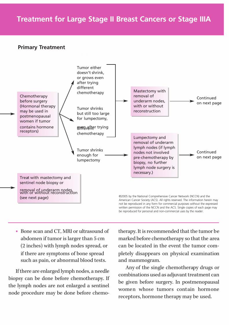
7/27/2019 NCCN Breast Guidelines
http://slidepdf.com/reader/full/nccn-breast-guidelines 59/84
• Bone scan and CT, MRI or ultrasound of
abdomen if tumor is larger than 5 cm
(2 inches) with lymph nodes spread, or
if there are symptoms of bone spread
such as pain, or abnormal blood tests.
If there are enlarged lymph nodes, a needle
biopsy can be done before chemotherapy. If
the lymph nodes are not enlarged a sentinel
node procedure may be done before chemo-
therapy. It is recommended that the tumor be
marked before chemotherapy so that the area
can be located in the event the tumor com-
pletely disappears on physical examination
and mammogram.
Any of the single chemotherapy drugs or
combinations used as adjuvant treatment can
be given before surgery. In postmenopausal
women whose tumors contain hormone
receptors, hormone therapy may be used.
©2005 by the National Comprehensive Cancer Network (NCCN) and the
American Cancer Society (ACS). All rights reserved. The information herein may
not be reproduced in any form for commercial purposes without the expressed
written permission of the NCCN and the ACS. Single copies of each page may
be reproduced for personal and non-commercial uses by the reader.
Treatment for Large Stage II Breast Cancers or Stage IIIA
Primary Treatment
Tumor either
doesn’t shrink,
or grows even
after trying
different
chemotherapy
Tumor shrinks
but still too large
for lumpectomy,
even after tryingdifferent
chemotherapy
Tumor shrinks
enough for
lumpectomy
Lumpectomy and
removal of underarm
lymph nodes (if lymph
nodes not involved
pre-chemotherapy by
biopsy, no further
lymph node surgery is
necessary.)
Mastectomy with
removal of
underarm nodes,
with or without
reconstruction
Chemotherapy
before surgery
(Hormonal therapy
may be used in
postmenopausal
women if tumor
contains hormonereceptors)
Treat with mastectomy and
sentinel node biopsy or
removal of underarm nodes,with or without reconstruction
(see next page)
Continued
on next page
Continued
on next page
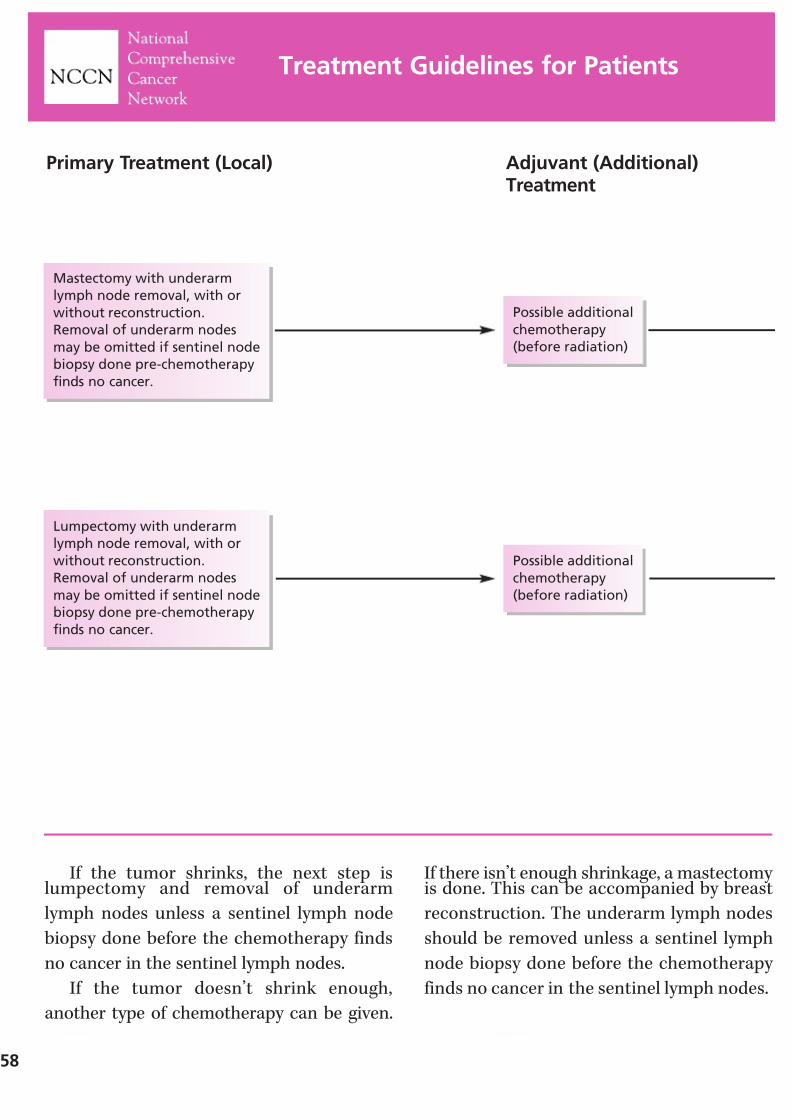
7/27/2019 NCCN Breast Guidelines
http://slidepdf.com/reader/full/nccn-breast-guidelines 60/84
If the tumor shrinks, the next step islumpectomy and removal of underarm
lymph nodes unless a sentinel lymph node
biopsy done before the chemotherapy finds
no cancer in the sentinel lymph nodes.
If the tumor doesn’t shrink enough,
another type of chemotherapy can be given.
If there isn’t enough shrinkage, a mastectomy is done. This can be accompanied by breast
reconstruction. The underarm lymph nodes
should be removed unless a sentinel lymph
node biopsy done before the chemotherapy
finds no cancer in the sentinel lymph nodes.
58
Treatment Guidelines for Patients
Primary Treatment (Local) Adjuvant (Additional)Treatment
Lumpectomy with underarm
lymph node removal, with or
without reconstruction.
Removal of underarm nodes
may be omitted if sentinel node
biopsy done pre-chemotherapy
finds no cancer.
Mastectomy with underarm
lymph node removal, with or
without reconstruction.
Removal of underarm nodes
may be omitted if sentinel node
biopsy done pre-chemotherapy
finds no cancer.
Possible additional
chemotherapy
(before radiation)
Possible additional
chemotherapy
(before radiation)
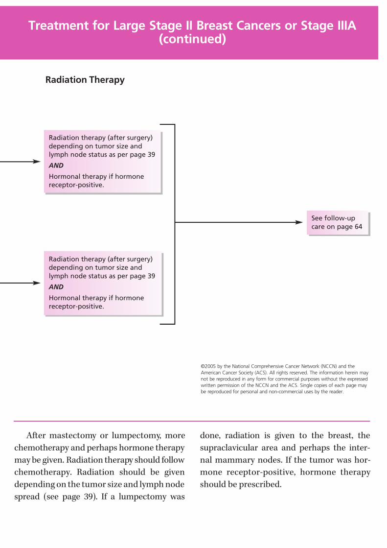
7/27/2019 NCCN Breast Guidelines
http://slidepdf.com/reader/full/nccn-breast-guidelines 61/84
After mastectomy or lumpectomy, more
chemotherapy and perhaps hormone therapy
may be given. Radiation therapy should follow
chemotherapy. Radiation should be given
depending on the tumor size and lymph node
spread (see page 39). If a lumpectomy was
done, radiation is given to the breast, the
supraclavicular area and perhaps the inter-
nal mammary nodes. If the tumor was hor-
mone receptor-positive, hormone therapy
should be prescribed.
Treatment for Large Stage II Breast Cancers or Stage IIIA(continued)
Radiation therapy (after surgery)
depending on tumor size and
lymph node status as per page 39
AND
Hormonal therapy if hormone
receptor-positive.
Radiation therapy (after surgery)
depending on tumor size and
lymph node status as per page 39
AND
Hormonal therapy if hormone
receptor-positive.
See follow-up
care on page 64
Radiation Therapy
©2005 by the National Comprehensive Cancer Network (NCCN) and the
American Cancer Society (ACS). All rights reserved. The information herein may
not be reproduced in any form for commercial purposes without the expressed
written permission of the NCCN and the ACS. Single copies of each page may
be reproduced for personal and non-commercial uses by the reader.
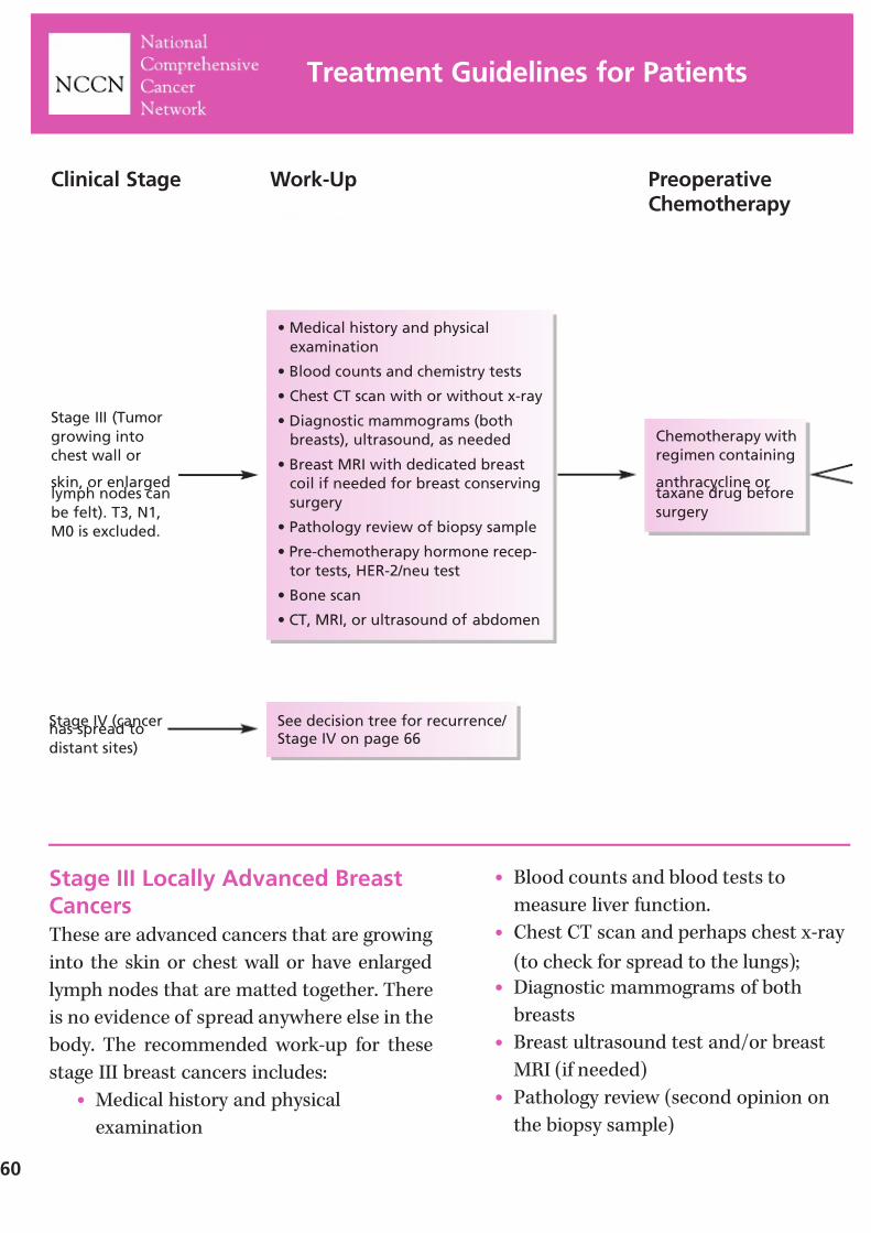
7/27/2019 NCCN Breast Guidelines
http://slidepdf.com/reader/full/nccn-breast-guidelines 62/84
60
Treatment Guidelines for Patients
Stage III Locally Advanced BreastCancersThese are advanced cancers that are growing
into the skin or chest wall or have enlarged
lymph nodes that are matted together. There
is no evidence of spread anywhere else in the
body. The recommended work-up for these
stage III breast cancers includes:
• Medical history and physical
examination
• Blood counts and blood tests to
measure liver function.
• Chest CT scan and perhaps chest x-ray
(to check for spread to the lungs);
• Diagnostic mammograms of both
breasts
• Breast ultrasound test and/or breast
MRI (if needed)
• Pathology review (second opinion on
the biopsy sample)
See decision tree for recurrence/ Stage IV on page 66
Stage III (Tumor
growing into
chest wall or
skin, or enlargedlymph nodes can
be felt). T3, N1,
M0 is excluded.
• Medical history and physical
examination
• Blood counts and chemistry tests
• Chest CT scan with or without x-ray
• Diagnostic mammograms (both
breasts), ultrasound, as needed
• Breast MRI with dedicated breast
coil if needed for breast conserving
surgery
• Pathology review of biopsy sample
• Pre-chemotherapy hormone recep-
tor tests, HER-2/neu test
• Bone scan
• CT, MRI, or ultrasound of abdomen
Clinical Stage Work-Up PreoperativeChemotherapy
Chemotherapy with
regimen containing
anthracycline ortaxane drug before
surgery
Stage IV (cancerhas spread to
distant sites)
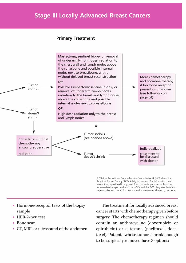
7/27/2019 NCCN Breast Guidelines
http://slidepdf.com/reader/full/nccn-breast-guidelines 63/84
• Hormone-receptor tests of the biopsy
sample
• HER-2/neu test
• Bone scan
• CT, MRI, or ultrasound of the abdomen
The treatment for locally advanced breast
cancer starts with chemotherapy given before
surgery. The chemotherapy regimen should
contain an anthracycline (doxorubicin or
epirubicin) or a taxane (paclitaxel, doce-
taxel). Patients whose tumors shrink enough
to be surgically removed have 3 options:
Stage III Locally Advanced Breast Cancers
©2005 by the National Comprehensive Cancer Network (NCCN) and the
American Cancer Society (ACS). All rights reserved. The information herein
may not be reproduced in any form for commercial purposes without the
expressed written permission of the NCCN and the ACS. Single copies of each
page may be reproduced for personal and non-commercial uses by the reader.
Primary Treatment
Tumor
shrinks
Tumor
doesn’t
shrink
Tumor shrinks –
(see options above)
Tumordoesn’t shrink
Individualized
treatment tobe discussed
with doctor
More chemotherapy
and hormone therapy
if hormone receptor
present or unknown
(see follow-up on
page 64)
Mastectomy, sentinel biopsy or removal
of underarm lymph nodes, radiation to
the chest wall and lymph nodes above
the collarbone and possible internal
nodes next to breastbone, with or
without delayed breast reconstruction
OR
Possible lumpectomy sentinel biopsy or
removal of underarm lymph nodes,
radiation to the breast and lymph nodes
above the collarbone and possible
internal nodes next to breasstbone
OR
High dose radiation only to the breast
and lymph nodes
Consider additional
chemotherapy
and/or preoperative
radiation

7/27/2019 NCCN Breast Guidelines
http://slidepdf.com/reader/full/nccn-breast-guidelines 64/84
62
• Mastectomy and sentinel node biopsy or removal of underarm lymph nodes.
This is followed by radiation therapy to
the chest wall, above the collarbone,
and if they are enlarged, internal nodes
next to the sternum or breastbone.
Breast reconstruction can be done
later if desired.
• Lumpectomy with sentinel biopsy orlymph node removal, if the cancer has
shrunk enough, followed by radiation
therapy to the breast and above the
collarbone, and if they are enlarged,
internal nodes next to the sternum or
breastbone.
Treatment Guidelines for Patients
NOTES

7/27/2019 NCCN Breast Guidelines
http://slidepdf.com/reader/full/nccn-breast-guidelines 65/84
• High dose radiation to the breast andlymph nodes. Among breast cancer
specialists, this option is controversial.
For these patients the guidelines recom-
mend adding more chemotherapy after surgery.
If the cancer contained hormone receptors,
hormone treatment is also recommended.
Women with stage IIIA or IIIB breastcancer whose tumor doesn’t shrink with a
chemotherapy regimen may be given another
chemotherapy regimen with or without radi-
ation. If they respond they can be treated as
outlined with standard breast cancer surgery.
If they do not respond, they should discuss
special, individualized treatment with their
doctor.
Stage III Locally Advanced Breast Cancers (continued)
NOTES

7/27/2019 NCCN Breast Guidelines
http://slidepdf.com/reader/full/nccn-breast-guidelines 66/84
64
Follow-up and Treatment of StageIV Disease or RecurrenceRoutine follow-up for all patients who have
had invasive breast cancer includes the fol-
lowing: a medical history and physical exam
every 4 to 6 months for 5 years, then once a year.
Women who have had a lumpectomy
should have a mammogram of the treated
breast 6 months after radiation therapy, and
then mammograms of both breasts every year.
Women who have had a mastectomy
should have a yearly mammogram of the
remaining breast after the surgery. Because
tamoxifen increases a woman’s risk of devel-
oping cancer of the endometrium (lining of
the upper part of the uterus), women taking this drug should have a yearly pelvic exam
and should promptly report any abnormal
bleeding from the vagina to their doctor.
Women on an aromatase inhibitor or who
went through early menopause on treatment
should have bone density testing.
Treatment Guidelines for Patients
• History and physical exam every 4 to
6 months for 5 years,then every year
• Mammogram every year. For
lumpectomy patients, the first one
should be 6 months after radiation
is completed
• Women taking tamoxifen: pelvic
exam every year if the uterus is
present
• Women on aromatase inhibitor or
who have gone through early
menopause with chemotherapy
should have bone density measured.
• Medical history and physical
examination
• Blood counts and chemistry
tests
• Chest x-ray
• Bone scan
• X-rays of bones that hurt and
weight-bearing bones thatare abnormal on bone scan
• CT or MRI of chest and
abdomen and/or PET scan
may be recommended
• Biopsy of suspected recur-
rence if possible
• ER/PR and HER-2/neu testing
if not known, hormone
receptor previously negative,
or HER-2 low
Routine Follow-Up Work-Up for Stage IV orSuspected Recurrence
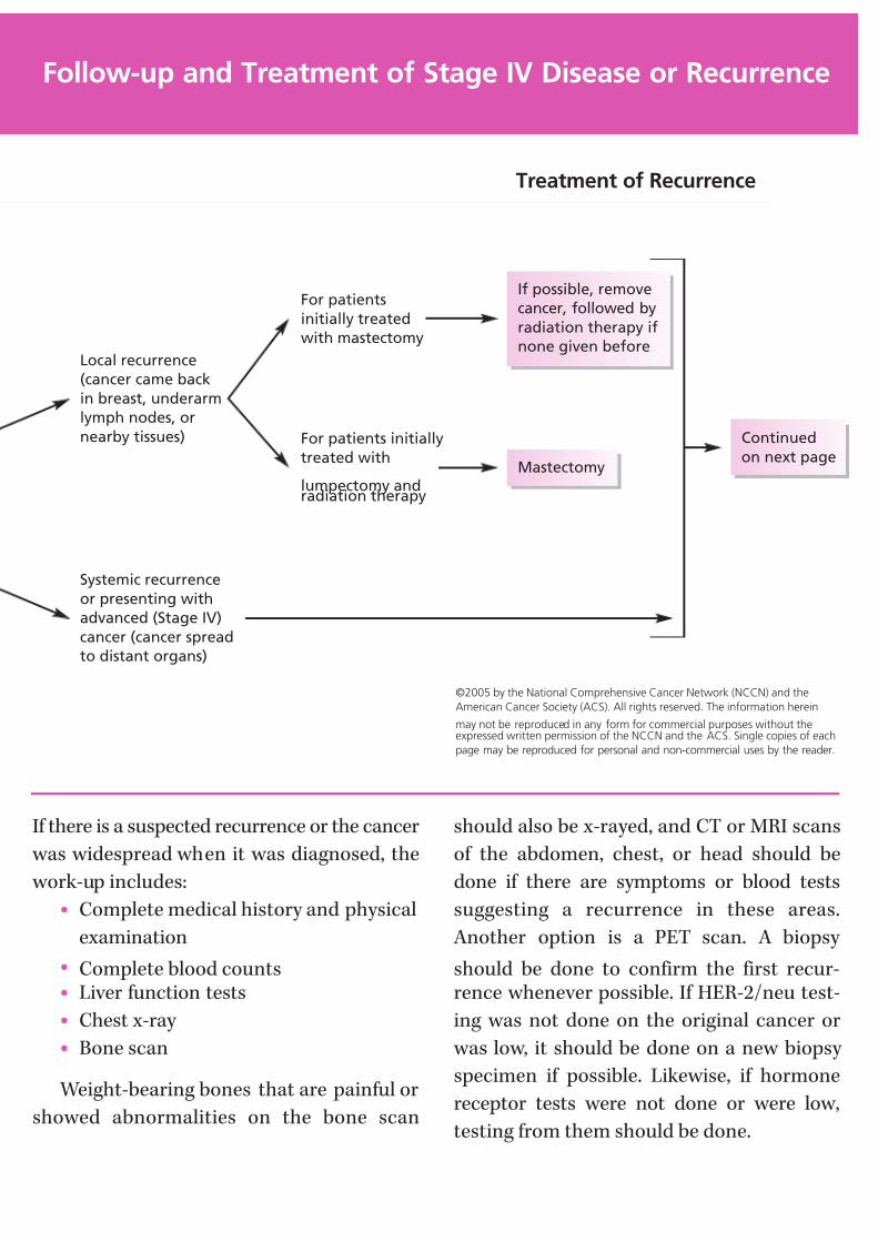
7/27/2019 NCCN Breast Guidelines
http://slidepdf.com/reader/full/nccn-breast-guidelines 67/84
If there is a suspected recurrence or the cancer
was widespread when it was diagnosed, the
work-up includes:
• Complete medical history and physical
examination
• Complete blood counts• Liver function tests
• Chest x-ray
• Bone scan
Weight-bearing bones that are painful or
showed abnormalities on the bone scan
should also be x-rayed, and CT or MRI scans
of the abdomen, chest, or head should be
done if there are symptoms or blood tests
suggesting a recurrence in these areas.
Another option is a PET scan. A biopsy
should be done to confirm the first recur-rence whenever possible. If HER-2/neu test-
ing was not done on the original cancer or
was low, it should be done on a new biopsy
specimen if possible. Likewise, if hormone
receptor tests were not done or were low,
testing from them should be done.
Follow-up and Treatment of Stage IV Disease or Recurrence
If possible, remove
cancer, followed by
radiation therapy if
none given before
Treatment of Recurrence
For patients
initially treated
with mastectomy
For patients initially
treated with
lumpectomy andradiation therapy
Mastectomy
Continued
on next page
Local recurrence
(cancer came back
in breast, underarm
lymph nodes, or
nearby tissues)
Systemic recurrence
or presenting with
advanced (Stage IV)
cancer (cancer spread
to distant organs)
©2005 by the National Comprehensive Cancer Network (NCCN) and the
American Cancer Society (ACS). All rights reserved. The information herein
may not be reproduced in any form for commercial purposes without theexpressed written permission of the NCCN and the ACS. Single copies of each
page may be reproduced for personal and non-commercial uses by the reader.
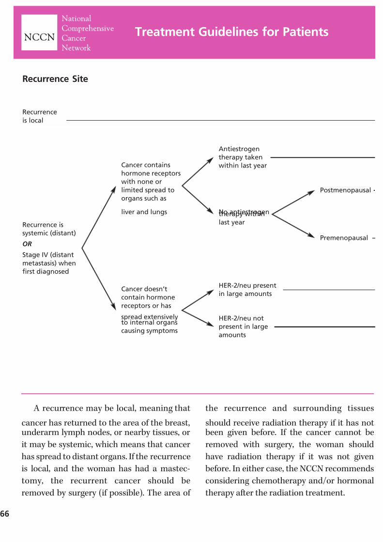
7/27/2019 NCCN Breast Guidelines
http://slidepdf.com/reader/full/nccn-breast-guidelines 68/84
A recurrence may be local, meaning that
cancer has returned to the area of the breast,underarm lymph nodes, or nearby tissues, or
it may be systemic, which means that cancer
has spread to distant organs. If the recurrence
is local, and the woman has had a mastec-
tomy, the recurrent cancer should be
removed by surgery (if possible). The area of
the recurrence and surrounding tissues
should receive radiation therapy if it has notbeen given before. If the cancer cannot be
removed with surgery, the woman should
have radiation therapy if it was not given
before. In either case, the NCCN recommends
considering chemotherapy and/or hormonal
therapy after the radiation treatment.
66
Treatment Guidelines for Patients
Postmenopausal
Premenopausal
Cancer contains
hormone receptors
with none or
limited spread to
organs such as
liver and lungs
Cancer doesn’t
contain hormone
receptors or has
spread extensivelyto internal organs
causing symptoms
No antiestrogentherapy within
last year
HER-2/neu present
in large amounts
HER-2/neu not
present in large
amounts
Recurrence
is local
Recurrence Site
Antiestrogen
therapy taken
within last year
Recurrence is
systemic (distant)
OR
Stage IV (distant
metastasis) when
first diagnosed
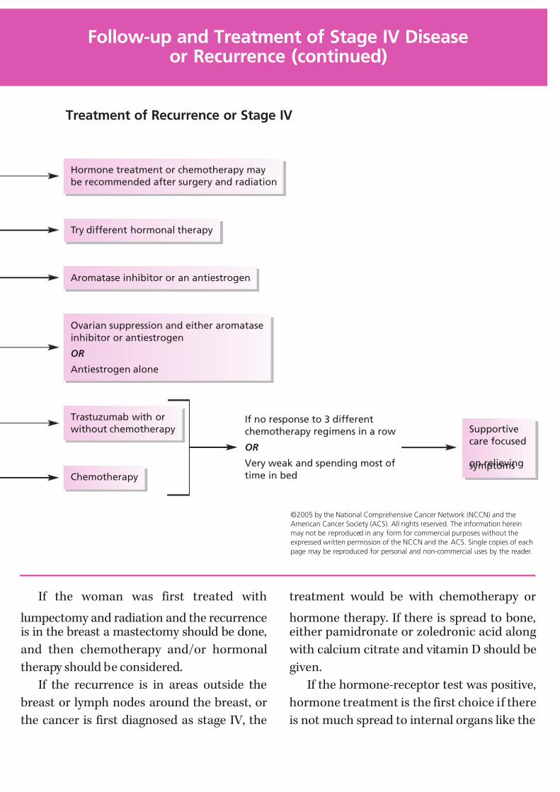
7/27/2019 NCCN Breast Guidelines
http://slidepdf.com/reader/full/nccn-breast-guidelines 69/84
If the woman was first treated with
lumpectomy and radiation and the recurrenceis in the breast a mastectomy should be done,
and then chemotherapy and/or hormonal
therapy should be considered.
If the recurrence is in areas outside the
breast or lymph nodes around the breast, or
the cancer is first diagnosed as stage IV, the
treatment would be with chemotherapy or
hormone therapy. If there is spread to bone,either pamidronate or zoledronic acid along
with calcium citrate and vitamin D should be
given.
If the hormone-receptor test was positive,
hormone treatment is the first choice if there
is not much spread to internal organs like the
©2005 by the National Comprehensive Cancer Network (NCCN) and the
American Cancer Society (ACS). All rights reserved. The information herein
may not be reproduced in any form for commercial purposes without the
expressed written permission of the NCCN and the ACS. Single copies of each
page may be reproduced for personal and non-commercial uses by the reader.
Follow-up and Treatment of Stage IV Diseaseor Recurrence (continued)
If no response to 3 different
chemotherapy regimens in a row
OR
Very weak and spending most of
time in bed
Treatment of Recurrence or Stage IV
Hormone treatment or chemotherapy may
be recommended after surgery and radiation
Try different hormonal therapy
Aromatase inhibitor or an antiestrogen
Ovarian suppression and either aromatase
inhibitor or antiestrogen
OR
Antiestrogen alone
Trastuzumab with or
without chemotherapy Supportive
care focused
on relievingsymptomsChemotherapy

7/27/2019 NCCN Breast Guidelines
http://slidepdf.com/reader/full/nccn-breast-guidelines 70/84
liver. Hormone therapy is not used if there ismajor involvement of the liver because it might
not act quickly enough to stop the damage to
that organ. For postmenopausal women, an
aromatase inhibitor or antiestrogen would be
the first choice. For premenopausal women,tamoxifen or blocking the ovaries from mak-
ing estrogen and treating with hormones as
are used for postmenopausal women would
be the first treatment. The ovaries may be
68
Treatment Guidelines for Patients
NOTES

7/27/2019 NCCN Breast Guidelines
http://slidepdf.com/reader/full/nccn-breast-guidelines 71/84
blocked with a luteinizing hormone-releasing hormone agonist (medicines that decrease
estrogen production in the ovary), with radi-
ation therapy or by removing them surgically.
In either case, an antiestrogen would not berecommended if it had been taken within the
previous year. Instead, another kind of hormone
treatment would be used (see next page).
Follow-up and Treatment of Stage IV Diseaseor Recurrence (continued)
NOTES
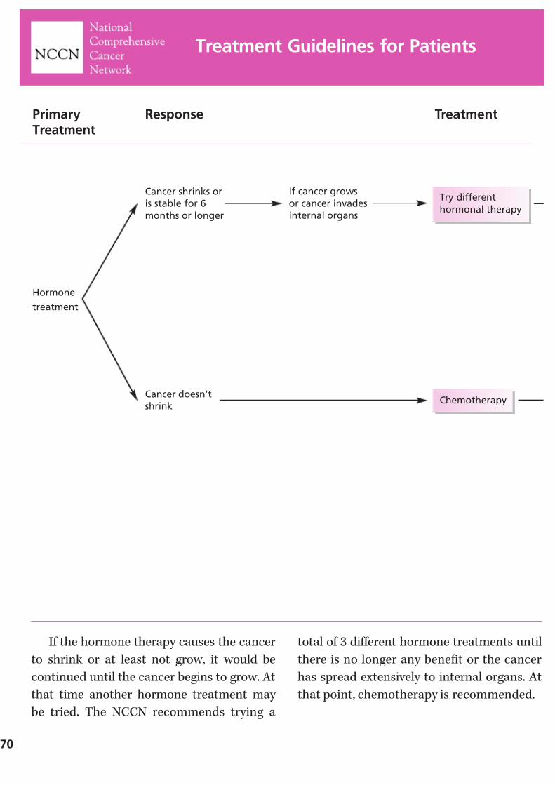
7/27/2019 NCCN Breast Guidelines
http://slidepdf.com/reader/full/nccn-breast-guidelines 72/84
If the hormone therapy causes the cancer
to shrink or at least not grow, it would be
continued until the cancer begins to grow. At
that time another hormone treatment may
be tried. The NCCN recommends trying a
total of 3 different hormone treatments until
there is no longer any benefit or the cancer
has spread extensively to internal organs. At
that point, chemotherapy is recommended.
70
Treatment Guidelines for Patients
Cancer shrinks or
is stable for 6
months or longer
If cancer grows
or cancer invades
internal organs
Cancer doesn’t
shrink
Hormone
treatment
PrimaryTreatment
Response Treatment
Try different
hormonal therapy
Chemotherapy
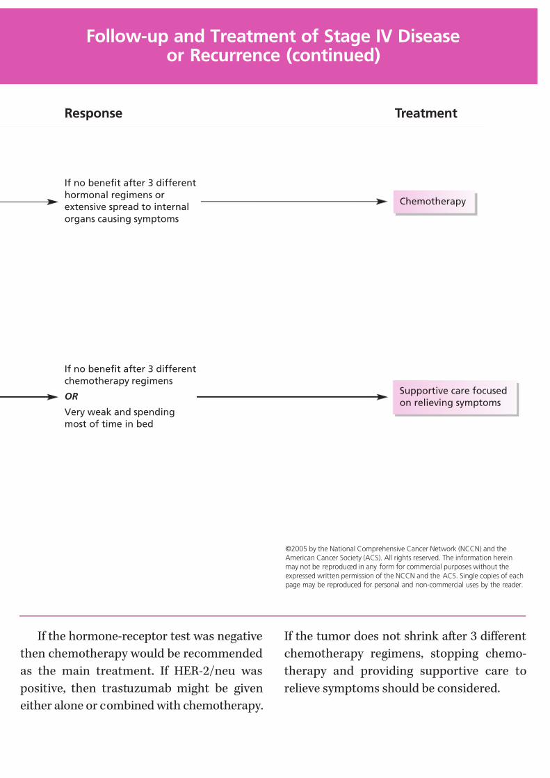
7/27/2019 NCCN Breast Guidelines
http://slidepdf.com/reader/full/nccn-breast-guidelines 73/84
If the hormone-receptor test was negative
then chemotherapy would be recommended
as the main treatment. If HER-2/neu was
positive, then trastuzumab might be given
either alone or combined with chemotherapy.
If the tumor does not shrink after 3 different
chemotherapy regimens, stopping chemo-
therapy and providing supportive care to
relieve symptoms should be considered.
©2005 by the National Comprehensive Cancer Network (NCCN) and the
American Cancer Society (ACS). All rights reserved. The information herein
may not be reproduced in any form for commercial purposes without the
expressed written permission of the NCCN and the ACS. Single copies of each
page may be reproduced for personal and non-commercial uses by the reader.
Follow-up and Treatment of Stage IV Diseaseor Recurrence (continued)
Response Treatment
If no benefit after 3 different
hormonal regimens or
extensive spread to internal
organs causing symptoms
Supportive care focused
on relieving symptoms
If no benefit after 3 different
chemotherapy regimens
OR
Very weak and spending
most of time in bed
Chemotherapy

7/27/2019 NCCN Breast Guidelines
http://slidepdf.com/reader/full/nccn-breast-guidelines 74/84
72
NOTES
7/27/2019 NCCN Breast Guidelines
http://slidepdf.com/reader/full/nccn-breast-guidelines 75/84
Adjuvant therapy
Treatment that is added to increase the effec-
tiveness of a primary therapy. It usually refers
to hormonal therapy, chemotherapy, or radi-
ation added after surgery to kill any cancer
cells still remaining and increase the chances
of curing the disease or keeping it in check.
Antiestrogen
A substance that blocks the effects of estrogen
on tumors ( for example, the drug tamoxifen).
Antiestrogens are used to treat breast cancers
that depend on estrogen for growth.
Aromatase inhibitors
Drugs that block production of estrogens from
hormones made by the adrenal gland. They are
used to treat hormone-sensitive breast cancerin postmenopausal women. These include
anastrozole, letrozole, and exemestane.
Axillary dissection
A surgical procedure in which the lymph
nodes in the armpit (axillary nodes) are
removed and examined to find out if breast
cancer has spread to those nodes and to
remove any cancerous lymph nodes.
Biopsy
A tissue sample that is removed and examined
under the microscope.
Bisphosphonates
Drugs that help strengthen bones weakened
by cancer by encouraging the deposition of
calcium. These include pamidronate and
zoledronate.
Breast-conserving therapy
Surgery to remove a breast cancer and a small
amount of benign tissue around the cancer,
without removing any other part of the breast.
This procedure is also called lumpectomy, seg-
mental excision, or limited breast surgery. The
method may require an axillary dissection and
usually requires radiation therapy in addition
to the breast conservation surgery.
Breast reconstruction
Surgery that rebuilds the breast contour aftermastectomy. A breast implant or the woman’s
own tissue provides the contour. If desired,
the nipple and areola may also be re-created.
Reconstruction can be done at same time as
the mastectomy or any time later.
Carcinoma in situ
An early stage of cancer, in which the tumor
is still only in the structures of the organ where it first developed, and the disease does
not invade other parts of the organ or spread
to distant sites. Most in situ carcinomas are
highly curable.
Glossary
7/27/2019 NCCN Breast Guidelines
http://slidepdf.com/reader/full/nccn-breast-guidelines 76/84
74
Chemotherapy
Treatment with drugs to destroy cancer cells.
Chemotherapy is often used in addition to
surgery or radiation to treat cancer when
metastasis is proven or suspected, when the
cancer has come back (recurred), or when
there is a strong likelihood that the cancercould recur.
Clinical stage
Stage includes evaluation of the size and
extension of the cancer, the presence or
absence of spread of cancer to lymph nodes,
and the presence or absence of spread to
other body organs. Clinical stage is the stage
determined only by physical examination,and x-ray or other imaging studies. This
includes determination of the size of the
cancer, and evaluation of lymph nodes by the
doctor’s examination of the armpit. The final
stage is the pathological stage which is deter-
mined from microscopic examination of the
tumor and lymph nodes. Clinical stage is
used for initial treatment planning.
Cyst
A fluid-filled mass that is usually benign. The
fluid can be removed for analysis.
Diagnostic mammography
Screening mammography is performed on
women with no evidence of lumps or other
symptoms. This includes 2 x-ray views of each
breast (top-bottom; side-to-side). Diagnosticmammography includes additional x-ray views
of areas of concern on physical examination
or on the screening mammogram to provide
more information about the size and character
of the abnormality.
Duct
A hollow passage for gland secretions. In the
breast, a passage through which milk passes
from the lobule (which makes the milk) to the
nipple. These ducts are the starting point for
most breast cancers.
Ductal carcinoma in situ
The most common type of non-invasive
breast cancer. Cancer cells have not spread
beyond the ducts.
Estrogen
A female sex hormone produced primarily
by the ovaries and in smaller amounts from
hormones produced by the adrenal gland
and fat cells. In breast cancer, estrogen may
promote the growth of breast cancer cells.
Fibroadenoma
A type of benign breast tumor composed of
fibrous tissue and glandular tissue. On clini-
cal examination or breast self-examination, it
usually feels like a firm, round, smooth lump.
These usually occur in young women.
Fibrocystic changes
A term that describes certain benign changes
in the breast, also called fibrocystic disease.
Symptoms of this condition are breast
swelling or pain. The breasts often feel lumpy
or nodular. Because these signs sometimes
mimic breast cancer, diagnostic mammogra-
phy or ultrasound or even a biopsy may be
needed to show that there is no cancer.
Fibrosis
Formation of fibrous (scar-like) tissue. This
can occur anywhere in the body.
7/27/2019 NCCN Breast Guidelines
http://slidepdf.com/reader/full/nccn-breast-guidelines 77/84
Grade
Cancer cells are graded by how much they
look like normal cells. Grade 1 (also called
well-differentiated) means the cancer cells
look like the normal cells; grade 3 (poorly
differentiated) cancer cells do not look like
normal cells at all. Grade 1 cancers aren’tconsidered aggressive. In other words, they
tend to grow more slowly and metastasize
slower. Grade 3 cancers are more likely to grow
faster and metastasize. A cancer’s grade along
with its stage is used to determine treatment.
Her-2/neu
A gene that produces a type of receptor that
helps cells grow. Breast cancer cells with toomany Her-2/neu receptors tend to be fast
growing and may respond to treatment with
an antibody called trastuzumab.
Hormone
A chemical substance released into the body by
glands, such as the thyroid, pituitary, or ovaries.
The substance travels through the bloodstream
and sets in motion various body functions. Forexample, prolactin, which is produced in the
pituitary gland, begins and sustains the pro-
duction of milk in the breast after childbirth.
Hormone receptor assay
A test to see whether a breast tumor is likely
to be affected by hormones or if it can be
treated with hormones.
Hormone therapyTreatment with hormones, drugs that interfere
with hormone production or hormone action,
or surgical removal of hormone-producing
glands to kill cancer cells or show their growth.
The most common hormonal therapy for
breast cancer is the drug tamoxifen. Other hor-
monal therapies include megestrol, aromatase
inhibitors, androgens and surgical removal of
the ovaries (oophorectomy ).
Internal mammary lymph nodes
Lymph nodes located inside the chest, next
to the junction of the sternum (breastbone)and the ribs.
Intraductal papillomas
Small, finger-like, polyp-like, noncancerous
growths in the breast ducts that may cause a
bloody nipple discharge. These are most
often found in women 45 to 50 years of age.
When many papillomas exist, breast cancer
risk is slightly increased.
Latissimus dorsi flap procedure
A method of breast reconstruction that uses
the long flat muscle of the back, by rotating it
to the chest area.
LHRH (luteinizing hormone-releasing
hormone) agonists
Drugs that block the ovaries from producing
estrogen.
Lobular carcinoma in situ
Also called lobular neoplasia. Non-invasive
cancer that has not spread beyond the lobules.
The lobules are the milk-producing parts of
the breast at the distant end of the ducts.
Lumpectomy
Surgery to remove the breast tumor and a
small amount of surrounding normal tissue.
Lymph nodes
Small bean-shaped collections of immune
system tissue such as lymphocytes, located
along lymphatic vessels. They remove waste
and fluids from lymph and help fight infec-
tions; also called lymph glands.
7/27/2019 NCCN Breast Guidelines
http://slidepdf.com/reader/full/nccn-breast-guidelines 78/84
76
Lymphedema
An infrequent complication after breast
cancer treatment. Swelling in the arm caused
by excess fluid that collects after lymph
nodes and vessels are removed by surgery or
treated by radiation.
Margin
The normal tissues around the tumor. Positive
means there is cancer at the margin or the
margin is less than 1 mm. More than 1 mm is
negative. If the margins are positive, repeat
surgery may be done to make them negative.
Mastectomy
Removal of the entire breast. In a simple or total
mastectomy surgeons do not cut away any
lymph nodes or muscle tissue; in a modified
radical mastectomy, surgeons remove the
breast and some armpit lymph nodes; in a
radical mastectomy (now rarely performed)
surgeons remove the breast, armpit lymph
nodes, and chest wall muscles under the breast.
Menopause
The time in a woman’s life when monthly
cycles of menstruation stop forever and the
level of hormones produced by the ovaries
decreases. Menopause usually naturally occurs
in a woman’s late 40s or early 50s, but it can
also be caused by surgical removal of both
ovaries (oophorectomy), or by chemotherapy,
which often destroys ovarian function.
MetastasisThe spread of cancer cells to distant areas of
the body by way of the lymph system or
bloodstream.
Preoperative therapy
Systemic therapy, such as chemotherapy or
hormone therapy, given before surgery.
Preoperative therapy can shrink some breast
cancers, so it can be removed with less exten-
sive surgery than would otherwise be needed.
Nodal status
Indicates whether a breast cancer has spread
(node-positive) or has not spread (node-
negative) to lymph nodes in the armpit (axillary
nodes). The number and site of positive lymph
nodes can help predict the risk of cancer
recurrence.
Oophorectomy
Surgery to remove the ovaries.
Ovary
Reproductive organ in the female pelvis.
Normally a woman has two ovaries. They
contain the eggs (ova) that, when joined with
sperm, result in pregnancy. Ovaries produce
most of a premenopausal woman’s estrogen.
PalpationUsing the hands to examine. A palpable mass
in the breast is one that can be felt.
PET (positron emission tomography)
scan
A total body scan that uses a radioactive form
of glucose to detect cancer.
Progesterone
A female sex hormone released by the ovariesduring every menstrual cycle to prepare the
uterus for pregnancy and the breasts for milk
production (lactation).
7/27/2019 NCCN Breast Guidelines
http://slidepdf.com/reader/full/nccn-breast-guidelines 79/84
Prognosis
A prediction of the course of disease – or the
outlook for the cure of the patient. For example,
women with breast cancer that is small and
does not involve the lymph nodes and receive
prompt treatment have a good prognosis.
Sentinel node mapping and biopsy
In a sentinel lymph node mapping and
biopsy, the surgeon injects a radioactive sub-
stance and/or a blue dye into the area around
the tumor. Lymphatic vessels carry these
materials to the sentinel lymph node (also
called the sentinel node). The doctor can see
the blue dye or detect the radioactivity (with
a Geiger counter) in the sentinel node, whichis cut out and examined. If the sentinel node
contains cancer, more axillary lymph nodes
are removed. But if it is free of cancer, the
patient can avoid additional axillary surgery
and its potential side effects.
Stage
A method of describing the size and location
of cancer based upon characteristics of thetumor, the lymph nodes, and whether there is
involvement of other organs.
Stereotactic needle biopsy
A method of needle biopsy that is useful in
some cases in which calcifications or a mass
can be seen on mammogram but cannot be
located by touch. Computerized equipment
maps the location of the mass and this isused as a guide to place the needle.
Supportive care
Measures taken to relieve symptoms and
improve quality of life, but that are not
expected to destroy the cancer. Pain medica-
tion is an example of supportive care.
Supraclavicular lymph nodes
Lymph nodes located in the area just above
the collarbone.
Systemic therapy
Treatment that reaches and affects cells
throughout the body; for example, chemo-therapy.
Tamoxifen
This drug blocks the effects of estrogen on
many organs, such as the breast. Blocking
estrogen is desirable in some cases of breast
cancer because estrogen promotes their
growth. Recent research suggests that tamox-
ifen may lower the risk of developing breast
cancer in women with certain risk factors.
Toremifene
Another anti-estrogen drug, similar to
tamoxifen.
Transverse rectus abdominus muscle
flap procedure (TRAM flap)
A method of breast reconstruction in which
tissue from the lower abdominal wall whichreceives its blood supply from the rectus
abdominus muscle is used. The tissue from
this area is moved up to the chest to create a
breast mound and usually does not require
an implant. Moving muscle and tissue from
the lower abdomen to the chest results in
flattening the lower abdomen (a “tummy
tuck”). Also called a TRAM flap or rectus
abdominus flap procedure.
Ultrasound
High frequency sound waves used to produce
images of the breast.
For a more comprehensive glossary, you may
visit the ACS Web site at www.cancer.org

7/27/2019 NCCN Breast Guidelines
http://slidepdf.com/reader/full/nccn-breast-guidelines 80/84
78
NOTES

7/27/2019 NCCN Breast Guidelines
http://slidepdf.com/reader/full/nccn-breast-guidelines 81/84
NOTES

7/27/2019 NCCN Breast Guidelines
http://slidepdf.com/reader/full/nccn-breast-guidelines 82/84
80
Current NCCN Treatment Guidelinesfor Patients
Advanced Cancer and Palliative Care Treatment Guidelines
(English and Spanish)
Bladder Cancer Treatment Guidelines for Patients (English and Spanish)
Breast Cancer Treatment Guidelines for Patients (English and Spanish)
Cancer Pain Treatment Guidelines for Patients (English and Spanish)
Cancer-Related Fatigue and Anemia Treatment Guidelines for Patients
(English and Spanish)
Colon and Rectal Cancer Treatment Guidelines for Patients
(English and Spanish)
Distress Treatment Guidelines for Patients (English and Spanish)
Fever and Neutropenia Treatment Guidelines for Cancer Patients
(English and Spanish)
Lung Cancer Treatment Guidelines for Patients (English and Spanish)
Melanoma Cancer Treatment Guidelines for Patients (English and Spanish)
Nausea and Vomiting Treatment Guidelines for Patients With Cancer
(English and Spanish)
Non-Hodgkin’s Lymphoma Treatment Guidelines for Patients
(English and Spanish)
Ovarian Cancer Treatment Guidelines for Patients (English and Spanish)
Prostate Cancer Treatment Guidelines for Patients (English and Spanish)
7/27/2019 NCCN Breast Guidelines
http://slidepdf.com/reader/full/nccn-breast-guidelines 83/84
Terri Ades, MS, APRN-BC, AOCN
American Cancer Society
Robert W. Carlson, MDStanford Hospital and Clinics
Stephen B. Edge, MD
Roswell Park Cancer Institute
Herman Kattlove, MD
American Cancer Society
Joan McClure, MSNational Comprehensive
Cancer Network
Shannan Rafine
National Comprehensive
Cancer Network Mary-Lou Smith
Y-ME National Breast Cancer
Organization
The Breast Cancer Treatment Guidelines for Patients were developed by a diverse group of experts
and were based on the NCCN clinical practice guidelines. These patient guidelines were translated,
reviewed, and published with help from the following individuals:
The original NCCN Breast Cancer Clinical Practice Guidelines were developed by the following
NCCN Panel Members:
Robert W. Carlson, MD/ChairStanford Hospital and Clinics
Benjamin O. Anderson, MD
Fred Hutchinson Cancer Research
Center/Seattle Cancer Care Alliance
Harold Burstein, MD, PhD
Dana-Farber/Partners CancerCare
W Bradford Carter, MD
H. Lee Moffitt Cancer Center &
Research Institute at the University
of South FloridaStephen B. Edge, MD
Roswell Park Cancer Institute
William B. Farrar, MD
Arthur G. James Cancer Hospital &
Richard J. Solove Research Institute
at The Ohio State University
Lori J. Goldstein, MD
Fox Chase Cancer Center
William Gradishar, MD
Robert H. Lurie ComprehensiveCancer Center of Northwestern
University
Daniel F. Hayes, MDUniversity of Michigan
Comprehensive Cancer Center
Clifford Hudis, MD
Memorial Sloan-Kettering
Cancer Center
Mohammed Jahanzeb, MD
St. Jude Children’s Research
Hospital/University of Tennessee
Cancer Institute
Britt-Marie Ljung, MDUCSF Comprehensive Cancer Center
Lawrence B. Marks, MD
Duke Comprehensive Cancer Center
Beryl McCormick, MD
Memorial Sloan-Kettering
Cancer Center
Lisle M. Nabell, MD
University of Alabama at Birmingham
Comprehensive Cancer Center
Lori J. Pierce, MDUniversity of Michigan
Comprehensive Cancer Center
Elizabeth Reed, MDUNMC Eppley Cancer Center
at The Nebraska Medical Center
Samuel M. Silver, MD, PhD
University of Michigan
Comprehensive Cancer Center
Mary Lou Smith
Y-ME National Breast Cancer
Organization
George Somlo, MD
City of Hope Cancer CenterRichard Theriault, DO, MBA
The University of Texas
M.D. Anderson Cancer Center
John Ward, MD
Huntsman Cancer Institute
at the University of Utah
Eric P. Winer, MD
Dana-Farber/Partners CancerCare
Antonio C. Wolff, MD
The Sidney Kimmel ComprehensiveCancer Center at Johns Hopkins

7/27/2019 NCCN Breast Guidelines
http://slidepdf.com/reader/full/nccn-breast-guidelines 84/84
©2005, American Cancer Society, Inc.
No.9405.07