Nature Reviews Neuroscience - PREA2k30
28
4/4/10 12:18 PM Neuroscience and education: from research to practice? : Article : Nature Reviews Neuroscience Page 1 of 28 http://www.nature.com/nrn/journal/v7/n5/full/nrn1907.html Perspective Nature Reviews Neuroscience 7, 406-413 (May 2006) | doi:10.1038/nrn1907 SCIENCE AND SOCIETY Neuroscience and education: from research to practice? Usha Goswami 1 About the author top Cognitive neuroscience is making rapid strides in areas highly relevant to education. However, there is a gulf between current science and direct classroom applications. Most scientists would argue that filling the gulf is premature. Nevertheless, at present, teachers are at the receiving end of numerous 'brain-based learning' packages. Some of these contain alarming amounts of misinformation, yet such packages are being used in many schools. What, if anything, can neuroscientists do to help good neuroscience into education? There is a hunger in schools for information about the brain. Teachers are keen to reap the benefits of the 'century of neuroscience' for their students. In neuroscience laboratories, considerable progress is being made in understanding the neurocognitive development underpinning essential skills taught by educators, such as numeracy and literacy. This progress is largely theoretical. The current gulf between neuroscience and education is being filled by packages and programmes claiming to be based on brain science. The
Transcript of Nature Reviews Neuroscience - PREA2k30

4/4/10 12:18 PMNeuroscience and education: from research to practice? : Article : Nature Reviews Neuroscience
Page 1 of 28http://www.nature.com/nrn/journal/v7/n5/full/nrn1907.html
Perspective
Nature Reviews Neuroscience 7, 406-413 (May 2006) | doi:10.1038/nrn1907
SCIENCE AND SOCIETY
Neuroscience and education: from research topractice?Usha Goswami1 About the author
top
Cognitive neuroscience is making rapid strides in areas highly
relevant to education. However, there is a gulf between current
science and direct classroom applications. Most scientists would
argue that filling the gulf is premature. Nevertheless, at present,
teachers are at the receiving end of numerous 'brain-based
learning' packages. Some of these contain alarming amounts of
misinformation, yet such packages are being used in many schools.
What, if anything, can neuroscientists do to help good neuroscience
into education?
There is a hunger in schools for information about the brain. Teachers are
keen to reap the benefits of the 'century of neuroscience' for their students. In
neuroscience laboratories, considerable progress is being made in
understanding the neurocognitive development underpinning essential skills
taught by educators, such as numeracy and literacy. This progress is largely
theoretical. The current gulf between neuroscience and education is being
filled by packages and programmes claiming to be based on brain science. The
speed with which such packages have gained widespread currency in schools
4/4/10 12:18 PMNeuroscience and education: from research to practice? : Article : Nature Reviews Neuroscience
Page 2 of 28http://www.nature.com/nrn/journal/v7/n5/full/nrn1907.html
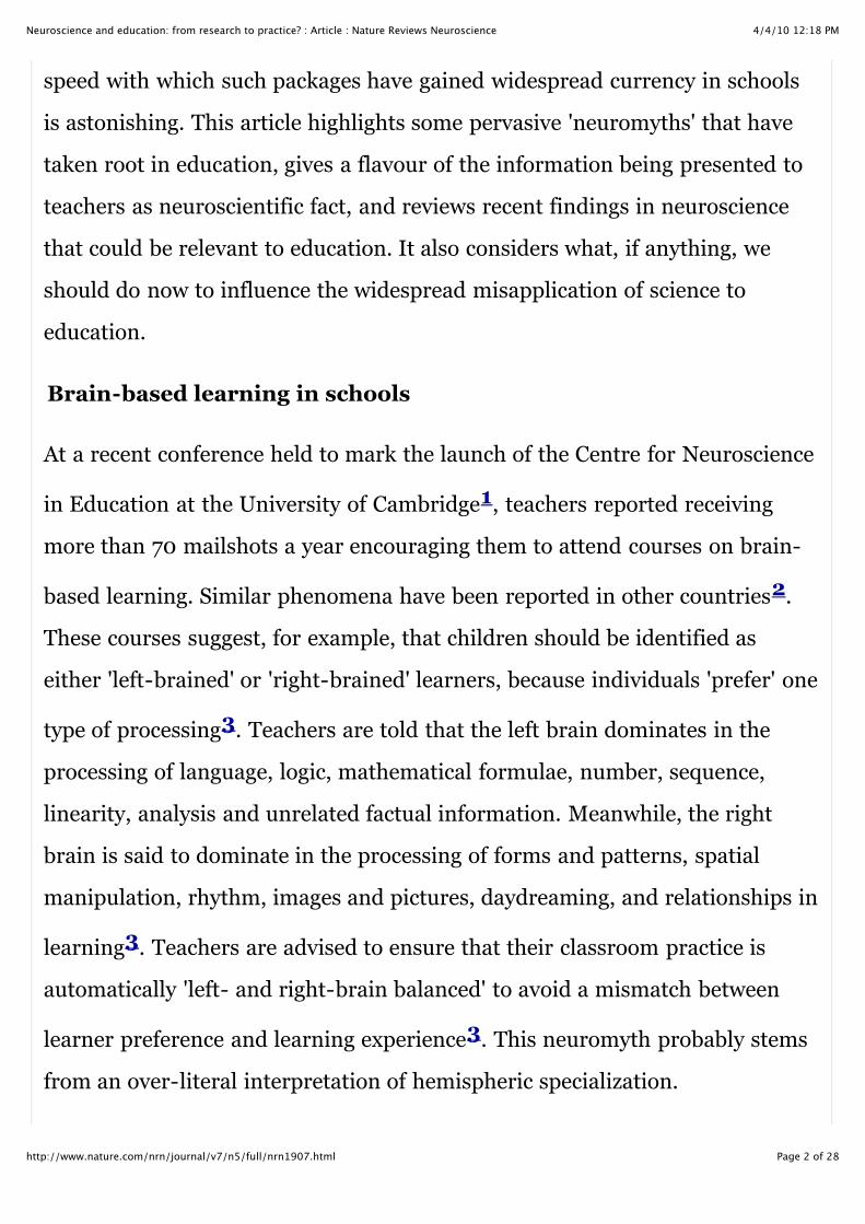
speed with which such packages have gained widespread currency in schools
is astonishing. This article highlights some pervasive 'neuromyths' that have
taken root in education, gives a flavour of the information being presented to
teachers as neuroscientific fact, and reviews recent findings in neuroscience
that could be relevant to education. It also considers what, if anything, we
should do now to influence the widespread misapplication of science to
education.
Brain-based learning in schools
At a recent conference held to mark the launch of the Centre for Neuroscience
in Education at the University of Cambridge1, teachers reported receiving
more than 70 mailshots a year encouraging them to attend courses on brain-
based learning. Similar phenomena have been reported in other countries2.
These courses suggest, for example, that children should be identified as
either 'left-brained' or 'right-brained' learners, because individuals 'prefer' one
type of processing3. Teachers are told that the left brain dominates in the
processing of language, logic, mathematical formulae, number, sequence,
linearity, analysis and unrelated factual information. Meanwhile, the right
brain is said to dominate in the processing of forms and patterns, spatial
manipulation, rhythm, images and pictures, daydreaming, and relationships in
learning3. Teachers are advised to ensure that their classroom practice is
automatically 'left- and right-brain balanced' to avoid a mismatch between
learner preference and learning experience3. This neuromyth probably stems
from an over-literal interpretation of hemispheric specialization.

4/4/10 12:18 PMNeuroscience and education: from research to practice? : Article : Nature Reviews Neuroscience
Page 3 of 28http://www.nature.com/nrn/journal/v7/n5/full/nrn1907.html
Other courses for teachers advise that children's learning styles should be
identified as either visual, auditory or kinaesthetic, and that children should
then wear a badge labelled either V, A or K while in school, showing their
learning style for the benefit of all of their teachers. Still others argue that
adoption of a commercial package 'Brain GymR' ensures that 'true' education
happens. Brain GymR prescribes a series of simple body movements4 "to
integrate all areas of the brain to enhance learning". Teachers are told that "in
technical terms, information is received by the brainstem as an 'impress', but
may be inaccessible to the front brain as an 'express'. This ... locks the student
into a failure syndrome. Whole-brain learning draws out the potential locked
in the body and enables students to access those areas of the brain previously
unavailable to them. Improvements in learning ... are often immediate". It is
even claimed that the child can press certain 'brain buttons' under their ribs4
to focus the visual system for reading and writing.
Many in education accept claims such as these as established fact5. Scientists
have already alerted society to the neuromyths that are dominant in education
at present6, 7, 8. In addition to the left brain/right brain learning myth,
neuromyths that relate to critical periods for learning and to synaptogenesis
can be identified. The critical period myth suggests that the child's brain will
not work properly if it does not receive the right amount of stimulation at the
right time (an insightful analysis is provided by Byrnes9). Direct teaching of
certain skills must occur during the critical period, or the window of
opportunity to educate will be missed. The synaptogenesis myth promotes the
idea that more will be learned if teaching is timed with periods of
synaptogenesis7. Educational interventions will be more effective if teachers
4/4/10 12:18 PMNeuroscience and education: from research to practice? : Article : Nature Reviews Neuroscience
Page 4 of 28http://www.nature.com/nrn/journal/v7/n5/full/nrn1907.html
synaptogenesis7. Educational interventions will be more effective if teachers
ensure that they coincide with increases in synaptic density. Educational
interventions are also sometimes suggested to be superior if they encourage
'neuroplasticity'10, and teachers are told that neural networks can be altered
by 'neuroplasticity training programmes'10. Teachers do not realize that,
although there might be sensitive periods for some forms of learning, the
effects of any type of training programme that changes behaviour will be
reflected in the 'remapping' of neural networks.
Neuroscience in the classroom
These neuromyths need to be eliminated. The dominance of these myths
obscures the important strides being made by cognitive neuroscience in many
areas relevant to education. For example, our understanding of the neural
bases of the '3 Rs' — reading, writing and arithmetic — is growing rapidly. So
is our understanding of how to optimize the brain's ability to benefit from
teaching. Good instructional practice can be undermined by brain-based
factors such as learning anxiety, attention deficits and poor recognition of
social cues. All of these factors disrupt an individual's capacity to learn, and
also have an effect on other learners in the same classroom.
Reading and dyslexia. From work with adults, it is well established that a
left-hemisphere network of frontal, temporoparietal and occipitotemporal
regions underpins mature reading11. However, cross-language imaging
studies show some interesting variations. These seem to depend on how the
orthography (the writing system) of a language represents phonology (the
sounds of the language). When learners of transparent writing systems (for

4/4/10 12:18 PMNeuroscience and education: from research to practice? : Article : Nature Reviews Neuroscience
Page 5 of 28http://www.nature.com/nrn/journal/v7/n5/full/nrn1907.html
sounds of the language). When learners of transparent writing systems (for
example, Italian) are contrasted with learners of non-transparent (for
example, English) or character-based (for example, Chinese) writing systems,
highly similar brain areas are found to be active during reading12, 13.
However, mature readers of transparent orthographies show greater activity in
the left planum temporale, a brain region involved in letter-sound conversion,
whereas mature English readers show greater activation of an area known as
the visual word form area (VWFA) in the left occipital temporal region12.
Although originally proposed as the substrate of visual word recognition14,
15, this neural area has also been proposed to involve phonology — for
example, through the computation of orthographic–phonological
connections16, 17. Its greater activation in English could reflect the several
levels of spelling-sound correspondence that are important for decoding
English18 (for example, reading BOMIC by letter-sound conversion or by
analogy to COMIC). Readers of Chinese show relatively more engagement of
visuospatial areas, presumably for recognizing complex characters13.
Developmentally, it is known from behavioural studies that pre-readers who
can recognize phonological similarity (for example, that CAT and HAT rhyme,
or that CAT and CUP share the first sound) become better readers. Imaging
studies have confirmed that young readers primarily depend on the left
posterior superior temporal cortex, the area identified in adult studies as the
locus of phonological decoding19 (Fig. 1). Activity in this region is also
modulated by children's phonological skills. As literacy is acquired, the VWFA
(described as a 'skill zone' by some developmental neuroscientists20) is more

4/4/10 12:18 PMNeuroscience and education: from research to practice? : Article : Nature Reviews Neuroscience
Page 6 of 28http://www.nature.com/nrn/journal/v7/n5/full/nrn1907.html
(described as a 'skill zone' by some developmental neuroscientists20) is more
engaged and areas initially active in the right hemisphere are disengaged.
Studies of children with developmental dyslexia (children who are failing to
learn to read normally despite average intelligence and educational
opportunity) show that, atypically, the right temporoparietal cortex continues
to be activated during reading21. Children with developmental dyslexia also
show significantly less activation in the usual left hemisphere sites. If targeted
remediation is provided, usually through intensive tuition in phonological
skills and in letter-sound conversion, activity in the left temporal and parietal
areas appears to normalize22, 23. So far, however, developmental
neuroimaging studies have been short term and mostly confined to English.
Theoretically motivated studies across languages are now required24.
Figure 1 | Brain areas involved in typical reading developmentand dyslexia measured with functional MRI.
a | Images in the top panel show the early reliance on
the left posterior superior temporal cortex, which is
known to be involved in phonological processing, in
children learning to read, and the expansive involvement
of the left parietal, temporal and frontal cortices in adult
readers. Correlations between brain activity during
reading and reading ability (measured on standardized
tests) demonstrate increased involvement of the left
temporal and frontal regions, associated with phonology
and semantics, as reading develops (bottom panel).
Right posterior activation declines as reading is
4/4/10 12:18 PMNeuroscience and education: from research to practice? : Article : Nature Reviews Neuroscience
Page 7 of 28http://www.nature.com/nrn/journal/v7/n5/full/nrn1907.html
Right posterior activation declines as reading is
acquired, presumably indicating reduced reliance on the
systems for recognizing non-lexical forms. b | Summary
of brain regions engaged during reading and reading-
related tasks in typically developing readers (left inferior
frontal gyrus, left temporoparietal cortex and left
inferotemporal cortex) and readers with dyslexia (left
inferior frontal gyrus only). Panel a reproduced, with
permission, from Ref. 19 © (2003) Macmillan
Publishers Ltd. Panel b courtesy of G. Eden, Centre for
the Study of Learning, Georgetown University,
Washington, DC, USA.
High resolution image and legend (27 KB)
Figures and tables index
Download PowerPoint slide (234 KB)
These developmental imaging studies show that we can begin to pin-point the
neural systems responsible for the acquisition of reading skills, and that we
can remediate inefficiencies in these systems. However, so far, these studies
do not tell teachers 'what works' in the classroom. Most training studies have
used interventions already known to be successful from educational research,
and have simply documented that neural changes in the expected areas
accompany behavioural changes22, 23. So far, neuroimaging tells us little
more, but, the potential is there. For example, imaging offers the possibility of
identifying neural indices of a child's potential difficulties, which may be

4/4/10 12:18 PMNeuroscience and education: from research to practice? : Article : Nature Reviews Neuroscience
Page 8 of 28http://www.nature.com/nrn/journal/v7/n5/full/nrn1907.html
identifying neural indices of a child's potential difficulties, which may be
hidden from view earlier in development. We can attempt to identify neural
markers for phonological sensitivity, such as brain responses to auditory cues
for rhythm25, to identify who is at risk of later reading difficulties.
Alternatively, we can seek general language markers for dyslexia26. In both
cases, early identification of infants with poor skills would enable language
interventions to prevent dyslexia long before schooling27.
Studies could also be designed to test neural hypotheses. For example, a
popular cognitive theory of developmental dyslexia proposes a cerebellar
deficit28. A commercial exercise-based treatment programme, the DDAT
(Dyslexia Dyspraxia Attention Deficit Treatment)29, aims to remediate
cerebellar difficulties. Children are encouraged to practise motor skills such as
catching beanbags while standing on one leg on a cushion. This is claimed to
benefit reading. Imaging studies could measure where neural changes occur in
response to such remediation, to see whether permanent changes to the neural
areas for reading are involved (this seems unlikely — any effects found for
reading are probably short-term placebo effects).
Number and dyscalculia. Progress in understanding the underpinnings of
arithmetic has been rapid since the proposal that the human brain has
dedicated circuits for recognizing numerosity30. This 'number sense' capacity
depends on parietal, prefrontal and cingulate areas, with the horizontal
segment of the bilateral intraparietal sulcus (HIPS) playing a central part in
the basic representation and manipulation of quantity31. In simple
paradigms, in which participants have to decide whether, for example, 3 is

4/4/10 12:18 PMNeuroscience and education: from research to practice? : Article : Nature Reviews Neuroscience
Page 9 of 28http://www.nature.com/nrn/journal/v7/n5/full/nrn1907.html
paradigms, in which participants have to decide whether, for example, 3 is
larger than 5, the HIPS might be the only region specifically engaged. Activity
in the HIPS is modulated by the semantic distance between numbers and by
the size of numbers32. Other arithmetic operations are more dependent on
language-based fact retrieval, such as simple multiplication, which activates
the angular gyrus33.
Some arithmetic operations depend on the mental 'number line'. This is an
apparently universal mental spatial representation of number, in which
smaller numbers are represented on the left side of space and larger numbers
are represented on the right34. The interactions revealed between number
and space in the parietal cortex have been particularly interesting. Manual
responses to large numbers are faster when the response is on the right side of
space, and vice versa for smaller numbers35. In line bisection tasks, in which
participants have to estimate the central point of a horizontal line, midpoint
estimation systematically deviates to the left if the line is made up of 2s
(222222222...) and to the right if the line is made up of 9s (999999999...)36.
The numbers automatically bias attention. Patients with visual neglect, a
disorder of spatial attention following right parietal damage, systematically
neglect the left side of space. These patients show a rightward bias in line
bisection tasks. This rightward bias was even found for oral estimation (for
example, when asked to state the numerical midpoint of 2 and 6, patients
tended to give answers like 5)37. Therefore, numerical manipulations seem to
depend crucially on intact spatial representations; indeed, blind adults who
acquire numbers spatially show normal parietal distance effects38.

4/4/10 12:18 PMNeuroscience and education: from research to practice? : Article : Nature Reviews Neuroscience
Page 10 of 28http://www.nature.com/nrn/journal/v7/n5/full/nrn1907.html
acquire numbers spatially show normal parietal distance effects38.
So far, findings from adult neuroimaging and neuropsychological studies
remain to be applied to understanding mathematical development in children.
One important electroencephalogram (EEG) study showed that when 5-year-
old children perform the number comparison task ("is 4 larger or smaller than
5?") they show effects at similar electrodes in the parietal cortex as adults,
with similar latencies39 (Fig. 2). However, reaction time data showed that
the children were three times slower to organize the key press response. This
imaging experiment raises the possibility that, neurally, young children can
extract numerical information as fast as adults. The slow acquisition of
calculation skills in the primary years might, therefore, reflect difficulties in
understanding arithmetic notation and place value, rather than difficulties in
understanding the relationship between digits and quantities. Neuroimaging
studies can help us to investigate this possibility. Also of interest to teachers is
the evidence for the spatial mental number line. At present, there are various
models in schools for teaching children ordinal knowledge of number — that
numbers come in an ordered scale of magnitude. The finding that the brain
has a preferred mode of representation suggests that teachers should build on
this spatial system when teaching ordinality and place value — for example,
through teaching tools such as the 'empty number line'40, 41.
Figure 2 | Electrophysiological recordings of activity duringnumber processing tasks in children and adults.
a | Participants were shown numbers, represented by
either dots or digits, and required to press a response
key with their left hand if the numbers were smaller
4/4/10 12:18 PMNeuroscience and education: from research to practice? : Article : Nature Reviews Neuroscience
Page 11 of 28http://www.nature.com/nrn/journal/v7/n5/full/nrn1907.html
key with their left hand if the numbers were smaller
than 5, or with their right hand if the numbers were
larger than 5. In adults, the typical finding in such tests
is that responses are faster when numbers are distant
(for example, 9 or 1) rather than close (6 or 4) to 5; this
is called the distance effect. Behavioural data indicated
distance effects for both adults and children in this task.
b | A schematic depiction of the event-related potential
(ERP) procedure. Recording of brain activity began 200
ms before and ended 800 ms after stimulus onset.
Within this recording epoch, voltage changes associated
with the behavioural distance effect for adults and
children were found at similar parietal electrode sites.
However, the schematic shows that the key press
response required 500 ms for the adults, but 1,600 ms
for the children. Whereas numbers seem to be
recognized at similar latencies by children and adults,
organization of the required response takes much longer
for children. c | Representative posterior channel (91)
comparing ERPs in adults and 5 year olds for the
number comparison task. The x-axis is in milliseconds
and corresponds to a 1-s epoch of recorded
electroencephalogram (EEG; 200 ms baseline, 800 ms
poststimulus). Top panel, notation effects (digits versus
dots). The two age groups show qualitatively similar
initial components (P1, N1 and P2p) with only slightly
4/4/10 12:18 PMNeuroscience and education: from research to practice? : Article : Nature Reviews Neuroscience
Page 12 of 28http://www.nature.com/nrn/journal/v7/n5/full/nrn1907.html
initial components (P1, N1 and P2p) with only slightly
delayed peaks in the 5 year olds. Middle panel, ERP
distance effect for digits in both age groups. Bottom
panel, ERP distance effect for dots in both age groups.
Significant differences associated with distance began in
children 50 ms after adults despite children having
reaction times (RTs) that were >1,000 ms longer.
Asterisk denotes significant differences at p < 0.5.
Modified, with permission, from Ref. 39 © (1998)
National Academy of Sciences.
High resolution image and legend (53 KB)
Figures and tables index
Download PowerPoint slide (262 KB)
Developmental dyscalculia occurs when a child experiences unexpected
difficulty in learning arithmetic in the absence of mental retardation despite
adequate schooling and social environment42. One possible neural
explanation is that the core quantity system in the HIPS has developed
abnormally. This possibility was investigated by a functional MRI (fMRI)
study of girls with Turner syndrome43, who typically present with
visuospatial and number processing deficits44. Sulcal morphometry using
new techniques45 revealed that the right intraparietal sulcal pattern of most
patients with Turner syndrome showed aberrant branching, abnormal
interruption and/or unusual orientation43. It was suggested that this
4/4/10 12:18 PMNeuroscience and education: from research to practice? : Article : Nature Reviews Neuroscience
Page 13 of 28http://www.nature.com/nrn/journal/v7/n5/full/nrn1907.html
interruption and/or unusual orientation43. It was suggested that this
anatomical disorganization could explain the visuospatial and arithmetic
impairments found behaviourally. A study of very low birthweight children
with arithmetical difficulties found reduced grey matter in the left
intraparietal sulcus46. Control studies are now required to determine whether
the parietal sulci are abnormal in other developmental syndromes that do not
present with arithmetical difficulties. If parietal abnormalities characterize
only children presenting with arithmetical impairments, this would imply a
direct link between the brain and behaviour. Children without apparent
developmental syndromes who present with unusually poor number
processing in the classroom would then need to be assessed for parietal
damage.
Attention, emotion and social cognition. The short attention spans of
some children pose continual problems for their teachers. Children with
attention deficit/hyperactivity disorder (ADHD) are particularly challenging to
educate, as they are inattentive and impulsive, cruising the classroom instead
of focusing on their work. Of course, all young children experience some
difficulties in sustaining attention and inhibiting impulses. Perhaps attentional
training might benefit all preschoolers47, leading to educational advantages?
A recent brain imaging study claimed that 5 days of attention training
significantly improved performance on tests of intelligence in 4- and 6-year-
old children48. The children were given training exercises to improve
stimulus discrimination, anticipation and conflict resolution. For example,
they learned to track a cartoon cat on a computer screen by using a joystick, to
4/4/10 12:18 PMNeuroscience and education: from research to practice? : Article : Nature Reviews Neuroscience
Page 14 of 28http://www.nature.com/nrn/journal/v7/n5/full/nrn1907.html
they learned to track a cartoon cat on a computer screen by using a joystick, to
anticipate the movement of a duck across a pond by moving the cat to where
the duck should emerge, and to select the larger of two arrays of digits when
conflict was introduced by using smaller digits to present the larger array.
Attention was tested before and after the training exercises by asking children
to press a computer key to indicate which direction the central fish in a row of
five fish was facing. Before training, the children were also given an
intelligence test, and the same test was repeated after 5 days of training
(which in itself would improve performance, due to item familiarity). Children
in the control group either received the attention and intelligence tests only,
or attended the laboratory for five sessions of watching popular videos. No
matched computer training with animal cartoons was provided to train a
control skill, such as memory. Even so, attention training did not improve
performance in attention. Instead, an effect of attention training was found for
one of the intelligence tests. Scores on the Matrices subtest improved by a
significant 6.5 points for the trained 4-year-olds only. EEG data were then
collected to determine whether neural conflict-related attentional effects
familiar from adults would be found in the trained children. The effect sought
was a larger frontal negativity for incongruent trials at the frontoparietal
electrodes, particularly at Cz. Despite the lack of behavioural effects, an
electrophysiological effect was found for the trained 6 year olds at the target
electrode (Cz). For the trained 4 year olds, a 'hint of an effect' was found at a
different frontal electrode (Fz). From these single electrode results, it was
argued that the executive attention network can be influenced by educational
interventions during development. However, as the attention intervention did
not affect the children's performance in the attention tasks, further research is

4/4/10 12:18 PMNeuroscience and education: from research to practice? : Article : Nature Reviews Neuroscience
Page 15 of 28http://www.nature.com/nrn/journal/v7/n5/full/nrn1907.html
not affect the children's performance in the attention tasks, further research is
needed to support this conclusion. Unusually, the authors offer their training
programme free through the Organization for Economic Cooperation and
Development, enabling other scientists to test its effectiveness. This is to be
highly commended.
The neural substrates for emotional processing are increasingly well
understood. For example, the amygdala is known to be important for the
interpretation of emotional and social signals, particularly from the face and
eyes49. In adults, the degree of amygdala activation is particularly correlated
with the intensity of facial expressions of fear49. Children, too, show
amygdala activity to fearful expressions, and children with autism (who have
impaired social cognition) have significantly increased amygdala volume50.
The anatomical system involved in fear processing could be abnormal from an
early age in autism, as was suggested by a recent EEG study with 3 year
olds51. The mirror neuron system in the inferior frontal gyrus is also involved
in understanding the emotional states of others52. The results of a recent
fMRI study showed no activity in this area in children with autism when
compared with typically developing children during the imitation of emotional
expressions53 (Fig. 3). Mirror neurons appear to mediate our understanding
of emotional states via imitation, allowing the translation of an observed
action (such as a facial expression) into its internally felt emotional
significance52. This translation appeared to be absent in autism.
Figure 3 | Neural activity during imitation and observation of

4/4/10 12:18 PMNeuroscience and education: from research to practice? : Article : Nature Reviews Neuroscience
Page 16 of 28http://www.nature.com/nrn/journal/v7/n5/full/nrn1907.html
emotional expressions for typically developing children andchildren with autism spectrum disorders.
a | Shows brain activation recorded during imitation of
emotional expressions. Activity in the bilateral pars
opercularis (stronger in the right) of the inferior frontal
gyrus is seen in the typically developing group (top
panel) but not in the group with autism spectrum
disorders (ASD; middle panel). A between-group
comparison (bottom panel) revealed that this difference
is significant (t >1.83, p <0.05, corrected for multiple
comparisons at the cluster level). RH, right hemisphere;
LH, left hemisphere. b | Activity in the mirror neuron
system during the observation of emotional
expressions53. The right pars opercularis showed
significantly greater activity in typically developing
children than in children with ASD (t >1.83, p <0.05,
small volume corrected). Reproduced, with permission,
from Ref. 53 © (2006) Macmillan Publishers Ltd.
High resolution image and legend (26 KB)
Figures and tables index
Download PowerPoint slide (236 KB)
Research such as this allows us to study the neural underpinnings of
emotional processing in children in mainstream schooling. For example,
children exposed to harsh discipline and physical abuse at home seem to

4/4/10 12:18 PMNeuroscience and education: from research to practice? : Article : Nature Reviews Neuroscience
Page 17 of 28http://www.nature.com/nrn/journal/v7/n5/full/nrn1907.html
children exposed to harsh discipline and physical abuse at home seem to
process emotions differently from other children54. In later childhood they
are also more likely to have conduct disorders that make them difficult to
teach55. Such children are prone to an anger attribution bias, tending to
(mis)attribute anger to the actions and statements of others54. So far, little
neuroimaging work has been done with such children. If atypical brain
development is found, and if training programmes can be devised to improve
these children's reading of social signals, this would be of benefit to education.
We already know that it might be possible to teach children with autism to
'read' emotions to some degree56. Optimal interventions for other groups of
children could also be designed, with imaging data helping to pin-point the
brain networks to be targeted.
A similar logic applies to learning anxiety. Neuroimaging studies of anxiety
disorders in adults focus particularly on structural and functional changes in
the orbitofrontal cortex (OFC) and the temporal lobes, including the
amygdala57. Anxiety disorders are known to increase following traumatic
brain injury (TBI). A neuroimaging study of children aged 4–19 years with
severe TBI showed that children with more damage to the OFC were less likely
to develop anxiety disorders58. The authors suggested that an imbalance in
the OFC–amygdala connection could influence the expression of anxiety, and
pointed out that in non-human primates these connections begin to develop
during gestation. Anxiety disorders can be treated, and neuroimaging in adults
suggests that some beneficial treatments target the amygdala59. As in adults,
anxiety in children appears to affect attentional systems, leading children to

4/4/10 12:18 PMNeuroscience and education: from research to practice? : Article : Nature Reviews Neuroscience
Page 18 of 28http://www.nature.com/nrn/journal/v7/n5/full/nrn1907.html
anxiety in children appears to affect attentional systems, leading children to
selectively shift attention towards threatening stimuli60. Again, it might be
possible to devise early interventions for such children, and to use
neuroimaging to identify who is most likely to benefit.
Can we bridge the gulf?
While we await such developments, can we bridge the gulf between
neuroscience and education by speaking directly to teachers, and sidestepping
the middlemen of the brain-based learning industry? We are trying to do this
in our UK seminar series, and through the International Mind, Brain and
Education Society1, 61. For example, at the Cambridge conference, prominent
neuroscientists working in areas such as literacy, numeracy, IQ, learning,
social cognition and ADHD spoke directly to teachers about the scientific
evidence being gathered in scientists' laboratories. The teachers were amazed
by how little was known. Although there was enthusiasm for and appreciation
of getting first-hand information, this was coupled with frustration at hearing
that many of the brain-based programmes currently in schools had no
scientific basis. The frustration arose because the neuroscientists were not
telling the teachers 'what works instead'. One delegate said that the
conference "Left teachers feeling [that] they had lots stripped away from them
and nothing put in [its] place". Another commented that "Class teachers will
take on new initiatives if they are sold on the benefits for the children.
Ultimately this is where brains live!".
This last comment surely provides an insight into the success of the brain-
based learning industry. Inspirational marketing ensures that teachers who

4/4/10 12:18 PMNeuroscience and education: from research to practice? : Article : Nature Reviews Neuroscience
Page 19 of 28http://www.nature.com/nrn/journal/v7/n5/full/nrn1907.html
based learning industry. Inspirational marketing ensures that teachers who
attend these conferences do get 'sold' on the supposed benefits of these
programmes for the children that they teach. Owing to placebo effects, these
programmes may indeed bring benefits to children in the short term.
However, such programmes are unlikely to yield benefits in the long term, and
so many will naturally fall out of use (one teacher commented "We no longer
make children wear their VAK badges"). The question for society is, should
neuroscientists do anything about this misuse of science? After all, each of
these programmes will have a natural life, and will then go away. Only
findings for the classroom that are really based on neuroscience will endure.
So should we do anything now?
At least two lessons for science and society have emerged from efforts to bring
together neuroscience and education1, 62, 63. The first is the immense
goodwill that teachers and educators have for neuroscience — they are very
interested in neuroscience, they feel that we have the potential to make
important discoveries about human learning, and they are eager to learn
about these discoveries and to contribute ideas and suggestions. Many
teachers have found attending these conferences an intellectually exhilarating
experience. The second lesson is that neuroscientists may not be those best
placed to communicate with teachers in any sustained way. The scientists are
seen as too concerned to establish the rigour of their experimental
manipulations, and as providing too much data. Most teachers prefer broad
brush messages with a 'big picture', and being 'told what works'.
Neuroscientists are not necessarily gifted at communicating with society at
large, and they are appropriately cautious about saying that something 'works'.

4/4/10 12:18 PMNeuroscience and education: from research to practice? : Article : Nature Reviews Neuroscience
Page 20 of 28http://www.nature.com/nrn/journal/v7/n5/full/nrn1907.html
It may be of most use to society if we as scientists foster and support a
network of communicators of our research — individuals who can bridge the
current gulf between neuroscience and education by providing high-quality
knowledge in a digestible form. These communicators could function in a
similar way to the information officers of medical charities, but, in this case,
explain what neuroscience breakthroughs mean for the child in the classroom.
Ideal communicators would be ex-scientists with an interest in education,
perhaps attached to universities or to national education departments. They
could fulfil a dual role: interpreting neuroscience from the perspective of and
in the language of educators, and feeding back research questions and ideas
from educators to neuroscientists. In my view, we should not remain quiet
when claims that we know to be spurious are made, such as that children can
organize themselves for reading and writing by pressing their 'brain buttons'.
Nevertheless, it might, ultimately, be of most value to society if we empower
our own middlemen, communicators who know who to consult for expert
advice on the latest claims of the brain-based learning industry, and who are
clearly working in the public interest and not for profit. A network of such
communicators would serve us all (and our children), and would prevent
society from pouring precious educational resources into scientifically
spurious applications.
Links
FURTHER INFORMATION
Learning Sciences and Brain Research The Centre for
Neuroscience in Education

4/4/10 12:18 PMNeuroscience and education: from research to practice? : Article : Nature Reviews Neuroscience
Page 21 of 28http://www.nature.com/nrn/journal/v7/n5/full/nrn1907.html
Article PubMed ISI ChemPort
Neuroscience in Education
Competing interests statement
The author declares no competing financial interests.
top
References
1. Economic and Social Research Council Teaching and Learning ResearchProgramme (ESRC TLRP) seminar series. Collaborative Frameworks forNeuroscience and Education. [online], Education and Brain Research:Neuroscience, Teaching and Learning conference. 25–27 July 2005,Faculty of Education, University of Cambridge, UK.
2. Stern, E. Pedagogy meets neuroscience. Science 310, 745 (2005).
3. Smith, A. Accelerated Learning in the Classroom (Network EducationalPress Ltd, Bodmin, UK, 1996).
4. Cohen, I. & Goldsmith, M. Hands On: How to Use Brain GymR in theClassroom (Hands On Books, Sea Point, South Africa, 2000).
5. Hoffman, E. Introducing Children to their Amazing Brains (LTL BooksLtd, Middlewich, UK, 2002).
6. Organisation for Economic Co-operation and Development.Understanding the Brain: Towards a New Learning Science (2002).
7. Bruer, J. T. Education and the brain: a bridge too far. Educ. Res. 26, 4–16(1997).
8. Blakemore, S. J. & Frith, U. The Learning Brain: Lessons for Education(Blackwell, Oxford, UK, 2005).
9. Byrnes, J. P. Minds, Brains and Learning (Guilford Press, New York,2001).
10. Tallal, P. Improving language and literacy is a matter of time. Nature Rev.Neurosci. 5, 721–728 (2004).
4/4/10 12:18 PMNeuroscience and education: from research to practice? : Article : Nature Reviews Neuroscience
Page 22 of 28http://www.nature.com/nrn/journal/v7/n5/full/nrn1907.html
Article
Article PubMed ChemPort
Article PubMed ISI ChemPort
Article PubMed ISI ChemPort
Article PubMed ISI
Article PubMed ISI
Article PubMed ISI ChemPort
Article PubMed ISI
Article
Article PubMed ISI ChemPort
11. Fiez, J. A. & Petersen, S. E. Neuroimaging studies of word reading. Proc.Natl Acad. Sci. USA 95, 914–921 (1998).
12. Paulesu, E. et al. Dyslexia: cultural diversity and biological unity. Science291, 2165–2167 (2001).
13. Siok, W. T., Perfetti, C. A., Jin, Z. & Tan, L. H. Biological abnormality ofimpaired reading is constrained by culture. Nature 431, 71–76 (2004).
14. Cohen, L. & Dehaene, S. Specialisation within the ventral stream: the casefor the visual word form area. Neuroimage 22, 466–476 (2004).
15. Dehaene, S. et al. The neural code for written words: a proposal. TrendsCogn. Sci. 9, 335–341 (2005).
16. Price, C. J. et al. Cortical localisation of the visual and auditory word formareas: a reconsideration of the evidence. Brain Lang. 86, 272–286(2003).
17. Goswami, U. & Ziegler, J. C. A developmental perspective on the neuralcode for written words. Trends Cogn. Sci. (in the press).
18. Ziegler, J. & Goswami, U. Reading acquisition, developmental dyslexia,and skilled reading across languages: a psycholinguistic grain size theory.Psychol. Bull. 131, 3–29 (2005).
19. Turkeltaub, P., Gareau, L., Flowers, D. L., Zeffiro, T. A. & Eden, G. F.Development of neural mechanisms for reading. Nature Neurosci. 6, 767–773 (2003).
20. Pugh, K. R. et al. Neurobiological studies of reading and reading disability.J. Commun. Disord. 34, 479–492 (2001).
21. Shaywitz, B. A. et al. Disruption of posterior brain systems for reading inchildren with developmental dyslexia. Biol. Psychiatry 52, 101–110(2002).

4/4/10 12:18 PMNeuroscience and education: from research to practice? : Article : Nature Reviews Neuroscience
Page 23 of 28http://www.nature.com/nrn/journal/v7/n5/full/nrn1907.html
Article PubMed ISI
Article PubMed ChemPort
PubMed ISI ChemPort
Article PubMed ISI ChemPort
ISI
Article
Article PubMed ISI
Article PubMed ISI ChemPort
Article PubMed ISI ChemPort
(2002).
22. Temple, E. et al. Neural deficits in children with dyslexia ameliorated bybehavioural remediation: evidence from functional fMRI. Proc. Natl Acad.Sci. USA 100, 2860–2865 (2003).
23. Simos, P. G. et al. Dyslexia-specific brain activation profile becomesnormal following successful remedial training. Neurology 58, 1203–1213(2002).
24. Ziegler, J. C. & Goswami, U. Becoming literate in different languages:similar problems, different solutions. Dev. Sci. (in the press).
25. Goswami, U. in Mind, Brain and Education (eds Fischer, K. & Batro, A.)(Pontifical Academy of Sciences, Rome, in the press).
26. Molfese, D. Predicting dyslexia at 8 years of age using neonatal brainresponses. Brain Lang. 72, 238–245 (2000).
27. Goswami, U. Neuroscience and Education. Brit. J. Educ. Psychol. 74, 1–14(2004).
28. Nicolson, R. I. & Fawcett, A. J. Developmental dyslexia: the role of thecerebellum. Dyslexia 5, 155–177 (1999).
29. Reynolds, D., Nicolson, R. I. & Hambly, H. Evaluation of an exercise-based treatment for children with reading difficulties. Dyslexia 9, 48–71(2003).
30. Dehaene, S. The Number Sense (Oxford Univ. Press, New York, 1997).
31. Dehaene, S., Molko, N., Cohen, L. & Wilson, A. J. Arithmetic and thebrain. Curr. Opin. Neurobiol. 14, 218–224 (2004).
32. Pinel, P., Dehaene, S., Riviere, D. & LeBihan, D. Modulation of parietalactivation by semantic distance in a number comparison task. Neuroimage14, 1013–1026 (2001).
4/4/10 12:18 PMNeuroscience and education: from research to practice? : Article : Nature Reviews Neuroscience
Page 24 of 28http://www.nature.com/nrn/journal/v7/n5/full/nrn1907.html
Article PubMed ISI ChemPort
Article ISI
Article ISI
Article
Article PubMed ISI ChemPort
Article PubMed ISI ChemPort
Article PubMed ChemPort
Article PubMed ISI ChemPort
33. Dehaene, S., Piazza, M., Pinel, P. & Cohen, L. Three parietal circuits fornumber processing. Cogn. Neuropsychol. 20, 487–506 (2003).
34. Dehaene, S. & Cohen, L. Towards an anatomical and functional model ofnumber processing. Math. Cogn. 1, 83–120 (1995).
35. Dehaene, S., Bossini, S. & Giraux, P. The mental representation of parityand numerical magnitude. J. Exp. Psychol. Gen. 122, 371–396 (1993).
36. Hubbard, E. M., Piazza, M., Pinel, P. & Dehaene, S. Interactions betweennumber and space in parietal cortex. Nature Rev. Neurosci. 6, 435–448(2005).
37. Zorzi, M., Priftis, K. & Umilta, C. Brain damage: neglect disrupts themental number line. Nature 417, 138–139 (2002).
38. Szucs, D. & Csépe, V. The parietal distance effect appears in both thecongenitally blind and matched sighted controls in an acoustic numbercomparison task. Neurosci. Lett. 384, 11–16 (2005).
39. Temple, E. & Posner, M. I. Brain mechanisms of quantity are similar in 5-year-old children and adults. Proc. Natl Acad. Sci. USA 95, 7836–7841(1998).
40. Bramald, R. Introducing the empty number line: the Dutch approach toteaching number skills. Education 3–13 28, 5–12 (2000).
41. Griffin, S. A., Case, R. & Siegler, R. S. in Classroom Lessons: IntegratingCognitive Theory (ed. McGilly, K.) 25–50 (MIT Press, Cambridge,Massachusetts, 1995).
42. Kosc, L. Developmental dyscalculia. J. Learn. Disabil. 7, 46–59 (1974).
43. Molko, N. et al. Functional and structural alterations of the intraparietalsulcus in a developmental dyscalculia of genetic origin. Neuron 40, 847–858 (2003).
44. Ross, J., Zinn, A. & McCauley, E. Neurodevelopmental and psychosocialaspects of Turner Syndrome. Ment. Retard. Dev. Disabil. Res. Rev. 6,

4/4/10 12:18 PMNeuroscience and education: from research to practice? : Article : Nature Reviews Neuroscience
Page 25 of 28http://www.nature.com/nrn/journal/v7/n5/full/nrn1907.html
Article PubMed ISI ChemPort
Article PubMed ISI
Article PubMed ISI ChemPort
Article PubMed ISI
Article PubMed ChemPort
Article PubMed ISI ChemPort
Article PubMed ISI ChemPort
Article PubMed ISI
Article PubMed ChemPort
aspects of Turner Syndrome. Ment. Retard. Dev. Disabil. Res. Rev. 6,135–141 (2000).
45. Riviere, D. et al. Automatic recognition of cortical sulci of the human brainusing a congregation of neural networks. Med. Image Anal. 6, 77–92(2002).
46. Isaacs, E. B., Edmonds, C. J., Lucas, A. & Gadian, D. G. Calculationdifficulties in children of very low birthweight: a neural correlate. Brain124, 1701–1707 (2001).
47. Posner, M. I. & Rothbart, M. K. Influencing brain networks: implicationsfor education. Trends Cogn. Sci. 9, 99–103 (2005).
48. Rueda, M. R., Rothbart, M. K., McCandliss, B. D., Saccomanno, L. &Posner, M. L. Training, maturation and genetic influences on thedevelopment of executive attention. Proc. Natl Acad. Sci. USA 102,14931–14936 (2005).
49. Morris, J. S. et al. A differential neural response in the human amygdala tofearful and happy facial expressions. Nature 383, 812–815 (1996).
50. Schumann, C. M. et al. The amygdala is enlarged in children but notadolescents with autism; the hippocampus is enlarged at all ages. J.Neurosci. 24, 6392–6401 (2004).
51. Dawson, G., Webb, S. J., Carver, L., Panagiotides, H. & McPartland, J.Young children with autism show atypical brain responses to fearful versusneutral facial expressions of emotion. Dev. Sci. 7, 340–359 (2004).
52. Carr, L. et al. Neural mechanisms of empathy in humans: a relay fromneural systems for imitation to limbic areas. Proc. Natl Acad. Sci. USA100, 5497–5502 (2003).
53. Dapretto, M. et al. Understanding emotions in others: mirror neurondysfunction in children with autism spectrum disorders. Nature Neurosci.9, 28–30 (2006).

4/4/10 12:18 PMNeuroscience and education: from research to practice? : Article : Nature Reviews Neuroscience
Page 26 of 28http://www.nature.com/nrn/journal/v7/n5/full/nrn1907.html
Article
PubMed ISI
PubMed
Article PubMed ISI ChemPort
Article PubMed ISI
PubMed
Article PubMed ISI ChemPort
9, 28–30 (2006).
54. Schultz, D., Izard, C. E. & Bear, G. Children's emotion processing: relationsto emotionality and aggression. Dev. Psychopathol. 16, 371–387 (2004).
55. Scott, S., Knapp, M., Henderson, J. & Maughan, B. Financial cost of socialexclusion: follow up study of antisocial children into adulthood. Brit. Med.J. 323, 1–5 (2001).
56. Golan, O. & Baren-Cohen, S. Systemizing empathy: teaching adults withAsperger syndrome and high functioning autism to recognise complexemotions using interactive media. Dev. Psychopathol. 18, 589–615(2006).
57. Adolphs, R. Neural systems for recognising emotion. Curr. Opin.Neurobiol. 12, 169–177 (2002).
58. Vasa, R. A. et al. Neuroimaging correlates of anxiety after pediatrictraumatic brain injury. Biol. Psychiatry 55, 208–216 (2004).
59. Rauch, S. L., Shin, L. M. & Wright, C. I. Neuroimaging studies of amygdalafunction in anxiety disorders. Ann. NY Acad. Sci. 985, 389–410 (2003).
60. Muris, P., Merckelbach, H. & Damsma, E. Threat perception bias innonreferred, socially anxious children. J. Clin. Child Psychol. 29, 348–359(2000).
61. International Mind, Brain and Education Society [online]
62. Mind, Brain and Education Useable Knowledge Conference, 7–8 October2004, Graduate School of Education, University of Harvard, [online]
63. International Mind, Brain and Education Summer School, 16–20 July2005, Erice, Sicily.
top

4/4/10 12:18 PMNeuroscience and education: from research to practice? : Article : Nature Reviews Neuroscience
Page 27 of 28http://www.nature.com/nrn/journal/v7/n5/full/nrn1907.html
Nature Reviews Neuroscience ISSN 1471-003X EISSN 1471-0048
Previous article Next article
Author affiliations
1. Usha Goswami is at the Centre for Neuroscience in Education, University of
Cambridge, 184 Hills Road, Cambridge CB2 2PQ, UK.
Email: [email protected]
top
MORE ARTICLES LIKE THIS
These links to content published by NPG are automatically generated.
REVIEWSEffects of development and enculturation on number representation inthe brain
Nature Reviews Neuroscience Review (01 Apr 2008)
Culture-sensitive neural substrates of human cognition: a transculturalneuroimaging approach
Nature Reviews Neuroscience Perspective (01 Aug 2008)
See all 6 matches for Reviews
NEWS AND VIEWSWhat makes a prodigy?
Nature Neuroscience News and Views (01 Jan 2001)
RESEARCHDevelopment of neural mechanisms for reading
Nature Neuroscience Article (01 Jul 2003)
Meaningful interactions can enhance visual discrimination of humanagents
Nature Neuroscience Article (01 Sep 2006)

4/4/10 12:18 PMNeuroscience and education: from research to practice? : Article : Nature Reviews Neuroscience
Page 28 of 28http://www.nature.com/nrn/journal/v7/n5/full/nrn1907.html
About NPGContact NPGRSS web feedsHelp
Privacy policyLegal noticeAccessibility statementTerms
Nature NewsNaturejobsNature AsiaNature Education
Search: go
© 2010 Nature Publishing Group, a division ofMacmillan Publishers Limited. All Rights Reserved.
partner of AGORA, HINARI, OARE, INASP, CrossRefand COUNTER