Naturally Acquired Bovine Besnoitiosis
13
Infectious Disease–Original Article Naturally Acquired Bovine Besnoitiosis: Histological and Immunohistochemical Findings in Acute, Subacute, and Chronic Disease M. C. Langenmayer 1 , N. S. Gollnick 2 , M. Majzoub-Altweck 1 , J. C. Scharr 3 , G. Schares 4 , and W. Hermanns 1 Abstract The pathogenesis of bovine besnoitiosis, a disease of increasing concern within Europe, is still incompletely understood. In this study, disease progression after natural infection with the causative apicomplexan Besnoitia besnoiti was monitored in histological skin sections of 5 individual female cattle over time. High-frequency skin sampling of 2 cattle with mild and 2 with severe acute, subacute, and chronic besnoitiosis, as well as from 1 animal during subclinical disease, enabled documentation from the beginning of the disease. Skin sections were stained with hematoxylin and eosin and Giemsa, periodic acid–Schiff reaction, and anti-Besnoitia immunohistochemistry. In all 4 clinically affected animals, tachyzoite-like endozoites could be detected for the first time by immu- nohistochemistry, and tissue cyst evolution was monitored. Besnoitiosis-associated lesions were not detected in the animal show- ing the subclinical course. Because of the inconsistency of the nomenclature of Besnoitia tissue cyst layers in the literature, a new nomenclature for B. besnoiti cyst wall layers is proposed: tissue cysts consist of a hypertrophied host cell with enlarged nuclei, an intracytoplasmic parasitophorous vacuole with bradyzoites, a sometimes vacuolated inner cyst wall, and an outer cyst wall in more developed cysts. Inner and outer cyst walls can be readily distinguished by using special stains. In 1 animal, extracystic B. besnoiti zoites were immunohistochemically detected during the chronic stage. At necropsy, the 2 severely affected cows dis- played large numbers of B. besnoiti cysts in a variety of tissues, including the corium of the claws, contributing mainly to the devel- opment of chronic laminitis in these 2 cases. Keywords Besnoitia besnoiti, bovine besnoitiosis, integument, cattle, immunohistochemistry, laminitis, tissue cyst evolution Bovine besnoitiosis is caused by Besnoitia besnoiti, an apicom- plexan parasite. 7 In Europe, the protozoal disease is spreading in the cattle population, causing first autochthonous cases also in countries not previously afflicted such as Germany, Hun- gary, Italy, and Switzerland. 1,16,20,21,30,34,45,46 The disease can be divided into an acute, a subacute, and a chronic stage. 3,6 In the acute stage, B. besnoiti tachyzoites proliferate within vascular endothelial cells, and clinical signs and histological alterations are mainly associated with vascular lesions. 3 In the subacute and chronic stages, brady- zoite proliferation occurs within mesenchymal host cells, resulting in typical tissue cyst formation in a variety of tissues. 3,4,19,35,36,38,49 Tachyzoites display different antigens compared with brady- zoites, which can be differentiated via serological tests. 17,47 However, differentiation of tachyzoites from bradyzoites in hematoxylin and eosin (HE)–stained histological sections is dif- ficult, and there are no reliable (immuno)histochemical tools to specifically detect tachyzoites so far. Bradyzoites can be identified by using antibodies against a bradyzoite-specific anti- gen (BAG) also present in the bradyzoites of other apicomplexan parasites (eg, Toxoplasma gondii or Neospora caninum). 14 Unless there is an overwhelming presence of tachyzoites in tissue sections, pathological diagnosis of acute besnoitiosis 1 Institute of Veterinary Pathology at the Centre for Clinical Veterinary Medicine, Ludwig-Maximilians-Universitaet Muenchen, Munich, Germany 2 Clinic for Ruminants with Ambulatory and Herd Health Services at the Centre for Clinical Veterinary Medicine, Ludwig-Maximilians-Universitaet Muenchen, Munich, Germany 3 Rammingen, Germany 4 Friedrich-Loeffler-Institut, Federal Research Institute for Animal Health, Institute of Epidemiology, Greifswald-Isle of Riems, Germany Corresponding Author: M. C. Langenmayer, Institute of Veterinary Pathology at the Centre for Clinical Veterinary Medicine, Ludwig-Maximilians-Universitaet Muenchen, Veterinaer- strasse 13, Munich, D-80539 Germany. Email: [email protected] Veterinary Pathology 2015, Vol. 52(3) 476-488 ª The Author(s) 2014 Reprints and permission: sagepub.com/journalsPermissions.nav DOI: 10.1177/0300985814541705 vet.sagepub.com
Transcript of Naturally Acquired Bovine Besnoitiosis

Infectious Disease–Original Article
Naturally Acquired Bovine Besnoitiosis:Histological and ImmunohistochemicalFindings in Acute, Subacute, andChronic Disease
M. C. Langenmayer1, N. S. Gollnick2, M. Majzoub-Altweck1,J. C. Scharr3, G. Schares4, and W. Hermanns1
AbstractThe pathogenesis of bovine besnoitiosis, a disease of increasing concern within Europe, is still incompletely understood. In thisstudy, disease progression after natural infection with the causative apicomplexan Besnoitia besnoiti was monitored in histologicalskin sections of 5 individual female cattle over time. High-frequency skin sampling of 2 cattle with mild and 2 with severe acute,subacute, and chronic besnoitiosis, as well as from 1 animal during subclinical disease, enabled documentation from the beginningof the disease. Skin sections were stained with hematoxylin and eosin and Giemsa, periodic acid–Schiff reaction, and anti-Besnoitiaimmunohistochemistry. In all 4 clinically affected animals, tachyzoite-like endozoites could be detected for the first time by immu-nohistochemistry, and tissue cyst evolution was monitored. Besnoitiosis-associated lesions were not detected in the animal show-ing the subclinical course. Because of the inconsistency of the nomenclature of Besnoitia tissue cyst layers in the literature, a newnomenclature for B. besnoiti cyst wall layers is proposed: tissue cysts consist of a hypertrophied host cell with enlarged nuclei, anintracytoplasmic parasitophorous vacuole with bradyzoites, a sometimes vacuolated inner cyst wall, and an outer cyst wall inmore developed cysts. Inner and outer cyst walls can be readily distinguished by using special stains. In 1 animal, extracysticB. besnoiti zoites were immunohistochemically detected during the chronic stage. At necropsy, the 2 severely affected cows dis-played large numbers of B. besnoiti cysts in a variety of tissues, including the corium of the claws, contributing mainly to the devel-opment of chronic laminitis in these 2 cases.
KeywordsBesnoitia besnoiti, bovine besnoitiosis, integument, cattle, immunohistochemistry, laminitis, tissue cyst evolution
Bovine besnoitiosis is caused by Besnoitia besnoiti, an apicom-
plexan parasite.7 In Europe, the protozoal disease is spreading
in the cattle population, causing first autochthonous cases also
in countries not previously afflicted such as Germany, Hun-
gary, Italy, and Switzerland.1,16,20,21,30,34,45,46
The disease can be divided into an acute, a subacute, and
a chronic stage.3,6 In the acute stage, B. besnoiti tachyzoites
proliferate within vascular endothelial cells, and clinical
signs and histological alterations are mainly associated with
vascular lesions.3 In the subacute and chronic stages, brady-
zoite proliferation occurs within mesenchymal host cells,
resulting in typical tissue cyst formation in a variety of
tissues.3,4,19,35,36,38,49
Tachyzoites display different antigens compared with brady-
zoites, which can be differentiated via serological tests.17,47
However, differentiation of tachyzoites from bradyzoites in
hematoxylin and eosin (HE)–stained histological sections is dif-
ficult, and there are no reliable (immuno)histochemical tools to
specifically detect tachyzoites so far. Bradyzoites can be
identified by using antibodies against a bradyzoite-specific anti-
gen (BAG) also present in the bradyzoites of other apicomplexan
parasites (eg, Toxoplasma gondii or Neospora caninum).14
Unless there is an overwhelming presence of tachyzoites
in tissue sections, pathological diagnosis of acute besnoitiosis
1Institute of Veterinary Pathology at the Centre for Clinical Veterinary
Medicine, Ludwig-Maximilians-Universitaet Muenchen, Munich, Germany2Clinic for Ruminants with Ambulatory and Herd Health Services at the Centre
for Clinical Veterinary Medicine, Ludwig-Maximilians-Universitaet Muenchen,
Munich, Germany3Rammingen, Germany4Friedrich-Loeffler-Institut, Federal Research Institute for Animal Health,
Institute of Epidemiology, Greifswald-Isle of Riems, Germany
Corresponding Author:
M. C. Langenmayer, Institute of Veterinary Pathology at the Centre for Clinical
Veterinary Medicine, Ludwig-Maximilians-Universitaet Muenchen, Veterinaer-
strasse 13, Munich, D-80539 Germany.
Email: [email protected]
Veterinary Pathology2015, Vol. 52(3) 476-488ª The Author(s) 2014Reprints and permission:sagepub.com/journalsPermissions.navDOI: 10.1177/0300985814541705vet.sagepub.com
is difficult to achieve, as dermal histological lesions are
nonspecific and tachyzoites can be rare.49 In the chronic
stage, diagnosis of besnoitiosis in cattle that display clin-
ical signs can be easily achieved by demonstration of
typical multilayered tissue cysts in HE-stained skin
sections.10,21
Current knowledge on the pathology of a B. besnoiti
infection is based on experimental infections in cattle and
rabbits,3,43 reports of subacute or chronic cases,10,14,20,45
or a synopsis of cases examined in different stages of the
disease.49 However, to our knowledge, as yet there is only
a single study that observed the same animals over the
course of all stages of bovine besnoitiosis and included his-
tological examinations of tissues during the different stages
of infection.3 Basson et al3 used cattle experimentally
infected with billions of parasites originating from cell cul-
ture or blood or tissue samples to study the evolution of
B. besnoiti tissue cysts in bovine skin sections and associ-
ated pathological findings. According to their definition, the
acute stage lasted until termination of dermal edema (3–25
days postinfection), and the chronic stage started with
demonstration of mature cysts >300 mm in diameter (>70
days postinfection). The subacute stage was in between and
was mainly characterized by cyst evolution. Further studies
analyzing naturally infected cattle histologically from the
acute to the chronic stage do not exist.
This study was part of a larger study on bovine besnoitio-
sis disease progression and transmission. We hypothesized
that different parasitic stages of B. besnoiti and associated
lesions can be demonstrated in tissue sections with histolo-
gical tools even after natural infection, which presumably
involves much lower parasite numbers than experimental
infection.3 Therefore, the objective of the study was to
investigate naturally acquired bovine besnoitiosis via histol-
ogy and immunohistochemistry in skin sections of the same
animals over a long period with special emphasis on early
stages of the disease. Furthermore, we monitored the transi-
tion to the subacute and chronic stages of the disease and
linked these findings to serological and polymerase chain
reaction (PCR) results of already published data on these
animals.48
Material and Methods
Abbreviations
The following abbreviations will be used throughout the manu-
script: das ¼ days ante seroconversion, dps ¼ days post sero-
conversion, ICW ¼ inner cyst wall, OCW ¼ outer cyst wall,
PV ¼ parasitophorous vacuole, SA ¼ study animal, and td ¼trial day.
B. besnoiti–Infected Animals
The animals included in this study were part of a longitudinal
study focusing on the different stages and disease progression
of naturally acquired bovine besnoitiosis combining data of
clinical examinations, histological investigations, and state-
of-the-art diagnostic tests (immunoblot, enzyme-linked immu-
nosorbent assay [ELISA], indirect fluorescent antibody test
[IFAT], PCR).48 In brief, 3 Simmental heifers, study animals
(SAs) 4, 6, and 8, acquired bovine besnoitiosis during an 84-
day cohabitation trial while housed together with chronically
B. besnoiti–infected cattle. After the acute and subacute phase
of disease, SAs 4 and 6 developed mild chronic bovine besnoi-
tiosis. SA 8 became subclinically infected only. SAs 20 and 22,
two Limousin cows belonging to Herd-BbGER1,48 became
infected on summer pasture and entered the study on tds 3 and
51 in the acute stage of disease. Subsequent to the subacute
stage, both animals developed a severe course of chronic bes-
noitiosis (Table 1). After completion of the study, animals
remained on the premises for fattening or breeding purposes
until submitted to slaughter or necropsy. SA 20 and SA 22 were
submitted to necropsy 291 and 188 days after study entry,
respectively.
Skin Sample Collection and Tissue Preparation
Skin biopsies were taken under local anesthesia in the femoral
region or, less often, laterally at the neck. Samples were fixed
in paraformaldehyde (4%) for 24 to 48 hours and subsequently
embedded in paraffin and plastic.24 Sections of 5 mm and 2 mm
were routinely stained with HE, according to Giemsa and per-
iodic acid–Schiff (PAS), reaction and examined by light
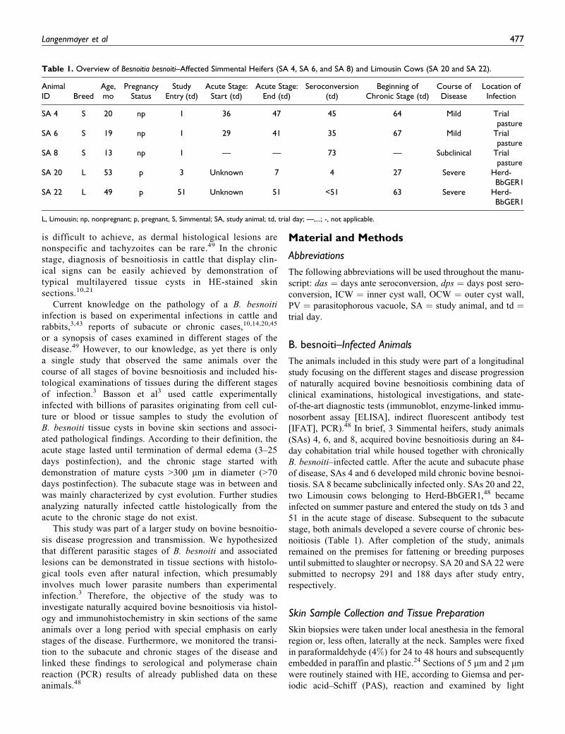
Table 1. Overview of Besnoitia besnoiti–Affected Simmental Heifers (SA 4, SA 6, and SA 8) and Limousin Cows (SA 20 and SA 22).
AnimalID Breed
Age,mo
PregnancyStatus
StudyEntry (td)
Acute Stage:Start (td)
Acute Stage:End (td)
Seroconversion(td)
Beginning ofChronic Stage (td)
Course ofDisease
Location ofInfection
SA 4 S 20 np 1 36 47 45 64 Mild Trialpasture
SA 6 S 19 np 1 29 41 35 67 Mild Trialpasture
SA 8 S 13 np 1 — — 73 — Subclinical Trialpasture
SA 20 L 53 p 3 Unknown 7 4 27 Severe Herd-BbGER1
SA 22 L 49 p 51 Unknown 51 <51 63 Severe Herd-BbGER1
L, Limousin; np, nonpregnant; p, pregnant, S, Simmental; SA, study animal; td, trial day; —,...; -, not applicable.
Langenmayer et al 477
microscopy. Selected skin sections of SA 6, SA 20, and SA 22
taken during subacute and chronic stages were also stained with
Masson’s trichrome stain and picrosirius red/Alcian blue.
SA 4, SA 6, and SA 8 were sampled twice a week during the
84-day trial. After entering the acute stage, SA 4 was sampled
every other day for 3 weeks. SA 6 was sampled daily for the
first week after entering the acute stage and every other day
in the following 2 weeks. Thereafter, SA 4 and SA 6 were
sampled twice a week again. SA 20 was sampled daily from the
day of admission for 3 weeks. Thereafter, SA 20 was sampled
once a week for the rest of the 84-day trial. Due to its unco-
operative behavior, SA 22 was sampled only on trial days 51,
56, 63, 70, and 80.
Tissues collected during necropsy were fixed, embedded,
cut, and stained as described above. Sections of claws were
decalcified for 1 week before plastic embedding.
Immunohistochemistry
Immunohistochemistry for detection of B. besnoiti zoites was
performed on skin sections of SA 4, SA 6, SA 8, SA 20, and
SA 22 and on selected parenchymatous organs of SA 20 and
SA 22 collected during necropsy. Briefly, sections of
paraformaldehyde-fixed, paraffin-embedded tissues were cut
at 5 mm. Primary antibody was a rabbit antiserum2 containing
polyclonal antibodies against B. besnoiti, used at a dilution of
1:2000. Tissues of experimentally infected g-interferon
knock-out mice21 comprising tachyzoites and skin of cattle
with confirmed bovine besnoitiosis, containing bradyzoites
within cysts, served as positive controls. An irrelevant primary
antibody (polyclonal rabbit anti–Escherichia coli, B0357;
Dako, Hamburg, Germany) used on tissue-containing parasites
served as a negative control. After deparaffinization and block-
ing with hydrogen peroxide, a blocking step using diluted nor-
mal goat serum for 30 minutes was applied. Primary antibody
incubation was performed for 60 minutes at room temperature.
Secondary antibody (biotinylated goat anti–rabbit immunoglo-
bulin, BA-1000; Vector, Burlingame, CA) incubation lasted for
45 minutes, followed by incubation with ABC-peroxidase (PK-
6100; Vector) and diaminobenzidine-hydrochloride as chromo-
gen (DAB, 4170; Biotrend, Cologne, Germany). Tissues were
counterstained with hematoxylin, dehydrated, and protected
by a coverslip. To exclude cross-reactions with other apicom-
plexan species, the anti–Besnoitia immunohistochemical stain-
ing was tested on mouse tissues containing T. gondii
tachyzoites, which had been made available from another study
on toxoplasmosis,25 and brain tissue from a cat containing T.
gondii bradyzoites (in both cases, T. gondii zoites had been
demonstrated in tissue sections via anti-Toxoplasma immuno-
histochemistry), as well as brain tissue from a cattle containing
N. caninum tachyzoites50 (dilution 1:1000) and brain tissue
from a dog containing N. caninum bradyzoites (in both cases,
N. caninum zoites had been demonstrated in tissue sections via
anti-Neospora immunohistochemistry), and muscle tissue
obtained from a cattle, necropsied at the Institute of Veterinary
Pathology, containing a not further characterized Sarcocystis
species.
Immunohistochemical staining for cellular markers was per-
formed on skin sections from the skin of SA 4, SA 6, SA 20,
and SA 22 obtained during the trial period. Briefly, sections
of paraformaldehyde-fixed, paraffin-embedded tissues were
cut at 5 mm. Primary antibody was a rabbit anti–CD3 antibody
(A045201; Dako) for detection of T lymphocytes, a mouse
anti–CD79a antibody (M7051; Dako) for detection of B lym-
phocytes, and a mouse anti–myeloid/histiocyte-antigen-
MAC387 antibody (M0747; Dako) for detection of histiocytes.
After deparaffinization, antigen retrieval was performed by
using Proteinase K incubation (MAC387) or a microwave
treatment (750 W, 2 � 10 minutes) with a Tris buffer at pH
9.0 (CD3, CD79a). Endogenous peroxidase was blocked with
hydrogen peroxide, followed by a blocking step using diluted
normal goat serum (CD3, CD79a) or normal rabbit serum
(MAC387) for 30 minutes. Primary antibody incubation was
performed for 60 minutes at room temperature. In the case of
CD3, secondary antibody was a biotinylated goat anti–rabbit
immunoglobulin (BA-1000; Vector), and secondary antibody
for CD79a was a biotinylated goat anti–mouse immunoglobu-
lin (E0433; Dako). Incubation lasted for 45 minutes, followed
by incubation with ABC-peroxidase (PK-6100; Vector) and
DAB as chromogen (4170; Biotrend). In the case of MAC387,
secondary antibody incubation lasted for 45 minutes with a rab-
bit anti–mouse immunoglobulin conjugated with peroxidase
(P0161; Dako), followed by DAB as chromogen (see above).
Tissues were counterstained with hematoxylin, dehydrated,
and protected with glass coverslips.
Measurement of Maximum Tissue Cyst Diameter
The maximum tissue cyst diameter (MTCD) was measured in
all plastic sections containing cysts of SA 4, SA 6, SA 20, and
SA 22. The number of sections measured per study animal and
the number of cysts in every measured section are depicted in
Fig. 13. Measurements were conducted by using a Videoplan
image analysis system (Zeiss-Kontron, Jena, Germany)
coupled to a light microscope (Orthoplan; Leitz, Stuttgart, Ger-
many) via a color video camera (CCTV WVCD132E; Mat-
sushita, Kadoma, Japan). Images of HE-stained sections were
displayed on a color monitor, and after calibration with an
object micrometer (Zeiss, Oberkochen, Germany), the profiles
of all tissue cysts in the sections were measured planimetrically
by circling their contours with a cursor on the digitizing tablet
of the image analysis system.
Serological Data
Data from a previous publication have been used to determine
B. besnoiti serological status.48 Four serological tests have been
performed as previously described47,48 by applying published
cutoffs.47,48 In IFAT, a positive cutoff titer of 1:200 was used;
in both immunoblots, recognition of at least 4 of 10 bands was
regarded positive.47 In the ELISA, a cut-off of Sample-to-
478 Veterinary Pathology 52(3)

Positive control ratio SP¼ 1.756 was applied.48 A positive ser-
oconversion was assumed when at least 3 of the 4 reference
tests were positive.48
Definitions
B. besnoiti parasites within skin sections were addressed as
zoites. Since there are no specific tools to unambiguously
identify tachyzoites selectively, zoites located within vessels
and/or intraendothelially were addressed as tachyzoite-like
endozoites.1,9,10,31 Zoites inside hypertrophied host cells with
distinguishable cyst wall(s) were addressed as brady-
zoites.1,9,10,13,31,40 As soon as a cyst wall was recognizable
in one of the used stains or in PAS reaction surrounding a
hypertrophied host cell containing a PV with bradyzoites, the
term tissue cyst was used. Developed cysts refer to tissue cysts
with only a small visible margin of host cell cytoplasm due to
a maximally enlarged PV containing bradyzoites.
The acute stage was classified according to clinical criteria.
Animals had to show fever (body temperature >39.0�C) or at
least 2 of the following clinical signs/diagnoses of acute bes-
noitiosis: depression, conjunctivitis, subcutaneous edema, lym-
phadenitis, or lameness.7,31,43,48 As soon as cysts were
clinically observed in the scleral conjunctivae, the term chronic
stage was used (Table 1). The subacute stage was defined as the
stage between the end of the acute and the beginning of the
chronic stage.
Results
In skin sections of all animals, there was a very mild basal
degree of eosinophilic and lymphoplasmacytic inflammation,
mainly located around small vessels and capillaries in the
superficial dermis and, if present, subcutis.
Other alterations that did not show correlations with known
besnoitiosis-derived lesions were demonstrable in varying
degrees in sections of all animals: sporadically occurring focal
epidermal edema and pigment incontinence as well as dilated
sweat glands, with the latter present in every section.
In tissue sections serving as positive controls for B. besnoiti,
immunohistochemical staining of the PV membrane and the
zoites displayed a brown positive signal, labeling membrane
and cytoplasm. Tissues in which an irrelevant primary antibody
was used as a negative control displayed no positive signals.
The polyclonal antiserum did not cross-react with T. gondii and
N. caninum tachyzoites and bradyzoites. A very faint signal
was observed with bradyzoites of Sarcocystis sp, which was
easily distinguishable from the strong signals B. besnoiti zoites
revealed.
Acute Stage
During the acute stage, all clinically affected animals showed
moderate dermal edema with separation of collagen fibers in
the papillary layer. In SA 4, dermal edema was detectable in
9 consecutive biopsies, starting 7 das; in SA 6, edema was
detectable in 10 consecutive biopsies, starting 2 das; and in
SA 20, edema was detectable in 7 consecutive biopsies, starting
1 das. SA 22 displayed dermal edema only on the day of admis-
sion (Fig. 1).
Accompanying the edema, multifocal to coalescing, perivas-
cular infiltrates containing moderate numbers of lymphocytes,
plasma cells, and eosinophils as well as a few macrophages were
demonstrable in all strata of the dermis in all clinically affected
animals (Fig. 4). SA 4 and SA 6 displayed low to moderate num-
bers of infiltrating inflammatory cells, whereas SA 20 and SA 22
displayed moderate to large numbers of infiltrating cells. Dermal
inflammation appeared simultaneously with dermal edema and
was detected in 6 consecutive biopsies in SA 4 and in 8 consec-
utive biopsies in SA 6 and SA 20. SA 22 displayed dermal
inflammation only on the day of admission (Fig. 1).
Multifocal to coalescing, mild to moderate, acute dermal
hemorrhages were detected in 6 consecutive biopsies in SA
4, starting 3 das; on 1 day (1 das) in SA 6; for 7 days in SA
20, starting 1 das; and on the day of admission and 5 days later
in SA 22.
In routine stains, tachyzoite-like endozoites were identified
only in HE plastic sections of SA 20 on the day of presentation
(1 das). Two to 5 sections of tachyzoite-like endozoites were
identified per PV, which was located inside endothelial cells
in the papillary layer of the dermis (Fig. 2). Longitudinally cut
tachyzoite-like endozoites displayed a crescent-like shape with
eosinophilic cytoplasm and a round basophilic nucleus.
Using immunohistochemistry, tachyzoite-like endozoites
were demonstrated in skin sections of SA 4, SA 6, SA 20, and
SA 22. Single or groups of up to 7 parasites were located inside
a PV within vascular endothelial cells mainly in the papillary
layer, whereas only a few were scattered in the reticular layer.
Within endothelial cells, closely spaced crescent-shaped
tachyzoite-like endozoites produced a typical banana-stalk pat-
tern (Fig. 3). Some zoites were also located in the neighbor-
hood of small vessels.
Immunohistochemically, tachyzoite-like endozoites were
demonstrable in 7 consecutive biopsies in SA 4 and SA 6, start-
ing 8 and 3 das, respectively. Tachyzoite-like endozoites were
demonstrable immunohistochemically for 6 days in SA 20 and
for 1 day in SA 22, starting on the day of admission (Fig. 1). In
skin sections of SA 4 and SA 6, only a few dermal tachyzoite-
like endozoites were detectable, whereas SA 20 and SA 22 dis-
played many tachyzoite-like endozoites per skin section.
Subacute and Chronic Stages
Edema and dermal infiltration with inflammatory cells sub-
sided a few days after the last immunohistochemical detection
of tachyzoite-like endozoites. Cyst formation was already initi-
alized during the acute stage, while tachyzoite-like endozoites
were still detectable (Fig. 1).
Tissue cyst evolution. The first evidence of beginning cyst forma-
tion was observed via immunohistochemistry. Hypertrophied
host cells possessed 1 or 2 round nuclei and displayed a diffuse tan
Langenmayer et al 479

labeling of the cytoplasm and intense brown labeling of the PV
with a single zoite. These hypertrophied host cells had no cyst
wall yet, as it was seen later in more evolved tissue cysts.
In HE sections, early tissue cysts consisted of hypertrophied
host cells with eosinophilic cytoplasm and 1 enlarged round
nucleus containing 1 to 3 nucleoli. The host cells in these young
cysts were surrounded by a thin, circular, gray-bluish layer with
irregular, lacerated borders, the ICW. The PV was small, taking
up about one-fifth of host cell cytoplasm, and contained only a
few bradyzoite sections with a crescent- or spindle-shaped
eosinophilic cytoplasm and 1 slightly darker nucleus per brady-
zoite (Fig. 5).
Tissue cyst evolution commenced by enlargement of the
host cell, which contained more hypertrophied round nuclei,
each with several nucleoli, and the enlargement of the PV,
which contained an increasing number of bradyzoites (Fig. 6).
The gray-bluish layer appeared thicker, still expressing an irre-
gular, lacerated outer border, and contained multiple small,
clear vacuoles. Especially in plastic sections stained with
Giemsa, this layer was colored brightly purple. In sections
stained with PAS reaction, the host cell cytoplasm was gray-
reddish and sometimes stained brightly PAS positive. The
ICW stained violet-reddish, and bradyzoites were gray-violet
with several small PAS-positive granules within the cytoplasm
of multiple bradyzoites. The ICW stained pale white in Mas-
son’s trichrome stain and blue-green in picrosirius red/Alcian
blue.
In skin sections of SA 20, taken 20 dps, extracystic parasites
were detected via immunohistochemistry in 1 location in the
papillary and in 1 location in the reticular layer of the dermis.
In the papillary layer, several zoites were grouped in a semicir-
cular shape in the periphery of a small vessel; a PV was not
detected. In the reticular layer, few zoites were detected intra-
luminally or associated with the vascular endothelium in a
small vessel (Fig. 7). Except for this single biopsy in SA 20,
extracystic zoites were not detected after initial intraendothelial
proliferation subsided.
Tissue cysts evolved further with consecutive enlargement
of the host cells; more host cell nuclei and nucleoli; a larger
PV, which still took up only a small part of host cell cytoplasm;
and more bradyzoite sections (Figs. 8, 9). Several rounded bod-
ies ranging from 3–4 mm to 11–12 mm, which stained green-
grayish in HE-stained sections and brightly positive in PAS
reaction, were observed within the PV 38 dps in SA 6, 27 dps
Figure 1. Chronology of histological skin lesions in study animal (SA) 4, SA 6, SA 20, and SA 22. The vertical line depicts day of seroconversion(0) for SA 4, SA 6, and SA 20 and day of admission for SA 22. The x-axis shows the days before (with negative signs) and after seroconversion (S4,SA 6, and SA 20) and day of admission (SA 22).
480 Veterinary Pathology 52(3)

in SA 20, and 29 days after admission in SA 22. In biopsies
taken on the following days and weeks, these bodies were occa-
sionally observed within the PV.
Rarely host cells contained 2 PVs, which were separated by
a thin septum of the host cell cytoplasm.
Multiplication of host cell nuclei occurred via endomitosis,
producing up to 12 large bizarre mitoses within the host cell
cytoplasm. Host cell nuclei showed marked anisokaryosis with
a wide array of shapes ranging from simple round or spindle
shape to large bizarre nuclei with multiple foldings and invagi-
nations of the nuclear membrane. A maximum of 17 nuclear
profiles was counted in a single tissue cyst, containing up to
6 nucleoli per nucleus.
In the periphery of the ICW, a secondary ivory-colored
layer, the OCW, began to develop in SA 6, SA 20, and SA
22. Formation of the OCW started with a brightening of the
gray-bluish (HE) or violet (Giemsa) ICW or blueing of the pale
(Masson’s trichrome) ICW from the periphery. In some sec-
tions, a patchy pattern of brightening was observed, emphasiz-
ing the lacerated borders of the ICW. The OCW was slightly
eosinophilic or ivory colored in HE-stained sections; pale
white, glassy, and nearly translucent in Giemsa-stained sec-
tions; and blue in Masson’s trichrome stain. In sections stained
with picrosirius red/Alcian blue, the OCW stained red with an
orange-colored birefringence in polarized light. A clearly visi-
ble OCW was present 35 dps in SA 6, 30 days after detection of
first cysts; 31 dps in SA 20, 29 days after detection of first
cysts; and 29 days after admission in SA 22, 24 days after
detection of first cysts, before cysts were fully developed. In
SA 4, an OCW was not detected.
Cysts were fully developed 49 dps in SA 6, 44 days after
first cyst detection, and 34 dps for SA 20, 32 days after first cyst
detection. Developed cysts were not detected in skin sections of
SA 4 and SA 22 during the first 84 days of the study. Developed
Figures 2–7. Besnoitia besnoiti infection, skin, bovine; study animal (SA) 20. Figure 2. One day ante seroconversion (das). Two B. besnoititachyzoite-like endozoites inside a parasitophorous vacuole (PV) within a vascular endothelial cell. Bar ¼ 25 mm. Hematoxylin and eosin(HE). Figure 3. One das. Multiple B. besnoiti tachyzoite-like endozoites within a vascular endothelial cell, producing a typical banana-stalk pat-tern. Same magnification as Fig. 2. Anti-Besnoitia immunohistochemistry with hematoxylin counterstain. Figure 4. Day of seroconversion.Edema with moderate, perivascular inflammation. HE. Figure 5. Four days post seroconversion (dps). Very young B. besnoiti tissue cyst withhypertrophied host cell and host cell nucleus, PV with 2 bradyzoites, and surrounding inner cyst wall (arrow). Same magnification as Fig. 2.HE. Figure 6. Twenty dps. B. besnoiti tissue cyst with multiple bradyzoites within a PV. Bar ¼ 25 mm. Anti-Besnoitia immunohistochemistry withhematoxylin counterstain. Figure 7. Twenty dps. Few B. besnoiti zoites within a small vessel in the reticular layer. Same section as Fig. 6, samemagnification as Fig. 2. Anti-Besnoitia immunohistochemistry with hematoxylin counterstain.
Langenmayer et al 481

cysts contained a maximally enlarged, central PV and only a small
rim of the host cell cytoplasm surrounding the PV. The host cell
nuclei were located in the periphery of the host cell cytoplasm and
were flattened. The ICW surrounded the host cell membrane like
a thin band. In some sections, the ICW was only merely visible
and even sometimes missing. The OCW consisted of several thick
eosinophilic laminae surrounding the host cell, with the outermost
lamina showing an irregular, lacerated shape blending into the
surrounding connective tissue (Figs. 10, 11).
Distribution of tissue cysts. Tissue cysts in the skin were predomi-
nantly randomly distributed in the papillary layer, in the direct
neighborhood of loose connective tissue. In many sections, tis-
sue cysts were closely arranged together, resulting in coales-
cing OCWs. Some cysts were detected directly beneath the
epidermis, bulging it outward or in the dermal sheath of a hair
follicle compressing the outer and inner root sheath of the hair
follicle. Intraepithelial cysts were not detected. Only a few
cysts were located in the reticular layer and the subcutis,
appearing to be lying directly in the periphery of small vessels
and loose connective tissue. Some tissue cysts were present in
the tunica media of larger vessels, bulging the tunica intima
inward into the lumen.
As in the case of tachyzoite-like endozoites, many more tis-
sue cysts were detected in skin sections of SA 20 and SA 22
compared with SA 4 and SA 6, which displayed only a few
cysts.
Pericystic inflammation. The immune system of SA 4 and SA 6
already responded to undeveloped tissue cysts. Immune
Figures 8–12. Besnoitia besnoiti infection, skin, bovine; study animal (SA) 20. Figure 8. Twenty-four days post seroconversion (dps). B. besnoititissue cyst with a single parasitophorous vacuole (PV), multiple bradyzoites, host cell nuclei and nucleoli, hypertrophied host cell, and surround-ing inner cyst wall (ICW). Bar ¼ 25 mm. Hematoxylin and eosin (HE). Figure 9. Twenty-four dps. Same cyst as in Fig. 8. Note: Bright purplestaining of the ICW. Same magnification as Fig. 8. Giemsa stain. Figure 10. Day of necropsy. Developed B. besnoiti tissue cyst with maximallyenlarged PV, numerous bradyzoites (inset: d), small rim of host cell cytoplasm with flattened host cell nuclei (inset: c), thin rim of the ICW (inset:b), and thick outer cyst wall (OCW; inset: a). Inset: Close-up of the different cyst layers. Same magnification as Fig. 12. HE. Figure 11. Day ofnecropsy. Same cyst as in Fig. 10. Inset: Close-up of the different cyst layers; same captions as in Fig. 10. Note: Thin rim of the purple ICW (inset:b) and pale white, nearly translucent OCW (inset: a). Same magnification as Fig. 12. Giemsa stain. Figure 12. Day of necropsy. Lysed B. besnoiticyst with necrotic inner structures and many macrophages, multinucleated giant cells, lymphocytes, and eosinophils in close proximity. Note:Thinning and breaching site of the OCW at the top and cyst-oriented macrophages and multinucleated giant cells directly adjacent to the OCW.Bar ¼ 50 mm. HE.
482 Veterinary Pathology 52(3)
response to cysts in skin sections of SA 20 started after
cysts were fully developed. In SA 22, pericystic infiltrates
were not found in all 4 biopsies containing tissue cysts. First
signs of inflammation surrounding cysts consisted of peri-
cystic infiltration with few lymphocytes, eosinophils, and
macrophages. In more pronounced pericystic inflammation,
especially the numbers of eosinophils and lymphocytes
increased. In severe pericystic inflammation, infiltrates con-
sisted of many lymphocytes, fewer eosinophils and macro-
phages, and a few multinucleated giant cells of foreign
body type (Fig. 12). Macrophages and giant cells adjusted
their cytoplasm toward the OCW, displaying a ruffled cyto-
plasmic border on the cyst side. Tissue cysts without any
signs of pericystic infiltrates coexisted adjacent to cysts
with mild to marked pericystic inflammation. Immunohisto-
chemistry revealed that pericystic lymphocytes consisted
predominantly of T lymphocytes with only a few B lympho-
cytes scattered in between. The host cell cytoplasm of
developed cysts tested negative for CD3, CD79a, and mye-
loid/histiocyte-antigen (MAC387).
Occasionally, lysis of tissue cysts occurred when pericys-
tic inflammation was breaching the hyaline layer (Fig. 12).
Some lysed cysts still contained necrotic debris with only few
histiocytic infiltrations; other cysts were completely infiltrated
with many histiocytes, eosinophils, and few multinucleated
giant cells. In many cases, the OCW was partially intact
and displayed a breaching site of variable dimension. Immu-
nohistochemically, intact bradyzoites were not detected in
such cysts, but brown granular fragments were observed in
the necrotic debris and within the cytoplasm of histiocytes.
The first lysis of tissue cysts was observed 25 dps in SA 4
and 59 dps in SA 6 and SA 20.
In skin sections of SA 8, lesions of acute besnoitiosis
(intraendothelial or intravascular tachyzoite-like endozoites,
interstitial edema, hemorrhages, or increased perivascular
inflammation) were not observed in routine stains and immu-
nohistochemistry in 4 consecutive skin biopsies before and
after seroconversion. Lesions associated with chronic
besnoitiosis-like tissue cysts or multifocal granulomatous
inflammation were not observed in routine stains and immuno-
histochemistry in skin biopsies taken after seroconversion.
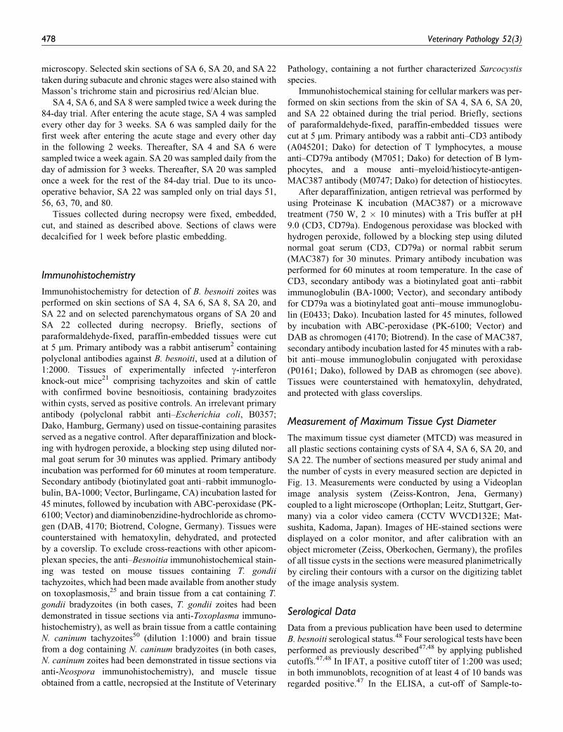
Tissue cyst size. The MTCDs were determined and means calcu-
lated for the individual animals and the different time points
(Fig. 13). During tissue cyst evolution, mean MTCDs became
progressively larger. The smallest MTCD measured out of all
animals was 29.3 mm (SA 6, 11 dps), and the largest was
409.9 mm (SA 20, 73 dps).
Figure 13. Aligned dot plot of the chronology of mean maximal cyst diameters in plastic skin sections of study animal (SA) 4, SA 6, SA 20, and SA22. Each replicate represents the diameter of the cyst(s) measured on the respective day. The connecting line represents mean. The vertical linesdepict day of seroconversion for SA 4, SA 6, and SA 20 and day of admission for SA 22.
Langenmayer et al 483

Necropsy results. At necropsy, the body condition score15 of SA
20 and SA 22 was 2.5 and 2.25, respectively. The skin was dif-
fusely thickened and seemed less elastic than normal, display-
ing folds and wrinkles especially in the neck region. The
peripheral lymph nodes were enlarged. Multiple, dissemi-
nated, miliary white foci, resembling B. besnoiti cysts, were
observed in the subcutis; musculature; fasciae of the head,
neck, trunk, and limbs; mucosa of the entire upper respiratory
tract; bronchi; vagina; and scleral conjunctivae (Figs. 14, 15).
Single cysts were found in the endocardium of the right atrium
and in the splenic capsule in SA 20 and in the veins, especially
of the distal limbs, in both animals. Moderate to severe rota-
tion of distal phalanges was present in the claws (Fig. 17). The
laminar corium was necrotic in several claws; in single ones,
necrotizing inflammation spread to the bone of the distal pha-
lanx, resulting in necrotizing osteomyelitis. In the reticuloru-
men of SA 22, multiple Paramphistomum spp were feeding on
the mucosa.
In both animals, developed cysts—many of them with gray-
greenish rounded bodies within the PV—with or without
pericystic, chronic lymphoplasmacytic, eosinophilic, and gran-
ulomatous inflammation were found in a variety of tissues (see
below). In tissues exposed to mechanical forces (fascia, skele-
tal muscle, laminar corium of the claws), the round structure of
the tissue cysts displayed impressions, and many cysts showed
a more elongated form. Especially in the fasciae, cysts were
surrounded by a layer of fibrocytes and vessels directly adja-
cent to the OCW, which were arranged in a circular fashion
around the cysts (Fig. 16).
In the case of SA 20, the lamellae of the laminar corium of
the claws were diffusely infiltrated by moderate numbers of
lymphocytes, plasma cells, and eosinophils directly beneath the
epidermis. In multiple locations, the horn appeared degenerate,
and islets of nuclear fragments (possibly neutrophils) were
present. In some locations, epidermal infiltration of the inflam-
matory cells was observed with epidermal edema and keratino-
cyte necrosis. In both animals, large numbers of B. besnoiti
cysts were observed in the laminar corium and the sole corium,
leading to a distention of the papillae and lamellae of the cor-
ium. A deviation or displacement of the infoldings of the
Figures 14–18. Besnoitia besnoiti infection; bovine. Figure 14. Scleral conjunctiva, study animal (SA) 22 (day of necropsy). Multiple B. besnoiticysts. Figure 15. Fascia and muscle, SA 22 (day of necropsy). Multiple B. besnoiti cysts in the fascia and muscle of the left hindlimb. Figure 16.Fascia, SA 22 (day of necropsy). Histological section of a tissue cyst within fascia with surrounding fibrovascular tissue. Hematoxylin and eosin(HE). Figure 17. Claw, SA 20 (day of necropsy). Rotation of the lateral third phalanx of the left forelimb due to chronic laminitis. Figure 18.Claw, SA 20 (day of necropsy). Multiple B. besnoiti cysts within the lamellae of the laminar corium, causing distention of the lamellae and deviationof the laminar epidermis. Note: Diffuse, subepidermal inflammation. HE.
484 Veterinary Pathology 52(3)

laminar epidermis, especially the lamellae of the stratum
lamellatum, occurred simultaneously (Fig. 18).
B. besnoiti tissue cysts were observed only within connec-
tive tissue and never inside epithelial layers. Extracystic zoites
were not detected in parenchymatous organs of either animal
via immunohistochemistry.
Sections of the following tissues contained cysts: skin, sub-
cutis, fasciae, muscles, scleral conjunctivae, sclera, nictitating
membrane, iris, nasal mucosa, trachea, bronchi, lung, medul-
lary cavity of nasal bones, oral mucosa, salivary glands, vagina,
capsule of abdominal hemal lymph node, and small and large
vessels. Sections of the following tissues were free of tissue
cysts: brain, meninges, thymus, thyroid glands, gastrointestinal
tract, liver, pancreas, spleen, kidneys, adrenal glands, uterus,
urinary bladder, lens, and cartilage.
Discussion
In this study, cases of spontaneous bovine besnoitiosis in 2 hei-
fers and 2 cows were examined from the acute to chronic stage
of infection by histological and immunohistological examina-
tions using paraffin- and plastic-embedded tissues. The high-
frequency sampling enabled documentation from the beginning
of the disease involving circulating tachyzoite-like endozoites,
up to tissue cyst evolution in subacute and chronic stages. To
our knowledge, this is the first study conducted on naturally
infected cattle over a long period.
There are several important differences between the find-
ings of this study and pathological observations already
published.3,35,49 For the first time, a Besnoitia-specific immu-
nohistological approach has been used to detect circulating
B. besnoiti tachyzoite-like endozoites, which enabled the
detection of single tachyzoite-like endozoites during the acute
stage, therefore making identification of mild cases possible
and enabling detection of extracystic proliferating zoites during
the cyst stage as well. Further identification of these zoites as
tachyzoites was not conducted, as specific antibodies to selec-
tively identify B. besnoiti tachyzoites are lacking, a point that
should be addressed in future studies. Furthermore, we
described an additional cyst wall layer, resulting in the distinc-
tion of 2 cyst wall layers, even in developed cysts. In addition,
for the first time, large numbers of tissue cysts were detected in
the corium of the claws, which was obviously responsible for
the occurrence of a chronic laminitis.
During ongoing chronic besnoitiosis, B. besnoiti zoites are
usually found only within a PV inside tissue cysts,3,14 since
extracystic proliferation ceases after initial intraendothelial
proliferation.8 Extracystic, intravascular B. besnoiti zoites dur-
ing ongoing chronic besnoitiosis have been described by
McCully et al,36 but they could not fully exclude artificial
translocation of bradyzoites into vascular lumina by cutting
fresh tissues for histological examination. Schulz49 reported
that bradyzoites, freed from tissue cysts after rupture, can be
easily distinguished from artificially translocated ones by the
presence of a cellular reaction. Lysis of B. besnoiti tissue cysts
has been observed in this study as well. However, close to these
cysts, no released bradyzoites were observed, and either the
content of the cyst was necrotic or the lumen of the cyst was
completely filled with cellular infiltrates. In our opinion, it is
unlikely that bradyzoites could reach the bloodstream after the
lysis of a tissue cyst caused by the cellular immune response
because they would have to pass a wall of inflammatory cells.
However, intravascular circulation of zoites during the chronic
stage may occur after mechanical rupture of tissue cysts lying
directly beneath a vascular endothelium or by reactivation of
tissue cysts and stage conversion to tachyzoites. Whether the
latter occurs in vivo is unknown.9,14 However, bradyzoites can
serve as an infectious agent to produce acute besnoitiosis (and
later cyst formation in cattle), as demonstrated in the artificial
infection of cattle7 and laboratory animals.2,46 Reactivation of
chronic rodent besnoitiosis is possible, since it has been
demonstrated via experimentally induced immunosuppression
in hamsters infected with Besnoitia jellisoni.18
Both necropsied animals revealed large numbers of tissue
cysts in the laminar corium as well as signs of chronic laminitis.
The pathogenesis of bovine laminitis is incompletely under-
stood.22,32,51 However, altered digital blood flow seems to be
one of many contributing factors in the development of lamini-
tis.23,32,51 An attempt to explain equine contralateral limb lami-
nitis evolves around local congestion, slow blood flow, platelet
activation, and formation of microthrombi, resulting in inade-
quate perfusion and ischemia of the lamellar tissue.32 During
the acute stage of bovine besnoitiosis, vascular changes, such
as congestion, endothelial injury, edema, and thromboses, are
common findings,3 and reluctance to move and stiff gait are
frequently described in diseased animals.9,31 Combining this
information, it is very likely that cattle with acute besnoitiosis
display lameness because of acute or subacute laminitis.23 In
chronic besnoitiosis, the presence of tissue cysts within vessel
walls of the limbs and within the corium most likely contributes
to the development of laminitis. Reduced digital blood flow or
localized digital congestion is easy to explain through the pres-
ence of multiple tissue cysts in the vessel walls of the distal
limbs. Local ischemia may occur because growing and devel-
oped tissue cysts compress arteries and capillaries. The multi-
focal pericystic inflammation may contribute to laminitis as
well.32 Ischemia, compression, and deviation of epithelial
lamellae may subsequently lead to production of a horn of
minor quality, predisposing the claw to ascending bacterial
infection and resulting in sole ulcers or necrotizing laminitis.
Basson et al3 measured maximum cyst diameter after
experimental infection of cattle with B. besnoiti; the largest
cyst they measured was 390 mm in diameter 77 days postinfec-
tion and 66 days after the first appearance of early tissue cysts.
In SA 20, the largest cyst (almost 410 mm) was measured 71
days after detection of first cysts. Comparing Basson and oth-
ers’ data with those of the animals in this study, there is a slight
deviation by a few days, but the time intervals are similar. The
difference in this study between detection of first cysts and first
measurement occurred because first cysts were observed in
paraffin sections, and measurements have been conducted only
in plastic sections to minimize the effect of shrinking artifacts.
Langenmayer et al 485

A real-time PCR conducted on the skin samples of SA 4, SA 6,
SA 20, and SA 22 displayed a decrease in cycle threshold (Ct)
values shortly after seroconversion, following a transient
increase in Ct values.48 This suggests a period of increased
parasite load within the skin,48 which coincides with the
increase of tissue cyst diameters in this study. Thus, it is very
likely that the previously observed decrease in the Ct values
reflected the intracystic growth of B. besnoiti bradyzoites.
Tissue cysts of Besnoitia species differ structurally from
cysts of other apicomplexan species. In the literature, nomen-
clature regarding the composition of Besnoitia tissue cysts and
cysts walls is not consistent. None of the published
schemes13,26,41,42 were fully applicable when looking at B. bes-
noiti tissue cyst development in this study. Therefore, a new
nomenclature that is fully applicable to B. besnoiti tissue cysts
is suggested: structures within the host cell are called by their
respective name—that is, bradyzoites, parasitophorous
vacuole, membrane of the parasitophorous vacuole, host cell
cytoplasm including organelles and nuclei, and host cell mem-
brane. For the outermost acellular layer, the term outer cyst
wall is proposed. And for the cyst wall of undeveloped B. bes-
noiti tissue cysts, which, in developed cysts, lies between the
OCW and the membrane of the host cell, the term inner cyst
wall is proposed. The whole hypertrophied host cell, including
the ICW and, if present, the OCW, is called a tissue cyst.
Interestingly, authors studying developed B. besnoiti tissue
cysts via light or transmission electron microscopy do not
report an ICW or layer between the membrane of the host cell
and the OCW.13,35,37 However, in light microscopic pictures,
the ICW can be seen as a blue37 or slightly eosinophilic layer.35
Perhaps this layer was interpreted as artifact, occurring after
processing of tissues for histological examination. Authors
studying B. besnoiti tissue cyst development report an ICW
directly on the outside of the host cell: a blue layer outside of
the host cell, interpreted as a developing cyst wall3 and a homo-
geneous capsule enclosing the host cell, staining basophilic in
Giemsa-stained sections.49 Later on they describe that the cyst
wall has transitioned into a hyaline49 or reddish appearance.3
The OCW has been studied in several publications with
transmission electron microscopy, identifying it as multiple
layers of collagen fibrils, arranged in a circular fashion.13,35,37
Dubey et al14 studied young B. besnoiti tissue cysts in 1
infected bull via electron microscopy, describing the outer
layer as a halo with connective tissue elements. The nature of
connective tissue elements surrounding the cysts was not
reported.
The collagenous nature of the OCW13,35,37 resulted in a blue
staining with Masson’s trichrome stain and a red staining with
orange-colored birefringence in polarized light in picrosirius
red/Alcian blue stain, with the latter suggesting that the OCW
is made up of collagen type I fibers.
The ICW, however, did stain pale white with Masson’s tri-
chrome stain and blue-green in picrosirius red/Alcian blue,
accentuating its different nature. According to the staining
reaction of the ICW in the Giemsa and picrosirius red/Alcian
blue stain, the ICW is most likely made up of elements of the
extracellular matrix (ECM), since other ECM elements (eg,
proteoglycans or glycosaminoglycans) show similar staining
with the Giemsa or Alcian blue stain as well. This ECM possi-
bly serves as template for the deposition of the collagen fibrils
of the OCW. The reason why some developed cysts do not have
an ICW may be due to a maximal amount of collagen fibrils
deposited within this matrix, connecting the OCW to the host
cell membrane. This theory is corroborated by a recent immu-
nohistochemical study on B. besnoiti cysts, suggesting myofi-
broblasts as host cells during cyst stage of B. besnoiti,14 since
myofibroblasts are capable of producing both abundant ECM
elements and different types of collagen, including collagen
type I.27,33
The distribution of tissue cysts within the skin and within the
whole body is striking. In skin, cysts are more frequently
observed in the papillary layer than in the reticular layer.
McCully et al36 suggest this is due to the better vascularization
of the papillary layer. They also report that the presence of B.
besnoiti tissue cysts is associated with small vessels, which is in
accordance with the observations in this study, where tissue
cysts were noted in the reticular layer of the skin. However, our
own observations and that of others show that highly vascular-
ized tissues such as brain, heart, spleen, kidney, or liver do not
harbor any or very seldom contain singular cysts.31,35,36,43
Furthermore, nonintestinal mucosae, fasciae, and subcutaneous
and intermuscular loose connective tissue contain large num-
bers of cysts.20,35,36 The reason for this distribution pattern is
unknown and is often associated with theories of transmission.
Shedding of zoites from ruptured cysts in respiratory mucosa to
water, grass, or soil29 and a vertical transmission of parasites
found within female genital organs39 have been suggested.
Cysts within the musculoskeletal system make a carnivorous
definitive host possible, but several feeding experiments could
not determine a definitive carnivorous host.2,12,44 Superficial
dermal cysts favor transmission via direct contact, insects, or
inanimate vectors; this mode of transmission has already been
shown experimentally.5,7,43
Myofibroblasts, recently suggested as host cells in chronic
disease,14 play a key role in physiological tissue repair, ECM
deposition, and wound contraction.11,28,33 In the acute stage
of bovine besnoitiosis, dermal inflammation inevitably leads
to local cytokine release and changes in ECM elements, which
are factors for promotion of myofibroblast differentiation.27 A
promotion of myofibroblasts during acute and subacute stages
and subsequent infestation of those cells may be another possi-
ble explanation for the distribution pattern of B. besnoiti cysts.
The current study is the first to use skin biopsies sampled
with high frequency to monitor histological lesions caused by
B. besnoiti after natural infection. The benefits of this study are
the completion of the fragmentary data of morphology of nat-
ural bovine besnoitiosis disease evolution, the characterization
of tachyzoite-like endozoites using immunohistochemistry dur-
ing the acute stage, and the detection of extracystic zoites dur-
ing the ongoing chronic stage, suggesting reactivation of B.
besnoiti tissue cysts. Furthermore, for the first time, the results
depicted here show that lesions of severe chronic besnoitiosis
486 Veterinary Pathology 52(3)
are correlated with laminitis, which is an important factor for
determining the prognosis of severely affected animals.
Acknowledgements
We acknowledge the excellent technical assistance of Teresa Hen-
neke, Elisabeth Kemper, Doris Merl, Michaela Nuetzel, and Heike
Sperling. We would like to thank Dr. Walter Basso from Institute of
Parasitology, Vetsuisse-Faculty University of Zurich for providing the
rabbit antiserum for immunohistochemistry. Further we would like to
express our gratitude to the owner of Limousin herd Herd-BbGER1
for his excellent collaboration and support over the years.
Declaration of Conflicting Interests
The author(s) declared no potential conflicts of interest with respect to
the research, authorship, and/or publication of this article.
Funding
The author(s) disclosed receipt of the following financial support for
the research, authorship and/or publication of this article: The study
was financially supported by Prionics AG, Schlieren, Switzerland.
NSG was supported by the BGF research stipend provided by the Ger-
man state of Bavaria.
References
1. Alvarez-Garcıa G, Frey CF, Ortega Mora LM, et al. A century of
bovine besnoitiosis: an unknown disease re-emerging in Europe.
Trends Parasitol. 2013;29(8):407–415.
2. Basso W, Schares G, Gollnick NS, et al. Exploring the life cycle
of Besnoitia besnoiti: experimental infection of putative defini-
tive and intermediate host species. Vet Parasitol. 2011;178(3–
4):223–234.
3. Basson PA, McCully RM, Bigalke RD. Observations on the
pathogenesis of bovine and antelope strains of Besnoitia besnoiti
(Marotel, 1912) infection in cattle and rabbits. Onderstepoort J
Vet Res. 1970;37(2):105–126.
4. Besnoit C, Robin V. Les lesions de la sarcosporidiose cutanee des
bovins dans leurs rapports avec l’histogenese du tubercule. Rev
Vet. 1914;71:193–207.
5. Bigalke RD. The artificial transmission of Besnoitia besnoiti
(Marotel, 1912) from chronically infected to susceptible cattle
and rabbits. Onderstepoort J Vet Res. 1967;34(2):303–316.
6. Bigalke RD. Besnoitiosis and globidiosis. In: Ristic M, McIntyre
I, eds. Diseases of Cattle in Tropics. The Hague, the Netherlands:
Martinus Nijhoff; 1981:429–442.
7. Bigalke RD. New concepts on the epidemiological features of
bovine besnoitiosis as determined by laboratory and field investi-
gations. Onderstepoort J Vet Res. 1968;35(1):3–137.
8. Bigalke RD. The present concept of the life cycle of Besnoitia
besnoiti of cattle. J Parasitol. 1970;56(4):29–30.
9. Bigalke RD, Prozesky L. Besnoitiosis. In: Coetzer JAW, Tustin
RC, eds. Infectious Diseases of Livestock. 2nd ed. Oxford, UK:
Oxford University Press; 2004:351–359.
10. Cortes H, Leitao A, Vidal R, et al. Besnoitiosis in bulls in Portu-
gal. Vet Rec. 2005;157(9):262–264.
11. Desmouliere A, Chaponnier C, Gabbiani G. Tissue repair, con-
traction, and the myofibroblast. Wound Repair Regen. 2005;
13(1):7–12.
12. Diesing L, Heydorn AO, Matuschka FR, et al. Besnoitia besnoiti:
studies on the definitive host and experimental infections in cattle.
Parasitol Res. 1988;75(2):114–117.
13. Dubey JP, Shkap V, Pipano E, et al. Ultrastructure of Besnoitia
besnoiti tissue cysts and bradyzoites. J Eukaryot Microbiol.
2003;50(4):240–244.
14. Dubey JP, Wilpe E, Blignaut DJC, et al. Development of early tis-
sue cysts and associated pathology of Besnoitia besnoiti in a natu-
rally infected bull (Bos taurus) from South Africa. J Parasitol.
2013;99(3):459–466.
15. Edmonson AJ, Lean IJ, Weaver LD, et al. A body condition scor-
ing chart for Holstein dairy cows. J Dairy Sci. 1989;72(1):68–78.
16. European Food Safety Authority (EFSA). Bovine besnoitiosis: an
emerging disease in Europe. EFSA J. 2010;8(2):1499.
17. Fernandez-Garcıa A, Alvarez-Garcıa G, Risco-Castillo V, et al.
Pattern of recognition of Besnoitia besnoiti tachyzoite and brady-
zoite antigens by naturally infected cattle. Vet Parasitol. 2009;
164(2–4):104–110.
18. Frenkel JK, Wilson HR. Effects of radiation on specific cellular
immunities: besnoitiosis and a herpesvirus infection of hamsters.
J Infect Dis. 1972;125(3):216–230.
19. Frey CF, Gutierrez-Exposito D, Ortega-Mora LM, et al. Chronic
bovine besnoitiosis: Intra-organ parasite distribution, parasite
loads and parasite-associated lesions in subclinical cases. Vet
Parasitol. 2013;197(1–2):95–103.
20. Gentile A, Militerno G, Bassi P, et al. Su di un episodio di besnoi-
tiosi bovina in Italia. Buiatria J Ital Ass Buiatrics. 2010;5(1):
3–15.
21. Gentile A, Militerno G, Schares G, et al. Evidence for bovine
besnoitiosis being endemic in Italy: first in vitro isolation of
Besnoitia besnoiti from cattle born in Italy. Vet Parasitol. 2012;
184(2–4):108–115.
22. Ginn PE, Mansell JEKL, Rakich PM. Skin and appendages. In:
Maxie MG, eds. Jubb, Kennedy, and Palmer’s Pathology of
Domestic Animals. 5th ed. 2007.
23. Greenough PR. The laminitis syndrome. In: Bergsten C, Brizzi A,
Muelling CKW, eds. Bovine Laminitis and Lameness. Philadel-
phia, PA: Saunders Elsevier; 2007:36–54.
24. Hermanns W, Liebig K, Schulz LC. Postembedding immunohis-
tochemical demonstration of antigen in experimental polyarthritis
using plastic embedded whole joints. Histochemistry. 1981;73(3):
439–446.
25. Herrmann DC, Barwald A, Maksimov A, et al. Toxoplasma gondii
sexual cross in a single naturally infected feline host: generation of
highly mouse-virulent and avirulent clones, genotypically differ-
ent from clonal types I, II and III. Vet Res. 2012;43(1):39.
26. Heydorn AO, Senaud J, Mehlhorn H, et al. Besnoitia sp. from
goats in Kenya. Z Parasitenkd. 1984;70(6):709–713.
27. Hinz B. Formation and function of the myofibroblast during tissue
repair. J Invest Dermatol. 2007;127(3):526–537.
28. Hinz B, Phan SH, Thannickal VJ, et al. The myofibroblast: one
function, multiple origins. Am J Pathol. 2007;170(6):1807–1816.
29. Hofmeyr CFB. Globidiosis in cattle. J S Afr Vet Med Assoc. 1945;
16:102–109.
30. Hornok S, Fedak A, Baska F, et al. Bovine besnoitiosis emerging in
Central-Eastern Europe, Hungary. Parasit Vectors. 2014;7(1):20.
Langenmayer et al 487

31. Jacquiet P, Lienard E, Franc M. Bovine besnoitiosis: epidemiolo-
gical and clinical aspects. Vet Parasitol. 2010;174(1–2):30–36.
32. Katz LM, Bailey SR. A review of recent advances and current
hypotheses on the pathogenesis of acute laminitis. Equine Vet J.
2012;44(6):752–761.
33. Klingberg F, Hinz B, White ES. The myofibroblast matrix: impli-
cations for tissue repair and fibrosis. J Pathol. 2013;229(2):
298–309.
34. Lesser M, Braun U, Deplazes P, et al. First cases of besnoitiosis in
cattle in Switzerland [in German]. Schweiz Arch Tierheilkd. 2012;
154(11):469–474.
35. Majzoub M, Breuer W, Gollnick NS, et al. Ein Ausbruch von Bes-
noitiose bei Rindern in Deutschland: pathomorphologische, ultra-
strukturelle und molekularbiologische Untersuchungen. Wien
Tieraerztl Mschr. 2010;97:9–15.
36. McCully RM, Basson PA, Van Niekerk JW, et al. Observations on
Besnoitia cysts in the cardiovascular system of some wild ante-
lopes and domestic cattle. Onderstepoort J Vet Res. 1966;33:
245–276.
37. Mehlhorn H, Klimpel S, Schein E, et al. Another African disease
in Central Europa: besnoitiosis of cattle. I. Light and electron
microscopical study. Parasitol Res. 2009;104(4):861–868.
38. Nobel TA, Klopfer U, Perl S, et al. Histopathology of genital
besnoitiosis of cows in Israel. Vet Parasitol. 1981;8(4):
271–276.
39. Nobel TA, Neumann M, Klopfer U, et al. Kystes de Besnoitia bes-
noiti dans les organes genitaux de la vache. Bull Acad Vet de
France. 1977;50:569–574.
40. Olias P, Schade B, Mehlhorn H. Molecular pathology, taxonomy
and epidemiology of Besnoitia species (Protozoa: Sarcocystidae).
Infect Genet Evol. 2011;11(7):1564–1576.
41. Oryan A, Azizi S. Ultrastructure and pathology of Besnoitia
caprae in the naturally infected goats of Kerman, east of Iran.
Parasitol Res. 2008;102(6):1171–1176.
42. Oryan A, Namazi F, Silver IA. Histopathologic and ultrastructural
studies on experimental caprine besnoitiosis. Vet Pathol. 2011;
48(6):1094–1100.
43. Pols JW. Studies on bovine besnoitiosis with special reference to
the aetiology. Onderstepoort J Vet Res. 1960;28:265–356.
44. Rommel M. New knowledge on the biology of Coccidia, Toxo-
plasma, Sarcosporidia and Besnoitia [in German]. Berl Muench
Tieraerztl Wochenschr. 1975;88(6):112–117.
45. Rostaher A, Mueller RS, Majzoub M, et al. Bovine besnoitiosis in
Germany. Vet Dermatol. 2010;21(4):329–339.
46. Schares G, Basso W, Majzoub M, et al. First in vitro isolation of
Besnoitia besnoiti from chronically infected cattle in Germany.
Vet Parasitol. 2009;163(4):315–322.
47. Schares G, Basso W, Majzoub M, et al. Comparative evaluation of
immunofluorescent antibody and new immunoblot test for the spe-
cific detection of antibodies against Besnoitia besnoiti tachyzoites
and bradyzoites in bovine sera. Vet Parasitol. 2010;171(1–2):32–40.
48. Schares G, Langenmayer MC, Scharr JC, et al. Novel tools for the
diagnosis and differentiation of acute and chronic bovine besnoi-
tiosis. Int J Parasitol. 2013;43(2):143–154.
49. Schulz KCA. A report on naturally acquired besnoitiosis with spe-
cial reference to its pathology. J S Afr Vet Med Assoc. 1960;31:
21–35.
50. Uzeda RS, Schares G, Ortega-Mora LM, et al. Combination of
monoclonal antibodies improves immunohistochemical diagnosis
of Neospora caninum. Vet Parasitol. 2013;197(3–4):477–486.
51. Vermunt JJ, Greenough PR. Predisposing factors of laminitis in
cattle. Br Vet J. 1994;150(2):151–164.
488 Veterinary Pathology 52(3)