Latex allergy Jay E. Slater, MD CBER/OVRR/DBPAP Laboratory of Immunobiochemistry.
Natural Rubber Latex Allergy
Cutaneous and Airway Responses in Mouse Modelsand Immune Responses in Latex-Allergic Patients
U N I V E R S I T Y O F T A M P E R E
ACADEMIC DISSERTATIONTo be presented, with the permission of
the Faculty of Medicine of the University of Tampere,for public discussion in the auditorium of Finn-Medi 1,
Biokatu 6, Tampere, on March 24th, 2007, at 12 o’clock.
MAILI LEHTO
DistributionBookshop TAJUP.O. Box 61733014 University of TampereFinland
Cover design byJuha Siro
Printed dissertationActa Universitatis Tamperensis 1215ISBN 978-951-44-6877-3 (print)ISSN 1455-1616
Tampereen Yliopistopaino Oy – Juvenes PrintTampere 2007
Tel. +358 3 3551 6055Fax +358 3 3551 [email protected]/tajuhttp://granum.uta.fi
Electronic dissertationActa Electronica Universitatis Tamperensis 601ISBN 978-951-44-6878-0 (pdf )ISSN 1456-954Xhttp://acta.uta.fi
ACADEMIC DISSERTATIONUniversity of Tampere, Medical SchoolFinnish Institute of Occupational HealthTampere University Hospital, Department of DermatologyFinland
Supervised byDocent Harri AleniusUniversity of TampereProfessor Timo ReunalaUniversity of Tampere
Reviewed byProfessor Ilkka HarvimaUniversity of KuopioProfessor Rauno MäntyjärviUniversity of Kuopio
3
CONTENTS
1 LIST OF ORIGINAL COMMUNICATIONS ............................................................................5
2 ABBREVIATIONS......................................................................................................................6
3 ABSTRACT .................................................................................................................................7
4 INTRODUCTION......................................................................................................................11
5 REVIEW OF THE LITERATURE...........................................................................................13
5.1 Allergy and allergens ...............................................................................................................................135.2 Mechanisms of allergic reactions .............................................................................................................13
5.2.1 Antigen presenting cells and T cells.....................................................................................................155.2.2 Immunoglobulin E in the allergic reaction............................................................................................175.2.3 Cytokines and chemokines...................................................................................................................18
5.3 Allergic disorders .....................................................................................................................................215.3.1 Cutaneous contact allergies..................................................................................................................215.3.2 Atopic eczema.....................................................................................................................................235.3.3 Airway allergies ..................................................................................................................................25
5.4 Natural rubber latex allergy ....................................................................................................................275.4.1 Prevalence and risk groups ..................................................................................................................275.4.2 Symptoms, diagnosis and outcome.......................................................................................................285.4.3 Natural rubber latex allergens ..............................................................................................................295.4.4 Peripheral blood mononuclear cell responses .......................................................................................33
5.5 Mouse models for allergic diseases ..........................................................................................................33
6 AIMS OF THE STUDY.............................................................................................................36
7 MATERIALS AND METHODS ...............................................................................................37
7.1 Natural rubber latex allergens.................................................................................................................377.1.1 NRL (I III) ........................................................................................................................................377.1.2 Purified NRL allergens (I IV) ............................................................................................................37
7.2 Experiments in mice.................................................................................................................................397.2.1 Dermatitis model for NRL allergy (I) ...................................................................................................397.2.2 Asthma model for NRL allergy (II)......................................................................................................407.2.3 Cytological, histological and immunohistological methods (I, II)..........................................................417.2.4 Cytokine and chemokine measurements by PCR (I, II).........................................................................427.2.5 IgE and IgG2a antibody measurements (I, II) .......................................................................................42
7.3 Studies in patients allergic to natural rubber latex .................................................................................437.3.1 Patients and controls (III, IV)...............................................................................................................437.3.2 Skin prick tests and IgE ELISA (III, IV) ..............................................................................................447.3.3 PBMC proliferation assay (III).............................................................................................................457.3.4 Cytokine and chemokine assays (III)....................................................................................................457.3.5 Flow cytometry (III) ............................................................................................................................46
8 RESULTS...................................................................................................................................47
8.1 Experiments in mice.................................................................................................................................478.1.1 NRL exposure to the skin: cutaneous and systemic responses (I) ..........................................................478.1.2 NRL exposure routes: respiratory and systemic responses (II) ..............................................................47
8.2 Studies in patients allergic to natural rubber latex .................................................................................498.2.1 PBMC responses to Hev b 5 and Hev b 6.01: proliferation, cytokines and chemokines (III) ..................498.2.2 Prevalence of IgE antibodies to Hev b 2 and Hev b 13 (IV) ..................................................................50
9 DISCUSSION.............................................................................................................................52
9.1 Experiments in mouse models of natural rubber latex allergy................................................................52

4
9.1.1 Cutaneous responses to NRL (I)...........................................................................................................529.1.2 Airway responses to NRL; effects of different exposure routes (II).......................................................549.1.3 IgE and IgG2a antibody responses (I, II) ..............................................................................................55
9.2 Studies in patients allergic to natural rubber latex .................................................................................569.2.1 PBMC responses to Hev b 5 and Hev b 6.01 (III).................................................................................579.2.2 Prevalence of IgE antibodies to Hev b 2 and Hev b 13 (IV) ..................................................................58
10 SUMMARY AND CONCLUSIONS..........................................................................................60
11 ACKNOWLEDGEMENTS .......................................................................................................62
12 REFERENCES ..........................................................................................................................64
5
1 LIST OF ORIGINAL COMMUNICATIONS
This thesis is based on the following articles, referred to in the text by Roman numerals (IIV).
I Lehto M, Koivuluhta M, Wang G, Amghaiab I, Majuri ML Savolainen K, Turjanmaa K,
Wolff H, Reunala T, Lauerma A, Palosuo T, Alenius H (2003): Epicutaneous natural rubber
latex sensitization induces T helper 2type dermatitis and strong proheveinspecific IgE
response. J Invest Dermatol 120:633640.
II Lehto M, Haapakoski R, Wolff H, Majuri ML, Mäkelä MJ, Leino M, Reunala T, Turjanmaa
K, Palosuo T, Alenius H (2005): Cutaneous, but not airway, latex exposure induces allergic
lung inflammation and airway hyperreactivity in mice. J Invest Dermatol 125:962968.
III Lehto M, Kotovuori A, Palosuo K, Varjonen E, Lehtimäki S, Kalkkinen N, Palosuo T,
Reunala T, Alenius H (2007): Hev b 6.01 and Hev b 5 induce proinflammatory cytokines
and chemokines from peripheral blood mononuclear cells in latex allergy. Clin Exp Allergy
37:133140. The definitive version is available at www.blackwell-synergy.com
IV Palosuo T, Lehto M, Kotovuori A, Kalkkinen N, Blanco C, Poza P, Carrillo T, Hamilton
RG, Alenius H, Reunala T, Turjanmaa K: Latex allergy: low prevalence of IgE to highly
purified Hev b 2 and Hev b 13 (submitted for publication).
6
2 ABBREVIATIONS
ACD allergic contact dermatitisACU allergic contact urticariaAE atopic eczemaAHR airway hyperreactivityAPC antigenpresenting cellsBAL bronchoalveolar lavageCC chemokine family (cysteinecysteine)CCL CC chemokine ligandCCR CC chemokine receptorCD cluster of differentiationcDNA complementary deoxyribonucleic acidCLA cutaneous lymphocyte antigenCXC chemokine family (cysteineamino acidcysteine)CXCL CXC chemokine ligandCXCR CXC chemokine receptorDC dendritic cellsEC epicutaneousELISA enzymelinked immunosorbent assayFc RI high affinity IgE receptorFoxp3 forkhead box 3 transcription factorHCW health care workersH&E haematoxylin and eosinHPF high power fieldHPLC high performance liquid chromatographyIC intracutaneousIFN interferonIg immunoglobulinIL interleukinIN intranasalIP intraperitonealMCh methacholinemRNA messenger ribonucleic acidNRL natural rubber latexPAS periodic acidSchiffPBMC peripheral blood mononuclear cellsPBS phosphate buffered salinePCD protein contact dermatitisPCR polymerase chain reactionPenh enhanced pausePHA phytohaemagglutininRU relative units (relative differences compared with the calibrator)SEB Staphylococcus aureus enterotoxin BSPT skin prick testTCR Tcell receptorTGF transforming growth factorTh1 T helper lymphocyte type 1Th2 T helper lymphocyte type 2TNF tumor necrosis factorTregs regulatory T cells
7
3 ABSTRACT
Natural rubber latex (NRL) allergy has been an important health issue for two decades. Gloves and
other NRL products cause contact urticaria, rhinitis and asthma in NRLallergic health care workers
(HCW) and may sensitize patients during surgical operations. In addition to type I immediate
symptoms, NRLallergic subjects often suffer from hand dermatitis. A major goal of the present
study was to investigate cutaneous and airway routes in sensitization and the subsequent
inflammatory responses to NRL in the skin and lungs. The specific goals included (i)
characterization of cutaneous inflammatory response to NRL allergens (ii) examination of the
impact of cutaneous exposure route in the development of airway hypersensitivity to NRL in mouse
models of NRL allergy and, (iii) investigation in NRLallergic patients of cytokine and chemokine
responses of circulating mononuclear cells to two of the most important NRL allergens, Hev b 6.01
and Hev b 5. Finally, the study aimed to determine IgE antibody prevalence rates in NRLallergic
patients to Hev b 2 and Hev b 13, two proteins, which have recently suggested to be major NRL
allergens.
When BALB/c mice were exposed to NRL patches for three oneweek periods, the
NRL exposed skin exhibited a Th2type dermatitis, characterized by infiltration of CD3+CD4+ T
cells, eosinophils and degranulated mast cells (I). This inflammatory response was associated with
enhanced expression of IL1 and IL4 mRNA, and with increased mRNA expression of CC
chemokines CCL2, CCL3, CCL4 and CCL11. Cutaneous NRL exposure induced significant total
and Hev b 6.01 specific IgE responses, whereas intraperitoneal exposure produced Hev b 1
specific and NRLspecific IgG2a responses.
Airway hyperresponsiveness in mice was measured by wholebody plethysmography
(II). Airway NRLchallenge (0.5% in PBS) after intracutaneous (IC) and intraperitoneal (IP)
sensitizations, but not after intranasal (IN) sensitization, induced a significant airway
hyperreactivity, lung mucus production and influx of mononuclear cells and eosinophils into the
lungs. Th2 cytokines (IL4 and IL13) and several CC chemokines (CCL1, CCL3, CCL8, CCL11,
CCL17 and CCL24) and chemokine receptors (CCR1, CCR3, CCR4 and CCR8) were similarly
induced in the lungs after IC and IP sensitization. IN sensitization of mice increased expression of
regulatory cytokine TGF 1 and regulatory Tcell marker Foxp3 mRNA in the lungs and produced
a negligible amount of Hev b 6.01specific IgE in the blood in contrast to IC and IP sensitization.
In human studies, PBMC from NRLallergic patients showed significant proliferation
responses to Hev b 6.01, but not to Hev b 5 compared with control subjects (III). Both allergens
induced a significant mRNA expression of proinflammatory (TNF, IL12p40) and Th2 (IL13)
cytokines. Regulatory cytokine expression in PBMC of NRLallergic patients exhibited diametrical
8
changes; IL10 was increased but TGF 1 mRNA was decreased compared with control subjects.
NRL allergens induced significant expression of inflammatory chemokines (CCL3, CCL4, and
CCL20). In addition, CCR4 expression was increased on CD3+CD8 T cells whereas CXCR3
expression decreased on CD3+CD8+ T cells.
IgE antibody prevalence to Hev b 2 and Hev b 13, and also to Hev b 5 and Hev b 6.01
was examined in the sera of 215 NRLallergic patients and 172 control subjects from Finland, Spain
and the USA (IV). The allergens were purified by chromatography under nondenaturating
conditions. A special effort was made to extensively purify native Hev b 2 and Hev b 13. By using
these purified allergens in ELISA, IgE prevalence was found to be relatively low both to Hev b 2
(from 5% to 15%) and Hev b 13 (from 18% to 30%). In contrast, IgE prevalence was high both to
Hev b 6.01 (from 54% to 74%) and Hev b 5 (from 28% to 71%) confirming that these NRL
allergens are the major allergens in Europe and in the USA.
In conclusion, the present experiments in mouse NRL allergy models reveal that
cutaneous NRL exposure induces Th2type skin and lung inflammation and strong humoral
allergenspecific IgE responses. Cutaneous exposure to NRL allergens could therefore be involved
in the development of hand eczema in NRLallergic patients and predispose sensitized subjects to
asthma. The observed PBMC responses to major NRL allergens suggest that allergenspecific
induction of inflammatory cytokines and chemokines is important in the pathophysiology of NRL
allergy. These findings also emphasize the important role of the regulatory cytokines. The IgE
antibody prevalence study emphasizes the importance of using highly purified NRL allergens and
furthermore provides clear evidence that neither Hev b 2 nor Hev b 13 can be considered as major
NRL allergens.
9
TIIVISTELMÄ
Luonnonkumiallergia on ollut merkittävä terveysongelma jo kahden vuosikymmenen ajan.
Kumikäsineet sekä muut luonnonkumivalmisteet herkistävät ihokosketuksessa ja aiheuttavat
nokkosihottumaa, allergista nuhaa ja astmaa erityisesti terveydenhuollon työntekijöille. Toistuvasti
leikatut potilaat voivat myös herkistyä luonnonkumille. Luonnonkumiallergikoilla on usein
välittömien (tyypin I) allergisten oireiden lisäksi käsiihottumaa, jonka syynä saattaa olla
viivästynyt allergia. Nykyään tiedetään melko vähän siitä, mitkä ovat luonnonkumiallergian iho ja
hengitystieherkistymiseen johtavat tulehdusvasteet. Tämän tutkimuksen yhtenä tavoitteena oli
selvittää hiirimallin avulla, millaisia ovat luonnonkumin aiheuttamat tulehdusvasteet iholla sekä
vastaainevasteet seerumissa. Toinen hiirimallin avulla toteutettu tutkimuskohde oli selvittää
luonnonkumin ihoaltistuksen mahdollinen vaikutus hengitysteiden herkistymiseen. Kolmantena
päämääränä oli tutkia sytokiini ja kemokiinivasteita luonnonkumin kahdella pääallergeenilla Hev
b 6.01:lla ja Hev b 5:lla. Vastikään on saatu tuloksia, joiden mukaan myös Hev b 2 ja Hev b 13
voisivat olla pääallergeeneja luonnonkumiallergiassa. Yksi tutkimuksen tavoitteista olikin määrittää
näille spesifisten IgE vastaaineiden esiintyvyys luonnonkumiallergikoilla.
BALB/c hiiriä herkistettiin luonnonkumille ihoon kiinnitettyjen testilappujen avulla
(I). Herkistämisaika oli kolme viikon pituista jaksoa. Altistettuun ihokohtaan kehittyi ihottuma, jota
luonnehtivat CD4 positiiviset T solut, eosinofiilit ja degranuloituneet syöttösolut. IL1 :n ja IL4:n
sekä CC kemokiinien (CCL2, CCL3, CCL4, CCL11) lähetti RNA pitoisuudet olivat merkittävästi
koholla tulehtuneella ihoalueella. KokonaisIgE sekä Hev b 6.01:lle spesifisten IgE vastaaineiden
pitoisuudet nousivat merkitsevästi. Vatsaonteloon tehty altistus käynnisti Hev b 1spesifisen sekä
luonnonkumille spesifisen IgG2a vasteen.
Hengitysteiden reaktioherkkyys (hyperreactivity) hiirillä mitattiin koko kehon
pletysmografilaitteella (II). Ihon ja vatsaontelon kautta herkistetyt hiiret reagoivat voimakkaasti
luonnonkumilla tehdyn hengitystiealtistuksen jälkeen. Se aiheutti merkittävän hengitysteiden
metakoliinivasteen, liman erittymisen keuhkoihin sekä mononukleaaristen solujen ja eosinofiilien
kerääntymisen keuhkokudokseen. Th2 sytokiinien (IL4, IL13), useiden CCkemokiinien (CCL1,
CCL3, CCL8, CCL11, CCL17, CCL24) sekä kemokiinireseptoreiden (CCR1, CCR3, CCR4,
CCR8) lähetti RNA määrät nousivat merkittävästi ihon ja vatsaontelon kautta herkistetyillä hiirillä.
Nenän kautta herkistettyjen hiirten keuhkoissa todettiin immuunivastetta hillitsevien tekijöiden
(TGF 1 ja Foxp3) lähetti RNA pitoisuuksien nousevan ja seerumissa esiintyvän vain hyvin
matalia, kontrollien tasolla olevia Hev b 6.01spesifisiä IgE vastaainepitoisuuksia.
10
Ihmistutkimuksissa luonnonkumiallergikkojen mononukleaariset solut (PBMC)
lisääntyivät merkitsevästi enemmän Hev b 6.01:llä aktivoinnin, mutta ei Hev b 5:llä aktivoinnin
jälkeen (III). Kumpikin allergeeni käynnisti proinflammatoristen (TNF, IL12p40) ja Th2
sytokiinien (IL13) lähetti RNA:n tuotannon. Säätelysytokiineista IL10:n määrä suureni, kun taas
TGF 1:n lähetti RNA:n määrä oli vähäinen. Luonnonkumiallergeenit aiheuttivat potilaiden PBMC
soluissa merkittävän CC tulehduskemokiinien (CCL3, CCL4, CCL20) tuotannon. Lisäksi havaittiin,
että kemokiinireseptori CCR4:n määrä oli suurempi allergikkojen CD3+CD8 soluissa ja
CXCR3:n määrä pienempi CD3+CD8+ soluissa verrattuna kontrolleihin.
Hev b 2 ja Hev 13spesifisten IgE vastaaineiden esiintyvyyttä tutkittiin 215
luonnonkumille allergisen potilaan ja 172 verrokin seeruminäytteistä, jotka olivat peräisin
Suomesta, Espanjasta ja Yhdysvalloista (IV). Nämä luonnonkumin allergeenit puhdistettiin
kromatografisin menetelmin eidenaturoivissa oloissa. Tämän jälkeen IgE vastaaineet mitattiin
ELISA:lla seerumeista käyttämällä näitä puhdistettuja allergeeneja, joille spesifisiä IgE vasta
aineita esiintyi vähän; Hev b 2 vastetta oli 5 % 15 %:lla ja Hev b 13 vastetta 18 % 30 %:lla
potilaista. Hev b 6.01 (54 % 74 %) ja Hev b 5 (28 % 71 %) IgE vastaaineiden esiintyvyys oli sen
sijaan selvästi korkeampi kaikissa kolmessa potilasaineistossa.
Luonnonkumiallergian hiirimalleja tutkimalla saadut tulokset osoittavat, että ihon
kautta tapahtuva herkistyminen aiheuttaa voimakkaan serologisen allergeenispesifisen IgE vasteen
ja altistaa hiiret allergiselle, Th2 tyypin iho ja keuhkotulehdukselle. Ihon altistuminen
luonnonkumille saattaa samalla mekanismilla aiheuttaa luonnonkumiallergikoille käsiihottumaa ja
toisaalta lisätä myös alttiutta luonnonkumin aiheuttamalle astmalle. Luonnonkumiallergikkojen
PBMC solujen vasteet luonnonkumin pääallergeeneille osoittavat, että tulehdussytokiinit ja
kemokiinit sekä säätelysytokiinit ovat todennäköisesti merkittäviä osallisia luonnonkumiallergian
aiheuttamassa tulehdustapahtumassa ja sen säätelyssä. Tutkimus IgE vastaaineiden esiintyvyydestä
osoittaa, että on tärkeää käyttää mahdollisimman huolellisesti puhdistettuja
luonnonkumiallergeeneja. Tulokset myös merkitsevät, että Hev b 2:ta ja Hev b 13:a ei voida pitää
luonnonkumiallergian pääallergeeneina.
11
4 INTRODUCTION
A worldwide epidemic of allergic diseases is ongoing in the developed countries (Asher et al.
2006). The most prevalent phenotypes are allergic rhinitis, atopic eczema, and asthma. An
international prevalence study showed that in Finland as many as one in every five adolescents
suffers from an allergic disease (ISAAC 1998). Environmental allergens such as pollens, house dust
mites, pet animals and food are the major sensitizing agents. Genetically predisposed individuals
are mainly affected and they exhibit pronounced IgE antibody production and inflammatory
responses with the appearance of eosinophils and T lymphocytes in the target tissues (Kay 2001a).
The epidemic of allergy to natural rubber latex (NRL) has been depicted as a special
“manmade” disease. This allergy has been an important health issue during the past 20 years
among health care workers (HCW) and other individuals in NRLglove using occupations (Sussman
et al. 2002). A recent metaanalysis found 4.3% prevalence of NRL allergy in HCW and 1.4%
prevalence in the general population (Bousquet et al. 2006). The onset of NRL allergy epidemic
seems to be linked to the HIV epidemic which demanded that health care personnel had to take
more careful personal protection and this led to a vast increase in the use of NRL gloves (Ownby
2002). In the mid 1990´s, the first sensitizing NRL allergens were identified and their presence in
the gloves documented. Soon after this, the policy of glove usage changed in many countries
towards powderfree gloves whose manufacture had simultaneously started to increase. The second
more difficult goal for quality control purposes was to develop reliable methods to measure the
NRL allergen content in the gloves (Palosuo et al. 2002).
Symptoms of NRL allergy range from contact urticaria, rhinitis and asthma to
anaphylaxis (Turjanmaa et al. 2002b, Vandenplas et al. 2002). In addition to type I immediate
symptoms, many NRLallergic HCW suffer from hand eczema which may disappear if they can
avoid any exposure to NRL gloves (Taylor and Praditsuwan 1996, Turjanmaa et al. 2002b). This
suggests that one phenotype of NRL allergy could be so called protein contact dermatitis (PCD).
This condition occurs particularly in food handlers, bakers and other occupations where animal or
plant proteins have repeated contact with the skin (Janssens et al. 1995). One fourth of the NRL
allergic HCW may suffer from rhinitis and a smaller proportion from asthma (Turjanmaa et al.
2002b, Bernstein et al. 2003a). Though skin is an obvious target for exposure to NRL allergens in
HCW and other individuals using gloves, the sensitization routes and mechanisms involved in
NRLinduced rhinitis and asthma are still poorly understood. In this thesis, mouse models for NRL
allergy were used to study whether repeated cutaneous application of NRL evokes specific
sensitization and the development of eczema, i.e. PCD, on the skin. The second approach using
12
mouse models for NRL allergy was to examine the significance of different NRL allergen exposure
routes in the development of lung inflammation and airway hyperreactivity.
The major NRL allergens in HCW are hevein (Hev b 6.02) and Hev b 5 (Wagner and
Breiteneder 2005). IgE antibody responses to these allergens are well defined but knowledge about
NRLinduced cellmediated immunity and especially to the chemokine responses are, however,
scanty. Therefore, one goal of the present thesis was to characterize PBMC responses in NRL
allergic patients to the major NRL allergens Hev b 5 and Hev b 6.02. Recent studies have indicated
that Hev b 2 and Hev b 13 could also be major NRL allergens (Bernstein et al. 2003a, Kurup et al.
2005). Confirmation of this claim would be important both for diagnostic and NRL glove quality
control purposes. Therefore, the final goal of the present thesis was to examine the prevalence of
IgE antibodies to Hev b 2 and Hev b 13, in comparison to Hev b 6.01 and Hev b 5, by using highly
purified native allergens to screen a large collection of sera from NRLallergic patients from three
different countries.
13
5 REVIEW OF THE LITERATURE
5.1 Allergy and allergens
Allergy (Greek allos, other and ergon, work) means symptoms or signs initiated by exposure to
common environmental antigens, which are tolerated by normal persons (Kay 2001a). Any
substance capable of eliciting an adaptive immune response is referred to as an antigen (antibody
generator). Allergy can be considered as a type of immune response commonly known as
hypersensitivity reactions (Janeway Jr et al. 2005). These reactions are classified by mechanism:
type I allergic reactions involve IgE antibodies; type II reactions involve IgG and/or IgM
antibodies; type III reactions involve antigenantibody complexes; and type IV reactions are T cell
mediated (Coombs and Gell 1964). Type I reactions are often referred to as immediate allergy and
type IV reactions as delayed allergy. Allergic reactions can only occur in individuals who have
mounted specific immune responses. The term atopy means an increased personal and / or familiar
tendency to become sensitized and to produce IgE antibodies to common environmental allergens
(Kay 2001a, Johansson et al. 2004). In addition to specific genes, environmental factors contribute
to the development of atopy and atopic diseases through their ability to influence gene expression
(Kay 2001a).
In immediate allergy an allergen is any substance stimulating the production of IgE in
a genetically susceptible individual (Aalberse 2000, Pomes 2002, Stewart and Robinson 2003).
Most allergens are proteins from complex sources (such as animals and plants) and their molecular
weight varies from 5 to 100 kDa. Recent data suggest that also carbohydrate components of
glycoproteins may be allergic, although IgE is usually produced against the protein part of
glycoproteins. Allergens that are recognized by more than 50% of allergic individuals are termed as
major allergens, and those recognized by less than 50% of these patients are considered as minor
allergens. The World Health Organization/International Union of Immunological Societies
(WHO/IUIS) maintains an official database of all identified allergens and their isoallergens at
http://www.allergen.org. In addition to proteins, other molecules can also function as allergens.
Small molecules (called haptens) such as nickel can bind to a carrier protein (for instance albumin)
and this complex can also sensitize subjects and elicit antibody (type I) and Tcell (type IV)
mediated hypersensitivity reactions (Divkovic et al. 2005).
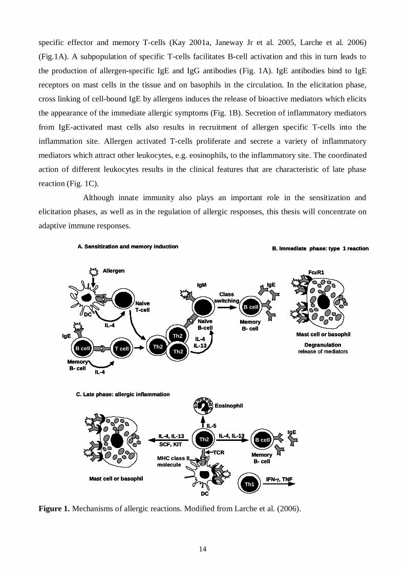
5.2 Mechanisms of allergic reactions
The first phase of an allergic reaction is called the sensitization phase, in which contact with
allergens leads to the activation of antigen presenting cells and subsequent generation of allergen
14
specific effector and memory Tcells (Kay 2001a, Janeway Jr et al. 2005, Larche et al. 2006)
(Fig.1A). A subpopulation of specific Tcells facilitates Bcell activation and this in turn leads to
the production of allergenspecific IgE and IgG antibodies (Fig. 1A). IgE antibodies bind to IgE
receptors on mast cells in the tissue and on basophils in the circulation. In the elicitation phase,
cross linking of cellbound IgE by allergens induces the release of bioactive mediators which elicits
the appearance of the immediate allergic symptoms (Fig. 1B). Secretion of inflammatory mediators
from IgEactivated mast cells also results in recruitment of allergen specific Tcells into the
inflammation site. Allergen activated Tcells proliferate and secrete a variety of inflammatory
mediators which attract other leukocytes, e.g. eosinophils, to the inflammatory site. The coordinated
action of different leukocytes results in the clinical features that are characteristic of late phase
reaction (Fig. 1C).
Although innate immunity also plays an important role in the sensitization and
elicitation phases, as well as in the regulation of allergic responses, this thesis will concentrate on
adaptive immune responses.
Figure 1. Mechanisms of allergic reactions. Modified from Larche et al. (2006).
Th2
B cell
T cellB cell
Th2
Th2
MemoryB cell
NaïveBcell
NaiveTcell
DC
Allergen
A. Sensitization and memory induction
IgE
MemoryB cellIL4
IL4
IL4IL13
IgM
IgE
B. Immediate phase: type 1 reaction
C. Late phase: allergic inflammation
Classswitching
Degranulationrelease of mediators
Mast cell or basophil
TCR
Fc R1
Eosinophil
IL5
DC
IL4, IL13B cell
MemoryB cell
IgE
Th1
IL4, IL13SCF, KIT
Mast cell or basophil IFN , TNF
Th2
MHC class IImolecule
Th2
B cell
T cellB cell
Th2
Th2
MemoryB cell
NaïveBcell
NaiveTcell
DC
Allergen
A. Sensitization and memory induction
IgE
MemoryB cellIL4
IL4
IL4IL13
IgM
IgE
B. Immediate phase: type 1 reaction
C. Late phase: allergic inflammation
Classswitching
Degranulationrelease of mediators
Mast cell or basophil
TCR
Fc R1
Eosinophil
IL5
DC
IL4, IL13B cell
MemoryB cell
IgE
Th1
IL4, IL13SCF, KIT
Mast cell or basophil IFN , TNF
Th2
Th2Th2
B cellB cell
T cellT cellB cellB cell
Th2Th2
Th2Th2
MemoryB cell
NaïveBcell
NaiveTcell
DC
Allergen
A. Sensitization and memory induction
IgE
MemoryB cellIL4
IL4
IL4IL13
IgM
IgE
B. Immediate phase: type 1 reaction
C. Late phase: allergic inflammation
Classswitching
Degranulationrelease of mediators
Mast cell or basophil
TCR
Fc R1
Eosinophil
IL5
DC
IL4, IL13B cellB cell
MemoryB cell
IgE
Th1Th1
IL4, IL13SCF, KIT
Mast cell or basophil IFN , TNF
Th2Th2
MHC class IImolecule
15
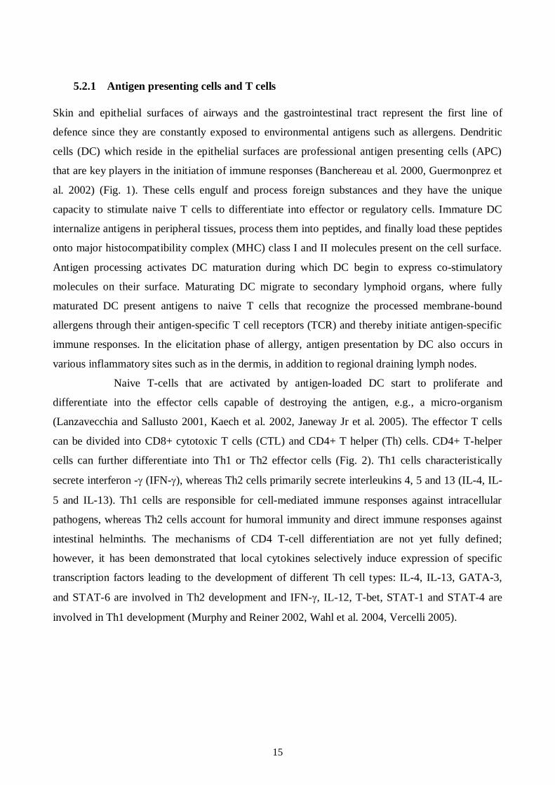
5.2.1 Antigen presenting cells and T cells
Skin and epithelial surfaces of airways and the gastrointestinal tract represent the first line of
defence since they are constantly exposed to environmental antigens such as allergens. Dendritic
cells (DC) which reside in the epithelial surfaces are professional antigen presenting cells (APC)
that are key players in the initiation of immune responses (Banchereau et al. 2000, Guermonprez et
al. 2002) (Fig. 1). These cells engulf and process foreign substances and they have the unique
capacity to stimulate naive T cells to differentiate into effector or regulatory cells. Immature DC
internalize antigens in peripheral tissues, process them into peptides, and finally load these peptides
onto major histocompatibility complex (MHC) class I and II molecules present on the cell surface.
Antigen processing activates DC maturation during which DC begin to express costimulatory
molecules on their surface. Maturating DC migrate to secondary lymphoid organs, where fully
maturated DC present antigens to naive T cells that recognize the processed membranebound
allergens through their antigenspecific T cell receptors (TCR) and thereby initiate antigenspecific
immune responses. In the elicitation phase of allergy, antigen presentation by DC also occurs in
various inflammatory sites such as in the dermis, in addition to regional draining lymph nodes.
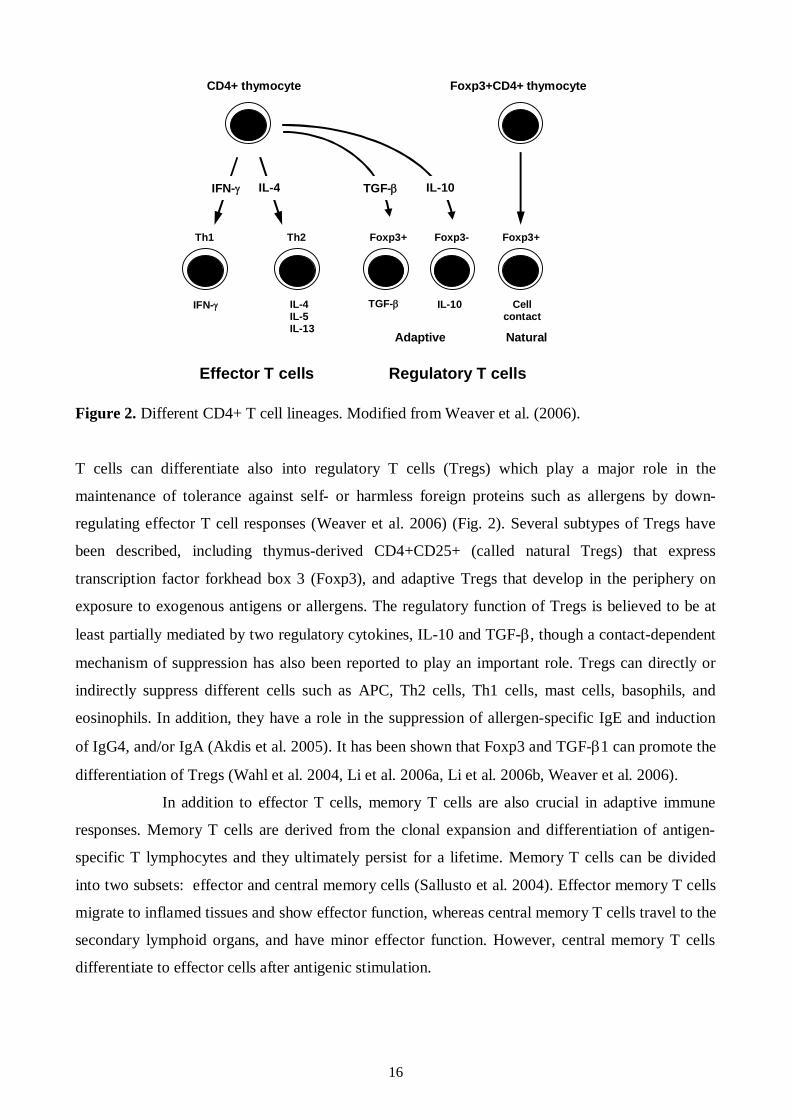
Naive Tcells that are activated by antigenloaded DC start to proliferate and
differentiate into the effector cells capable of destroying the antigen, e.g., a microorganism
(Lanzavecchia and Sallusto 2001, Kaech et al. 2002, Janeway Jr et al. 2005). The effector T cells
can be divided into CD8+ cytotoxic T cells (CTL) and CD4+ T helper (Th) cells. CD4+ Thelper
cells can further differentiate into Th1 or Th2 effector cells (Fig. 2). Th1 cells characteristically
secrete interferon (IFN ), whereas Th2 cells primarily secrete interleukins 4, 5 and 13 (IL4, IL
5 and IL13). Th1 cells are responsible for cellmediated immune responses against intracellular
pathogens, whereas Th2 cells account for humoral immunity and direct immune responses against
intestinal helminths. The mechanisms of CD4 Tcell differentiation are not yet fully defined;
however, it has been demonstrated that local cytokines selectively induce expression of specific
transcription factors leading to the development of different Th cell types: IL4, IL13, GATA3,
and STAT6 are involved in Th2 development and IFN , IL12, Tbet, STAT1 and STAT4 are
involved in Th1 development (Murphy and Reiner 2002, Wahl et al. 2004, Vercelli 2005).
16
Figure 2. Different CD4+ T cell lineages. Modified from Weaver et al. (2006).
T cells can differentiate also into regulatory T cells (Tregs) which play a major role in the
maintenance of tolerance against self or harmless foreign proteins such as allergens by down
regulating effector T cell responses (Weaver et al. 2006) (Fig. 2). Several subtypes of Tregs have
been described, including thymusderived CD4+CD25+ (called natural Tregs) that express
transcription factor forkhead box 3 (Foxp3), and adaptive Tregs that develop in the periphery on
exposure to exogenous antigens or allergens. The regulatory function of Tregs is believed to be at
least partially mediated by two regulatory cytokines, IL10 and TGF , though a contactdependent
mechanism of suppression has also been reported to play an important role. Tregs can directly or
indirectly suppress different cells such as APC, Th2 cells, Th1 cells, mast cells, basophils, and
eosinophils. In addition, they have a role in the suppression of allergenspecific IgE and induction
of IgG4, and/or IgA (Akdis et al. 2005). It has been shown that Foxp3 and TGF 1 can promote the
differentiation of Tregs (Wahl et al. 2004, Li et al. 2006a, Li et al. 2006b, Weaver et al. 2006).
In addition to effector T cells, memory T cells are also crucial in adaptive immune
responses. Memory T cells are derived from the clonal expansion and differentiation of antigen
specific T lymphocytes and they ultimately persist for a lifetime. Memory T cells can be divided
into two subsets: effector and central memory cells (Sallusto et al. 2004). Effector memory T cells
migrate to inflamed tissues and show effector function, whereas central memory T cells travel to the
secondary lymphoid organs, and have minor effector function. However, central memory T cells
differentiate to effector cells after antigenic stimulation.
IFN
Th1 Th2 Foxp3+
IL4 TGF
Foxp3+CD4+ thymocyte
Foxp3 Foxp3+
CD4+ thymocyte
IL10
IFN IL4IL5IL13
IL10 Cellcontact
TGF
Regulatory T cellsEffector T cells
Adaptive Natural
17
5.2.2 Immunoglobulin E in the allergic reaction
B cells produce antibodies against foreign agents and maintain a pool of memory cells (Manz et al.
2005, Kalia et al. 2006, Radbruch et al. 2006). When a B lymphocyte has recognized its antigen,
with the help of T helper cells, it becomes activated and is transformed into a lymphoblast that starts
to divide. The dividing lymphoblast produces a clone of cells with identical specificity that in the
end differentiates into antibody secreting plasma cells. Antibodies are secreted into blood and other
extracellular fluids where they affect by binding to their corresponding antigens. This marks the
antigen for elimination by other cells. After the antigen has been eliminated, a part of the
differentiated plasma cells remains, building up the so called immunological memory. These
memory lymphocytes react more rapidly and effectively to the presence of the antigen in secondary
infections. In mammals, there are five classes of antibodies, IgG, IgM, IgD, IgA and IgE, each with
its own class of heavy chain , , , , and , respectively. The basic structural unit of an antibody
molecule consists of four polypeptide chains, two identical light chains and two identical heavy
chains.
Immunoglobulin E (IgE) is the key molecule mediating type I allergic reactions, such
as allergic asthma, allergic rhinitis and insect venom allergy (PlattsMills 2001, Geha et al. 2003,
Gould et al. 2003). IgE is a Yshaped molecule with two distinct regions. One is the constant region
(Fc), which determines the type of immunoglobulin (e.g. Fc for IgE) and the other is the variable
region that forms two identical antigenbinding sites (Fab). The Fc region switches on the effector
mechanisms that are activated by antigens. The concentration of IgE in serum is the lowest of the
five immunoglobulins and its halflife in human serum is only 2 days. On the other hand, IgE is
mostly bound to tissue mast cells as well as to circulating basophils and activated eosinophils and
therefore its halflife is considerably longer. The synthesis of IgE by B cells occurs at a low rate
compared with that of the other antibodies, even in allergic subjects.
Most IgE is bound to cells via its highaffinity receptor, Fc RI, expressed by tissue
mast cells and circulating basophils (Galli et al. 2005a, Galli et al. 2005b, Gibbs 2005). Cross
linking of IgE bound to Fc RI on these cells by specific antigen results in a local release of
inflammatory mediators (for example, histamine, prostaglandins and leukotrienes), enzymes and
cytokines, such as IL4, 5, 6, 10, and 13 and TNF, which are important in the pathogenesis of
various allergic reactions. In particular, IL5 production is reported to be critical for the
pathogenesis of eosinophilic inflammation in the lung (Foster et al. 1996, Hamelmann and Gelfand
2001, Rothenberg and Hogan 2006). In addition, the lowaffinity IgE receptor, Fc RII (CD23),
which is expressed by a wide variety of immune cell types, including B cells, macrophages and
dendritic cells can also bind IgE (Geha et al. 2003). IgE bound to the highaffinity IgE receptor
18
Fc RI or Fc RII can also facilitate allergen uptake by antigenpresenting cells (APC) and augment
secondary immune responses.
5.2.3 Cytokines and chemokines
Cytokines
Various cells secrete soluble proteins or glycoproteins called cytokines (Greek cyto, cell, and
kinesis, movement) that act as intercellular (between cells) mediators or signaling molecules
(Prescott et al. 2002, Borish and Steinke 2003, Steinke and Borish 2006). Cytokines affect growth,
differentiation and activation properties of cells, regulating the nature of immune responses. They
participate in nearly every aspect of immunity and inflammation, and their production is induced by
a variety of nonspecific stimuli such as, different infections, cancers and inflammations, or by
specific interactions between T cells and antigens. The biological functions of cytokines are
expressed when they bind to their highaffinity receptors on target cells. In general, cytokines
function locally, but some cytokines can affect distant cells because of their ability to enter the
circulation. Cytokines can operate in an autocrine way (on the cell producing them), in a paracrine
way (on adjacent cells) or in an endocrine way (on the cells attainable via the circulation) (Prescott
et al. 2002, Borish and Rosenwasser 2003).
Cytokines can be divided roughly according to their major functions into different
subtypes e.g. chemokines, proinflammatory cytokines, growth factors, regulatory cytokines, Th1
type and Th2 type cytokines. However, it is not always clear to what category each cytokine
belongs and sometimes it is possible to allocate the same cytokine into more than one category
(Borish and Steinke 2003). Main cytokines and chemokines contributing to the allergic reaction and
which are relevant to the present study are presented in Table 1.
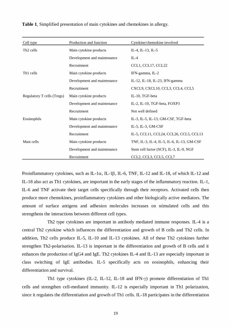
19
Table 1. Simplified presentation of main cytokines and chemokines in allergy.
Cell type Production and function Cytokine/chemokine involved
Th2 cells Main cytokine products IL4, IL13, IL5
Development and maintenance IL4
Recruitment CCL1, CCL17, CCL22
Th1 cells Main cytokine products IFNgamma, IL2
Development and maintenance IL12, IL18, IL23, IFNgamma
Recruitment CXCL9, CXCL10, CCL3, CCL4, CCL5
Regulatory T cells (Tregs) Main cytokine products IL10, TGFbeta
Development and maintenance IL2, IL10, TGFbeta, FOXP3
Recruitment Not well defined
Eosinophils Main cytokine products IL3, IL5, IL13, GMCSF, TGFbeta
Development and maintenance IL5, IL3, GMCSF
Recruitment IL5, CCL11, CCL24, CCL26, CCL5, CCL13
Mast cells Main cytokine products TNF, IL3, IL4, IL5, IL6, IL13, GMCSF
Development and maintenance Stem cell factor (SCF), IL3, IL9, NGF
Recruitment CCL2, CCL3, CCL5, CCL7
Proinflammatory cytokines, such as IL1 , IL1 , IL6, TNF, IL12 and IL18, of which IL12 and
IL18 also act as Th1 cytokines, are important in the early stages of the inflammatory reaction. IL1,
IL6 and TNF activate their target cells specifically through their receptors. Activated cells then
produce more chemokines, proinflammatory cytokines and other biologically active mediators. The
amount of surface antigens and adhesion molecules increases on stimulated cells and this
strengthens the interactions between different cell types.
Th2 type cytokines are important in antibody mediated immune responses. IL4 is a
central Th2 cytokine which influences the differentiation and growth of B cells and Th2 cells. In
addition, Th2 cells produce IL5, IL10 and IL13 cytokines. All of these Th2 cytokines further
strengthen Th2polarisation. IL13 is important in the differentiation and growth of B cells and it
enhances the production of IgG4 and IgE. Th2 cytokines IL4 and IL13 are especially important in
class switching of IgE antibodies. IL5 specifically acts on eosinophils, enhancing their
differentiation and survival.
Th1 type cytokines (IL2, IL12, IL18 and IFN ) promote differentiation of Th1
cells and strengthen cellmediated immunity. IL12 is especially important in Th1 polarization,
since it regulates the differentiation and growth of Th1 cells. IL18 participates in the differentiation
20
of Th1 cells and, in addition, it stimulates the production of chemokines and proinflammatory
cytokines. Two cytokines, IL12 and IL18, act synergistically to increase IFN production. IFN
is the most important Th1 cytokine produced, especially by Th1 cells. IFN activates macrophages
and inhibits the growth of Th2 cells.
Regulatory cytokines IL10 and TGF weaken the intensity of the inflammatory
response. They are especially produced by regulatory Tcells. These cytokines diminish the
production of proinflammatory cytokines and reduce cell stimulation by IL12 and IFN .
Chemokines
Chemokines are members of a family of chemoattractant cytokines released by tissues in the early
phases of inflammation (Zlotnik and Yoshie 2000, Rot and von Andrian 2004, Esche et al. 2005,
Pease and Williams 2006). This family contains over 40 proteins which are divided into four
subclasses, on the basis of the arrangement of the first two amino terminal cysteine residues: CXC
( family), CC ( family), C ( family), and CX3C ( family) (where "X" is an amino acid).
Chemokines interact with G proteincoupled receptors that initiate signal transduction pathways
leading to a plethora of cellular responses, in particular leukocyte chemotaxis and adhesion.
Chemokine receptor expression patterns differ between different leukocyte types and their
maturation levels. In general, the expression of CCR7 (receptor of CCL19/MIP3 and
CCL21/6Ckine) guides immature leukocytes (naive T cells, B cells, immature DC) toward the
lymph nodes. Cells of inflammatory tissues (keratinocytes, endothelial cells, T cells) produce
various sets of chemokines, which attract different effector cells, such as T cells, mast cells,
eosinophils and APC cells.
After allergen contact in the lymph nodes, naive T cells differentiate and start to
express the appropriate chemokine receptors that help them to travel to the sites of infection. Th1
cells express preferentially CXCR3 (receptor of CXCL9/Mig, CXCL10/IP10, CXCL11/ITAC)
and CCR5 (receptor of CCL3/MIP1 , CCL4/MIP1 , CCL5/RANTES, CCL11/eotaxin,
CCL14/HCCI, CCL16/HCC4), whereas Th2 cells express CCR4 (receptor of CCL17/TARC,
CCL22/MDC) and CCR8 (receptor of CCL1/I309) (Bonecchi et al. 1998). Eosinophils and mast
cells express a wide range of chemokine receptors of which CCR3 has been most extensively
studied in the context of allergy. CCR3 is a receptor for CCL11/eotaxin, CCL24/eotaxin2,
CCL26/eotaxin3, CCL7/MCP3, CCL5/RANTES, CCL8/MCP2, CCL13/MCP4) and plays
critical role in the recruitment of eosinophils to the site of allergic inflammation. In addition to the
cellspecific expression of chemokine receptors, also tissuespecific expression of chemokines
exists. For example, epidermal keratinocytes express selectively CCL27 which attracts the skin
21
homing CCR10 positive T cells to the inflamed skin (Sallusto and Mackay 2004, Homey et al.
2006).
5.3 Allergic disorders
Allergens can enter the body through skin, airways and gastrointestinal tract and in sensitized
individuals they evoke a variety of allergic manifestations (Kay 2001a, Kay 2001b). Allergic
contact urticaria (ACU) and contact dermatitis (ACD) are typical cutaneous responses to topical
allergen exposure. Aeroallergens cause airway responses such as allergic rhinitis and asthma.
Ingested food allergens can cause gastrointestinal, cutaneous or more generalized symptoms. The
most severe systemic reaction is anaphylaxis which may occur for example in peanut, bee sting and
NRL allergies (Chiu and Kelly 2005, Sicherer and Leung 2006). In addition to IgEmediated
allergies, there are allergic disorders with delayed symptoms. ACD is a typical example which is a
cellmediated, type IV hypersensitivity reaction (SaintMezard et al. 2004b). Delayed symptoms are
seen also in protein contact dermatitis (PCD) but the pathophysiological events leading to this
disorder are not fully understood (Doutre 2005). Atopic eczema (AE) is often associated with food
allergy, especially in children, and it often precedes allergic rhinitis and asthma. AE is, however,
best regarded as a delayed, T cell dependent cutaneous inflammation (Leung and Bieber 2003).
5.3.1 Cutaneous contact allergies
Intact skin barrier is important in the protection of the human body from the outer environment.
Small chemical molecules can easily penetrate into the skin, but penetration of larger protein
molecules usually requires that the skin barrier is damaged (Smith Pease et al. 2002). The entry of
allergens into the skin in a sensitized subject can cause an immediate type I reaction manifesting as
ACU or a delayed response such as ACD. The pathophysiology of AE is complex and seems to
depend on genetic, immunological and environmental factors (Leung and Bieber 2003, Homey et al.
2006, Simpson and Hanifin 2006).
Contact urticaria and protein contact dermatitis
Contact urticaria can be caused either by allergic or nonallergic mechanisms (Doutre 2005). The
wheal and flare reaction occurs in nonallergic contact urticaria after nonimmunologic release of
vasoactive substances from mast cells, whereas in ACU, previous sensitization to an allergen is
mandatory. After sensitization, allergen binding to IgE molecules on the mast cell surfaces leads to
release of histamine and other mediators (Geha et al. 2003, Gould et al. 2003, Galli et al. 2005a,
22
Galli et al. 2005b). When allergy is severe, the wheal and flare reaction can be associated with
systemic symptoms such as generalized urticaria, asthma and even anaphylaxis. Contact urticaria
syndrome is the term which covers the whole range of symptoms in ACU (Maibach and Johnson
1975). A contact urticaria reaction can sometimes be followed by delayed symptoms, such as local
erythematic swelling, appearing a few hours after the wheal and flare reaction. This delayed
reaction may be a latephase type I hypersensitivity reaction attributable to the influx of eosinophils,
basophils, mast cells and other lymphocytes (Macfarlane et al. 2000, Larche et al. 2006).
Protein contact dermatitis (PCD) is a rather new term and its pathophysiology is far
from clear (Janssens et al. 1995). By definition, PCD is caused by protein contact and the
phenotype is dermatitis, i.e. eczema, which is a delayed inflammatory response in the skin. Since
PCD occurs mostly in atopic subjects with IgE antibodies, it could be a delayed IgEassociated
reaction caused by penetration of protein allergens through damaged skin (Hjorth and Roed
Petersen 1976, Johansson et al. 2004). Epidermal Langerhans cells (and other cutaneous DC)
express high affinity IgE receptors (Fc RI) on their surface and possibly participate in this allergic
disorder. These cells can internalize allergens via Fc RIassociated IgE and further process them for
antigen presentation to T cells. IgE binding to cutaneous Fc R1 expressing Langerhans cells has
been detected in patients with active AE, asthma or rhinitis, but not in individuals with disease
remission or healthy controls (Semper et al. 2003). Fc RI expression of these cells are specifically
found in the lesional skin of AE and they seem to affect the outcome of T cell responses in AE
(Novak et al. 2004). Patch testing with mite, pollen and food allergens on the intact skin has often
revealed delayed eczematous reactions in sensitized atopic subjects (Darsow et al. 2004, Turjanmaa
2005). In addition to subjects with IgE antibodies, positive atopy patch test reactions can also be
seen in AE patients who have no IgE antibodies to the allergen tested, a typical example being a
positive reaction to cow´s milk (Isolauri and Turjanmaa 1996). Positive atopy patch test reactions to
protein allergens have been shown to be associated with allergenspecific T cell responses,
suggesting that these eczematous reactions could be caused by Tcell mediated immune reactions
(WistokatWulfing et al. 1999, Johansson et al. 2002).
ACU and PCD are particularly prevalent in occupations where animal or plant
proteins are handled (Doutre 2005). Examples of this can be found in the food industry (fish
handlers, butchers), in bakeries (exposure to wheat, egg) and in kitchen work where the skin is
exposed to all kinds of foods including also fruits and vegetables (Hjorth and RoedPetersen 1976,
Hannuksela and Lahti 1977, Janssens et al. 1995, Wuthrich 1996). Farmers, veterinarians and
laboratory workers may acquire ACU and PCD from animal allergens, such as cow´s dander and rat
urine (Janssens et al. 1995). ACU is a prominent symptom in NRLallergic health care workers and
these subjects often present also with hand eczema. Recently, Turjanmaa and coworkers
23
(Turjanmaa et al. 2002b) reported that after avoiding the use of NRL gloves, the hand eczema
seemed to heal, suggesting that eczema could also be a PCD caused by NRL allergens.
Allergic contact dermatitis
Allergic contact dermatitis ACD (also called contact hypersensitivity in animal models) is caused
mostly by small molecules (haptens), such as nickel, which penetrate into the skin, combine with
protein and then cause a twophase immunological response leading to ACD (Divkovic et al. 2005).
Most contact allergens are themselves irritant, providing both antigen and danger signals when they
contact the skin. In the sensitization phase, epidermal Langerhans cells (and possibly other
cutaneous DC) take up and process the allergen and then migrate to regional lymph nodes where
they can activate naive T cells after their maturation (Girolomoni et al. 2004, SaintMezard et al.
2004a, SaintMezard et al. 2004b, Cavani 2005). The subsequent event is the production of
allergenspecific memory and effector T cells. A further allergen exposure leads to the elicitation
phase in which haptenspecific T cells migrate to the site of hapten challenge and are activated.
Activated CD4+ and CD8+ T cells then release cytokines, such as IFN (Grabbe and Schwarz
1998, Girolomoni et al. 2004). Experiments in animals have indicated that allergenspecific type 1
CD4+ and CD8+ T cells act as effector cells and type 2 CD4+ T cells act as regulatory cells in ACD
(Cavani et al. 2001, Kimber and Dearman 2002, Vocanson et al. 2006). Keratinocytes are also
activated in the elicitation phase of ACD; after activation they release cytokines such as IL1, IL6,
TNF and GMCSF, and also CXC chemokines, such as CXCL8 (IL8), CXCL9 (Mig) and CXCL10
(IP). A further event is the influx of monocytes and their maturation to macrophages which then
secrete inflammatory mediators, such as IL1 and TNF (Janeway Jr et al. 2005). Results obtained
from human skin biopsy samples suggest that certain DC subgroups also participate in the
pathogenesis of ACD (Bangert et al. 2003). The end point of this complex cascade of inflammatory
events is ACD. This appears at the site of allergen contact but in severe allergy, simultaneous flare
ups can also occur in other areas of the skin. ACD can easily be diagnosed by patch testing in which
the suspected allergen in petrolatum is applied on the skin for 48 hours (Mowad 2006). When
positive, an eczema reaction appears at the test site and is still present at day 5 when the last reading
is performed.
5.3.2 Atopic eczema
Atopic eczema (AE, also called as atopic dermatitis) is a chronic, very itchy and relapsing
inflammatory skin disease characterized by typically distributed eczematous skin lesions and dry
skin in the noninvolved areas (Leung and Bieber 2003, Leung et al. 2004, Simpson and Hanifin
24
2006). AE is a very common disease that affects people in all age groups worldwide (Williams
2000, Schultz Larsen 2002, Williams and Flohr 2006). Its prevalence varies between 7% and 17%
in children and 13% in adults and its incidence has been on the rise in parallel with asthma (Schultz
Larsen 2002, Kupper and Fuhlbrigge 2004, Boguniewicz 2005, Simpson and Hanifin 2006). AE is
often the initial step in the socalled atopic march, i.e. about a half of young children with AD
subsequently develop allergic rhinitis or asthma (Spergel and Paller 2003, Hahn and Bacharier
2005). AE is classified into two main types: an extrinsic type associated with the presence of IgE
antibodies to various environmental allergens; and an intrinsic type in which there is no evidence of
IgEmediated sensitization (Akdis and Akdis 2003). According to the new nomenclature, the term
AE can only be used for those patients who have associated IgE antibodies and the term nonAE is
reserved for those patients who have no evidence of IgE antibodies to environmental allergens
(Akdis and Akdis 2003, Johansson et al. 2004).
Clinically, AE is characterized by the development of erythematous, exudative lesions
in the skin folds that are associated with intense itching. Histopathological specimens show
perivascular infiltration of lymphocytes and macrophages in the dermis, and lymphocytes and
spongiotic vesicles in the epidermis. In acute AE, there is a prominent infiltration of T cells
whereas in more chronic lesions, various amounts of eosinophils are also seen (Akdis et al. 2006).
In the acute phase of AE, Th2type cytokines, IL4, IL5 and IL13 characterize the inflammatory
response in the inflamed skin. In the chronic phase of the disease, however, Th1type cytokines, IL
12 and IFN , are highly expressed and predominate over Th2 cytokines.
Since approximately 7080 % of patients with AE have elevated levels of serum IgE
and specific IgE antibodies to environmental allergens, this points to an important role for allergens
in AE. It has been hypothesised that aeroallergens, such as housedust mite and pollens allergens,
may penetrate into the skin in sensitized atopic subjects, bind to IgE receptors on the Langerhans
cells and then cause eosinophil and T cell mediated inflammatory responses leading to the eczema
reaction (BruijnzeelKoomen et al. 1989). In support of this theory, housedust mite specific T cells
have been found in the inflamed and noninflamed skin of AE patients sensitized to this mite (Bohle
et al. 1998). There is a substantial body of evidence that specific elimination diets can be beneficial
in those AE children who are sensitized to food allergens (Sicherer and Sampson 1999). Moreover,
doubleblind placebocontrolled oral food challenges have confirmed that flareups of eczema are
specific reactions to food allergens, such as cow´s milk, egg and wheat (Isolauri and Turjanmaa
1996, Niggemann et al. 2001), but the mechanism by which they are transferred from the gut to the
skin is at present incompletely understood.
The skin of more than 90 % of AE patients is colonized with superantigenproducing
strains of Staphylococcus aureus, whereas only 510 % of healthy individuals carry these micro
25
organisms (Leyden et al. 1974, Aly et al. 1977, Michie and Davis 1996, Breuer et al. 2000). It is
possible that an innate immunity reaction initiated by bacterial superantigens leads to the release of
inflammatory cytokines and then to new lesions in the skin of subjects with AE. Topical exposure
to staphylococcal superantigens may also critically contribute to the development of Th1 type skin
inflammation in AE patients during the chronic phase of the disease (Breuer et al. 2005). A
deficiency in the expression of antimicrobial peptides (e.g. defensin and cathelicidin) in inflamed
skin may contribute to the increased susceptibility to S. aureus colonization in patients with AE
(Michie and Davis 1996, Ong et al. 2002).
Although significant progress has been made in the understanding of AE, its cause is
still unknown, and much remains to be learned about the complex interrelationship of genetic,
environmental and immunological factors in this disease (Leung et al. 2004).
5.3.3 Airway allergies
Airways are continuously exposed to inhaled particles, microbes and harmless antigens to which
either immunity or tolerance is induced. Allergic rhinitis and allergic asthma are two important
allergic disorders, which affect the upper and lower parts of the respiratory tract, respectively
(Howarth 2003, Cohn et al. 2004, Greiner 2006). Similar inflammatory features are found in these
diseases, such as vasodilation and local infiltration of mast cells, macrophages, eosinophils,
dendritic cells and T cells. However, the importance of affected structures differs: smooth muscles
are essential components in lower airways and blood vessels in upper airways. In addition, the
surface of the epithelium plays an important role in the development of asthma (Cookson 2004).
Allergic rhinitis
Allergic rhinitis is a common, but often underestimated, inflammatory condition of the nasal
mucosa characterized by itching, sneezing, increased nasal secretion and nasal stuffiness. If the
symptoms occur at a particular time of the year, e.g. polleninduced allergic rhinitis, it is called
seasonal allergic rhinitis. In perennial allergic rhinitis, the individual is sensitized all year round to
allergens, such as dust mites, pets, cockroaches and molds. Epidemiological studies support the
concept that allergic rhinitis is a part of the systemic inflammatory process and is associated with
other mucosal inflammatory diseases, such as asthma, rhinosinusitis and allergic conjunctivitis
(Bousquet et al. 2003, Greiner 2006, Watelet et al. 2006).
The basis for the development of allergic rhinitis is the overproduction of IgE and the
interaction between IgE and allergens. Most atopic subjects exhibit a systemic sensitization, where
the B cell synthesizes IgE in the draining lymph nodes, when the antigen has been presented to T
26
cells by APC within the nasal mucosa. However, the B cell can synthesize IgE also locally within
the nasal mucosa (Durham et al. 1997). The local production of IgE provides an explanation for the
rarely encountered individuals who have a history of seasonal rhinitis but exhibit negative skin
prick tests. In addition to these local events within the nose, there is also a systemic component to
the allergic response, with stimulation of the synthesis and maturation of bone marrow precursors
for eosinophils, basophils, and mast cells. Consistent with this concept, it has been demonstrated
that nasal allergen challenge can increase inflammatory cell recruitment within the lower airways,
and that a lower airway allergen challenge enhances upper airway cell recruitment in allergic
rhinitis (Braunstahl et al. 2001a, Braunstahl et al. 2001b).
Asthma
Asthma is commonly divided into IgEmediated allergic asthma and nonIgE mediated asthma
(nonallergic asthma) (Humbert et al. 1999, Johansson et al. 2004). Eighty percent of childhood
asthma and over 50% of adult asthma has been reported to be allergic in this manner. The
mechanisms initiating nonallergic asthma are not welldefined, although similar inflammatory
changes occur in both forms of asthma.
Asthma is a phenotypically heterogeneous disorder that results from complex
interactions between environmental and genetic factors (Maddox and Schwartz 2002, Umetsu et al.
2002, Bel 2004, WillsKarp and Ewart 2004). Allergic asthma is triggered by allergeninduced
activation of submucosal mast cells in the lower airways (Bousquet et al. 2000, Busse and
Lemanske 2001, Cohn et al. 2004). This leads to immediate bronchial constriction and amplified
secretion of fluid and mucus, causing respiratory distress. A central aspect of asthma is chronic
airway inflammation which is characterized by the continuous presence of Th2 lymphocytes,
eosinophils, neutrophils, and other leukocytes. These cells work together to cause improper
remodelling of the airways, accompanied by augmented mucus production. Th2 cytokines such as
IL13 may directly affect airway epithelial cells and cause the induction of gobletcell metaplasia
and the secretion of mucus (Kuperman et al. 2002). Bronchial epithelial cells express the chemokine
receptor CCR3 and also produce at least two of the ligands for this receptor CCL5 (RANTES) and
CCL11 (eotaxin 1). These chemokines attract more Th2 cells and eosinophils to the damaged lungs
which can increase the Th2 response. Additionally, recent studies indicate that CCL11 has a
profibrogenic effect on human airway epithelial cells and fibroblasts through the chemokine
receptor CCR3 (Beck et al. 2006, Puxeddu et al. 2006). Th2 cytokines and chemokines also have a
direct effect on airway smooth muscle cells and lung fibroblasts leading to airway remodelling.
Remodelling comprises thickening of the airway walls by hyperplasia and hypertrophy of the
smooth muscle layer and mucous glands, with the final development of fibrosis.
27
Initially, allergic asthma is driven by a response to a specific allergen, but chronic
inflammation seems to continue unabated even in the absence of allergen exposure. The airways
become hyperreactive, and factors other than reexposure to antigen can trigger asthma symptoms.
For instance, environmental irritants, such as cigarette smoke, typically induce airway
hyperreactivity. In addition, viral or bacterial respiratory infections can worsen asthma by inducing
a Th2 dominated local response (Friedlander and Busse 2005, Lemanske and Busse 2006).
5.4 Natural rubber latex allergy
NRL allergy has been an important health issue over the past 20 years (Sussman et al. 2002). The
first NRLallergic patients were described at the beginning of the 80’s (Nutter 1979, Turjanmaa et
al. 1984). Subsequently, it was noted that sensitization to NRL was common especially in HCW and
in patients with spina bifida (Turjanmaa 1988, Slater 1989). The diagnosis of NRL allergy is based
on skin prick testing and measuring of IgE antibodies in the blood (Turjanmaa 2001). Awareness of
NRL allergens and their quantification in NRL products has significantly improved during the last
few years (Palosuo et al. 2002, TomazicJezic and Lucas 2002). However, there is still some dispute
about what are the major NRL allergens and furthermore, little is known about the cellular and
molecular mechanisms involved in NRL allergy.
5.4.1 Prevalence and risk groups
Estimates of NRL allergy in the general population vary extensively, from less than 1 % to 12 %
(Liss and Sussman 1999, Turjanmaa et al. 2002a). In the health care sector, the risk groups for NRL
allergy are HCW and multioperated patients. Up to 22 % of HCW and 60 % children with spina
bifida have been reported to have become sensitized to NRL (Kelly et al. 1994, Ylitalo et al. 1997,
Poley and Slater 2000, Mazon et al. 2005). A recent metaanalysis comparing the prevalence of
NRL allergy in HCW and in the general population (Bousquet et al. 2006) found NRL allergy in
4.3% of HCW and in 1.4% of the general population. Sensitization to NRL was even more
common, since positive SPT varied from 6.9% to 7.8% in HCW and from 2.1% to 3.7% in the
general population. The NRL sensitized or allergic HCW showed an increased risk of hand
dermatitis (OR 2.5), asthma or wheezing (OR 1.6) and rhinoconjunctivitis (OR 2.7) (Bousquet et al.
2006). Recent German and Italian studies have indicated that education about NRL allergy
combined with the use of powderfree NRL gloves with reduced protein levels have led to a steady
decline in the numbers of sensitized HCW (Allmers et al. 2002, Allmers et al. 2004, Filon and
Radman 2006).
28
NRL products in the general environment are considered nowadays as an important
source of sensitization. NRL cleaning gloves, balloons, condoms, pacifiers and hotwater bottles
can evoke allergic reactions (Axelsson et al. 1988, Wrangsjo et al. 1988, Levy et al. 1992, Ylitalo et
al. 2000, Wakelin 2002). It seems evident that more attention should be paid to the protection of
NRLallergic children in the domestic environment (Ylitalo et al. 2000).
5.4.2 Symptoms, diagnosis and outcome
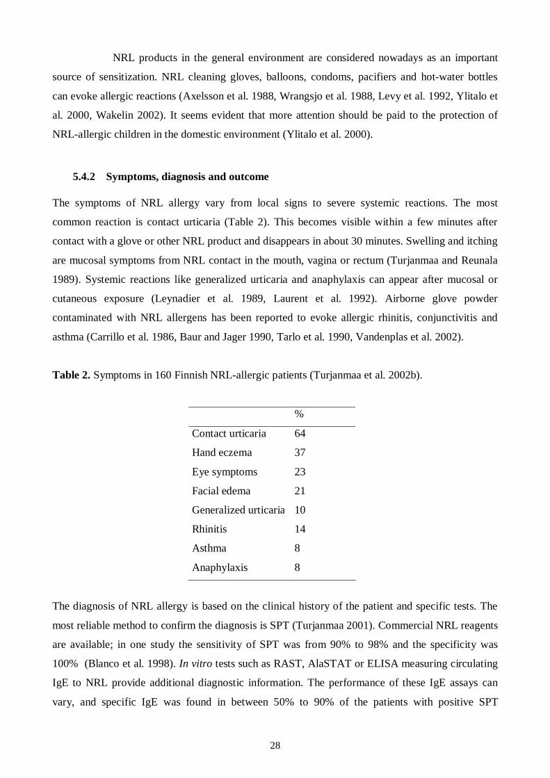
The symptoms of NRL allergy vary from local signs to severe systemic reactions. The most
common reaction is contact urticaria (Table 2). This becomes visible within a few minutes after
contact with a glove or other NRL product and disappears in about 30 minutes. Swelling and itching
are mucosal symptoms from NRL contact in the mouth, vagina or rectum (Turjanmaa and Reunala
1989). Systemic reactions like generalized urticaria and anaphylaxis can appear after mucosal or
cutaneous exposure (Leynadier et al. 1989, Laurent et al. 1992). Airborne glove powder
contaminated with NRL allergens has been reported to evoke allergic rhinitis, conjunctivitis and
asthma (Carrillo et al. 1986, Baur and Jager 1990, Tarlo et al. 1990, Vandenplas et al. 2002).
Table 2. Symptoms in 160 Finnish NRLallergic patients (Turjanmaa et al. 2002b).
%
Contact urticaria 64
Hand eczema 37
Eye symptoms 23
Facial edema 21
Generalized urticaria 10
Rhinitis 14
Asthma 8
Anaphylaxis 8
The diagnosis of NRL allergy is based on the clinical history of the patient and specific tests. The
most reliable method to confirm the diagnosis is SPT (Turjanmaa 2001). Commercial NRL reagents
are available; in one study the sensitivity of SPT was from 90% to 98% and the specificity was
100% (Blanco et al. 1998). In vitro tests such as RAST, AlaSTAT or ELISA measuring circulating
IgE to NRL provide additional diagnostic information. The performance of these IgE assays can
vary, and specific IgE was found in between 50% to 90% of the patients with positive SPT
29
(Hamilton et al. 1999). NRL allergens produced by recombinant technology are becoming
commercially available and their use seems to increase the sensitivity of the IgE testing (Lundberg
et al. 2001). When SPT or IgE assays produce results suspected to be falsenegative or positive, the
final diagnosis of NRL allergy should be based on a provocation or usage test (Turjanmaa 2001).
This test is started by placing a piece of highallergenic NRL glove on the moistened skin. If the test
is negative in this phase, it is continued by putting the whole glove on the hand. The appearance of
a wheal and flare reaction on the test site confirms that the patient is allergic, i.e. reacts clinically to
NRL. When needed, NRL challenges can also be performed in the airways (Vandenplas et al.
2002).
The outcome of NRLallergic HCW is generally favourable after the institution of
intervention measures such as use of non or lowallergen NRL gloves. A followup study in a
Finnish University Hospital showed that utilization of NRL gloves of a low allergen category made
it possible that all NRLallergic employees, whose most common symptom was contact urticaria,
could continue working in their previous positions (Turjanmaa et al. 2002b). In a European study,
the outcome of NRLinduced asthma in HCW was favourable after reduction of exposure to NRL
(Vandenplas et al. 2002). In a study from the USA, a minority of HCW, especially those with
asthma, had to change to NRLsafe workplaces, resulting in a mean 24% reduction in their annual
income (Bernstein et al. 2003b).
Treatment options for NRL allergy are at present limited. Trials have been performed
with antiIgE (Omalizumab) treatment and with immunotherapy, but the clinical efficacy has been
rather low (Sutherland et al. 2002b, Leynadier et al. 2004). Crude NRL preparations used in
immunotherapy have produced frequently adverse events (Rolland et al. 2005, Sastre et al. 2006).
Tailored hevein molecules with a low potential for adverse events have been generated but not yet
evaluated in immunotherapy trials (Karisola et al. 2004).
5.4.3 Natural rubber latex allergens
Natural rubber is a highly processed plant product derived from the cytosol, or latex, of the
commercial rubber tree, Hevea brasiliensis. Noncoagulated, ammoniated latex is mostly used in the
manufacture of rubber gloves and other soft rubber products such as condoms and balloons. The
natural function of latex is to seal damaged sites on the surface of the rubber tree (Ko et al. 2003).
Sealing is a coagulation process involving aggregation of rubber particles by hevein (Hev b 6.02)
and rubber elongation factor (Hev b 1) (Dennis and Light 1989). Prenyltransferase and Hev b 3 link
short isoprene units into cis1,4polyisoprene chains, which are responsible for the structural
integrity of latex (Oh et al. 1999). In addition to these proteins involved in rubber latex
30
biosynthesis, NRL contains between 200 250 different polypeptides (Alenius et al. 1994, Posch et
al. 1997, Petsonk 2000), many of which have unknown functions. Analysis of public domain
databases of known H. brasiliensis proteins revealed 102 different polypeptide amino acid
sequences (Guarneri et al. 2006).
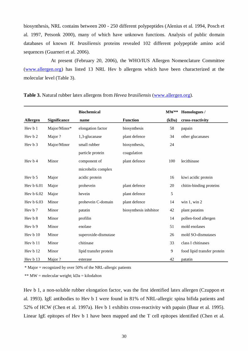
At present (February 20, 2006), the WHO/IUS Allergen Nomenclature Committee
(www.allergen.org) has listed 13 NRL Hev b allergens which have been characterized at the
molecular level (Table 3).
Table 3. Natural rubber latex allergens from Hevea brasiliensis (www.allergen.org).
Biochemical MW** Homologues /
Allergen Significance name Function (kDa) crossreactivity
Hev b 1 Major/Minor* elongation factor biosynthesis 58 papain
Hev b 2 Major ? 1,3glucanase plant defence 34 other glucanases
Hev b 3 Major/Minor small rubber biosynthesis, 24
particle protein coagulation
Hev b 4 Minor component of plant defence 100 lecithinase
microhelix complex
Hev b 5 Major acidic protein 16 kiwi acidic protein
Hev b 6.01 Major prohevein plant defence 20 chitinbinding proteins
Hev b 6.02 Major hevein plant defence 5
Hev b 6.03 Minor prohevein Cdomain plant defence 14 win 1, win 2
Hev b 7 Minor patatin biosynthesis inhibitor 42 plant patatins
Hev b 8 Minor profilin 14 pollenfood allergen
Hev b 9 Minor enolase 51 mold enolases
Hev b 10 Minor superoxidedismutase 26 mold SOdismutases
Hev b 11 Minor chitinase 33 class I chitinases
Hev b 12 Minor lipid transfer protein 9 food lipid transfer protein
Hev b 13 Major ? esterase 42 patatin
* Major = recognized by over 50% of the NRLallergic patients
** MW = molecular weight; kDa = kilodalton
Hev b 1, a nonsoluble rubber elongation factor, was the first identified latex allergen (Czuppon et
al. 1993). IgE antibodies to Hev b 1 were found in 81% of NRLallergic spina bifida patients and
52% of HCW (Chen et al. 1997a). Hev b 1 exhibits crossreactivity with papain (Baur et al. 1995).
Linear IgE epitopes of Hev b 1 have been mapped and the T cell epitopes identified (Chen et al.
31
1996, RaulfHeimsoth et al. 1998). IgE antibodies to Hev b 3, a small rubber particle protein, have
been found in 7683 % of NRLallergic spina bifida patients (Alenius et al. 1993, Lu et al. 1995,
Wagner et al. 1999). The hydrophobic NRL allergens Hev b 1 and Hev b 3 can sensitize spina
bifida and other multioperated patients, apparently because NRL catheters and other devices
containing these allergens are often in direct contact for long times with the mucosal membranes
(Yeang et al. 1998).
Hev b 5, a major NRL allergen, was reported to be recognized by sera from 56 % of
spina bifida patients and 92 % of HCW suffering from latex allergy (Slater et al. 1996). During
glove manufacture, highly acidic and proline rich Hev b 5 may degrade and form aggregates and
interact with other NRL proteins, leading up to the high levels of Hev b 5 present in NRL gloves
(Sutherland et al. 2002a). The B cell and T cells epitopes of Hev b 5 have been mapped and this
knowledge has been used to generate a pilot immunotherapy molecule for NRL allergy (Beezhold et
al. 1999, Slater et al. 1999, de Silva et al. 2000, Beezhold et al. 2001).
Hevein (Hev b 6.02) at the Nterminus of prohevein (Hev b 6.01) is a major NRL
allergen, recognized by over 50% of all kinds of NRL allergic patients. Initial studies indicated that
83 % of NRL allergic patients, 75 % of HCW and 27 % of spina bifida patients possessed IgE to
prohevein (Alenius et al. 1995, Chen et al. 1997b) . The water soluble hevein is abundant and easily
eluted from NRL gloves (Alenius et al. 1996a). The immunological role of hevein has been well
characterized e.g. the Bcell and Tcell epitopes have been identified (Banerjee et al. 1997,
Beezhold et al. 1997, de Silva et al. 2004), and the B cell and T cell responses clarified (Raulf
Heimsoth et al. 2004).
Hev b 4, a component of the microhelix complex, is a glycoprotein and its IgE
reactivity is dependent upon glycosylation (Sunderasan et al. 2005). SPT with native Hev b 4 has
been positive in 39 % of NRLallergic HCW (Bernstein et al. 2003a). Hev b 7 isoallergens exhibit
structural homology with potato patatins (Kostyal et al. 1998, Sowka et al. 1998), but cross
reactivity between these proteins is still somewhat unclear (Sowka et al. 1999, Seppala et al. 2000).
Hev b 7 is classified as a minor NRL allergen, although it may sensitize patients with spina bifida
similarly to Hev b 1 and Hev b 3 (Sowka et al. 1998, Kurup et al. 2000, Wagner et al. 2001a,
Bernstein et al. 2003a). Hev b 8 (NRL profilin) and rubber tree enolase, Hev b 9, are minor
allergens in NRL. Hev b 8 is recognized by 20 % of HCW and by 12% of spina bifida patients
(Rihs et al. 2000), whereas 14.5 % of NRL allergic patients recognize Hev b 9 (Wagner et al. 2000).
Sensitization to birch and other plant profilins can result in falsepositive IgE tests to NRL
according to a recent study (Ebo et al. 2004). Hev b 10, a manganese superoxidedismutase, is a
minor NRL allergen which exhibits crossreactivity to molds (Posch et al. 1997, Wagner et al.
2001b). Recombinant Hev b 10 was recognized only by two out of 20 spina bifida patients and by
32
none of the 20 HCW (Rihs et al. 2001). Hev b 11 is also a minor NRL allergen that has recently
been cloned and shown to be a heveinlike protein belonging to class I chitinases (O'Riordain et al.
2002). Recombinant Hev b 11 was recognized by 19 % and 29% of NRLallergic patients
(O'Riordain et al. 2002, Rihs et al. 2003). Hev b 12, a lipid transfer protein, is a minor NRL
allergen, which in immunoblot analysis was recognized by 24 % of NRLallergic patients
(Beezhold et al. 2003). Potential new NRL allergens have recently been sought by proteomic
analysis. This identified five new allergen candidates i.e. UDPglucose pyrophosphorylase,
isoflavone reductase, rotamase, thioredoxin and citratebinding protein (Yagami et al. 2004).
Up to 50% of NRLallergic patients have been reported to react with lip swelling or
other immediate allergic and even systematic symptoms to several fruits and vegetables (Makinen
Kiljunen 1994, Beezhold et al. 1996, Blanco et al. 1999, Blanco 2003). This reactivity is now
termed as latexfruit syndrome and is most often evoked by consumption of banana, kiwi fruit,
chestnut and avocado (Beezhold et al. 1996, Blanco et al. 1999, Wagner and Breiteneder 2002). The
molecular basis for this latexfruit syndrome is well documented. NRL allergens such as Hev b 2,
Hev b 5, Hev b 6.01, Hev b 7, Hev b 8 and Hev b 9 have homologous proteins in other plants (Table
3). Class I chitinases in avocado and banana and other fruits are the most likely allergens involved
in the latexfruit syndrome (Chen et al. 1998, DiazPerales et al. 1998, Mikkola et al. 1998). In
these fruits, the Nterminal chitinbinding heveinlike domains exhibit 7080 % identity to hevein
(Wagner and Breiteneder 2002). Homologous classII chitinases, lacking Nterminal chitinbinding
heveinlike domains, are also reported to be responsible for allergic crossreactions (Subroto et al.
1999). A recent study revealed that isolated heveinlike domains but not intact endochitinases are
responsible for the IgE mediated in vitro and in vivo reactions in the latexfruit syndrome (Karisola
et al. 2005). It is also known that Hev b 5 shows homology with an acidic protein present in kiwi
fruit (Slater et al. 1996) and there is Hev b 7 homology with patatin (Sol t1) in potato (Seppala et al.
2000).
At present there is a general agreement that the most important NRL allergens for
HCW and other adult NRLallergic subjects are Hev b 6.02 and Hev b 5 (Alenius et al. 1996a,
Slater et al. 1996, Chen et al. 1997b). The major allergens for the patients with spina bifida and
other congenital anomalies are Hev b 1 and Hev b 3 (Alenius et al. 1993, Lu et al. 1995, Chen et al.
1997a, Rihs et al. 1998, Wagner et al. 1999). There is also a general agreement that in medical NRL
gloves, the most abundant allergens are Hev b 6.02 and Hev b 5 but Hev b 3 and Hev b 1 are also
commonly found (Kujala et al. 2002, Palosuo et al. 2002, Sutherland et al. 2002a, Koh et al. 2005).
It has recently been suggested that the major NRL allergens should be expanded to include Hev b 2
and Hev b 13 (Table 3) (Yeang 2004). The role of Hev b 2 and Hev b 13 as major NRL allergens is,
however, controversial and needs to be confirmed in further studies.
33
5.4.4 Peripheral blood mononuclear cell responses
There are much fewer studies on NRLinduced cellmediated immunity than there are for IgE
antibody responses. Proliferation of peripheral blood mononuclear cells (PBMC) in NRLallergic
patients have been performed using NRL extracts, purified native or recombinant allergens (Murali
et al. 1994, RaulfHeimsoth et al. 1996, Ebo et al. 1997, Johnson et al. 1999, RaulfHeimsoth et al.
2004). Purified Hev b 1 was shown to induce lymphocyte proliferation in 52 % of patients
sensitized to NRL and in 25% of NRLexposed subjects (RaulfHeimsoth et al. 1996). T cell
responses to Hev b 3 have been studied in NRLallergic patients with spina bifida using poly,
oligo, and monoclonal T lymphocyte cultures (Lu et al. 1995, Bohle et al. 2000). Further studies
with Hev b 5 have indicated that certain Hev b 5 peptides induce proliferation of T cell lines from
NRLallergic patients (de Silva et al. 2000) as well as from NRLsensitized mice (Slater et al.
1999).
5.5 Mouse models for allergic diseases
Experimental animal models are crucial in elucidating the pathophysiology of different diseases and
in the development of novel therapies. Different animal species such as dog, monkey, rat and guinea
pig have been used as models for allergic disorders but mouse models are most commonly used,
probably due to the availability of immunological tools, such as transgenic and knock out mouse
strains and antibodies. Mice have other advantages e.g. short breeding periods, a wellknown
genome, a fully characterized immune system and relatively low maintenance costs (Gutermuth et
al. 2004, Taube et al. 2004).
AE is a pruritic inflammatory skin disease characterized by dermal infiltration of
lymphocytes and eosinophils, expression of Th2 cytokines and chemokines and increased levels of
total and specific IgE (Leung et al. 2004). Several experimental models have been developed and
used to examine specific features of AE, for instance genetic regulation, immune deficiencies, the
significance of various cytokines and the role of bacteria in the course of inflammation (Gutermuth
et al. 2004, Shiohara et al. 2004). Mouse models of AE can be roughly categorized into the
following groups: a) mouse models (NC/Nga) with spontaneous development of AE (Matsuda et al.
1997); b) genetically modified mice, transgenic or knockout e.g. with regard to IL4 and IL18
(Chan et al. 2001, Konishi et al. 2002) c) AElike skin lesions after epicutaneous ovalbumin
sensitization (Wang et al. 1996, Spergel et al. 1998) and d) humanized mouse models of AE such as
the SCID mutant mice (Carballido et al. 2003).
34
For the time being, the limited availability and the lack of standardized mice housing
conditions make the use of NC/Nga mice inconvenient. Humanised SCID and genetically modified
mice are important tools when examining specific mechanisms of diseases. On the other hand, a
mouse model making use of repeated epicutaneous protein exposure (Spergel et al. 1998) imitate
reasonably well naturally occurring cutaneous sensitization. In this model, tape stripping is an
important feature provoking the development of general skin injury characteristic of AE. This
protein exposure model has been modified by using NRL instead of ovalbumin to study the
cutaneous route exposure to NRL allergens in this thesis (I).
Allergic asthma is a complex condition characterized by increased amounts of
systemic IgE, elevated allergenspecific Th2 cells and their products, airway hyperreactivity
(AHR), and structural changes in the lung (Cohn et al. 2004). Animal models of both acute and
chronic allergic airway responses have been described, although none of these models encompasses
all aspects of human asthma (Kips et al. 2003, Epstein 2004, Taube et al. 2004, Boyce and Austen
2005, Fulkerson et al. 2005). Moreover, considerable differences in the airway reactivity between
different mice strains exist (Takeda et al. 2001, Whitehead et al. 2003). The most widely used
mouse model of acute asthma involves intraperitoneal injections with ovalbumin combined with
alum, followed by repeated ovalbumin challenges intratracheally, intranasally, or by inhalation.
This evokes a marked eosinophilic inflammation and AHR, which is independent of IgE, B cells, or
mast cells, but dependent on CD4+ T cells (Corry et al. 1998). The presence of effector T cells is
essential and sufficient to provide the necessary Th2 cytokines to evoke both histological changes
and induced AHR (Boyce and Austen 2005). Chronic asthma models with remodelling of lung
tissue have been reported but these models are difficult to standardize (McMillan and Lloyd 2004,
Fulkerson et al. 2005, Wegmann et al. 2005).
A few studies have used mouse models to examine the pathological mechanisms
involved in NRL allergy (reviewed in (Meade and Woolhiser 2002, Herz et al. 2004). BALB/c mice
have been most commonly investigated mice strain, but in two studies also C57BL/6 and B6C3F1
mice were used (Kurup et al. 1994, Woolhiser et al. 1999). Crude NRL has been the allergen used
in almost all studies (Kurup et al. 1994, Thakker et al. 1999, Woolhiser et al. 1999, Xia et al. 1999)
but recombinant Hev b 5 has also been administered in two studies (Slater et al. 1998, Slater et al.
1999). One group has been interested in the effects of NRL in the lungs which they have
investigated by using intranasal and intraperitoneal sensitization (Kurup et al. 1994, Thakker et al.
1999, Xia et al. 1999). They found increased levels of total IgE, IL4, IL5 and eosinophils in the
blood (Kurup et al. 1994). Eosinophils were detected in the lungs and AHR occurred after
methacholine (MCh) challenge (Thakker et al. 1999, Xia et al. 1999). Experiments in IL4 knockout
BALB/c mice showed no eosinophils nor IgE antibodies, confirming the importance of this
35
cytokine in the initiation of this response (Xia et al. 1999). It is, however, of interest that IL4
knockout mice exhibited a weak AHR to MCh (Thakker et al. 1999, Xia et al. 1999).
Woolhiser et al. (1999) studied the effects of IN, subcutaneous and EC exposure by
using crude NRL as allergen. They found increased total IgE levels by all exposure routes and
detected the presence of several IgE bands with cutaneous exposure. However, the results of
different exposure routes are not comparable because NRL dosage and exposure times were
different. Slater and coworkers (Slater et al. 1998, Slater et al. 1999) used recombinant Hev b 5
with maltose binding protein to sensitize BALB/c mice by the IN route. Hev b 5 specific IgG1 or
IgE antibodies were found in the blood. A significant lymphoid cell infiltration, but no mucus
secretion, was seen in the lungs when the allergen was given together with LPS. These mouse
model studies indicate that IgE antibodies appear after cutaneous, intranasal and intraperitoneal
exposure to NRL. It seems evident that IN exposure to NRL can evoke eosinophilic lung
inflammation and AHR in the sensitized mice. None of these mouse experiments have examined
cutaneous inflammation to NRL and studied whether sensitization through the skin can predispose
the animal to NRLallergic asthma.
36
6 AIMS OF THE STUDY
The aims of the present study were:
1. To examine the role of cutaneous exposure to NRL in the development of skin inflammation
and the humoral IgE antibody response in mice.
2. To study the effect of different exposure routes of NRL in the development of lung
inflammation and airway hyperreactivity in mice.
3. To characterize cytokine and chemokine profiles to major NRL allergens Hev b 5 and Hev b
6.01 in peripheral blood mononuclear cells of NRLallergic patients.
4. To investigate the prevalence of IgE antibodies to highly purified Hev b 2 and Hev b 13 in
relation to Hev b 5 and Hev b 6.01 in NRLallergic patients from Finland, Spain and the USA.
37
7 MATERIALS AND METHODS
7.1 Natural rubber latex allergens
7.1.1 NRL (I III)
NRL (Yeang et al. 2002) was obtained from fresh Malaysian nonammoniated latex (Ansell
International, Melbourne, Australia) (I, II). The liquid latex from H. brasiliensis was collected and
stored at 700C. For the extraction of soluble proteins, latex was diluted 1:2 with phosphatebuffered
saline (PBS) and centrifuged at 100,000 g for 90 min after which supernatant was stored at 700C
(Alenius et al. 1993). Buffer exchange to PBS was achieved by highperformance liquid
chromatography (HPLC) on a Fast Desalting Column HR 10/10 (Amersham Biosciences, Uppsala,
Sweden) for mice experiments (I, II). Pooled protein fractions were concentrated in a vacuum
centrifuge (SpeedVac, Savant Instruments, Inc., Farmingdale, NY, USA) (I, II). Native proteins
were used for human studies and buffer exchange was conducted on a BioGelP6 desalting gel
column (BioRad Laboratories, Hercules, CA, USA) in PBS (III). The source material for isolation
of native NRL allergens (Bserum and Cserum of nonammoniated NRL, clone RRI 600) was
obtained from the Rubber Research Institute of Malaysia (Kuala Lumpur, Malaysia) (III, IV).
7.1.2 Purified NRL allergens (I IV)
Hev b 1 (I)
Hev b 1 was purified from NRL with electroelution using sodium dodecyl sulphatepolyacrylamide
gel electrophoresis (SDSPAGE) as previously described (Alenius et al. 1996b).
Hev b 2 (IV)
Native Hev b 2 was isolated and purified from Bserum of NRL (Yeang et al. 2002). The first
purification step was cation exchange chromatography on a MonoS (4.6/100 PE, Tricorn) column
(Amersham Biosciences) equilibrated with 50 mM sodium phosphate, pH 7.5 and eluted with a
linear gradient of NaCl (0 to 1 M in 30 min) at a flow rate of 1.0 ml/min (IV, Fig. 1A). Gel filtration
chromatography of pooled and concentrated Hev b 2 fractions was conducted on a Superdex 75
(HR 10/30) column (Amersham Biosciences) in PBS (0.5 ml/min) (IV, Fig. 1B). Monitoring of
these chromatographs was made at 214 nm.
38
Hev b 5 (III, IV)
The starting material for native Hev b 5 was the Cserum of NRL (Yeang et al. 2002) and
purification was undertaken via buffer exchange and cation exchange chromatography. A BioGelP6
desalting gel column in 20 mM Nacitrate, pH 3.8 was used for buffer exchange and cation
exchange was performed on a MonoS (4.6/100 PE) column equilibrated with the same buffer.
Elution was done with a linear gradient of NaCl (0 to 1 M in 30 minutes) in the equilibration buffer.
Hev b 6.01 (IIV)
For purification of Hev b 6.01, chitinbinding proteins of NRL were attached to chitin (Sigma
Chemical Co, St.Louis, MO, USA) and detached from it with 0.25 M and 0.6 M acetic acid
(Hanninen et al. 1999) (I, II). Hev b 6.01 fractions were then obtained by HPLC on a ReSource (3
ml) column (Amersham Biosciences) (linear gradient of 060% acetonitrile in 30 min, flow rate 1
ml/min). The fractions were pooled and concentrated in a vacuum centrifuge. For human studies
(III, IV) native Hev b 6.01 was purified from the Bserum of NRL by gel filtration and anion
exchange chromatography (Karisola et al. 2005). Gel filtration was performed on a Superdex 75
(HR 16/60) column (Amersham Biosciences) in PBS. Prohevein fractions were concentrated and
the buffer was changed to 20 mM TrisHCl, pH 8.5, by means of a BioGelP6 column. Anion
exchange chromatography was conducted on a MonoQ (HR5/5) column (Amersham Biosciences)
equilibrated with 20 mM TrisHCl, pH 8.5 and eluted with a linear gradient of NaCl (0 to 1 M in 30
minutes) in the same buffer.
Hev b 13 (IV)
Bserum of NRL was at first chromatographed on a Superdex 75 (HR 16/60) column to obtain Hev
b 13 containing fractions; flow rate was 1.5 ml/min and with monitoring at 280 nm (IV, Fig. 1D).
Buffer exchange to 50 mM sodium acetate, pH 4.5 was then made for pooled and concentrated Hev
b 13 containing fractions on a BioGelP6 column. Subsequently, a cation exchange column, MonoS
(HR 5/5, Amersham Biosciences) was used for further purification (IV, Fig 1E). Elution was
performed with a linear gradient of NaCl (0 to 1 M in 40 min) at a flow rate of 1.0 ml/min and the
chromatography was monitored at 214 nm.
Identification and characterization of the purified allergens (III, IV)
Identity of the purified native allergens Hev b 2, Hev 5, Hev b 6.01 and Hev b 13 was confirmed
with monoclonal antibodies and commercial ELISA kits for Hev b 5 and Hev b 6.02 (Fit Biotech
Ltd., Tampere, Finland). In addition, reversedphase chromatography on a C1 column (2 x 20 mm,
Tosoh Corp, Tokyo, Japan) was used to check the purity and to quantify the purified allergens
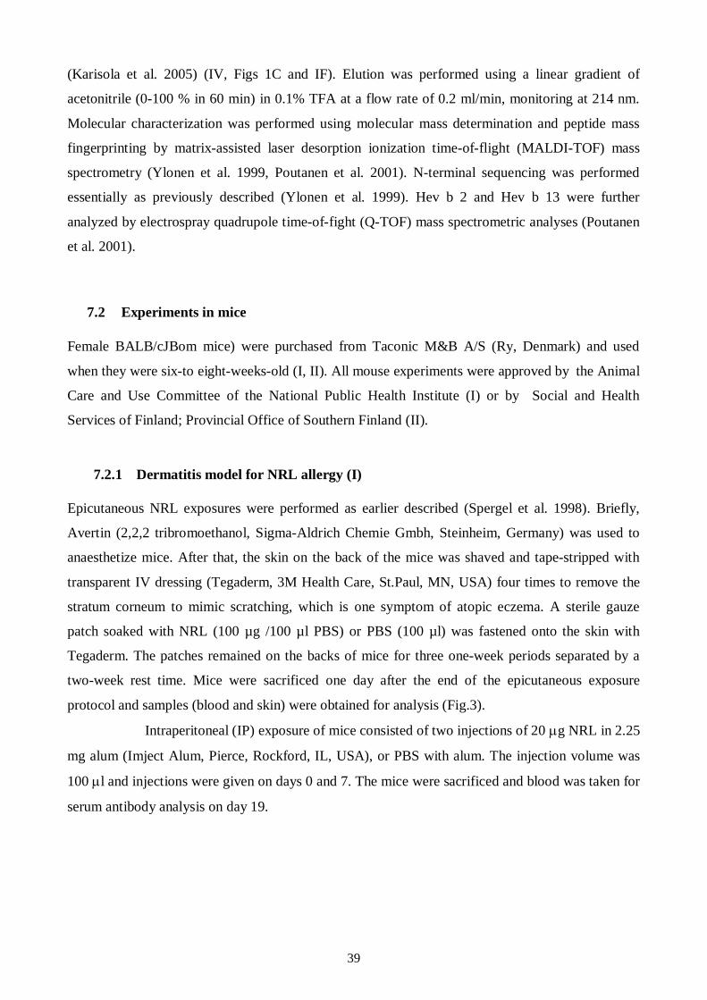
39
(Karisola et al. 2005) (IV, Figs 1C and IF). Elution was performed using a linear gradient of
acetonitrile (0100 % in 60 min) in 0.1% TFA at a flow rate of 0.2 ml/min, monitoring at 214 nm.
Molecular characterization was performed using molecular mass determination and peptide mass
fingerprinting by matrixassisted laser desorption ionization timeofflight (MALDITOF) mass
spectrometry (Ylonen et al. 1999, Poutanen et al. 2001). Nterminal sequencing was performed
essentially as previously described (Ylonen et al. 1999). Hev b 2 and Hev b 13 were further
analyzed by electrospray quadrupole timeoffight (QTOF) mass spectrometric analyses (Poutanen
et al. 2001).
7.2 Experiments in mice
Female BALB/cJBom mice) were purchased from Taconic M&B A/S (Ry, Denmark) and used
when they were sixto eightweeksold (I, II). All mouse experiments were approved by the Animal
Care and Use Committee of the National Public Health Institute (I) or by Social and Health
Services of Finland; Provincial Office of Southern Finland (II).
7.2.1 Dermatitis model for NRL allergy (I)
Epicutaneous NRL exposures were performed as earlier described (Spergel et al. 1998). Briefly,
Avertin (2,2,2 tribromoethanol, SigmaAldrich Chemie Gmbh, Steinheim, Germany) was used to
anaesthetize mice. After that, the skin on the back of the mice was shaved and tapestripped with
transparent IV dressing (Tegaderm, 3M Health Care, St.Paul, MN, USA) four times to remove the
stratum corneum to mimic scratching, which is one symptom of atopic eczema. A sterile gauze
patch soaked with NRL (100 µg /100 µl PBS) or PBS (100 µl) was fastened onto the skin with
Tegaderm. The patches remained on the backs of mice for three oneweek periods separated by a
twoweek rest time. Mice were sacrificed one day after the end of the epicutaneous exposure
protocol and samples (blood and skin) were obtained for analysis (Fig.3).
Intraperitoneal (IP) exposure of mice consisted of two injections of 20 g NRL in 2.25
mg alum (Imject Alum, Pierce, Rockford, IL, USA), or PBS with alum. The injection volume was
100 l and injections were given on days 0 and 7. The mice were sacrificed and blood was taken for
serum antibody analysis on day 19.
40
Fig. 3. Epicutaneous exposure schedule.
7.2.2 Asthma model for NRL allergy (II)
Total exposure time to NRL (NRL groups) or PBS (SAL or saline groups) via intracutaneous (IC),
intraperitoneal (IP) and intranasal (IN) routes lasted for four weeks (II, Fig 6). Anaesthesia with
isoflurane (Abbott Laboratories Ltd, Queenborough, UK) was used, when IC or IN administration
of NRL or PBS was made. Before the IC exposures, the backs of the mice were shaved and tape
stripped as earlier described for epicutaneous exposures. Mice were given once a week NRL (40 g
/100 l PBS) during the IC exposures. Intranasally exposed mice received also 40 g NRL in 50 l
PBS once per week. IP exposure consisted injections of 80 g NRL in 2 mg alum (Pierce,
Rockford, IL, USA) on days 1 and 14 or 100 l PBS with alum. IC and IN control mice received
100 l PBS instead of NRL. Inhalation challenges of NRL (0.5% in PBS for 20 min) were achieved
with an ultrasonic nebulizator (Aerogen Ltd., Galway, Ireland) on days 28, 29, and 30.
Airway hyperreactivity
Airway hyperreactivity (AHR) to methacholine (MCh) (SigmaAldrich Co, St.Louis, MO, USA)
was assessed 24 hours after the last NRL challenge (on day 31) by wholebody plethysmography
(Buxco Electronics, Inc., Sharon, CT, USA) using MCh concentrations 0 100 mg/ml. The average
enhanced pause (Penh) values were measured by a exposure time of 5 min to each concentration.
The results are expressed as compared to Penh values following PBS exposure.
The mice were killed with carbon dioxide and samples (blood, BAL fluids and lung
biopsies) collected. The blood was used to measure antibodies, BAL fluids to analyze inflammatory
cells and tissue biopsies to study mRNA expression and inflammatory cells.
Shaving +tape stripping
PatchingNRL 100 g /100 l in PBS
Schedule of sensitization:
Sensitization Sensitization SensitizationDay 23 29 51
Skin biopsieswk1 wk2 wk3
71 44 50
Blood
Shaving +tape stripping
PatchingNRL 100 g /100 l in PBS
Schedule of sensitization:
Sensitization Sensitization SensitizationDay 23 29 51
Skin biopsieswk1 wk2 wk3
71 44 50
Blood
41
7.2.3 Cytological, histological and immunohistological methods (I, II)
BAL cytology (II)
Bronchoalveolar lavage (BAL) samples were prepared by lavaging lungs once with 0.8 ml of PBS
via the trachea. The samples were cytocentrifuged (Shandon Scientific Ltd., Runcorn,UK; 150 g, 5
min), and stained with MayGrünwaldGiemsa. Cells were counted in 40 highpower fields (HPF)
at x 1000 using light microscopy.
Lung histology (II)
The lung samples in 10% buffered formalin were embedded into paraffin, cut into sections of 4 µm,
and stained with haematoxylin and eosin (H&E) or with periodic acidSchiff (PAS) solution. Lung
inflammation was scored after H&E staining (1: slightly inflamed tissue; 2: inflammation of the
area over 20%; 3: inflammation of the area over 50%; 4: inflammation of the area over 75%; 5:
entire tissue inflamed). Mucus producing cells per 100 m were counted after PAS staining. The
slides were analyzed in a blind manner with a Nikon Eclipse E800 microscope (Leica Microsystems
GmbH, Wetzlar, Germany).
Skin histology (I)
Skin biopsies from patch areas were treated like the lung samples and after that the slides were
stained with H&E or otoluidin blue for mast cell counting. Inflammatory cell types were counted at
x 1000 and mast cells at x 400 magnification. A scaled ocular lens was used to measure the
thickness of the skin. The measurements were adjusted for the magnification optics and expressed
in micrometers. The slides were examined in a blind manner using a light microscope (Leitz Dialux
20 EB, Wetzlar, Germany).
Skin immunohistology (I)
Skin samples were embedded in a TissueTek OCT compound (Sakura, AA Zoeterwoude, The
Netherlands) on dry ice. Subsequently, skin sections (4 m) were stained immunohistochemically.
Briefly, the sections were incubated with rat antimouse monoclonal antibodies (antiCD3, anti
CD4, antiCD8) (BD PharMingen) followed by biotinylated rabbit antirat Ig (Vector Laboratories,
Burlingame, CA, USA). Peroxidaseblocking solution (Dako, Carpinteria, CA, USA) was used to
block endogenous peroxidase activity. Sections were then incubated with peroxidaseconjugated
streptavidin (Dako) and stained in a substrate solution containing hydrogen peroxide (Dako) and 3
amino9ethylcarbazole (Dako), followed by haematoxylin incubation (Dako). Mounting of the skin
sections was achieved with Aquamount (BDH, Gurr, Poole, UK). Cell counting was done in 10
HPFs at x 1000 with positively stained cells being expressed as cells per HPF.
42
7.2.4 Cytokine and chemokine measurements by PCR (I, II)
RNA isolation (I, II)
Skin and lung biopsies were removed and immediately frozen in dry ice. Total RNA was isolated
using Trizol (Gibco BRL, Paisley, UK) according to the manufacturer's instructions.
Homogenization of the samples was done by UltraTurrax T8 (IKA Labortechnik, Staufen,
Germany). After RNA isolation, genomic DNA was removed with DNaseI (RNasefree) (Gibco
BRL) treatment and final RNA (in nucleasefree water) was obtained on the Trizol according to
guidelines.
cDNA synthesis and realtime quantitative PCR (I, II)
Total RNA was reversetranscribed into cDNA with MultiScribe Reverse transcriptase (PE
Applied Biosystems, Foster City, CA, USA) and random hexamers as primers according to the
manufacturer's instructions. The realtime quantitative polymerase chain reaction (PCR) was
performed with an AbiPrism 7700 Sequence Detector System (SDS) (PE Applied Biosystems).
PCR primers and probes were predeveloped reagents or designed by PrimerExpress (version 2.0)
software and obtained from PE Applied Biosystems. The PCR products were detected by
monitoring fluorescence levels of the reporter dyes, FAM or VIC . The FAM dye of the sample
cDNA was standardized to the VIC dye of the endogenous reference (18S rRNA) to control sample
loading and to allow normalization between samples according to the manufacturer's instructions
(PE Applied Biosystems). The final results were expressed as relative units (RU) i.e. ratio of PCR
amplification efficiency in the sample cDNA compared to that of the calibrator. These were
calculated by the comparative CT method according to the manufacturer's instructions. An FAM CT
value of 40, reflecting the situation in which the gene is not expressed at the mRNA level, was used
as the calibrator.
7.2.5 IgE and IgG2a antibody measurements (I, II)
Total IgE (I, II)
Total IgE levels in serum were measured by ELISA. Briefly, 96well plates (Nunc, Roskilde,
Denmark) were coated with rat antimouse IgE mAb (BD PharMingen, San Diego, CA, USA).
Diluted serum samples were added to the plate and incubated overnight. Bound IgE was detected
using biotinconjugated rat antimouse IgE (BD PharMingen), streptavidinconjugated horseradish
peroxidase (BD PharMingen) and peroxidase substrate reagents (KPL, Kirkegaard & Perry
Laboratories, Gaithersburg, MD, USA). Absorbance values at 405 nm were obtained by an ELISA
43
plate reader (Titertek Multiscan, Eflab, Turku, Finland). Total IgE levels were calculated from IgE
standard curve.
Specific IgE and IgG2a (I)
For measurement of specific IgE and IgG2a antibodies, plates were coated (50 l/well) with purified
antigen (Hev b 6.01 or Hev b 1, 2 g/ml) or 20 g/ml of NRL in 0.05M NaHCO3, pH 9.6 at 40C
overnight. Briefly, the plates were then blocked (2 hours) and 100 l of serial dilutions of sera were
incubated overnight at 40C. Subsequently, the biotinlabeled antimouse isotypespecific antibody
(BD PharMingen, 2 g/ml) was added. Streptavidinhorseradish peroxidase (BD PharMingen) was
used for detection and finally peroxidase substrate (KPL) was added, and absorption at 405 nm was
read with an automated ELISA reader.
Specific IgE to Hev b 6.01 (II)
A slightly modified specific ELISA for Hev b 6.01 was used in the second study (II) compared to
the first study (I). Plates were first coated for 3 hours at +200C and then overnight at +40C. The
blocking time was 1 hour and 1:10 diluted samples were incubated for 2 hours at +200C. Biotin
labeled antimouse isotypespecific antibody (BD PharMingen) was added. Streptavidinconjugated
alkaline phosphatase (Zymed, San Francisco, CA, USA) and colour substrate (pnitrophenyl
phosphate, Sigma) were used for detection and absorption values were read at 405 nm.
7.3 Studies in patients allergic to natural rubber latex
7.3.1 Patients and controls (III, IV)
Fourteen NRLallergic patients participated in the study of PBMC responses (III) (Table 2). All of
them were occupational NRLglove users and six of them were HCW. Altogether eleven of these
individuals suffered from different atopic diseases (asthma, allergic rhinitis and atopic eczema). All
patients had a positive skin prick test to a commercial latex extract (Stallergenes, Antony Cedex,
France) and a positive NRL glove challenge test (Turjanmaa et al. 2002b). Induction of cytokines,
chemokines and chemokine receptors was investigated in 10 NRLallergic patients with high levels
of antigenspecific IgE, as well as positive SPT and PBMC proliferative responses to NRL
allergens. Ten healthy subjects served as controls. The study was approved by the ethics committee
of Helsinki University Central Hospital.
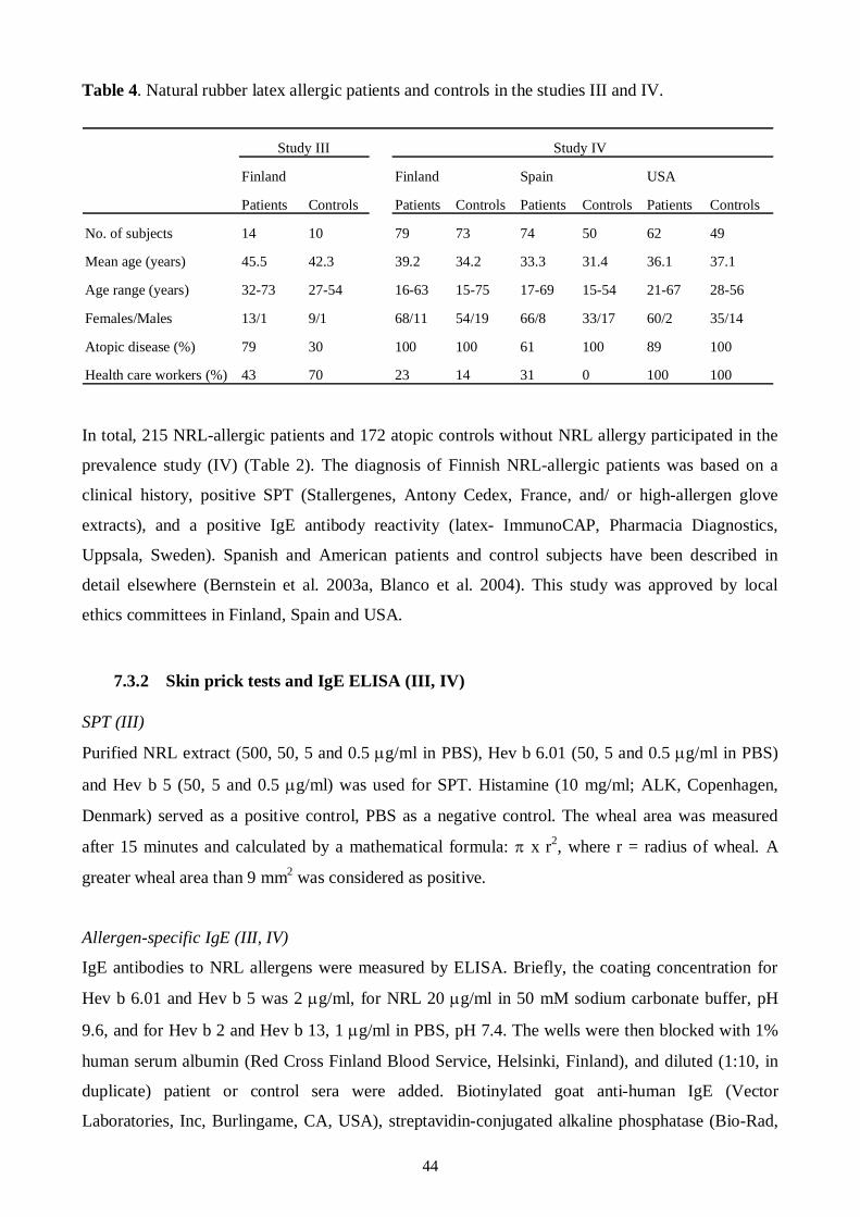
44
Table 4. Natural rubber latex allergic patients and controls in the studies III and IV.
Study III Study IV
Finland Finland Spain USA
Patients Controls Patients Controls Patients Controls Patients Controls
No. of subjects 14 10 79 73 74 50 62 49
Mean age (years) 45.5 42.3 39.2 34.2 33.3 31.4 36.1 37.1
Age range (years) 3273 2754 1663 1575 1769 1554 2167 2856
Females/Males 13/1 9/1 68/11 54/19 66/8 33/17 60/2 35/14
Atopic disease (%) 79 30 100 100 61 100 89 100
Health care workers (%) 43 70 23 14 31 0 100 100
In total, 215 NRLallergic patients and 172 atopic controls without NRL allergy participated in the
prevalence study (IV) (Table 2). The diagnosis of Finnish NRLallergic patients was based on a
clinical history, positive SPT (Stallergenes, Antony Cedex, France, and/ or highallergen glove
extracts), and a positive IgE antibody reactivity (latex ImmunoCAP, Pharmacia Diagnostics,
Uppsala, Sweden). Spanish and American patients and control subjects have been described in
detail elsewhere (Bernstein et al. 2003a, Blanco et al. 2004). This study was approved by local
ethics committees in Finland, Spain and USA.
7.3.2 Skin prick tests and IgE ELISA (III, IV)
SPT (III)
Purified NRL extract (500, 50, 5 and 0.5 g/ml in PBS), Hev b 6.01 (50, 5 and 0.5 g/ml in PBS)
and Hev b 5 (50, 5 and 0.5 g/ml) was used for SPT. Histamine (10 mg/ml; ALK, Copenhagen,
Denmark) served as a positive control, PBS as a negative control. The wheal area was measured
after 15 minutes and calculated by a mathematical formula: x r2, where r = radius of wheal. A
greater wheal area than 9 mm2 was considered as positive.
Allergenspecific IgE (III, IV)
IgE antibodies to NRL allergens were measured by ELISA. Briefly, the coating concentration for
Hev b 6.01 and Hev b 5 was 2 g/ml, for NRL 20 g/ml in 50 mM sodium carbonate buffer, pH
9.6, and for Hev b 2 and Hev b 13, 1 g/ml in PBS, pH 7.4. The wells were then blocked with 1%
human serum albumin (Red Cross Finland Blood Service, Helsinki, Finland), and diluted (1:10, in
duplicate) patient or control sera were added. Biotinylated goat antihuman IgE (Vector
Laboratories, Inc, Burlingame, CA, USA), streptavidinconjugated alkaline phosphatase (BioRad,
45
Hercules, CA, USA) and color substrate (pnitrophenyl phosphate, ICN Biomedicals Inc., Aurora,
Ohio, USA) were used to detect bound IgE. The absorbance values were read at 405 nm, when
pooled control sera had reached predetermined ODvalues. Positive reactivity was defined as
absorbance values exceeding the 98th percentile of local atopic controls (approximately the mean +
3 SD) (IV).
Inhibition ELISA tests (IV)
The assay was performed using the allergenspecific ELISA protocol described above, except that a
competitive inhibition step was added (Mikkola et al. 1998). Inhibition was performed by
incubating sera (final dilution 1/10) for 1 hour at room temperature with an equal volume of
inhibitor solution. Different NRL allergens were used to inhibit the binding of patients’ IgE to
solidphase antigens (IV).
7.3.3 PBMC proliferation assay (III)
PBMC were cultured in complete RPMI 1640 containing 5% heat inactivated human AB serum as
earlier described (Lehto et al. 2003). Briefly, Ficoll gradient centrifugation of peripheral blood was
used to obtain PBMC. Isolated cells were washed twice with PBS. PBMC were stimulated alone or
together with allergens (NRL 100, 50, 10, 5, 1 g/ml, Hev b 6.01 10, 5, 1 and 0.1 g/ml, Hev b 5
10, 5, 1 and 0.1 g/ml). Phytohaemagglutinin was used as a positive control (PHA, 45 g/ml;
Murex Biotech Ltd., Dartford, UK). Proliferation was determined by tritiated methylthymidine
(Amersham Biosciences, Little Chalfont, UK) incorporation after 6 days of culturing. The
proliferation results were estimated as stimulation indexes (uptake of isotope in stimulated
culture/uptake of isotope in nonstimulated control culture).
7.3.4 Cytokine and chemokine assays (III)
Cell culture
PBMC were cultured in complete RPMI 1640 on 24well plates (Costar, Corning Incorporated,
Corning, NY, USA) with or without Hev b 6.01 (10 g/ml) or Hev b 5 (10 g/ml). PHA (45 g/ml)
and Staphylococcus aureus enterotoxin B (SEB; 1 g/ml; SigmaAldrich Co Ltd., Gillingham, UK)
were utilized as polyclonal stimulators of PBMC. The cell culture time was 6 hours for flow
cytometry analysis and isolation of RNA, whereas cell culture supernatants were collected after 24
hours.
46
Cytokine and chemokine mRNA measurements by PCR
Total RNA was isolated from PBMC according to Trizol instructions (Gibco, BRL). Subsequently,
synthesis of cDNA and realtime quantitative PCR with AbiPrism 7700 Sequence Detector System
(Applied Biosystems) were performed as earlier described (I, II). PCR primers and probes were
obtained from Applied Biosystems, where they were also designed. Cytokine and chemokine
mRNA results have been expressed as relative units (RU), which were calculated by the
comparative CT method as earlier described (I).
Cytokine and chemokine protein measurements by ELISA
Commercial ELISA kits were used to measure protein levels of TNF, IL10 and CCL3. The
sensitivities of TNF and IL10 kits were 4 pg/ml and 2 pg/ml, respectively (eBioscience, Inc.; San
Diego, CA, USA). The lower detection limit of the CCL3 kit (R&D Systems, Minneapolis, MN,
USA) was 8 pg/ml.
7.3.5 Flow cytometry (III)
Measurements of Tcell surface markers were made by FACSCalibur (BD Biosciences, San Jose,
CA, USA) using FACSComp software version 4.01 (BD Biosciences). Monoclonal antibodies and
corresponding isotype controls were obtained from BD Biosciences. In brief, PBMC were incubated
with the fluorochromeconjugated monoclonal antibodies, washed, and fixed in 1%
paraformaldehyde (BD Biosciences). A total of 20 000 events within the lymphocyte gate were
collected. The numbers of CCR4 and CXCR3 positive cells within the CD3+, CD3+CD8+ and
CD3+CD8 populations were analyzed by BD CELLQuestPro software (BD Biosciences).
47
8 RESULTS
8.1 Experiments in mice
8.1.1 NRL exposure to the skin: cutaneous and systemic responses (I)
Repeated EC exposure to NRL induced strong epidermal and dermal thickening in sensitized skin
sites in mice (I, Figs 1 and 2). Infiltration of inflammatory cells, especially eosinophils, increased
significantly into the NRLsensitized skin sites, when compared to PBStreated sites (I, Fig. 3). A
significant increase in the number of degranulated mast cells was also seen in NRLsensitized skin
sites, although the total number of mast increased insignificantly (I, Figs 4 and 5). In addition, a
significant 5fold increase in the numbers of CD4 positive cells and a 7fold increase in CD3
positive cells were observed after EC NRL exposure (I, Fig. 6).
Epicutaneous exposure to NRL caused enhanced expression of proinflammatory and
Th2 type cytokines. After NRL sensitization, mRNA expression of IL1 and IL4 increased
significantly in skin, whereas the enhancement of IFN mRNA expression was insignificant as
compared to PBStreated skin (I, Fig. 7). In agreement with the evidence of inflammatory cell
recruitment into NRLtreated skin, mRNA expression of several chemokines increased
significantly in NRL exposed skin sites. Expression of CCL3, CCL4, CCL2, and CCL11 mRNA
was induced significantly after epicutaneous sensitization compared to PBStreated skin (I, Fig. 8).
On the other hand, mRNA expression of CCL17 and CCL24 was moderate and comparable in both
mice groups, whereas expression of CCL27 mRNA was strong, but there were no differences
between the animal groups.
In contrast to intraperitoneal exposure, epicutaneous exposure to NRL induced high
amounts of total and specific IgE to NRL and Hev b 6.01 (I, Figs 9 and 10). Both EC sensitization
and IP immunization with NRL induced low but comparable levels of Hev b 1specific IgE
antibodies. On the other hand, intraperitoneal, but not epicutaneous, NRLtreatment caused
enhancement of the NRL and Hev b 1specific IgG2a antibody response.
8.1.2 NRL exposure routes: respiratory and systemic responses (II)
IC and IP exposure caused perivascular and peribronchial inflammation of lung tissues after airway
NRL challenge, whereas after IN exposure, only a slight, mostly perivascular inflammation was
detected (II, Figs 1A and S1). The number of eosinophils increased dramatically after IC treatment,
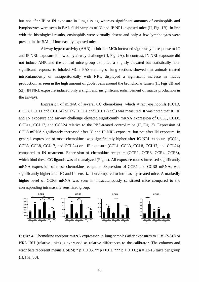
48
but not after IP or IN exposure in lung tissues, whereas significant amounts of eosinophils and
lymphocytes were seen in BAL fluid samples of IC and IP NRLexposed mice (II, Fig. 1B). In line
with the histological results, eosinophils were virtually absent and only a few lymphocytes were
present in the BAL of intranasally exposed mice.
Airway hyperreactivity (AHR) to inhaled MCh increased vigorously in response to IC
and IP NRL exposure followed by airway challenge (II, Fig. 2A). In contrast, IN NRL exposure did
not induce AHR and the control mice group exhibited a slightly elevated but statistically non
significant response to inhaled MCh. PASstaining of lung sections showed that animals treated
intracutaneously or intraperitoneally with NRL displayed a significant increase in mucus
production, as seen in the high amount of goblet cells around the bronchiolar lumen (II, Figs 2B and
S2). IN NRL exposure induced only a slight and insignificant enhancement of mucus production in
the airways.
Expression of mRNA of several CC chemokines, which attract eosinophils (CCL3,
CCL8, CCL11 and CCL24) or Th2 (CCL1 and CCL17) cells was measured. It was noted that IC, IP
and IN exposure and airway challenge elevated significantly mRNA expression of CCL1, CCL8,
CCL11, CCL17, and CCL24 relative to the PBStreated control mice (II, Fig. 3). Expression of
CCL3 mRNA significantly increased after IC and IP NRL exposure, but not after IN exposure. In
general, expression of most chemokines was significantly higher after IC NRL exposure (CCL1,
CCL3, CCL8, CCL17, and CCL24) or IP exposure (CCL1, CCL3, CCL8, CCL17, and CCL24)
compared to IN treatment. Expression of chemokine receptors (CCR1, CCR3, CCR4, CCR8),
which bind these CC ligands was also analyzed (Fig. 4). All exposure routes increased significantly
mRNA expression of these chemokine receptors. Expression of CCR1 and CCR8 mRNAs was
significantly higher after IC and IP sensitization compared to intranasally treated mice. A markedly
higher level of CCR3 mRNA was seen in intracutaneously sensitized mice compared to the
corresponding intranasally sensitized group.
Figure 4. Chemokine receptor mRNA expression in lung samples after exposures to PBS (SAL) or
NRL. RU (relative units) is expressed as relative differences to the calibrator. The columns and
error bars represent means SEM; * p < 0.05, ** p< 0.01, *** p < 0.001; n = 1215 mice per group
(II, Fig. S3).
CCR1
SAL NRL SAL NRL SAL NRL0
5000
10000
15000
20000
25000
30000
35000
IC IP IN
****
**
***
***
CCR3
SAL NRL SAL NRL SAL NRL
100
300
500
700
900
1100
IC IP IN
*** ****
***
CCR4
SAL NRL SAL NRL SAL NRL
500
1500
2500
3500
4500
IC IP IN
**
**
***
CCR8
SAL NRL SAL NRL SAL NRL0
20
40
60
80
100
120
IC IP IN
**
49
The cytokine expression profile at the mRNA level revealed differences between different exposure
routes. IC and IP exposure to NRL clearly increased the expression levels of IL4 and IL13
mRNA, but only IC exposure increased significantly the expression of IL5 mRNA in lung
compared to PBS treated controls (II, Fig. 4A). In addition, the IL13 mRNA level was elevated (8
fold) in the intranasally NRL exposed mice in comparison with the controls. IC exposure elicited
significantly higher expression of IL4, IL5, and IL13 mRNA compared to IN group. The mRNA
levels of regulatory cytokine IL10 enhanced significantly in all NRL exposed groups, especially in
intranasally treated mice (II, Fig. 4B).On the other hand, a significant elevation in the levels of
TGF 1 mRNA was observed after IN and IP NRL exposure but not after IC exposure. In addition,
the TGF 1 mRNA levels were significantly higher in IN and IP exposed mice than in IC exposed
mice (II, Fig. 4B). Forkhead box 3 (Foxp3) transcription factor mRNA expression also increased
significantly after IP and IN NRL administration but not after IC exposure (II, Fig. 4B) with the
elevation in expression being most marked after IN NRL exposure.
Total and Hev b 6.01specific IgE antibodies were strongly elevated after IC and IP
exposure, but not after IN exposure to NRL (II, Fig. 5).
8.2 Studies in patients allergic to natural rubber latex
8.2.1 PBMC responses to Hev b 5 and Hev b 6.01: proliferation, cytokines and
chemokines (III)
Two highly purified native NRL allergens Hev b 5 and Hev b 6.01 were used to stimulate PBMC of
NRLallergic patients and controls. IgE reactivity of the study groups was studied by SPT and
allergenspecific IgE ELISA. All 14 patients had positive (wheal area 9 mm2) SPT to NRL, 12 to
Hev b 6.01 and 4 to Hev b 5. In addition, 2 patients showed borderline positive SPT responses
(wheal area 79 mm2) to Hev b 5 (III, Fig. 1A). In ELISA, 13 patients demonstrated increased IgE
to NRL, 12 to Hev b 6.01 and 3 to Hev b 5 (III, Fig. 1B). All control sera were negative against all
NRL allergens (III, Fig. 1B)
Proliferative responses of PBMC to Hev b 6.01 were significantly higher in NRL
allergic patients than in controls, whereas stimulation indexes against NRL and Hev b 5 revealed no
differences between the two groups (III, Fig. 1C). There was a strong proliferative response against
PHA but no differences were seen between responses in patients and controls.
Hev b 6.01, and to a lesser extent Hev b 5, stimulated PBMC of the NRLallergic
patients and were found to express more TNF mRNA and protein on PBMC compared with the
50
nonallergic controls (III, Figs 2A, 2B). They also clearly induced the expression of IL12p40
mRNA in NRLallergic patients (III, Fig. 2C). In addition, expression of the Th2 cytokine IL13
mRNA increased significantly after allergenspecific stimulation (III, Fig. 2E), whereas the
expression of the Th1 cytokine, IFN , did not change (III, Fig. 2D). Finally, PBMC from NRL
allergic patients expressed significantly lower amounts of the regulatory cytokine TGF 1 mRNA
(III, Fig. 2F) after allergen stimulation compared with the controls, while the expressions of IL10
mRNA and protein (III, Figs 2G and H) were upregulated. Polyclonal stimulators (PHA and SEB)
induced the expression of several cytokines (i.e., TNF, IFNgamma, IL13, and IL10) in NRL
allergic patients as well as in control subjects (III, Figs 2 A, B, D, E, G, H), but the only significant
difference was that SEB downregulated the expression of TGF 1 mRNA in the patients’ PBMC
(III, Fig. 2F).
Hev b 6.01 and Hev b 5 stimulation of PBMC caused significantly higher mRNA
expression of the proinflammatory chemokines CCL3 (both mRNA and protein), CCL4 and CCL20
in NRLallergic patients than in controls (III, Fig. 3). On the contrary, expression of the Th1type
chemokine CXCL10 mRNA was significantly induced only after polyclonal stimulation, and there
were no differences in the expression levels between patients and the controls (III, Fig. 3E).
Expression of the Th2type chemokine CCL17 mRNA was enhanced to a lesser degree after
allergenspecific stimulation compared to the situation after polyclonal stimulation (III, Fig. 3F).
Levels of CCL17 mRNA were similar in the patient and control groups (III, Fig. 3F).
Surface expression of the chemokine receptors CCR4 and CXCR3 on CD3+CD8 and
CD3+CD8+ T cells did not change after any stimulation (III, Figs 4A and B). However, baseline
expression of CCR4 on CD3+CD8 T cells was significantly enhanced and that of CXCR3 on
CD3+CD8+ T cells was significantly reduced in the patient group compared to the control group
(III, Fig. 4).
8.2.2 Prevalence of IgE antibodies to Hev b 2 and Hev b 13 (IV)
One important aspect in the prevalence studies of allergenspecific IgE antibodies is the identity and
purity of allergen preparations. Homogeneity of Hev b 2, Hev b 5, Hev b 6.01, and Hev b 13 was
ensured by reversedphase chromatography (IV, e.g. Fig. 1), and their identity was confirmed using
monoclonal antibodies specific for Hev b 2, Hev b 5, Hev b 6.01, and Hev b 13. It is of interest,
however, that low binding of monoclonal antiHev b 6.02 antibodies to purified Hev b 13 was seen
indicating that minute amounts of Hev b 6.02 reactivity were still present even in the highly purified
Hev b 13 (IV, Fig. 2).
51
There were welldefined differences in the prevalence of IgE specific for the four
NRL allergens in serum from NRLallergic patients from Finland, Spain and the USA (IV, Fig. 3).
IgE antibodies specific for Hev b 2 were rare in Spain (5 %), and seen only in a minority of patients
from Finland (15%) and USA (11%). The prevalence of IgE to Hev b 13 ranged between 18 to 30%
in all three countries, suggesting a relatively low overall frequency of sensitization to this allergen.
The prevalence of IgE antibodies to Hev b 5 was unexpectedly low in Finland (28%), in contrast to
the sera collected from Spanish and American patients (49% and 71%, respectively). Meanwhile,
the frequency of IgE antibodies to Hev b 6.01 was > 50% in the serum of NRLallergic patients in
all three countries (IV, Fig. 3).
Inhibition ELISA tests established that purified soluble Hev b 6.01 inhibited the
binding of patient specific IgE to immobilized Hev b 13 in a dosedependent manner with most
studied sera. There were three different modes of inhibition (IV, Fig. 5). Either soluble Hev b 6.01
could not inhibit antiHev b 13 IgE binding (IV, Fig. 5A) or inhibited binding partially (IV, Fig. 5B)
or totally (IV, Fig. 5C). Inhibition experiments were carried out for 8 sera, which displayed
moderate to strong IgE antibody response to Hev b 13. Two of these sera produced a 24%
inhibition of the binding of IgE to solidphase Hev b 13, when the concentration of Hev b 6.01 was
0.1 g/ml (III, such as Fig. 5A). Two other serum samples produced 2654% inhibition (IV, such as
Fig. 5B), and with the remaining four sera the inhibition was between 69 to 95% after exposure to
soluble Hev b 6.01(IV, like Fig. 5C).
52
9 DISCUSSION
The peak of the epidemic of NRL allergy in the health care sector seems to have passed in Western
Countries but this allergy is still a problem among HCW and people using gloves or other NRL
products (Ahmed et al. 2003, Reunala et al. 2004). Proteins eluting from NRL gloves cause in the
skin type I immediate symptoms such as contact urticaria but hand eczema may also be one
phenotypic manifestation of NRL allergy (Turjanmaa et al. 2002b). On the other hand, airborne
glove powder contaminated with NRL allergens can cause asthma and allergic rhinitis in sensitized
subjects (Vandenplas et al. 2002). In this thesis, the significance of different exposure routes in the
development of NRLallergy was examined by using mouse models. The findings highlight the
importance of cutaneous exposure in inducing a strong allergenspecific IgE antibody response,
development of Th 2type dermatitis and marked airway sensitization predisposing to NRLallergic
asthma (I, II). Cellular immune mechanisms in NRLallergic patients were studied by investigating
PBMC responses to the major NRL allergens, Hev b 6.01 and Hev b 5. The results demonstrate that
allergenspecific stimulation of PBMC induces upregulation of several inflammatory cytokines and
chemokines. The prevalence of IgE antibodies was studied in NRLallergic patients from Finland,
Spain and USA using highly purified native NRL allergens (IV). In contrast to Hev b 6.01 and Hev
b 5, the prevalence rates of IgE antibodies to Hev b 2 and Hev b 13 were in the range of 5 to 30%
showing that these two allergens cannot be considered as major NRL allergens.
9.1 Experiments in mouse models of natural rubber latex allergy
9.1.1 Cutaneous responses to NRL (I)
Repeated EC exposure to NRL led to dermatitis characterized by skin thickening and dermal
infiltration of eosinophils and CD4+ T cells. Degranulated mast cells were also increased in NRL
sensitized skin, suggesting that these cells may also participate in the development of dermatitis. In
support of this theory, it has been previously reported that mast cells crucially affect IFNgamma
expression in a mouse model (Alenius et al. 2002). EC exposure to NRL also induced significant
expression of mRNA of proinflammatory (IL1beta) and Th2 (IL4) cytokines. A previous study
using a mouse model of allergic dermatitis showed that eosinophils were decreased and T cells
increased in the skin of ovalbuminsensitized IL4 deficient mice suggesting that this cytokine is
important in the modulation of protein induced skin inflammation (Spergel et al. 1999).
In line with enhanced traffic of inflammatory cells to the sensitized skin, epicutaneous
NRL exposure induced higher mRNA expression of CCL2, CCL3 and CCL4, which attract T cells,
53
monocytes, NK cells, and dendritic cells. Expression of CCL11, which is especially important in
the recruitment of eosinophils, mast cells and Th2 cells (Luster and Rothenberg 1997, Rot and von
Andrian 2004, Charo and Ransohoff 2006), was also enhanced in NRL sensitized skin. In addition,
mRNA expression of CCL17 and CCL27 was found, although the expression levels were similar
between control and NRL sensitized groups. CCL27 and CCL17 are important chemoattractants for
CCR10+CLA+T cells and CCR4+Th2 cells, respectively, during skin inflammation (Reiss et al.
2001, Homey et al. 2002). Cutaneous expression of CCL17 and CCL27 can then attract NRL
specific CD4+Th2 cells from the circulation to the inflamed skin. In summary, our results show
that EC exposure to NRL significantly enhances the expression of various chemokines in the skin
contributing to the recruitment of different inflammatory cells, especially T cells, eosinophils and
mast cells to the NRLsensitized skin sites.
Disruption of the skin barrier is likely to be an important factor influencing the
development of dermatitis in NRL allergy. Stratum corneum, the outer layer of the skin normally
prevents foreign substances from penetrating into and through the skin (Cookson 2004, Elias 2005,
Cork et al. 2006). In our mouse model, mechanical skin injury is induced by tape stripping
mimicking the effects of skin scratching in humans. Scratching is a common consequence of
pruritus, typical symptom of eczema patients and is thought to participate in the pathophysiology of
AE (Homey et al. 2006). Moreover, a dysfunction of skin barrier function of stratum corneum
strongly associates with AE (Cork et al. 2006, Morar et al. 2006). Especially two lossoffunction
mutations of the epidermal barrier protein filaggrin predispose an individual to AE (Hudson 2006,
Marenholz et al. 2006, Palmer et al. 2006, Weidinger et al. 2006).
Epicutaneous sensitization to NRL proteins described here presents a model for
protein contact dermatitis. This model incorporates several features that are likely to be important in
the pathogenesis of NRL allergy in humans. Induction of dermatitis manifested by epidermal and
dermal thickening and characterized by the dermal accumulation of eosinophils and degranulated
mast cells required cutaneous barrier disruption by tape stripping (injury) and repeated application
of NRL proteins. NRLsensitized skin exhibited an increase in the expression of Th2 cytokine
mRNA. These results in mouse indicate that epicutaneous exposure to protein antigens induces
dermatitis with a striking Th2 bias. To our knowledge, no biopsy studies have been performed from
hand eczema (dermatitis) in NRLallergic patients. However, NRL patch testing has evoked
eczematous reactions (Sommer et al. 2002). This suggests that repeated exposure to NRL proteins
similarly to the present mouse NRL model in humans could also cause delayed allergic responses in
skin, such as hand eczema or protein contact dermatitis. This is indirectly further supported by the
clinical findings in NRLallergic patients in most of whom avoidance of highallergenic NRL
gloves has resulted in improvement of hand dermatitis (Turjanmaa et al. 2002b).
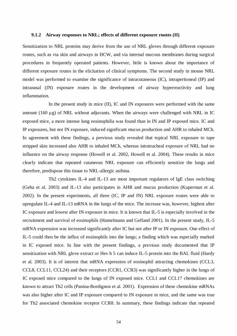
54
9.1.2 Airway responses to NRL; effects of different exposure routes (II)
Sensitization to NRL proteins may derive from the use of NRL gloves through different exposure
routes, such as via skin and airways in HCW, and via internal mucous membranes during surgical
procedures in frequently operated patients. However, little is known about the importance of
different exposure routes in the elicitation of clinical symptoms. The second study in mouse NRL
model was performed to examine the significance of intracutaneous (IC), intraperitoneal (IP) and
intranasal (IN) exposure routes in the development of airway hyperreactivity and lung
inflammation.
In the present study in mice (II), IC and IN exposures were performed with the same
amount (160 g) of NRL without adjuvants. When the airways were challenged with NRL in IC
exposed mice, a more intense lung eosinophilia was found than in IN and IP exposed mice. IC and
IP exposures, but not IN exposure, induced significant mucus production and AHR to inhaled MCh.
In agreement with these findings, a previous study revealed that topical NRL exposure to tape
stripped skin increased also AHR to inhaled MCh, whereas intratracheal exposure of NRL had no
influence on the airway response (Howell et al. 2002, Howell et al. 2004). These results in mice
clearly indicate that repeated cutaneous NRL exposure can efficiently sensitize the lungs and
therefore, predispose this tissue to NRLallergic asthma.
Th2 cytokines IL4 and IL13 are most important regulators of IgE class switching
(Geha et al. 2003) and IL13 also participates in AHR and mucus production (Kuperman et al.
2002). In the present experiments, all three (IC, IP and IN) NRL exposure routes were able to
upregulate IL4 and IL13 mRNA in the lungs of the mice. The increase was, however, highest after
IC exposure and lowest after IN exposure in mice. It is known that IL5 is especially involved in the
recruitment and survival of eosinophils (Hamelmann and Gelfand 2001). In the present study, IL5
mRNA expression was increased significantly after IC but not after IP or IN exposure. One effect of
IL5 could then be the influx of eosinophils into the lungs; a finding which was especially marked
in IC exposed mice. In line with the present findings, a previous study documented that IP
sensitization with NRL glove extract or Hev b 5 can induce IL5 protein into the BAL fluid (Hardy
et al. 2003). It is of interest that mRNA expression of eosinophil attracting chemokines (CCL3,
CCL8, CCL11, CCL24) and their receptors (CCR1, CCR3) was significantly higher in the lungs of
IC exposed mice compared to the lungs of IN exposed mice. CCL1 and CCL17 chemokines are
known to attract Th2 cells (PaninaBordignon et al. 2001). Expression of these chemokine mRNAs
was also higher after IC and IP exposure compared to IN exposure in mice, and the same was true
for Th2 associated chemokine receptor CCR8. In summary, these findings indicate that repeated
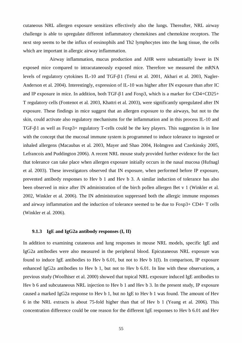
55
cutaneous NRL allergen exposure sensitizes effectively also the lungs. Thereafter, NRL airway
challenge is able to upregulate different inflammatory chemokines and chemokine receptors. The
next step seems to be the influx of eosinophils and Th2 lymphocytes into the lung tissue, the cells
which are important in allergic airway inflammation.
Airway inflammation, mucus production and AHR were substantially lower in IN
exposed mice compared to intracutaneously exposed mice. Therefore we measured the mRNA
levels of regulatory cytokines IL10 and TGF 1 (Terui et al. 2001, Akbari et al. 2003, Nagler
Anderson et al. 2004). Interestingly, expression of IL10 was higher after IN exposure than after IC
and IP exposure in mice. In addition, both TGF 1 and Foxp3, which is a marker for CD4+CD25+
T regulatory cells (Fontenot et al. 2003, Khattri et al. 2003), were significantly upregulated after IN
exposure. These findings in mice suggest that an allergen exposure to the airways, but not to the
skin, could activate also regulatory mechanisms for the inflammation and in this process IL10 and
TGF 1 as well as Foxp3+ regulatory Tcells could be the key players. This suggestion is in line
with the concept that the mucosal immune system is programmed to induce tolerance to ingested or
inhaled allergens (Macaubas et al. 2003, Mayer and Shao 2004, Holmgren and Czerkinsky 2005,
Lefrancois and Puddington 2006). A recent NRL mouse study provided further evidence for the fact
that tolerance can take place when allergen exposure initially occurs in the nasal mucosa (Hufnagl
et al. 2003). These investigators observed that IN exposure, when performed before IP exposure,
prevented antibody responses to Hev b 1 and Hev b 3. A similar induction of tolerance has also
been observed in mice after IN administration of the birch pollen allergen Bet v 1 (Winkler et al.
2002, Winkler et al. 2006). The IN administration suppressed both the allergic immune responses
and airway inflammation and the induction of tolerance seemed to be due to Foxp3+ CD4+ T cells
(Winkler et al. 2006).
9.1.3 IgE and IgG2a antibody responses (I, II)
In addition to examining cutaneous and lung responses in mouse NRL models, specific IgE and
IgG2a antibodies were also measured in the peripheral blood. Epicutaneous NRL exposure was
found to induce IgE antibodies to Hev b 6.01, but not to Hev b 1(I). In comparison, IP exposure
enhanced IgG2a antibodies to Hev b 1, but not to Hev b 6.01. In line with these observations, a
previous study (Woolhiser et al. 2000) showed that topical NRL exposure induced IgE antibodies to
Hev b 6 and subcutaneous NRL injection to Hev b 1 and Hev b 3. In the present study, IP exposure
caused a marked IgG2a response to Hev b 1, but no IgE to Hev b 1 was found. The amount of Hev
6 in the NRL extracts is about 75fold higher than that of Hev b 1 (Yeang et al. 2006). This
concentration difference could be one reason for the different IgE responses to Hev b 6.01 and Hev
56
b 1 in the present study. However, different antibody responses could be explained even better by
biochemical properties of the NRL allergens. Hev b 6.01 is hydrophilic and can therefore easily
penetrate into the skin. In contrast, Hev b 1 is hydrophobic and seems to be capable of causing
marked IgE response only when injected into the skin or intraperitoneally. In agreement with these
findings in the mouse model, Hev b 6.01 is known to be a major sensitizing allergen for gloveusing
HCW, whereas it seems that Hev b 1 needs to be delivered from NRL gloves or catheters to the
peritoneal or other mucous membranes before it can cause a significant IgE response. Apparently
this is the reason, why Hev b 1 is a significant allergen for spina bifida and other multioperated
patients (Alenius et al. 1996a, Alenius et al. 1996b, Chen et al. 1997a).
In the second study (II), IP exposure induced specific IgE antibodies to Hev b 6.01 in
contrast to the first study (I). One possible explanation for this discrepancy could be different IP
exposure protocols. In the first study (I), a booster injection was given after one week and serum
was obtained on day 19. In the second study (II), a booster injection was provided after two weeks
and the blood was collected after airway challenge on day 31. Therefore, in the first study, the IP
immunisation time may have been too short to elicit proper IgE responses. Another difference was
the amount of NRL antigen used during the IP exposures. The total amount was 40 g in the first
and 160 g in the second study. In line with the results in the second study (II), NRL specific IgE
antibodies have also been found after IP exposure in two other studies (Hardy et al. 2003, Hufnagl
et al. 2003).
9.2 Studies in patients allergic to natural rubber latex
Earlier studies on cellmediated immune responses with NRLallergic patients have focused on the
measurement of PBMC proliferation responses against different NRL allergens using crude NRL
extracts or purified native proteins or recombinant NRL allergens. One goal of these studies has
been to resolve, which allergens are relevant for clinical and diagnostic purposes. One aim of this
thesis was to obtain information about allergenspecific cytokine and chemokine responses in NRL
allergic patients by stimulating their peripheral blood mononuclear cells (PBMC) with highly
purified native major allergens Hev b 5 and Hev b 6.01. An additional aim was to assess IgE class
antibody prevalence to two recently characterized NRL allergens, Hev b 2 and Hev b 13, in three
different geographical areas to resolve whether these proteins truly meet the criteria for major
allergens.
57
9.2.1 PBMC responses to Hev b 5 and Hev b 6.01 (III)
In NRLallergic patients, diagnosed by positive SPT and glove challenge test, Hev b 6.01, but not
Hev b 5, induced significantly higher PBMC proliferation when compared to the control subjects.
This finding suggests that the patients had been mainly sensitized to Hev b 6.01. This result is in
agreement with the results of the fourth study of the present thesis (IV) which revealed that Finnish
NRLallergic patients had a markedly higher IgE antibody prevalence to Hev b 6.01 than to Hev b
5. A previous study indicated that the NRL proliferation test had a low sensitivity (39%) but high
specificity (95%) in NRL allergy (Ebo et al. 1997). It is also well documented that cellmediated
responses do not correlate with SPT reactivity or specific IgE antibody levels (RaulfHeimsoth et al.
1996, Ebo et al. 1997, Johnson et al. 1999).
Manifestations of allergic symptoms are greatly dependent on the cytokine and
chemokine milieu, where allergenspecific cells meet their corresponding allergens (Borish and
Rosenwasser 2003, Borish and Steinke 2003, Homey et al. 2006). The present study revealed that
the major NRL allergens Hev b 6.01 and Hev b 5 induced expression of proinflammatory cytokines
(TNF and IL12p40) and Th2type cytokine IL13. Interestingly, NRL allergen stimulation did not
induce IFN mRNA, although IL12 was upregulated and this cytokine is known to be a major
inducer of IFN (Hunter 2005). Most of the present NRLallergic patients were atopic subjects, and
it is of interest that after IL12 stimulation, IFN production has been shown to be low especially in
atopic patients (Matsui et al. 2000). Moreover, Th2 cell differentiation can occur even though in the
presence of IL12 signalling (Nishikomori et al. 2000). Thus, IL12 activation is clearly required for
Th1 immune responses, but it is not sufficient to suppress Th2 responses (Heath et al. 2000,
Nishikomori et al. 2000, Berenson et al. 2004). However, IL12 expression was measured at the
mRNA level using primers and probes specific for the IL12p40 subunit. Human IL12p40 subunit
is a part of heterodimeric proinflammatory cytokines IL12 and IL23, both of which induce Th1
type immune responses (Hunter 2005). Further studies on protein expression level are needed to
clarify which one of these related cytokines or perhaps both of them are important in the outcome of
NRL allergy. Overall, the present results imply that, in addition to Th2 cytokines (IL13), also
proinflammatory cytokines (IL12/IL23 and TNF) are involved in the induction of NRLspecific
immune responses.
IL10 and TFG 1 are important suppressors of immune responses and TGF
signaling by T cells is essential for tolerance and T cell homeostasis (Jutel et al. 2003, Hawrylowicz
2005, Hawrylowicz and O'Garra 2005, Horwitz 2006, Li et al. 2006a, Li et al. 2006b, Marie et al.
2006). The present finding of decreased expression of TGF 1 mRNA both after allergen and SEB
stimulations focuses attention onto the role of this regulatory cytokine in NRL allergy. On the other
58
hand, IL10 mRNA expression increased significantly in PBMC after allergen stimulation. This
suggests that IL10 may downregulate Th1 responses and as a result, the Th2 response is more
prominent in NRLallergic patients. In agreement, a recent study with IL10 deficient mice in a
mouse model of allergic dermatitis revealed that IL10 was crucial for the development of
cutaneous Th2 response and eosinophilia (Laouini et al. 2003).
Allergen stimulation markedly increased the expression of the proinflammatory
chemokines CCL3, CCL4 and CCL20 mRNA in the NRLallergic patients. CCL3 seems to be
important for optimal mast cell degranulation (Miyazaki et al. 2005) and CCL3 and CCL4 co
stimulate T cell responses (Molon et al. 2005, Trautmann 2005). It is of interest that allergen
specific stimulation did not induce the Th2type chemokine CCL7 or the Th1type chemokine
CXCL10. These results indicate that proinflammatory chemokines, rather than Th1 or Th2
chemokines, seem to play an important role in the immune responses in NRL allergy.
Flow cytometric analysis of CCR4 (Th2 type) and CXCR3 (Th1 type) chemokine
receptors on the T cells did not exhibit changes after stimulation with NRLallergens. However,
there were differences in the expression pattern of these chemokine receptors in the T cell
subpopulations. NRLallergic patients showed increased CCR4 expression on CD3+CD4+ T cells,
whereas CXCR3 expression was decreased on CD3+CD8+ T cells. In agreement with these
findings in NRLallergic patients, previous studies in AE have been shown an increase of CCR4
expression on peripheral blood CD4+ T cells (Wakugawa et al. 2001) and a decrease of CXCR3 in
PBMC (Hatano et al. 2001).
9.2.2 Prevalence of IgE antibodies to Hev b 2 and Hev b 13 (IV)
In the present study, the prevalence rates of IgE antibodies to Hev b 6.01 were high (5474%) in
Finland, Spain and the USA. This result from these three countries confirms that Hev b 6.01 is the
most important NRL allergen, especially in HCW a fact that was also documented in a recent meta
analysis (Bousquet et al. 2006). Hev b 5 is the second major NRL allergen. The prevalence of IgE
to Hev b 5 was found to be high in the USA (71%) and Spain (49%) but rather low (28%) in
Finland. The reasons for these differences in the patients series may also be due to the variable
amounts of Hev b 5 in the NRL gloves used in different countries (Palosuo et al. 2002).
The prevalence rates of IgE antibodies to both Hev b 2 (515%) and Hev b 13 (18
30%) were low, showing that these NRL proteins should not be classified as major NRL allergens.
These prevalence rates which are obtained by using extensively purified allergens are markedly
lower than those previously reported (Kurup et al. 2000, Bernstein et al. 2003a, Yeang 2004, Kurup
et al. 2005). It is noteworthy that in all of the studies cited above, the native Hev b 2 and Hev b 13
59
allergen preparations used were obtained from one single laboratory. In the present study, it was
shown that binding of IgE to insufficiently purified Hev b 13 could be partially or totally inhibited
in most sera studied by the addition of soluble Hev b 6.01. This suggests that there is some impurity
expressing or resembling Hev b 6.01 or Hev b 6.02 epitopes in the conventionally purified Hev b 13
allergen and the high prevalence rates obtained earlier may be explained by contamination by Hev b
6–like components. Since no sequence homology between Hev b 13 and Hev b 6.02 has been found
significant crossreactivity between these proteins is unlikely. Moreover, if crossreactivity could
explain this phenomenon, it should remain at the same or even higher level when the purity of Hev
b 13 increases. Consistent with the present results is a recent report that shows that only 10% of
Taiwanese HCW had IgE binding to Hev b 13 that had been quality controlled with peptide mass
fingerprinting and analyzed by immunoblotting after twodimensional gel electrophoresis (Lee et al.
2006).
60
10 SUMMARY AND CONCLUSIONS
Allergy to NRL has been an important health issue during the last 20 years. In particular, HCW
have been sensitized to NRL proteins diffusing from the protective gloves. NRL causes type I
allergy symptoms such as contact urticaria but it is not known whether delayed symptoms such as
hand eczema could be one phenotypic manifestation of NRL allergy. Airborne NRL allergens could
theoretically cause asthma but little is known about cutaneous and airway route sensitization and
inflammatory responses to NRL in the skin and lung. At present, IgE mediated responses are well
characterized in NRL allergy but allergenspecific cellmediated immune responses are poorly
understood. Considerable progress has been made in the characterization of NRL allergens and
there is agreement that the major allergens present in the gloves are hevein (Hev b 6.02) and Hev b
5. Recently, it has been claimed that Hev b 2 and Hev b 13 could also be major NRL allergens.
Cutaneous inflammatory responses to NRL were explored in a mouse model of NRL
allergy (I). Mouse skin exposed to NRL developed a Th2 type dermatitis consisting of CD4+ T
cells, eosinophils and degranulated mast cells. This inflammatory response was associated with
increased expression of IL1 , IL4 and inflammatory chemokines. Cutaneous NRL exposure
induced also significant humoral Hev b 6.01 specific IgE responses. On the basis of these findings,
it can be concluded that in addition to producing significant allergenspecific IgE responses,
cutaneous NRL exposure can also induce Th2type dermatitis. Whether the common hand eczema
in NRLallergic patients would have a similar pathophysiology and could be termed as protein
contact dermatitis needs to be assessed in human studies. Mouse models have been invaluable in
studies on pathophysiology of different diseases. Nonetheless, the many differences existing
between mice and human immunological responses necessitate that any results obtained in mouse
models need to be confirmed in human studies.
A NRL mouse model was also used to examine the impact of cutaneous NRL
exposure in the development of airway hypersensitivity (II). NRL airway challenge caused
significant AHR, lung mucus production and influx of mononuclear cells and eosinophils into the
lungs in intracutaneously but not in intranasally exposed mice. The expressions of mRNA of Th2
cytokines (IL4 and IL13), and several CC chemokines and chemokine receptors were similarly
induced in the lungs. Intranasally exposed mice exhibited an increase in the expression of the
downregulatory TGF 1 and Foxp3 mRNA in the lungs but negligible levels of IgE to Hev b 6.01
in the blood. These results suggest that repeated cutaneous NRL exposure predisposes mice to
NRLallergic asthma.
61
In human studies, PBMC from NRLallergic patients underwent significant
proliferation responses to Hev b 6.01 but not to Hev b 5 (III). Both allergens induced significant
expression of mRNA of proinflammatory (TNF, IL12p40) and Th2 (IL13) cytokines. Regulatory
cytokine expression was complex; levels of IL10 were increased but TGF 1 decreased as
compared to control subjects. NRL allergens also induced significant expression of mRNA of
inflammatory CCchemokines. It is important that major NRL allergens Hev b 6.01 and Hev b 5
were shown to induce proinflammatory cytokines and chemokines and that regulatory cytokines
also seemed to be involved in the NRLspecific immune responses. These findings increase our
knowledge of the immune mechanisms involved in NRL allergy and also highlight the possible role
of regulatory cytokines in the maintenance of this allergy. Whether tolerance to NRL allergens can
be achieved in NRLallergic subjects by natural mechanisms or by immune therapy remains an
open question that needs to be addressed in future studies.
To reassess the importance and role of two NRL allergens, Hev b 2 and Hev b 13,
which were recently proposed to be major NRL allergens, a large IgE prevalence study was
undertaken in NRLallergic patients from Europe and North America. In this study, extensively
purified native Hev b 2 and Hev b 13 allergen preparations were used. The prevalence was shown to
be low to both allergens (from 5% to 30%) in contrast to Hev b 6.01 and Hev b 5 (from 28% to
74%). These results indicate that Hev b 6.01 and Hev b 5, but not Hev b 2 and Hev b 13, are major
NRL allergens both in Europe and in the USA. This study highlights the importance of using
extensively purified native NRL allergens when characterizing IgE responses in patient populations.
Thus caution is necessary in interpretation of results where native NRL allergens have been used for
diagnostic purposes. Whether recombinant Hev b 2 and Hev b 13 allergens, if in the future they
were produced as immunologically reactive molecules, would exhibit better performance than the
native compounds in prevalence assessment and diagnosis of NRL allergy needs to be elucidated in
further studies.

62
11 ACKNOWLEDGEMENTS
The laboratory work of this study was carried out in the Unit of Excellence for Immunotoxicology
at the Finnish Institute of Occupational Health and in the Laboratory of Immunobiology at the
National Public Health Institute. The clinical work was carried out in the Department of
Dermatology at the Helsinki University Hospital for Skin and Allergic Diseases. I thank directors
of these institutes, Professors Harri Vainio (the present director) and Jorma Rantanen (the previous
director) from the Finnish Institute of Occupational Health, Professors Pekka Puska (the present
director) and Jussi Huttunen (the previous director) from the National Public Health Institute and
Professor Annamari Ranki the Head of the Department of Dermatology, for excellent research
facilities.
I owe my deepest gratitude to my supervisors Professor Harri Alenius and Professor Timo Reunala.
It has been stimulating to work with them, as both of them are deeply interested in new advances in
science. Their support and positive attitude have encouraged my progress in the scientific world of
immunology and allergology.
I thank my referees Professor Ilkka Harvima and Professor Rauno Mäntyjärvi for careful reading of
this thesis and for their valuable comments and suggestions in the final version.
I kindly thank all my coauthors of the original publications. My special thanks go to Timo Palosuo
whose kindness and enthusiasm have been invaluable in the guidance given in scientific writing
and humoral immunology. I am grateful to Kristiina Turjanmaa for her support for my studies. She
has also succeeded to bring geniality to the world of research. Meetings of the old latex group in
her summer house at Ruhala have been one example of how to mix pleasure with the research
world. I thankfully remember the whole latex group which has been an important support group for
my thesis. Especially I thank Kati Palosuo, Piia Karisola, Kirsi Vaali, Tuija Puumalainen and Ulla
Seppälä for many enlightening discussions and their pleasant company during scientific meetings
in Finland and abroad. I am grateful to Elina Varjonen and Leena Petman who contributed to the
clinical part of this study. Nisse Kalkkinen and Annika Kotovuori have provided invaluable help in
protein purification. In addition, I am grateful to Antti Lauerma and Mika J Mäkelä for their
expertise knowledge of cutaneous and airway diseases, respectively. Henrik Wolff, Guoying Wang,
Iman Amghaiab and Tuula Stjernvall have been indispensable in histological sample analysis. Kai
Savolainen and other coworkers in the Finnish Institute of Occupational Health have created a

63
relaxing and innovative environment for research work. I warmly thank all my friends and
colleagues in this institute. My special thanks go to our study group for creating such a good
working atmosphere and so many cheerful moments in the laboratory and outside it. My dear
friends and colleagues Minna Anthoni, Nanna FyhrquistVanni, Rita Haapakoski, Helena
Honkasalo, Sari Lehtimäki, Marina Leino, MarjaLeena Majuri, Lea Pylkkänen, Johanna
Rintahaka, Terhi Savinko, Helene StockmannJuvala and Sari Tillander are acknowledged for their
expert help and advice in addition being good company. I also want to thank Ewen MacDonald for
English language revision of this thesis.
I am thankful to all the patients who participated in these studies. Their contribution has been
invaluable when studying clinically important aspects of NRL allergy.
I want to thank all my friends and relatives for their patience, because I have had so little time for
them during these years of intensive research work. Especially my late motherinlaw Kaarina and
my fatherinlaw Toivo have supported me. My warmest thanks go to my own family, especially to
my dear husband Jokke, my wonderful children Jukka and Sarianne, and the newest member of our
family, my delightful daughterinlaw Helena. They all have made it possible for me to have a
family life in addition to doing research work. My husband Jokke has excellently taken care of our
home and children while I was doing long days in the laboratory. In addition, he has patiently
listened to my work problems, offering neverending support and encouragement. My children
have produced many happy moments in my life and clarified what is the most important and
relevant in life. I believe that this study would not have been possible without the support and
presence of my family in my life.
This study was financially supported by grants from the Academy of Finland, the Allergy Research
Foundation, the Finnish Work Environment Fund, and the Allergy Foundation. The Ansell
Healthcare Corporation also funded this work.
Helsinki, March 2007
Maili Lehto
64
12 REFERENCES
Aalberse RC (2000): Structural biology of allergens. J Allergy Clin Immunol 106:228238.
Ahmed DD, Sobczak SC and Yunginger JW (2003): Occupational allergies caused by latex.Immunol Allergy Clin North Am 23:205219.
Akbari O, Stock P, DeKruyff RH and Umetsu DT (2003): Role of regulatory T cells in allergy andasthma. Curr Opin Immunol 15:627633.
Akdis CA and Akdis M (2003): Immunological differences between intrinsic and extrinsic types ofatopic dermatitis. Clin Exp Allergy 33:16181621.
Akdis M, Blaser K and Akdis CA (2005): T regulatory cells in allergy: novel concepts in thepathogenesis, prevention, and treatment of allergic diseases. J Allergy Clin Immunol 116:961968;quiz 969.
Akdis CA, Akdis M, Bieber T, BindslevJensen C, Boguniewicz M, Eigenmann P, Hamid Q, KappA, Leung DY, Lipozencic J, Luger TA, Muraro A, Novak N, PlattsMills TA, Rosenwasser L,Scheynius A, Simons FE, Spergel J, Turjanmaa K, Wahn U, Weidinger S, Werfel T and ZuberbierT (2006): Diagnosis and treatment of atopic dermatitis in children and adults: European Academyof Allergology and Clinical Immunology/American Academy of Allergy, Asthma andImmunology/PRACTALL Consensus Report. Allergy 61:969987.
Alenius H, Palosuo T, Kelly K, Kurup V, Reunala T, MakinenKiljunen S, Turjanmaa K and Fink J(1993): IgE reactivity to 14kD and 27kD natural rubber proteins in latex allergic children withspina bifida and other congenital anomalies. Int Arch Allergy Immunol 102:6166.
Alenius H, Kurup V, Kelly K, Palosuo T, Turjanmaa K and Fink J (1994): Latex allergy: frequentoccurrence of IgE antibodies to a cluster of 11 latex proteins in patients with spina bifida andhistories of anaphylaxis. J Lab Clin Med 123:712720.
Alenius H, Kalkkinen N, Lukka M, Reunala T, Turjanmaa K, MakinenKiljunen S, Yip E andPalosuo T (1995): Prohevein from the rubber tree (Hevea brasiliensis) is a major latex allergen. ClinExp Allergy 25:659665.
Alenius H, Kalkkinen N, Reunala T, Turjanmaa K and Palosuo T (1996a): The main IgEbindingepitope of a major latex allergen, prohevein, is present in its Nterminal 43amino acid fragment,hevein. J Immunol 156:16181625.
Alenius H, Kalkkinen N, Yip E, Hasmin H, Turjanmaa K, MakinenKiljunen S, Reunala T andPalosuo T (1996b): Significance of rubber elongation factor as a latex allergen. Int Arch AllergyImmunol 109:362368.
Alenius H, Laouini D, Woodward A, Mizoguchi E, Bhan AK, Castigli E, Oettgen HC and Geha RS(2002): Mast cells regulate IFNgamma expression in the skin and circulating IgE levels in allergeninduced skin inflammation. J Allergy Clin Immunol 109:106113.
65
Allmers H, Schmengler J and Skudlik C (2002): Primary prevention of natural rubber latex allergyin the German health care system through education and intervention. J Allergy Clin Immunol110:318323.
Allmers H, Schmengler J and John SM (2004): Decreasing incidence of occupational contacturticaria caused by natural rubber latex allergy in German health care workers. J Allergy ClinImmunol 114:347351.
Aly R, Maibach HI and Shinefield HR (1977): Microbial flora of atopic dermatitis. Arch Dermatol113:780782.
Asher MI, Montefort S, Bjorksten B, Lai CK, Strachan DP, Weiland SK and Williams H (2006):Worldwide time trends in the prevalence of symptoms of asthma, allergic rhinoconjunctivitis, andeczema in childhood: ISAAC Phases One and Three repeat multicountry crosssectional surveys.Lancet 368:733743.
Axelsson IG, Eriksson M and Wrangsjo K (1988): Anaphylaxis and angioedema due to rubberallergy in children. Acta Paediatr Scand 77:314316.
Banchereau J, Briere F, Caux C, Davoust J, Lebecque S, Liu YJ, Pulendran B and Palucka K(2000): Immunobiology of dendritic cells. Annu Rev Immunol 18:767811.
Banerjee B, Wang X, Kelly KJ, Fink JN, Sussman GL and Kurup VP (1997): IgE from latexallergic patients binds to cloned and expressed B cell epitopes of prohevein. J Immunol 159:57245732.
Bangert C, Friedl J, Stary G, Stingl G and Kopp T (2003): Immunopathologic features of allergiccontact dermatitis in humans: participation of plasmacytoid dendritic cells in the pathogenesis of thedisease? J Invest Dermatol 121:14091418.
Baur X and Jager D (1990): Airborne antigens from latex gloves. Lancet 335:912.
Baur X, Chen Z, Rozynek P, Duser M and RaulfHeimsoth M (1995): Crossreacting IgE antibodiesrecognizing latex allergens, including Hev b 1, as well as papain. Allergy 50:604609.
Beck LA, Tancowny B, Brummet ME, Asaki SY, Curry SL, Penno MB, Foster M, Bahl A andStellato C (2006): Functional analysis of the chemokine receptor CCR3 on airway epithelial cells. JImmunol 177:33443354.
Beezhold DH, Sussman GL, Liss GM and Chang NS (1996): Latex allergy can induce clinicalreactions to specific foods. Clin Exp Allergy 26:416422.
Beezhold DH, Kostyal DA and Sussman GL (1997): IgE epitope analysis of the hevein preprotein;a major latex allergen. Clin Exp Immunol 108:114121.
Beezhold DH, Hickey VL, Slater JE and Sussman GL (1999): Human IgEbinding epitopes of thelatex allergen Hev b 5. J Allergy Clin Immunol 103:11661172.
Beezhold DH, Hickey VL and Sussman GL (2001): Mutational analysis of the IgE epitopes in thelatex allergen Hev b 5. J Allergy Clin Immunol 107:10691076.
66
Beezhold DH, Hickey VL, Kostyal DA, Puhl H, Zuidmeer L, van Ree R and Sussman GL (2003):Lipid transfer protein from Hevea brasiliensis (Hev b 12), a crossreactive latex protein. AnnAllergy Asthma Immunol 90:439445.
Bel EH (2004): Clinical phenotypes of asthma. Curr Opin Pulm Med 10:4450.
Berenson LS, Ota N and Murphy KM (2004): Issues in Thelper 1 developmentresolved andunresolved. Immunol Rev 202:157174.
Bernstein DI, Biagini RE, Karnani R, Hamilton R, Murphy K, Bernstein C, Arif SA, Berendts Band Yeang HY (2003a): In vivo sensitization to purified Hevea brasiliensis proteins in health careworkers sensitized to natural rubber latex. J Allergy Clin Immunol 111:610616.
Bernstein DI, Karnani R, Biagini RE, Bernstein CK, Murphy K, Berendts B, Bernstein JA andBernstein L (2003b): Clinical and occupational outcomes in health care workers with natural rubberlatex allergy. Ann Allergy Asthma Immunol 90:209213.
Blanco C (2003): Latexfruit syndrome. Curr Allergy Asthma Rep 3:4753.
Blanco C, Carrillo T, Ortega N, Alvarez M, Dominguez C and Castillo R (1998): Comparison ofskinprick test and specific serum IgE determination for the diagnosis of latex allergy. Clin ExpAllergy 28:971976.
Blanco C, DiazPerales A, Collada C, SanchezMonge R, Aragoncillo C, Castillo R, Ortega N,Alvarez M, Carrillo T and Salcedo G (1999): Class I chitinases as potential panallergens involved inthe latex fruit syndrome. J Allergy Clin Immunol 103:507513.
Blanco C, SanchezGarcia F, TorresGalvan MJ, Dumpierrez AG, Almeida L, Figueroa J, Ortega N,Castillo R, Gallego MD and Carrillo T (2004): Genetic basis of the latexfruit syndrome:association with HLA class II alleles in a Spanish population. J Allergy Clin Immunol 114:10701076.
Boguniewicz M (2005): Atopic dermatitis: beyond the itch that rashes. Immunol Allergy Clin NorthAm 25:333351, vii.
Bohle B, Schwihla H, Hu HZ, FriedlHajek R, Sowka S, Ferreira F, Breiteneder H, BruijnzeelKoomen CA, de Weger RA, Mudde GC, Ebner C and Van Reijsen FC (1998): Longlived Th2clones specific for seasonal and perennial allergens can be detected in blood and skin by their TCRhypervariable regions. J Immunol 160:20222027.
Bohle B, Wagner B, Vollmann U, Buck D, Niggemann B, Szepfalusi Z, Fischer G, Scheiner O,Breiteneder H and Ebner C (2000): Characterization of T cell responses to Hev b 3, an allergenassociated with latex allergy in spina bifida patients. J Immunol 164:43934398.
Bonecchi R, Bianchi G, Bordignon PP, D'Ambrosio D, Lang R, Borsatti A, Sozzani S, Allavena P,Gray PA, Mantovani A and Sinigaglia F (1998): Differential expression of chemokine receptors andchemotactic responsiveness of type 1 T helper cells (Th1s) and Th2s. J Exp Med 187:129134.
Borish L and Rosenwasser LJ (2003): Cytokines in allergic inflammation. In: Middleton's allergyprinciples and practice. 6th., pp. 135157. Eds. NF Adkinson Jr., JW Yunginger, WW Busse, BSBochner, ST Holgate and FER Simons, Mosby, Inc., Philadelphia.
67
Borish LC and Steinke JW (2003): 2. Cytokines and chemokines. J Allergy Clin Immunol111:S460475.
Bousquet J, Jeffery PK, Busse WW, Johnson M and Vignola AM (2000): Asthma. Frombronchoconstriction to airways inflammation and remodeling. Am J Respir Crit Care Med161:17201745.
Bousquet J, Vignola AM and Demoly P (2003): Links between rhinitis and asthma. Allergy 58:691706.
Bousquet J, Flahault A, Vandenplas O, Ameille J, Duron JJ, Pecquet C, Chevrie K and AnnesiMaesano I (2006): Natural rubber latex allergy among health care workers: A systematic review ofthe evidence. J Allergy Clin Immunol 118:447454.
Boyce JA and Austen KF (2005): No audible wheezing: nuggets and conundrums from mouseasthma models. J Exp Med 201:18691873.
Braunstahl GJ, Overbeek SE, Fokkens WJ, Kleinjan A, McEuen AR, Walls AF, Hoogsteden HCand Prins JB (2001a): Segmental bronchoprovocation in allergic rhinitis patients affects mast celland basophil numbers in nasal and bronchial mucosa. Am J Respir Crit Care Med 164:858865.
Braunstahl GJ, Overbeek SE, Kleinjan A, Prins JB, Hoogsteden HC and Fokkens WJ (2001b):Nasal allergen provocation induces adhesion molecule expression and tissue eosinophilia in upperand lower airways. J Allergy Clin Immunol 107:469476.
Breuer K, Wittmann M, Bosche B, Kapp A and Werfel T (2000): Severe atopic dermatitis isassociated with sensitization to staphylococcal enterotoxin B (SEB). Allergy 55:551555.
Breuer K, Wittmann M, Kempe K, Kapp A, Mai U, DittrichBreiholz O, Kracht M, MrabetDahbi Sand Werfel T (2005): Alphatoxin is produced by skin colonizing Staphylococcus aureus andinduces a T helper type 1 response in atopic dermatitis. Clin Exp Allergy 35:10881095.
BruijnzeelKoomen CA, Fokkens WJ, Mudde GC and Bruijnzeel PL (1989): Role of Langerhanscells in atopic disease. Int Arch Allergy Appl Immunol 90 Suppl 1:5156.
Busse WW and Lemanske RF, Jr. (2001): Asthma. N Engl J Med 344:350362.
Carballido JM, Biedermann T, Schwarzler C and de Vries JE (2003): The SCIDhu Skin mouse as amodel to investigate selective chemokine mediated homing of human Tlymphocytes to the skin invivo. J Immunol Methods 273:125135.
Carrillo T, Cuevas M, Munoz T, Hinojosa M and Moneo I (1986): Contact urticaria and rhinitisfrom latex surgical gloves. Contact Dermatitis 15:6972.
Cavani A (2005): Breaking tolerance to nickel. Toxicology 209:119121.
Cavani A, Albanesi C, Traidl C, Sebastiani S and Girolomoni G (2001): Effector and regulatory Tcells in allergic contact dermatitis. Trends Immunol 22:118120.
68
Chan LS, Robinson N and Xu L (2001): Expression of interleukin4 in the epidermis of transgenicmice results in a pruritic inflammatory skin disease: an experimental animal model to study atopicdermatitis. J Invest Dermatol 117:977983.
Charo IF and Ransohoff RM (2006): The many roles of chemokines and chemokine receptors ininflammation. N Engl J Med 354:610621.
Chen Z, Van Kampen V, RaulfHeimsoth M and Baur X (1996): Allergenic and antigenicdeterminants of latex allergen Hev b 1: peptide mapping of epitopes recognized by human, murineand rabbit antibodies. Clin Exp Allergy 26:406415.
Chen Z, Cremer R, Posch A, RaulfHeimsoth M, Rihs HP and Baur X (1997a): On the allergenicityof Hev b 1 among health care workers and patients with spina bifida allergic to natural rubber latex.J Allergy Clin Immunol 100:684693.
Chen Z, Posch A, Lohaus C, RaulfHeimsoth M, Meyer HE and Baur X (1997b): Isolation andidentification of hevein as a major IgEbinding polypeptide in Hevea latex. J Allergy Clin Immunol99:402409.
Chen Z, Posch A, Cremer R, RaulfHeimsoth M and Baur X (1998): Identification of hevein (Hev b6.02) in Hevea latex as a major cross reacting allergen with avocado fruit in patients with latexallergy. J Allergy Clin Immunol 102:476481.
Chiu AM and Kelly KJ (2005): Anaphylaxis: drug allergy, insect stings, and latex. ImmunolAllergy Clin North Am 25:389405.
Cohn L, Elias JA and Chupp GL (2004): Asthma: mechanisms of disease persistence andprogression. Annu Rev Immunol 22:789815.
Cookson W (2004): The immunogenetics of asthma and eczema: a new focus on the epithelium.Nat Rev Immunol 4:978988.
Coombs R and Gell P (1964): The classification of allergic reactions underlying disease. In:Clinical aspects of immunology. 2th edition, pp. 317337. Eds. P Gell and R Coombs, BlackwellScientific Publications, Oxford.
Cork MJ, Robinson DA, Vasilopoulos Y, Ferguson A, Moustafa M, MacGowan A, Duff GW, WardSJ and TaziAhnini R (2006): New perspectives on epidermal barrier dysfunction in atopicdermatitis: geneenvironment interactions. J Allergy Clin Immunol 118:321; quiz 2223.
Corry DB, Grunig G, Hadeiba H, Kurup VP, Warnock ML, Sheppard D, Rennick DM and LocksleyRM (1998): Requirements for allergeninduced airway hyperreactivity in T and B celldeficientmice. Mol Med 4:344355.
Czuppon AB, Chen Z, Rennert S, Engelke T, Meyer HE, Heber M and Baur X (1993): The rubberelongation factor of rubber trees (Hevea brasiliensis) is the major allergen in latex. J Allergy ClinImmunol 92:690697.
Darsow U, Laifaoui J, Kerschenlohr K, Wollenberg A, Przybilla B, Wuthrich B, Borelli S, Jr.,Giusti F, Seidenari S, Drzimalla K, Simon D, Disch R, Borelli S, Devillers AC, Oranje AP, DeRaeve L, Hachem JP, Dangoisse C, Blondeel A, Song M, Breuer K, Wulf A, Werfel T, Roul S,Taieb A, Bolhaar S, BruijnzeelKoomen C, Bronnimann M, Braathen LR, Didierlaurent A, Andre C
69
and Ring J (2004): The prevalence of positive reactions in the atopy patch test with aeroallergensand food allergens in subjects with atopic eczema: a European multicenter study. Allergy 59:13181325.
de Silva HD, Sutherland MF, Suphioglu C, McLellan SC, Slater JE, Rolland JM and O'Hehir R E(2000): Human Tcell epitopes of the latex allergen Hev b 5 in health care workers. J Allergy ClinImmunol 105:10171024.
de Silva HD, Gardner LM, Drew AC, Beezhold DH, Rolland JM and O'Hehir RE (2004): Thehevein domain of the major latexglove allergen Hev b 6.01 contains dominant T cell reactive sites.Clin Exp Allergy 34:611618.
Dennis MS and Light DR (1989): Rubber elongation factor from Hevea brasiliensis. Identification,characterization, and role in rubber biosynthesis. J Biol Chem 264:1860818617.
DiazPerales A, Collada C, Blanco C, SanchezMonge R, Carrillo T, Aragoncillo C and Salcedo G(1998): Class I chitinases with heveinlike domain, but not class II enzymes, are relevant chestnutand avocado allergens. J Allergy Clin Immunol 102:127133.
Divkovic M, Pease CK, Gerberick GF and Basketter DA (2005): Haptenprotein binding: fromtheory to practical application in the in vitro prediction of skin sensitization. Contact Dermatitis53:189200.
Doutre MS (2005): Occupational contact urticaria and protein contact dermatitis. Eur J Dermatol15:419424.
Durham SR, Gould HJ, Thienes CP, Jacobson MR, Masuyama K, Rak S, Lowhagen O, SchotmanE, Cameron L and Hamid QA (1997): Expression of epsilon germline gene transcripts and mRNAfor the epsilon heavy chain of IgE in nasal B cells and the effects of topical corticosteroid. Eur JImmunol 27:28992906.
Ebo DG, Stevens WJ, Bridts CH and De Clerck LS (1997): Latexspecific IgE, skin testing, andlymphocyte transformation to latex in latex allergy. J Allergy Clin Immunol 100:618623.
Ebo DG, Hagendorens MM, Bridts CH, De Clerck LS and Stevens WJ (2004): Sensitization tocrossreactive carbohydrate determinants and the ubiquitous protein profilin: mimickers of allergy.Clin Exp Allergy 34:137144.
Elias PM (2005): Stratum corneum defensive functions: an integrated view. J Invest Dermatol125:183200.
Epstein MM (2004): Do mouse models of allergic asthma mimic clinical disease? Int Arch AllergyImmunol 133:84100.
Esche C, Stellato C and Beck LA (2005): Chemokines: key players in innate and adaptiveimmunity. J Invest Dermatol 125:615628.
Filon FL and Radman G (2006): Latex allergy: a follow up study of 1040 healthcare workers.Occup Environ Med 63:121125.
Fontenot JD, Gavin MA and Rudensky AY (2003): Foxp3 programs the development and functionof CD4+CD25+ regulatory T cells. Nat Immunol 4:330336.
70
Foster PS, Hogan SP, Ramsay AJ, Matthaei KI and Young IG (1996): Interleukin 5 deficiencyabolishes eosinophilia, airways hyperreactivity, and lung damage in a mouse asthma model. J ExpMed 183:195201.
Friedlander SL and Busse WW (2005): The role of rhinovirus in asthma exacerbations. J AllergyClin Immunol 116:267273.
Fulkerson PC, Rothenberg ME and Hogan SP (2005): Building a better mouse model: experimentalmodels of chronic asthma. Clin Exp Allergy 35:12511253.
Galli SJ, Kalesnikoff J, Grimbaldeston MA, Piliponsky AM, Williams CM and Tsai M (2005a):Mast cells as "tunable" effector and immunoregulatory cells: recent advances. Annu Rev Immunol23:749786.
Galli SJ, Nakae S and Tsai M (2005b): Mast cells in the development of adaptive immuneresponses. Nat Immunol 6:135142.
Geha RS, Jabara HH and Brodeur SR (2003): The regulation of immunoglobulin E classswitchrecombination. Nat Rev Immunol 3:721732.
Gibbs BF (2005): Human basophils as effectors and immunomodulators of allergic inflammationand innate immunity. Clin Exp Med 5:4349.
Girolomoni G, Gisondi P, Ottaviani C and Cavani A (2004): Immunoregulation of allergic contactdermatitis. J Dermatol 31:264270.
Gould HJ, Sutton BJ, Beavil AJ, Beavil RL, McCloskey N, Coker HA, Fear D and Smurthwaite L(2003): The biology of IGE and the basis of allergic disease. Annu Rev Immunol 21:579628.
Grabbe S and Schwarz T (1998): Immunoregulatory mechanisms involved in elicitation of allergiccontact hypersensitivity. Immunol Today 19:3744.
Greiner AN (2006): Allergic rhinitis: impact of the disease and considerations for management.Med Clin North Am 90:1738.
Guarneri F, Guarneri C, Guarneri B and Benvenga S (2006): In silico identification of potential newlatex allergens. Clin Exp Allergy 36:916919.
Guermonprez P, Valladeau J, Zitvogel L, Thery C and Amigorena S (2002): Antigen presentationand T cell stimulation by dendritic cells. Annu Rev Immunol 20:621667.
Gutermuth J, Ollert M, Ring J, Behrendt H and Jakob T (2004): Mouse models of atopic eczemacritically evaluated. Int Arch Allergy Immunol 135:262276.
Hahn EL and Bacharier LB (2005): The atopic march: the pattern of allergic disease development inchildhood. Immunol Allergy Clin North Am 25:231246, v.
Hamelmann E and Gelfand EW (2001): IL5induced airway eosinophiliathe key to asthma?Immunol Rev 179:182191.
71
Hamilton RG, Biagini RE and Krieg EF (1999): Diagnostic performance of Food and DrugAdministrationcleared serologic assays for natural rubber latexspecific IgE antibody. The MultiCenter Latex Skin Testing Study Task Force. J Allergy Clin Immunol 103:925930.
Hanninen AR, Mikkola JH, Kalkkinen N, Turjanmaa K, Ylitalo L, Reunala T and Palosuo T (1999):Increased allergen production in turnip (Brassica rapa) by treatments activating defensemechanisms. J Allergy Clin Immunol 104:194201.
Hannuksela M and Lahti A (1977): Immediate reactions to fruits and vegetables. Contact Dermatitis3:7984.
Hardy CL, Kenins L, Drew AC, Rolland JM and O'Hehir RE (2003): Characterization of a mousemodel of allergy to a major occupational latex glove allergen Hev b 5. Am J Respir Crit Care Med167:13931399.
Hatano Y, Katagiri K and Takayasu S (2001): Decreased levels of CXCR3 transcripts in peripheralblood mononuclear cells from patients with atopic dermatitis and with cutaneous diseasesassociated with eosinophilia. Arch Dermatol Res 293:319322.
Hawrylowicz CM (2005): Regulatory T cells and IL10 in allergic inflammation. J Exp Med202:14591463.
Hawrylowicz CM and O'Garra A (2005): Potential role of interleukin10secreting regulatory Tcells in allergy and asthma. Nat Rev Immunol 5:271283.
Heath VL, Showe L, Crain C, Barrat FJ, Trinchieri G and O'Garra A (2000): Cutting edge: ectopicexpression of the IL12 receptorbeta 2 in developing and committed Th2 cells does not affect theproduction of IL4 or induce the production of IFNgamma. J Immunol 164:28612865.
Herz U, Renz H and Wiedermann U (2004): Animal models of type I allergy using recombinantallergens. Methods 32:271280.
Hjorth N and RoedPetersen J (1976): Occupational protein contact dermatitis in food handlers.Contact Dermatitis 2:2842.
Holmgren J and Czerkinsky C (2005): Mucosal immunity and vaccines. Nat Med 11:S4553.
Homey B, Alenius H, Muller A, Soto H, Bowman EP, Yuan W, McEvoy L, Lauerma AI, AssmannT, Bunemann E, Lehto M, Wolff H, Yen D, Marxhausen H, To W, Sedgwick J, Ruzicka T,Lehmann P and Zlotnik A (2002): CCL27CCR10 interactions regulate T cellmediated skininflammation. Nat Med 8:157165.
Homey B, Steinhoff M, Ruzicka T and Leung DY (2006): Cytokines and chemokines orchestrateatopic skin inflammation. J Allergy Clin Immunol 118:178189.
Horwitz DA (2006): Transforming Growth Factorbeta: Taking Control of T Cells' Life and Death.Immunity 25:399401.
Howarth PH (2003): Allergic and nonallergic rhinitis. In: Middleton's allergy: principles andpractice. 6th, pp. 13911410. Eds. NFJ Adkinson, JW Yunginger, WW Busse, BS Bochner, STHolgate and FER Simons, Mosby, Inc., Philadelphia.
72
Howell MD, Weissman DN and Jean Meade B (2002): Latex sensitization by dermal exposure canlead to airway hyperreactivity. Int Arch Allergy Immunol 128:204211.
Howell MD, Tomazic VJ, Leakakos T, Truscott W and Meade BJ (2004): Immunomodulatoryeffect of endotoxin on the development of latex allergy. J Allergy Clin Immunol 113:916924.
Hudson TJ (2006): Skin barrier function and allergic risk. Nat Genet 38:399400.
Hufnagl K, Wagner B, Winkler B, Baier K, Hochreiter R, Thalhamer J, Kraft D, Scheiner O,Breiteneder H and Wiedermann U (2003): Induction of mucosal tolerance with recombinant Hev b1 and recombinant Hev b 3 for prevention of latex allergy in BALB/c mice. Clin Exp Immunol133:170176.
Humbert M, Menz G, Ying S, Corrigan CJ, Robinson DS, Durham SR and Kay AB (1999): Theimmunopathology of extrinsic (atopic) and intrinsic (nonatopic) asthma: more similarities thandifferences. Immunol Today 20:528533.
Hunter CA (2005): New IL12family members: IL23 and IL27, cytokines with divergentfunctions. Nat Rev Immunol 5:521531.
ISAAC (1998): Worldwide variation in prevalence of symptoms of asthma, allergicrhinoconjunctivitis, and atopic eczema: ISAAC. The International Study of Asthma and Allergies inChildhood (ISAAC) Steering Committee. Lancet 351:12251232.
Isolauri E and Turjanmaa K (1996): Combined skin prick and patch testing enhances identificationof food allergy in infants with atopic dermatitis. J Allergy Clin Immunol 97:915.
Janeway Jr C, Travers P, Walport M and Shlomchik M (2005): Allergy and hypersensitivity. In:Immunobiology the immune system in health and disease. 6th edition, pp. 517555. GarlandScience Publishing, New York.
Janssens V, Morren M, DoomsGoossens A and Degreef H (1995): Protein contact dermatitis: mythor reality? Br J Dermatol 132:16.
Johansson C, Eshaghi H, Linder MT, Jakobson E and Scheynius A (2002): Positive atopy patch testreaction to Malassezia furfur in atopic dermatitis correlates with a T helper 2like peripheral bloodmononuclear cells response. J Invest Dermatol 118:10441051.
Johansson SG, Bieber T, Dahl R, Friedmann PS, Lanier BQ, Lockey RF, Motala C, Ortega MartellJA, PlattsMills TA, Ring J, Thien F, Van Cauwenberge P and Williams HC (2004): Revisednomenclature for allergy for global use: Report of the Nomenclature Review Committee of theWorld Allergy Organization, October 2003. J Allergy Clin Immunol 113:832836.
Johnson BD, Kurup VP, Sussman GL, Arif SA, Kelly KJ, Beezhold DH and Fink JN (1999):Purified and recombinant latex proteins stimulate peripheral blood lymphocytes of latex allergicpatients. Int Arch Allergy Immunol 120:270279.
Jutel M, Akdis M, Budak F, AebischerCasaulta C, Wrzyszcz M, Blaser K and Akdis CA (2003):IL10 and TGFbeta cooperate in the regulatory T cell response to mucosal allergens in normalimmunity and specific immunotherapy. Eur J Immunol 33:12051214.
73
Kaech SM, Wherry EJ and Ahmed R (2002): Effector and memory Tcell differentiation:implications for vaccine development. Nat Rev Immunol 2:251262.
Kalia V, Sarkar S, Gourley TS, Rouse BT and Ahmed R (2006): Differentiation of memory B and Tcells. Curr Opin Immunol 18:255264.
Karisola P, Mikkola J, Kalkkinen N, Airenne KJ, Laitinen OH, Repo S, Pentikainen OT, Reunala T,Turjanmaa K, Johnson MS, Palosuo T, Kulomaa MS and Alenius H (2004): Construction of hevein(Hev b 6.02) with reduced allergenicity for immunotherapy of latex allergy by comutation of sixamino acid residues on the conformational IgE epitopes. J Immunol 172:26212628.
Karisola P, Kotovuori A, Poikonen S, Niskanen E, Kalkkinen N, Turjanmaa K, Palosuo T, ReunalaT, Alenius H and Kulomaa MS (2005): Isolated heveinlike domains, but not 31kd endochitinases,are responsible for IgEmediated in vitro and in vivo reactions in latexfruit syndrome. J AllergyClin Immunol 115:598605.
Kay AB (2001a): Allergy and allergic diseases. First of two parts. N Engl J Med 344:3037.
Kay AB (2001b): Allergy and allergic diseases. Second of two parts. N Engl J Med 344:109113.
Kelly KJ, Pearson ML, Kurup VP, Havens PL, Byrd RS, Setlock MA, Butler JC, Slater JE,Grammer LC, Resnick A and et al. (1994): A cluster of anaphylactic reactions in children withspina bifida during general anesthesia: epidemiologic features, risk factors, and latexhypersensitivity. J Allergy Clin Immunol 94:5361.
Khattri R, Cox T, Yasayko SA and Ramsdell F (2003): An essential role for Scurfin inCD4+CD25+ T regulatory cells. Nat Immunol 4:337342.
Kimber I and Dearman RJ (2002): Allergic contact dermatitis: the cellular effectors. ContactDermatitis 46:15.
Kips JC, Anderson GP, Fredberg JJ, Herz U, Inman MD, Jordana M, Kemeny DM, Lotvall J,Pauwels RA, Plopper CG, Schmidt D, Sterk PJ, Van Oosterhout AJ, Vargaftig BB and Chung KF(2003): Murine models of asthma. Eur Respir J 22:374382.
Ko JH, Chow KS and Han KH (2003): Transcriptome analysis reveals novel features of themolecular events occurring in the laticifers of Hevea brasiliensis (para rubber tree). Plant Mol Biol53:479492.
Koh D, Ng V, Leow YH and Goh CL (2005): A study of natural rubber latex allergens in glovesused by healthcare workers in Singapore. Br J Dermatol 153:954959.
Konishi H, Tsutsui H, Murakami T, YumikuraFutatsugi S, Yamanaka K, Tanaka M, Iwakura Y,Suzuki N, Takeda K, Akira S, Nakanishi K and Mizutani H (2002): IL18 contributes to thespontaneous development of atopic dermatitislike inflammatory skin lesion independently ofIgE/stat6 under specific pathogenfree conditions. Proc Natl Acad Sci U S A 99:1134011345.
Kostyal DA, Hickey VL, Noti JD, Sussman GL and Beezhold DH (1998): Cloning andcharacterization of a latex allergen (Hev b 7): homology to patatin, a plant PLA2. Clin ExpImmunol 112:355362.
74
Kujala V, Alenius H, Palosuo T, Karvonen J, Pfaffli P and Reijula K (2002): Extractable latexallergens in airborne glove powder and in cut glove pieces. Clin Exp Allergy 32:10771081.
Kuperman DA, Huang X, Koth LL, Chang GH, Dolganov GM, Zhu Z, Elias JA, Sheppard D andErle DJ (2002): Direct effects of interleukin13 on epithelial cells cause airway hyperreactivity andmucus overproduction in asthma. Nat Med 8:885889.
Kupper TS and Fuhlbrigge RC (2004): Immune surveillance in the skin: mechanisms and clinicalconsequences. Nat Rev Immunol 4:211222.
Kurup VP, Kumar A, Choi H, Murali PS, Resnick A, Kelly KJ and Fink JN (1994): Latex antigensinduce IgE and eosinophils in mice. Int Arch Allergy Immunol 103:370377.
Kurup VP, Yeang HY, Sussman GL, Bansal NK, Beezhold DH, Kelly KJ, Hoffman DR, WilliamsB and Fink JN (2000): Detection of immunoglobulin antibodies in the sera of patients using purifiedlatex allergens. Clin Exp Allergy 30:359369.
Kurup VP, Sussman GL, Yeang HY, Elms N, Breiteneder H, Arif SA, Kelly KJ, Bansal NK andFink JN (2005): Specific IgE response to purified and recombinant allergens in latex allergy. ClinMol Allergy 3:11.
Lanzavecchia A and Sallusto F (2001): Antigen decoding by T lymphocytes: from synapses to fatedetermination. Nat Immunol 2:487492.
Laouini D, Alenius H, Bryce P, Oettgen H, Tsitsikov E and Geha RS (2003): IL10 is critical forTh2 responses in a murine model of allergic dermatitis. J Clin Invest 112:10581066.
Larche M, Akdis CA and Valenta R (2006): Immunological mechanisms of allergenspecificimmunotherapy. Nat Rev Immunol 6:761771.
Laurent J, Malet R, Smiejan JM, Madelenat P and Herman D (1992): Latex hypersensitivity afternatural delivery. J Allergy Clin Immunol 89:779780.
Lee MF, Chen YH, Lin HC, Wang HL, Hwang GY and Wu CH (2006): Identification of hevamineand hev B 1 as major latex allergens in taiwan. Int Arch Allergy Immunol 139:3844.
Lefrancois L and Puddington L (2006): Intestinal and pulmonary mucosal T cells: local heroes fightto maintain the status quo. Annu Rev Immunol 24:681704.
Lehto M, Palosuo K, Varjonen E, Majuri ML, Andersson U, Reunala T and Alenius H (2003):Humoral and cellular responses to gliadin in wheatdependent, exerciseinduced anaphylaxis. ClinExp Allergy 33:9095.
Lemanske RF, Jr. and Busse WW (2006): 6. Asthma: Factors underlying inception, exacerbation,and disease progression. J Allergy Clin Immunol 117:S456461.
Leung DY and Bieber T (2003): Atopic dermatitis. Lancet 361:151160.
Leung DY, Boguniewicz M, Howell MD, Nomura I and Hamid QA (2004): New insights intoatopic dermatitis. J Clin Invest 113:651657.
75
Levy DA, Charpin D, Pecquet C, Leynadier F and Vervloet D (1992): Allergy to latex. Allergy47:579587.
Leyden JJ, Marples RR and Kligman AM (1974): Staphylococcus aureus in the lesions of atopicdermatitis. Br J Dermatol 90:525530.
Leynadier F, Pecquet C and Dry J (1989): Anaphylaxis to latex during surgery. Anaesthesia 44:547550.
Leynadier F, Doudou O, Gaouar H, Le Gros V, Bourdeix I, GuyomarchCocco L and Trunet P(2004): Effect of omalizumab in health care workers with occupational latex allergy. J Allergy ClinImmunol 113:360361.
Li MO, Sanjabi S and Flavell RA (2006a): Transforming Growth Factorbeta ControlsDevelopment, Homeostasis, and Tolerance of T Cells by Regulatory T CellDependent and Independent Mechanisms. Immunity 25:455471.
Li MO, Wan YY, Sanjabi S, Robertson AK and Flavell RA (2006b): Transforming growth factorbeta regulation of immune responses. Annu Rev Immunol 24:99146.
Liss GM and Sussman GL (1999): Latex sensitization: occupational versus general populationprevalence rates. Am J Ind Med 35:196200.
Lu LJ, Kurup VP, Hoffman DR, Kelly KJ, Murali PS and Fink JN (1995): Characterization of amajor latex allergen associated with hypersensitivity in spina bifida patients. J Immunol 155:27212728.
Lundberg M, Chen Z, Rihs HP and Wrangsjo K (2001): Recombinant spiked allergen extract.Allergy 56:794795.
Luster AD and Rothenberg ME (1997): Role of the monocyte chemoattractant protein and eotaxinsubfamily of chemokines in allergic inflammation. J Leukoc Biol 62:620633.
Macaubas C, DeKruyff RH and Umetsu DT (2003): Respiratory tolerance in the protection againstasthma. Curr Drug Targets Inflamm Allergy 2:175186.
Macfarlane AJ, Kon OM, Smith SJ, Zeibecoglou K, Khan LN, Barata LT, McEuen AR, BuckleyMG, Walls AF, Meng Q, Humbert M, Barnes NC, Robinson DS, Ying S and Kay AB (2000):Basophils, eosinophils, and mast cells in atopic and nonatopic asthma and in latephase allergicreactions in the lung and skin. J Allergy Clin Immunol 105:99107.
Maddox L and Schwartz DA (2002): The pathophysiology of asthma. Annu Rev Med 53:477498.
Maibach HI and Johnson HL (1975): Contact urticaria syndrome. Contact urticaria todiethyltoluamide (immediatetype hypersensitivity). Arch Dermatol 111:726730.
MakinenKiljunen S (1994): Banana allergy in patients with immediatetype hypersensitivity tonatural rubber latex: characterization of crossreacting antibodies and allergens. J Allergy ClinImmunol 93:990996.
Manz RA, Hauser AE, Hiepe F and Radbruch A (2005): Maintenance of serum antibody levels.Annu Rev Immunol 23:367386.
76
Marenholz I, Nickel R, Ruschendorf F, Schulz F, EsparzaGordillo J, Kerscher T, Gruber C, Lau S,Worm M, Keil T, Kurek M, Zaluga E, Wahn U and Lee YA (2006): Filaggrin lossoffunctionmutations predispose to phenotypes involved in the atopic march. J Allergy Clin Immunol 118:866871.
Marie JC, Liggitt D and Rudensky AY (2006): Cellular Mechanisms of Fatal EarlyOnsetAutoimmunity in Mice with the T CellSpecific Targeting of Transforming Growth FactorbetaReceptor. Immunity 25:441454.
Matsuda H, Watanabe N, Geba GP, Sperl J, Tsudzuki M, Hiroi J, Matsumoto M, Ushio H, Saito S,Askenase PW and Ra C (1997): Development of atopic dermatitislike skin lesion with IgEhyperproduction in NC/Nga mice. Int Immunol 9:461466.
Matsui E, Kaneko H, Teramoto T, Fukao T, Inoue R, Kasahara K, Takemura M, Seishima M andKondo N (2000): Reduced IFNgamma production in response to IL12 stimulation and/or reducedIL12 production in atopic patients. Clin Exp Allergy 30:12501256.
Mayer L and Shao L (2004): Therapeutic potential of oral tolerance. Nat Rev Immunol 4:407419.
Mazon A, Nieto A, Pamies R, Felix R, Linana JJ, Lanuza A, Caballero L, Estornell F, GarciaIbarraF and AlvarezGarijo JA (2005): Influence of the type of operations on the development of latexsensitization in children with myelomeningocele. J Pediatr Surg 40:688692.
McMillan SJ and Lloyd CM (2004): Prolonged allergen challenge in mice leads to persistent airwayremodelling. Clin Exp Allergy 34:497507.
Meade BJ and Woolhiser M (2002): Murine models for natural rubber latex allergy assessment.Methods 27:6368.
Michie CA and Davis T (1996): Atopic dermatitis and staphylococcal superantigens. Lancet347:324.
Mikkola JH, Alenius H, Kalkkinen N, Turjanmaa K, Palosuo T and Reunala T (1998): Heveinlikeprotein domains as a possible cause for allergen cross reactivity between latex and banana. JAllergy Clin Immunol 102:10051012.
Miyazaki D, Nakamura T, Toda M, CheungChau KW, Richardson RM and Ono SJ (2005):Macrophage inflammatory protein1alpha as a costimulatory signal for mast cellmediatedimmediate hypersensitivity reactions. J Clin Invest 115:434442.
Molon B, Gri G, Bettella M, GomezMouton C, Lanzavecchia A, Martinez AC, Manes S and ViolaA (2005): T cell costimulation by chemokine receptors. Nat Immunol 6:465471.
Morar N, WillisOwen SA, Moffatt MF and Cookson WO (2006): The genetics of atopic dermatitis.J Allergy Clin Immunol 118:2434; quiz 3526.
Mowad CM (2006): Patch testing: pitfalls and performance. Curr Opin Allergy Clin Immunol6:340344.
Murali PS, Kelly KJ, Fink JN and Kurup VP (1994): Investigations into the cellular immuneresponses in latex allergy. J Lab Clin Med 124:638643.
77
Murphy KM and Reiner SL (2002): The lineage decisions of helper T cells. Nat Rev Immunol2:933944.
NaglerAnderson C, Bhan AK, Podolsky DK and Terhorst C (2004): Control freaks: immuneregulatory cells. Nat Immunol 5:119122.
Niggemann B, Reibel S, Roehr CC, Felger D, Ziegert M, Sommerfeld C and Wahn U (2001):Predictors of positive food challenge outcome in nonIgEmediated reactions to food in childrenwith atopic dermatitis. J Allergy Clin Immunol 108:10531058.
Nishikomori R, Ehrhardt RO and Strober W (2000): T helper type 2 cell differentiation occurs inthe presence of interleukin 12 receptor beta2 chain expression and signaling. J Exp Med 191:847858.
Novak N, Valenta R, Bohle B, Laffer S, Haberstok J, Kraft S and Bieber T (2004): FcepsilonRIengagement of Langerhans celllike dendritic cells and inflammatory dendritic epidermal celllikedendritic cells induces chemotactic signals and different Tcell phenotypes in vitro. J Allergy ClinImmunol 113:949957.
Nutter AF (1979): Contact urticaria to rubber. Br J Dermatol 101:597598.
Oh SK, Kang H, Shin DH, Yang J, Chow KS, Yeang HY, Wagner B, Breiteneder H and Han KH(1999): Isolation, characterization, and functional analysis of a novel cDNA clone encoding a smallrubber particle protein from Hevea brasiliensis. J Biol Chem 274:1713217138.
Ong PY, Ohtake T, Brandt C, Strickland I, Boguniewicz M, Ganz T, Gallo RL and Leung DY(2002): Endogenous antimicrobial peptides and skin infections in atopic dermatitis. N Engl J Med347:11511160.
O'Riordain G, Radauer C, HoffmannSommergruber K, Adhami F, Peterbauer CK, Blanco C,GodnicCvar J, Scheiner O, Ebner C and Breiteneder H (2002): Cloning and molecularcharacterization of the Hevea brasiliensis allergen Hev b 11, a class I chitinase. Clin Exp Allergy32:455462.
Ownby DR (2002): A history of latex allergy. J Allergy Clin Immunol 110:S2732.
Palmer CN, Irvine AD, TerronKwiatkowski A, Zhao Y, Liao H, Lee SP, Goudie DR, SandilandsA, Campbell LE, Smith FJ, O'Regan GM, Watson RM, Cecil JE, Bale SJ, Compton JG,DiGiovanna JJ, Fleckman P, LewisJones S, Arseculeratne G, Sergeant A, Munro CS, El Houate B,McElreavey K, Halkjaer LB, Bisgaard H, Mukhopadhyay S and McLean WH (2006): Commonlossoffunction variants of the epidermal barrier protein filaggrin are a major predisposing factorfor atopic dermatitis. Nat Genet 38:441446.
Palosuo T, Alenius H and Turjanmaa K (2002): Quantitation of latex allergens. Methods 27:5258.
PaninaBordignon P, Papi A, Mariani M, Di Lucia P, Casoni G, Bellettato C, Buonsanti C, MiottoD, Mapp C, Villa A, Arrigoni G, Fabbri LM and Sinigaglia F (2001): The CC chemokine receptorsCCR4 and CCR8 identify airway T cells of allergenchallenged atopic asthmatics. J Clin Invest107:13571364.
78
Pease JE and Williams TJ (2006): Chemokines and their receptors in allergic disease. J Allergy ClinImmunol 118:305318; quiz 319320.
Petsonk EL (2000): Couriers of asthma: antigenic proteins in natural rubber latex. Occup Med15:421430.
PlattsMills TA (2001): The role of immunoglobulin E in allergy and asthma. Am J Respir CritCare Med 164:S15.
Poley GE, Jr. and Slater JE (2000): Latex allergy. J Allergy Clin Immunol 105:10541062.
Pomes A (2002): Intrinsic properties of allergens and environmental exposure as determinants ofallergenicity. Allergy 57:673679.
Posch A, Chen Z, Wheeler C, Dunn MJ, RaulfHeimsoth M and Baur X (1997): Characterizationand identification of latex allergens by two dimensional electrophoresis and proteinmicrosequencing. J Allergy Clin Immunol 99:385395.
Poutanen M, Salusjarvi L, Ruohonen L, Penttila M and Kalkkinen N (2001): Use of matrixassistedlaser desorption/ionization timeofflight mass mapping and nanospray liquidchromatography/electrospray ionization tandem mass spectrometry sequence tag analysis for highsensitivity identification of yeast proteins separated by twodimensional gel electrophoresis. RapidCommun Mass Spectrom 15:16851692.
Prescott L, Harley J and Klein D (2002): Microbiology. McGrawHill Companies, Inc., New York.
Puxeddu I, Bader R, Piliponsky AM, Reich R, LeviSchaffer F and Berkman N (2006): The CCchemokine eotaxin/CCL11 has a selective profibrogenic effect on human lung fibroblasts. J AllergyClin Immunol 117:103110.
Radbruch A, Muehlinghaus G, Luger EO, Inamine A, Smith KG, Dorner T and Hiepe F (2006):Competence and competition: the challenge of becoming a longlived plasma cell. Nat RevImmunol 6:741750.
RaulfHeimsoth M, Chen Z, Liebers V, Allmers H and Baur X (1996): Lymphocyte proliferationresponse to extracts from different latex materials and to the purified latex allergen Hev b 1 (rubberelongation factor). J Allergy Clin Immunol 98:640651.
RaulfHeimsoth M, Chen Z, Rihs HP, Kalbacher H, Liebers V and Baur X (1998): Analysis of Tcell reactive regions and HLADR4 binding motifs on the latex allergen Hev b 1 (rubber elongationfactor). Clin Exp Allergy 28:339348.
RaulfHeimsoth M, Rozynek P, Bruning T and Rihs HP (2004): Characterization of B and Tcellresponses and HLADR4 binding motifs of the latex allergen Hev b 6.01 (prohevein) and its posttranscriptionally formed proteins Hev b 6.02 and Hev b 6.03. Allergy 59:724733.
Reiss Y, Proudfoot AE, Power CA, Campbell JJ and Butcher EC (2001): CC chemokine receptor(CCR)4 and the CCR10 ligand cutaneous T cell attracting chemokine (CTACK) in lymphocytetrafficking to inflamed skin. J Exp Med 194:15411547.
Reunala T, Alenius H, Turjanmaa K and Palosuo T (2004): Latex allergy and skin. Curr OpinAllergy Clin Immunol 4:397401.
79
Rihs HP, Cremer R, Chen Z and Baur X (1998): Molecular analysis of DRB and DQB1 alleles inGerman spina bifida patients with and without IgE responsiveness to the latex major allergen Hev b1. Clin Exp Allergy 28:175180.
Rihs HP, Chen Z, Rozynek P, Baur X, Lundberg M and Cremer R (2000): PCRbased cloning,isolation, and IgEbinding properties of recombinant latex profilin (rHev b 8). Allergy 55:712717.
Rihs HP, Chen Z, Rozynek P and Cremer R (2001): Allergenicity of rHev b 10 (manganesesuperoxide dismutase). Allergy 56:8586.
Rihs HP, Dumont B, Rozynek P, Lundberg M, Cremer R, Bruning T and RaulfHeimsoth M(2003): Molecular cloning, purification, and IgEbinding of a recombinant class I chitinase fromHevea brasiliensis leaves (rHev b 11.0102). Allergy 58:246251.
Rolland JM, Drew AC and O'Hehir RE (2005): Advances in development of hypoallergenic lateximmunotherapy. Curr Opin Allergy Clin Immunol 5:544551.
Rot A and von Andrian UH (2004): Chemokines in innate and adaptive host defense: basicchemokinese grammar for immune cells. Annu Rev Immunol 22:891928.
Rothenberg ME and Hogan SP (2006): The eosinophil. Annu Rev Immunol 24:147174.
SaintMezard P, Berard F, Dubois B, Kaiserlian D and Nicolas JF (2004a): The role of CD4+ andCD8+ T cells in contact hypersensitivity and allergic contact dermatitis. Eur J Dermatol 14:131138.
SaintMezard P, Rosieres A, Krasteva M, Berard F, Dubois B, Kaiserlian D and Nicolas JF (2004b):Allergic contact dermatitis. Eur J Dermatol 14:284295.
Sallusto F and Mackay CR (2004): Chemoattractants and their receptors in homeostasis andinflammation. Curr Opin Immunol 16:724731.
Sallusto F, Geginat J and Lanzavecchia A (2004): Central memory and effector memory T cellsubsets: function, generation, and maintenance. Annu Rev Immunol 22:745763.
Sastre J, RaulfHeimsoth M, Rihs HP, FernandezNieto M, Barber D, Lombardero M, Martin S andQuirce S (2006): IgE reactivity to latex allergens among sensitized healthcare workers before andafter immunotherapy with latex. Allergy 61:206210.
Schultz Larsen F (2002): Epidemiology of atopic dermatitis. Immunol Allergy Clin North Am 22:124.
Semper AE, Heron K, Woollard AC, Kochan JP, Friedmann PS, Church MK and Reischl IG(2003): Surface expression of Fc epsilon RI on Langerhans' cells of clinically uninvolved skin isassociated with disease activity in atopic dermatitis, allergic asthma, and rhinitis. J Allergy ClinImmunol 112:411419.
Seppala U, Palosuo T, Kalkkinen N, Ylitalo L, Reunala T and Turjanmaa K (2000): IgE reactivityto patatinlike latex allergen, Hev b 7, and to patatin of potato tuber, Sol t 1, in adults and childrenallergic to natural rubber latex. Allergy 55:266273.
80
Shiohara T, Hayakawa J and Mizukawa Y (2004): Animal models for atopic dermatitis: are theyrelevant to human disease? J Dermatol Sci 36:19.
Sicherer SH and Sampson HA (1999): Food hypersensitivity and atopic dermatitis:pathophysiology, epidemiology, diagnosis, and management. J Allergy Clin Immunol 104:S114122.
Sicherer SH and Leung DY (2006): Advances in allergic skin disease, anaphylaxis, andhypersensitivity reactions to foods, drugs, and insects. J Allergy Clin Immunol 118:170177.
Simpson EL and Hanifin JM (2006): Atopic dermatitis. Med Clin North Am 90:149167, ix.
Slater JE (1989): Rubber anaphylaxis. N Engl J Med 320:11261130.
Slater JE, Vedvick T, ArthurSmith A, Trybul DE and Kekwick RG (1996): Identification, cloning,and sequence of a major allergen (Hev b 5) from natural rubber latex (Hevea brasiliensis). J BiolChem 271:2539425399.
Slater JE, Paupore EJ, Elwell MR and Truscott W (1998): Lipopolysaccharide augments IgG andIgE responses of mice to the latex allergen Hev b 5. J Allergy Clin Immunol 102:977983.
Slater JE, Paupore EJ and O'Hehir RE (1999): Murine Bcell and Tcell epitopes of the allergenHev b 5 from natural rubber latex. Mol Immunol 36:135143.
Smith Pease CK, White IR and Basketter DA (2002): Skin as a route of exposure to proteinallergens. Clin Exp Dermatol 27:296300.
Sommer S, Wilkinson SM, Beck MH, English JS, Gawkrodger DJ and Green C (2002): Type IVhypersensitivity reactions to natural rubber latex: results of a multicentre study. Br J Dermatol146:114117.
Sowka S, Wagner S, Krebitz M, ArijaMadArif S, Yusof F, Kinaciyan T, Brehler R, Scheiner Oand Breiteneder H (1998): cDNA cloning of the 43kDa latex allergen Hev b 7 with sequencesimilarity to patatins and its expression in the yeast Pichia pastoris. Eur J Biochem 255:213219.
Sowka S, Hafner C, Radauer C, Focke M, Brehler R, Astwood JD, Arif SAM, Kanani A, SussmanGL, Scheiner O, Beezhold DH and Breiteneder H (1999): Molecular and immunologiccharacterization of new isoforms of the Hevea brasiliensis latex allergen Hev b 7: evidence of nocrossreactivity between Hev b 7 isoforms and potato patatin and proteins from axcocado andbanana. J Allergy Clin Immunol 104:13021310.
Spergel JM and Paller AS (2003): Atopic dermatitis and the atopic march. J Allergy Clin Immunol112:S118127.
Spergel JM, Mizoguchi E, Brewer JP, Martin TR, Bhan AK and Geha RS (1998): Epicutaneoussensitization with protein antigen induces localized allergic dermatitis and hyperresponsiveness tomethacholine after single exposure to aerosolized antigen in mice. J Clin Invest 101:16141622.
Spergel JM, Mizoguchi E, Oettgen H, Bhan AK and Geha R (1999): Roles of TH1 and TH2cytokines in a murine model of allergic dermatitis. J Clin Invest 103:11031111.
81
Steinke JW and Borish L (2006): 3. Cytokines and chemokines. J Allergy Clin Immunol 117:S441445.
Stewart GA and Robinson C (2003): Allergen structure and function. In: Middleton's allergyprinciples and practice. 6th, pp. 585609. Eds. NF Adkinson Jr, JW Yunginger, WW Busse, BSBochner, ST Holgate and FER Simons, Mosby, Inc., Philadelphia.
Subroto T, Sufiati S and Beintema JJ (1999): Papaya (Carica papaya) lysozyme is a member of thefamily 19 (basic, class II) chitinases. J Mol Evol 49:819821.
Sunderasan E, Bahari A, Arif SA, Zainal Z, Hamilton RG and Yeang HY (2005): Molecularcloning and immunoglobulin E reactivity of a natural rubber latex lecithinase homologue, the majorallergenic component of Hev b 4. Clin Exp Allergy 35:14901495.
Sussman GL, Beezhold DH and Liss G (2002): Latex allergy: historical perspective. Methods 27:39.
Sutherland MF, Drew A, Rolland JM, Slater JE, Suphioglu C and O'Hehir RE (2002a): Specificmonoclonal antibodies and human immunoglobulin E show that Hev b 5 is an abundant allergen inhigh protein powdered latex gloves. Clin Exp Allergy 32:583589.
Sutherland MF, Suphioglu C, Rolland JM and O'Hehir RE (2002b): Latex allergy: towardsimmunotherapy for health care workers. Clin Exp Allergy 32:667673.
Takeda K, Haczku A, Lee JJ, Irvin CG and Gelfand EW (2001): Strain dependence of airwayhyperresponsiveness reflects differences in eosinophil localization in the lung. Am J Physiol LungCell Mol Physiol 281:L394402.
Tarlo SM, Wong L, Roos J and Booth N (1990): Occupational asthma caused by latex in a surgicalglove manufacturing plant. J Allergy Clin Immunol 85:626631.
Taube C, Dakhama A and Gelfand EW (2004): Insights into the pathogenesis of asthma utilizingmurine models. Int Arch Allergy Immunol 135:173186.
Taylor JS and Praditsuwan P (1996): Latex allergy. Review of 44 cases including outcome andfrequent association with allergic hand eczema. Arch Dermatol 132:265271.
Terui T, Sano K, Shirota H, Kunikata N, Ozawa M, Okada M, Honda M, Tamura G and Tagami H(2001): TGFbetaproducing CD4+ mediastinal lymph node cells obtained from mice tracheallytolerized to ovalbumin (OVA) suppress both Th1 and Th2induced cutaneous inflammatoryresponses to OVA by different mechanisms. J Immunol 167:36613667.
Thakker JC, Xia JQ, Rickaby DA, Krenz GS, Kelly KJ, Kurup VP and Dawson CA (1999): Amurine model of latex allergyinduced airway hyperreactivity. Lung 177:89100.
TomazicJezic VJ and Lucas AD (2002): Protein and allergen assays for natural rubber latexproducts. J Allergy Clin Immunol 110:S4046.
Trautmann A (2005): Chemokines as immunotransmitters? Nat Immunol 6:427428.
82
Turjanmaa K (1988): Latex glove contact urticaria. Thesis. Acta Universitatis Tamperensis ser A254: 186. University of Tampere, Tampere.
Turjanmaa K (2001): Diagnosis of latex allergy. Allergy 56:810812.
Turjanmaa K (2005): The role of atopy patch tests in the diagnosis of allergy in atopic dermatitis.Curr Opin Allergy Clin Immunol 5:425428.
Turjanmaa K and Reunala T (1989): Condoms as a source of latex allergen and cause of contacturticaria. Contact Dermatitis 20:360364.
Turjanmaa K, Reunala T, Tuimala R and Karkkainen T (1984): Severe IgEmediated allergy tosurgical gloves. Abstract. Allergy 39(Suppl 2):2.
Turjanmaa K, Alenius H, Reunala T and Palosuo T (2002a): Recent developments in latex allergy.Curr Opin Allergy Clin Immunol 2:407412.
Turjanmaa K, Kanto M, Kautiainen H, Reunala T and Palosuo T (2002b): Longterm outcome of160 adult patients with natural rubber latex allergy. J Allergy Clin Immunol 110:S7074.
Umetsu DT, McIntire JJ, Akbari O, Macaubas C and DeKruyff RH (2002): Asthma: an epidemic ofdysregulated immunity. Nat Immunol 3:715720.
Vandenplas O, Jamart J, Delwiche JP, Evrard G and Larbanois A (2002): Occupational asthmacaused by natural rubber latex: outcome according to cessation or reduction of exposure. J AllergyClin Immunol 109:125130.
Vercelli D (2005): Genetic regulation of IgE responses: Achilles and the tortoise. J Allergy ClinImmunol 116:6064.
Vocanson M, Hennino A, CluzelTailhardat M, SaintMezard P, Benetiere J, Chavagnac C, BerardF, Kaiserlian D and Nicolas JF (2006): CD8+ T cells are effector cells of contact dermatitis tocommon skin allergens in mice. J Invest Dermatol 126:815820.
Wagner B, Krebitz M, Buck D, Niggemann B, Yeang HY, Han KH, Scheiner O and Breiteneder H(1999): Cloning, expression, and characterization of recombinant Hev b 3, a Hevea brasiliensisprotein associated with latex allergy in patients with spina bifida. J Allergy Clin Immunol104:10841092.
Wagner B, Buck D, Hafner C, Sowka S, Niggemann B, Scheiner O and Breiteneder H (2001a): Hevb 7 is a Hevea brasiliensis protein associated with latex allergy in children with spina bifida. JAllergy Clin Immunol 108:621627.
Wagner S and Breiteneder H (2002): The latexfruit syndrome. Biochem Soc Trans 30:935940.
Wagner S and Breiteneder H (2005): Hevea brasiliensis Latex Allergens: Current Panel and ClinicalRelevance. Int Arch Allergy Immunol 136:9097.
Wagner S, Breiteneder H, SimonNobbe B, Susani M, Krebitz M, Niggemann B, Brehler R,Scheiner O and HoffmannSommergruber K (2000): Hev b 9, an enolase and a new crossreactiveallergen from hevea latex and molds. Purification, characterization, cloning and expression. Eur JBiochem 267:70067014.
83
Wagner S, Sowka S, Mayer C, Crameri R, Focke M, Kurup VP, Scheiner O and Breiteneder H(2001b): Identification of a Hevea brasiliensis latex manganese superoxide dismutase (Hev b 10) asa crossreactive allergen. Int Arch Allergy Immunol 125:120127.
Wahl SM, Vazquez N and Chen W (2004): Regulatory T cells and transcription factors: gatekeepersin allergic inflammation. Curr Opin Immunol 16:768774.
Wakelin SH (2002): Contact anaphylaxis from natural rubber latex used as an adhesive for hairextensions. Br J Dermatol 146:340341.
Wakugawa M, Nakamura K, Kakinuma T, Onai N, Matsushima K and Tamaki K (2001): CCchemokine receptor 4 expression on peripheral blood CD4+ T cells reflects disease activity ofatopic dermatitis. J Invest Dermatol 117:188196.
Wang LF, Lin JY, Hsieh KH and Lin RH (1996): Epicutaneous exposure of protein antigen inducesa predominant Th2like response with high IgE production in mice. J Immunol 156:40774082.
Watelet JB, Van Zele T, Gjomarkaj M, Canonica GW, Dahlen SE, Fokkens W, Lund VJ, ScaddingGK, Mullol J, Papadopoulos N, Bonini S, Kowalski ML, Van Cauwenberge P and Bousquet J(2006): Tissue remodelling in upper airways: where is the link with lower airway remodelling?Allergy 61:12491258.
Weaver CT, Harrington LE, Mangan PR, Gavrieli M and Murphy KM (2006): Th17: an effectorCD4 T cell lineage with regulatory T cell ties. Immunity 24:677688.
Wegmann M, Fehrenbach H, Fehrenbach A, Held T, Schramm C, Garn H and Renz H (2005):Involvement of distal airways in a chronic model of experimental asthma. Clin Exp Allergy35:12631271.
Weidinger S, Illig T, Baurecht H, Irvine AD, Rodriguez E, DiazLacava A, Klopp N, Wagenpfeil S,Zhao Y, Liao H, Lee SP, Palmer CN, Jenneck C, Maintz L, Hagemann T, Behrendt H, Ring J,Nothen MM, McLean WH and Novak N (2006): Lossoffunction variations within the filaggringene predispose for atopic dermatitis with allergic sensitizations. J Allergy Clin Immunol 118:214219.
Whitehead GS, Walker JK, Berman KG, Foster WM and Schwartz DA (2003): Allergeninducedairway disease is mouse strain dependent. Am J Physiol Lung Cell Mol Physiol 285:L3242.
Williams HC (2000): Epidemiology of atopic dermatitis. Clin Exp Dermatol 25:522529.
Williams H and Flohr C (2006): How epidemiology has challenged 3 prevailing concepts aboutatopic dermatitis. J Allergy Clin Immunol 118:209213.
WillsKarp M and Ewart SL (2004): Time to draw breath: asthmasusceptibility genes areidentified. Nat Rev Genet 5:376387.
Winkler B, Baier K, Wagner S, Repa A, Eichler HG, Scheiner O, Kraft D and Wiedermann U(2002): Mucosal tolerance as therapy of type I allergy: intranasal application of recombinant Bet v1, the major birch pollen allergen, leads to the suppression of allergic immune responses and airwayinflammation in sensitized mice. Clin Exp Allergy 32:3036.
84
Winkler B, Hufnagl K, Spittler A, Ploder M, Kallay E, Vrtala S, Valenta R, Kundi M, Renz H andWiedermann U (2006): The role of Foxp3+ T cells in longterm efficacy of prophylactic andtherapeutic mucosal tolerance induction in mice. Allergy 61:173180.
WistokatWulfing A, Schmidt P, Darsow U, Ring J, Kapp A and Werfel T (1999): Atopy patch testreactions are associated with T lymphocytemediated allergenspecific immune responses in atopicdermatitis. Clin Exp Allergy 29:513521.
Woolhiser MR, Munson AE and Meade BJ (1999): Role of sensitization routes in the developmentof type I hypersensitivity to natural rubber latex in mice. Am J Ind Med Suppl 1:139141.
Woolhiser MR, Munson AE and Meade BJ (2000): Immunological responses of mice followingadministration of natural rubber latex proteins by different routes of exposure. Toxicol Sci 55:343351.
Wrangsjo K, Wahlberg JE and Axelsson IG (1988): IgEmediated allergy to natural rubber in 30patients with contact urticaria. Contact Dermatitis 19:264271.
Wuthrich B (1996): Protein contact dermatitis. Br J Dermatol 135:332333.
Xia JQ, Rickaby DA, Kelly KJ, Choi H, Dawson CA and Kurup VP (1999): Immune response andairway reactivity in wild and IL4 knockout mice exposed to latex allergens. Int Arch AllergyImmunol 118:2329.
Yagami T, Haishima Y, Tsuchiya T, TomitakaYagami A, Kano H and Matsunaga K (2004):Proteomic analysis of putative latex allergens. Int Arch Allergy Immunol 135:311.
Yeang HY (2004): Natural rubber latex allergens: new developments. Curr Opin Allergy ClinImmunol 4:99104.
Yeang HY, Ward MA, Zamri AS, Dennis MS and Light DR (1998): Amino acid sequencesimilarity of Hev b 3 to two previously reported 27 and 23kDa latex proteins allergenic to spinabifida patients. Allergy 53:513519.
Yeang HY, Arif SA, Yusof F and Sunderasan E (2002): Allergenic proteins of natural rubber latex.Methods 27:3245.
Yeang HY, Hamilton RG, Bernstein DI, Arif SA, Chow KS, Loke YH, RaulfHeimsoth M, WagnerS, Breiteneder H and Biagini RE (2006): Allergen concentration in natural rubber latex. Clin ExpAllergy 36:10781086.
Ylitalo L, Turjanmaa K, Palosuo T and Reunala T (1997): Natural rubber latex allergy in childrenwho had not undergone surgery and children who had undergone multiple operations. J AllergyClin Immunol 100:606612.
Ylitalo L, Alenius H, Turjanmaa K, Palosuo T and Reunala T (2000): Natural rubber latex allergyin children: a followup study. Clin Exp Allergy 30:16111617.
Ylonen A, Rinne A, Herttuainen J, Bogwald J, Jarvinen M and Kalkkinen N (1999): Atlanticsalmon (Salmo salar L.) skin contains a novel kininogen and another cysteine proteinase inhibitor.Eur J Biochem 266:10661072.

85
Zlotnik A and Yoshie O (2000): Chemokines: a new classification system and their role inimmunity. Immunity 12:121127.
ORIGINAL ARTICLESee related Commentary on page viii
Epicutaneous Natural Rubber Latex Sensitization Induces THelper 2-Type Dermatitis and Strong Prohevein-Speci¢c IgEResponse
Maili Lehto,n Minna Koivuluhta,n GuoyingWang,n Iman Amghaiab,n Marja-Leena Majuri,n Kai Savolainen,n
Kristiina Turjanmaa,w HenrikWol¡,nzTimo Reunala,w Antti Lauerma,yTimo Palosuo,z and Harri AleniusnnDepartments of Industrial Hygiene and Toxicology and Occupational Medicine, the Finnish Institute of Occupational Health, Helsinki, Finland;wDepartment of Dermatology, University and University Hospital of Tampere,Tampere, Finland; zKymenlaakso Central Hospital, Kotka, Finland;ySkin and Allergy Hospital, Helsinki University Central Hospital, Helsinki, Finland; zNational Public Health Institute, Helsinki, Finland
In addition to immediate type I allergy symptoms, nat-ural rubber latex allergy may manifest as protein con-tact dermatitis on the hands of health-care workers andother natural rubber latex glove users. We examinedwhether repeated application of natural rubber latex onmouse skin causes sensitization to natural rubber latexand dermatitis. Epicutaneous sensitization with naturalrubber latex produced a signi¢cant in£ux of mono-nuclear cells, CD4þ CD3þ cells, and eosinophils to thesensitized skin sites. The number of degranulated mastcells in natural rubber latex-sensitized skin sites was sig-ni¢cantly higher compared with control sites treatedwith phosphate-bu¡ered saline.The expression of inter-leukin-1b and interleukin-4 mRNA was markedlyincreased in natural rubber latex-sensitized skin sites.Moreover, signi¢cant increases in the mRNA expressionof chemokines CCL2 (monocyte chemoattractantprotein-1), CCL11 (eotaxin-1), CCL3 (macrophagein£ammatory protein-1a), and CCL4 (macrophage in-£ammatory protein-1b) were found. In addition to thecutaneous in£ammatory response, epicutaneous sensiti-zation with natural rubber latex induced a striking in-crease in the total and speci¢c immunoglobulin Elevels but not in the immunoglobulin G2a levels.
Intraperitoneal immunization with natural rubber latexinduced a strong natural rubber latex-speci¢c immuno-globulin G2a response, but only a weak immunoglobu-lin E response. We also studied the role of two majornatural rubber latex allergens, the highly hydrophilicprohevein and the hydrophobic rubber elongation fac-tor. Cutaneous application of natural rubber latex eli-cited a strong immunoglobulin E response againstprohevein, but not against rubber elongation factor.On the contrary, intraperitoneal immunization withnatural rubber latex elicited strong immunoglobulinG2a production to rubber elongation factor but not toprohevein. These results demonstrate that epicutaneoussensitization with natural rubber latex induces T helper2-dominated dermal in£ammation and strong immu-noglobulin E response in this murine model of naturalrubber latex induced protein contact dermatitis. Epicu-taneous sensitization to natural rubber latex proteinseluting from latex gloves may therefore contribute tothe development of hand dermatitis and also naturalrubber latex-speci¢c immunoglobulin E antibodies.Key words: chemokines/cytokines/immunohistochemistry/nat-ural rubber latex allergy/protein contact dermatitis. J InvestDermatol 120:633 ^640, 2003
Immediate hypersensitivity to natural rubber latex (NRL)has become increasingly common during the last 20 y andis currently one of the most frequently encountered occupa-tional diseases among health-care workers (HCW) andothers using protective gloves (Liss and Sussman, 1999).
Several groups at high risk for developing NRL allergy havebeen de¢ned, including HCW and children with spina bi¢daor other congenital malformations with histories of multiple
surgeries (Alenius et al, 2002b). In addition, hand dermatitis hasbeen proposed to be a risk factor for latex allergy (Nettis et al,2002). The wide spectrum of clinical manifestations of NRL al-lergy range from contact urticaria and rhinitis to severe systemicreactions, e.g., asthma and anaphylaxis (Alenius et al, 2002b).Previous studies have demonstrated that in addition to type
I immediate symptoms such as contact urticaria, at least 50%of NRL-allergic HCW su¡er from hand dermatitis (Taylor andPraditsuwan, 1996; Alenius et al, 2002b). A recent follow-up studyshowed that avoidance of NRL gloves containing high levels ofallergenic proteins signi¢cantly reduced the occurrence of handdermatitis in HCW (Turjanmaa et al, 2002). These results suggestthat NRL allergy may also manifest as protein contact dermatitison the hands of HCWand other NRL glove users. Protein con-tact dermatitis has been described previously in food handlers,bakers, farmers, and other occupations where animal or plantproteins have repeated contact with the skin (Nethercott and
Reprint requests to: Harri Alenius, PhD, Laboratory of Immunotoxicol-ogy, Finnish Institute of Occupational Health, Topeliuksenkatu 41 aA,FIN-00250 Helsinki, Finland. Email: harri.alenius@ttl.¢Abbreviations: HCW, health-care workers; Hev b1, rubber elongation
factor (REF); Hev b6.01, prohevein; HPF, high power ¢eld; NRL, naturalrubber latex.
Manuscript received July 8, 2002; revised October 8, 2002; accepted forpublication November 11, 2002
0022-202X/03/$15.00 . Copyright r 2003 by The Society for Investigative Dermatology, Inc.
633
Holness, 1989; Susitaival et al, 1995); however, the pathomechan-ism of protein contact dermatitis is still poorly understood (Tostiet al, 1990; Janssens et al, 1995).In this study we used repeated applications of NRL extract on
to murine skin to assess the role of cutaneous route sensitizationto NRL proteins in the development of skin in£ammation andantibody responses.
MATERIALS AND METHODS
Mice and sensitization BALB/CJBOM mice were obtained fromM&B (Ry, Denmark) and maintained under pathogen-free conditions.All procedures performed on the mice were in accordance with theAnimal Care and Use Committee of the National Public HealthInstitute.Epicutaneous sensitization of 6^8 wk old female mice was performed as
described previously (Spergel et al, 1998). Brie£y, mice were anesthetizedwith Avertin (2,2,2 tribromoethanol; Sigma-Aldrich Chemie GmbH,Steinheim, Germany) and their backs were shaved with an electric razor.The shaved areas were tape-stripped four times by transparent IV dressing(Tegaderm, 3M Health Care, St Paul, MN) to remove loose hair and tointroduce a standardized skin injury. A dose of 100 mg of antigen in 100 mlof phosphate-bu¡ered saline (PBS) or placebo (100 ml of PBS) was placedon a patch of sterile gauze (1�1 cm), which was secured to the skin withTegaderm. The patches were placed for a 1 wk period and then removed.Two weeks later, an identical patch was reapplied to the same skin site.Each mouse had a total of three 1 wk exposures to the patch, separatedfrom each other by 2 wk intervals. Mice were killed and specimens werecollected 1 d after the end of the series of three epicutaneous sensitizations.Blood was collected (by cardiac puncture) and used for serum antibodyanalysis. Skin biopsies from sensitized areas were used for histology,immunohistology, and RNA isolation.Intraperitoneal (IP) sensitization of mice was carried out by IP injection
of 20 mg NRL in 2.25 mg alum (Imject Alum, Pierce, Rockford, IL), or theanimals received PBS with alum in a total volume of 100 ml on days 0 and 7.On day 19 the mice were killed and blood was collected for serumantibody analysis.
Antigens NRL used for epicutaneous sensitization and enzyme-linkedimmunosorbent assay (ELISA) analysis was prepared from freshMalaysian nonammoniated latex, a gift from Ansell International, asearlier described (Alenius et al, 1995). Further puri¢cation by high-performance liquid chromatography was performed on a Fast DesaltingColumn HR 10/10 (Amersham Pharmacia Biotech, AB, Uppsala,Sweden). Protein fractions were pooled and concentrated in a vacuumcentrifuge (SpeedVac, Savant Instruments, Inc., Farmingdale, NY).Puri¢ed prohevein (Hev b6.01) and rubber elongation factor (REF or
Hev b1) were used as solid phase NRL allergens in ELISA analysis. Hevb6.01 was puri¢ed from NRL using the chitin-binding method(Hanninen et al, 1999). Brie£y, chitin-binding proteins in NRL wereattached to chitin (Sigma, St Louis, MO) and detached from chitinwith 0.25 M and 0.6 M acetic acid. Supernatants were pooled and ¢ltered(0.22 mm). Hev b6.01-containing fractions were isolated with high-performance liquid chromatography using a ReSource 3 ml column(Amersham Pharmacia Biotech) (linear gradient of 0^60% acetonitrile in30 min, £ow rate 1 ml per min). Hev b6.01-containing fractions werepooled and concentrated in a vacuum centrifuge. Hev b1 was puri¢edfrom NRLwith electroelution as described previously (Alenius et al, 1996b).
Antibodies All antibodies were purchased from BD PharMingen (SanDiego, CA). Rat and hamster monoclonal antibodies (MoAb) againstmouse CD3 (145^2C11), CD4 (H129.19), and CD8 (53^6.7) were used forimmunohistochemical analysis. The MoAb used in antigen-speci¢c ELISAwere biotin-conjugated rat anti-mouse IgE MoAb clone R35-92 andbiotin-conjugated rat anti-mouse IgG2a (clone R19-15). For total IgEmeasurements, rat anti-mouse IgE MoAb clone R35-72 was used forcoating the plates, and biotin-conjugated rat anti-mouse IgE MoAb cloneR35-92 was used for detection.
ELISA The standard PharMingen protocol for sandwich ELISA wasused to quantify the total amount of IgE in mice sera, and puri¢edmouse IgE (clone C38-2) (BD PharMingen) was used as a standard (200,100, 50, 25, 12.5, and 6.25 ng per ml). Ninety-six-well microtiter plates(Nunc, Roskilde, Denmark) were coated with rat anti-mouse IgE MoAb(clone R35-72) and bound IgE was detected with biotin-conjugated ratanti-mouse IgE (clone R35-92). Streptavidin-horseradish peroxidase waspurchased from BD PharMingen and peroxidase substrate reagents fromKirkegaard & Perry Laboratories (Gaithersburg, MD).
NRL-speci¢c IgE and IgG2a antibodies were measured as describedpreviously (Spergel et al, 1998). Plates were coated (50 ml per well) withpuri¢ed antigen (Hev b6.01 or Hev b1, 2 mg per ml) or 20 mg per ml ofNRL in 0.05 M NaHCO3, pH 9.6 at 41C overnight. The plates werewashed with PBS-Tween 20 (0.05%), blocked with PBS-3% bovineserum albumin for 2 h at 201C and washed again. One hundredmicroliters of serial dilutions of sera (1 : 10, 1 : 20, 1 : 40, 1 : 80 for speci¢cIgE and 1 : 60, 1 : 180, 1 : 540, 1 : 1620 for IgG2a) in 1% bovine serumalbumin/PBS were incubated overnight at 41C. After washing with PBS-Tween 20, biotin-labeled anti-mouse isotype-speci¢c antibody (2 mg perml) was added for 2 h. After washing with PBS-Tween 20, streptavidin^horseradish peroxidase (BD PharMingen), diluted 1 : 4000 in 1% bovineserum albumin/PBS was incubated for 30 min at room temperature. Theplates were then washed with PBS-Tween 20, peroxidase substrate wasadded, and absorption at 405 nm was read with an automated ELISAreader (Titertek Multiscan, E£ab,Turku, Finland).
Histologic analysis For histologic examination, skin biopsies wereobtained from patched areas 24 h after the patch from the thirdsensitization was removed. Specimens were ¢xed in 10% bu¡eredformalin and embedded in para⁄n. Multiple 4 mm sections were stainedwith hematoxylin and eosin for in£ammatory cell counting or witho-toluidine blue for mast cell counting. Individual in£ammatory cell typeswere counted blinded in 15^20 high-power ¢elds (HPF) at�1000 andexpressed as cells per HPF, with mean and SEM calculated. The HPF wereplaced directly subepidermally in the histologic and immunohistologicanalysis. Mast cell counting was made at� 400 magni¢cation. The slideswere analyzed with a Leitz Dialux 20 EB microscope (WeŒ tzlar, Germany).The thickness of the epidermis and dermis was measured by means of an
ocular micrometer. One to three sections from each biopsy were evaluated;at least ¢ve evaluations were performed per slide. The measured valueswere adjusted for the magni¢cation optics, and are expressed as thethickness in micrometers, with mean and SEM calculated.
Immunohistologic analysis Skin sections were embedded in Tissue-Tek oxacalcitriol compound (Sakura, AA Zoeterwoude, the Netherlands)on dry ice. Sections of 4 mm were prepared and stored at ^801C. Sectionswere stained by a streptavidin^biotin method. Brie£y, the sections were¢xed in cold acetone for 2 min, air-dried, and incubated with puri¢ed ratanti-mouse MoAb (anti-CD3, anti-CD4, anti-CD8) (BD PharMingen)followed by incubation with biotinylated rabbit anti-rat immunoglobulin(Vector Laboratories, Burlingame, CA). Endogenous peroxidase activitywas blocked by incubation with peroxidase-blocking solution (Dako,Carpinteria, CA). Specimens were then incubated with peroxidase-conjugated streptavidin (Dako) and stained with a solution of hydrogenperoxide (Dako) and 3-amino-9-etylcarbazole (Dako) followed by 1 minincubation in hematoxylin (Dako). The tissue sections were mounted withAquamount (BDH, Gurr, Poole, U.K.). The positively stained cells werecounted in 10 HPF at�1000 and expressed as cells per HPF, with meanand SEM calculated.
RNA isolation Skin biopsies were obtained 24 h after the third patchwas removed and they were immediately frozen in dry ice. To extract theRNA, the samples were homogenized inTrizol (Gibco BRL, Paisley, U.K.)using a Ultra-Turrax T8 (IKA Labortechnik, Staufen, Germany). FurtherRNA extraction was performed according to the Trizol instructions.Contaminating genomic DNA was removed from isolated RNA withDNaseI (RNase-free) (Gibco BRL) treatment, after which RNA wasextracted with phenol/chloroform (1 : 1) (Gibco BRL) and reprecipitated,washed, and redissolved according to the Trizol instructions. The quantityand purity of the RNA was determined by measuring the absorbance atA260 and A280 nm in a spectrophotometer [GeneQuant II, PharmaciaBiotech (Biochrom) Ltd, Cambridge, U.K.]. Isolated total RNA wasstored at ^ 701C in nuclease-free water.
cDNA synthesis and polymerase chain reaction (PCR)ampli¢cation of reverse-transcribed cDNA cDNA was synthesizedfrom 1 mg of total RNA in a 50 ml reaction mix using MultiScribet
Reverse transcriptase and random hexamers (PE Applied Biosystems,Foster City, CA) according to the manufacturer’s instructions. Thereaction was performed for 60 min at 481C, and the enzyme wasinactivated at 951C for 5 min in a thermal cycler (Mastercycler gradient,Eppendorf, Hamburg, Germany). PCR primers and probes werepurchased from PE Applied Biosystems as predeveloped reagents [18Sribosomal RNA, interleukin (IL)-1b, IL-4, interferon (IFN)-g, CCL2,CCL3] or were designed [CCL4, CCL11, CCL24 (eotaxin-2), CCL17(TARC), CCL27 (CTACK)], Table I by PrimerExpress version v2.0software (PE Applied Biosystems, Foster City, CA). Whenever possible,primer pairs were designed to span exon^exon borders. The real-time
634 LEHTO ETAL THE JOURNAL OF INVESTIGATIVE DERMATOLOGY
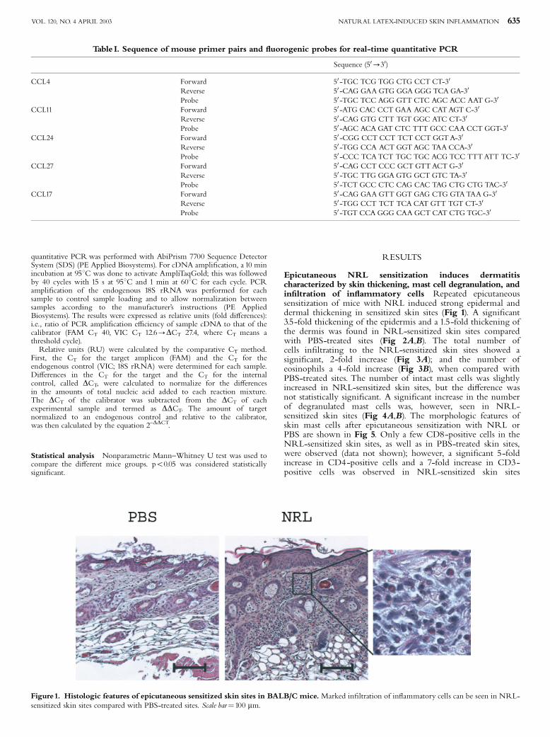
quantitative PCRwas performed with AbiPrism 7700 Sequence DetectorSystem (SDS) (PE Applied Biosystems). For cDNA ampli¢cation, a 10 minincubation at 951C was done to activate AmpliTaqGold; this was followedby 40 cycles with 15 s at 951C and 1 min at 601C for each cycle. PCRampli¢cation of the endogenous 18S rRNA was performed for eachsample to control sample loading and to allow normalization betweensamples according to the manufacturer’s instructions (PE AppliedBiosystems). The results were expressed as relative units (fold di¡erences):i.e., ratio of PCR ampli¢cation e⁄ciency of sample cDNA to that of thecalibrator (FAM CT 40, VIC CT 12.6-DCT 27.4, where CT means athreshold cycle).Relative units (RU) were calculated by the comparative CT method.
First, the CT for the target amplicon (FAM) and the CT for theendogenous control (VIC; 18S rRNA) were determined for each sample.Di¡erences in the CT for the target and the CT for the internalcontrol, called DCT, were calculated to normalize for the di¡erencesin the amounts of total nucleic acid added to each reaction mixture.The DCT of the calibrator was subtracted from the DCT of eachexperimental sample and termed as DDCT. The amount of targetnormalized to an endogenous control and relative to the calibrator,was then calculated by the equation 2^DDCT.
Statistical analysis Nonparametric Mann^Whitney U test was used tocompare the di¡erent mice groups. po0.05 was considered statisticallysigni¢cant.
RESULTS
Epicutaneous NRL sensitization induces dermatitischaracterized by skin thickening, mast cell degranulation, andin¢ltration of in£ammatory cells Repeated epicutaneoussensitization of mice with NRL induced strong epidermal anddermal thickening in sensitized skin sites (Fig 1). A signi¢cant3.5-fold thickening of the epidermis and a 1.5-fold thickening ofthe dermis was found in NRL-sensitized skin sites comparedwith PBS-treated sites (Fig 2A,B). The total number ofcells in¢ltrating to the NRL-sensitized skin sites showed asigni¢cant, 2-fold increase (Fig 3A); and the number ofeosinophils a 4 -fold increase (Fig 3B), when compared withPBS-treated sites. The number of intact mast cells was slightlyincreased in NRL-sensitized skin sites, but the di¡erence wasnot statistically signi¢cant. A signi¢cant increase in the numberof degranulated mast cells was, however, seen in NRL-sensitized skin sites (Fig 4A,B). The morphologic features ofskin mast cells after epicutaneous sensitization with NRL orPBS are shown in Fig 5. Only a few CD8-positive cells in theNRL-sensitized skin sites, as well as in PBS-treated skin sites,were observed (data not shown); however, a signi¢cant 5-foldincrease in CD4-positive cells and a 7-fold increase in CD3-positive cells was observed in NRL-sensitized skin sites
Table I. Sequence of mouse primer pairs and £uorogenic probes for real-time quantitative PCR
Sequence (50-30)
CCL4 Forward 50 -TGC TCG TGG CTG CCT CT-30
Reverse 50 -CAG GAA GTG GGA GGG TCA GA-30
Probe 50 -TGC TCC AGG GTT CTC AGC ACC AAT G-30
CCL11 Forward 50 -ATG CAC CCT GAA AGC CATAGT C-30
Reverse 50 -CAG GTG CTT TGT GGC ATC CT-30
Probe 50 -AGC ACA GAT CTC TTT GCC CAACCT GGT-30
CCL24 Forward 50 -CGG CCT CCT TCT CCT GGTA-30
Reverse 50 -TGG CCA ACT GGTAGC TAACCA-30
Probe 50 -CCC TCATCT TGC TGC ACG TCC TTTATT TC-30
CCL27 Forward 50 -CAG CCT CCC GCT GTTACT G-30
Reverse 50 -TGC TTG GGA GTG GCT GTC TA-30
Probe 50 -TCT GCC CTC CAG CAC TAG CTG CTG TAC-30
CCL17 Forward 50 -CAG GAA GTT GGT GAG CTG GTATAA G-30
Reverse 50 -TGG CCT TCT TCACAT GTT TGT CT-30
Probe 50 -TGT CCA GGG CAA GCT CAT CTG TGC-30
Figure1. Histologic features of epicutaneous sensitized skin sites in BALB/C mice.Marked in¢ltration of in£ammatory cells can be seen in NRL-sensitized skin sites compared with PBS-treated sites. Scale bar¼100 mm.
NATURAL LATEX-INDUCED SKIN INFLAMMATION 635VOL. 120, NO. 4 APRIL 2003
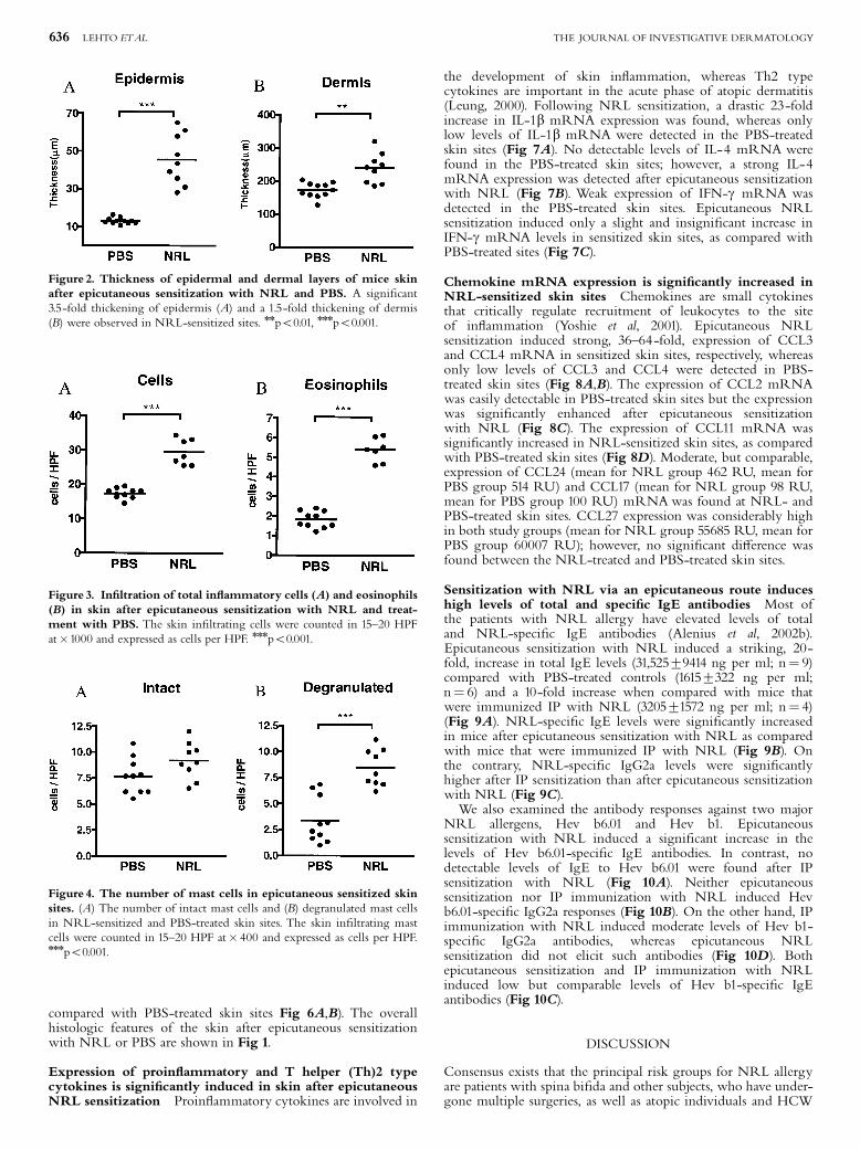
compared with PBS-treated skin sites Fig 6A,B). The overallhistologic features of the skin after epicutaneous sensitizationwith NRL or PBS are shown in Fig 1.
Expression of proin£ammatory and T helper (Th)2 typecytokines is signi¢cantly induced in skin after epicutaneousNRL sensitization Proin£ammatory cytokines are involved in
the development of skin in£ammation, whereas Th2 typecytokines are important in the acute phase of atopic dermatitis(Leung, 2000). Following NRL sensitization, a drastic 23-foldincrease in IL-1b mRNA expression was found, whereas onlylow levels of IL-1b mRNA were detected in the PBS-treatedskin sites (Fig 7A). No detectable levels of IL-4 mRNA werefound in the PBS-treated skin sites; however, a strong IL-4mRNA expression was detected after epicutaneous sensitizationwith NRL (Fig 7B). Weak expression of IFN-g mRNA wasdetected in the PBS-treated skin sites. Epicutaneous NRLsensitization induced only a slight and insigni¢cant increase inIFN-g mRNA levels in sensitized skin sites, as compared withPBS-treated sites (Fig 7C).
Chemokine mRNA expression is signi¢cantly increased inNRL-sensitized skin sites Chemokines are small cytokinesthat critically regulate recruitment of leukocytes to the siteof in£ammation (Yoshie et al, 2001). Epicutaneous NRLsensitization induced strong, 36^64-fold, expression of CCL3and CCL4 mRNA in sensitized skin sites, respectively, whereasonly low levels of CCL3 and CCL4 were detected in PBS-treated skin sites (Fig 8A,B). The expression of CCL2 mRNAwas easily detectable in PBS-treated skin sites but the expressionwas signi¢cantly enhanced after epicutaneous sensitizationwith NRL (Fig 8C). The expression of CCL11 mRNA wassigni¢cantly increased in NRL-sensitized skin sites, as comparedwith PBS-treated skin sites (Fig 8D). Moderate, but comparable,expression of CCL24 (mean for NRL group 462 RU, mean forPBS group 514 RU) and CCL17 (mean for NRL group 98 RU,mean for PBS group 100 RU) mRNAwas found at NRL- andPBS-treated skin sites. CCL27 expression was considerably highin both study groups (mean for NRL group 55685 RU, mean forPBS group 60007 RU); however, no signi¢cant di¡erence wasfound between the NRL-treated and PBS-treated skin sites.
Sensitization with NRL via an epicutaneous route induceshigh levels of total and speci¢c IgE antibodies Most ofthe patients with NRL allergy have elevated levels of totaland NRL-speci¢c IgE antibodies (Alenius et al, 2002b).Epicutaneous sensitization with NRL induced a striking, 20-fold, increase in total IgE levels (31,52579414 ng per ml; n¼ 9)compared with PBS-treated controls (16157322 ng per ml;n¼ 6) and a 10-fold increase when compared with mice thatwere immunized IP with NRL (320571572 ng per ml; n¼ 4)(Fig 9A). NRL-speci¢c IgE levels were signi¢cantly increasedin mice after epicutaneous sensitization with NRL as comparedwith mice that were immunized IP with NRL (Fig 9B). Onthe contrary, NRL-speci¢c IgG2a levels were signi¢cantlyhigher after IP sensitization than after epicutaneous sensitizationwith NRL (Fig 9C).We also examined the antibody responses against two major
NRL allergens, Hev b6.01 and Hev b1. Epicutaneoussensitization with NRL induced a signi¢cant increase in thelevels of Hev b6.01-speci¢c IgE antibodies. In contrast, nodetectable levels of IgE to Hev b6.01 were found after IPsensitization with NRL (Fig 10A). Neither epicutaneoussensitization nor IP immunization with NRL induced Hevb6.01-speci¢c IgG2a responses (Fig 10B). On the other hand, IPimmunization with NRL induced moderate levels of Hev b1-speci¢c IgG2a antibodies, whereas epicutaneous NRLsensitization did not elicit such antibodies (Fig 10D). Bothepicutaneous sensitization and IP immunization with NRLinduced low but comparable levels of Hev b1-speci¢c IgEantibodies (Fig 10C).
DISCUSSION
Consensus exists that the principal risk groups for NRL allergyare patients with spina bi¢da and other subjects, who have under-gone multiple surgeries, as well as atopic individuals and HCW
Figure 2. Thickness of epidermal and dermal layers of mice skinafter epicutaneous sensitization with NRL and PBS. A signi¢cant3.5-fold thickening of epidermis (A) and a 1.5-fold thickening of dermis(B) were observed in NRL-sensitized sites. nnpo0.01, nnnpo0.001.
Figure 3. In¢ltration of total in£ammatory cells (A) and eosinophils(B) in skin after epicutaneous sensitization with NRL and treat-ment with PBS. The skin in¢ltrating cells were counted in 15^20 HPFat�1000 and expressed as cells per HPF. nnnpo0.001.
Figure 4. The number of mast cells in epicutaneous sensitized skinsites. (A) The number of intact mast cells and (B) degranulated mast cellsin NRL-sensitized and PBS-treated skin sites. The skin in¢ltrating mastcells were counted in 15^20 HPF at� 400 and expressed as cells per HPF.nnnpo0.001.
636 LEHTO ETAL THE JOURNAL OF INVESTIGATIVE DERMATOLOGY
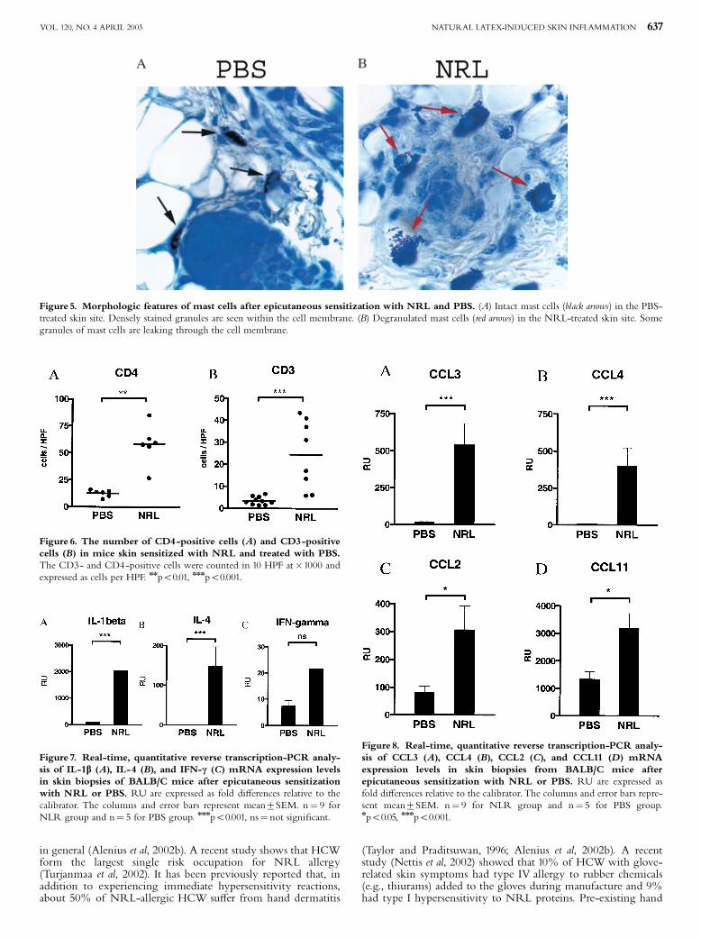
in general (Alenius et al, 2002b). A recent study shows that HCWform the largest single risk occupation for NRL allergy(Turjanmaa et al, 2002). It has been previously reported that, inaddition to experiencing immediate hypersensitivity reactions,about 50% of NRL-allergic HCW su¡er from hand dermatitis
(Taylor and Praditsuwan, 1996; Alenius et al, 2002b). A recentstudy (Nettis et al, 2002) showed that 10% of HCWwith glove-related skin symptoms had type IV allergy to rubber chemicals(e.g., thiurams) added to the gloves during manufacture and 9%had type I hypersensitivity to NRL proteins. Pre-existing hand
Figure 5. Morphologic features of mast cells after epicutaneous sensitization with NRL and PBS. (A) Intact mast cells (black arrows) in the PBS-treated skin site. Densely stained granules are seen within the cell membrane. (B) Degranulated mast cells (red arrows) in the NRL-treated skin site. Somegranules of mast cells are leaking through the cell membrane.
Figure 6. The number of CD4-positive cells (A) and CD3-positivecells (B) in mice skin sensitized with NRL and treated with PBS.The CD3- and CD4-positive cells were counted in 10 HPF at�1000 andexpressed as cells per HPF. nnpo0.01, nnnpo0.001.
Figure 7. Real-time, quantitative reverse transcription-PCR analy-sis of IL-1b (A), IL-4 (B), and IFN-c (C) mRNA expression levelsin skin biopsies of BALB/C mice after epicutaneous sensitizationwith NRL or PBS. RU are expressed as fold di¡erences relative to thecalibrator. The columns and error bars represent mean7SEM. n¼ 9 forNLR group and n¼ 5 for PBS group. nnnpo0.001, ns¼ not signi¢cant.
Figure 8. Real-time, quantitative reverse transcription-PCR analy-sis of CCL3 (A), CCL4 (B), CCL2 (C), and CCL11 (D) mRNAexpression levels in skin biopsies from BALB/C mice afterepicutaneous sensitization with NRL or PBS. RU are expressed asfold di¡erences relative to the calibrator. The columns and error bars repre-sent mean7SEM. n¼ 9 for NLR group and n¼ 5 for PBS group.npo0.05, nnnpo0.001.
NATURAL LATEX-INDUCED SKIN INFLAMMATION 637VOL. 120, NO. 4 APRIL 2003
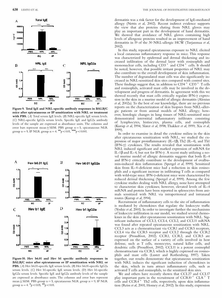
dermatitis was a risk factor for the development of IgE-mediatedallergy (Nettis et al, 2002). Recent indirect evidence supportsthe view that also proteins eluting from NRL gloves mayplay an important part in the development of hand dermatitis.We showed that avoidance of NRL gloves containing highlevels of allergenic proteins resulted in an improvement of handdermatitis in 19 of the 30 NRL-allergic HCW (Turjanmaa et al,2002).In this study, repeated epicutaneous exposure to NRL elicited
a local cutaneous in£ammatory response in mice. This responsewas characterized by epidermal and dermal thickening and in-creased in¢ltration of the dermal layer with eosinophils andmononuclear cells, including CD3þ and CD4þ cells. It shouldbe noted, however, that possible irritant properties of NRL mayalso contribute to the overall development of skin in£ammation.The number of degranulated mast cells was also signi¢cantly in-creased in NRL-sensitized skin sites compared with control sites.These ¢ndings suggest that, in addition to CD4þ CD3þ T cellsand eosinophils, activated mast cells may be involved in the de-velopment and progress of dermatitis. In agreement with this weshowed recently that mast cells critically regulate IFN-g expres-sion in the skin in a murine model of allergic dermatitis (Aleniuset al, 2002a). To the best of our knowledge, there are no previousreports on the characterization of skin biopsies from NRL-aller-gic patients or from animal models of NRL allergy; how-ever, histologic changes in lung tissues of NRL-sensitized micedemonstrated interstitial in£ammatory in¢ltrates consistingof lymphocytes, histiocytes, plasma cells, and eosinophils(Kurup et al, 1994; Slater et al, 1998; Thakker et al, 1999; Xia et al,1999).In order to examine in detail the cytokine milieu in the skin
after epicutaneous sensitization with NRL, we studied the ex-pression of major proin£ammatory (IL-1b), Th2 (IL-4), and Th1(IFN-g) cytokines. The results revealed that sensitization withNRL induced signi¢cant and marked expression of mRNA forIL-1b and IL-4, but not for IFN-g. A recent study utilizing a no-vel murine model of allergic dermatitis suggests that both IL-4and IFN-g critically contribute to the development of ovalbu-min-induced skin in£ammation (Spergel et al, 1999). Sensitizedskin from IL-4-de¢cient mice had a reduction in skin eosino-phils and a signi¢cant increase in in¢ltrating T cells as comparedwith wild-type mice. IFN-g-de¢cient mice were characterized byreduced dermal thickening (Spergel et al, 1999). Among the fewcytokine studies dealing with NRL allergy, none have attemptedto characterize skin cytokines; however, elevated levels of IL-4mRNA and protein have been reported in splenocytes from ani-mals sensitized with NRL via intraperitoneal and intranasalroutes (Kurup et al, 1994).Recruitment of in£ammatory cells to the site of in£ammation
is mediated by chemokines that regulate the leukocyte tra⁄c(Yoshie et al, 2001). In order to investigate further the mechanismsof leukocyte in¢ltration in our model, we studied several chemo-kines in the skin after epicutaneous sensitization with NRL. Sig-ni¢cant induction of CCL3, CCL4, CCL2, and CCL11 mRNAwas found after repeated epicutaneous sensitization with NRL.CCL3 acts as a chemoattractant via CCR1 and CCR5 receptors,CCL4 via the CCR5 receptor and CCL2 through the CCR2receptor (Proudfoot, 2002). CCR1, CCR2, and CCR5 areexpressed on the surface of a variety of cells involved in hostdefense, such as T cells, monocytes, natural killer cells, anddendritic cells (Proudfoot, 2002). CCL11 is a potent eosinophilchemoattractant via CCR3, which is highly expressed on eosino-phils and mast cells (Luster and Rothenberg, 1997). Takentogether, our results demonstrate that epicutaneous sensitizationwith NRL induces the signi¢cant induction of chemokines inthe skin, which in turn attract in£ammatory cells, such asactivated T cells and eosinophils, to the sensitized skin sites.We and others have recently shown that CCL27 and CCL17
play important parts in the recruitment of CCR10þ CLAþ Tcells and CCR4þ Th2 cells, respectively, upon skin in£amma-tion (Reiss et al, 2001; Homey et al, 2002). In this study, expression
Figure 9. Total IgE and NRL-speci¢c antibody responses in BALB/Cmice after epicutaneous or IP sensitization with NRL or treatmentwith PBS. (A) Total serum IgE levels. (B) NRL-speci¢c IgE serum levels.(C) NRL-speci¢c IgG2a serum levels. Speci¢c IgE and IgG2a antibodylevels of the sample are expressed as absorbance units. The columns anderror bars represent mean7SEM. PBS group n¼ 5, epicutaneous NLRgroup n¼ 9, IP NLR group n¼ 4. nnpo0.01, nnnpo0.001.
Figure10. Hev b6.01 and Hev b1 speci¢c antibody responses inBALB/C mice after epicutaneous or IP sensitization with NRL orPBS. (A) Hev b6.01-speci¢c IgE serum levels. (B) Hev b6.01-speci¢c IgG2aserum levels. (C) Hev b1-speci¢c IgE serum levels. (D) Hev b1-speci¢cIgG2a serum levels. Speci¢c IgE and IgG2a antibody levels of the sampleare expressed as absorbance units. The columns and error bars representmean7SEM. PBS group n¼ 5, epicutaneous NLR group n¼ 9, IP NLRgroup n¼ 4. npo0.05, nnpo0.01.
638 LEHTO ETAL THE JOURNAL OF INVESTIGATIVE DERMATOLOGY
of CCL17 was comparable in NRL-sensitized and PBS-treatedskin sites. Similarly, strong but comparable expression of CCL27mRNAwas found in the skin of PBS- and NRL-treated groups.Because the majority of skin-homing Th2 cells express CCR10and CCR4 on their surface (Homey et al, 2002; Reiss et al, 2001)it is possible that NRL-speci¢c CD4þ CD3þ Th2 cells areattracted to the in£amed skin partially through local but consti-tutive expression of CCL27 and CCL17. Thus, in¢ltration of Tcells to the skin may be regulated, not only by the induction ofskin chemokines, but also by the expression of chemokine recep-tors on the surface of antigen-speci¢c skin-homing T cells. Onthe other hand also other factors, e.g., adhesion molecules andcytokines, contribute to the cell in¢ltration as well (Harlan andWinn, 2002).Atopy and the presence of NRL-speci¢c IgE antibodies are
strongly associated with NRL allergy (Alenius et al, 2002b). Inthis study, epicutaneous sensitization, but not IP immunization,with NRL elicited a signi¢cant elevation of total serum IgE andNRL-speci¢c IgE antibodies. On the contrary, an elevation ofNRL-speci¢c IgG2a antibodies was found only after IP immuni-zation. In agreement with the present ¢ndings a recent animalstudy byWoolhiser et al (2000) demonstrated that di¡erent sensi-tization routes can induce di¡erences in NRL-speci¢c IgE pro-¢les. Consistent with our results, Kurup et al (1994) showed thatIP immunization with NRL induced a marked increase in IgG2aantibodies. The present results suggest that epicutaneous sensitiza-tion with NRL favors the development of a Th2 type response,whereas IP immunization favors the development of a Th1 typeantibody response.Hev b6.01 is a major allergen for HCW, whereas Hev b1
is a major allergen for children with spina bi¢da (Aleniuset al, 1996a,b; Wagner et al, 1999). For the time being, node¢nitive explanation exists for the observed di¡erencesin IgE reactivity between the two groups. In this, epicutaneoussensitization with NRL elicited a strong IgE response againstHev b6.01 but not against Hev b1. On the contrary, IPimmunization with NRL elicited strong IgG2a production toHev b1 but not to Hev b6.01. In line with our observationsWoolhiser et al (2000) showed indirectly by immunoblottingthat mice sensitized intratracheally or topically with NRLproduced IgE antibodies against Hev b6, Hev b2, and Hev b4(Microhelix protein complex), whereas those sensitizedsubcutaneously produced IgE antibodies against Hev b1 andHev b3 (small rubber particle protein). The di¡erent antibodypro¢les against Hev b6.01 and Hev b1 may re£ect di¡erencesin the biochemical nature of the allergens. Hev b6.01is a highly hydrophilic protein, whereas Hev b1 is a highlyhydrophobic one. This may cause di¡erences in thepenetration of these allergens through the skin. Di¡erences inthe concentrations of Hev b6.01 and Hev b1 in NRL extractmay also a¡ect the antibody responses. The amount of Hevb6.01 and its N-terminal fragment hevein is about 30-fold higherthan that of Hev b1 in fresh NRL (ML, HA, unpublishedobservation). It has previously been suggested that high proteinantigen concentrations favor the development of a Th2 type IgEresponse, whereas low antigen concentrations preferably induce aTh1 type IgG2a response (Constant and Bottomly, 1997; Jankovicet al, 2001).In conclusion, the present results demonstrate that epicuta-
neous sensitization with NRL elicits in mice Th2 type systemicimmune response and a local Th2 dominating skin in£ammation.The in£ammatory cell in¢ltration to the skin correlates with aninduction of proin£ammatory and Th2 cytokines as well aschemokines. Epicutaneous sensitization to proteins eluting fromNRL gloves may therefore play an important part in thedevelopment of NRL-speci¢c IgE antibodies. Moreover, NRLproteins, in addition to rubber chemicals, may also have anin£uence on the development of hand dermatitis. The murine ex-periments described here present a model for protein contact der-matitis that can be utilized for studying the mechanisms ofallergen-induced skin in£ammation as well as for determination
of which protein allergens are capable of sensitizing mice via thecutaneous route.
This study was supported by grants from the Academy of Finland (grant numbers37852 and 50809).We thank SariTillander, Helena Honkasalo, andTuula Stjernvallfor their expert and excellent technical assistance.
REFERENCES
Alenius H, Kalkkinen N, Lukka M, et al: Puri¢cation and partial amino acid sequen-cing of a 27-kD natural rubber allergen recognized by latex-allergic childrenwith spina bi¢da. Int Arch Allergy Immunol 106:258^262, 1995
Alenius H, Kalkkinen N, ReunalaT,Turjanmaa K, PalosuoT:The main IgE-bindingepitope of a major latex allergen, prohevein, is present in its N-terminal43-amino acid fragment, hevein. J Immunol 156:1618^1625, 1996a
Alenius H, Kalkkinen N, Yip E, et al: Signi¢cance of rubber elongation factor as alatex allergen. Int Arch Allergy Immunol 109:362^368, 1996b
Alenius H, Laouini D,Woodward A, et al: Mast cells regulate IFN-gamma expressionin the skin and circulating IgE levels in allergen-induced skin in£ammation.J Allergy Clin Immunol 109:106^113, 2002a
Alenius H,Turjanmaa K, Palosuo T: Natural rubber latex allergy. Occup Environ Med59:419^424, 2002b
Constant SL, Bottomly K: Induction of Th1 and Th2 CD4þ T cell responses: Thealternative approaches. Annu Rev Immunol 15:297^322, 1997
Hanninen AR, Mikkola JH, Kalkkinen N,Turjanmaa K,Ylitalo L, Reunala T, Palo-suoT: Increased allergen production in turnip (Brassica rapa) by treatments acti-vating defense mechanisms. J Allergy Clin Immunol 104:194^201, 1999
Harlan JM,Winn RK: Leukocyte^endothelial interactions: Clinical trials of anti-ad-hesion therapy. Crit Care Med 30:S214^S219, 2002
Homey B, Alenius H, Muller A, et al: CCL27^CCR10 interactions regulate T cell-mediated skin in£ammation. Nat Med 8:157^165, 2002
Jankovic D, Liu Z, Gause WC: Th1- and Th2-cell commitment during infectiousdisease: Asymmetry in divergent pathways.Trends Immunol 22:450^457, 2001
Janssens V, Morren M, Dooms-Goossens A, Degreef H: Protein contact Dermatitis:Myth or reality? Br J Dermatol. 132:1^6, 1995
KurupVP, Kumar A, Choi H, Murali PS, Resnick A, Kelly KJ, Fink JN: Latex anti-gens induce IgE and eosinophils in mice. Int Arch Allergy Immunol 103:370^377,1994
Leung DY: Atopic dermatitis: New insights and opportunities for therapeutic inter-vention. J Allergy Clin Immunol 105:860^876, 2000
Liss GM, Sussman GL: Latex sensitization: Occupational versus general populationprevalence rates. AmJ Ind Med 35:196^200, 1999
Luster AD, Rothenberg ME: Role of the monocyte chemoattractant protein and eo-taxin subfamily of chemokines in allergic in£ammation. J Leukoc Biol 62:620^633, 1997
Nethercott JR, Holness DL: Occupational dermatitis in food handlers and bakers.J Am Acad Dermatol 21:485^490, 1989
Nettis E, Assennato G, Ferrannini A,Tursi A: Type I allergy to natural rubber latexand type IV allergy to rubber chemicals in health care workers with glove-re-lated skin symptoms. Clin Exp Allergy 32:441^447, 2002
Proudfoot AE: Chemokine receptors: Multifaceted therapeutic targets. Nature RevImmunol 2:106^115, 2002
ReissY, Proudfoot AE, Power CA, Campbell JJ, Butcher EC: CC chemokine recep-tor (CCR) 4 and the CCR10 ligand cutaneous T cell-attracting chemokine(CTACK) in lymphocyte tra⁄cking to in£amed skin. J Exp Med 194:1541^1547, 2001
Slater JE, Paupore E, ZhangYT, Colberg-PoleyAM:The latex allergen Hev b 5 tran-script is widely distributed after subcutaneous injection in BALB/c mice of itsDNAvaccine. J Allergy Clin Immunol 102:469^475, 1998
Spergel JM, Mizoguchi E, Brewer JP, MartinTR, Bhan AK, Geha RS: Epicutaneoussensitization with protein antigen induces localized allergic dermatitis and hy-perresponsiveness to methacholine after single exposure to aerosolized antigenin mice. J Clin Invest 101:1614^1622, 1998
Spergel JM, Mizoguchi E, Oettgen H, Bhan AK, Geha R: Roles of TH1 and TH2cytokines in a murine model of allergic dermatitis. J Clin Invest 103:1103^1111,1999
Susitaival P, Husman L, Hollmen A, Horsmanheimo M, Husman K, Hannuksela M:Hand eczema in Finnish farmers. A questionnaire-based clinical study. ContactDermatitis 32:150^155, 1995
Taylor JS, Praditsuwan P: Latex allergy. Review of 44 cases including outcome andfrequent association with allergic hand eczema. Arch Dermatol 132:265^271, 1996
Thakker JC, Xia JQ, Rickaby DA, Krenz GS, Kelly KJ, Kurup VP, Dawson CA: Amurine model of latex allergy-induced airway hyperreactivity. Lung 177:89^100, 1999
NATURAL LATEX-INDUCED SKIN INFLAMMATION 639VOL. 120, NO. 4 APRIL 2003

Tosti A, Fanti PA, Guerra L, Piancastelli E, Poggi S, Pileri S: Morphological andimmunohistochemical study of immediate contact dermatitis of the handsdue to foods. Contact Dermatitis 22:81^85, 1990
Turjanmaa K, Kanto M, Kautiainen H, ReunalaT, PalosuoT: Long-term outcome of160 adult patients with natural rubber latex allergy. J Allergy Clin Immunol110:S70^S74, 2002
Wagner B, Krebitz M, Buck D, et al: Cloning, expression, and characterization ofrecombinant Hev b 3, a Hevea brasiliensis protein associated with latex allergyin patients with spina bi¢da. J Allergy Clin Immunol 104:1084^1092, 1999
Woolhiser MR, Munson AE, Meade BJ: Immunological responses of mice follow-ing administration of natural rubber latex proteins by di¡erent routes of expo-sure.Toxicol Sci 55:343^351, 2000
Xia JQ, Rickaby DA, Kelly KJ, et al: Immune response and airway reactivity in wildand IL-4 knockout mice exposed to latex allergens. Int Arch Allergy Immunol118:23^29, 1999
Yoshie O, Imai T, Nomiyama H: Chemokines in immunity. Adv Immunol 78:57^110,2001
640 LEHTO ETAL THE JOURNAL OF INVESTIGATIVE DERMATOLOGY
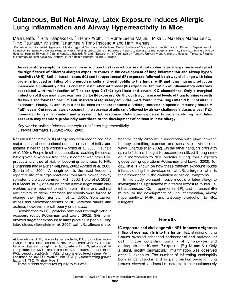
Cutaneous, But Not Airway, Latex Exposure Induces AllergicLung Inflammation and Airway Hyperreactivity in Mice
Maili Lehto,�1 Rita Haapakoski,�1 Henrik Wolff,�wz Marja-Leena Majuri,� Mika J. Makela,y Marina Leino,�
Timo Reunala,z Kristiina Turjanmaa,z Timo Palosuo,# and Harri Alenius,��Departments of Industrial Hygiene and Toxicology and Occupational Medicine, Finnish Institute of Occupational Health, Helsinki, Finland; wDepartment ofPathology, Kymenlaakso Central Hospital, Kotka, Finland; zDepartment of Pathology, Helsinki University Central Hospital, Helsinki, Finland; ySkin and AllergyHospital, Helsinki University Central Hospital, Helsinki, Finland; zDepartment of Dermatology, Tampere University and University Hospital, Tampere, Finland;#Laboratory of Immunobiology, National Public Health Institute, Helsinki, Finland
As respiratory symptoms are common in addition to skin reactions in natural rubber latex allergy, we investigated
the significance of different allergen exposure routes in the development of lung inflammation and airway hyper-
reactivity (AHR). Both intracutaneous (IC) and intraperitoneal (IP) exposure followed by airway challenge with latex
proteins induced an influx of mononuclear cells and eosinophils to the lungs. AHR and lung mucus production
increased significantly after IC and IP but not after intranasal (IN) exposure. Infiltration of inflammatory cells was
associated with the induction of T-helper type 2 (Th2) cytokines and several CC chemokines. Only a marginal
induction of these mediators was found after IN exposure. On the contrary, increased levels of transforming growth
factor-b1 and forkhead box 3 mRNA, markers of regulatory activities, were found in the lungs after IN but not after IC
exposure. Finally, IC and IP, but not IN, latex exposure induced a striking increase in specific immunoglobulin E
(IgE) levels. Cutaneous latex exposure in the absence of adjuvant followed by airway challenge induces a local Th2-
dominated lung inflammation and a systemic IgE response. Cutaneous exposure to proteins eluting from latex
products may therefore profoundly contribute to the development of asthma in latex allergy.
Key words: asthma/chemokines/cytokines/latex hypersensitivityJ Invest Dermatol 125:962 –968, 2005
Natural rubber latex (NRL) allergy has been recognized as amajor cause of occupational contact urticaria, rhinitis, andasthma in health care workers (Ahmed et al, 2003; Reunalaet al, 2004). People in other occupations requiring the use oflatex gloves or who are frequently in contact with other NRLproducts are also at risk of becoming sensitized to NRL(Turjanmaa and Makinen-Kiljunen, 2002; Ahmed et al, 2003;Sparta et al, 2004). Although skin is the most frequentlyreported site of allergic reactions from latex gloves, airwaysymptoms are also common (Fish, 2002; Nolte et al, 2002).In a recent study, one-fourth of the latex-allergic health careworkers were reported to suffer from rhinitis and asthmaand several of these asthmatic individuals were forced tochange their jobs (Bernstein et al, 2003). Sensitizationroutes and pathomechanisms of NRL-induced rhinitis andasthma, however, are still poorly understood.
Sensitization to NRL proteins may occur through variousexposure routes (Weissman and Lewis, 2002). Skin is anobvious target for exposure to latex proteins in people usinglatex gloves (Bernstein et al, 2003) but NRL allergens also
become easily airborne in association with glove powder,thereby permitting exposure and sensitization via the air-ways (Charous et al, 2002). On the other hand, children withspina bifida are thought to become sensitized through mu-cous membranes to NRL proteins eluting from surgeon’sgloves during operations (Weissman and Lewis, 2002). To-day, little is known on how these different exposure routesinteract during the development of NRL allergy or what istheir importance in the elicitation of clinical symptoms.
In this study, we used mouse models of latex allergy toinvestigate the significance of different exposure routes, i.e.intracutaneous (IC), intraperitoneal (IP), and intranasal (IN)routes, to the development of lung inflammation, airwayhyperreactivity (AHR), and antibody production to NRLallergens.
Results
IC exposure and challenge with NRL induces a vigorousinflux of eosinophils into the lungs H&E staining of lungtissues revealed enhanced peribronchial and perivascularcell infiltrates consisting primarily of lymphocytes andeosinophils after IC and IP exposure (Fig 1A and S1). Onlya slight, mostly perivascular, inflammation was observedafter IN exposure. The number of infiltrating eosinophilsboth in perivascular and in peribronchial areas of lungtissues showed a dramatic increase in intracutaneously1These authors contributed equally to this work.
Abbreviations: AHR, airway hyperreactivity; BAL, bronchoalveolarlavage; Foxp3, forkhead box 3; Hev b6.01, prohevein; IC, intracu-taneous; IgE, immunoglobulin E; IL, interleukin; IN, intranasal; IP,intraperitoneal; MCh, methacholine; NRL, natural rubber latex;PAS, periodic acid-Schiff; PBS, phosphate-buffered saline; Penh,enhanced pause; RU, relative units; TGF-b1, transforming growthfactor-b1; Th2, T-helper type 2
Copyright r 2005 by The Society for Investigative Dermatology, Inc.
962
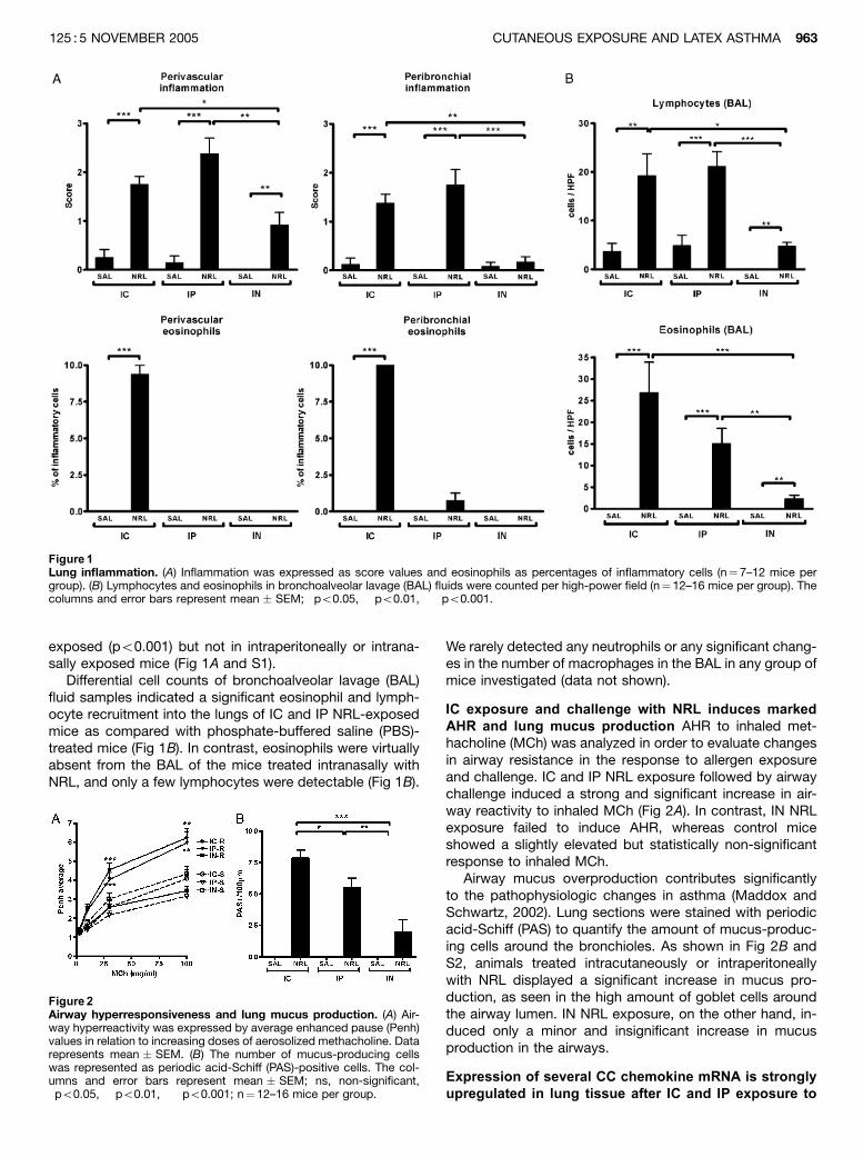
exposed (po0.001) but not in intraperitoneally or intrana-sally exposed mice (Fig 1A and S1).
Differential cell counts of bronchoalveolar lavage (BAL)fluid samples indicated a significant eosinophil and lymph-ocyte recruitment into the lungs of IC and IP NRL-exposedmice as compared with phosphate-buffered saline (PBS)-treated mice (Fig 1B). In contrast, eosinophils were virtuallyabsent from the BAL of the mice treated intranasally withNRL, and only a few lymphocytes were detectable (Fig 1B).
We rarely detected any neutrophils or any significant chang-es in the number of macrophages in the BAL in any group ofmice investigated (data not shown).
IC exposure and challenge with NRL induces markedAHR and lung mucus production AHR to inhaled met-hacholine (MCh) was analyzed in order to evaluate changesin airway resistance in the response to allergen exposureand challenge. IC and IP NRL exposure followed by airwaychallenge induced a strong and significant increase in air-way reactivity to inhaled MCh (Fig 2A). In contrast, IN NRLexposure failed to induce AHR, whereas control miceshowed a slightly elevated but statistically non-significantresponse to inhaled MCh.
Airway mucus overproduction contributes significantlyto the pathophysiologic changes in asthma (Maddox andSchwartz, 2002). Lung sections were stained with periodicacid-Schiff (PAS) to quantify the amount of mucus-produc-ing cells around the bronchioles. As shown in Fig 2B andS2, animals treated intracutaneously or intraperitoneallywith NRL displayed a significant increase in mucus pro-duction, as seen in the high amount of goblet cells aroundthe airway lumen. IN NRL exposure, on the other hand, in-duced only a minor and insignificant increase in mucusproduction in the airways.
Expression of several CC chemokine mRNA is stronglyupregulated in lung tissue after IC and IP exposure to
Figure 1Lung inflammation. (A) Inflammation was expressed as score values and eosinophils as percentages of inflammatory cells (n¼ 7–12 mice pergroup). (B) Lymphocytes and eosinophils in bronchoalveolar lavage (BAL) fluids were counted per high-power field (n¼12–16 mice per group). Thecolumns and error bars represent mean � SEM; �po0.05, ��po0.01, ���po0.001.
Figure 2Airway hyperresponsiveness and lung mucus production. (A) Air-way hyperreactivity was expressed by average enhanced pause (Penh)values in relation to increasing doses of aerosolized methacholine. Datarepresents mean � SEM. (B) The number of mucus-producing cellswas represented as periodic acid-Schiff (PAS)-positive cells. The col-umns and error bars represent mean � SEM; ns, non-significant,�po0.05, ��po0.01, ���po0.001; n¼ 12–16 mice per group.
CUTANEOUS EXPOSURE AND LATEX ASTHMA 963125 : 5 NOVEMBER 2005
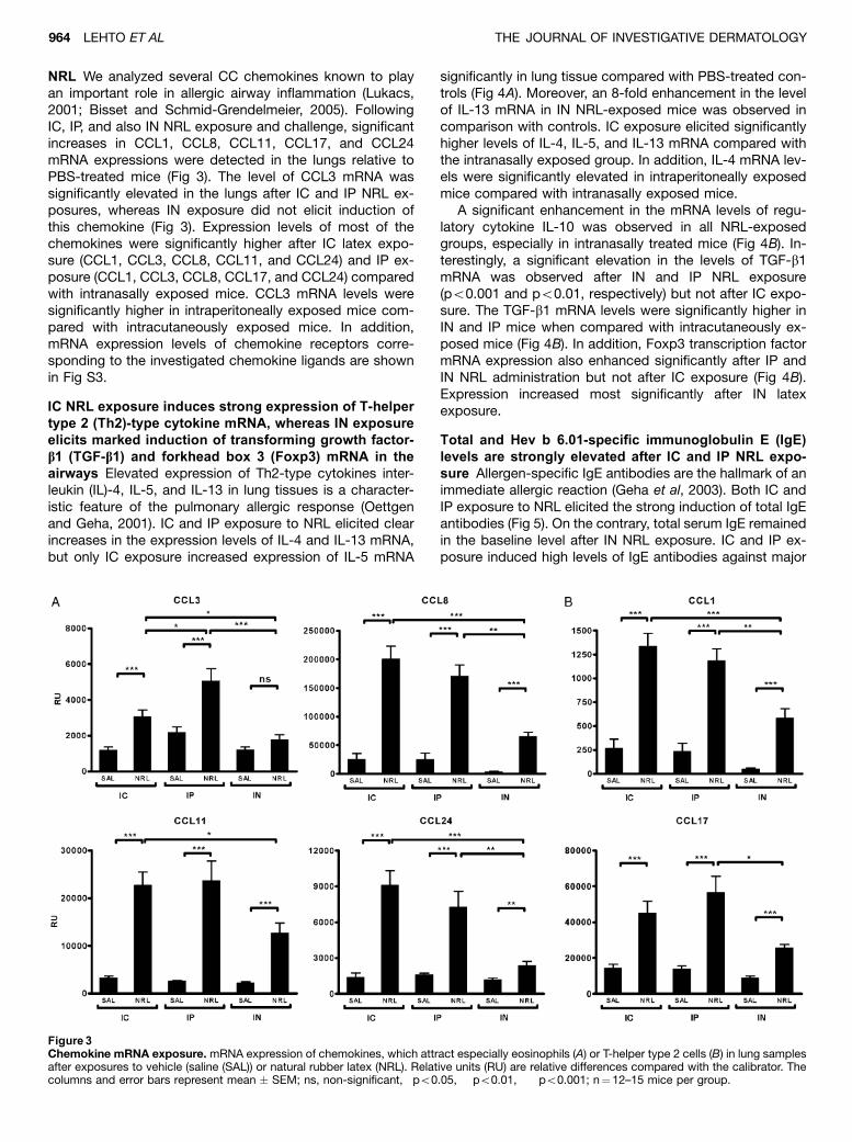
NRL We analyzed several CC chemokines known to playan important role in allergic airway inflammation (Lukacs,2001; Bisset and Schmid-Grendelmeier, 2005). FollowingIC, IP, and also IN NRL exposure and challenge, significantincreases in CCL1, CCL8, CCL11, CCL17, and CCL24mRNA expressions were detected in the lungs relative toPBS-treated mice (Fig 3). The level of CCL3 mRNA wassignificantly elevated in the lungs after IC and IP NRL ex-posures, whereas IN exposure did not elicit induction ofthis chemokine (Fig 3). Expression levels of most of thechemokines were significantly higher after IC latex expo-sure (CCL1, CCL3, CCL8, CCL11, and CCL24) and IP ex-posure (CCL1, CCL3, CCL8, CCL17, and CCL24) comparedwith intranasally exposed mice. CCL3 mRNA levels weresignificantly higher in intraperitoneally exposed mice com-pared with intracutaneously exposed mice. In addition,mRNA expression levels of chemokine receptors corre-sponding to the investigated chemokine ligands are shownin Fig S3.
IC NRL exposure induces strong expression of T-helpertype 2 (Th2)-type cytokine mRNA, whereas IN exposureelicits marked induction of transforming growth factor-b1 (TGF-b1) and forkhead box 3 (Foxp3) mRNA in theairways Elevated expression of Th2-type cytokines inter-leukin (IL)-4, IL-5, and IL-13 in lung tissues is a character-istic feature of the pulmonary allergic response (Oettgenand Geha, 2001). IC and IP exposure to NRL elicited clearincreases in the expression levels of IL-4 and IL-13 mRNA,but only IC exposure increased expression of IL-5 mRNA
significantly in lung tissue compared with PBS-treated con-trols (Fig 4A). Moreover, an 8-fold enhancement in the levelof IL-13 mRNA in IN NRL-exposed mice was observed incomparison with controls. IC exposure elicited significantlyhigher levels of IL-4, IL-5, and IL-13 mRNA compared withthe intranasally exposed group. In addition, IL-4 mRNA lev-els were significantly elevated in intraperitoneally exposedmice compared with intranasally exposed mice.
A significant enhancement in the mRNA levels of regu-latory cytokine IL-10 was observed in all NRL-exposedgroups, especially in intranasally treated mice (Fig 4B). In-terestingly, a significant elevation in the levels of TGF-b1mRNA was observed after IN and IP NRL exposure(po0.001 and po0.01, respectively) but not after IC expo-sure. The TGF-b1 mRNA levels were significantly higher inIN and IP mice when compared with intracutaneously ex-posed mice (Fig 4B). In addition, Foxp3 transcription factormRNA expression also enhanced significantly after IP andIN NRL administration but not after IC exposure (Fig 4B).Expression increased most significantly after IN latexexposure.
Total and Hev b 6.01-specific immunoglobulin E (IgE)levels are strongly elevated after IC and IP NRL expo-sure Allergen-specific IgE antibodies are the hallmark of animmediate allergic reaction (Geha et al, 2003). Both IC andIP exposure to NRL elicited the strong induction of total IgEantibodies (Fig 5). On the contrary, total serum IgE remainedin the baseline level after IN NRL exposure. IC and IP ex-posure induced high levels of IgE antibodies against major
Figure 3Chemokine mRNA exposure. mRNA expression of chemokines, which attract especially eosinophils (A) or T-helper type 2 cells (B) in lung samplesafter exposures to vehicle (saline (SAL)) or natural rubber latex (NRL). Relative units (RU) are relative differences compared with the calibrator. Thecolumns and error bars represent mean � SEM; ns, non-significant, �po0.05, ��po0.01, ���po0.001; n¼12–15 mice per group.
964 LEHTO ET AL THE JOURNAL OF INVESTIGATIVE DERMATOLOGY
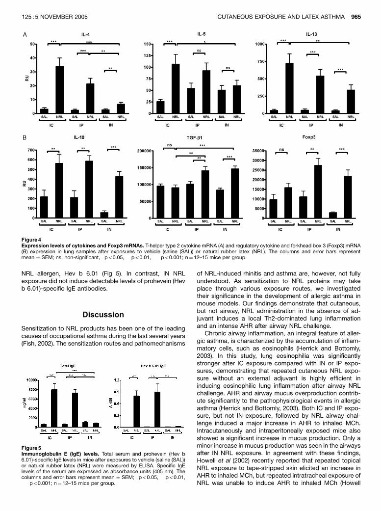
NRL allergen, Hev b 6.01 (Fig 5). In contrast, IN NRLexposure did not induce detectable levels of prohevein (Hevb 6.01)-specific IgE antibodies.
Discussion
Sensitization to NRL products has been one of the leadingcauses of occupational asthma during the last several years(Fish, 2002). The sensitization routes and pathomechanisms
of NRL-induced rhinitis and asthma are, however, not fullyunderstood. As sensitization to NRL proteins may takeplace through various exposure routes, we investigatedtheir significance in the development of allergic asthma inmouse models. Our findings demonstrate that cutaneous,but not airway, NRL administration in the absence of ad-juvant induces a local Th2-dominated lung inflammationand an intense AHR after airway NRL challenge.
Chronic airway inflammation, an integral feature of aller-gic asthma, is characterized by the accumulation of inflam-matory cells, such as eosinophils (Herrick and Bottomly,2003). In this study, lung eosinophilia was significantlystronger after IC exposure compared with IN or IP expo-sures, demonstrating that repeated cutaneous NRL expo-sure without an external adjuvant is highly efficient ininducing eosinophilic lung inflammation after airway NRLchallenge. AHR and airway mucus overproduction contrib-ute significantly to the pathophysiological events in allergicasthma (Herrick and Bottomly, 2003). Both IC and IP expo-sure, but not IN exposure, followed by NRL airway chal-lenge induced a major increase in AHR to inhaled MCh.Intracutaneously and intraperitoneally exposed mice alsoshowed a significant increase in mucus production. Only aminor increase in mucus production was seen in the airwaysafter IN NRL exposure. In agreement with these findings,Howell et al (2002) recently reported that repeated topicalNRL exposure to tape-stripped skin elicited an increase inAHR to inhaled MCh, but repeated intratracheal exposure ofNRL was unable to induce AHR to inhaled MCh (Howell
Figure 4Expression levels of cytokines and Foxp3 mRNAs. T-helper type 2 cytokine mRNA (A) and regulatory cytokine and forkhead box 3 (Foxp3) mRNA(B) expression in lung samples after exposures to vehicle (saline (SAL)) or natural rubber latex (NRL). The columns and error bars representmean � SEM; ns, non-significant, �po0.05, ��po0.01, ���po0.001; n¼12–15 mice per group.
Figure 5Immunoglobulin E (IgE) levels. Total serum and prohevein (Hev b6.01)-specific IgE levels in mice after exposures to vehicle (saline (SAL))or natural rubber latex (NRL) were measured by ELISA. Specific IgElevels of the serum are expressed as absorbance units (405 nm). Thecolumns and error bars represent mean � SEM; �po0.05, ��po0.01,���po0.001; n¼12–15 mice per group.
CUTANEOUS EXPOSURE AND LATEX ASTHMA 965125 : 5 NOVEMBER 2005
et al, 2004). On the other hand, Woolhiser et al (2000) pre-viously demonstrated IgE response and AHR following INexposure of 50 mg of NRL administered 5 d per wk for10 wk. Our results, however, clearly demonstrate that der-mal route exposure using much lower allergen dosage andlength of exposure leads to vigorous IgE response, AHR,and airway inflammation after airway challenge.
In order to investigate the mechanisms of leukocyte re-cruitment to the airways, in detail we studied several CCchemokines in lung tissue. Eosinophils are known to re-spond to CCL3, CCL5, CCL8, CCL11, and CCL24 (Zimmer-mann et al, 2003). In this study, expressions of CCL8,CCL11, and CCL24 mRNA were significantly higher in in-tracutaneously exposed mice compared with intranasallyexposed mice with marked airway eosinophilia after cuta-neous exposure. These chemokines were, however, equallyexpressed in intraperitoneally and intracutaneously ex-posed groups although lung eosinophilia was significantlymore intense in the intracutaneously exposed mice. Ex-pression levels of CCL1 and CCL17, which are known toattract Th2 cells (Panina-Bordignon et al, 2001), were alsohigher after IC and IP exposure compared with IN exposure.Taken together, our results demonstrate that cutaneous andIP NRL exposure elicits induction of various chemokines inlung tissue, which in turn may recruit Th2-type inflammatorycells, a characteristic of allergic airway inflammation.
A consensus exists that IL-4 and IL-13 are key regulatorsof IgE class switching (Geha et al, 2003). In addition, it hasbeen shown that IL-13 contributes to AHR and mucusoverproduction (Kuperman et al, 2002). Expression levels ofIL-4 and IL-13 mRNA were significantly enhanced after allroutes of exposure with NRL in lung tissue in this study. Butincreases were markedly higher after IC exposure and to alesser extent after IP exposure, in comparison with IN ex-posure. The expression level of IL-5, a key cytokine regu-lating eosinophil recruitment and survival (Hamelmann andGelfand, 2001), was significantly elevated only after IC NRLexposure. Thus, the greater number of eosinophils in thelungs of intracutaneously exposed mice may be because ofan increased eosinophil survival rate induced by increasedlevels of IL-5 in the airways. On the other hand, CCL24 andIL-5 have been shown to cooperatively promote eosinophilaccumulation into airways and to increase AHR to inhaledMCh (Yang et al, 2003). Only a few studies exist in the lit-erature describing cytokine expression in lungs after NRLadministration. Hardy et al (2003) recently reported thatmice sensitized intraperitoneally with NRL allergens, andwith NRL glove extract demonstrated elevated levels of IL-5protein in the BAL fluid. They found no significant differ-ences in the levels of IL-4 protein between controls andlatex-sensitized groups. Our results clearly indicate that re-peated cutaneous NRL exposure induces Th2-dominatedcytokine expression in the lungs after airway challenge.
Increased levels of total and allergen-specific IgE anti-bodies in the patient serum are characteristic of allergicasthma (Busse and Lemanske, 2001; Lemanske and Busse,2003). In this study, both IC and IP NRL exposure elicited asignificant elevation of total serum IgE levels. As an exampleof allergen-specific IgE response, we investigated antibodyresponses against Hev b 6.01, which is a major NRL aller-gen (Alenius et al, 1996; Wagner and Breiteneder, 2005).
The sera from intracutaneously or intraperitonelly exposedmice contained high levels of IgE antibodies to Hev b 6.01.On the contrary, IN exposure failed to induce elevation oftotal and specific IgE antibodies. In agreement with thesefindings, Hufnagl et al (2003) also reported increased NRLallergen-specific IgE levels after IP immunization, and arecent study by Woolhiser et al (2000) demonstrated thatdifferent exposure routes can induce differences in NRL-specific IgE profiles in mice.
It is of interest that airway inflammation, AHR, and IgElevels were substantially lower after IN exposure comparedwith IC exposure. To address the role of regulatorycytokines in the induction of airway inflammation, we analy-zed the mRNA levels of IL-10 and TGF-b1, both of which areknown to play important roles in the downregulation of im-mune responses (Terui et al, 2001; Akbari et al, 2003; Nag-ler-Anderson et al, 2004). Proportional increase (allergen-exposed mice vs PBS-exposed control) in the expression ofIL-10 mRNA was markedly more prominent in intranasallyexposed mice (7.4-fold increase) compared with intracuta-neously (2.6-fold increase) and intraperitoneally exposed(2.8-fold increase) mice. In line with these results, Akbariet al (2001) have reported that respiratory exposure to al-lergens can induce T cell tolerance, which appears to bemediated by IL-10 production. On the other hand, we foundsignificant induction of TGF-b1 mRNA only after IN and IPNRL exposure but not after IC exposure. Furthermore, pro-portional expression of transcription factor Foxp3, which isexpressed predominantly by CD4þCD25þ T regulatorycells (Fontenot et al, 2003; Khattri et al, 2003), was signif-icantly upregulated after IN exposure (7.3-fold) but to alesser extent after IP exposure (2.5-fold) and IC exposure(1.6-fold). Thus, it is possible that increased levels of IL-10and TGF-b1 as well as Foxp3þ regulatory T cells in thelungs of intranasally exposed mice suppress the inflamma-tory responses, resulting in diminished airway inflammation.The link between TGF-b and regulatory T cells is supportedby the finding that TGF-b is able to promote differentiationof T cells into Foxp3þ regulatory T cells (Chen et al, 2003;Fantini et al, 2004).
These findings underline the role of cutaneous route al-lergen exposure in the elicitation of allergic airway inflam-mation and AHR. Instead of epicutaneous (Spergel et al,1998; Lehto et al, 2003) allergen application, we used ICallergen exposure assuring that the actual allergen doseentering the body was the same in all exposure routes. Thepresent observations are of importance when consideringprevention of airway hypersensitivity to NRL allergens.Glove powder is the most important carrier of NRL aller-gens to airways, and thus primary prevention has mainlyfocused on the use of non-powdered latex gloves (Charouset al, 2002). As the exposure via the cutaneous route couldbe an important way for sensitizing airways, attention willneed to be paid to the use of low- or non-allergen NRLgloves instead of using only non-powdered gloves.
Materials and Methods
Allergens NRL and Hev b 6.01 were purified as earlier described(Lehto et al, 2003).
966 LEHTO ET AL THE JOURNAL OF INVESTIGATIVE DERMATOLOGY
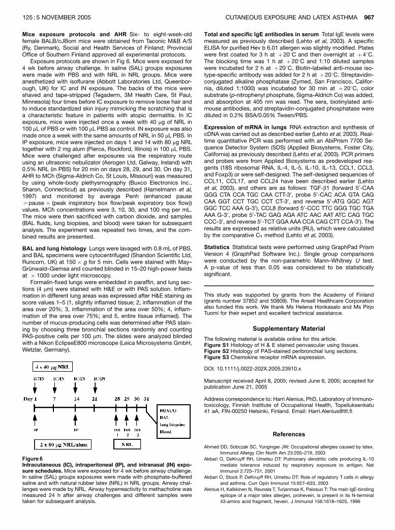
Mice exposure protocols and AHR Six- to eight-week-oldfemale BALB/cJBom mice were obtained from Taconic M&B A/S(Ry, Denmark), Social and Health Services of Finland; ProvincialOffice of Southern Finland approved all experimental protocols.
Exposure protocols are shown in Fig 6. Mice were exposed for4 wk before airway challenge. In saline (SAL) groups exposureswere made with PBS and with NRL in NRL groups. Mice wereanesthetized with isoflurane (Abbott Laboratories Ltd, Queenbor-ough, UK) for IC and IN exposure. The backs of the mice wereshaved and tape-stripped (Tegaderm, 3M Health Care, St Paul,Minnesota) four times before IC exposure to remove loose hair andto induce standardized skin injury mimicking the scratching that isa characteristic feature in patients with atopic dermatitis. In ICexposure, mice were injected once a week with 40 mg of NRL in100 mL of PBS or with 100 mL PBS as control. IN exposure was alsomade once a week with the same amounts of NRL in 50 mL PBS. InIP exposure, mice were injected on days 1 and 14 with 80 mg NRLtogether with 2 mg alum (Pierce, Rockford, Illinois) in 100 mL PBS.Mice were challenged after exposures via the respiratory routeusing an ultrasonic nebulizator (Aerogen Ltd, Galway, Ireland) with0.5% NRL (in PBS) for 20 min on days 28, 29, and 30. On day 31,AHR to MCh (Sigma-Aldrich Co, St Louis, Missouri) was measuredby using whole-body plethysmography (Buxco Electronics Inc.,Sharon, Connecticut) as previously described (Hamelmann et al,1997) and monitored by average Penh (enhanced pause¼pause � (peak inspiratory box flow/peak expiratory box flow))values. MCh concentrations were 3, 10, 30, and 100 mg per mL.The mice were then sacrificed with carbon dioxide, and samples(BAL fluids, lung biopsies, and blood) were taken for subsequentanalysis. The experiment was repeated two times, and the com-bined results are presented.
BAL and lung histology Lungs were lavaged with 0.8 mL of PBS,and BAL specimens were cytocentrifuged (Shandon Scientific Ltd,Runcorn, UK) at 150 � g for 5 min. Cells were stained with May–Grunwald–Giemsa and counted blinded in 15–20 high-power fieldsat � 1000 under light microscopy.
Formalin-fixed lungs were embedded in paraffin, and lung sec-tions (4 mm) were stained with H&E or with PAS solution. Inflam-mation in different lung areas was expressed after H&E staining asscore values 1–5 (1, slightly inflamed tissue; 2, inflammation of thearea over 20%; 3, inflammation of the area over 50%; 4, inflam-mation of the area over 75%; and 5, entire tissue inflamed). Thenumber of mucus-producing cells was determined after PAS stain-ing by choosing three bronchial sections randomly and countingPAS-positive cells per 100 mm. The slides were analyzed blindedwith a Nikon EclipseE800 microscope (Leica Microsystems GmbH,Wetzlar, Germany).
Total and specific IgE antibodies in serum Total IgE levels weremeasured as previously described (Lehto et al, 2003). A specificELISA for purified Hev b 6.01 allergen was slightly modified. Plateswere first coated for 3 h at þ 201C and then overnight at þ 41C.The blocking time was 1 h at þ 201C and 1:10 diluted sampleswere incubated for 2 h at þ 201C. Biotin-labeled anti-mouse iso-type-specific antibody was added for 2 h at þ 201C. Streptavidin-conjugated alkaline phosphatase (Zymed, San Francisco, Califor-nia, diluted 1:1000) was incubated for 30 min at þ 201C, colorsubstrate (p-nitrophenyl phosphate, Sigma-Aldrich Co) was added,and absorption at 405 nm was read. The sera, biotinylated anti-mouse antibodies, and streptavidin-conjugated phosphatase werediluted in 0.2% BSA/0.05% Tween/PBS.
Expression of mRNA in lungs RNA extraction and synthesis ofcDNA was carried out as described earlier (Lehto et al, 2003). Real-time quantitative PCR was performed with an AbiPrism 7700 Se-quence Detector System (SDS) (Applied Biosystems, Foster City,California) as previously described (Lehto et al, 2003). PCR primersand probes were from Applied Biosystems as predeveloped rea-gents (18S ribosomal RNA, IL-4, IL-5, IL-10, IL-13, CCL1, CCL3,and Foxp3) or were self-designed. The self-designed sequences ofCCL11, CCL17, and CCL24 have been described earlier (Lehtoet al, 2003), and others are as follows: TGF-b1 (forward 50-CAAGGG CTA CCA TGC CAA CTT-30, probe 50-CAC ACA GTA CAGCAA GGT CCT TGC CCT CT-30, and reverse 50-ATG GGC AGTGGC TCC AAA G-30), CCL8 (forward 50-CCC TTC GGG TGC TGAAAA G-30, probe 50-TAC GAG AGA ATC AAC AAT ATC CAG TGCCCC-30, and reverse 50-TCT GGA AAA CCA CAG CTT CCA-30). Theresults are expressed as relative units (RU), which were calculatedby the comparative CT method (Lehto et al, 2003).
Statistics Statistical tests were performed using GraphPad PrismVersion 4 (GraphPad Software Inc.). Single group comparisonswere conducted by the non-parametric Mann–Whitney U test.A p-value of less than 0.05 was considered to be statisticallysignificant.
This study was supported by grants from the Academy of Finland(grants number 37852 and 50809). The Ansell Healthcare Corporationalso funded this work. We thank Ms Helena Honkasalo and Ms PirjoTuomi for their expert and excellent technical assistance.
Supplementary Material
The following material is available online for this article.Figure S1 Histology of H & E stained penvascular using tissues.Figure S2 Histology of PAS-stained peribronchial lung sections.Figure S3 Chemokine receptor mRNA expression.
DOI: 10.1111/j.0022-202X.2005.23910.x
Manuscript received April 8, 2005; revised June 6, 2005; accepted forpublication June 21, 2005
Address correspondence to: Harri Alenius, PhD, Laboratory of Immuno-toxicology, Finnish Institute of Occupational Health, Topeliuksenkatu41 aA, FIN-00250 Helsinki, Finland. Email: [email protected]
References
Ahmed DD, Sobczak SC, Yunginger JW: Occupational allergies caused by latex.
Immunol Allergy Clin North Am 23:205–219, 2003
Akbari O, DeKruyff RH, Umetsu DT: Pulmonary dendritic cells producing IL-10
mediate tolerance induced by respiratory exposure to antigen. Nat
Immunol 2:725–731, 2001
Akbari O, Stock P, DeKruyff RH, Umetsu DT: Role of regulatory T cells in allergy
and asthma. Curr Opin Immunol 15:627–633, 2003
Alenius H, Kalkkinen N, Reunala T, Turjanmaa K, Palosuo T: The main IgE-binding
epitope of a major latex allergen, prohevein, is present in its N-terminal
43-amino acid fragment, hevein. J Immunol 156:1618–1625, 1996
Figure 6Intracutaneous (IC), intraperitoneal (IP), and intranasal (IN) expo-sure schedules. Mice were exposed for 4 wk before airway challenge.In saline (SAL) groups exposures were made with phosphate-bufferedsaline and with natural rubber latex (NRL) in NRL groups. Airway chal-lenges were made by NRL. Airway hyperreactivity to methacholine wasmeasured 24 h after airway challenges and different samples weretaken for subsequent analysis.
CUTANEOUS EXPOSURE AND LATEX ASTHMA 967125 : 5 NOVEMBER 2005
Bernstein DI, Biagini RE, Karnani R, et al: In vivo sensitization to purified Hevea
brasiliensis proteins in health care workers sensitized to natural rubber
latex. J Allergy Clin Immunol 111:610–616, 2003
Bernstein DI, Karnani R, Biagini RE, et al: Clinical and occupational outcomes in
health care workers with natural rubber latex allergy. Ann Allergy Asthma
Immunol 90:209–213, 2003
Bisset LR, Schmid-Grendelmeier P: Chemokines and their receptors in the
pathogenesis of allergic asthma: Progress and perspective. Curr Opin
Pulm Med 11:35–42, 2005
Busse WW, Lemanske RF Jr: Asthma. N Engl J Med 344:350–362, 2001
Charous BL, Blanco C, Tarlo S, et al: Natural rubber latex allergy after 12 y:
Recommendations and perspectives. J Allergy Clin Immunol 109:31–34,
2002
Chen W, Jin W, Hardegen N, et al: Conversion of peripheral CD4þCD25� naive T
cells to CD4þCD25þ regulatory T cells by TGF-beta induction of tran-
scription factor Foxp3. J Exp Med 198:1875–1886, 2003
Fantini MC, Becker C, Monteleone G, Pallone F, Galle PR, Neurath MF: Cutting
edge: TGF-beta induces a regulatory phenotype in CD4þCD25� T cells
through Foxp3 induction and down-regulation of Smad7. J Immunol 172:
5149–5153, 2004
Fish JE: Occupational asthma and rhinoconjunctivitis induced by natural rubber
latex exposure. J Allergy Clin Immunol 110:S75–S81, 2002
Fontenot JD, Gavin MA, Rudensky AY: Foxp3 programs the development and
function of CD4þCD25þ regulatory Tcells. Nat Immunol 4:330–336, 2003
Geha RS, Jabara HH, Brodeur SR: The regulation of immunoglobulin E class-
switch recombination. Nat Rev Immunol 3:721–732, 2003
Hamelmann E, Gelfand EW: IL-5-induced airway eosinophilia—the key to asth-
ma? Immunol Rev 179:182–191, 2001
Hamelmann E, Schwarze J, Takeda K, Oshiba A, Larsen GL, Irvin CG, Gelfand
EW: Noninvasive measurement of airway responsiveness in allergic mice
using barometric plethysmography. Am J Respir Crit Care Med 156:
766–775, 1997
Hardy CL, Kenins L, Drew AC, Rolland JM, O’Hehir RE: Characterization of a
mouse model of allergy to a major occupational latex glove allergen Hev b
5. Am J Respir Crit Care Med 167:1393–1399, 2003
Herrick CA, Bottomly K: To respond or not to respond: T cells in allergic asthma.
Nat Rev Immunol 3:405–412, 2003
Howell MD, Tomazic VJ, Leakakos T, Truscott W, Meade BJ: Immunomodulatory
effect of endotoxin on the development of latex allergy. J Allergy Clin
Immunol 113:916–924, 2004
Howell MD, Weissman DN, Jean Meade B: Latex sensitization by dermal expo-
sure can lead to airway hyperreactivity. Int Arch Allergy Immunol 128:
204–211, 2002
Hufnagl K, Wagner B, Winkler B, et al: Induction of mucosal tolerance with re-
combinant Hev b 1 and recombinant Hev b 3 for prevention of latex
allergy in BALB/c mice. Clin Exp Immunol 133:170–176, 2003
Khattri R, Cox T, Yasayko SA, Ramsdell F: An essential role for Scurfin in
CD4þCD25þ T regulatory cells. Nat Immunol 4:337–342, 2003
Kuperman DA, Huang X, Koth LL, et al: Direct effects of interleukin-13 on ep-
ithelial cells cause airway hyperreactivity and mucus overproduction in
asthma. Nat Med 8:885–889, 2002
Lehto M, Koivuluhta M, Wang G, et al: Epicutaneous natural rubber latex sen-
sitization induces T helper 2-type dermatitis and strong prohevein-
specific IgE response. J Invest Dermatol 120:633–640, 2003
Lemanske RF Jr, Busse WW: 6. Asthma. J Allergy Clin Immunol 111:S502–S519,
2003
Lukacs NW: Role of chemokines in the pathogenesis of asthma. Nat Rev
Immunol 1:108–116, 2001
Maddox L, Schwartz DA: The pathophysiology of asthma. Annu Rev Med
53:477–498, 2002
Nagler-Anderson C, Bhan AK, Podolsky DK, Terhorst C: Control freaks: Immune
regulatory cells. Nat Immunol 5:119–122, 2004
Nolte H, Babakhin A, Babanin A, et al: Prevalence of skin test reactions to natural
rubber latex in hospital personnel in Russia and eastern Europe. Ann
Allergy Asthma Immunol 89:452–456, 2002
Oettgen HC, Geha RS: IgE regulation and roles in asthma pathogenesis. J Allergy
Clin Immunol 107:429–440, 2001
Panina-Bordignon P, Papi A, Mariani M, et al: The C–C chemokine receptors
CCR4 and CCR8 identify airway T cells of allergen-challenged atopic
asthmatics. J Clin Invest 107:1357–1364, 2001
Reunala T, Alenius H, Turjanmaa K, Palosuo T: Latex allergy and skin. Curr Opin
Allergy Clin Immunol 4:397–401, 2004
Sparta G, Kemper MJ, Gerber AC, Goetschel P, Neuhaus TJ: Latex allergy in
children with urological malformation and chronic renal failure. J Urol
171:1647–1649, 2004
Spergel JM, Mizoguchi E, Brewer JP, Martin TR, Bhan AK, Geha RS: Ep-
icutaneous sensitization with protein antigen induces localized allergic
dermatitis and hyperresponsiveness to methacholine after single expo-
sure to aerosolized antigen in mice. J Clin Invest 101:1614–1622, 1998
Terui T, Sano K, Shirota H, et al: TGF-beta-producing CD4þ mediastinal lymph
node cells obtained from mice tracheally tolerized to ovalbumin (OVA)
suppress both Th1- and Th2-induced cutaneous inflammatory responses
to OVA by different mechanisms. J Immunol 167:3661–3667, 2001
Turjanmaa K, Makinen-Kiljunen S: Latex allergy: Prevalence, risk factors, and
cross-reactivity. Methods 27:10–14, 2002
Wagner S, Breiteneder H: Hevea brasiliensis latex allergens: Current panel and
clinical relevance. Int Arch Allergy Immunol 136:90–97, 2005
Weissman DN, Lewis DM: Allergic and latex-specific sensitization: Route, fre-
quency, and amount of exposure that are required to initiate IgE pro-
duction. J Allergy Clin Immunol 110:S57–S63, 2002
Woolhiser MR, Munson AE, Meade BJ: Immunological responses of mice fol-
lowing administration of natural rubber latex proteins by different routes
of exposure. Toxicol Sci 55:343–351, 2000
Yang M, Hogan SP, Mahalingam S, et al: Eotaxin-2 and IL-5 cooperate in the lung
to regulate IL-13 production and airway eosinophilia and hyperreactivity.
J Allergy Clin Immunol 112:935–943, 2003
Zimmermann N, Hershey GK, Foster PS, Rothenberg ME: Chemokines in asth-
ma: Cooperative interaction between chemokines and IL-13. J Allergy
Immunol Clin 111:227–242; 2003 quiz 243
968 LEHTO ET AL THE JOURNAL OF INVESTIGATIVE DERMATOLOGY
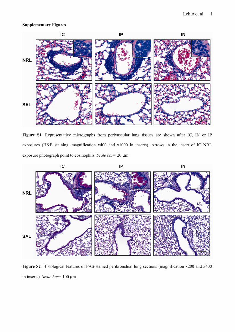
Lehto et al. 1
Supplementary Figures
Figure S1. Representative micrographs from perivascular lung tissues are shown after IC, IN or IP
exposures (H&E staining, magnification x400 and x1000 in inserts). Arrows in the insert of IC NRL
exposure photograph point to eosinophils. Scale bar= 20 μm.
Figure S2. Histological features of PAS-stained peribronchial lung sections (magnification x200 and x400
in inserts). Scale bar= 100 μm.
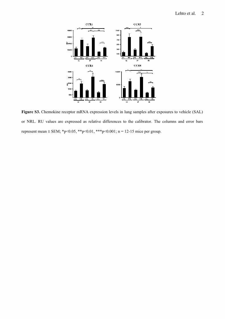
Lehto et al. 2
Figure S3. Chemokine receptor mRNA expression levels in lung samples after exposures to vehicle (SAL)
or NRL. RU values are expressed as relative differences to the calibrator. The columns and error bars
represent mean ± SEM; *p<0.05, **p<0.01, ***p<0.001; n = 12-15 mice per group.
ORIGINAL PAPER
Hev b 6.01 and Hev b 5 induce pro-inflammatory cytokines and chemokinesfrom peripheral blood mononuclear cells in latex allergyM. Lehto�, A. Kotovuoriw, K. Palosuoz, E. Varjonenz, S. Lehtimaki�, N. Kalkkinenw, T. Palosuo‰, T. Reunalaz and H. Alenius��Unit of Excellence for Immunotoxicology, Finnish Institute of Occupational Health, Helsinki, Finland, wInstitute of Biotechnology, University of Helsinki, Helsinki,
Finland, zSkin and Allergy Hospital, Helsinki University Central Hospital, Helsinki, Finland, ‰Laboratory of Immunobiology, National Public Health Institute, Helsinki,
Finland, and zDepartment of Dermatology, Tampere University and University Hospital, Tampere, Finland
Clinical andExperimental
Allergy
Correspondence:Maili Lehto, Unit of Excellence inImmunotoxicology, Finnish Institute ofOccupational Health, Topeliuksenkatu41 aA, FIN-00250 Helsinki, Finland.E-mail: [email protected]
Summary
Background Hev b 6.01 (prohevein) and Hev b 5 [acidic natural rubber latex (NRL) protein] aremajor IgE-binding allergens in NRL allergy.Objective To examine allergen-specific cytokine and chemokine responses in NRL-allergicpatients.Methods Fourteen NRL-allergic patients and 10 healthy controls participated in the study. Hevb 6.01 and Hev b 5 were purified under non-denaturating conditions by chromatographicmethods. Specific IgE antibodies were measured by ELISA and proliferation of peripheralblood mononuclear cells (PBMC) by 3H-thymidine incorporation assay. Allergen-specificinduction of cytokine and chemokine mRNA in PBMC was measured by real-time PCR andprotein levels by ELISA. Surface expression of chemokine receptors was analysed by flowcytometry.Results Twelve (86%) NRL-allergic patients had positive skin prick test reactions and IgEantibodies against Hev b 6.01, but less than 30% responded to Hev b 5. Cell proliferationagainst Hev b 6.01, but not against Hev b 5, was significantly increased. Both allergenselicited significantly higher expression of pro-inflammatory and T-helper type 2 cytokines(TNF, IL-12p40, IL-13) and chemokines (CCL3, CCL4, CCL20) in the NRL-allergic patients thanin controls. Interestingly, mRNA expression of the regulatory cytokine TGF-b1 was reduced,whereas IL-10 expression was enhanced after allergen stimulations in patients with NRLallergy. Finally, the NRL-allergic patients showed increased CCR4 expression on CD31CD8�
T cells and decreased CXCR3 expression on CD31CD81 T cells.Conclusion Allergen-specific induction of cytokines and chemokines in PBMC and chemokinereceptor expression on circulating T cells may contribute to the pathogenesis of NRL allergy.
Keywords chemokine receptor, chemokines, cytokines, latex hypersensitivity, PBMCSubmitted 31 March 2006; revised 14 July 2006; accepted 2 October 2006
Introduction
Natural rubber latex (NRL) allergy has been one of themost frequently encountered occupational diseases espe-cially among the health care workers (HCW) [1]. Theprevalence of NRL allergy in HCW has been reported tobe between 0.6% and 17% [2]. However, recent studiesshow that incidence decreases when attention is paid tothe quality of NRL gloves [3, 4]. Symptoms of NRL allergyvary from mild localized reactions, e.g. contact urticariaand rhinitis, to more severe symptoms, e.g. generalizedurticaria, asthma and anaphylaxis [2, 5]. In addition to
type I symptoms, NRL allergy may present with eczema onthe hands [6]. This finding is further supported by experi-ments in mice showing that cutaneous NRL exposurecauses T-helper type 2 dermatitis [7].
In contrast to IgE-mediated reactions, knowledge ofNRL-induced cell-mediated immunity is scanty. Research-ers have mainly studied proliferation of peripheral bloodmononuclear cells (PBMC) against NRL allergens [8–11].Evidence exists that the major IgE-binding NRL allergenHev b 6.02 (hevein domain of prohevein) [12, 13] containsdominant T cell epitopes [14]. In addition, Hev b 6.03 [12]
doi: 10.1111/j.1365-2222.2006.02622.x Clinical and Experimental Allergy, 37, 133–140
�c 2007 The Authors
Journal compilation �c 2007 Blackwell Publishing Ltd
(C domain of prohevein) has HLA-DR-binding motifs andinduces T cell proliferation [10]. Certain Hev b 5 peptideshave been shown to induce proliferation of peripheralT cell lines of patients with NRL allergy [15]. However,only little is known about cytokine and chemokineresponses to NRL allergens.
In the present study, we examined responses ofPBMC from NRL-allergic patients against the two majorNRL allergens, i.e. Hev b 6.01 and Hev b 5 [12]. Ourresults suggest that allergen-induced production of pro-inflammatory and regulatory cytokines and chemokinesmay play an important role in the pathogenesis of NRLallergy.
Materials and methods
Patients
Fourteen patients with NRL allergy (13 women and oneman; mean age 46 years), who were diagnosed at the Skinand Allergy Hospital, Helsinki University Central Hospital,in 2003 participated in the study. Six of them were HCWand seven were other occupational NRL-glove users.Three patients had asthma, five allergic rhinitis and threeatopic eczema. All patients had a positive skin prick test(SPT; weal diameter at least 3 mm and half of histaminecontrol, when negative control was 0) to a commerciallatex extract (Stallergenes, Antony Cedex, France) and apositive NRL-glove challenge test [6]. Ten healthy subjects(nine women and one man; mean age 42 years), seven ofwhom were HCW, served as controls. The study wasapproved by the ethics committee of Helsinki UniversityCentral Hospital.
Natural rubber latex allergens
NRL [13, 16] was subjected to buffer exchange by using aBioGelP6 desalting gel column (1.6�14 cm; Bio-RadLaboratories, Hercules, CA, USA) in PBS.
Hev b 6.01 was purified from the B-serum of latex [17]by gel filtration and anion exchange chromatographywithout reversed phase chromatography [18]. Briefly, gelfiltration was performed on a Superdex 75 HR 16/60column (Amersham Biosciences, Uppsala, Sweden) inPBS. Prohevein-containing fractions were concentratedand the buffer was changed to 20 mM Tris-HCl, pH 8.5, byusing a BioGelP6 desalting column. Anion-exchangechromatography was performed on a MonoQ column(HR5/5, Amersham Biosciences) equilibrated with 20 mM
Tris-HCl, pH 8.5. Elution was performed with a lineargradient of NaCl (0–1 M in 30 min) in the equilibrationbuffer.
Hev b 5 was purified from the C-serum of latex [17] bybuffer exchange and cation exchange chromatography.C-serum was first subjected to buffer exchange by using a
BioGelP6 desalting gel column in 20 mM Na citrate, pH3.8. Cation exchange chromatography was performednext on a MonoS 4.6/100 PE (Tricorn, Amersham Bios-ciences) column equilibrated with 20 mM Na citrate, pH3.8. Elution was performed with a linear gradient of NaCl(0–1 M in 30 min) in the equilibration buffer.
Commercial ELISA kits utilizing monoclonal antibodiesto Hev b 5 and Hev b 6.02 (Fit Biotech Ltd, Tampere,Finland) were used to analyse the molecular identity of thepurified NRL proteins. For purity check and quantifica-tion, the allergens were first subjected to reversed phasechromatography. For further confirmation of their iden-tity and for additional molecular characterization, theallergens were subjected to molecular mass determinationand peptide mass fingerprinting by matrix-assisted laserdesorption ionization time-of-flight (MALDI-TOF) massspectrometry [19, 20]. N-terminal sequencing was per-formed essentially as previously described [20].
Skin prick test and immunoglobulin E enzyme-linkedimmunosorbent assay
SPT was performed with a purified NRL extract (500, 50, 5and 0.5 mg/mL in PBS), Hev b 6.01 (50, 5 and 0.5 mg/mL inPBS) and Hev b 5 (50, 5 and 0.5 mg/mL). Histamine (10 mg/mL; ALK, Copenhagen, Denmark) was used as a positivecontrol, and PBS as a negative control.
IgE ELISA was performed as previously described [21].Briefly, the concentration of Hev b 6.01 and Hev b 5 wasadjusted to 2 mg/mL and that of NRL to 20 mg/mL forcoating microtitre plates (100 mL/well). The wells werethen post-coated with 1% human serum albumin (RedCross Finland Blood Service, Helsinki, Finland) in 50 mM
sodium carbonate buffer, and 100 mL of patient or controlserum (diluted 1 : 10) was added. Biotinylated goat anti-human IgE (Vector Laboratories Inc., Burlingame, CA,USA; diluted 1 : 1000) was added, followed by streptavi-din-conjugated alkaline phosphatase (Bio-Rad, Hercules,CA, USA; diluted 1 : 3000) and colour substrate (p-nitro-phenyl phosphate, ICN Biomedicals Inc., Aurora, OH,USA). The optical density was measured at 405 nm withan automated ELISA reader (Titertek Multiscan, Eflab,Turku, Finland).
Proliferation of peripheral blood mononuclear cells
Proliferation assays with PBMC were performed in com-plete RPMI 1640 containing 5% heat-inactivated humanAB serum as described earlier [22]. PBMC were isolatedfrom heparinized venous blood by using Ficoll densitygradient centrifugation and washed twice with PBS.PBMC proliferation was determined by stimulation of cellsin triplicate (105 cells/200 mL/well) alone or togetherallergens (NRL 100, 50, 10, 5, 1 mg/mL, Hev b 6.01 10, 5, 1and 0.1 mg/mL, Hev b 5 10, 5, 1 and 0.1 mg/mL). Phyto-
�c 2007 The AuthorsJournal compilation �c 2007 Blackwell Publishing Ltd, Clinical and Experimental Allergy, 37 : 133–140
134 M. Lehto et al
haemagglutinin (PHA, 45 mg/mL; Murex Biotech Ltd,Dartford, UK) was used as a positive control. After 6 daysof culturing, proliferation was measured by tritiatedmethyl-thymidine ([methyl-3H]-TdR, Amersham Bios-ciences, Little Chalfont, UK) incorporation. The resultswere expressed as stimulation indexes (SI; uptake ofisotope in stimulated culture/uptake of isotope in non-stimulated control culture).
Cytokine and chemokine measurements
Induction of cytokines, chemokines and chemokine re-ceptors was investigated in 10 NRL-allergic patients withhigh levels of antigen-specific IgE, as well as positive SPTand PBMC proliferative responses to NRL allergens. Ofthese 10 patients, nine had positive SPT responses to Hev b6.01 and four to Hev b 5 (weal area 4 9 mm2). Besides,two additional patients showed borderline SPT reactivityto Hev b 5 (weal area 7–9 mm2). Isolated and washedPBMC were stimulated in complete RPMI 1640 medium on24-well plates (Costar, Corning Incorporated, Corning, NY,USA) in the presence or absence of Hev b 6.01 (10 mg/mL)or Hev b 5 (10 mg/mL). PHA (45 mg/mL) and Staphylococ-cus aureus enterotoxin B (superantigen, SEB; 1 mg/mL;Sigma-Aldrich Co. Ltd, Gillingham, UK) were used aspolyclonal activators of PBMC. Each 1.5 mL well con-tained 3�106cells, the total number of cells per stimula-tion being 6�106. Cell pellets were collected after 6 h ofincubation and used for RNA isolation or flow cytometryanalysis. Cell culture supernatants were obtained after24 h of incubation and stored at –70 1C before measure-ment of cytokine or chemokine protein levels.
RNA extraction, synthesis of cDNA and real-time quan-titative PCR with an AbiPrism 7700 Sequence DetectorSystem (Applied Biosystems, Foster City, CA, USA) wereperformed as described earlier [7, 16, 22]. Applied Biosys-tems, from where PCR primers and probes were purchased,also designed them. The results were expressed as relativeunits (RU), which were calculated by the comparative CTmethod [7].
Protein levels of TNF, IL-10 and CCL3 were assayed bycommercial ELISA kits. TNF and IL-10 kits were fromeBioscience Inc. (San Diego, CA, USA) and their sensitivitywas 4 and 2 pg/mL, respectively. CCL3 kit was from R&DSystems (Minneapolis, MN, USA) and its lower detectionlimit was 8 pg/mL.
Flow cytometry
Three-colour flow cytometric analysis on T cell surfacemarkers was performed with FACSCalibur (BD Bios-ciences, San Jose, CA, USA) using FACSComp softwareversion 4.01 (BD Biosciences). Monoclonal antibodies(phycoerythrin-Cy5 (PE-Cy5)-conjugated anti-CD3,clone UCHT1; fluorescein isothiocyanate (FITC)-conjugated
anti-CD8, clone SK1; PE-conjugated anti-CXCR3, clone1C6/CXCR3; and PE-conjugated anti-CCR4, clone 1G1)and corresponding isotype controls were purchased fromBD Biosciences. We used CD8 expression to discriminatebetween T cell subtypes, as activation of human peripheralblood T cells can result in a virtually complete loss of thesurface CD4 marker, but only in a partial loss of CD8surface expression [23]. Furthermore, the vast majority ofCD31CD8� T cells are CD31CD41 T cells. In brief, 2�105
PBMC were incubated with the fluorochrome-conjugatedmAbs at 14 1C in the dark for 30 min; washed withstaining buffer (0.45 mm filter-sterilized PBS with 0.1%bovine serum albumin and 0.1% Na-azide); and fixed in1% paraformaldehyde (BD Biosciences). 20 000 eventswithin a lymphocyte gate were collected. CD31 cellswithin the lymphocyte gate were further gated to deter-mine the number of chemokine receptor-positive cellswithin CD31, CD31CD81 and CD31CD8� populations.Data were subsequently analysed with BD CELLQuestProsoftware (BD Biosciences).
Statistics
A non-parametric Mann–Whitney U-test was used tocompare the two groups using the GraphPadPrism Version4 (GraphPad Software Inc., San Diego, CA, USA). Datawere expressed as means� SEMs unless otherwise speci-fied. A P-value o 0.05 was considered to be statisticallysignificant.
Results
Analysis of the purity and identity of the allergens used forthe studies
Both purified allergens Hev b 6.01 and Hev b 5 gave singlepeaks in reversed phase chromatography when monitoredat the wavelength 214 nm. The integrated peak areas werealso utilized to determine the allergen concentrations forthe experiments. MALDI-TOF mass spectrometric analysisof Hev b 6.01 gave a single mass of 18 517 Da (and itsdouble charged form), which is within the mass accuracyof the used instrument, in agreement with the molecularmass calculated from the sequence (O49860, residues1–173) present in the databases. The identity of Hev b6.01 was further confirmed by peptide mass fingerprintingafter digestion with trypsin as well as with sequenceanalysis where it gave a pure N-terminal sequenceEQCGRQAGGK. Analysis of Hev b 5 by MALDI-TOF massspectrometry gave a single molecular mass of 15 952 Da(and its double-charged form), which corresponds to themolecular mass calculated from the sequence (Q39967)present in the databases. The identity of Hev b 5 wasfurther confirmed by peptide mass fingerprintingafter cleavage with trypsin. According to the database
�c 2007 The AuthorsJournal compilation �c 2007 Blackwell Publishing Ltd, Clinical and Experimental Allergy, 37 : 133–140
Cytokines and chemokines in latex allergy 135
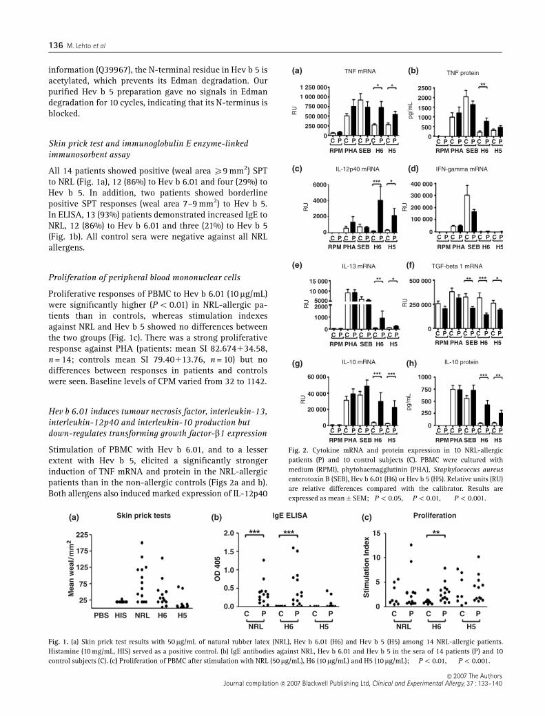
information (Q39967), the N-terminal residue in Hev b 5 isacetylated, which prevents its Edman degradation. Ourpurified Hev b 5 preparation gave no signals in Edmandegradation for 10 cycles, indicating that its N-terminus isblocked.
Skin prick test and immunoglobulin E enzyme-linkedimmunosorbent assay
All 14 patients showed positive (weal area X9 mm2) SPTto NRL (Fig. 1a), 12 (86%) to Hev b 6.01 and four (29%) toHev b 5. In addition, two patients showed borderlinepositive SPT responses (weal area 7–9 mm2) to Hev b 5.In ELISA, 13 (93%) patients demonstrated increased IgE toNRL, 12 (86%) to Hev b 6.01 and three (21%) to Hev b 5(Fig. 1b). All control sera were negative against all NRLallergens.
Proliferation of peripheral blood mononuclear cells
Proliferative responses of PBMC to Hev b 6.01 (10 mg/mL)were significantly higher (Po 0.01) in NRL-allergic pa-tients than in controls, whereas stimulation indexesagainst NRL and Hev b 5 showed no differences betweenthe two groups (Fig. 1c). There was a strong proliferativeresponse against PHA (patients: mean SI 82.674134.58,n = 14; controls mean SI 79.40113.76, n = 10) but nodifferences between responses in patients and controlswere seen. Baseline levels of CPM varied from 32 to 1142.
Hev b 6.01 induces tumour necrosis factor, interleukin-13,interleukin-12p40 and interleukin-10 production butdown-regulates transforming growth factor-b1 expression
Stimulation of PBMC with Hev b 6.01, and to a lesserextent with Hev b 5, elicited a significantly strongerinduction of TNF mRNA and protein in the NRL-allergicpatients than in the non-allergic controls (Figs 2a and b).Both allergens also induced marked expression of IL-12p40
225
(a) (b) (c)
175
125
25
PBS HIS NRL H50.0
C C C PPP C C C PPP
NRL H6 NRL H6H5 H5
0.5
OD
405
Mea
n w
eal/
mm
2
Sti
mu
lati
on
Ind
ex
1.0
1.5
2.0 15
ProliferationlgE ELISASkin prick tests
10
5
0H6
75
Fig. 1. (a) Skin prick test results with 50mg/mL of natural rubber latex (NRL), Hev b 6.01 (H6) and Hev b 5 (H5) among 14 NRL-allergic patients.Histamine (10 mg/mL, HIS) served as a positive control. (b) IgE antibodies against NRL, Hev b 6.01 and Hev b 5 in the sera of 14 patients (P) and 10control subjects (C). (c) Proliferation of PBMC after stimulation with NRL (50mg/mL), H6 (10mg/mL) and H5 (10mg/mL); ��Po 0.01,���Po 0.001.
0
0
0
0
0
C
RPM PHA SEB H6 H5 RPM PHA SEB H6 H5
CP P P P PC C C
CRPM PHA SEB H6 H5
CP P P P PC C C
C
RPM PHA SEB H6 H5
CP P P P PC C C C
RPM PHA SEB H6 H5
CP P P P PC C C
C
RPM PHA SEB H6 H5
CP P P P PC C C
C CP P P P PC C C
250 000 500
400 000
200 000
100 000
6000
4000
2000
0C
RPM PHA SEB H6 H5
CP P P P PC C C C
RPMPHA SEB H6 H5
CP P P P PC C C
60 000 1000
750
500
250
0
40 000
20 000
0
15 000 500 000
250 000
10 000
50002000
1000
300 000
1000
1500
2000
2500
500 000
750 000
1 000 000
1 250 000
(a) (b)
(d)(c)
(e) (f)
(h)(g)
TNF mRNA TNF protein
IFN-gamma mRNAIL-12p40 mRNA
RU
RU
RU
RU
RU
RU
pg/m
Lpg
/mL
IL-13 mRNA
IL-10 mRNA IL-10 protein
TGF-beta 1 mRNA
Fig. 2. Cytokine mRNA and protein expression in 10 NRL-allergicpatients (P) and 10 control subjects (C). PBMC were cultured withmedium (RPMI), phytohaemagglutinin (PHA), Staphylococcus aureusenterotoxin B (SEB), Hev b 6.01 (H6) or Hev b 5 (H5). Relative units (RU)are relative differences compared with the calibrator. Results areexpressed as mean�SEM; �Po 0.05, ��Po 0.01, ���Po 0.001.
�c 2007 The AuthorsJournal compilation �c 2007 Blackwell Publishing Ltd, Clinical and Experimental Allergy, 37 : 133–140
136 M. Lehto et al
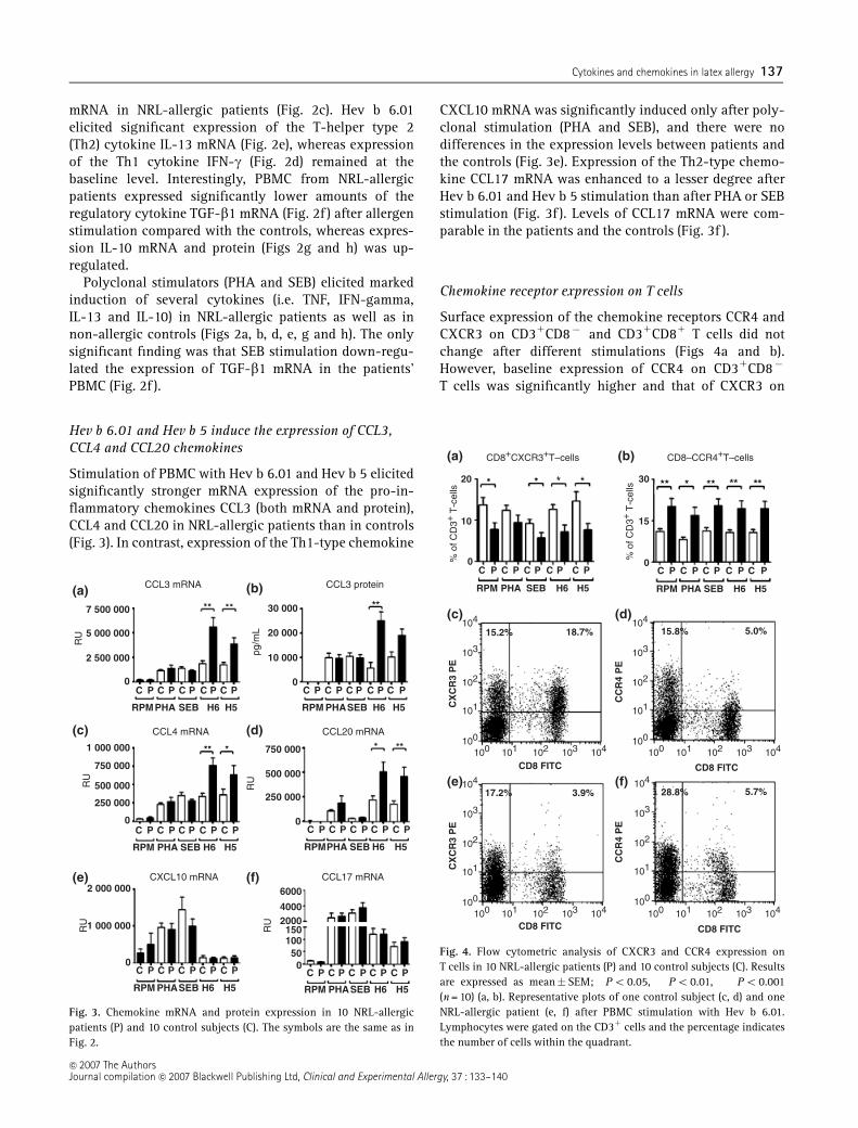
mRNA in NRL-allergic patients (Fig. 2c). Hev b 6.01elicited significant expression of the T-helper type 2(Th2) cytokine IL-13 mRNA (Fig. 2e), whereas expressionof the Th1 cytokine IFN-g (Fig. 2d) remained at thebaseline level. Interestingly, PBMC from NRL-allergicpatients expressed significantly lower amounts of theregulatory cytokine TGF-b1 mRNA (Fig. 2f ) after allergenstimulation compared with the controls, whereas expres-sion IL-10 mRNA and protein (Figs 2g and h) was up-regulated.
Polyclonal stimulators (PHA and SEB) elicited markedinduction of several cytokines (i.e. TNF, IFN-gamma,IL-13 and IL-10) in NRL-allergic patients as well as innon-allergic controls (Figs 2a, b, d, e, g and h). The onlysignificant finding was that SEB stimulation down-regu-lated the expression of TGF-b1 mRNA in the patients’PBMC (Fig. 2f ).
Hev b 6.01 and Hev b 5 induce the expression of CCL3,CCL4 and CCL20 chemokines
Stimulation of PBMC with Hev b 6.01 and Hev b 5 elicitedsignificantly stronger mRNA expression of the pro-in-flammatory chemokines CCL3 (both mRNA and protein),CCL4 and CCL20 in NRL-allergic patients than in controls(Fig. 3). In contrast, expression of the Th1-type chemokine
CXCL10 mRNA was significantly induced only after poly-clonal stimulation (PHA and SEB), and there were nodifferences in the expression levels between patients andthe controls (Fig. 3e). Expression of the Th2-type chemo-kine CCL17 mRNA was enhanced to a lesser degree afterHev b 6.01 and Hev b 5 stimulation than after PHA or SEBstimulation (Fig. 3f ). Levels of CCL17 mRNA were com-parable in the patients and the controls (Fig. 3f ).
Chemokine receptor expression on T cells
Surface expression of the chemokine receptors CCR4 andCXCR3 on CD31CD8� and CD31CD81 T cells did notchange after different stimulations (Figs 4a and b).However, baseline expression of CCR4 on CD31CD8�
T cells was significantly higher and that of CXCR3 on
0
0
0
0
0
C
RPMPHA SEB H6 H5 RPM PHASEB H6 H5
CP P P P PC C C
C
RPM PHA SEB H6 H5
CP P P P PC C C
C
RPM PHASEB H6 H5
CP P P P PC C C C
RPM PHASEB H6 H5
CP P P P PC C C
C
RPMPHA SEB H6 H5
CP P P P PC C C
C CP P P P PC C C
2 500 000
500 000
750 000
250 000
750 000
250 000
500 000
30 000
20 000
10 000
6000
40002000
150100500
5 000 000
7 500 000
1 000 000
2 000 000
1 000 000
(a) (b)
(d)
(f)(e)
(c)
CCL3 mRNA CCL3 protein
CCL4 mRNA CCL20 mRNA
CCL17 mRNACXCL10 mRNA
RU
RU
RU
RU
RU
pg/m
L
Fig. 3. Chemokine mRNA and protein expression in 10 NRL-allergicpatients (P) and 10 control subjects (C). The symbols are the same as inFig. 2.
0 0C
RPM
104
104
103
103
102
102
101
101
CD8 FITC
CX
CR
3 P
E
CC
R4
PE
CD8 FITC
100
100
104
104
103
103
102
102
101
101100
100
104
104
103
103
102
102
101
101100
100
104
104
103
103
102
102
101
101100
100
CD8 FITC
CX
CR
3 P
E
CC
R4
PE
CD8 FITC
PHA
15.2% 18.7% 15.8% 5.0%
28.8%17.2% 3.9% 5.7%
SEB H6 H5 RPM PHA SEB H6 H5
CP P P P PC C C C CP P P P PC C C
20
10
30
15
(a)
(c) (d)
(e) (f)
(b)CD8+CXCR3+T–cells CD8–CCR4+T–cells%
of C
D3+
T-c
ells
% o
f CD
3+ T
-cel
ls
Fig. 4. Flow cytometric analysis of CXCR3 and CCR4 expression onT cells in 10 NRL-allergic patients (P) and 10 control subjects (C). Resultsare expressed as mean� SEM; �Po 0.05, ��Po 0.01, ���Po 0.001(n = 10) (a, b). Representative plots of one control subject (c, d) and oneNRL-allergic patient (e, f) after PBMC stimulation with Hev b 6.01.Lymphocytes were gated on the CD31 cells and the percentage indicatesthe number of cells within the quadrant.
�c 2007 The AuthorsJournal compilation �c 2007 Blackwell Publishing Ltd, Clinical and Experimental Allergy, 37 : 133–140
Cytokines and chemokines in latex allergy 137
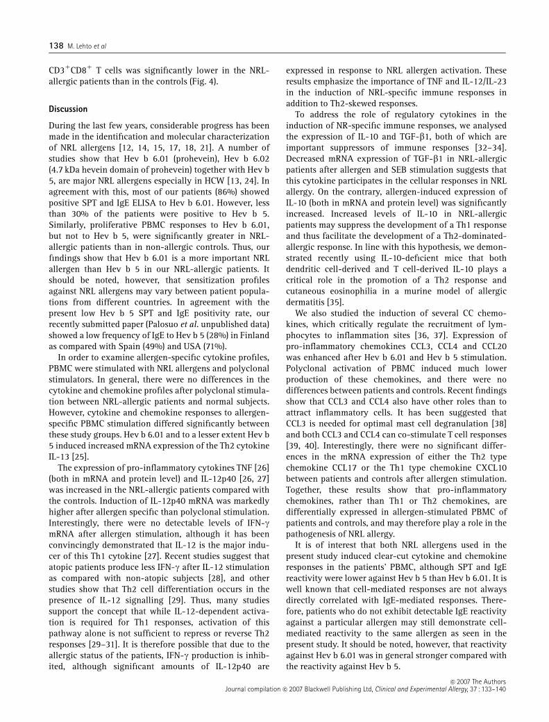
CD31CD81 T cells was significantly lower in the NRL-allergic patients than in the controls (Fig. 4).
Discussion
During the last few years, considerable progress has beenmade in the identification and molecular characterizationof NRL allergens [12, 14, 15, 17, 18, 21]. A number ofstudies show that Hev b 6.01 (prohevein), Hev b 6.02(4.7 kDa hevein domain of prohevein) together with Hev b5, are major NRL allergens especially in HCW [13, 24]. Inagreement with this, most of our patients (86%) showedpositive SPT and IgE ELISA to Hev b 6.01. However, lessthan 30% of the patients were positive to Hev b 5.Similarly, proliferative PBMC responses to Hev b 6.01,but not to Hev b 5, were significantly greater in NRL-allergic patients than in non-allergic controls. Thus, ourfindings show that Hev b 6.01 is a more important NRLallergen than Hev b 5 in our NRL-allergic patients. Itshould be noted, however, that sensitization profilesagainst NRL allergens may vary between patient popula-tions from different countries. In agreement with thepresent low Hev b 5 SPT and IgE positivity rate, ourrecently submitted paper (Palosuo et al. unpublished data)showed a low frequency of IgE to Hev b 5 (28%) in Finlandas compared with Spain (49%) and USA (71%).
In order to examine allergen-specific cytokine profiles,PBMC were stimulated with NRL allergens and polyclonalstimulators. In general, there were no differences in thecytokine and chemokine profiles after polyclonal stimula-tion between NRL-allergic patients and normal subjects.However, cytokine and chemokine responses to allergen-specific PBMC stimulation differed significantly betweenthese study groups. Hev b 6.01 and to a lesser extent Hev b5 induced increased mRNA expression of the Th2 cytokineIL-13 [25].
The expression of pro-inflammatory cytokines TNF [26](both in mRNA and protein level) and IL-12p40 [26, 27]was increased in the NRL-allergic patients compared withthe controls. Induction of IL-12p40 mRNA was markedlyhigher after allergen specific than polyclonal stimulation.Interestingly, there were no detectable levels of IFN-gmRNA after allergen stimulation, although it has beenconvincingly demonstrated that IL-12 is the major indu-cer of this Th1 cytokine [27]. Recent studies suggest thatatopic patients produce less IFN-g after IL-12 stimulationas compared with non-atopic subjects [28], and otherstudies show that Th2 cell differentiation occurs in thepresence of IL-12 signalling [29]. Thus, many studiessupport the concept that while IL-12-dependent activa-tion is required for Th1 responses, activation of thispathway alone is not sufficient to repress or reverse Th2responses [29–31]. It is therefore possible that due to theallergic status of the patients, IFN-g production is inhib-ited, although significant amounts of IL-12p40 are
expressed in response to NRL allergen activation. Theseresults emphasize the importance of TNF and IL-12/IL-23in the induction of NRL-specific immune responses inaddition to Th2-skewed responses.
To address the role of regulatory cytokines in theinduction of NR-specific immune responses, we analysedthe expression of IL-10 and TGF-b1, both of which areimportant suppressors of immune responses [32–34].Decreased mRNA expression of TGF-b1 in NRL-allergicpatients after allergen and SEB stimulation suggests thatthis cytokine participates in the cellular responses in NRLallergy. On the contrary, allergen-induced expression ofIL-10 (both in mRNA and protein level) was significantlyincreased. Increased levels of IL-10 in NRL-allergicpatients may suppress the development of a Th1 responseand thus facilitate the development of a Th2-dominated-allergic response. In line with this hypothesis, we demon-strated recently using IL-10-deficient mice that bothdendritic cell-derived and T cell-derived IL-10 plays acritical role in the promotion of a Th2 response andcutaneous eosinophilia in a murine model of allergicdermatitis [35].
We also studied the induction of several CC chemo-kines, which critically regulate the recruitment of lym-phocytes to inflammation sites [36, 37]. Expression ofpro-inflammatory chemokines CCL3, CCL4 and CCL20was enhanced after Hev b 6.01 and Hev b 5 stimulation.Polyclonal activation of PBMC induced much lowerproduction of these chemokines, and there were nodifferences between patients and controls. Recent findingsshow that CCL3 and CCL4 also have other roles than toattract inflammatory cells. It has been suggested thatCCL3 is needed for optimal mast cell degranulation [38]and both CCL3 and CCL4 can co-stimulate T cell responses[39, 40]. Interestingly, there were no significant differ-ences in the mRNA expression of either the Th2 typechemokine CCL17 or the Th1 type chemokine CXCL10between patients and controls after allergen stimulation.Together, these results show that pro-inflammatorychemokines, rather than Th1 or Th2 chemokines, aredifferentially expressed in allergen-stimulated PBMC ofpatients and controls, and may therefore play a role in thepathogenesis of NRL allergy.
It is of interest that both NRL allergens used in thepresent study induced clear-cut cytokine and chemokineresponses in the patients’ PBMC, although SPT and IgEreactivity were lower against Hev b 5 than Hev b 6.01. It iswell known that cell-mediated responses are not alwaysdirectly correlated with IgE-mediated responses. There-fore, patients who do not exhibit detectable IgE reactivityagainst a particular allergen may still demonstrate cell-mediated reactivity to the same allergen as seen in thepresent study. It should be noted, however, that reactivityagainst Hev b 6.01 was in general stronger compared withthe reactivity against Hev b 5.
�c 2007 The AuthorsJournal compilation �c 2007 Blackwell Publishing Ltd, Clinical and Experimental Allergy, 37 : 133–140
138 M. Lehto et al
Th1 and Th2 cells differ in their chemokine receptorexpression and their responsiveness to various chemo-kines [36]. We therefore studied the expression of Th1(CXCR3) and Th2 (CCR4) type chemokine receptors on thesurface of circulating T lymphocytes. No changes in theamounts of these receptors were found after any stimula-tion. However, CCR4 expression was constitutively higheron the patients’ CD31CD8� T cells (most of which areCD31CD41 T cells), whereas CXCR3 expression was con-stitutively lower on their CD31CD81 T cells. In line withthese results, it has been observed that CCR4 expressionon peripheral blood CD41 T cells is enhanced especially insevere atopic dermatitis [41], and that levels of CXCR3transcripts are decreased in PBMC from such patients [42].These results suggest that differences in the expression ofCCR4 and CXCR3 on circulating T cells may have animpact on the development and maintenance of NRLallergy.
In conclusion, the present results demonstrate thatallergen-specific stimulation of PBMC induces clear-cutdifferences in the cytokine and chemokine profiles ofNRL-allergic patients and non-allergic control subjects. Itis of interest that pro-inflammatory cytokines andchemokines in particular are predominantly expressedafter Hev b 6.01 and Hev b 5 stimulation and thatregulatory cytokines also seem to be involved in NRL-specific immune responses. These findings may reflect themolecular mechanisms involved in the development andmaintenance of NRL allergy and may open new avenuesfor disease prevention.
Acknowledgements
We thank Leena Petman for her expert and excellentassistance both in recruitment of patients and clinicalobservations. We also thank Elina Ahola for excellenttechnical assistance in purification and analysis ofthe Hev b 6.01 and Hev b 5 allergens. This study wassupported by grants from the Academy of Finland (grantsnumber 37 852 and 50 809). The Ansell HealthcareCorporation also funded this work.
References
1 Ahmed DD, Sobczak SC, Yunginger JW. Occupational allergiescaused by latex. Immunol Allergy Clin North Am 2003;23:205–19.
2 Reunala T, Alenius H, Turjanmaa K, Palosuo T. Latex allergy andskin. Curr Opin Allergy Clin Immunol 2004; 4:397–401.
3 Hunt LW, Kelkar P, Reed CE, Yunginger JW. Management ofoccupational allergy to natural rubber latex in a medical center:the importance of quantitative latex allergen measurement andobjective follow-up. J Allergy Clin Immunol 2002; 110:S96–106.
4 Allmers H, Schmengler J, John SM. Decreasing incidence ofoccupational contact urticaria caused by natural rubber latex
allergy in German health care workers. J Allergy Clin Immunol2004; 114:347–51.
5 Ranta PM, Ownby DR. A review of natural-rubber latex allergy inhealth care workers. Clin Infect Dis 2004; 38:252–6.
6 Turjanmaa K, Kanto M, Kautiainen H, Reunala T, Palosuo T.Long-term outcome of 160 adult patients with natural rubberlatex allergy. J Allergy Clin Immunol 2002; 110:S70–4.
7 Lehto M, Koivuluhta M, Wang G et al. Epicutaneous naturalrubber latex sensitization induces T helper 2-type dermatitis andstrong prohevein-specific IgE response. J Invest Dermatol 2003;120:633–40.
8 Murali PS, Kelly KJ, Fink JN, Kurup VP. Investigations into thecellular immune responses in latex allergy. J Lab Clin Med 1994;124:638–43.
9 Raulf-Heimsoth M, Chen Z, Liebers V, Allmers H, Baur X.Lymphocyte proliferation response to extracts from differentlatex materials and to the purified latex allergen Hev b 1 (rubberelongation factor). J Allergy Clin Immunol 1996; 98:640–51.
10 Raulf-Heimsoth M, Rozynek P, Bruning T, Rihs HP. Characteriza-tion of B- and T-cell responses and HLA-DR4 binding motifs ofthe latex allergen Hev b 601 (prohevein) and its post-transcrip-tionally formed proteins Hev b 6.02 and Hev b 6.03. Allergy2004; 59:724–33.
11 Johnson BD, Kurup VP, Sussman GL et al. Purified and recombi-nant latex proteins stimulate peripheral blood lymphocytesof latex allergic patients. Int Arch Allergy Immunol 1999;120:270–9.
12 Wagner S, Breiteneder H. Hevea brasiliensis latex allergens:current panel and clinical relevance. Int Arch Allergy Immunol2005; 136:90–7.
13 Alenius H, Kalkkinen N, Reunala T, Turjanmaa K, Palosuo T. Themain IgE-binding epitope of a major latex allergen, prohevein, ispresent in its N-terminal 43-amino acid fragment, hevein.J Immunol 1996; 156:1618–25.
14 de Silva HD, Gardner LM, Drew AC et al. The hevein domain ofthe major latex-glove allergen Hev b 6.01 contains dominantT cell reactive sites. Clin Exp Allergy 2004; 34:611–8.
15 de Silva HD, Sutherland MF, Suphioglu C et al. Human T-cellepitopes of the latex allergen Hev b 5 in health care workers.J Allergy Clin Immunol 2000; 105:1017–24.
16 Lehto M, Haapakoski R, Wolff H et al. Cutaneous, but not airway,latex exposure induces allergic lung inflammation and airwayhyperreactivity in mice. J Invest Dermatol 2005; 125:962–8.
17 Yeang HY, Arif SA, Yusof F, Sunderasan E. Allergenic proteins ofnatural rubber latex. Methods 2002; 27:32–45.
18 Karisola P, Kotovuori A, Poikonen S et al. Isolated hevein-likedomains, but not 31-kd endochitinases, are responsible for IgE-mediated in vitro and in vivo reactions in latex-fruit syndrome.J Allergy Clin Immunol 2005; 115:598–605.
19 Poutanen M, Salusjarvi L, Ruohonen L, Penttila M, Kalkkinen N.Use of matrix-assisted laser desorption/ionization time-of-flightmass mapping and nanospray liquid chromatography/electro-spray ionization tandem mass spectrometry sequence tag analy-sis for high sensitivity identification of yeast proteins separatedby two-dimensional gel electrophoresis. Rapid Commun MassSpectrom 2001; 15:1685–92.
20 Ylonen A, Rinne A, Herttuainen J et al. Atlantic salmon (Salmosalar L.) skin contains a novel kininogen and another cysteineproteinase inhibitor. Eur J Biochem 1999; 266:1066–72.
�c 2007 The AuthorsJournal compilation �c 2007 Blackwell Publishing Ltd, Clinical and Experimental Allergy, 37 : 133–140
Cytokines and chemokines in latex allergy 139
21 Karisola P, Alenius H, Mikkola J et al. The major conformationalIgE-binding epitopes of hevein (Hev b 6.02) are identified by anovel chimera-based allergen epitope mapping strategy. J BiolChem 2002; 277:22656–61.
22 Lehto M, Palosuo K, Varjonen E et al. Humoral and cellularresponses to gliadin in wheat-dependent, exercise-induced ana-phylaxis. Clin Exp Allergy 2003; 33:90–5.
23 Anderson SJ, Coleclough C. Regulation of CD4 and CD8 expres-sion on mouse T cells. Active removal from the cell surface bytwo mechanisms. J Immunol 1993; 151:5123–34.
24 Bernstein DI, Biagini RE, Karnani R et al. In vivo sensitization topurified Hevea brasiliensis proteins in health care workerssensitized to natural rubber latex. J Allergy Clin Immunol 2003;111:610–6.
25 Wynn TA. IL-13 effector functions. Annu Rev Immunol 2003;21:425–56.
26 Baud V, Karin M. Signal transduction by tumor necrosis factorand its relatives. Trends Cell Biol 2001; 11:372–7.
27 Hunter CA. New IL-12-family members: IL-23 and IL-27, cyto-kines with divergent functions. Nat Rev Immunol 2005;5:521–31.
28 Matsui E, Kaneko H, Teramoto T et al. Reduced IFNgammaproduction in response to IL-12 stimulation and/or reducedIL-12 production in atopic patients. Clin Exp Allergy 2000;30:1250–6.
29 Nishikomori R, Ehrhardt RO, Strober W. T helper type 2 celldifferentiation occurs in the presence of interleukin 12 receptorbeta2 chain expression and signaling. J Exp Med 2000; 191:847–58.
30 Heath VL, Showe L, Crain C et al. Cutting edge: ectopicexpression of the IL-12 receptor-beta 2 in developing andcommitted Th2 cells does not affect the production of IL-4 orinduce the production of IFN-gamma. J Immunol 2000; 164:2861–5.
31 Berenson LS, Ota N, Murphy KM. Issues in T-helper 1 develop-ment-resolved and unresolved. Immunol Rev 2004; 202:157–74.
32 Schmidt-Weber CB, Blaser K. Regulation and role of transform-ing growth factor-beta in immune tolerance induction andinflammation. Curr Opin Immunol 2004; 16:709–16.
33 Hawrylowicz CM, O’Garra A. Potential role of interleukin-10-secreting regulatory T cells in allergy and asthma. Nat RevImmunol 2005; 5:271–83.
34 Jutel M, Akdis M, Budak F et al. IL-10 and TGF-beta cooperate inthe regulatory T cell response to mucosal allergens in normalimmunity and specific immunotherapy. Eur J Immunol 2003;33:1205–14.
35 Laouini D, Alenius H, Bryce P et al. IL-10 is critical for Th2responses in a murine model of allergic dermatitis. J Clin Invest2003; 112:1058–66.
36 Moser B, Wolf M, Walz A, Loetscher P. Chemokines: multiplelevels of leukocyte migration control. Trends Immunol 2004;25:75–84.
37 Rot A, von Andrian UH. Chemokines in innate and adaptive hostdefense: basic chemokinese grammar for immune cells. AnnuRev Immunol 2004; 22:891–928.
38 Miyazaki D, Nakamura T, Toda M et al. Macrophage inflamma-tory protein-1alpha as a co-stimulatory signal for mast cell-mediated immediate hypersensitivity reactions. J Clin Invest2005; 115:434–42.
39 Trautmann A. Chemokines as immunotransmitters? Nat Immu-nol 2005; 6:427–8.
40 Molon B, Gri G, Bettella M et al. T cell costimulation bychemokine receptors. Nat Immunol 2005; 6:465–71.
41 Wakugawa M, Nakamura K, Kakinuma T et al. CC chemokinereceptor 4 expression on peripheral blood CD41 T cells reflectsdisease activity of atopic dermatitis. J Invest Dermatol 2001;117:188–96.
42 Hatano Y, Katagiri K, Takayasu S. Decreased levels of CXCR3transcripts in peripheral blood mononuclear cells frompatients with atopic dermatitis and with cutaneous diseasesassociated with eosinophilia. Arch Dermatol Res 2001; 293:319–22.
�c 2007 The AuthorsJournal compilation �c 2007 Blackwell Publishing Ltd, Clinical and Experimental Allergy, 37 : 133–140
140 M. Lehto et al

Latex allergy: low prevalence of IgE to highly purified Hev b 2 and Hev b 13
Timo Palosuo, MD,a Maili Lehto, MSc,b Annika Kotovuori, MSc,c Nisse Kalkkinen,
PhD,c Carlos Blanco, MD, PhD,d Paloma Poza, MD, d Teresa Carrillo, MD, PhD,d
Robert G Hamilton, PhD,e Harri Alenius, PhD,b Timo Reunala, MD,f Kristiina
Turjanmaa, MD f
Helsinki and Tampere, Finland, Las Palmas de Gran Canaria, Spain, and Baltimore,
MD, USA
From a Laboratory of Immunobiology, National Public Health Institute , Helsinki, b
Unit of Excellence for Immunotoxicology, Finnish Institute of Occupational Health,
Helsinki, c Institute of Biotechnology, University of Helsinki, Helsinki, d Servicio de
Alergia, Hospital Universitario de Gran Canaria Dr. Negrín, Las Palmas de Gran
Canaria, e Division of Allergy and Clinical Immunology, Johns Hopkins University
School of Medicine, Baltimore, MD,f Department of Dermatology, Tampere University
and University Hospital, Tampere
Correspondence and reprint requests: Timo Palosuo, MD, a Laboratory of
Immunobiology, National Public Health Institute, Mannerheimintie 166, FIN00300
Helsinki, Finland Tel: +35894744 8263, cell phone +35840723 0284, fax: +3589
4744 8281, Email: [email protected]
Running head: Low prevalence of IgE to Hev b 2 and Hev b 13
Palosuo 2
Abstract
Background: Hev b 2 and Hev b 13 have been recently identified as major latex
allergens by detecting specific IgE antibodies in >50% of sera from Hevea brasiliensis
latexallergic individuals.
Objective: We assessed the prevalence rates for sensitization to extensively purified
latex allergens in patients from three diverse geographical areas.
Methods: Native Hev b 2, Hev b 5, Hev b 6.01 and Hev b 13 were purified by non
denaturating chromatography and used in ELISAs to assess sera from 215 latexallergic
patients and 172 atopic nonsensitized controls from Finland, Spain and USA to detect
allergenspecific IgE antibodies.
Results: Unexpectedly, even highly purified Hev b 13 contained epitope(s) to which
Hev b 6 specific human IgE antibodies bound effectively. Further purification,
however, reduced the prevalence of IgE antibody reactivity to low levels: 15%, 5% and
11% for Hev b 2 and, 18%, 30% and 27% for Hev b 13 among latexallergic Finnish,
Spanish and American patients, respectively. Interestingly, Finnish patients had a lower
prevalence of Hev b 5specific IgE antibody (28%) as compared to Spanish (49%) and
American (71%) patients. The prevalence of Hev b 6.01specific IgE reactivity was
uniformly >50% in all three populations.
Conclusion: Neither Hev b 2 nor Hev b 13 appear to be major latex allergens when
evaluated in serological assays using highly purified allergens. The reason(s) for the
observed differences in published sensitization rates in various geographic regions
requires further study. Purity of the allergen preparations has a marked impact on the
accuracy of latexspecific IgE antibody detection in epidemiological studies and in the
serological diagnosis of latex allergy.

Palosuo 3
Keywords: Allergens, Hev b 2, Hev b 13, IgE antibodies, latex hypersensitivity,
prevalence
Palosuo 4
Introduction
Immediatetype hypersensitivity to natural rubber latex (NRL) continues to be
an important occupational disease among health care workers (HCW) and other
personnel using NRL gloves 14 . Of the more than 250 different polypeptides detected
from NRL, only about one fourth appear to be capable of binding IgE antibodies 57 .
Knowledge of the principal allergens in Hevea latex can facilitate the development of
more reliable in vivo and in vitro diagnostic reagents for use in assessing individuals
with a suspected NRL allergy based on their clinical history. Moreover, knowledge of
which Hevea brasiliensis (Hev b) allergenic proteins drive the sensitization to NRL aids
in the design of assays for monitoring the allergenicity of NRL containing products. The
WHO/IUIS Allergen Nomenclature Committee (www.allergen.org) in February 2006
listed 13 NRL Hev b allergens as having been characterized at the molecular level.
Several NRL proteins reportedly represent clinicallyimportant allergens 810 .
However, the current knowledge about which allergenic proteins or their fragments are
present in extracts of manufactured NRL products is still limited This has made it
difficult to define the relevance of each allergen in the induction of clinical disease. Of
the wellcharacterized NRL allergens, Hev b 6 (Hev b 6.01 [prohevein], and Hev b 6.02
[hevein]) and Hev b 5 have been unequivocally identified as major allergens for adults
as evidenced by the presence of specific IgE antibodies in the sera of more than half of
sensitized individuals in any defined population of patients 5, 1113 . Moreover, Hev b
1 and Hev b 3 are important allergens for children with spina bifida or other congenital
malformations who are sensitized as a result of exposure during their multiple surgeries
at an early age 1417 . Recently, Hev b 2 and Hev b 13 have been reclassified as major
allergens in NRL allergy 8, 10, 18 , based on a positive skin prick test (SPT) in 63% of
Palosuo 5
NRLallergic patients. However, preliminary studies in a Finnish population have
indicated to the contrary that IgE to purified native Hev b 2 and Hev b 13 are relatively
rare (e.g.,~20% of Finnish patients). In this study, we examine the prevalence of IgE
antibodies using highly purified Hev b 2 and Hev b 13, and contrast it to the presence of
IgE antibody specific for Hev b 5 and Hev b 6.01. Sera from three latex sensitized
populations in diverse geographic areas were evaluated to control for the possibility of
differential exposure to latex products containing variable distributions of individual
NRL allergens.
Palosuo 6
Methods
Patients and controls
A total of 215 NRLallergic adult patients and 172 atopic controls without NRL
allergy were included in the study (Table I). For the Finnish patients, the diagnosis of
NRL allergy was based on a compatible clinical history, positive SPT (Stallergenes,
Antony Cedex, France, and/ or highallergen glove extracts), and a positive IgE
antibody serology (latex ImmunoCAP, Phadia, Uppsala, Sweden). Descriptions of
patients and control subjects from Spain and the USA have been reported in detail
elsewhere 8, 19 . This study was approved by local ethics committees in Finland
(Tampere University Hospital and National Public Health Institute), Spain (Hospital
Universitario de Gran Canaria Dr. Negrín) and USA (the Research and Ethics
Committees of the Johns Hopkins Medical Institutions).
Purification of NRL allergens
The source latex for isolation of the NRL allergens (Bserum and Cserum of
nonammoniated NRL, clone RRI 600) was purchased from the Rubber Research
Institute, Kuala Lumpur, Malaysia. Hev b 2, Hev b 6.01 and Hev b 13 were purified
from Bserum of NRL (diluted 1:5 with 50 mM sodium phosphate, pH 7.5), whereas
Hev b 5 was isolated from Cserum 9 .
Purification of Hev b 2 began with cation exchange chromatography using a MonoS
(Tricorn 4.6/100PE, Amersham Biosciences, Uppsala, Sweden) column that had been
equilibrated with 50 mM sodium phosphate, pH 7.5 (Fig 1, A). Elution was performed
using a linear gradient of NaCl (0 to 1 M in 30 min). The flow rate was 1.0 ml/min and
the chromatography was monitored at 214 nm. Hev b 2 containing fractions (marked
Palosuo 7
grey in Fig 1, A) were pooled and concentrated Hev b 2 containing preparation was
subjected to gel filtration chromatography on a Superdex 75 HR 10/30 column
(Amersham Biosciences) in phosphatebuffered saline (PBS) (Fig 1, B). The flow rate
was 0.5 ml/min and the chromatography was monitored at 214 nm. The Hev b 2
containing fractions (marked grey in Fig 1, B) were again pooled and concentrated.
Hev b 5 was purified by cation exchange chromatography. Cserum was first
subjected to a buffer exchange of 20 mM Nacitrate, pH 3.8 using a BioGelP6 desalting
gel column (BioRad Laboratories, Hercules, CA, USA). Cation exchange
chromatography was then performed on a MonoS 4.6/100 PE column (Tricorn,
Amersham Biosciences) that had been equilibrated with 20 mM Nacitrate, pH 3.8.
Elution was performed with a linear gradient of NaCl (0 to 1 M in 30 minutes) in the
equilibration buffer.
Purification of Hev b 6.01 involved gel filtration and anion exchange
chromatography 20 . Briefly, gel filtration was performed on a Superdex 75 HR 16/60
column (Amersham Biosciences) in phosphate buffered saline. Proheveincontaining
fractions were concentrated and the buffer was changed to 20 mM TrisCl, pH 8.5 using
a BioGelP6 column. Anionexchange chromatography was performed on a MonoQ
HR5/5 column (Amersham Biosciences) equilibrated with 20 mM TrisCl, pH 8.5.
Elution was performed with a linear gradient of NaCl (0 to 1 M in 30 minutes) in the
equilibration buffer.
For purification of Hev b 13, 1.5 ml of NRL Bserum was chromatographed on a
Superdex 75 (HR 16/60) column at a flow rate of 1.5 ml/min and monitored at 280 nm
(Fig 1, D). Hev b 13 containing fractions (marked grey) were pooled and concentrated.
The buffer was changed to 50 mM sodium acetate, pH 4.5 on a BioGelP6DG column.
Palosuo 8
Hev b 13 containing fractions were applied to a cation exchange column, MonoS (HR
5/5, Amersham Biosciences) (Fig 1, E). Elution was performed using a linear gradient
of NaCl (0 to 1 M in 40 min). The flow rate was 1.0 ml/min and the chromatography
was monitored at 214 nm. Hev b 13 eluted as a peak marked in the figure (Fig 1, E).
The purity of all four allergen preparations was ascertained by reversedphase
chromatography on a C1 column (2x20mm, Tosoh Corp, Tokyo, Japan) as described
previously 20 . Elution was performed with a linear gradient of acetonitrile (0100 % in
60 min) in 0.1% trifluoroacetic acid (TFA). The flow rate was 0.2 ml/min and detection
was performed at 214 nm. The eluted peaks for Hev b2 and Hev b13 are shown in Fig.
1 C and F, respectively.
Identification and characterization of the purified allergens
Monoclonal antibodies to the four allergens and commercial ELISA kits for Hev
b 5 and Hev b 6.02 (Fit Biotech Ltd., Tampere, Finland) were used to identify and
quantify the purified allergens. For molecular characterization of the purified allergens,
the molecular masses and peptide mass fingerprint analyses were performed by matrix
assisted laser desorption ionization timeofflight (MALDITOF) and electrospray
quadrupole timeoffight (QTOF) mass spectrometric analyses 21 . Nterminal
sequencing was performed as previously described 22 .
IgE ELISA
IgE antibodies to Hev b 5 and Hev b 6.01 were measured by ELISA as
previously described 23 . Briefly, Hev b 6.01 and Hev b 5 were separately coated on
microtiter plates at 2 g/ml (100 l/well). The wells were then blocked with 1% human
Palosuo 9
serum albumin (Red Cross Finland Blood Service, Helsinki, Finland) in 50 mM sodium
carbonate buffer, pH 9.6, and 100 l of patient or control serum (in duplicate, diluted
1:10) was added. Biotinylated goat antihuman IgE (Vector Laboratories, Inc,
Burlingame, CA; diluted 1:1000) was added, followed sequentially by streptavidin
conjugated alkaline phosphatase (BioRad, Hercules, CA; diluted 1:3000) and substrate
(pnitrophenyl phosphate, ICN Biomedicals Inc., Aurora, Ohio). When internal pooled
control sera had reached predetermined ODvalues, the optical density was read at 405
nm with an automated ELISA reader (Titertek Multiscan, Eflab, Turku, Finland). IgE
antibodies to Hev b 2 and Hev b 13 were measured using the same assay format except
that the coating concentration of these antigens was 1 g/ml in PBS, pH 7.4. Positive
reactions in each assay were determined as ODvalues exceeding the 98th percentile of
local atopic control subjects, which approximately corresponded to the mean + 3 SD.
Inhibition ELISA analyses were performed for specificity assessment as described
earlier 24 , using different NRL allergens to inhibit the binding of patients’ IgE to
solidphase antigens.
Data analysis
Nonparametric statistics (MannWhitney Utest) were used to perform statistical
tests because the majority of OD values were not normally distributed. A P value of
0.05 or less was considered a statistically significant difference between groups.
Correlation between individual serum IgE antibody levels to the various allergens was
assessed by a nonparametric Spearman r test.
Palosuo 10
Results
Identification and assessment of the purity of Hev b 2 and Hev b 13 preparations
The Hev b 2preparationf used in this study for serological evaluation was
shown to be homogeneous by in reversedphase chromatography (Fig 1, C).. Its identity
was further confirmed using monoclonal antiHev b 2 antibody reactivity, Nterminal
sequence assessment and spectrometric mass fingerprint analysis. Monoclonal
antibodies specific for Hev b 6.02, Hev b 5, or Hev b 13 had no detectable
immunoreactivity to the finalized native purified Hev b 2 (data not shown).
The Hev b 13 used in the study required extensive repurification. Monoclonal
antiHev b 6.02 antibodies and human sera containing IgE antibodies specific for Hev b
6.01 or Hev b 6.02 bound to the Hev b 13 preparations at various stages in the
purification process. Figure 2 shows that even after the final purification step, Hev b
6.02 reactivity was still present in the Hev b 13 preparation. However, there was no
indication of the presence of mature hevein (Hev b 6.02), prohevein (Hev b 6.01) or
other proteins as analyzed by reversedphase chromatography or peptide mass
fingerprint analyses in this final Hev b 13 preparation. Thus, the nature of the binding of
antiHev b 6.01 to epitopes in the Hev b 13 remained elusive. Monoclonal antibodies
specific for Hev b 13 bound effectively to the purified protein in ELISA while
monoclonal antibodies specific for Hev b 2 and Hev b 5 displayed no reactivity to the
Hev b 13 (data not shown). Mass fingerprint analyses and Nterminal sequencing
confirmed the identity of the purified protein as Hev b 13. Sequence analysis of the
purified protein gave an Nterminal sequence of ETCDFPA, which resulted from the
cleavage of the Hev b 13’s 27 amino acid signal sequence. MALDITOF mass
spectrometric analysis of the purified Hev b 13 produced an average molecular mass of
Palosuo 11
43,375 Da. The mass of the protein polypeptide chain, as calculated from the known
deduced sequence 18 and the Nterminus determined in the present study was 40313.2
Da. This indicated that the protein is posttranslationally modified.
The Hev b 13 polypeptide chain contains 3 putative Nglycosylation sites. The
last one (N 286) contained at least 3 different forms of glycans, composed of Nacetyl
glucosamine and varying amounts of mannose, fucose and xylose (data not shown). The
mass of these glycans was not enough to cover the suggested mass for the post
translational modifications. The tryptic peptide containing the two remaining
glycosylation sites was not further analyzed due to its low recovery and large size.
Prevalence of IgE antibodies to four purified NRL allergens in three different
geographic areas
There were welldefined differences in the prevalence of IgE specific for the
four NRL allergens in serum from NRLallergic patients from Finland, Spain and the
USA (Fig 3). IgE antibodies specific for Hev b 2 were rare in Spain (5 %), and seen
only in a minority of patients from Finland (15%) and USA (11%). The prevalence of
IgE to Hev b 13 ranged between 18 to 30% in all three geographic regions, suggesting a
relatively low overall frequency of sensitization to this allergen. Prevalence of IgE
antibodies to Hev b 5 was unexpectedly low in Finland (28%), which contrasted with
sera collected from Spanish and American patients (49% and 71%, respectively).
Meanwhile, the frequency of IgE antibodies to Hev b 6.01 was > 50% in the serum of
NRLallergic patients in all three countries (Fig 3).
Significant differences were noted among the three geographic areas in the mean
optical density levels that reflected the relative quantity of IgE antibodies in the serum
Palosuo 12
that was reactive to the individual NRL allergens (Fig 4). The highest mean response
values for antiHev b 2, antiHev b 5 and antiHev b 13 were seen with sera from the
American patients and highest mean antiHev b 6.01 level was present in the sera from
the Spanish patients. Lowest mean antiHev b 2, antiHev b 5 and antiHev b 13 levels
were observed with sera from the Finnish patients, and lowest mean antiHev b 6.01
level was present in American sera.
IgE binding to solidphase Hev b 13 can be inhibited by adding soluble Hev b 6.01
Purified soluble Hev b 6.01 inhibited the binding of patient specific IgE to immobilized
Hev b 13 in a dosedependent manner with most of the sera that were studied. Figure 5
depicts representative examples of the three modes of inhibition. Fig 5A shows a ”pure”
antiHev b 13 IgE response that was not inhibitable by Hev b 6.01. In Fig 5B, IgE anti
Hev b 13 binding was partially inhibited by the addition of soluble Hev b 6.01. In
contrast, Fig 5C displays the binding of human IgE antiHev b 13 that was totally
inhibited by Hev b 6.01. Inhibition experiments were carried out for 8 sera chosen for
their moderate to strong IgE antibody responses to Hev b 13. Of these, two sera
belonged to the Fig 5Alike category in which soluble Hev b 6.01 at 0.1 µg/ml produced
a 24% inhibition of the binding of IgE to solidphase Hev b 13. Two other sera
mimicked Figure 5Blike category in which 2654% inhibition was observed with the
addition of soluble Hev b 6.01. The remaining four sera fell in the Fig 5Clike category
with 6995% inhibition produced by soluble Hev b 6.01 (data not shown).
Palosuo 13
Discussion
Hev b 6.01 and Hev b 5 have been known as the clinically most important
“major” NRL allergens 7 . Recent studies have suggested that Hev b 2 10, 25, 26 and
Hev b 13 18, 27 may also be clinically important NRL allergens. Accordingly,
Bernstein et al. recently reported that 63% of NRLallergic patients had a positive SPT
to both purified Hev b 2 and Hev b 13. This was similar to the frequencies of positive
skin reactions seen with Hev b 6.01 8 . Kurup et al. used similar allergen preparations
in serological assays and reported that ~70 to 80% of NRL allergic adults had IgE
antibodies reactive with Hev b 2 and Hev b 13 10 . Importantly, the Hev b 13 that had
been produced in E.coli by recombinant DNA technology did not bind human IgE 18 .
Since the removal of the glycans from native Hev b 13 protein abolished its IgE
binding reactivity, it was proposed that the epitopes mediating IgE reactivity appeared
to reside in the glycan residues of the proteins 18, 28 . However, these epitopes have
not been characterized or clearly defined, especially in relation to the relative purity of
the proteins that were used to assess their allergenicity. Parenthetically, assessment of
the prevalence of IgE antibodies to the 9 Hev b allergens other than Hev b 2, 5, 6.01,
and 13 was not undertaken in this study because it is generally agreed based on the
literature that these allergens represent minor players in terms of inducing IgE
sensitization in adult healthcare workers 8, 27, 29 .
During our studies involving the purification of multiple NRL allergens, we faced
the problem that Hev b 6.02 (mature hevein) readily copurified with Hev b 13. This
unfortunately resulted in falsepositive serologic and skin prick tests results in
otherwise IgE antiHev b 13negative individuals. In the present study, we paid
exceptional attention to the relative purity of the Hev b 13 and strove to remove all Hev
Palosuo 14
b 6.02 prior to its use in the serology studies. Our previous experience with partially
impure Hev b 13 and Hev b 2 preparations revealed the presence of other NRL
allergens, or their fragments such as Hev b 6.02, Hev b 5 and, in some instances, Hev b
1 when analyzed using a commercial capture enzyme immunoassay (FIT kit, FIT
Biotech Ltd).
One may argue that the extensively purified allergen preparations were devoid of
in vivo reactivity and therefore unsuitable for IgE prevalence assessment. This,
however, was shown to not be the case since skin prick tests carried out to eight Finnish
NRLallergic individuals (at allergen concentration of 50 µg/ml, data not shown)
revealed two patients positive to Hev b 2 and four patients positive to Hev b 13
(diameter of the wheal at least 3 mm). Three of the eight patients were positive for Hev
b 5 and six for Hev b 6.01. Among the four Hev b 13positive patients, one was
negative to Hev b 6.01 and to other allergens tested, indicating the presence of a pure
Hev b 13specific reaction.
During the purification process of Hev b 13, binding of antiHev b 5 and antiHev
b 1 progressively decreased to undetectable levels. In contrast, monoclonal anti Hev b
6.02 reactivity to the final Hev b 13 preparation could not be totally abolished. When
the purity of the final Hev b 13 preparation was assessed with different protein
chemical methods, such as reversed phase chromatography, Nterminal sequencing,
electrospray or MALDITOF mass spectrometry or mass spectrometric peptide mass
fingerprinting, no other structures than Hev b 13 could be detected. However, the
relative Hev b 6.02 levels below approximately 12% could not have been detected
using these techniques.
Palosuo 15
There is no significant homology between the primary sequences of Hev b 6.02
(or Hev b 6.01) and Hev b 13. The exact nature of the binding of the Hev b 6.02
reactive antibodies to the Hev b 13 preparation has not yet been determined.
Fortunately, with extensive purification, we were able to reduce this phenomenon from
significant to very low levels, enabling the use of this protein to assess human IgE
antibodies specific for native Hev b 13 in serological assays.
We chose to use the 98th percentile of local atopic controls to define positive
threshold for the presence of Hev b allergen reactive IgE antibody. This approach
enables one to control for possible differences associated with variability resulting from
the local geographic background such as exposure to different NRL containing
products. Moreover, the actual potency of the immunological response (level of
antibody) is unlikely to influence the determination of prevalence. In addition, this
study contended with the skewed distribution of the low absorbance values obtained
with the sera from the control subjects.
The sensitization rates to purified Hev b 13 were generally low (1830%, Fig 3)
when the ultrapure Hev b 13 was used in the analysis. Furthermore, the binding of
patients’ IgE to Hev b 13 could be partially or totally inhibited by the addition of
soluble Hev b 6.01. When the binding of IgE to solidphase Hev b 13 was not inhibited
by Hev b 6.01 (Fig 5A) the response was considered specific to Hev b 13. However,
sera with a clearly defined IgE antiHev b 13 response in the absence of measurable IgE
antiHev b 6.01 antibodies were rare. Inhibition experiments were performed only to a
small subset of the sera evaluated in the study, and thus these results should be
interpreted with care. Interestingly, the binding of IgE was partially inhibited (up to
54%) by soluble Hev b 6.01 in some sera, suggesting that the measured IgE binding
Palosuo 16
was a result of a mixture of both Hev b 13 and Hev b 6.01specific antibodies (Fig 5B).
In half of the sera evaluated for inhibition, up to 95% of the binding activity to Hev b
13 was inhibited by the addition of soluble Hev b 6.01 (Fig 5C). In these cases of
essentially total inhibition, it is likely that the serum does not contain IgE antibodies
that are specific antiHev b 13. It is therefore possible that NRL allergic patients with a
restricted Hev b 13specific IgE antibody response are even rarer than suggested by the
present results (1830%).Consistent with our observations is a recent report that shows
that only 10% of Taiwanese HCW had IgE binding to Hev b 13 that had been quality
controlled with peptide mass fingerprinting and analyzed by immunoblotting after two
dimensional gel electrophoresis 30 .
Since the binding of IgE to solidphase Hev b 13 can be influenced by a minor
impurity of Hev b 6.01 or Hev b 6.02, one might expect to see a correlation between the
levels of IgE antiHev b 13 and antiHev b 6.01 in the patients’ sera. Even though the
number of antiHev b 13 positive sera was low, this was indeed the case. In the whole
patient series (n=215), the correlation coefficient (r) between the presence of Hev b
6.01 and Hev b 13 reactive antibodies as assessed by the Spearman rank test was 0.517
(p < 0.0001). Alternatively, no correlation was observed between antiHev b 6.01 and
antiHev b 2 antibodies (data not shown).
The purification procedure for Hev b 2 generated a clean, native protein that
displayed no reactivity with monoclonal antibodiesspecific for Hev b allergens other
than Hev b 2. Using this protein as an ELISA coating antigen in the present study and
previously with immunoblotting 31 , we have been able to verify markedly lower
prevalence rates for IgE antiHev b 2 in NRLallergic patients than previously reported
elsewhere 8, 10, 28, 29 . The prevalence of IgE to Hev b 2 and Hev b 13 in NRL
Palosuo 17
allergic patients in the present study also differed considerably in across three different
geographic areas. Among the Spanish patients, the prevalence of IgE to Hev b 2 and
Hev b 13 was low (5% and 30%, respectively).
While the present study focused on the significance of Hev b 2 and Hev b 13 as
NRL allergens in three geographical areas, we also included a comparable analysis of
prevalence rates for Hev b 6.01 and Hev b 5 which are known to be major NRL
allergens. Prevalence of IgE to Hev b 5 was unexpectedly low (28%) in Finland while it
was higher in Spain (49%) and the USA (71%). In contrast, IgE to Hev b 6.01 was high
(5474%) across all three geographic areas. The reasons for the different sensitization
rates may be due to different properties of NRL gloves used in these countries, and
differences in glove usage habits. We noted significant differences also in the relative
levels of IgE antibodies specific for the four Hev b allergens (Fig 4). These may reflect
local differences in exposure to these and other possibly crossreactive allergens.
Differences may exist in patient selection and demographics in the three series of
specimens.
In conclusion, the low frequency of IgE to exhaustively purified Hev b 2 and Hev
b 13 in NRLallergic patients from three different geographic areas suggests that the
rates of sensitization to these allergens are relatively low in comparison to previous
reports. This observation implies that these allergens should not be classified as major
NRL allergens. Although our purification protocols led to reasonable but not complete
purity of Hev b 13 and to chromatographically and immunologicallypure Hev b 2,
production of these allergens in immunologically active form by recombinant DNA
technology is needed to ultimately assess their clinical significance in NRL allergy.
Palosuo 18
Acknowledgments
The authors thank Dr. Vladimir Ovod of FITBiotech Ltd, Tampere, Finland, for
monoclonal antibodies to Hev b 1, 2, 3, 5, 6.02 and 13. We also thank Helena
Honkasalo and Marko Hukka for expert technical assistance. This study was supported
by grants from the Academy of Finland (grants number 37852 and 50809), the Johns
Hopkins University School of Medicine, Baltimore, MD USA (NIH latex
immunotherapy grant) and from Instituto de Salud Carlos III (ISCiii RTIC RedRespira
C03/011 and RTIC Vegetalia G03/094). The Ansell Healthcare Corporation also
funded this work.
Palosuo 19
References
1 Turjanmaa K, Alenius H, MakinenKiljunen S, Reunala T, Palosuo T. Natural
rubber latex allergy. Allergy 1996; 51:593602.
2 Liss GM, Sussman GL. Latex sensitization: occupational versus general
population prevalence rates. Am J Ind Med 1999; 35:196200.
3 Charous BL, Blanco C, Tarlo S et al. Natural rubber latex allergy after 12 years:
recommendations and perspectives. J Allergy Clin Immunol 2002; 109:314.
4 Ahmed DD, Sobczak SC, Yunginger JW. Occupational allergies caused by
latex. Immunol Allergy Clin North Am 2003; 23:20519.
5 Alenius H, Kalkkinen N, Reunala T, Turjanmaa K, Palosuo T. The main IgE
binding epitope of a major latex allergen, prohevein, is present in its Nterminal
43amino acid fragment, hevein. J Immunol 1996; 156:161825.
6 Posch A, Chen Z, Wheeler C, Dunn MJ, RaulfHeimsoth M, Baur X.
Characterization and identification of latex allergens by two dimensional
electrophoresis and protein microsequencing. J Allergy Clin Immunol 1997;
99:38595.
7 Wagner S, Breiteneder H. Hevea brasiliensis Latex Allergens: Current Panel and
Clinical Relevance. Int Arch Allergy Immunol 2005; 136:907.
8 Bernstein DI, Biagini RE, Karnani R et al. In vivo sensitization to purified
Hevea brasiliensis proteins in health care workers sensitized to natural rubber
latex. J Allergy Clin Immunol 2003; 111:6106.
9 Yeang HY, Arif SA, Yusof F, Sunderasan E. Allergenic proteins of natural
rubber latex. Methods 2002; 27:3245.
Palosuo 20
10 Kurup VP, Sussman GL, Yeang HY et al. Specific IgE response to purified and
recombinant allergens in latex allergy. Clin Mol Allergy 2005; 3:11.
11 Chen Z, Posch A, Lohaus C, RaulfHeimsoth M, Meyer HE, Baur X. Isolation
and identification of hevein as a major IgEbinding polypeptide in Hevea latex. J
Allergy Clin Immunol 1997; 99:4029.
12 Akasawa A, Hsieh LS, Martin BM, Liu T, Lin Y. A novel acidic allergen, Hev b
5, in latex. Purification, cloning and characterization. J Biol Chem 1996;
271:2538993.
13 Slater JE, Vedvick T, ArthurSmith A, Trybul DE, Kekwick RG. Identification,
cloning, and sequence of a major allergen (Hev b 5) from natural rubber latex
(Hevea brasiliensis). J Biol Chem 1996; 271:253949.
14 Alenius H, Turjanmaa K, Palosuo T. Natural rubber latex allergy. Occup
Environ Med 2002; 59:41924.
15 Alenius H, Palosuo T, Kelly K et al. IgE reactivity to 14kD and 27kD natural
rubber proteins in latex allergic children with spina bifida and other congenital
anomalies. Int Arch Allergy Immunol 1993; 102:616.
16 Lu LJ, Kurup VP, Hoffman DR, Kelly KJ, Murali PS, Fink JN. Characterization
of a major latex allergen associated with hypersensitivity in spina bifida patients.
J Immunol 1995; 155:27218.
17 Yeang HY, Cheong KF, Sunderasan E et al. The 14.6 kd rubber elongation
factor (Hev b 1) and 24 kd (Hev b 3) rubber particle proteins are recognized by
IgE from patients with spina bifida and latex allergy. J Allergy Clin Immunol
1996; 98:62839.
Palosuo 21
18 Arif SA, Hamilton RG, Yusof F et al. Isolation and characterization of the early
nodulespecific protein homologue (Hev b 13), an allergenic lipolytic esterase
from Hevea brasiliensis latex. J Biol Chem 2004; 279:2393341.
19 Blanco C, SanchezGarcia F, TorresGalvan MJ et al. Genetic basis of the latex
fruit syndrome: association with HLA class II alleles in a Spanish population. J
Allergy Clin Immunol 2004; 114:10706.
20 Karisola P, Kotovuori A, Poikonen S et al. Isolated heveinlike domains, but not
31kd endochitinases, are responsible for IgEmediated in vitro and in vivo
reactions in latexfruit syndrome. J Allergy Clin Immunol 2005; 115:598605.
21 Poutanen M, Salusjarvi L, Ruohonen L, Penttila M, Kalkkinen N. Use of matrix
assisted laser desorption/ionization timeofflight mass mapping and nanospray
liquid chromatography/electrospray ionization tandem mass spectrometry
sequence tag analysis for high sensitivity identification of yeast proteins
separated by twodimensional gel electrophoresis. Rapid Commun Mass
Spectrom 2001; 15:168592.
22 Ylonen A, Rinne A, Herttuainen J, Bogwald J, Jarvinen M, Kalkkinen N.
Atlantic salmon (Salmo salar L.) skin contains a novel kininogen and another
cysteine proteinase inhibitor. Eur J Biochem 1999; 266:106672.
23 Karisola P, Alenius H, Mikkola J et al. The major conformational IgEbinding
epitopes of hevein (Hev b6.02) are identified by a novel chimerabased allergen
epitope mapping strategy. J Biol Chem 2002; 277:2265661.
24 Mikkola JH, Alenius H, Kalkkinen N, Turjanmaa K, Palosuo T, Reunala T.
Heveinlike protein domains as a possible cause for allergen cross reactivity
between latex and banana. J Allergy Clin Immunol 1998; 102:100512.
Palosuo 22
25 Yagami T, Osuna H, Kouno M, Haishima Y, Nakamura A, Ikezawa Z.
Significance of carbohydrate epitopes in a latex allergen with beta1,3glucanase
activity. Int Arch Allergy Immunol 2002; 129:2737.
26 Wagner S, Radauer C, Hafner C et al. Characterization of crossreactive bell
pepper allergens involved in the latexfruit syndrome. Clin Exp Allergy 2004;
34:173946.
27 Yeang HY, Arif SA, RaulfHeimsoth M et al. Hev b 5 and Hev b 13 as allergen
markers to estimate the allergenic potency of latex gloves. J Allergy Clin
Immunol 2004; 114:5938.
28 Yeang HY. Natural rubber latex allergens: new developments. Curr Opin
Allergy Clin Immunol 2004; 4:99104.
29 Kurup VP, Yeang HY, Sussman GL et al. Detection of immunoglobulin
antibodies in the sera of patients using purified latex allergens. Clin Exp Allergy
2000; 30:35969.
30 Lee MF, Chen YH, Lin HC, Wang HL, Hwang GY, Wu CH. Identification of
hevamine and hev B 1 as major latex allergens in Taiwan. Int Arch Allergy
Immunol 2006; 139:3844.
31 Alenius H, Kalkkinen N, Lukka M et al. Prohevein from the rubber tree (Hevea
brasiliensis) is a major latex allergen. Clin Exp Allergy 1995; 25:65965.
Palosuo 23
Legends for the Figures and the Table
Table 1. Demographic properties of NRLallergic patients and control subjects (NRL,
natural rubber latex; HCW, healthcare workers).
Figure 1. Purification steps and confirmation of purity by reversedphase
chromatography of Hev b 2 (A, B and C) and Hev b 13 (D, E and F) proteins.
Figure 2. Dosedependent binding of a murine monoclonal antiHev b 6.02 antibody
(diluted 1:1000) onto Hev b 13 coated (1 µg/ml) microtiter wells. The binding of the
antibody to wells coated with purified Hev b 6.02, (1 g/ml) is shown for comparison.
Figure 3. Distribution of IgE antibodies to Hev b 2, Hev b 5, Hev b 6.01 and Hev b 13
in three patient populations. The frequency of positive reactions (percentage of values
exceeding the 98th percentile of respective local control subjects) is given for each
allergen on the xaxis.
Figure 4. Comparison of mean IgE antibody levels to Hev b 2, Hev b 5, Hev b 6.01 and
Hev b 13 in three patient populations. Number of patients in Finland, Spain and USA
were 79, 74 and 62, respectively. The columns and the error bars represent means ±
SEMs. * p<0.05, ** p<0.01, *** p<0.001, ns = not significant.
Figure 5. Illustration of patterns of competitive inhibition of IgE binding to solidphase
Hev b 13 (1 g/ml) with soluble Hev b 6.01 and Hev b 13. Sera from three antiHev b

Palosuo 24
13 positive NRLallergic patients were analyzed. Panel A denotes no inhibition, Panel
B, partial inhibition and, Panel C, total inhibition by the presence of soluble Hev b 6.01.
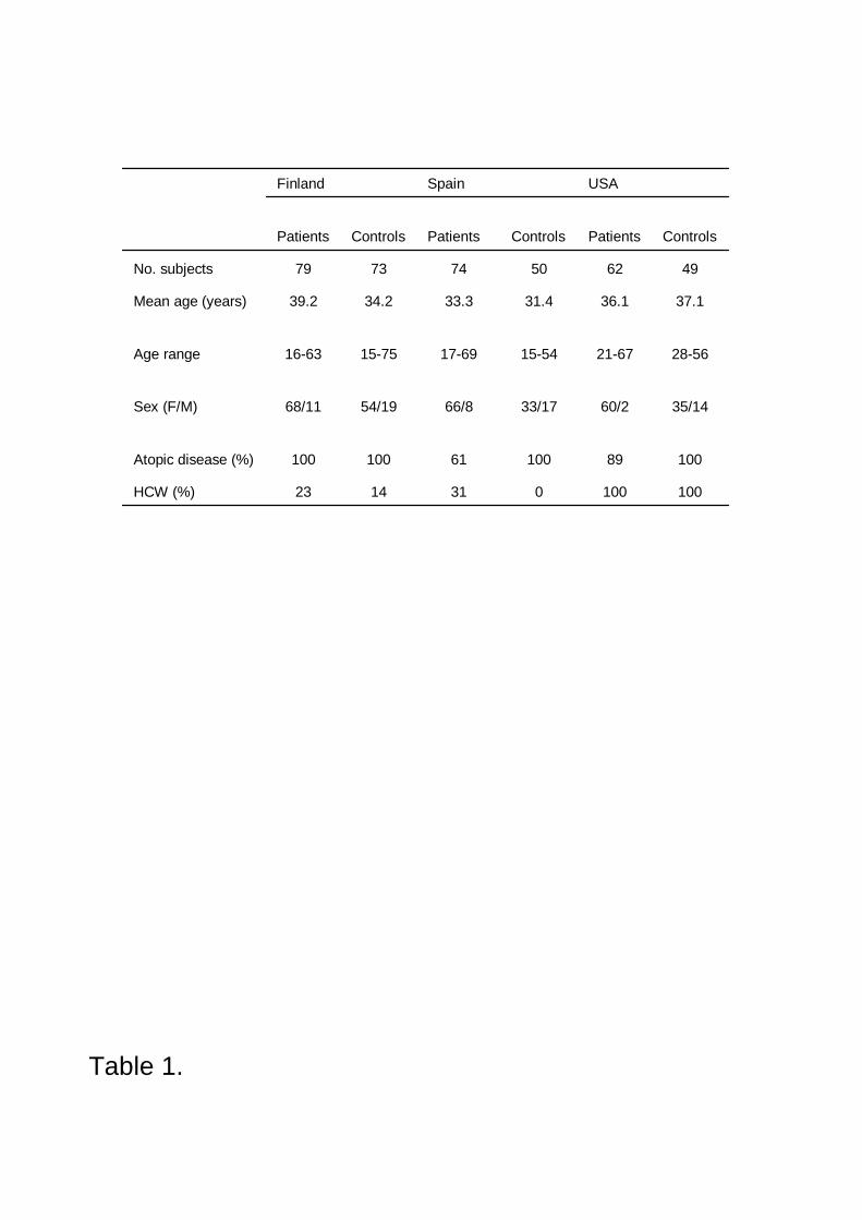
1001000311423HCW (%)
1008910061100100Atopic disease (%)
35/1460/233/1766/854/1968/11Sex (F/M)
285621671554176915751663Age range
37.136.131.433.334.239.2Mean age (years)
496250747379No. subjects
ControlsPatientsControlsPatientsControlsPatients
USASpainFinland
Table 1.
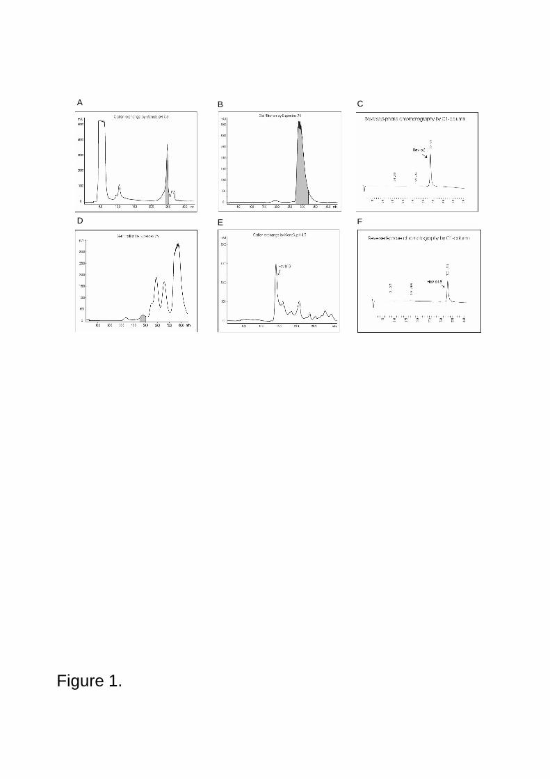
Figure 1.
B CA
E FD
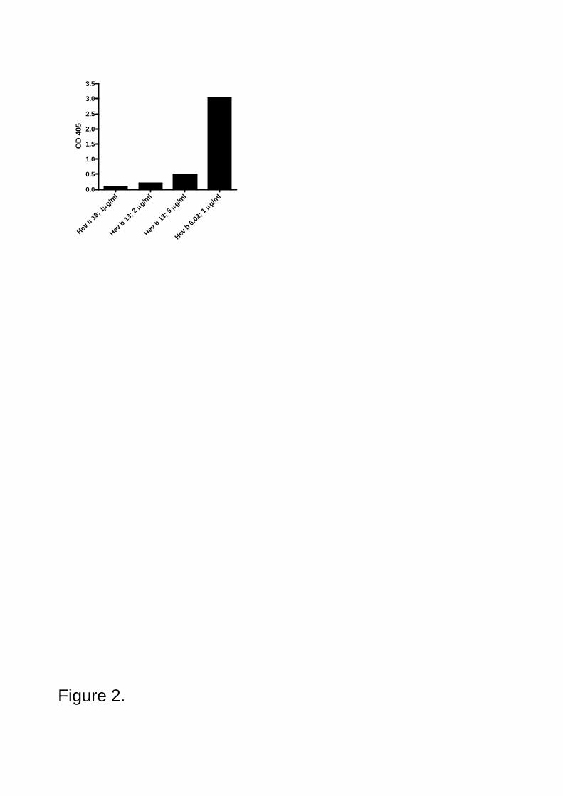
Figure 2.
g/ml
Hev b 13
; 1g/m
l
Hev b 13
; 2g/m
l
Hev b 13
; 5g/m
l
Hev b 6.
02; 1
0.0
0.5
1.0
1.5
2.0
2.5
3.0
3.5O
D 4
05
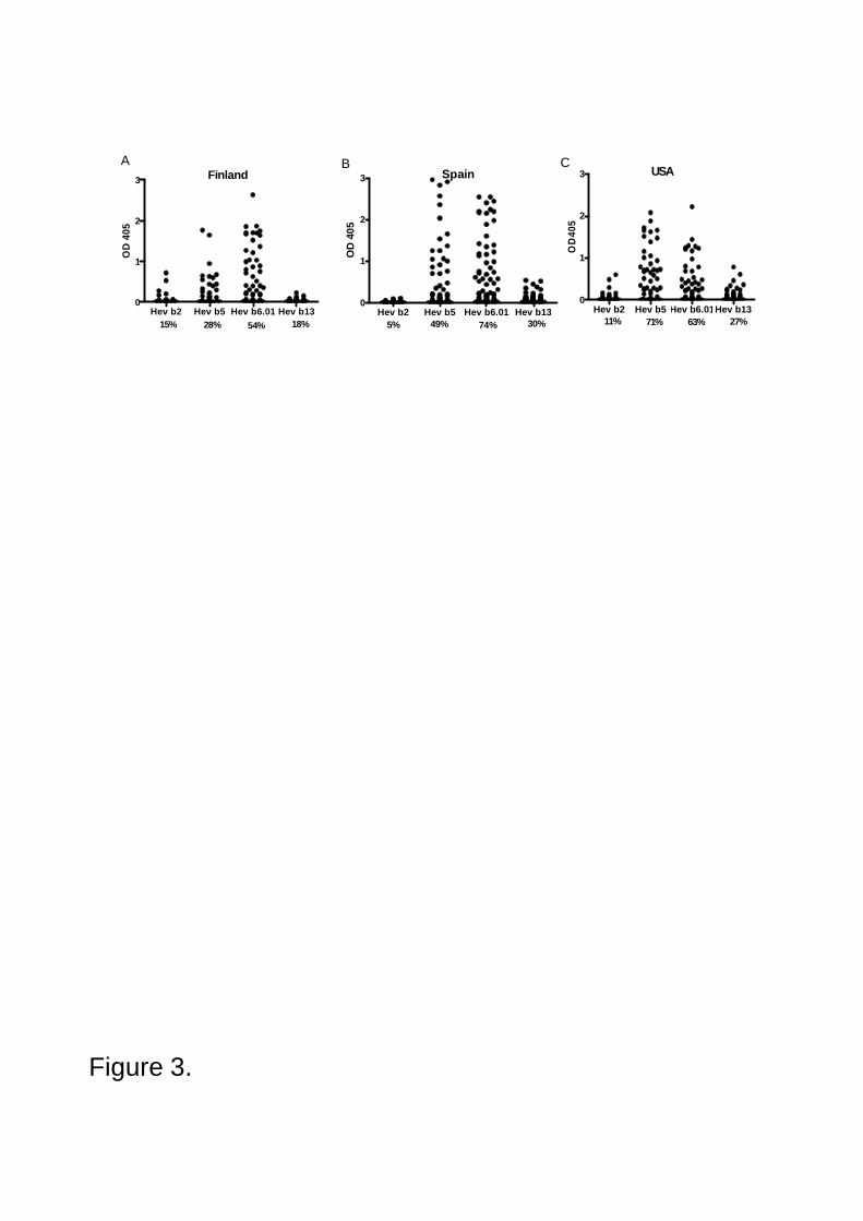
Figure 3.
B CA
Hev b2 Hev b5 Hev b6.01 Hev b130
1
2
3 Finland
15% 18%54%28%
OD
405
Hev b2 Hev b5 Hev b6.01 Hev b130
1
2
3 Spain
5% 30%74%49%
OD
405
Hev b2 Hev b5 Hev b6.01Hev b130
1
2
3 USA
11% 27%63%71%
OD
405
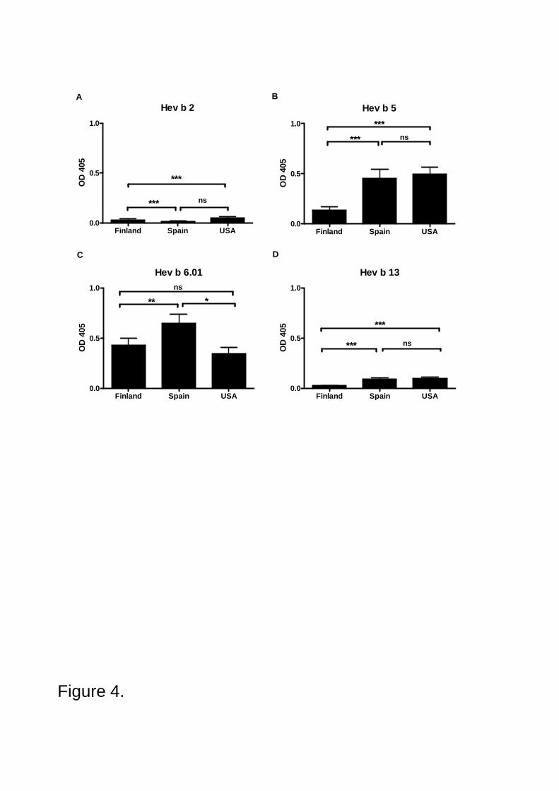
Figure 4.
A B
C D
Hev b 2
Finland Spain USA0.0
0.5
1.0
ns***
***OD
405
Hev b 5
Finland Spain USA0.0
0.5
1.0 ****** ns
OD
405
Hev b 6.01
Finland Spain USA0.0
0.5
1.0
** *ns
OD
405
Hev b 13
Finland Spain USA0.0
0.5
1.0
***
*** ns
OD
405
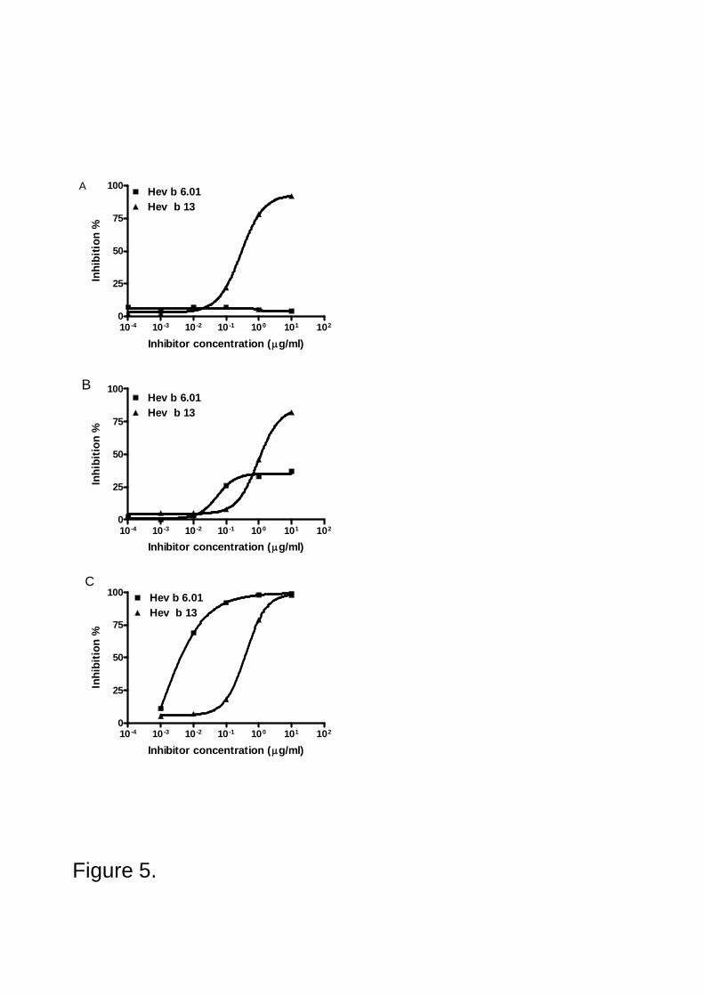
Figure 5.
A
10 4 10 3 10 2 10 1 100 101 1020
25
50
75
100 Hev b 6.01Hev b 13
Inhibitor concentration ( g/ml)
Inhi
bitio
n %
10 4 10 3 10 2 10 1 100 101 1020
25
50
75
100Hev b 6.01Hev b 13
Inhibitor concentration ( g/ml)
Inhi
bitio
n %
10 4 10 3 10 2 10 1 100 101 1020
25
50
75
100 Hev b 6.01Hev b 13
Inhibitor concentration ( g/ml)
Inhi
bitio
n %
B
C