NATIONAL HEALTHCAREER ASSOCIATION - Amazon S3 · Artifacts of Ambulatory EKG ... These layers and...
41
NATIONAL HEALTHCAREER ASSOCIATION Certified EKG Technician Study Guide This document is property of the National Healthcareer Association. This cannot be reproduced for any reason without written consent of the National Healthcareer Association.
Transcript of NATIONAL HEALTHCAREER ASSOCIATION - Amazon S3 · Artifacts of Ambulatory EKG ... These layers and...
NATIONAL HEALTHCAREER
ASSOCIATION
Certified EKG Technician
Study Guide
This document is property of the
National Healthcareer Association.
This cannot be reproduced for any
reason without written consent of the
National Healthcareer Association.
National Healthcareer Association EKG Study Guide (Eb) 2
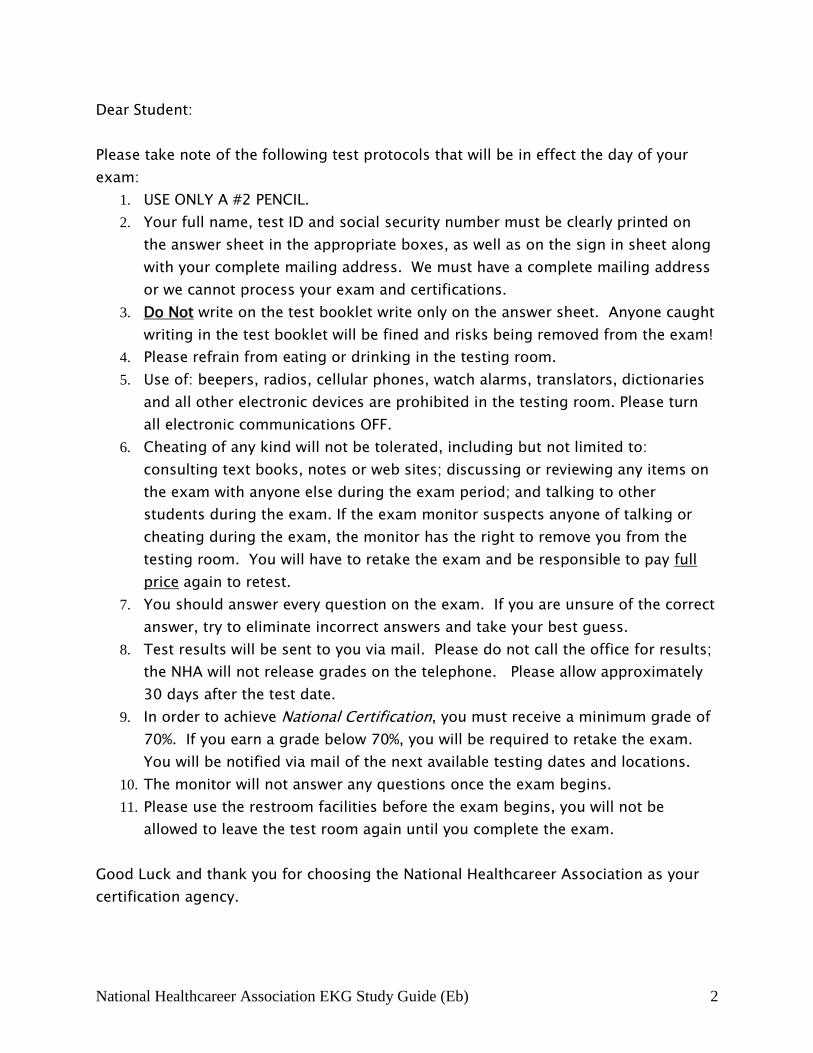
Dear Student:
Please take note of the following test protocols that will be in effect the day of your
exam:
1. USE ONLY A #2 PENCIL.
2. Your full name, test ID and social security number must be clearly printed on
the answer sheet in the appropriate boxes, as well as on the sign in sheet along
with your complete mailing address. We must have a complete mailing address
or we cannot process your exam and certifications.
3. Do Not write on the test booklet write only on the answer sheet. Anyone caught
writing in the test booklet will be fined and risks being removed from the exam!
4. Please refrain from eating or drinking in the testing room.
5. Use of: beepers, radios, cellular phones, watch alarms, translators, dictionaries
and all other electronic devices are prohibited in the testing room. Please turn
all electronic communications OFF.
6. Cheating of any kind will not be tolerated, including but not limited to:
consulting text books, notes or web sites; discussing or reviewing any items on
the exam with anyone else during the exam period; and talking to other
students during the exam. If the exam monitor suspects anyone of talking or
cheating during the exam, the monitor has the right to remove you from the
testing room. You will have to retake the exam and be responsible to pay full
price again to retest.
7. You should answer every question on the exam. If you are unsure of the correct
answer, try to eliminate incorrect answers and take your best guess.
8. Test results will be sent to you via mail. Please do not call the office for results;
the NHA will not release grades on the telephone. Please allow approximately
30 days after the test date.
9. In order to achieve National Certification, you must receive a minimum grade of
70%. If you earn a grade below 70%, you will be required to retake the exam.
You will be notified via mail of the next available testing dates and locations.
10. The monitor will not answer any questions once the exam begins.
11. Please use the restroom facilities before the exam begins, you will not be
allowed to leave the test room again until you complete the exam.
Good Luck and thank you for choosing the National Healthcareer Association as your
certification agency.
National Healthcareer Association EKG Study Guide (Eb) 3
SPECIAL ACCOMMODATIONS
Special exam accommodations are available for persons with disabilities or
other special needs. The participants or their representatives can submit a
request, in writing, to the National Healthcareer Association. The request
should include an explanation of the disability and the participants’ specific
requirements. Special accommodations may include additional testing time,
use of a private room or physical assistance in completing the examination. If
you have questions about special accommodations, please call the NHA’s
Corporate Office at 1-800-499-9092. Requests for special accommodations
must be submitted to the NHA at least 45 days prior to the exam date and may
be sent via certified mail or faxed to our corporate offices.
EXAM CHALLENGES:
If you believe a question on an exam was misleading, unfair or contained
errors, you may submit an exam question challenge. Any challenges to exam
questions must be submitted in writing to NHA’s Corporate Office. Challenges
to exams must be submitted within 5 business days of the completion of the
exam. No action can be taken on exam challenges submitted after that date,
and no challenge will be considered viable unless submitted in writing to our
Corporate Office. This policy allows everyone to benefit from any legitimate
challenges before grades are posted, while avoiding any unreasonable delays in
the NHA’s ability to process and deliver participants’ grades.
The NHA does not provide individual responses to challenges; however, every
challenge is considered and acted on accordingly. Exam challenges may be
faxed to NHA, Attn. Cynthia Orr, (973) 644-4797, or may be sent via overnight
courier to the following address:
National Healthcareer Association
Attn.: Cynthia Orr
7 Ridgedale Avenue, Ste. 203
Cedar Knolls, New Jersey 07927
National Healthcareer Association EKG Study Guide (Eb) 4
Dear Graduate:
Thank you for choosing the NATIONAL HEALTHCAREER ASSOCIATION as your certifying
agency. The Certified EKG Technician exam consists of 100 multiple-choice questions.
The following is a study guide meant to assist you achieve a focus in your review for the exam. It
is not intended to replace the text books or notes from your classes. In addition to the topics in
this guide, you are expected to have an understanding of basic EKG tracing, rate, rhythm and
common abnormalities.
Contents General Anatomy of the Heart ........................................................................................................ 5
Internal Heart Structure............................................................................................................... 5
Coronary Circulation .................................................................................................................. 7 Heart Physiology ......................................................................................................................... 7
Basic Electrophysiology ................................................................................................................. 9
Conduction System of the Heart ............................................................................................... 10 Fundamentals of Electrocardiogram ............................................................................................. 11
The Electrocardiographic Grid and Waves ............................................................................... 13 Definition of Waves, Segments and Intervals ........................................................................... 14
The Normal EKG Waves and Complexes ................................................................................ 14 The Normal EKG Segments, Intervals and Junctions .............................................................. 15
Analyzing the EKG Strip involves the following steps ............................................................ 15 EKG Interpretation and Pathology Recordings ........................................................................ 16
Artifacts of Ambulatory EKG Recording ..................................................................................... 28
Medical Terminology.................................................................................................................... 29 Resources: ..................................................................................................................................... 37
Reference books: ........................................................................................................................... 37 Sample EKG Exam ....................................................................................................................... 38
Answer Key: ................................................................................................................................. 41
National Healthcareer Association EKG Study Guide (Eb) 5
General Anatomy of the Heart
The heart is a hollow muscular organ located in the thoracic cavity between the lungs in a
space called Mediastenum, just behind the sternum. The heart Base is located at the level
of the second intercostal and the tip of the heart (Apex) is located at the level of 5th
intercostal and mid-clavicular line on the left.
Internal Heart Structure
Layers of the heart
Endocardium - the innermost layer of the heart. It is a thin layer of epithelium very
similar to vessels’ endothelium, which covers the inside part of the heart. It forms the
lining and folds back onto itself to form the heart valves and also covers the papillary
muscles that anchor chordae tendinae, strings of connective tissue that keep in place
the AV valves. The function of endocardium is to prevent blood cell destruction and
clotting. The endocardium is also the layer in which the heart’s conduction system is
embedded.
Myocardium - the middle and contractile layer of the heart. It is made up of special
striated muscle fibers with strong connection with each other (intercalated disks) and
branches that ensure a unified and simultaneous contraction of all the muscle fibers.
There is a high concentration of calcium ions in the space between the muscle fibers
(interstitial space), which influences the force of the muscle contraction.
“Heart Skeleton”- is made up of four rings of thick connective tissue. These rings
which surround the base of the heart and large vessels, create the cardiac septum, and
provide a solid connection between the heart chambers and a strong attachment for
the heart valves.
Pericardium – is the outermost layer of the heart. Pericardium is attached with
ligaments to the spinal column and diaphragm fixing the heart in its position.
Pericardium is built by two layers of connective tissue. The outside layer is called
parietal pericardium and the inner layer is called visceral pericardium or
epicardium. Two layers of pericardium are separated by a thin layer of fluid to
prevent friction. These layers and the fluid between them are referred to as the
pericardial sac.
The Heart Chambers
A structure in the middle of the heart called the septum, divides the heart into two sides.
The right side pumps deoxygenated blood with low pressure from the veins into the lungs
(pulmonary circulation) and left side, that pumps oxygenated blood with high pressure
(blood pressure) toward the tissues through arteries (systemic circulation). The heart has
four chambers: they are the right and left atria (smaller, thin-walled chambers that are
situated on top of the ventricles and receive blood from the lungs and veins) and the right
and left ventricles (larger, more muscular chambers that eject blood out to the systemic
circulation and to the two lungs).
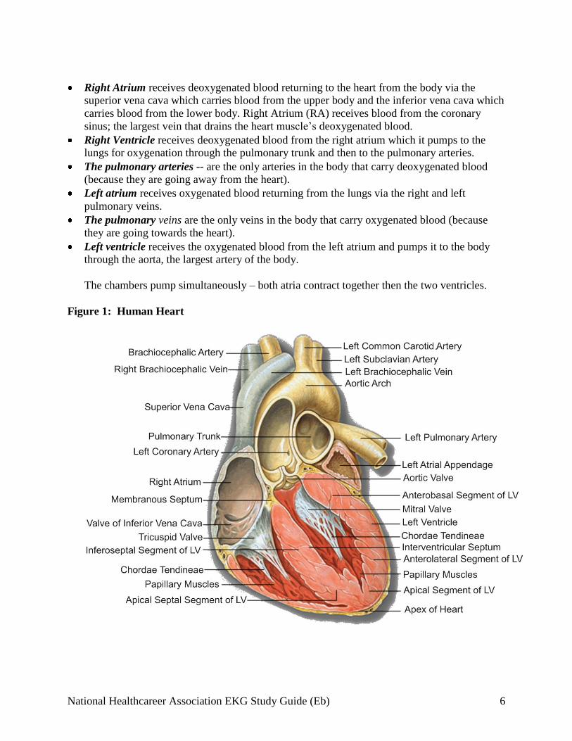
National Healthcareer Association EKG Study Guide (Eb) 6
Right Atrium receives deoxygenated blood returning to the heart from the body via the
superior vena cava which carries blood from the upper body and the inferior vena cava which
carries blood from the lower body. Right Atrium (RA) receives blood from the coronary
sinus; the largest vein that drains the heart muscle’s deoxygenated blood.
Right Ventricle receives deoxygenated blood from the right atrium which it pumps to the
lungs for oxygenation through the pulmonary trunk and then to the pulmonary arteries.
The pulmonary arteries -- are the only arteries in the body that carry deoxygenated blood
(because they are going away from the heart).
Left atrium receives oxygenated blood returning from the lungs via the right and left
pulmonary veins.
The pulmonary veins are the only veins in the body that carry oxygenated blood (because
they are going towards the heart).
Left ventricle receives the oxygenated blood from the left atrium and pumps it to the body
through the aorta, the largest artery of the body.
The chambers pump simultaneously – both atria contract together then the two ventricles.
Figure 1: Human Heart
National Healthcareer Association EKG Study Guide (Eb) 7
The Heart Valves
The purpose of the heart valves is to prevent backflow of blood thereby assuring
uni-directional flow thru the heart.
A. The atrioventricular valves (AV): are located between the atria and ventricles. AV
cusped valves characteristics are:
They have tough fibrous rings
Long and strong leaflets (cuspids)
They have accessory organs, such as papillary muscles and chordae tendinae.
a.) Tricuspid valve is located between the right atrium and the right ventricle. As the
name connotes, it has three cusps (or leaflets).
b.) Bicuspid Mitral valve is located between the left atrium and the left ventricle. It has
two cusps (or leaflets) and it also called the mitral valve.
B. The semilunar valves: called semilunar because they have half-moon shaped leaflets,
with the following characteristics:
Three leaflets
Shallow in depth
They have no accessory organs
a.) Pulmonic valve – located between the right ventricle and the pulmonary trunk.
b.) Aortic valve - located between the left ventricle and aorta
Coronary Circulation
The right and left coronary arteries are the first branches coming out of Aorta and supply
the heart with oxygenated blood. The blood runs through these arteries during diastole.
Coronary arteries are located on the epicardium.
The left coronary artery has two branches Left Anterior Descending (LAD) artery and
Left Circumflex (LCX) artery. There is only one main artery that supplies the right side
of the heart RCA (Right Coronary Artery) artery.
Heart Physiology
Cardiac Cycle
Systole is the period of contractions of both Arial and Ventricles
Diastole is the period of relaxation and filling of all cardiac chambers.
Heart Sounds
Heart sounds are caused by the closure of the heart valves
S1 first heart sound (Lubb) occurs during ventricle contraction and the closure of AV
valves.
S2 second heart sound (Dupp) occurs during ventricular relaxation when SL valves
National Healthcareer Association EKG Study Guide (Eb) 8
(Pulmonary and Aortic valves) close. Murmurs are caused by diseases of the valves or
other structural abnormalities.
Heart Rate is the number of heart contractions per minute. The normal heart rate is 60 to
100 bpm (beat per minute). HR is controlled by Chemo-receptors (chemical sensors) and
Baro-receptors (pressure receptors) located in Aortic Arch and Carotid arteries. The heart
is under the influence by the autonomic nervous system (ANS) which is subdivided into
the sympathetic and parasympathetic nervous systems.
Parasympathetic (Vagus Nerve) generally has an inhibitory effect via the
neurotransmitter Acetylcholine which may cause the following to happen:
Slows SA pacemaker and HR
Slows the conduction of electricity in AV node
Decreases the strength of atrial and ventricular contraction
Sympathetic via the neurotransmitter Norepinephrine results:
Increases the HR
Increases the force of contraction
Increases the blood pressure
Via dopaminergic receptors increases the diameter of the visceral blood vessels and
consequently the visceral blood flow.
Heart as a Pump
The blood volume ejected outside the heart is equal to the blood volume returning back I
into the heart.
Stroke Volume (Preload) is the blood volume ejected outside the ventricle after each
contraction. The stroke volume depends on
The volume of blood returning into the heart.
The force of the myocardium contraction
Vascular resistance (After Load)
Starling Law: “The greater the volume of blood inside the heart during diastole, the
stronger the heart contraction force during the systole. (Stroke Volume).
The other main factor influencing the stroke volume is vascular resistance (after load).
The lower the resistance in the vessels, the more easily blood can be ejected outside the
heart through the circulation.
Cardiac Output:: The amount of blood ejected outside the heart per minute.
Cardiac Output = (Stroke volume) x (HR per/min)
Peripheral Vascular Resistance: Is the force exerted against the blood flow and is
determined by the diameter of the vessel. The lower the vascular resistance the less force
is needed to eject the blood out of the heart during systole.
National Healthcareer Association EKG Study Guide (Eb) 9
Blood Pressure: The force exerted by circulating blood volume on the walls of the artery
during circulation.
BP = (Cardiac Output) x (Vascular Resistance)
Higher Cardiac output will result in a higher BP
High vascular resistance will also result in a higher BP.
Therefore, lower cardiac output OR lower vascular resistance will result in a lower
BP.
Basic Electrophysiology
EKG = graphical presentation of heart electricity (voltage) over time. This electricity is created
by specialized cells called pacemaker cells. These cells generate electrical impulses
spontaneously (without outside influence) and rhythmically (automaticity). The electricity is
created by passing of ions (charged particles) through the cell membrane. The electricity is than
conducted, transmitted to other specialized cells that together with the pacemaker cells create the
conductive system of the heart, the necessary wires and switches to stimulate cardiac muscle
fibers for a synchronized contraction.
Cardiac Cell properties:
Automaticity: the ability to spontaneously trigger electrical impulses without being
stimulated by another source.
Excitability: (also called irritability) the ability to respond and react to a stimulus
Conductivity: the ability to receive and transmit electrical impulses to adjacent cells.
Contractility: a myocardial cell’s ability to shorten (or contract) in response to a
stimulus.
Depolarization occurs when positively charged ions (such as sodium and calcium) rapidly move
from outside the myocardial cell membrane to the inside, changing the overall charge from
negative to positive. This process results in a “chain reaction” that spreads from cell to cell very
rapidly. This electrical event is expected to result in contraction. Depolarization flows from the
endocardium to the myocardium to the epicardium (or from the innermost layer to the
outermost).
Repolarization occurs immediately after depolarization and is the movement of positively
charged ions back to the outside of the cell, returning the cell back to its original polarized state.
A cell must repolarize before it can depolarize again. Whereas depolarization results in
myocardial contraction, repolarization does not result in any actual muscle movement…it is
strictly an electrochemical event.
Absolute Refractory Period: is the 1st phase of repolarization in which a myocardial cell is
unable to react to any electrical stimulus. This period falls during the depolarization and

National Healthcareer Association EKG Study Guide (Eb) 10
contraction of the ventricles, thus protecting the heart from any abnormal electrical stimulus that
might result in loss of rhythmic contractions.
Relative Refractory Period: is the 2nd
phase of repolarization during which time a strong
enough electrical stimulus might cause new depolarization and contraction. This could result in
a chaotic, possibly lethal rhythm disturbance.
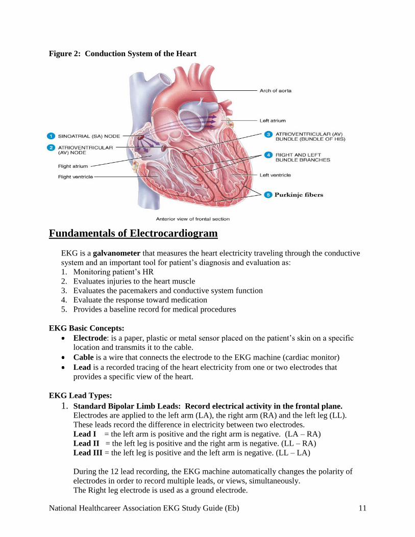
Conduction System of the Heart
Conduction system of the heart is the system that generates and delivers (conducts) the
electricity to all the muscle fibers of the heart resulting in a smooth, complete contraction of the
cardiac muscle fibers, which forcefully ejects the blood outside the heart. This electrical delivery
system consists of two nodes (which generate and control the rhythmic impulses) and conduction
pathways that connect the nodes and deliver the electrical impulses to the myocardium.
The Conduction system of the heart consists of:
SA Node (Sino-Atrial Node or Sinus Node) Found in the upper posterior portion of the right
atrial wall just below the opening of the superior vena cava. It is the primary pacemaker of the
heart and has a normal firing rate of 60-100 beats per minute (BPM).
Internodal Pathways: Consists of anterior, middle and posterior divisions that distribute electrical
impulses generated by the SA node throughout the right and left atria to the atrio-ventricular
(AV) node.
AV Junction (AV node): This node is located at the posterior septal wall of the right atrium just
above the tricuspid valve. There is normally a .12 - .20 second delay of electrical activity at this
level to allow blood to flow from the atria and fill the ventricles with blood. The AV node also
functions as the backup pacemaker (at a slower heart rate) if the Sinus Node fails to fire. Its
intrinsic firing rate is between 40-60 beats per minute.
Bundle of His: Found at the superior portion of the interventricular septum, it is the pathway
that leads out of the AV node and connects to the Bundle Branches. It has an ability to initiate
electrical impulses with an intrinsic firing rate of 40-60 beats per minute.
Bundle branches: Located at the interventricular septum, the Bundle of His divides into the
right and left bundle branches, the function of which is to conduct the electrical impulse to the
Purkinje fibers throughout the ventricles.
Purkinje fibers: Found within the ventricular endocardium, it consists of a network of small
conduction fibers that deliver the electrical impulses from the Bundle Branches to the ventricular
myocardium. This network has the ability to initiate electrical impulses and act as a pacemaker
if the higher level pacemakers fail. The intrinsic firing rate is 20-40 beats per minute.
National Healthcareer Association EKG Study Guide (Eb) 11
Figure 2: Conduction System of the Heart
Fundamentals of Electrocardiogram
EKG is a galvanometer that measures the heart electricity traveling through the conductive
system and an important tool for patient’s diagnosis and evaluation as:
1. Monitoring patient’s HR
2. Evaluates injuries to the heart muscle
3. Evaluates the pacemakers and conductive system function
4. Evaluate the response toward medication
5. Provides a baseline record for medical procedures
EKG Basic Concepts:
Electrode: is a paper, plastic or metal sensor placed on the patient’s skin on a specific
location and transmits it to the cable.
Cable is a wire that connects the electrode to the EKG machine (cardiac monitor)
Lead is a recorded tracing of the heart electricity from one or two electrodes that
provides a specific view of the heart.
EKG Lead Types:
1. Standard Bipolar Limb Leads: Record electrical activity in the frontal plane.
Electrodes are applied to the left arm (LA), the right arm (RA) and the left leg (LL).
These leads record the difference in electricity between two electrodes.
Lead I = the left arm is positive and the right arm is negative. (LA – RA)
Lead II = the left leg is positive and the right arm is negative. (LL – RA)
Lead III = the left leg is positive and the left arm is negative. (LL – LA)
During the 12 lead recording, the EKG machine automatically changes the polarity of
electrodes in order to record multiple leads, or views, simultaneously.
The Right leg electrode is used as a ground electrode.
National Healthcareer Association EKG Study Guide (Eb) 12
2. Augmented Unipolar-also records electrical activity in the frontal plane.
Augmented Unipolar Leads record the heart electricity from one limb and compare it
with a zero voltage lead in the center of the heart. The EKG machine uses a midpoint
between the two other limbs as a negative reference point. These leads augment
(magnify) the voltage up to 50% compared to the standard leads. AV stands for
augmented voltage; R stands for Right Arm (RA), L stands for Left Arm (LA) and F
stands for Left Leg (LL).
Lead aVR = the right arm is positive and the other limbs are negative.
Lead aVL = the left arm is positive and the other limbs are negative.
Lead aVF = the left leg (or foot) is positive and the other limbs are negative.
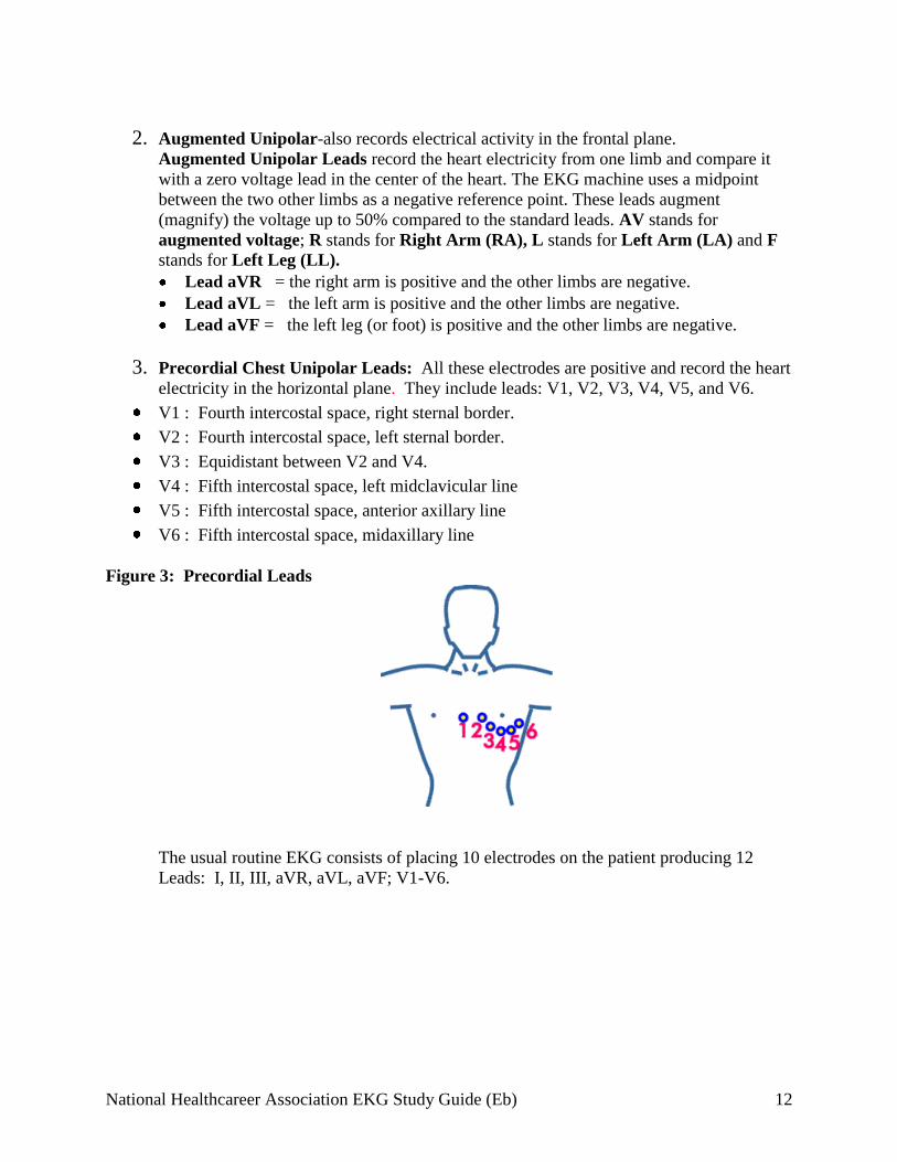
3. Precordial Chest Unipolar Leads: All these electrodes are positive and record the heart
electricity in the horizontal plane. They include leads: V1, V2, V3, V4, V5, and V6.
V1 : Fourth intercostal space, right sternal border.
V2 : Fourth intercostal space, left sternal border.
V3 : Equidistant between V2 and V4.
V4 : Fifth intercostal space, left midclavicular line
V5 : Fifth intercostal space, anterior axillary line
V6 : Fifth intercostal space, midaxillary line
Figure 3: Precordial Leads
The usual routine EKG consists of placing 10 electrodes on the patient producing 12
Leads: I, II, III, aVR, aVL, aVF; V1-V6.
National Healthcareer Association EKG Study Guide (Eb) 13
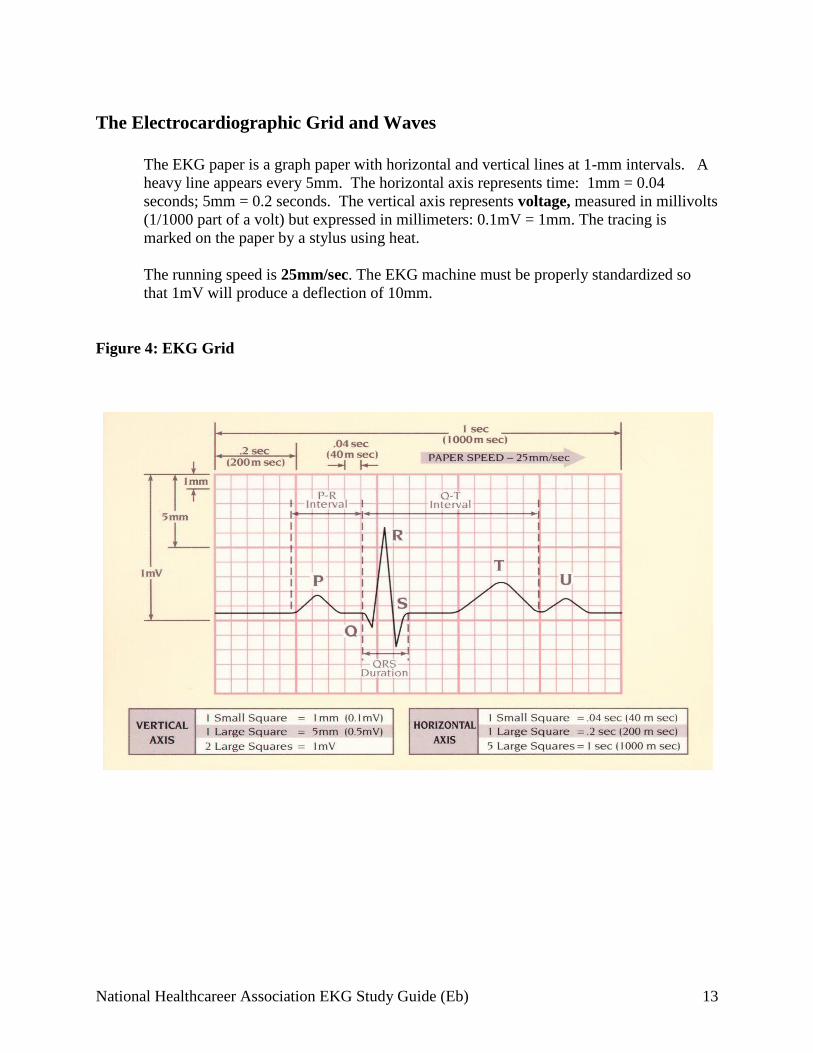
The Electrocardiographic Grid and Waves
The EKG paper is a graph paper with horizontal and vertical lines at 1-mm intervals. A
heavy line appears every 5mm. The horizontal axis represents time: 1mm = 0.04
seconds; 5mm = 0.2 seconds. The vertical axis represents voltage, measured in millivolts
(1/1000 part of a volt) but expressed in millimeters: 0.1mV = 1mm. The tracing is
marked on the paper by a stylus using heat.
The running speed is 25mm/sec. The EKG machine must be properly standardized so
that 1mV will produce a deflection of 10mm.
Figure 4: EKG Grid
National Healthcareer Association EKG Study Guide (Eb) 14
Definition of Waves, Segments and Intervals
Waveform: refers to movement away from the isoelectric line with either upward
(positive) deflection or downward (negative) deflection.
Segment: line between two waveforms.
Interval: waveform plus a segment.
Complex: several waveforms
The Normal EKG Waves and Complexes
1. Atrial Depolarization:
a. P wave: is the first deflection after the diastole, produced by atrial depolarization.
It is a smooth, round, not more than 2.5 mm high and no more than 0.11 sec
Positive in I,II, and V2 to V6
The normal P wave in standard, limb, and precordial leads does not exceed 0.11s in
duration or 2.5mm in height.
b. There is no wave for atrial repolarization, because is obscured by the larger QRS
complex
2. Ventricular Depolarization:
a. QRS complex
Represents ventricular depolarization (activation).
The ventricle is depolarized from the endocardium to the myocardium, to the
epicardium.
Normal duration is no more than 0.1 sec (otherwise stated as “less than .12 sec”).
b. Q (q) wave: the initial negative deflection produced by ventricular depolarization.
c. R (r) wave: the first positive deflection produced by ventricular depolarization.
c. S (s) wave: the first negative deflection produced by the ventricular depolarization that
follows the first positive deflection, (R) wave.
3.Ventricular Repolarization:
a. T wave: The first wave after the QRS complex has the following characteristics
The deflection produced by ventricular repolarization.
It is slightly asymmetric
No more than 5 mm in height
b. U wave: Is the deflection seen following the T wave but preceding the diastole.
Represents repolarization of Purkinje fibers
Round and symmetric less than 1.5 mm in height
A prominent U wave is due to hypokalemia (low potassium, blood level).
National Healthcareer Association EKG Study Guide (Eb) 15
The Normal EKG Segments, Intervals and Junctions
1. Normal EKG Segments: Segments: are lines between waveforms.
a. PR segment this segment is measured from the end of the P wave to the beginning of the
QRS complex. Represents depolarization of AV node and its delay and depolarization of
the Bundle of His and the Bundle Branches.
b. ST segment This segment represents the time of ventricular contraction and the
beginning of repolarization of both ventricles. It is measured from end of QRS to the
beginning of the T wave. The point where QRS complex and the ST segment meet is
called “the junction” or “J point”. ST segment is the most sensitive part of EKG changed
by cardiac ischemia.
2. Normal Interval and Junctions: By definition an interval is a segment plus a waveform.
a. PR Interval Is defined as P wave and PR segment and is measured
from the beginning of P wave to the beginning of QRS complex. The
normal interval is 0.12 – 0.2 sec.
b. QT Interval It represents the total ventricular activity (ventricular
depolarization PLUS ventricular repolarization), and it is measured from
the beginning of QRS to the end of T wave. The normal duration of this
interval depends on the age and the HR.
c. RR Interval. It is important to determine the HR and its regularity..
RR interval: this is the interval between two R waves.
d. J (RST) junction: point at which QRS complex ends and ST segment
begins.
e. ST segment: from J point to the onset of the T wave. This segment is
compared to the PR segment to help identify myocardial ischemia or
injury.
Analyzing the EKG Strip involves the following steps
To analyze the EKG strip for its quality and to identify any emergency pathology you can
follow the following guidelines:
1. Assesses the HR
1. 6 second Method: The number of QRS complexes between 6 sec marks on the EKG
paper is multiplied by 10. Used generally for estimating slow or irregular rhythms.
2. Large Boxes Method: count the number of large boxes between two consecutive RR
(one RR interval) and divide into 300 for the ventricular rate; and count large boxes
between two consecutive P waves for the atrial rate. Used mainly in regular rhythms.
National Healthcareer Association EKG Study Guide (Eb) 16
3. Small Boxes: One minute has 1500 small boxes (0.04 sec). Count the number of small
boxes between an RR interval and divide into 1500.This method is more accurate and is
used for regular rhythms only.
4. Sequence Method: Select the R that falls on a dark vertical line. Number the next
consecutive dark line as 300, 150, 100, 75, 60, and 50. Note where the next R wave falls
in relation to the dark lines. That is the heart rate.
2. Assess Rhythm/ Regularity
The HR is considered regular if all the RR or PP intervals on the EKG leads are equal. If there
are changes in their durations the rhythm is irregular.
3. Identify and examine the P waves: Identify the P waves, PP interval and measure the size of
the P wave in different leads.
4. Assess intervals (PR, QRS, QT): Measure each of these intervals and determine if they are
normal.
5. Evaluate ST segments and T waves. ST segment elevation or depression and/or T wave
abnormalities can suggest the presence of myocardial ischemia or injury.
6. General Evaluation and Conclusion: Notify the doctor for any abnormality that you can find
on the EKG strip.
EKG Interpretation and Pathology Recordings
Cardiac arrhythmias are due to the following mechanisms:
Arrhythmias of sinus origin - where electrical flow follows the usual conduction
pathway but is too fast, too slow, or irregular. Normal sinus rate is 60-100 beats
per minute. If the rate goes beyond 100 per minute, it is called sinus tachycardia.
If the rate goes below 60 per minute, it is referred to as sinus bradycardia.
Ectopic rhythms - electrical impulses originate from somewhere else other than
the sinus node.
Conduction blocks - electrical impulses go down the usual pathway but encounter
blocks and delays.
Pre-excitation syndromes - the electrical impulses bypass the normal pathway
and, instead, go down an accessory shortcut.
National Healthcareer Association EKG Study Guide (Eb) 17
A. Sinus Rhythm
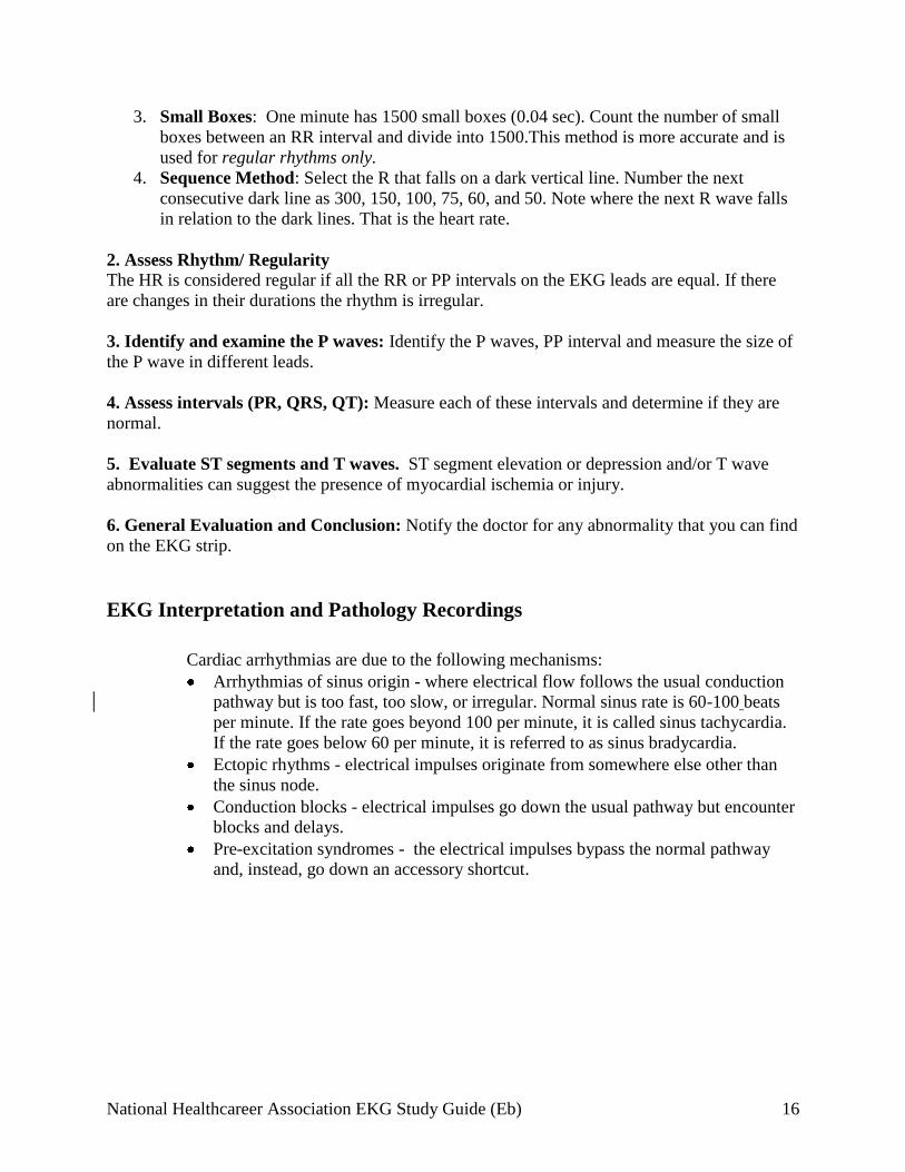
1. Normal Sinus Rhythm
Fig. 5. Normal Sinus rhythm. Notice that all PQRST waves are equal and
present. Diastolic period can be easily identified.
The rhythm originated from the SA has the following characteristics
a. HR 60 – 100 bpm
b. Similar P in all the leads in front of all QRS (0.1 sec)
c. A constant PR (0.12 to 0.2) sec interval in all the leads, regular rhythm
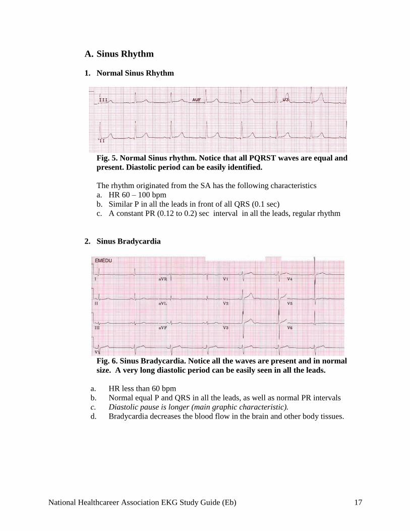
2. Sinus Bradycardia
Fig. 6. Sinus Bradycardia. Notice all the waves are present and in normal
size. A very long diastolic period can be easily seen in all the leads.
a. HR less than 60 bpm
b. Normal equal P and QRS in all the leads, as well as normal PR intervals
c. Diastolic pause is longer (main graphic characteristic).
d. Bradycardia decreases the blood flow in the brain and other body tissues.

National Healthcareer Association EKG Study Guide (Eb) 18
3. Sinus Tachycardia
Fig 7. Sinus Tachycardia. The diastolic period cannot be seen well, P and T
waves are very close to each other.
a. HR over 100 bpm
b. Normal equal P and QRS in all the leads, as well as normal PR intervals
c. Diastolic pause is very small or nonexistent (main graphic Characteristic)
d. Tachycardia reduces the blood supply to the cardiac muscle.
4. Sinus Arrhythmia:
Fig. 8. Sinus Arrhythmia. Notice that the PQRST waves are equal and
normal in size, but diastolic periods are different after each heart systole.
a. HR 60 to 100 bpm
b. Normal equal P and QRS in all the leads, as well as normal PR intervals
c. Different diastolic pause after each systole. If there are changes with the
respiration is named Respiratory Sinus Arrhythmia, otherwise is called
Nonrespiratory Arrhythmia
B. Important Atrial Rhythms
1. Supraventricular Tachycardias.
Fig. 10. Supraventricular Tachycardia
National Healthcareer Association EKG Study Guide (Eb) 19
Atrial Tachycardia (AT) it is caused by an irritable focus in the atria that
fires electrical impulses after the normal firing of the SA node pacemaker.
HR is regular between 100 and 150 bpm.
AV Reentry Tachycardia is caused when the electrical impulse passes
through a passage other than AV node. Cardiac rhythm is regular but up to
250 bpm. P waves are often hidden by the QRS complexes or the QRS
complexes that follow a P wave are different and with different PR
interval (AV Nodal Reentry Tachycardia AVNRT).
In cases with AV Reentry Tachycardia (AVRT) QRS complexes are
greater than 0.12 sec with a slurred up strike (delta wave) seen in one or
more leads.
It is an EMERGENCY. NOTIFY THE DOCTOR.
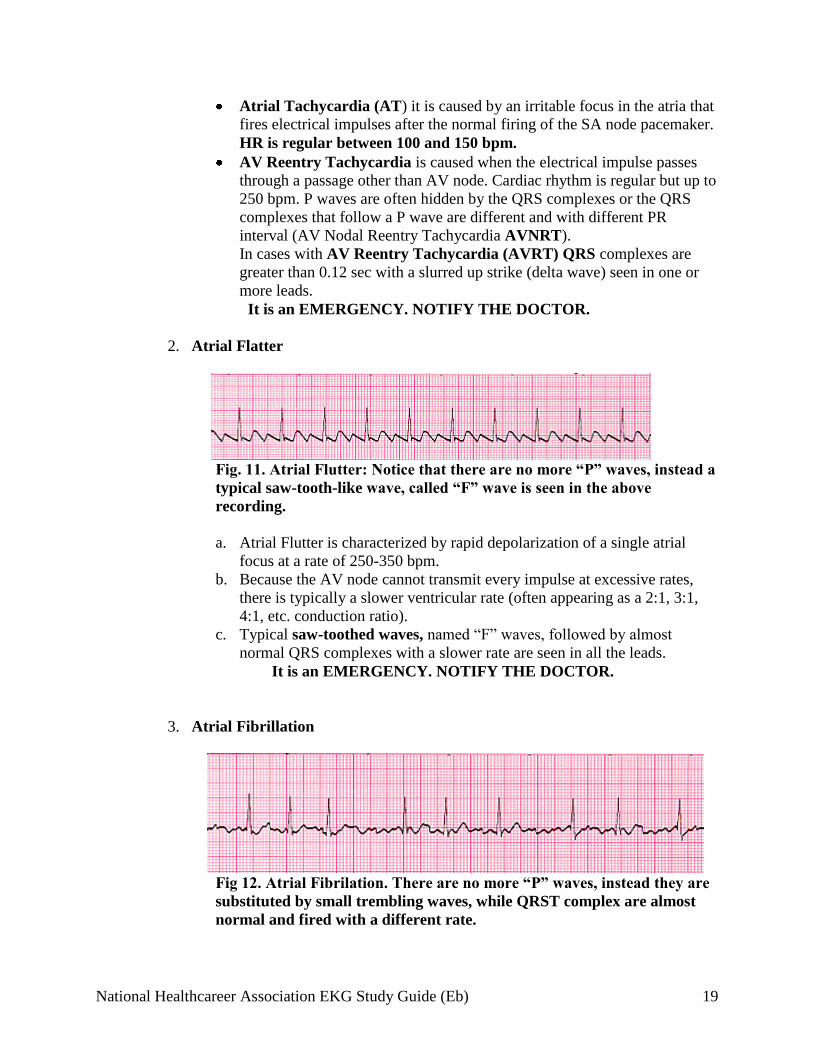
2. Atrial Flatter
Fig. 11. Atrial Flutter: Notice that there are no more “P” waves, instead a
typical saw-tooth-like wave, called “F” wave is seen in the above
recording.
a. Atrial Flutter is characterized by rapid depolarization of a single atrial
focus at a rate of 250-350 bpm.
b. Because the AV node cannot transmit every impulse at excessive rates,
there is typically a slower ventricular rate (often appearing as a 2:1, 3:1,
4:1, etc. conduction ratio).
c. Typical saw-toothed waves, named “F” waves, followed by almost
normal QRS complexes with a slower rate are seen in all the leads.
It is an EMERGENCY. NOTIFY THE DOCTOR.
3. Atrial Fibrillation
Fig 12. Atrial Fibrilation. There are no more “P” waves, instead they are
substituted by small trembling waves, while QRST complex are almost
normal and fired with a different rate.
National Healthcareer Association EKG Study Guide (Eb) 20
a. Atrial fibrillation is caused by multiple irritable sites all over the atria firing at a
rate exceeding 350 bpm. These rapid impulses cause quivering (fibrillation) of
the muscular fibers, which results in a drastic decrease in the cardiac output,
blood stagnation and the formation of a clot.
b. No identifiable P waves can be seen, fibrillatory erratic “f” waves are seen in all
the leads. Ventricular rhythm is very irregular, with a much slower rate than the
atria. This is seen in all leads.
c. Controlled atrial fibrillation: Average ventricular rate is less than 100 bpm.
d. Uncontrolled atrial fibrillation: Average ventricular rate is over 100 bpm.
It is an EMERGENCY. NOTIFY THE DOCTOR.
C. Ventricular Rhythms
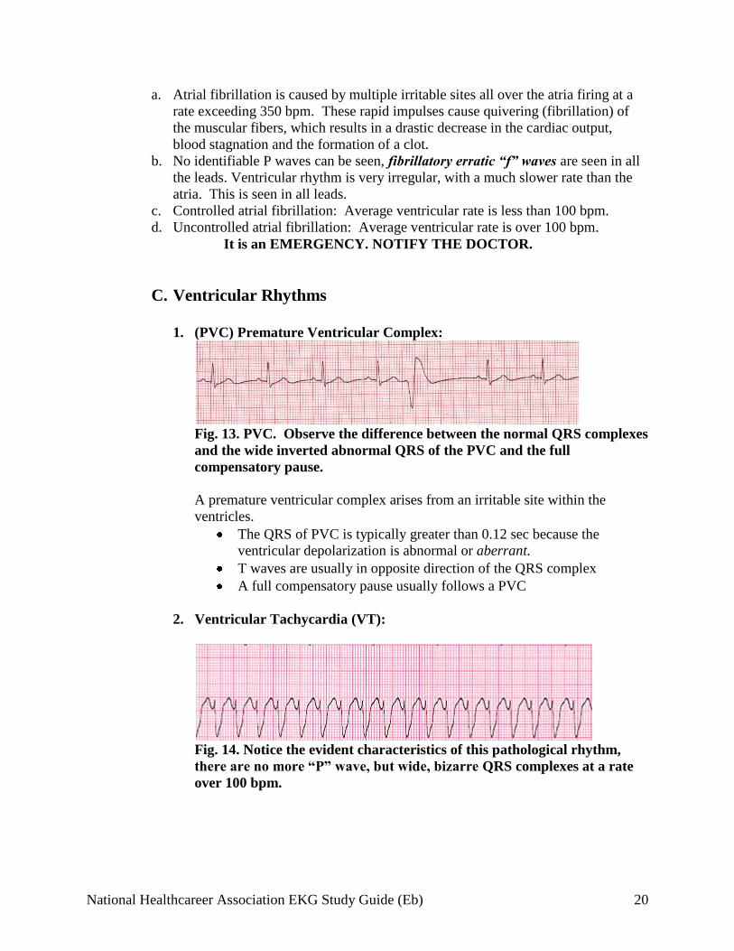
1. (PVC) Premature Ventricular Complex:
Fig. 13. PVC. Observe the difference between the normal QRS complexes
and the wide inverted abnormal QRS of the PVC and the full
compensatory pause.
A premature ventricular complex arises from an irritable site within the
ventricles.
The QRS of PVC is typically greater than 0.12 sec because the
ventricular depolarization is abnormal or aberrant.
T waves are usually in opposite direction of the QRS complex
A full compensatory pause usually follows a PVC
2. Ventricular Tachycardia (VT):
Fig. 14. Notice the evident characteristics of this pathological rhythm,
there are no more “P” wave, but wide, bizarre QRS complexes at a rate
over 100 bpm.
National Healthcareer Association EKG Study Guide (Eb) 21
Ventricular Tachycardia (V-Tach) is characterized by 3 or more PVC’s in a row
at a rate over 100 bpm. If V-Tach occurs for more than 30 sec is called sustained
Ventricular Tachycardia. The main characteristics of this rhythm are:
Regular fast rhythm 100 to 250 bpm
No P waves
Wide, bizarre QRS complexes with T waves pointing in opposite direction from
main QRS direction (T waves may be difficult to identify). If QRS complexes are
different in size it is called Polymorphic V-Tach or “Torsades de Pointes”.
It is an EMERGENCY. NOTIFY THE DOCTOR.
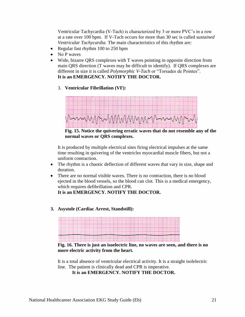
3. Ventricular Fibrillation (VF):
Fig. 15. Notice the quivering erratic waves that do not resemble any of the
normal waves or QRS complexes.
It is produced by multiple electrical sites firing electrical impulses at the same
time resulting in quivering of the ventricles myocardial muscle fibers, but not a
uniform contraction.
The rhythm is a chaotic deflection of different waves that vary in size, shape and
duration.
There are no normal visible waves. There is no contraction, there is no blood
ejected in the blood vessels, so the blood can clot. This is a medical emergency,
which requires defibrillation and CPR.
It is an EMERGENCY. NOTIFY THE DOCTOR.
3. Asystole (Cardiac Arrest, Standstill):
Fig. 16. There is just an isoelectric line, no waves are seen, and there is no
more electric activity from the heart.
It is a total absence of ventricular electrical activity. It is a straight isolelectric
line. The patient is clinically dead and CPR is imperative.
It is an EMERGENCY. NOTIFY THE DOCTOR.

National Healthcareer Association EKG Study Guide (Eb) 22
D. Atrio-Ventricular Blocks (AV Blocks)
AV blocks are defined as a delay or interruption of the electric impulse
conduction beyond the AV node. It is evaluated by measuring the PR interval in
EKG traces. The PR interval is the key of differentiation and classification of the
AV blocks.
1. Type I First Degree AV block. It is characterized by a delay of impulses at
the level of AV node. . PR interval is prolonged and is greater than 0.2 sec
Fig. 17. Notice the prolonged PR interval after each “P” wave in the
recording. This is the main feature of the First degree Av Block. Other than
the PR prolongation, the EKG may otherwise appear normal.
2. Type II Second Degree AV blocks. Some of the atrial impulses, but not all,
are blocked at the AV node level. Because SA node fires regular rhythmic
impulses, each P wave occurs in a regular interval across the EKG strip, but not
all P waves will be followed by a QRS complex
a. Type I second Degree AV Block (Wenckebach, Mobitz I)
PR interval lengthens in each interval until one QRS disappears
Fig. 18. Notice the gradual increase in the PR interval, and then a sudden
disappearance of a QRS complex. In a Type I, there is occasionally an extra
P wave, not always.
b. Type II Second Degree AV Block (Mobitz II)
It is a more serious pathology.
Conducted P waves have a constant PR interval; but there are always non-
conducted P waves between cardiac cycles, usually producing a “conduction
ratio” between atria and ventricles (i.e. 2 P waves for each QRS, or 3 P waves
for each QRS)
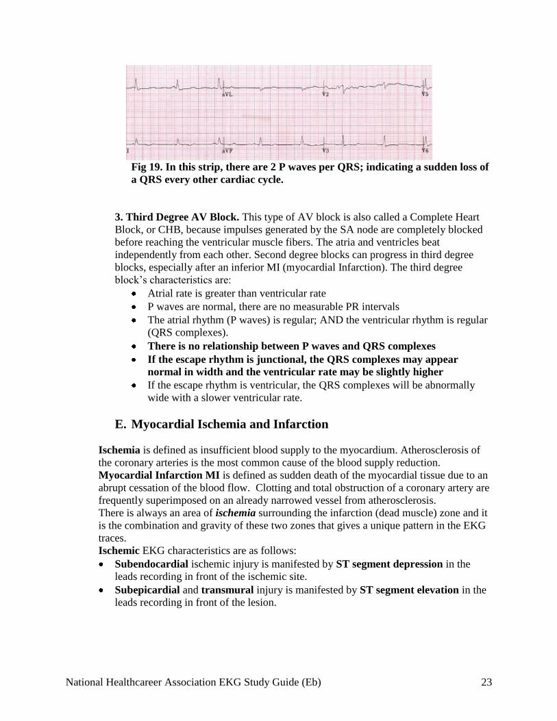
National Healthcareer Association EKG Study Guide (Eb) 23
Fig 19. In this strip, there are 2 P waves per QRS; indicating a sudden loss of
a QRS every other cardiac cycle.
3. Third Degree AV Block. This type of AV block is also called a Complete Heart
Block, or CHB, because impulses generated by the SA node are completely blocked
before reaching the ventricular muscle fibers. The atria and ventricles beat
independently from each other. Second degree blocks can progress in third degree
blocks, especially after an inferior MI (myocardial Infarction). The third degree
block’s characteristics are:
Atrial rate is greater than ventricular rate
P waves are normal, there are no measurable PR intervals
The atrial rhythm (P waves) is regular; AND the ventricular rhythm is regular
(QRS complexes).
There is no relationship between P waves and QRS complexes
If the escape rhythm is junctional, the QRS complexes may appear
normal in width and the ventricular rate may be slightly higher
If the escape rhythm is ventricular, the QRS complexes will be abnormally
wide with a slower ventricular rate.
E. Myocardial Ischemia and Infarction
Ischemia is defined as insufficient blood supply to the myocardium. Atherosclerosis of
the coronary arteries is the most common cause of the blood supply reduction. Myocardial Infarction MI is defined as sudden death of the myocardial tissue due to an
abrupt cessation of the blood flow. Clotting and total obstruction of a coronary artery are
frequently superimposed on an already narrowed vessel from atherosclerosis.
There is always an area of ischemia surrounding the infarction (dead muscle) zone and it
is the combination and gravity of these two zones that gives a unique pattern in the EKG
traces.
Ischemic EKG characteristics are as follows:
Subendocardial ischemic injury is manifested by ST segment depression in the
leads recording in front of the ischemic site.
Subepicardial and transmural injury is manifested by ST segment elevation in the
leads recording in front of the lesion.
National Healthcareer Association EKG Study Guide (Eb) 24
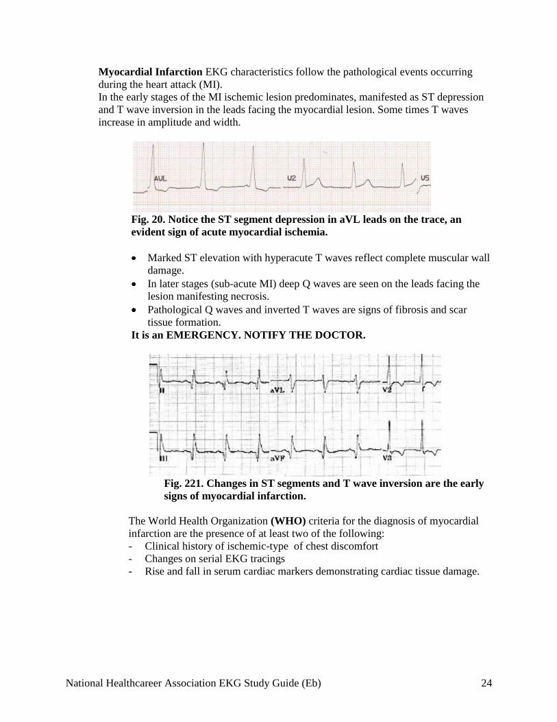
Myocardial Infarction EKG characteristics follow the pathological events occurring
during the heart attack (MI).
In the early stages of the MI ischemic lesion predominates, manifested as ST depression
and T wave inversion in the leads facing the myocardial lesion. Some times T waves
increase in amplitude and width.
Fig. 20. Notice the ST segment depression in aVL leads on the trace, an
evident sign of acute myocardial ischemia.
Marked ST elevation with hyperacute T waves reflect complete muscular wall
damage.
In later stages (sub-acute MI) deep Q waves are seen on the leads facing the
lesion manifesting necrosis.
Pathological Q waves and inverted T waves are signs of fibrosis and scar
tissue formation.
It is an EMERGENCY. NOTIFY THE DOCTOR.
Fig. 221. Changes in ST segments and T wave inversion are the early
signs of myocardial infarction.
The World Health Organization (WHO) criteria for the diagnosis of myocardial
infarction are the presence of at least two of the following:
- Clinical history of ischemic-type of chest discomfort
- Changes on serial EKG tracings
- Rise and fall in serum cardiac markers demonstrating cardiac tissue damage.
National Healthcareer Association EKG Study Guide (Eb) 25
F. EKG Artifacts
Artifact is an unwanted interference or jitter on the EKG recording. This makes the EKG
reading difficult or impossible, as well as can lead to a misdiagnosis.
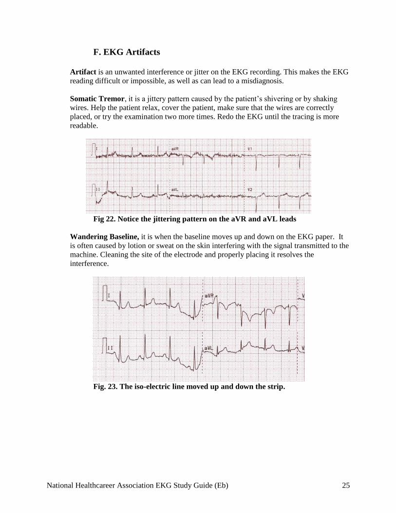
Somatic Tremor, it is a jittery pattern caused by the patient’s shivering or by shaking
wires. Help the patient relax, cover the patient, make sure that the wires are correctly
placed, or try the examination two more times. Redo the EKG until the tracing is more
readable.
Fig 22. Notice the jittering pattern on the aVR and aVL leads
Wandering Baseline, it is when the baseline moves up and down on the EKG paper. It
is often caused by lotion or sweat on the skin interfering with the signal transmitted to the
machine. Cleaning the site of the electrode and properly placing it resolves the
interference.
Fig. 23. The iso-electric line moved up and down the strip.
National Healthcareer Association EKG Study Guide (Eb) 26
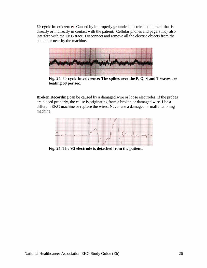
60-cycle Interference: Caused by improperly grounded electrical equipment that is
directly or indirectly in contact with the patient. Cellular phones and pagers may also
interfere with the EKG trace. Disconnect and remove all the electric objects from the
patient or near by the machine.
Fig. 24. 60-cycle Interference: The spikes over the P, Q, S and T waves are
beating 60 per sec.
Broken Recording can be caused by a damaged wire or loose electrodes. If the probes
are placed properly, the cause is originating from a broken or damaged wire. Use a
different EKG machine or replace the wires. Never use a damaged or malfunctioning
machine.
Fig. 25. The V2 electrode is detached from the patient.
National Healthcareer Association EKG Study Guide (Eb) 27
G. Other EKG Related Tests
1. Stress Testing
Stress testing is a noninvasive diagnostic procedure to determine indirectly, the presence
and severity of coronary artery disease and the heart muscle lack of blood supply. The
test is performed through exercise (by having the patient walk on a treadmill or by
pedaling on a bicycle), or pharmacologically (by administration of medication that
causes increase in heart rate), while hooked up to an EKG monitor. The limb leads are
applied to the torso of the patient rather than on the extremities themselves. A rhythm
strip is run continuously throughout the test and a complete 12-lead EKG is recorded
usually every 90 seconds during exercise and every minute in the recovery period
post-exercise. The most common indication for a stress test is chest pain during
exercise or physical effort (Angina Pectoris).
Some indications for stress testing are:
Exercise stress test
This test is performed until at least 85% of the target heart rate is reached or symptoms or
EKG changes develop which requires the test to be terminated. Target heart rate is: 220
minus patient’s age. For example, the target heart rate for a 40 year old patient is 180
(220 – 40). 85% of 180 or 153 is required for the test to be valid for interpretation.
Pharmacologic stress test
This test is appropriate for patients with physical limitation, e.g. amputees, or those who
could not exercise to reach the target heart rate, e.g. elderly. Medications such as
adenosine, dipyridamole, or dobutamine are given intravenously through an IV line to
cause the heart rate to climb to the target level or the same symptoms and EKG changes
as the exercise test develop. The test is concluded after 85% of the target heart rate is
achieved.
2. Ambulatory EKG Monitoring
Ambulatory EKG monitoring is mainly, but not only, used for diagnosis of cardiac
dysrhythmias (disorders of the heart rhythm). It also can evaluate the heart rate, rhythm
and function during daily activities.
Holter Monitor: This is an ambulatory EKG done to rule out intermittent
arrhythmias or ischemia that could be missed on a routine EKG. This may be
done as an in-patient or outpatient procedure. The patient is hooked-up to a Holter
monitor and EKG signals are recorded on a magnetic tape or digital flash media.
After the prescribed duration, the patient returns the monitor to the facility and the
data is entered into a computer and scanned for abnormalities.
Five electrodes are attached to the patient’s trunk instead of the arms and leg
to prevent muscle artifact. The skin is especially prepped by abrading a thin
layer of skin and then the electrodes are taped to the skin so it will adhere better
and prevent from dislodging since the entire procedure will be on for 24 hours or
longer. Before the ambulatory recording starts, EKG tracings are taken with the
National Healthcareer Association EKG Study Guide (Eb) 28
patient lying, sitting, and standing in order to be able to identify these positional
changes which can bring about substantial variation in QRST morphology upon
playback of the tape.
Typical electrode placement for Holter monitoring:
Two exploring electrodes are placed over bone (to minimize motion artifact) near the V1
(over the 4th
or 5th
rib to the right of the sternum) and V5 (over the 5th
rib at the left
midaxillary line).
Two indifferent electrodes placed over the sternal head.
One ground electrode placed over the 9th
or 10th
rib at the right midaxillary line
Artifacts of Ambulatory EKG Recording Recording artifacts can result from the following:
With an analog system: Incomplete tape erasure - this can result in EKG tracings
belonging to two different patients confounding both the scanner and the interpreter.
With an analog system: Tape drag within the apparatus - this will result in recording of
spuriously rapid cardiac rhythms. A narrowing of all EKG complexes and intervals
should give clue to this situation.
Battery depletion - this may result in varying QRS amplitude
Loose connection - intermittently loose connection in the insertion of the electrodes
into the recording apparatus can result in the absence of all EKG signals which may
mimic bradycardia-tachycardia syndrome. Clue to this artifact is the attenuated QRST
morphology of the complexes beginning and ending the pause in rhythm.
Movement of electrodes - this may occur during scratching the chest near the electrodes
and can produce tracings that look like malignant ventricular arrhythmias. However, the
underlying rhythm and rate remain undisturbed and should give clue to this artifact.
Event Monitoring Some patients have symptoms very infrequently that a Holter monitor
yields little useful data. These patients are best suited for an event recorder, a hand held
device carried in the patient’s pocket or purse which is switched only when the patient is
actually experiencing the symptom. The EKG is recorded from the anterior chest wall on
magnetic tape or computer chip which is scanned later the same way as that of the Holter
monitor or it can be transmitted by telephone to a receiving station for immediate
attention. Since the event recorder is used only when symptoms occur, multiple
recordings can be made over the course of a prolonged period of time.
Commonly Used Cardiovascular Drugs:
1. Oxygen: Given to all patients with angina pectoris (acute severe chest pain).
Causes vasodilatation and protects the tissues from hypoxia. It can be administered
through a cannula or a facial mask.
2. Epinephrine: A sympathetic drug used to manage cardiac arrest, because increases
heart contractibility.
3. Isoproterenol (Isuprel): Isoproterenol produces an overall increase in heart rate and
myocardial contractility, but newer agents have replaced it in most clinical settings.
4. Dopamine (Intropin): This drug is indicated and is used in cases with hypotension
(systolic blood pressure is less than 90 mmHg). It causes vasoconstriction (narrowing
National Healthcareer Association EKG Study Guide (Eb) 29
of the blood vessel). It should be used at the lowest dose that produces adequate
perfusion of vital organs.
5. Beta Blockers (Propranolol, Metoprolol, Atenolol, and Esmolol): Beta blockers
reduce heart rate, blood pressure, myocardial contractility, and myocardial oxygen
consumption which make them effective in the treatment of angina pectoris and
hypertension. They are also useful in preventing atrial fibrillation, atrial flutter, and
paroxysmal supra-ventricular tachycardia. Adverse effects of beta blockers are
hypotension, congestive heart failure and broncho-spasm.
6. Lidocaine: Lidocaine is the drug of choice for the suppression of ventricular ectopy
(a beat located outside the conductive system) contractions (PVC), including
ventricular tachycardia and ventricular flutter. Excessive doses can produce
neurological changes, such as drowsiness, disorientation, decreased hearing ability,
paresthesia, muscle twitching, and eventual seizures. In large doses it causes
myocardial depression, and circulatory depression.
7. Verapamil: Verapamil is used in the treatment of paroxysmal supraventricular
tachycardia (PSVT), effective in terminating more than 90% of episodes of PVST in
adults and infants. Verapamil is also useful in slowing ventricular response to atrial
flutter and fibrillation. Vigilant monitoring of blood pressure is recommended due to
hypotension that could occur.
8. Digitalis: Digitalis increases the force of cardiac contraction as well as cardiac
output.. Digitalis is a drug with high toxicity, therefore patients require constant
monitoring for signs and symptoms of toxicity such as: yellow vision, nausea,
vomiting, and drowsiness.
9. Morphine Sulfate: It is the traditional drug of choice for the pain and anxiety
associated with acute myocardial infarction. In high doses, morphine sulfate may
cause respiratory depression. It is a controlled substance and has a tendency for abuse
and addiction.
10. Nitroglycerin: Nitroglycerin is a powerful smooth muscle relaxant effective in
relieving angina pectoris. Headache is a common consequence following the
administration of this drug. Hypotension may occur and patients should be instructed
to sit or lie down while taking nitroglycerin.
Medical Terminology
Word Analysis
Healthcare terminology is broken down into word roots, prefixes, suffixes and combining
vowels and forms. Word roots, or base words, are the foundation of the healthcare term. A
suffix is a word ending, a prefix is a word beginning, and a combining vowel, (usually o), links
the root to the suffix or to another root. The combining form is word root plus the appropriate
combining vowel.
For example: oste /o/ athr/itis
Combining Forms and Their Meanings
Some combining forms and their meanings:

National Healthcareer Association EKG Study Guide (Eb) 30
Arthr/o joint
Bi/o life
Cardi/o heart
Carcin/o cancerous, cancer
Cephal/o head
Cerebr/o cerebrum (largest part of the brain)
Dent/I teeth
Derm/o skin
Electr/o electrical activity
Enter/o intestines
Fet/o fetus
Gastr/o stomach
Hepat/o liver
Iatr/o treatment, physician
Leuk/o white
Nephr/o kidney
Oste/o bone
Path/o disease
Ren/o kidney
Rhin/o nose
Sarc/o flesh
Thromb/o clotting
Ur/o urinary tract
Some suffixes and their meanings:
-al pertaining to
-algia pain
-dynia pain
-ectomy excision, removal
-emia blood condition
-genic produced by, pertaining to producing
-globin protein
-itis inflammation
-oma tumor, mass swelling
-osis condition, usually abnormal
-pathy disease condition
-sis state of; condition
Some Prefixes and their meanings:
Ante- before, in front of
Anti - against
Dia - through, complete
End, endo within
Epi - above, upon
Hyper- excessive, above more than normal
National Healthcareer Association EKG Study Guide (Eb) 31
Hypo - deficient, below, under less than normal
Peri - surrounding, around
Pre- before
Sub- under, below
Suffixes used to describe therapeutic interventions
-ectomy excision
-graphy process of recording
-metry process of measurement
-scopy a visual examination
-stomy a new opening
-tomy incision
-tripsy process of crushing
Suffixes used to describe instruments
-graph an instrument/machine to record
-meter an instrument to measure
-scope an instrument to visually or aurally examine
-tome an instrument to cut
-tripter an instrument to crush
-trite an instrument to crush
Organs
Organs are comprised of several types of tissue. The stomach is made up of muscle tissue, nerve
tissue, and epithelial tissue. The medical term for internal organs is viscera.
Systems are groups of organs working together to perform complex functions.
Body Systems Functions Organs
Musculoskeletal support, movement muscles, bones, joints, bone
protection marrow
Integumentary protection skin, hair, nails
Gastrointestinal nutrition stomach, intestines
Urinary elimination of nitrogenous kidneys, bladder, ureters,
waste urethra
Reproductive reproduction ovaries, testes
Blood/Lymphatic transportation blood cells
Immune protection
National Healthcareer Association EKG Study Guide (Eb) 32
Cardiovascular transportation lymph glands
heart, vessels
Respiratory delivers oxygen to cells lungs, bronchi, trachea
removes carbon dioxide
Nervous/Behavioral receive/process information brain, nerves, mind
Endocrine effects changes through pancreas, thyroid
chemical messengers
Planes of the Body
Dividing the body into planes or flat surfaces is an additional way to describe the body. These
descriptions listed below are useful when doing magnetic imaging, CT scans, and other imaging
techniques.
Sagittal planes are vertical planes that separate the sides from each other.
Midsagittal plane separates the body into right and left halves.
The frontal plane divides the body into front and back portions.
The transverse plane divides the body horizontally into an upper and lower part.
Positional and Directional Terms
Anterior (ventral) – front surface of the body
Posterior (dorsal) – back side of the body
Deep – away from the surface
Proximal –near the point of attachment to the trunk or near the beginning of a structure.
Distal – far from the point of attachment to the trunk or far from the beginning of a structure.
Inferior – below another structure
Superior – above another structure
Medial – pertaining to the middle or nearer the medial plane of the body
Lateral – pertaining to the side
Supine – lying on the back
Prone – lying on the belly

National Healthcareer Association EKG Study Guide (Eb) 33
4. Ethics and Legal Considerations .
Ethics are standards of right and wrong that regulate the behavior of any professional in their daily
activities and communication. PCTs are required to have the highest ethical standards.
Law is defined as a regulation or action enforced by a controlling authority, such as local, state and
federal government. A tort is defined as a civil wrong committed against a person or property that
causes damage or deprives someone of his or her personal liberty and freedom. Torts are punishable
by law.
Some intentional torts are:
Assault: an open threat of bodily harm to someone
Battery: any body contact made without permission
False imprisonment: unlawful restraint or confinement of one person from another
Invasion of privacy: an interference with the person’s right to be left alone
Negligence is an unintentional tort. Negligence is charged when a health care professional fails to
provide ordinary and standard care and the patient suffers injuries. As a tort, negligence is punishable
by law.
Patient’s Bill of Rights was created in 1973 and revised in 1993. Patient’s Bill of Rights is a list of
standards that a patient can expect from any health care provider. Preserving the confidentiality of a
patient is one of the main requirements of the Patient’s Bill of Rights. Patient’s Confidentiality is also
at the foundation of HIPAA (Health Insurance Portability and Accountability Act).
HIPAA is a US law designed to provide privacy standards to protect patients’ medical records and
other health information provided to health insurance agencies, doctors, hospitals and other health care
providers, while transmitted or recorded by electronic or other media.
Informed Consent involves the patient’s right to receive complete information and understanding
relative to his or her condition and the obligation to make decisions and cooperate in his or her
treatment based on this knowledge. The person that signs the informed consent must be legally and
mentally healthy and responsible.
Implied Consent is a self-understood action of permission not expressed in words or in writing.
Extending an arm for blood withdrawal, to a health care provider, is an example of the implied
consent.
Advance Directives or Advanced Health Care Directives are instructions given by individuals
specifying what actions should be taken for their health in the event that they are no longer capable to
make the proper decisions due to an illness or incapacitating condition. A living will and Power of
Attorney are also forms of advance directives and usually cover specific directives as to the treatment
provided by caregivers. Abuse of any person who is incapable of self-protection is punishable by law.
It may include children, adults or the elderly. Abuse could be physical, psychological or sexual. A
patient’s confidential rights are waived when a report of abuse is made. If you suspect a child or any
individual is abused, neglected or maltreated, relay your suspicion to the physician.
Chain of Custody reflects a timed written record of different individuals who have custody of a test or
item from its initial acquisition until its final deposition in court as evidence. For medical test results
the chain is maintained until there is an official disclosure of the results.
National Healthcareer Association EKG Study Guide (Eb) 34
D. OSHA Regulations and Infection Control
1. OSHA (Occupational Safety and Health Administration) requires basic safety practices
to protect any employee in the working place from different types of hazards
Biological Hazards: damage caused by infectious agents such as bacteria, viruses,
fungus, or other parasites. Allergic reactions are part of the biological hazards and
can be caused by different allergens. Latex sensitivity can cause allergic reactions
ranging from simple dermatitis to anaphylaxis.
Chemical Hazards: damage is caused by different chemicals used in the medical
laboratory. There is possible exposure to toxic, carcinogenic or caustic substances
in the medical lab and offices. All chemicals and reagents, containing hazardous
ingredients in a concentration greater than 1%, must have a Material Safety Sheet
(MSDS) on file in the laboratory. The MSDS contains information on physical and
chemical characteristics; fire, explosion, reactivity, and health hazards; primary
routes of entry, exposure limits and carcinogenic potential; precautions for safe
handling; clean-up and emergency first aid information.
Radiological Hazards: damage caused by radiating X-rays or atomic particles.
Radiation is used widely in the medical field for diagnostic and therapeutic
purposes. Radiation it is not recognized by the senses and must be detected by
specialized equipment.
Electrical Hazards: high-voltage equipment can cause burns and shock. Simple
steps, such as avoiding extension cords, grounding and maintaining electrical
equipment, drying hands before using electrical equipment and positioning
electrical devices away from sinks, faucets and other sources of water can prevent
electrical injuries.
Fire or explosive Hazards: Bunsen burners, oxygen and chemicals can cause
burns or dismemberment. To prevent damage from fire you should know and follow
the policies of the office. You should know the routes of exit and evacuation and
remember the word RACE (R = Rescue persons immediately in danger, A = Alarm
the nearest fire alarm, C = Confine the fire closing doors and windows, and E =
Extinguish a small fire and stop it from spreading).
Physical Hazards: wet floors, heavy lifting can cause falls, sprains and
strains. To avoid physical injury you should wear proper attire, walk do not
run in the office or laboratories unless it is an emergency, do not overextend
your reach, use the transportation belt while transporting a patient, lift with
your legs when lifting heavy objects, etc.
2. Infection, Body Protection and Infection Control
Infectious diseases are damages to the tissues or organs resulting from the activity or presence
of living micro-organisms. Infection is a result of the interaction between the human body’s
defense mechanisms and the microorganisms. A pathogen is defined as a disease causing
microorganism Infections acquired in a medical environment are called nosocomial infections.
The goals of infection control are to limit or prevent the presence of infectious agents, to create
barriers against transmission and to reduce the risk to other individuals of being infected.
National Healthcareer Association EKG Study Guide (Eb) 35
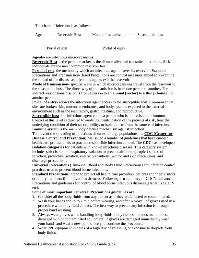
The chain of infection is as follows:
Agent ---------Reservoir Host-------- Mode of transmission ------- Susceptible host
: :
: :
Portal of exit Portal of entry
Agents- are infectious microorganisms.
Reservoir Host-is the person that keeps the disease alive and transmits it to others. Sick
individuals are the most common reservoir host.
Portal of exit- the method by which an infectious agent leaves its reservoir. Standard
Precautions and Transmission-Based Precautions are control measures aimed at preventing
the spread of the disease as infectious agents exit the reservoir.
Mode of transmission- specific ways in which microorganisms travel from the reservoir to
the susceptible host. The direct way of transmission is from one person to another. The
indirect way of transmission is from a person to an animal (vector) to a thing (fomite) to
another person.
Portal of entry- allows the infectious agent access to the susceptible host. Common entry
sites are broken skin, mucous membranes, and body systems exposed to the external
environment such as the respiratory, gastrointestinal, and reproductive.
Susceptible host- the infectious agent enters a person who is not resistant or immune.
Control at this level is directed towards the identification of the patients at risk, treat the
underlying condition of their susceptibility, or isolate them from the source of infection.
Immune system is the main body defense mechanism against infection.
To prevent the spreading of infectious diseases to large populations the CDC (Center for
Disease Control and Prevention) has issued a number of guidelines that have enabled
health care professionals to practice responsible infection control. The CDC has developed
isolation categories for patients with known infectious diseases. This category system
includes strict isolation, respiratory isolation to prevent air borne (droplet) spread of
infection, protective isolation, enteric precautions, wound and skin precautions, and
discharge precautions.
Universal Precautions (Universal Blood and Body Fluid Precautions) are infection control
practices used to prevent blood borne infections.
Standard Precautions intend to protect all health care providers, patients and their visitors
or family members from infectious diseases. Following is a summary of CDC’s Universal
Precautions and guidelines for control of blood borne infectious diseases (Hepatitis B, HIV
etc):
Some of most important Universal Precautions guidelines are:
1. Consider all the body fluids from any patient as if they are infected or contaminated.
2. Wash your hands for up to 2 min before wearing, and after removal, of gloves used in a
procedure with body fluid contact. The best way to prevent any infection is through
proper hand washing.
3. Always wear gloves when handling body fluids, body tissues, mucous membranes,
damaged skin or contaminated equipment. If gloves are damaged immediately wash
your hands and wear a new pair before you continue the procedure.
4. Wear PPE equipment in cases of a high risk of splashing or exposure to droplets from
body fluids.
National Healthcareer Association EKG Study Guide (Eb) 36
5. Never recap a needle. Always cover the used needle with the protective device and
dispose it immediately into a biohazard sharp container.
6. In cases of body fluid spills spray the spill first with a disinfectant or most frequently
with 10% bleach.
7. Always properly dispose soiled soft materials into biohazard bags or boxes.
8. Properly report needle stick injuries, splashes, and wound secretion contact and
contamination
9. Health care workers with open wounds or lesions, dermatitis or other infectious
diseases should avoid direct contact with the patients.
10. All the health care workers in contact or at risk of body fluid contamination should be
vaccinated for Hepatitis B virus.
3. Personal Protective Equipment (PPE): includes gloves, eye and facial protection,
protective clothing and resuscitation equipment.
a. Gloves: are used to protect against contact with infected body fluids, mucous
membranes, non intact or damaged skin, contaminated surfaces, equipment and
instruments or when performing vascular access procedures such as phlebotomy.
b. Mask and Facial Protection are required whenever there is a potential of splashing,
spraying or splattering of contaminated body fluids, such as centrifugation (equipment
used to divide particles by spinning and on their weight).
c. Protective Clothing must be worn whenever splashing or splattering to skin or
clothing may occur. They include hats, gown and boots (shoe coverage).
National Healthcareer Association EKG Study Guide (Eb) 37
Resources:
Web Sites:
1. http://rnbob.tripod.com
2. http://davidge2.umaryland.edu
3. http://info.med.yale.edu
4. http://www.waycross.edu
5. http://www.usfca.edu
Reference books:
1. Principles of Clinical Electrocardiography, 13th edition, Goldschlager, Nora, MD.; Goldman Mervin J., MD
2. Basic Arrhythmias, 5th edition. Walraven, Gale
3. The only EKG Book You’ll Ever Need, 4th Edition. Thaler, Malcom S., MD
4. Understanding 12-lead EKG’s. Beasley, Brenda M., West, Michael C.,
5. ECG’s Made Easy, 2nd Edition. Aehlert, Barbara
6. Gray’s Anatomy. Gray, Henry. FRS., Carter, HV., MD
National Healthcareer Association EKG Study Guide (Eb) 38
Sample EKG Exam
Choose the best answer:
1. A 60-year-old male patient has a history of on and off chest pain. His resting ECG is
normal. It is decided to put him thru an exercise stress test. Which of the following will
be the target heart rate for this patient?
A. 180 beats per minute
B. 160 beats per minute
C. 140 beats per minute
D. 120 beats per minute
2. This part of the conduction system of the heart is located at the superior portion of the
interventricular septum and has the ability to function as a pacemaker with an intrinsic
firing rate of 40-60 beats per minute.
A. SA node
B. AV node
C. Bundle of His
D. Purkinje fibers
3. This represents the time it takes for electrical impulse to travel from the atria to the AV
node, bundle of His, bundle branches and to the Purkinje fibers.
A. PR interval
B. PP interval
C. RR interval
D. QT interval
4. Which of the following is an indication for stress testing?
A. Angina at rest
B. Acute myocardial infarction
C. Severe hypertension
D. Evaluation of chest pain in a patient with normal baseline EKG
5. Which of the following is considered a negative Holter?
A. Pauses
B. Bradycardias or tachycardias
C. No significant arrhythmias or ST changes.
D. ST segment elevation or depression
6. The number of R waves in a six-second strip is 9. The heart rate is:
A. 72 per minute
B. 45 per minute
C. 90 per minute
D. 54 per minute
National Healthcareer Association EKG Study Guide (Eb) 39
7. Mr. Adam Edwards came to the clinic complaining of occasional chest pains, substernal
in location and radiating to the left arm. The physician prescribed him medication to be
taken during such chest pain episodes. A few days later, the patient came back with
complain of headaches occurring after taking the medication. The medication prescribed
is most likely:
A. Procainamide
B. Propranolol
C. Nitroglycerin
D. Digitalis
8. The following valves are called semilunar because they have half-moon shaped cusps:
A. Pulmonic and tricuspid valves
B. Aortic and mitral valves
C. Pulmonic and mitral valves
D. Aortic and pulmonic valves
9. Which of the following is incorrect of the augmented unipolar extremity leads?
A. They represent the difference in the electrical potential between two extremities.
B. The midpoint between two limbs is used as a negative reference point.
C. Only one electrode from one limb makes a lead.
D. The amplitude of deflections is increased by 50%.
10. This is an arrhythmia produced by an electrical impulse originating from a site other than
the sinus node:
A. Pre-excitation syndromes
B. Arrhythmia of sinus origin
C. Conduction blocks
D. Ectopic rhythms
11. Which of the following statements is incorrect regarding the limb leads?
A. Electrodes and leads are applied to the right and left arms and legs.
B. The right leg functions as a ground.
C. The right leg plays a significant role in the production of the electrocardiogram.
D. Where the electrode is placed in the extremities will make no difference in the
electrical potential recorded.
12. The vertical axis of the EKG paper measures:
A. Time
B. Voltage/Amplitude
C. Rhythm
D. Heart rate
13. A normal PR interval should measure:
A. .08 - .12 seconds
B. < .12 seconds
C. .12 - .20 seconds
D. > .20 seconds
National Healthcareer Association EKG Study Guide (Eb) 40
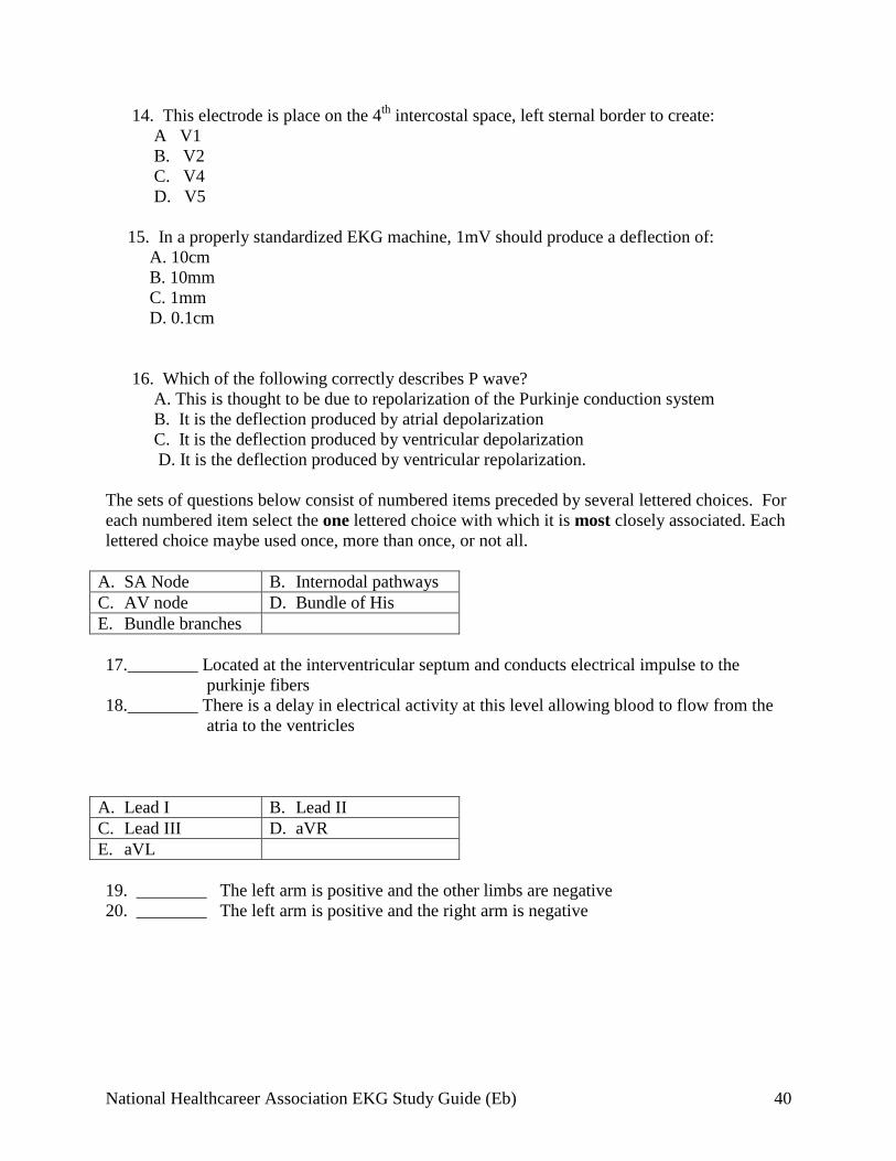
14. This electrode is place on the 4th
intercostal space, left sternal border to create:
A V1
B. V2
C. V4
D. V5
15. In a properly standardized EKG machine, 1mV should produce a deflection of:
A. 10cm
B. 10mm
C. 1mm
D. 0.1cm
16. Which of the following correctly describes P wave?
A. This is thought to be due to repolarization of the Purkinje conduction system
B. It is the deflection produced by atrial depolarization
C. It is the deflection produced by ventricular depolarization
D. It is the deflection produced by ventricular repolarization.
The sets of questions below consist of numbered items preceded by several lettered choices. For
each numbered item select the one lettered choice with which it is most closely associated. Each
lettered choice maybe used once, more than once, or not all.
A. SA Node B. Internodal pathways
C. AV node D. Bundle of His
E. Bundle branches
17.________ Located at the interventricular septum and conducts electrical impulse to the
purkinje fibers
18.________ There is a delay in electrical activity at this level allowing blood to flow from the
atria to the ventricles
A. Lead I B. Lead II
C. Lead III D. aVR
E. aVL
19. ________ The left arm is positive and the other limbs are negative
20. ________ The left arm is positive and the right arm is negative
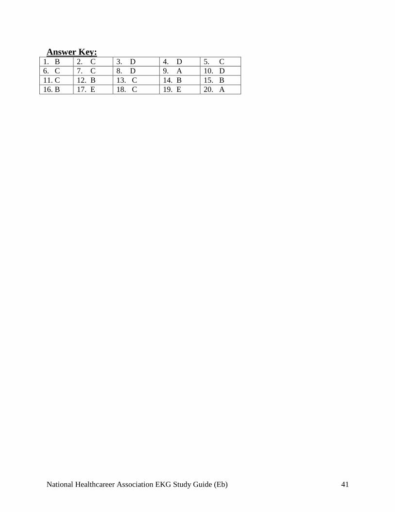
National Healthcareer Association EKG Study Guide (Eb) 41
Answer Key: 1. B 2. C 3. D 4. D 5. C
6. C 7. C 8. D 9. A 10. D
11. C 12. B 13. C 14. B 15. B
16. B 17. E 18. C 19. E 20. A