
Nasotemporal Overlap in Visual Field Projected to Lateral Geniculate Nucleus...
14
Nasotemporal Overlap in Visual Field Projected to Lateral Geniculate Nucleus in the Cat K. J. SANDERSON AND S. MURRAY SHERMAN Department of Physiology, John Curtin School of Medical Research, Australian National University, Canberra, Australia 2601 IN ANIMALS with welI-developed binocular vision, such as the cat, monkey, and man, the retina is divided into temporal and na- sal halves which, respectively, send optic fibers back to the same and opposite halves of the brain so that the left hemiretinas of both eyes project to the left hemisphere of the brain and, similarly, the right hemi- retinas to the right hemisphere. This means that an object in the left visual field is rep- resented in the right half of the brain and objects in the right visual field are repre- sented in the left half of the brain. There has been some doubt, however, about the projection of the vertical midline (zero vertical meridian), and recent wo,rk has shown that in the cat, a strip of visual field straddling the midline projects to both sides of the brain (12, 23, 29). It is generally agreed that the naso#tem- poral partition of the retina and the partial decussation at the optic chiasma are fea- tures of the organization of the visual sys- tem that are essential for binocular depth discrimination or stereopsis. Based on the concept of receptive-field disparity (1, 25) a detailed neural theory of stereopsis has recently been develo#ped (6-8, 21). An es- sential element in this theory is that there should be a nasotemporal overlap pro- jected to the visual cortex (6, 12) as part of the general retinocerebral projection through the lateral geniculate nucleus. There are two related aspects of this theory that require a nasoItemporal projectiosn. These will be considered in DISCUSSION and only brief mention is needed here. Nearly all the cells in the striate cortex have two receptive fields, one for each eye. Consider a receptive field for one eye being Received for publication November 19, 1970. located precisely on the vertical meridian. The concept of receptive-field disparity in- volves the possibility that the receptive field for the other eye be located to one or other side of the corresponding point for this second eye. This necessarily involves the further possibility that receptive fields be located across the midline into the “wrong” hemifield. The other related as- pect requiring a nasotemporal overlap con- cerns the stereoscopic viewing of objects situated in the vicinity of the fixation point. If the retina were sharply divided and without overlap, then there would be regions in the visual field between the two visual axes both in front of and behind the fixatiomn point that would be repre- sented in both cerebral hemispheres, but only from one eye in each case (6, 12). In other words, neither of these regions could be viewed binocularly by the one hemi- sphere. This is a matter of some concern since these are regions of known high stereoacuity. The presence of a nasotempo- ral overlap would resolve this difficulty. In the cat, a nasotemporal overlap has been shown in the retina by histoIogica1 methods (29) and in the projection to the visual co,rtex by neurophysiological methods (12, 23). Some doubt exists, how- ever, as to whether the overlap in the cere- bra1 cortex is produced by fibers which cross in the corpus callosum or whether it is projected from the lateral geniculate nucleus. The uncertainty about the pro- jection of the vertical meridian in the cat’s brain has arisen because of the absence of a specialized fovea in the retina, and the position of the vertical meridian has usu- ally been inferred, often with an error of I” or so, froIm the position of the blind 453
Transcript of Nasotemporal Overlap in Visual Field Projected to Lateral Geniculate Nucleus...

Nasotemporal Overlap in Visual Field Projected to
Lateral Geniculate Nucleus in the Cat
K. J. SANDERSON AND S. MURRAY SHERMAN
Department of Physiology, John Curtin School of Medical Research, Australian National University, Canberra, Australia 2601
IN ANIMALS with welI-developed binocular vision, such as the cat, monkey, and man, the retina is divided into temporal and na- sal halves which, respectively, send optic fibers back to the same and opposite halves of the brain so that the left hemiretinas of both eyes project to the left hemisphere of the brain and, similarly, the right hemi- retinas to the right hemisphere. This means that an object in the left visual field is rep- resented in the right half of the brain and objects in the right visual field are repre- sented in the left half of the brain. There has been some doubt, however, about the projection of the vertical midline (zero vertical meridian), and recent wo,rk has shown that in the cat, a strip of visual field straddling the midline projects to both sides of the brain (12, 23, 29).
It is generally agreed that the naso#tem- poral partition of the retina and the partial decussation at the optic chiasma are fea- tures of the organization of the visual sys- tem that are essential for binocular depth discrimination or stereopsis. Based on the concept of receptive-field disparity (1, 25) a detailed neural theory of stereopsis has recently been develo#ped (6-8, 21). An es- sential element in this theory is that there should be a nasotemporal overlap pro- jected to the visual cortex (6, 12) as part of the general retinocerebral projection through the lateral geniculate nucleus. There are two related aspects of this theory that require a nasoItemporal projectiosn. These will be considered in DISCUSSION and only brief mention is needed here.
Nearly all the cells in the striate cortex have two receptive fields, one for each eye. Consider a receptive field for one eye being
Received for publication November 19, 1970.
located precisely on the vertical meridian. The concept of receptive-field disparity in- volves the possibility that the receptive field for the other eye be located to one or other side of the corresponding point for this second eye. This necessarily involves the further possibility that receptive fields be located across the midline into the “wrong” hemifield. The other related as- pect requiring a nasotemporal overlap con- cerns the stereoscopic viewing of objects situated in the vicinity of the fixation point. I f the retina were sharply divided and without overlap, then there would be regions in the visual field between the two visual axes both in front of and behind the fixatiomn point that would be repre- sented in both cerebral hemispheres, but only from one eye in each case (6, 12). In other words, neither of these regions could be viewed binocularly by the one hemi- sphere. This is a matter of some concern since these are regions of known high stereoacuity. The presence of a nasotempo- ral overlap would resolve this difficulty.
In the cat, a nasotemporal overlap has been shown in the retina by histoIogica1 methods (29) and in the projection to the visual co,rtex by neurophysiological methods (12, 23). S ome doubt exists, how- ever, as to whether the overlap in the cere- bra1 cortex is produced by fibers which cross in the corpus callosum or whether it is projected from the lateral geniculate nucleus. The uncertainty about the pro- jection of the vertical meridian in the cat’s brain has arisen because of the absence of a specialized fovea in the retina, and the position of the vertical meridian has usu- ally been inferred, often with an error of I” or so, froIm the position of the blind
453

454 K. J. SANDERSON AND S, M. SHERMAN
spot. This paper sets out to show that a nasotemporal overlap projected to both lateral geniculate nuclei is sufficient to ac- count for much of the nasotemporal over- lap to be found in the striate cortex in the vicinity of the 17/ 18 border. We have re- corded from single units having receptive fields in the vicinity of the vertical mid- line and, in order to avoid problems aris- ing from uncertainty about the precise location of the vertical meridian, we have recorded in both lateral genicuIate bodies of each animal.
METHODS
The general methods have been described previously (21, 22, 25). Ten cats (2.5-4.0 kg) were anesthetized with ether for initial surgery and subsequently with N&)/O, (70%/30%) for the course of the experiment. The animals were paralyzed with a continuous intravenous infu- sion of muscle relaxant, gallamine triethiodide (Flaxedil, May and Baker, 16.2 mg/hr) and C-Toxiferine I (toxiferine dichloride, Hoffmann- La Roche, I mg/hr) in 0.9% saline (6.5 ml/h+ The body temperature was maintained at 38 Cl with an electric heating blanket. Eye movements were reduced to a very low level by the paralysis of the extraocular muscles coupled with bilateral cervical sympathectomy (27).
We recorded single units in both lateral geniculate nuclei using tungsten-in-glass elec- trodes with an exposed tip of 8-10 p length, Electrolytic lesions (10 pa for IO set) were placed at the bottom of some electrode tracks to aid subsequent histological identification. At the end of each experiment the brain was fixed in formal saline and, following standard pro- cedures (22), serial histological sections through the lateral geniculate body were stained with cresyl violet. Outline drawings were made of the sections which contained electrode tracks, and the positions of the units recorded in each electrode track were superimposed on the draw- ing.
Receptive fields were plotted on a tangent screen placed 1 m in front of the nodal points of the eyes. Moving targets were used to locate the position of each receptive field before care- fully mapping the field center with small flash- ing spots (0.143”). The location of each receptive field was given by the visual direction of its center and expressed in terms of the two angles, azimuth and elevation, of the system of spherical polar coordinates described in detail by Bishop, Kozak, and Vakkur (11).
Azimuth is the angle between the visual axis and the projection of the center of the receptive
field on to the fixation plane (positive to the right of the visual axis, negative to the left).
Elevation is the angle between the fixation plane and the center of the receptive field (positive, upward from the fixation plane: negative, downward).
In the above system of spherical polar CO-
ordinates the reference meridian (vertical mid- line in the visual field) should be referred to as the zero meridian since all the meridia are vertical. We will find it useful however to refer to the vertical midline as the vertical meridian since the latter term is already in common use in the literature (20, 23).
The blind spots were also plotted on the tan- gent screen, using the ophthalmoscopic facilities of a Zeiss fundus camera modified for rear pro- jection (9). Plotting the blind spots at 15-min intervals enabled us to monitor any slow drifts in eye position, the amount of eye movement over a 15-min period being negligible under our experimental conditions (21).
RESULTS
Design of experiment
The general design of the experiment is represented diagr’ammatically in Fig. 1. The projection of the visual field onto coronal sections through the middle of the anteroposterior extent of the dorsal nu- cleus of the lateral geniculate body (LGNd) and medial interlaminar nucleus (MIN) is shown for the nuclei on both sides of the brain. Because paralysis of the extraocular muscles fixes the eyes in divergence, the nasotemporal part of the visual field is represented separately above each eye. The vertical meridian projects onto the medial edge of the LGNd on both sides (10). The first electrode track was usually inserted into the left LGNd, being so positioned as to record cells having receptive fields with azimuths of abo,ut + 3 to 4-5” (22). There- after successive microelectroSde penetrations were made through the nucleus in the one corona1 plane, each penetration being placed more medially than its predecessor in steps usually of 0.3 mm. Cells in the three main layers of the nucleus were recorded in each penetration, the cells in layers A and B having receptive fields belonging to the contralateral (right) eye and those of the cells in layer A, belo,nging to the ipsilateral (left) eye. As the electrode was moved medially, the locations of the receptive fields moved approximately in a

NASOTEMPORAL OVERLAP IN GENICULATE 455
Left Eye
Left Rqht
FIG. 1. Diagram of the general design of the experiment. Electrode penetrations were placed in a series across the LGNd/MIN border on each side. Vertical lines on the coronal sections of each lateral geniculate body are lines of isoazimuth, with azimuth values given above each line. The shaded part of each LGNd is the region where cells recorded commonly had receptive fields with center points in the wrong visual hemifield. For details see text.
horizontal line closer and closer to the vertical meridian until, in the LGNd/MIN border region, cells were found having re- ceptive-field centers on the other side of the midline into the wrong hemifield. It should be appreciated however that, at this stage of the experiment, there could be no certainty whether or not the receptive-field centers were, in fact, located over the verti- cal meridian.
There is a “mirror-image” representation of the visual field in the MIN (5, 22, 28) so that, as the electrode was moved still more medially, the receptive fields of the cells in the MIN now began to reverse their progression and move steadily away from the vertical meridian. This progression of receptive-field locations, first toward the midline, then over the midline and, finally, back again away from the center, is rep- resented in Fig. 1 by the recurved arrows.
The arrows go slightly beyond the vertical meridian into the ipsilateral hemifield be- fore curving back into the contralateral hemifield. As the coronal sections indicate, the progression of receptive-field locations away from the vertical meridian is much more rapid with medial movement into the MIN than it is with lateral movement in the LGNd. It can be seen also that the direction of the progression for the one LGNd is the same for the two eyes, but reverses when the change is made to the other LGNd.
The above procedure was then repeated on the right LGNd/MIN complex. Pro- vided appropriate correction is applied for the small eye movements that may occur, any overlap of receptive-field centers from the two sides can be taken to indicate that a nasotemporal overlap is projected to the LGNd/MIN complex. By recording from cells in the nuclei of both sides of the brain, it is possible to reach this conclusion with- out any knowledge of the location of the vertical meridian. However, in order to de- termine the visual directions of the recep- tive fields near the midline and the amount of their overlap into the wrong hemifield, an estimate of the position of the vertical meridian for each eye is needed. Two sepa- rate methods were used for this purpose. 1) Midpoint of nasotemporal overlap. The vertical meridian must fall within the re- gion of overlap and, if one assumes bi- lateral symmetry, the line will be the verti- cal through the midpoint of the overlap. In using this method we have restricted the overlap to receptive-field centers of cells recorded in laminae A and Al. These mat- ters will be discussed in more detail below. 2) Receptive-field separation method. This method, developed originally for experi- ments on the visual cortex (21, 25) has been adapted here for use at the geniculate level. The main features of this method relevant to our experiments are set out below.
Since we studied the part of the LGNd concerned with the central visual field, the cells recorded along a vertical electrode track all had receptive fields with approxi- mately the same visual direction (10; un- published observatio’ns). Although the term projection line was originally applied to a column of cells in the LGNd having recep-
456 K. Jm SANDERSON AND S. bi. SHERMAN
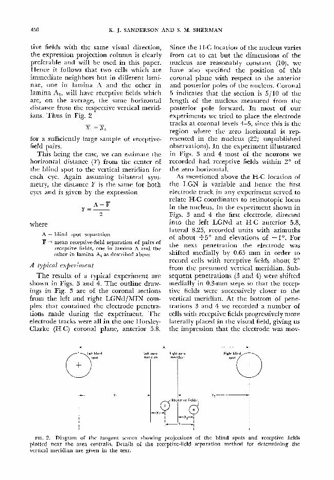
tive fields with the same visual direction, the expression projection column is cIearly preferable and will be used in this paper. Hence it follows that two cells which are immediate neighbors but in different Iami- nae, one in lamina A and the other in lamina Al, wiI1 have receptive fields which are, on the average, the same horizo’ntal distance from the respective vertical merid- ians. Thus in Fig. 2
x, =x.) folr a sufficiently large sample of receptive- field pairs.
This being the case, we can estimate the horizontal distance (Y) from the center of . the blind spot to the vertical meridian for each eye. Again assuming bilateral sym- metry, the distance Y is the same for both eyes and is given by the expression
A-F Y =-
2
where
A = blind spot separation F= mean receptive-field separation of pairs of
receptive fields, one in lamina A and the other in lamina A, as described above
A ty@d expen’ment
The results of a typical experiment are shown in Figs. 3 and 4. The outline draw- ings in Fig. 3 are of the coronal sections from the left and right LGNd/MIN com- plex that contained the electrode penetra- tions made during the experiment. The electrode tracks were all in the one Horslev- Clarke (H-C) coronal plane, anterior 5.8.
Since the H-C location o,f the nucleus varies from cat to cat but the dimensions of the nucleus are reasonably constant (lo), we have aIso specified the position ‘of this coronal plane with respect to the anterior and post-&i or poles of the n ucleus. &ronaI 5 indicates th at the section is 5/I 0 of the length of the nucleus measured frolm the posterior pole forward. In most of our experiments we tried to place the electrode tracks at coronal levels 4-5, since this is the region where the zero horizolntaI is rep- resented in the nucleus (22; unpublished observations). In t he ex peri ment illustra ted in Figs. 3 and 4 most of the neurons we recorded had receptive fields within 2’ of the zero horizontal.
As mentioned above the H-C location of the LGN is variabIe and hence the first electrode track in any experiment served to relate H-C coordinates to retinotopic locus in the nucleus. In the experiment shown in Figs, 3 and 4 the first electrode, directed into the left LGNd at H-C anterior 5.8, lateral 8.25, recorded units with azimuths of about +5” and elevations of - lo, For the next penetration the electrode was shifted medially by 0.65 mm in olrder to record cells with receptive fields about 2” from the presumed vertical meridian. Sub- sequent penetrations (3 and 4) were shifted medially-in 0.3-mm steps so that the recep- tive fields were successivelv closer to the vertical meridian. At the bottom of pene- trations 3 and 4 we recorded a number of cells with receptive fields progressively more laterally placed in the visual field, giving us the impression that the electrode was mov-
Y,-- Y,-----y
FIG, 2. Diagram of the tangent screen showing projections of the blind spots and receptive fields plotted near the area centralis. Details of the receptive-field separation method for determir-iing the vertical meridian are given in the text.

NASOTEMPORAL OVERLAP IN GENICULATE 457
left LGN
Cat 3 Coronal 5 Right LGN
1 2 3 45 l
l l
FLI:. 3. A typical experiment. Locations of units which were recorded close to the LGNd/MIN border on each side have been shown on outline drawings of the coronal sections which contained the electrode penetrations placed in the nucleus. For details see text.
ing out of the main layers of the LGNd into the MIN (see Fig. 3). In electrode pene- tration 5, which was in the MIN, all the receptive fields had azimuths of about HO”, so we terminated the penetratio’ns in the left LGNd and moved to the right LGNd where we repeated the mapping procedure.
At the end elf the mapping of the LGNd/ MIN border region on the two sides and following appropriate correction for any
m a oom l
l
Cat 3
l Right LGN
0 Left LGN
l B
0 0
eye movement, the distribution of receptive fields for each eye was examined for an overlap of fields from the two sides. As we have aheady observed, paralysis o,f the ex- traocular muscles fixes the eyes in diver- gence and so the receptive-field distrib,u- tions for the two eyes were separated on the tangent screen. Figure 4 sho,ws the loca- tion of receptive-field center points of units recorded in the experiment described above. For both eyes there was an overlap of
Right eye
0
FIG. 4. Distribution of receptive-field center points of units shown in Fig. 3. For each. eye there is an overlap, indicated by the striped zone, of the center points of receptive fields belonging to cells re- corded in the two halves of the brain.

458 K. J. SANDERSON AND S. M. SHERMAN
receptive-field center points fro’m the two sides for units recorded in lavers A and A,. This overlap is indicated by the striped regions on the diagram, being 0.3” for the left eye and 1.2” for the right eye. This is a minimal estimate o,f the nasotemporal overlap since the sample of receptive fields in the vicinitv of the vertical meridian was fairly small, and in acldition it does not include the overlap of the general boidy of each receptive field. The difference between the two eyes is probably due to sampling error. We also found a nasotemporal over- lap projected both to layer B and to the MIN, but it was of a rather different kind from that projected to layers A and A,. In this experiment there were three contra- lateral units from layer B whose receptive- field center points were, respectively, 2.5, 4, and 16” across the vertical meridian. In general the receptive fields from layer B and the MIN, which overlapped the mid- line, were frolm the contralateral eye and, as we found here, the center points were often as far as loo into the wrong hemifield. This overlap will be discussed in more detail below.
We estimated the location of the vertical meridian both by the receptive-field separa- tion method and from the midpo’int of the nasotemporal overlap, In Fig. 4 the vertical line in the vicinity of the overlap region of each eye is the estimate of the vertical meridian obtained by the receptive-field separation method. In this experiment the two estimates of the vertical meridian agreed well, as they were within 0.5” of each other for the two eyes. The horizontal distance (Y) from the center of the blind spot to the vertical meridian was estimated by the receptive-field separatio’n method to be 25.6 cm (14.4”) for this cat.
Bilaterally refh-esented stl-ip Of visual field
There were 20 eyes in this study, and fo,r each eye there was an overlap of the center points of receptive fields from the two sides of the brain. Accordingly, we concluded that there is a central strip of the visual field of each eye represented in both lateral geniculate nuclei.
Receptive fields with center points across the midline were found in all the layers of
the LGNd and also in the MIN. In the analysis of results however, we have treated the receptive fields from layers A and A, separately from those recorded in layer I3 and the MIN. This division was made because the kind of overlap recorded in the two groups was different and, in addi- tion, the two overlaps, one projected to layers A and A, and the other to layer B and the MIN, probably represent separate neural pathways in the brain.
In layers A and A, we recorded cells with receptive fields having center points up to about 15’ * across the vertical meridian, there being no difference between ipsilat- era1 and contralateral units. It seems likely that these cells, like the others in layers A and A,, are part of the main neural path- way from the retina to the visual cortex, particularly since the overlap projected to layers A and A1 is similar in size to the nasotemporal strip of overlap which has been shoSwn in the retina (29) and at the 17/ 18 border in the cerebral cortex (12, 23). One might conclude therefore, that the binocular cortical cells from the 17/ 18 bor- der region which are thought to be impor- tant for vertical midline stereopsis (6) will have receptive fields formed in approxi- mately the same way as for olther cortical units, that is from a number of lateral geniculate neuro#ns having closely adjacent receptive fields (18) (see DISCUSSION).
In layer B and the MIN, as mentioned above, we found a population of cells with receptive fields having center points a con- siderable distance into the wrong hemifield. Almost all of these receptive fields were for the contralateral eye, so it is probable that this population of cells receives its projec- tion from the scattered population of gan- glion cells in the temporal retina whose axons enter the contralateral (wrong) optic tract (29). It is unlikely that the overlap in layer B and the MIN projects to the visual cortex, simply because it is much larger than the overlap recorded at the 17/M bo’r- der. In addition, it is known that the MIN projects not to the primary visual cortex (area 17) but to area 19 (16, 34) and pro’b- ably area 18 (13, 17, 24), and not all the projections from layer B go to the cortex (4, 26). Thus we have treated the overlap recorded in layers A and A, as the thalamic
NASOTEMYORAL OVERLAY IN GENICULATE
input of the overlap seen at the level elf the visu; 1 cortex, the overlap projected to layer B : nd the MIN belonging to a separate pathway.
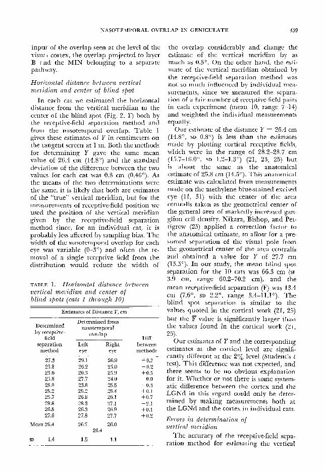
Horizontal distance bet ween vertical meridian a zd center of blind spot
In each cat we estimated the horizontal distance from the vertical meridian to the center of the blind spot (Fig. 2, Y) both by the receptive-field separation method and from the naso8temporal o,verlap. Table 1 gives these estimates of Y in centimeters on the tangent screen at 1 m. Both the metholds for determining Y gave the same mean value of 26.4 cm (14.8”) and the standard deviation of the difference between the two values for each cat was 0.8 cm (0.46”). As the means of the two determinations were the same, it is likely that both are estimates of the “true” vertical meridian, but for the measurements of receptive-field posi tioa we used the position of the vertical meridian given by the receptive-field separatio,n method since, fo’r an individual cat, it is probably less affected by sampling bias. The width of the nasotemporal overlap for each eye was variable (O-3”) and often the re- moval of a single receptive field from the distribution would reduce the width of
TABLE 1, Horizontal distance between vertical meridian and center of blind spots (cats 1 through IO)
Estimates of Distance Y, cm -
Determined from Determined
by receptive- nasotemporal
field overlap
separation Left Right DifI-
between method eye eye methods
27.3 29.1 26.9 +0.7 25.8 26.2 25.0 -0.2 25.6 26.3 25.9 + 0.5 25.8 27.7 24.0 0.0 25.0 23.8 25.5 -0.3 25.2 25.2 25.4 +0.1 25.7 26.8 26.1 +0.7 29.8 28.3 27.1 -2.1 26.5 26.3 26.9 +0.1 27.6 27.8 27.7 +0,2
Mean 26.4 26.7 26.0
SD 1.4 1.5 1.1
the overlap considerably and change the estimate of the vertical meridian by as much as 0.5’. On the o#ther hand, the esti- mate of the vertical meridian obtained by the receptive-field separation method was not so much influenced by individual mea- surements, since we measured the separa- tion of a fair number of receptive-field pairs in each experiment (mean 10, range 7-14) and weighted the individual measurements equally.
Our estimate of the distance Y = 26.4 cm (14.8”, SD 0.8”) is less than the estimates made by plotting cortical receptive fields, which were in the range of 28.2-28.7 cm (15.7-16.0”, SD 1.2-1.3”) (21, 23, 25) but is about the same as the anatomical estimate of 25.8 cm (14.5”). This anatomical estimate was calculated from measurements made on the methylene blue-stained excised eye (11, 31) with the center of the area centralis taken as the geometrical center of the general area of markedly increased gan- glion cell density. Nikara, Bishop, and Pet- tigrew (25) applied a correction factor to the anatolmicaI estimate, to allow for a pre- sumed separation o,f the visual pole frolm the geometrical center of the area centralis and obtained a value for Y of 27.7 cm (15.5”). In our study, the mean blind spot separation for the 10 cats was 66.3 cm (sr 3.9 cm, range 60.2-70.2 cm), and the
mean receptive-field separation (@) was 13.4 cm (7.6”, SD 2.2’, range 3.4~11.P). The blind spot separation is similar to the values quoted in the cortical work (21, 25)
but the F value is significantly larger thair the values found in the cortical work (21,
25) . Our estimates of Y and the corresponding
estimates at the cortical level are signifi- cantly different at the 2% level (Student’s t test). This difference was not expected, and there seems to be no o,bvious explanation for it. Whether or not there is some system- atic difference between the cortex and the LGNd in this regard could only be deter- mined by makin, u measurements both at the LGNd and the cortex in individual cats.
Errors in determination of vertical meridian
The accuracy of the receptive-field sepa- ration methold for estimating the vertical

460 K. J. SANI)ERSON AND S, M. SHERMAN
meridian is limited by two factors, namely a) the error induced by assuming symmetry of the two eyes, and b) the error in deter- mining the average receptive-field separa-
tion (F). As the errors are independent, the variance of the total error is equal to the sum of the individual variances. The recep- tive-field separation method assumes that the horizoatal distances from the center of the blind spot to the vertical meridian in each eye (Fig, 2, distances Y,, Y2) are the same in the one animal, but in fact they are often different (23, 25). We determined the vertical meridians independently for each eye from the nasotemporal overlap (Table 1) and found that the distribution of differences between the estimates of Y for the two eyes of each animal had a stan- dard deviation of 0.9”. As the asymmetry is a property oIf the two eyes taken together, the error in determining the po,sition of the vertical meridian in each eye by assuming symmetry has a standard deviation which is half the standard deviation of the asym- metry. Our estimate of the standard devia- tion of this error is therefore 0.45” and the probable maximum size of the error is 0.9”. Leicester (23) and Nikara, Bishop, and Pet- tigrew (25) also measured this error and obtained a slightly larger estimate of its size (1.2”).
The second source of error in the recep- tive-field separation method, namely the error in determining the average receptive-
field separation F, decreases towards zero as the s,ample of receptive-field separations becomes large. In this study the standard
deviation of the F distribution for each cat had a mean value of 0.83”, calculated from 102 measurements made in the 10 cats, and the standard error of the mean varied fro,m 0.08 to 0.56”, with an average value of 0.23”. The mean sampling error in determining
F for all the cats had a standard deviation
of 0.83O/fl = 0.26”, since the average number of receptive-field separations mea- sured per cat was 10. The probable maxi-
mum error in determining F for any one cat was therefore 0.52” (two standard devia- tions) and the corresponding maximum error in determining Y, which is half of tnt
error in F, since Y = (A - F)/Z, was 0.26”.
The total error in determining the posi- tion of the vertical meridian in each eye, made up of the error induced by assuming retinal symmetry and the sampling error in determining Y, had a standard deviation ranging from 0.45” to 0.53” in the 10 cats, with an expected value of 0.47” fo,r any one cat. Since the error induced by assuming symmetry had a standard deviatio,n of 0.45”,
any error in the determination of F in these experiments was of no significance and we concluded that the estimate of the position of the vertical meridian in each eye by the receptive-field separation method was ac- curate to Alo.
The overlap of receptive-field center points for cells recorded in layers A and A, in each experiment is sho,wn in Table 2. There were 20 distributions, for each eye, of receptive fields scattered art lnd the verti- cal midline, and in I6 of thes there was an overlap of center points for 1 Cts reco,rded in layers A and AI on the t’ 3 sides. The mean overlap of center points 3r cells from the two LGNd’s was 0.8’ and e maximum 3.0”. The overlap of receptiv fields them- selves was larger, as much a: 5” for some eyes. The amount of overlap ‘, clried greatly from one experiment to the next, probably because in any one cat only a small number of cells (say 10) were recorded from laminae A and AI, having receptive fields over the c vertical midline.
TABLE. 2. Width of lzasotem~oral overlap of recefitive-field center points of cells from laminae A and A, (rats I thyough 10)
Left Eye, deg
1.4 - -0.1
0.3 3.0 0.9 0.0 2.2 0.1)
-0.7 -0.2
Mean 0.7
0.8
Right Eye, deg _-- .- +---
0.1 0.3 1.2 I .(i 1.3 1.3
- 0.5 12.0 I A 0.7
0.9

NASOTEMPORAL OVERLAP IN GENICULATE 461
To give a better idea elf the overlap we have pooled the results of a11 experiments, the resulting distribution in Fig. 5 being of all the cells in laminae A and A, whose receptive-field center points fell over the vertical meridian. In Fig. 5 the overlap of receptive fields of cells from laminae A and A, has been shown as if they were all recorded in the left LGNd-hence the cen- ter points of their receptive fields lie in the left (wrong) visual hemifield. The maxi- mum overlap by any receptive-field center point beyond the vertical meridian was 3*5”, and for all the receptive fields in the wrong hemifield (total 94) the mean overlap was 0.44”. There was no difference between cells from lamina A and those from lamina A,. In Fig. 6 we graphed nasotemporal
I I
I I I I” 17" 2" 3"
Nasotemporal overlap
FIG. 6. Cumulative frequency diagram of the overlap distribution shown in Fig. 5.
overlap against the progressive total of receptive fields, so that for any given 08ver- lap value, say lo, the percentages of recep- tive-field center points overlapping the ver- tical midline by up to lo and by more than 1 O can be stated. Of the total of overlapping center points, 95% were within 1.7’ of the vertical midline, so one estimate of the overlap is 1.7O. The standard deviation of
Zero vertical meridian
l
Left hemifield
A -5” Zero horizontal
I-103 0 Q
FIG. 5. Distribution of receptive-field center points over the midline of cells recorded in laminae A and A, in all cats. All the receptive-
field center points are plotted as though they were from cells recorded in the left LGNd, so the over- lap is to the left of the vertical meridian.
the overlap was 0.76” treating the distribu- tion as normal, with mean 0’ and an over- lap distributed on both sides of the vertica1 midline. Hence one statistical estimate of the overlap of center points over the mid- line is 1.5” (2 standard deviations from the mean), and the width of the nasotemporal strip including receptive-field center points in both LGNd’s is approximately 3.0°, that is 1.5” on either side of the vertical meridian
Since the vertical meridian was deter- mined by the receptive-field separation method, the estimate elf the width of the nasotemporal strip must take account of the possible error in determination of the vertical meridian. The probable maximuti size of this error was found to be 0.94”, so the maximum error in determining the width of the strip of overlap is twice that value, namely l.9”. The minimum estimate of the width of the strip of overlap would therefore be 1.1 O, but this is almost cer- tainly too small since 9 o,f the 20 eyes studied (Table 2) had an overlap of recep- tive-field center po,ints from the two sides which was larger than this. It can be argued also that errors in determining the vertical

462 K. J. SANDERSON AND S. M. SHERMAN
meridian are just as likely to decrease the overlap as increase it, so we concluded that 3” is probably a good estimate o’f the width of the nasotempo,ral strip co#ntaining recep- tive-field center points from both lateral geniculate nuclei.
Projection of naso temporal overlap to lateral geniedate nucleus
The nasotemporal strip of visual field is represented in the main nucleus (LGNd) along its medial edge and in the immedi- ately-adjacent part of the MIN. In the main nucleus the representation o,f the overlap extends from the medial edge, where we recorded cells with receptive fields having center midlin
points e, to a
as far a region
-
35 0 .
abou over t he vertical
t 0.75 mm more lateral in the nucleus where cells with re- ceptive fields with azimuths elf -I-L5” are to bi found. Units with receptive-field center points in the wrong visual hemifield were found as far as 0.5 mm from the medial edge o,f the main nucleus, but it was on the border between the LGNd and the MIN that we observed cells having the greatest receptive-field. overlap across the vertical midline. In a previous histological study (30) no representation of the central retina was folund in layer B or the MIN, but in subsequent single-unit microelectro,de studies (22; unpublished observatiomns) the central area of the retina was found to project to all the layers, A, Al, and B of the LGNd and the adjoining part of the MIN.
In the MIN units with receptive fields across the vertical midline were found close to the border of the MIN next to the main nucleus. Further medial in the MIN we recorded units with receptive fields in the contralateral (“normal”) visual hemifield. Figure 7 shows the distribution of receptive fields of cells in lamina B and the MIN whose center points overlapped the vertical meridian. As in Fig. 5, the center points have been shown as if all the cells were recorded in the left LGN. We have not attempted to distinguish between units from lamina B and the MIN, partly be- cause the boundary separating these two regions was often rather uncertain histo- logically and partly because there was no obvious difference between the properties of receptive fields from the two regions. The mean overlap of center points across the vertical meridian was 3.8’, the maxi- mum was 36”, and all but one of the units were contralateral.
As we mentioned before, a large part of this overlap to layer I3 and the MIN pre- sumably comes from the scattered popula- tion of ganglion cells in the temporal retina which project contralaterally, that is to the wrong half of the brain. Kinston, Vadas, and Bishop (22) also reco#rded a co#nsider- able overlap in the MIN, but they found ipsilateral units as well as contralateral units with center points o,verlapping the vertical meridian. Ho’wever much of the overlap of ipsiIatera1 field centers which
Zero vertical meridian
FIG. 7. Distribution of receptive-field center points over the midline of ceiis recorded in lamina 13 almost all units are contralateral and the MIN in all cats. Note that
than that seen in laminae A and Al, and the overlap is much greater

NASOTEMPORAL OVERLAP 1;1J GENICULATE 463
they reported may have been due to errors in the determination of the vertical merid- ian, since they recorded from the lateral geniculate nucleus on one side only.
DISCUSSION
Exfzrimental detewrkation of uertical meridian
Since the cat does not have a specialized fovea, a number of different ways for deter- mining the vertical meridian in each eye have been developed for studies of visual receptive fields in this animal. Until re- cently, in many studies, the vertical merid- ian of each eye was inferred from the posi- tion of the blind spot-the horizontal distance between the vertical meridian and the center of the blind spot (distance Y) being taken as 16.0” (21). However, since the distance Y varies considerably between cats, in recent studies the vertical meridians in each cat have been estimated from the positions of receptive fields near the fixa- tion points. Leicester (23) and Blakemore (12) recorded from the projection of the vertical meridian to the cortex on both sides and took as their true vertical merid- ian the line down the center of the strip of bilateral representation for each eye. In other studies, however, where units only from the one side were recorded, it has been necessary to define the vertical merid- ian differently. Nikara, BishoIp, and Pet- tigrew (25) and Joshua and Bishop (21) defined the vertical meridian for each eye so that the binocular receptive fields of cortical neurons are, on the average, the same horizontal distance from the respec- tive vertical meridians.
In this study we recorded from the LGNd on both sides and determined the vertical meridian by two independent methods, from the nasotemporal overlap in each eye and by a receptive-field separation method adapted from the co’rtical work. However, the latter method has a more general appli- cation since it can be used also in experi- ments where cells are recorded from the LGNd on one side only. Just a few mea- surements of receptive-fielb separation are required to estimate the position of the vertical meridian, so we have calculated the accuracy of this estimate when the number
of measurements varies from 1 to 10. If the error due to asymmetry is taken to have a
standard deviation of 0.45” and the F dis- tribution in each cat a standard deviation of 0.83’, then the standard deviation of the error incurred when n measurements are made is given by the expression
I 0.422 s = I1
v( 0,452 + -
11 > degrees of visual angIe
The figure 0.42” is half the standard devia-
tion for the F distribution since this error is spread between the two eyes.
The accuracy in determining the vertical meridian for one measurement is 1.24’, for two measurements LOS”, and for three mea- surements 1.02? Further measurements of receptive-field separation only slightly in- crease the accuracy as there is a residual error of 0.90” due to the retinal asymmetry.
Width of nasotempornl owl-lap in wtina, LGNd, a72d cortex
Stone (29) studied the nasotemporal over- lap in the retina, and found that the strip of retina about 0.2 mm (0.9”) wide, strad- dling the vertical meridian, projected equa1Iy to both optic tracts. This overlap is much less than the overlap measured at the level of the LGNd and the co,rtex (see below), but Bishop (6) has interpreted Stone’s data as indicating that t-he width of the overlap in the retina is, in fact, greater than 0.9”. Further evidence that the overlap in the retina was underestimated comes from our results, as the overlap ob- served in the LGNd is presumably pro- jected via the optic tract on the same side.
We found that the center points of receptive fields belonging to cells recorded in layers A and A, extended up to 3.5” over the midline and the overlap of the body of each receptive field was about lo larger. Most of our experiments were concerned wit11 the part of the LGNd near the rep- resentation of the zero horizontal, but we recorded sufficient: cells with receptive fields having large positive or negative elevations to be fairly sure that the overlap projected to the LGNd consists of a strip of visual field parallel to the zero vertical meridian. Kinston, Vadas, and Bishop (22) also found an overlap proiected to the LGNd, as they

464 K. J. SANDERSON AND S. M. SHERMAN
recorded cells from the main laminae with receptive-field center points up to 2’ across the assumed vertical meridian. Ho:wever, they recorded from the LGNd on one side only and since they took the vertical merid- ian in each eye to be 28.2 cm (15.7”) from the center of the blind spo’t, it is likely that their measurements of overlap were often slightly in error.
The overlap at the level of the visual cortex has been studied by recolrding single units in both hemispheres along the 17/ 18 border of the visual cortex (12, 23) and also in experiments where units were recorded in the visual cortex on one side o,nly (21, 25). In the two hemisphere experiments the cells recorded near the projection of the vertical meridian had the general body of the receptive fields commonly up to 5” across the midline, and the range of recep- tive-field center points across the midline was 1.2” (23) and 1.5” (12). ln the experi- ments where only oIne cortex was studied the range of center points over the vertical meridian was fo#und to be 2.5” (25) and 2.9” (21), the larger overlap observed in these experiments being due perhaps to errors in the determinatio;n elf the vertical meridian for each eye. Other studies (2, 3, 14, 20) have provided evidence that the re- ceptive fields elf solme cortical cells overlap into the ipsilateral (wrong) visual hemifield.
There has been solme doubt about the origin of the bilateral projection o,f the central strip of visual field to the visual cortex, as the 17/ 18 border of the visual cortex receives its input both from the medial edge of the ipsilateral LGNd (16, 34) and from callosal fibers coming from the corresponding part o’f the 17/ 18 border in the opposite hemisphere (3, 14, 15, 19, 32, 33). In the cat probably the main co,n- tribution to the cortical receptive fields overlapping the vertical meridian is from the ipsilateral LGNd, since Leicester (23) found that section of the corpus callosum in one animal did not noticeably change the width of the overlap observed at the cortex and, in addition, the o#verlap we measured at the level of the LGNd is suffi- cient to account for most of the overlap of the cortical receptive fields, except per- haps the overlap of the edges and surround of the larger ones. The range of overlap
of the center po’ints of geniculate receptive fields is larger than the overlap of the cen- ter points of cortical receptive fields, but the maximum overlap of cortical field edges (6”) (12) is larger than the overlap of the body of any geniculate receptive field (4”) recorded from laminae A and Al. We did not inc1ud.e cells from lamina B and the MIN which had receptive fields overlap- ping the midline since must oif these prob- ably do not project to the primary visual cortex.
It can be concluded then that most of the cortical receptive fields which overlap the vertical meridian are built up from the concentric receptive fields o’f lateral genicu- late neurons, supplemented by an input from the cortical fields in the other hemi- sphere. This view is in accordance with the current theories concerning vertical midline receptive-field disparity and stere- opsis (6, 8). As we mentioned previously, it is the binocular cortical cells fro,m the 17/ 18 bo,rder region which are thought to be important for binocular depth percep- tion at the midline (6). Midline stereopsis requires that there be horizontal receptive- f’ield disparity at the midline, that is a receptive field situated precisely on the midline in olne eye should have its com- panion receptive field in the other eye situated on either side of the midline. The projection of a nasotemporal overlap to the cortex makes this possible, and in the two hemisphere experiments (12, 23) it has been shown that there are indeed cells with the receptive fields for the two eyes on either side o’f the midline. In the general theory of stereopsis, it is proposed that in addi- tio’n to there being receptive-field disparity, the two receptive fields oif a striate neuron should have precisely the same o#ptimal stimulus parameters so that they will re- spond to the same feature in the visual world (6-8). Presumably this will apply especially to the vertical meridian in the vicinity of the fixatioa point since this is a region of known high stereoacuity. It is not unreasonable to suggest that cortical receptive-field pairs situated near the mid- line will have basically the same kind o’f input to the two fields even if o’ne receptive field of the pair is across the midline. It is our contention therefore that the naso.
NASOTEMPORAL OVERLAP IN GENICULATE 465
temporal overlap, projected from the retina to the LGNd, for& the basis of the recep- tive-field disparity that is to be found on either side of the vertical meridian, at the level of the cortex. This receptive-field dis- parity, in turn, provides the basis for binoc- ular depth discrimination in the vicinity of the fixation point.
SUMMARY
By recording from single units in both lateral geniculate nuclei (dorsal nucleus, LGNd) in each of 10 cats, it has been shown that a strip of retina, straddling the vertical meridian, projects onto the medial edge of both nuclei. This nasotemporal overlap projects to the three main laminae (A 49 and B) of the LGNd and to the adjoining part of the medial interlaminar nucleus (MIN). The overlap seen in layers A and A1 is probablv derived from the I ten tral strip of retina which sends fibers equally to both optic tracts and is similar to the overlap seen in the cortex in the border region of areas 17 and 18, The cen- ter points of the receptive fields of cells recorded in layers A and A, overlapped the vertical meridian by up to 3.5’, the overlap of the body oaf each receptive field being about lo larger. The distribution of recep- tive-field center points over the midline had a standard deviation of 0.76”. The investi- gation was mainly concerned with central vision but also included the region near the vertical meridian for about 10” above and below the zero horizontal. The over- lap seen in layer B and the MIN was dif-
REFERENCES
1. BARLOW, H. B,, BLAKEMORE, C., AND PETTICREW, J. D. The neural mechanism of binocular depth discrimination. J, Physiol., London 193: 327-342, 1967.
2. BERLUCCHI, G., GAZZANIGA, M. S., AND RIZZO- LATTI, G. Microelectrode analysis of transfer of visual information by the corpus callosum. Arch. Ital. Biol. 105: 583-596, 1967.
3. BERLUCCHI, G. AND RIZZOLATTI, G. Binocu- larly driven neurons in visual cortex of split- chiasm cats. Science 159: 308-310, 1968.
4. BISHOP, G. H, AND CLARE, M. H. Organiza- tion and distribution of fibres in t-he optic tract of the cat. J. Cornp. Newel. 103: 269- 304, 1955.
5. BISHOP, P. 0. The nature of the representa- tion of the visual fields in the lateral genicu-
ferent from that projecting to layers A and A,, since most of the overlapping receptive fields were from the contralateral eye, and their center points extend up to 36” into the wrong hemifield. These cells presum- ably receive their input from the scattered ganglion cells in the temporal retina which project to the wrong optic tract, and prob- ably do not project to the cortex in the border region of areas 17 and 18 as no overlap of this kind has been found there.
For each cat the position of the vertical meridian was determined both from the midpoint of the nasotemporal overlap and also by a receptive-field separation method. The two estimates gave the same mean value for the 10 cats, the mean horizontal distance from the vertical meridian to the center of the blind spot being 14.EI”.
It is concluded that a large part of the nasotemporal overlap to be found in the visual cortex is projected from the lateral geniculate nucleus on the same side, The significance of the nasotemporal overlap for vertical midline receptive-field disparity and binocular depth discrimination is discussed.
ACKNOWLEDGMENTS
The authors thank Professor P. 0. Bishop for his help and advice during the course of this in- vestigation. The authors are grateful for the skilled assistance of Mr. Lionel Davies, Mr. R. Tupper, and the members of the technical staff. We are also grateful to Mrs. Eva Elekessy for help with the preparation of the figures and to Miss Marga- ret Wells and Mrs. Lyn Speight for much secre- tarial assistance. Toxiferine dichloride was kindly supplied by Roche Products Ltd., Sydney.
late nucleus. Proc. Australian Assoc. Neurologists 3: 15-25, 1965.
6. BISHOP, P. 0. Neurophysiology of binocular single vision and stereopsis. In: Handbook of Sensory Physiology, edited by R. Jung. Berlin: Springer, 1970, vol. 7.
7. BISHOP, P. 0. Beginning of form vision and binocular depth discrimination in cortex. In: The Neurosciences: Second Study Program, edited by F. 0. Schmitt. New York: Rockefeller Univ. Press, 1970, p. 471-485.
8. BISHOP, P. 0. AND HENRY, G. I-I. Spatial vi- sion. Ann. Rev. Psychol. In press.
9. BISHOP, P. O., HENRY, G. H., AND SMITH, C. J. Binocular interaction fields of single units in the cat striate cortex. J. Physiol., London. In press.
K. J. SANDERSON AND S. M. SHERMAN 466
10.
II.
12*
13.
14.
15.
16.
17.
18.
19.
20.
21.
BISHOP, P. O., KOZAK, W., LEVICK, W. R., AND VAKKUR, G. J, The determination of the pro- jection of the visual field on to the lateral geniculate nucleus in the cat. J. PhysioE., Lon- don 163: 503-539, 1962. BISHOP, P. O., KOZAK, W., AND VAKKUR, G. J. Some quantitative aspects of the cat’s eye: axis and plane of refercncc, visual field co- ordinates and optics. J. Physiol., London 163: 466-502, 1962. BLAKEMORE, C. Binocular depth discrimina- tion and the nasotemporal division. J. PhysioZ,, London 205: 471-497, 1969, BURROWS, G. R. AND HAYROW, W. R. The or- ganization of the thalamo-cortical visual path- ways in the cat. An experimental degeneration study. Brain, Beha-uior, Evolution. In psess. CHOUDHURY, B. P),, WHITTERIDCE, D., ANI) WIL- SON, M. E. The function of the callosal con- nections of the visual cortex, Quart. J. ExptZ. Physiol. 50: 214-219, 1965. ERNER, F. F. AND MYERS, R. E. Distribution of corpus callosum and anterior commissure in cat and raccoon. J* Conzp. Neural. 124: 353-366, 1965. GAREY, L. J. AND POWELL,, T. P, S. The pro-
jection of the lateral geniculate nucleus upon the cortex in the cat. Proc. Roy. Sot. London, Ser. B 169: 107-126, 1967. GLICKSTEIN, M., KING, IX. A., MILLER, J., AND BERKLEY, M. Cortical projections from the dorsal lateral geniculate nucleus of cats. J. Camp, Neural. 130: 55-76, 1967. HUBEL, D. H, AND WIESEL, T. N. Receptive fields, binocular interaction and functional architecture in the cat’s visual cortex. J. Physiol., London 160: 106-154, 1562. HUBEL, D. H. AND WIESEL, T. N. Receptive fields and functional architecture in two non- striate visual areas (18 and 19) of the cat. J. Neurophysiol. 28: 229-289, 1965. HUBEL, D. H. AND WXESEL, T. N. Cortical ~ntl callosal connections concerned with the vertccal meridian of visual fields in the cat. J. Ne~w- physiol. 30: 1561-1573, 1967. JOSHUA, D. E. AND BISHOP, P. 0. Binocular single vision and depth discrimination. Recep- tive field disparities for central and peripheral vision and binocular interaction on peripher;l I
22.
23.
24.
25.
26.
27.
28.
29.
30.
31.
32.
33.
34.
single units in cat striate cortex, Ex@ Brain Kes. 10: 389-416, 1970. KINSTON, W. J., VXDAS, M. A., AND B~SHOI’, P. 0. MuItiple projection of the visual field to the medial portion of the dorsal lateral ge- niculatc nucleus and the adjacent nuclei of the thalamus of the cat. J. Con+ Newel. 136: 295-316, 1969. LEICESTER, J + Projection of the visual vcrtica1 meridian to cerebral cortex of the cat. J. NC -ophysioE. 31: 371-382, 1968. n’xmvrr, K. AND SPRAGUE, J. M. Thalamo-corti- cal organization of the visual system in the cat. J. Camp. Nwrol. 138: 219-250, 1970. ~~IKAKA, T., Bxsnor, I?, O., AND PI’TTICREW, J. D. Analysis of retinal correspondence by studying reccptivc fields of binocular single units in cat striate cortex. Expt E. Brain Res. 6: 353-372, 1968. O’LEARY, J. L, A structural analysis of the lateral geniculate nucleus of the cat. J+ Corn@ hreuroZ. 73: 405-430, 1940. RODIECK, R. W., PFETTI~REW, J. D., BISHOP, P. O;, AND NIKARA, T. Residual eye movemcri ts in receptive field studies of paralyzed cats. Vision Res. 7: 107-110, 1967.
SENEVIRATNE, K. N. AND WHITTERIDGE, D. Vi- sual evoked responses in the lateral geniculate nucleus. EEectl-oe?zccphu~o~. Clin. hTeurophysiol. 14: 785, 1962. STONE, J. The nasotemporal division of the cat’s retina. J. Camp. h7ewoZ. 126: 585-600, 1966. STONE, J. AND HANSEN, S. M. The projection of the cat’s retina on the lateral gen’iculate nucleus. J. Corn@ Neural. 126: 601-624, 1966. VAKKUR, G. J., BISHOP, P. O., AND KOZAK, W. Visual optics in the cat, including posterior nodal distance and retinal landmarks. Vision Res. 3: 289-314, 1963. VESBAESYA, C., WHITTERIDGE, D., AND WILSON, M. E+ Callosal connexions of the cortex rep- resenting the area contralis. J. Physiol., Lon- don 191: 79-8OP, 1967. ~~ILSON, M. E. Cortico-cortical connexions of the cat visual areas. J. Amt. 102: 375-386, 1968. WIUON, M. E. AND CRAGG, B. G. Projections from the lateral gcniculate nucleus in the cat and monkey. J. Anat, 101: 677-692, 1967.