Nanosized Delivery of Radiopharmaceuticals - IAEA 2nd RCM...C-4 1 13) Nanosized Delivery of...
161
C-41 (May 13) Nanosized Delivery of Radiopharmaceuticals 2nd Research Coordination Meeting of the CRP 5–9 October 2015 Padua, Italy
Transcript of Nanosized Delivery of Radiopharmaceuticals - IAEA 2nd RCM...C-4 1 13) Nanosized Delivery of...

C-4
1 (
May
13)
Nanosized Delivery of
Radiopharmaceuticals
2nd Research Coordination Meeting of the CRP
5–9 October 2015
Padua, Italy

FOREWORD
The currently used therapeutic agents in nuclear medicine continue to pose medical challenges
mainly due to the limited uptake of radiocompound within tumour sites. This limited accumulation
accounts for the fact that current beta-emitting therapeutic nuclear medicine agents have failed to
deliver optimum therapeutic payloads at tumour sites. Actually, no other radiolabelled therapeutic
agent has been capable to get close to the remarkably high target accumulation that was demonstrated
by the long-lasting radionuclide I-131 in thyroid cancer. This means that metastases of several
different types of aggressive cancers cannot be controlled, making it difficult to treat cancer patients.
Therefore, new delivery modalities that result in (i) effective delivery of therapeutic probes with
optimum payloads, site specifically at the tumour sites, minimal/tolerable systemic toxicity, and (ii)
higher tumour retention, would bring about a clinically measurable shift in the way cancers are
diagnosed and treated.
Nanotechnology has the potential to bring about this paradigm shift in the early detection and
therapy of various forms of human cancers because radioactive nanoparticles of optimum sizes for
penetration across tumour cell membranes can be engineered through a myriad of interdisciplinary
approaches, involving teams of experts from nuclear medicine, materials sciences, physics, chemistry,
tumour biology and oncologists. Therefore, the overall approach which encompasses application of
nanoparticulate radioactive probes in combination with polymeric nanomaterials has a realistic
potential to generate the next generation of tumour specific theranostic nanoradiopharmaceuticals and
minimize/eliminate delivery and tumour accumulation problems associated with the existing
traditional nuclear medicine agents.
This CRP was formulated based on the conclusions and recommendations of a Consultants
Meeting (27−31 May 2013), and is utilizing the knowledge and expertise in synthesizing polymer
nanoparticles using radiation technologies developed under the framework of a completed CRP
“Nanoscale Radiation Engineering of Advanced Materials for Potential Biomedical Applications”.
The first RCM was held on 7–11 July 2014 in Vienna, Austria, where the participants reviewed
the relevant work being carried out in their respective institutions, as well as discussed and adopted the
work plan for the next period. The meeting report is available at: http://www-
naweb.iaea.org/napc/iachem/working_materials/RCM1-F22064_REPORT.pdf.
The second RCM was held on 5–9 October 2015 in Padua, Italy. The meeting report from that
event is this Working Material.
The IAEA wishes to thank all participants for their valuable contributions. The IAEA officer
responsible for this publication was Agnes Safrany of the Division of Physical and Chemical Sciences.

C-4
1 (
May
13)
CONTENTS
1. SUMMARY ...................................................................................................................... 3
INTRODUCTION .............................................................................................. 3 1.1.
1.1.1. Remaining Challenges ............................................................................ 3 1.1.2. Role of nanotechnology .......................................................................... 4 1.1.3. Research already in progress in Member States ..................................... 4
CRP OVERALL OBJECTIVE ........................................................................... 5 1.2.
1.2.1. Specific Objectives ................................................................................. 5 1.2.2. Expected research outputs ...................................................................... 5
REVIEW OF THE PROGRESS OF THE WORK IN INDIVIDUAL 1.3.
INSTITUTIONS ............................................................................................... 12
1.3.1. Argentina .............................................................................................. 12 1.3.2. Brazil ..................................................................................................... 12 1.3.3. Egypt ..................................................................................................... 13
1.3.4. Iran ........................................................................................................ 14 1.3.5. Italy (Milan) .......................................................................................... 14 1.3.6. Italy (Padova) ........................................................................................ 15 1.3.7. Italy (Palermo) ...................................................................................... 16
1.3.8. Malaysia ................................................................................................ 16 1.3.9. Mexico .................................................................................................. 17
1.3.10. Pakistan ................................................................................................. 17 1.3.11. Poland ................................................................................................... 17
1.3.12. Poland- POLATOM .............................................................................. 18 1.3.13. Singapore .............................................................................................. 19
1.3.14. Thailand ................................................................................................ 19 1.3.15. United States of America ...................................................................... 20
4. CONCLUSIONS AND RECOMMENDATIONS ....................................... 21 1.4.
2. PREPARATION OF ALBUMIN AND GOLD/ALBUMIN NANO-PARTICLES
BY RADIATION-INDUCED CROSS-LINKING ......................................................... 22
M. Grasselli, S. del Valle Alonso, E. Achilli, M. Sir, C. Flores
3. RADIO-INDUCED CROSSLINKING OF ALBUMIN NANOPARTICLES FOR
RADIOPHARMACEUTICALS DELIVERY SYSTEM ............................................... 31
A. Benévolo Lugão , G.H.C. Varca; R.G. Queiróz; L. Goulart; A. Geraldes; J.G. Batista; A. Silva; Jessica
Leal
4. RADIOSYNTHESIS, QUALITY CONTROL AND BIOLOGICAL STUDIES
OF NANOSIZED RESVERATROL-AU-198 ............................................................... 38
T. Hafez, T.M. Sakr, H.A. Abd El-Rehim K.K. Katti, L. Watkinson, T. Carmack, C.J. Smith, C.S. Cutler,
K.V. Katti,
5. DEVELOPMENT OF CHITOSAN-BASED NANOPARTICLES FOR
MONOCLONAL ANTIBODY RADIOPHARMACEUTICALS DELIVERY ............ 42
S. Shanehsazzadeh, A.R. Jalilian

6. RADIOLABELED NANOSIZED DELIVERY SYSTEMS FOR
THERANOSTICS OF PRIMARY AND METASTATIC COLORECTAL
CANCER ........................................................................................................................ 48
L.Melendez Alafort, A.Rosato G., Pasut, C. Bolzati, S. Mocellin, N.Salverese, D.Carpanese
7. CANCER-FIGHTING DIAGNOSTIC AND THERAPEUTIC NANOGELS .............. 59
C.Dispenza, M.A. Sabatino, N. Grimaldi, L. Ditta, E. Murugan, M. Jonsson
8. NANOSIZED DELIVERY SYSTEMS FOR RADIOPHARMACEUTICALS ............ 72
S.N.M. Janib
9. NANOSIZED DELIVERY SYSTEMS FOR RADIOPHARMACEUTICALS:
NANOSIZED RADIOLABELED POLYAMIDOAMINE DENDRIMERS FOR
TUMOR IMAGING AND TARGETED THERAPY .................................................... 78
B.E. Ocampo-García, G. Ferro-Flores, F.D.E.M. Ramírez De La Cruz, C.L. Santos-Cuevas, E.P. Azorín-
Vega, E. Orocio-Rodríguez
10. NANOSIZED DELIVERY SYSTEMS FOR RADIOPHARMACEUTICALS ............ 89
I.U. Khan, R. Zahoor, A. Shahid, F. Iram1,
11. NANOSIZED DELIVERY SYSTEMS BASED RADIOPHARMACEUTICALS
IN POLAND ................................................................................................................... 99
M. Maurin
12. POLYMER-BASED NANOCARRIERS FOR RADIOPHARMACEUTICALS ....... 107
P.Ulanski
13. DEVELOPING DELIVERY SYSTEMS FOR RADIOPHARMACEUTICALS........ 130
S.C.J. Loo
14. GREEN SYNTHESIS OF POLYSACCHARIDE AND POLYPEPTIDE-BASED
NANOPARTICLE TOWARD THE CREATION OF STABLE AND
FUNCTIONALIZED RADIOPHARMACEUTICS FOR DIAGNOSIS AND
THERAPY .................................................................................................................... 139
W. Pasanphan, T. Rattanawongwiboon, S.Wongkrongsak, T. Tangthong
15. IN VIVO STUDIES AND PRODUCTION OF NANOSIZED DELIVERY
SYSTEMS FOR RADIOPHARMACEUTICALS ....................................................... 153
K.V. Katti

1

2

3
1. SUMMARY
INTRODUCTION 1.1.
Nuclear medicine is an important medical specialty involving the application of
radioactive substances in the diagnosis and treatment of diseases. There are a number of
diagnostic and therapeutic radiopharmaceuticals that are FDA approved for use in human
patients. These radiopharmaceuticals, once administered to the patient, can localize to specific
organs or cellular receptors. This property of radiopharmaceuticals allows nuclear medicine the
ability to image the extent of a disease process in the body, based on the cellular function and
physiology, rather than relying on physical changes in the tissue anatomy. Nuclear medicine has
the ability to identify medical problems at an earlier stage than other diagnostic tests. Nuclear
medicine is often compared to "radiology done inside out", or "endo-radiology", because it
records radiation emitting from within the body rather than radiation that is generated by
external sources such as X-rays. The therapeutic effect of radiopharmaceuticals relies on the
tissue-destructive power originating from the emission of massive, short-range ionizing
radiation by some selected radionuclides. The combined use of a pair of diagnostic and
therapeutic radiopharmaceuticals for the treatment of the same disease constitutes the basic
paradigm of the field of theranostics as applied to nuclear medicine.
Currently used diagnostic PET and SPECT agents in Nuclear Medicine utilize
radiolabelled small-molecules, proteins and peptides as targeting vectors. On the therapy front,
β-emitting radionuclides conjugated with tumor specific peptides or monoclonal antibodies, are
routinely used to ablate tumours and metastatic lesions. Only a small number of small-molecule
radiolabelled compounds are routinely employed in therapy mostly for pain palliation of
metastatic bone cancer. There exist very few examples of radionuclides administered under a
simple ionic form; the most widely employed being I-131 injected as iodide salt for the
treatment of thyroid cancer. Strontium-89 is employed as Sn2+
ion for treating metastatic bone
cancer and, recently, the alpha-emitting 223
Ra as Ra2+
ion has been approved for the same
indication.
1.1.1. Remaining Challenges
The currently used therapeutic agents in nuclear medicine continue to pose vexing
medical challenges mainly due to the limited uptake of radiocompound within tumour sites.
This limited accumulation accounts for the fact that current beta-emitting therapeutic nuclear
medicine agents have failed to deliver optimum therapeutic payloads at tumour sites. Actually,
no other radiolabelled therapeutic agent has been capable to get close to the remarkably high
target accumulation that was demonstrated by the long-lasting radionuclide I-131 in thyroid
cancer. This means that metastases of several different types of aggressive cancers cannot be
controlled, thus resulting in propagation of cancers to various other organs including bone,
making it difficult to treat cancer patients. These challenges have resulted in minimal or no
improvement in the quality of life for cancer patients. 177
Lu-AuNPs conjugated to different
peptides have been proposed as a new class of theragnostic radiopharmaceuticals. These
radioconjugates may function as diagnostic molecular imaging agents, radiotherapeutic systems
and thermal-ablation nanodevices. Therefore, new delivery modalities that result in (i) effective
delivery of therapeutic probes with optimum payloads, site specifically at the tumour sites, with

4
minimal/tolerable systemic toxicity, and (ii) higher tumour retention, would bring about a
clinically measurable shift in the way cancers are diagnosed and treated. Such oncological
advances in targeted delivery of radioactive therapeutic probes would provide significant
benefits to patient population worldwide.
1.1.2. Role of nanotechnology
Nanotechnology has the potential to bring about a paradigm shift in the early detection
and therapy of various forms of human cancers because radioactive nanoparticles of optimum
sizes for penetration across tumour cell membranes can be engineered through a myriad of
interdisciplinary approaches, involving teams of experts from nuclear medicine, materials
sciences, physics, chemistry, tumour biology and oncologists. In particular, intervention of
nanotechnology in nuclear medicine is poised to offer tremendous benefits in site-specific
delivery of both diagnostic and therapeutic probes site specifically at tumour sites. This is
possible because gamma and beta emitting isotopes can be converted into their corresponding
nanoparticles. The sizes of diagnostic and therapeutic probes can be engineered to be within the
10–50 nm range so that they can penetrate tumour cells (which are 5–10 microns in size) to
provide optimum diagnostic and therapeutic payloads for efficient diagnostic imaging and
therapy.
Radiation induced production of polymeric nanoparticles using both the natural and
synthetic polymers and proteins in combination with theranostic radioactive Au-198/199 (and
other beta and gamma emitting radionuclides of Re, Sm, Rh etc.) would afford
nanoradiopharmaceuticals within the 30–50 nm range. This size range would allow facile
penetration of diagnostic and therapeutic payloads across tumour vasculature to achieve (i)
effective delivery of optimum diagnostic/therapeutic payloads accompanied by
minimal/tolerable systemic toxicity, and (ii) higher tumour retention using homogeneously
dispersed diagnostic/therapy agents. Therefore, the overall approach which encompasses
application of nanoparticulate radioactive probes in combination with polymeric nanomaterials
has a realistic potential to generate the next generation of tumour specific theranostic
nanoradiopharmaceuticals and minimize/eliminate delivery and tumour accumulation problems
associated with the existing traditional nuclear medicine agents.
In this context, nanostructured antibodies when tagged with nanostructured
diagnostic/therapeutic probes may also offer significant benefits over conventional radio-
immunotherapy, because nanoparticulates can penetrate tumour vasculature and being retained
by the tumour mass more efficiently than traditional antibody labelled radiopharmaceuticals.
1.1.3. Research already in progress in Member States
Within the framework of successfully completed CRP “Nanoscale Radiation Engineering
of Advanced Materials for Potential Biomedical Applications” considerable progress has been
made in the Member States in synthesizing polymer nanoparticles using radiation technique.
These new nanomaterials include, among others, protein nanoparticles, nanogels of various
physical and chemical properties, as well as nanoparticles based on polysaccharides (Argentina,
Brazil, China, Egypt, Hungary, India, Italy, Korea, Malaysia, Poland, Serbia, Thailand and
Turkey). These approaches were aimed at precise control of the structure, size, shape and
functionality of the nanoscale products. In some cases, procedures were developed for preparing
“hybrid” products, e.g., where the surfaces of nanoparticles and nanogels were decorated with
functional polymers and biomolecules. It will be very valuable to build up on this accumulated

5
knowledge and experience for creating new products where radiation-engineered nanoparticles
are the basis of new generation of diagnostic and therapeutic systems for use in nuclear
medicine.
This CRP was formulated based on the conclusions and recommendations of a
Consultants Meeting (27−31 May 2013), and is utilizing the knowledge and expertise in
synthesizing polymer nanoparticles using radiation technologies developed under the
framework of a completed CRP “Nanoscale Radiation Engineering of Advanced Materials for
Potential Biomedical Applications”. It is expected to result in new nanoradiopharmaceuticals,
nanoparticles capable of forming stable bonds with diagnostic and therapeutic radioisotopes,
and with tumor specific biomolecules and proteins (including monoclonal antibodies) leading to
well-defined delivery devices.
CRP OVERALL OBJECTIVE 1.2.
The overall objective of this CRP is to exploit the unique properties of materials at the
nanometer scale for developing nanocarriers of radioactivity capable of selectively targeting and
penetrating cancerous cells.
1.2.1. Specific Objectives
To synthesize polymeric (both synthetic and natural) nanoparticles capable of forming in
vitro and in vivo-stable bonds, through chelate interactions, with diagnostic and therapeutic
radioisotopes—for the creation of new generation of nanoparticle-based tumour specific
radiopharmaceuticals.
To formulate tumour specific proteins (including monoclonal antibodies) and generate
well-defined nanoparticles, through radiation methods, for use in conjugation reactions
with diagnostics and therapeutic radioisotopes (Tc-99m, Re-186/188)—for the generation
of nanosized tumour specific radiopharmaceuticals.
To formulate nanoparticles derived from inherently diagnostic/ therapeutic radioisotopes
(Au-198, Au-199, Re-186/188, Rh-105) and to conjugate them with tumour specific
peptide(s)—in order to generate radioactive nanoradiopharmaceuticals for diagnostic
imaging and therapy.
To develop a functional in vivo platform for efficient testing of various radiolabelled
nanoconstructs using animal models that mimic human tumours—all aimed at clinical
translation of diagnostic/therapeutic efficacy from animal for human applications.
1.2.2. Expected research outputs
Polymeric (synthetic and natural) nanoparticles for the creation of new generation of
nanoparticle-based tumour specific radiopharmaceuticals.
Protocols for conjugation reactions with diagnostics and therapeutic radioisotopes (Tc-
99m, Re-186/188)—for the generation of nanosized tumour specific radiopharmaceuticals.
Nanoparticles derived from inherently diagnostic/ therapeutic radioisotopes (Au-198, Au-
199, Re-186/188, Rh-105) and conjugated with tumour specific peptide(s).
Normalized in vivo platform for efficient testing of various radiolabelled nanoconstructs
using animal models that mimic human tumours—all aimed at clinical translation of
diagnostic/therapeutic efficacy from animal for human applications.

6
The first Research Coordination Meeting (RCM) was held in Vienna, on 7–11 July 2014
and was attended by 14 participants and 3 external observers coming from thirteen different
countries. During the meeting, presentations were given by all participants showing the main
results of their research activity in the development of new nanomaterials and in their
applications to the design and production of novel diagnostic and therapeutic
radiopharmaceuticals. The meeting report is available as Working Material at: http://www-
naweb.iaea.org/napc/iachem/working_materials/RCM1-F22064_REPORT.pdf.
The second RCM was held in Padua, Italy, on 5–9 October 2015, attended by all chief
scientific investigators and numerous external observers (the list of participants is given at the
end of this publication). The participants gave a thorough account of the progress made since
the first RCM, discussed the challenges encountered and ways to overcome them, and agreed
upon the road map for the next 18 months. Table 1.1. summarizes the achievements since the
first RCM as reported by participants, as well as the work planned for the next period.
TABLE 1.1. SUMMARY OF THE ACHIEVEMENTS AND FUTURE WORK OF PARTICIPANTS
Country
Achievements
Future work
ARGENTINA Synthetized Albumin and
Gold/Albumin NPs (by radiation-
induced crosslinking) with
Dh=20–80 nm, colloidal stability
in aqueous solutions, and
redispersablility from freeze-
dried form (Albumin NPs).
Developed protocols for
conjugation with DOTA-BBN
peptide.
Further characterisation by
MS (collaboration required)
and SLS (in progress).
Quantification of ligands in
the NPs (in progress).
BRASIL Synthetized solvent-free Albumin
nanoparticles as PBS buffer
dispersion and lyophilized solid
with sugar with controlled the
size (6 to 16 nm) and narrow size
distribution.
Synthetized papain nanoparticles
(as dispersion in PBS and
lyophilized with cyclodextrin)
with controlled size (5 to 20nm.
Further development of the
production of laboratory
amount of Au–198 and Au-
199 NP conjugated with
CD163 mimic peptide;
galectin-4 mimic peptide; and
Au-198 NP with mangoferin
and other regional extracts
EGYPT Resveratrol Au-198 nanoparticle
(RES-Au-198NP) had been
prepared with high
radiochemical purity (97%) in
Cooperate with Prof. Dr
Wanvimol for labeling her
prepared nanoparticles using
Tc-99m and test them for in

7
reproducible batches. RES-Au-
198NP showed stability up to
10days in serum.
RES-Au-198NP showed high
accumulation and retention in
tumor, 252.11±97 %ID/g at
30min, which increased to
378.9±60.99 % ID/g at 24h with
no uptake of NP, was observed in
other organs confirming the
successful targeting of the
therapeutic payload to the tumor
with minimal toxic side effects.
vitro stability and do some
biodistribution studies.
Cooperate with Prof Dr
Kattesh and Prof Dr Ademar
for preparation of Au-198 NP
and its biodistribution studies.
Prepare PVP-I-131 NP
IRAN We tried some different iron
oxide nanoparticles(NPs)
(USPIO) and done the
biodistribution studies
The Dextran Coated NPS usually
taken up with RES (more than
80 %ID/g)
Although we get good and
promising results by Dextran
coated NPs in in-vitro but the in-
vivo results were not promising
and disappointing
Now we are conjugated different
size of PEGylated NPs with
BBN and we are ready to
radiolabeled them with 68
Ga
We Conjugate and make this
probe BBN-NODA-Ga-
USPIO
We are ready to run in-vitro
and in-vivo tests
ITALY-MILAN Development of a new conjugate
of human albumin and p-SCN-
Bn-DOTA for radioguided
occult lesion localization in
breast cancer (ROLL)
radiolabeled with radionuclides
showing t1/2 > 99m
Tc.
Preclinical evaluation of a new
Bombesin Analogue with 177Lu
(Cooperation with Saudi
Arabia).
The labeling of DOTA-
Bombesin nanoparticles with 177
Lu and 111
In .
The Quality Control of
labeled-nanoparticles by
different methods (ITLC,
HPLC, SE-HPLC).
The stability studies in human
serum.
ITALY-PADOVA Preliminary binding tests (flow
cytometry) analysis on naked
and bombesin-functionalized
HSA/BSA NP and Au-
HAS/BSA NP from Argentina
177Lu-labelled anti-PSCA and
Design and develop
multifunctional Radiolabeled
Nanosize Delivery Systems
(RNDS) with defined
chemical structures, in which
the heterobifunctional PEG

8
antimesothelin using DOTA as
chelating agent were efficiently
obtained; they were found to be
stable able to detect efficiently
pancreas carcinoma cells in
vitro.
177Lu-DOTA-mAbs conjugates
can be attached to the gold NP
surface.
will be conjugated suitable
coordinating system for
radiometals, and GE11
dodecaptide selective for
EGFR overexpressed in CRC
cells.
Perform 99m
Tc-labelling, by
means of 99m
Tc-HYNIC and
[99m
Tc(N)(PNP)]-
technologies, of several NP
(sent us by the various units),
both naked and Bombesin-
functionalized. The NP must
be adequately functionalized
with suitable chelating
systems for both labelling
technologies.
Check the in vitro stability of
the radiolabeled NPs.
Perform cellular uptake
assays of the radiolabeled NPs
ITALY-PALERMO Synthetized a family of carboxyl
functionalised PVP nanogel
particles (by e-beam radiation
processing) with Dh in the 20 -
150 nm diameter range
(PDI=0.2-0.3), negative surface
charge density (-20 - -40 mV),
colloidal stability in aqueous
solutions upon storage at 5°C
and 37°C up to three months,
redispersability after freeze-
drying, biocompatibility and
hemocompatibility.
Developed protocols for
conjugation with BBN, DOTA-
BBN peptides and DTPA.
Expand the library of
available nanogel particles
(simpler manufacturing),
improve methods for
quantification of available
functional groups and
conjugation degree, and test
thermal stability up to 85°C.
MALAYSIA Synthesized and characterized
epoxidized palm olein (EPOo) as
a palm oil based precursor for
polymeric drug vehicles.
Prepared and characterized
nanosized gels from
polyethylene glycol diacrylate
(PEGDA), N-
isopropylacrylamide (NIPAAM)
Functionalization of the
prepared nanogels with
bombesin and performing
subsequent in vitro
evaluation.

9
and vinylpyrollidone (VP). Drug
encapsulation and release was
evaluated.
MEXICO In this period two nanosized
multimeric systems for
neuroendocrine tumor imaging, 99m
Tc-PAMAM-Tyr3-Octreotide
and 99m
Tc-AuNP-Tyr3-Octreotide
were prepared and evaluated in
vitro uptake in SR-positive
AR42J cancer cells as well as
their biodistribution profile in
athymic mice bearing AR42J
tumors.
In vitro binding studies were
carried out in AR42J cancer
cells. Biodistribution studies
were accomplished in athymic
mice with AR42J-induced
tumors with blocked and
unblocked receptors. Both
radiopharmaceuticals
demonstrated properties suitable
for use as target-specific agents
for molecular imaging of tumors
that overexpressed SR.
To evaluate in vitro and in
vivo PEGMA-DCWSCS-
DOTA-BBN (Thailand).
PAKISTAN Establishing Radiolabeling and
Quality Control of Nanoparticles
with Au-198 and Lu-177
Imaging and Biodistribution
Studies with Au-198 and Lu-177
Optimizing the radiolabeling
and imaging protocols with
further nanoconstructs in
collaboration other members
of CRP group.
POLAND-LODZ Synthetized gold nanoparticles
by radiation-induced reduction
of chloroaurate ions in the
presence of chitosan, zeta
potential >+30 mV, Au core
diameter ca. 10–12 nm,
hydrodynamic diameter 12–37
nm.
Synthetized nanogels of
poly(acrylic acid) – PAA (via
intramolecular cross-linking of
linear PAA initiated by pulse
irradiation with electron beam)
with radius of gyration in the
range 40–90 nm, colloidal
stability in neutral aqueous
Further model tests on
functionalization of PAA
nanogels – optimization of the
coupling procedure:
Sub-stoichiometric ratios of
coupling agents
Targeting low degree of
coupling (below 1%)
Reproducibility.
Training in BBN-DOTA
quantification procedures
Establishing and testing
purification procedures
Coupling of BBN-DOTA
with PAA nanogels,

10
solutions and redispersability
after freeze-drying
Developed two protocols to link
an amino-containing model
molecule (tryptamine) to the
PAA nanogels. Both of them led
to conjugated products, albeit the
control on the conjugation
degree is still to be achieved.
characterization
POLAND-
POLATOM
There are no outcome from the
cooperation with another
participants of the RCM. In the
Palatom the AGuIX
nanoparticles polisiloxan core
with DOTA chelators where
studied. The 90Y, 177Lu
radiolabeling protocols were
established and in vitro and in
vivo properties of functionalized
particles with TATE peptide and
native particles were established.
Participation in the IAEA project
– fields of cooperation
The Polatom can cooperate in
field of radiopharmaceutical
study.
1. Labelling of nanoparticles
with 68Ga, 90Y, Lu177,
99mTc
2. Labelling and characterization
of particles with “cold”
metals Ga, Y, Lu and 99Tc.
3. Stability study of labelled
samples
4. Study on radiochemical purity
5. Preliminary in vivo study on
healthy rats
The other filed of cooperation is
the activation of gold and
preparation of radioactive
gold nanoparticles (protocol
from Kattesh Katti).
SINGAPORE Synthesized PLGA-NH2
nanoparticles of sized ~200nm.
Conjugated DOTA-BBN onto
PLGA-NH2 nanoparticles of
sized 200nm.
Continue to produce these
nanoparticles for animal
studies
Optimize the conjugation
protocol of DOTA-BBN to
NPs
THAILAND Synthetized amphiphilic core-
shell polyethyleneglycol
monomethacrylate-grafted-
deoxycholate water soluble
chitosan NPs (PEGMA-
Further characterisation of
AuNPs in WSSF-DOTA-
BBN in terms of
hydrodynamic size.

11
DCWSCS NPs) with a bimodal
size distribution (Dh1 = 90 and
Dh2=664 nm) and no surface
charge.
Developed protocols to
conjugate the PEGMA-
DCWSCS NPs with DOTA-
BBN (hydrodynamic size after
conjugation Dh1=122 nm (15%)
and Dh2= 836 nm (85%)).
Synthetized water soluble
chitosan nanoparticle (WSCS
NPs, 192 nm, 1.49 mV) and
water-soluble silk fibroin
nanoparticle (WSSF NPs, 284
nm, -11.3 mV)
Developed protocols to
conjugate WSCSNPs and WSSF
NPs with DOTA-BBN.
Note: These types of conjugated
water-soluble natural polymers
will be used as reducing,
stabilizing and targeting
compound for radioactive 198
AuNPs production. The size
of the non-radioactive AuNPs
produced in WSCS-DOTA-BBN
and WSSF-DOTA-BBN are 86
nm (+22.89 mV) and 257 nm (-
12 mV), respectively.
Further development of
synthetic protocols for
reducing particle size.
Further development of
protocols for quantification of
conjugated DOTA-BBN.
USA Detailed In vivo toxicity profiles
of Gum-Arabic functionalized
Au-198 nanoparticles fully
established. Results published in
a peer reviewed journal;
Theranostic efficacy studies of
EGCG-Au-198 nanoparticles for
treating prostate cancer (in dogs)
and osteosarcoma (in guinea
pigs) completed.
Future studies for the coming
year (2015-2016) include
completion of theranostic
efficacy of Mangiferin –Au-
198 gold nanoparticles in
mice and dogs.

12
The summary of the work reported at this meeting is given in the alphabetical order of the
participating Member States. Only the recent progress is highlighted; the general background is
available in the Meeting Report of the 1st RCM.
REVIEW OF THE PROGRESS OF THE WORK IN INDIVIDUAL 1.3.
INSTITUTIONS
1.3.1. Argentina
Achievements
During the first period a novel gold/Albumin-NPs (core/shell) have been prepared by
radiation-induced crosslinking. NPs in the range of 60 to 80 nm have been characterized by
using UV-Vis, DLS (Dynamic Light Scattering), TEM and AFM. In addition FT-IR
spectroscopy, Zeta potential, dot-blot (antibody recognition) and available thiol groups have
been determined in the nanostructured material. Centrifugation method and Size Exclusion
Chromatography have been compared for NP purification. The chromatographic technique
shows a better purification performance, reducing the NP aggregation.
Decoration of Albumin-NPs and gold/Albumin-NPs with DOTA-Bombesin derivative
(DOTA-BBN) has been achieved by using a hetero-bifunctional linker. In a first step, DOTA-
BBN-linker was prepared and characterized by ESI-Mass spectroscopy. In a second step, this
compound was linked to the NPs by the free thiols available on NPs. A novel technique, based
on Time-Resolved Fluorescence, was analysed as a potential method to quantify the DOTA-
BBN moiety linked to the NPs.
NP samples were sent to Italy-Padova and Pakistan for biological analysis. Also a
collaboration work has been done with Brazil to develop a protocol to prepare novel
198/199Au/Albumin NPs.
Work plan for the next period
It is proposed for the next period to improve the purification method of NPs in order to
obtain samples free of other proteins. It will be performed Static Light Scattering measurements
and ligand quantification by fluorescence techniques. Others characterization techniques, such
as Mass spectroscopy will be done through collaboration work. NP samples will be prepared for
other participants according to the proposed collaborative research work. NP internalization will
be study by in-vitro cell cultures.
1.3.2. Brazil
GENERAL GOAL
The general goal is to develop process free from solvents and free from shear and high
pressure for the production of protein nanoparticles with size control based on the recently
discovered radiation-induced technique for potential loading of chemo and
radiopharmaceuticals. Alternatively, a platform for the synthesis of gold nanoparticles as drug
carriers will also be synthesized.
ACHIEVEMENTS
13
Development of the capability for controlling the size and size distribution and
large scale production of protein nanoparticles: The radiation-induced synthesis of
Albumin nanoparticles led to the formation of protein based nanoparticles with size
ranging from 6 to 68 nm. Scale up experiments revealed that the radiation induced
technique may be carried out under a wide protein concentration range with minimum
size variation, for both BSA and papain.
Development of the capability for production of large amount of gold and
radioactive gold nanoparticles: The synthesis of gold nanoparticles was developed by
the use of green technology, through the stabilization with natural gums and
compounds. Irradiation was also applied for the process. The radioactive nanoparticles
were produced in the neutron reactor. Particle size ranged from 20- 70 nm.
FUTURE WORK PLAN
Encapsulation of Yttrium by protein nanoparticles including, encapsulation stability and
crosslinking of new peptides for radiopharmaceuticals encapsulation;
Protein crosslinking onto gold nanoparticles surface.
COLABORATIONS
This work is being performed in collaboration with distinct institutes in Brazil -
Universidade Federal de Uberlândia - UFU and IPEN/CNEN-SP, with dr. Luis Goulart (UFU –
MG) and dr. Rosemeire Silva from the Heart Institute – FMUSP. International collaboration is
being performed by technical visits from Prof. Kattesh Katti, dr. Menka Khoobchandani -
Missou.Univ. USA, dr. Tamer Sakr (Egypt), dr. Piotr Ulansky, dr. Slawomir Kadlubousky
(IMTR- Lodz, PL) and dr. Mariano Grasselli (UNQ – Ar)
1.3.3. Egypt
Main objective:
The preparation of nanosized Resveratrol-198
Au nanoparticles (RES-198
AuNPs) in high
radiochemical purity. RES-198
AuNPs are of interest for treatment of neuro degenerative diseases
including rheumatoid and osteoarthritis and cancer.
Summary:
Resveratrol-198
AuNP Preparation and QC:
About 0.5mg Resveratrol was dissolved in 0.3ml absolute ethanol. Next 1477.5µg of gold
(198
Au + NaAuCl4) followed by 1.2ml Milli Q water were added while stirring for 2h until a
dark ruby red color was obtained. The UV-Vis and radiochemical purity were assayed and the
pH was adjusted to pH 7–7.5. The in-vitro stability of the RES-198
AuNP was tested. After
complete decay, the particle core diameter was checked using TEM. Normal female CF1 mice
were injected via the tail vein with 100µCi in 100µL and euthanized at the following time points
30min, 1h, 2h, 4h and 24h. Organs were harvested, weighed and counted with standards to
calculate the % ID/g and % ID/organ.

14
Results:
RES-198
AuNP were successfully prepared with high radiochemical purity (97%) in reproducible
batches. They were stable out to 10 days in serum. Its λmax was at 540nm confirming their
formation. The TEM showed a particle core diameter of 20–50nm. The biodistribution study in
normal CFI female mice showed liver and spleen initial average uptake of 55.63±4.48 and
63.95±26.66 %ID/g at 30min which increased to 61.47±8.14, and 69.8±33.12 %ID/g at 24h
respectively, (Fig 1.1) with nearly no uptake in the other organs. While blood showed 1.61±0.49
%ID/g at 30min decreasing to 0.61±0.37 %ID/g at 24h confirming the in-vivo stability of RES-198
AuNP. The biodistribution study in PC-3 tumor bearing SCID mice model showed high
accumulation and retention of RES-198
AuNPs in tumor, 252.11±97 %ID/g at 30min which
increased to 378.9±60.99 %ID/g at 24h, Figure (1.3). Nearly no uptake of NP was observed in
other organs confirming the successful targeting of the therapeutic payload to the tumor with
minimal toxic side effects.
Conclusion:
RES-198
AuNP is a new nanosized radiopharmaceutical with potential applications in
radiosynovectomy agent and/or as a radiotherapeutic agent for various cancers.
Acknowledgment:
This research was supported by IAEA and MURR.
1.3.4. Iran
Highlights of our works:
We tried some different iron oxide nanoparticles(NPs) (USPIO) and done the
biodistribution studies
The Dextran Coated NPS usually taken up with RES (more than 80 %ID/g)
Although we get good and promising results by Dextran coated NPs in in-vitro but the
in-vivo results were not promising and disappointing
Now we are conjugated different size of PEGylated NPs with BBN and we are ready to
radiolabeled them with 68
Ga
1.3.5. Italy (Milan)
Achievements
The 177
Lu-labeling procedure of the new bombesin analogue (DOTA-Glu-BNU), provided
through the collaboration with Saudi Arabia, was optimized at different Specific Activities and
then the stability of the compound with the greater RCP value was checked in serum up to 24
hours. The Radiochemical purity of the 177
Lu-Bombesin was found greater than 95% for the
compounds labelled at As 0.05 and 0.1 mCi/g. In addition high stability and higher affinity for
cancer cells was observed. This seems to be coherent with the goal of this CRP in order to
achieve a Bombesin-nanoparticle compounds useful to label with different radionuclides.

15
Future Plans
Development of a new nanopharmaceuticals for lymphoscintigraphy (LS) with targeting
properties in order to identify, during the radioguided LS, not only the sentinel node but
also any other diseased lymphonodes.
Perform the labeling of a new DOTA-Bombesin analogue and in vitro stability studies,
after the suppling by the Agency.
Test any new DOTA-Bombesin nanoparticles which may become available through the
CRP network.
1.3.6. Italy (Padova)
Flow cytometry analysis of albumin nanoparticles.
Flow cytometry analysis was carried out on 2 cancer cell line expressing bombesin receptors
MDA-MB-231 and PC3. Interaction of nanoparticles with tumor cells was evaluated by
incubation of 3×10⁵ cell with medium containing BODIPY-labeled albumin nanoparticles
(naked and functionalized with bombesin) at different dilutions for 2 h at 37°C. Then cells were
washed and analyzed by flow cytometer. Interaction of nanoparticles with cells was also
evaluated over time. Samples of 3×10⁵ cell containing a protein concentration of 100 µg/ml
BODIPY-labeled nanoparticles were incubated at 37°C at different time points and analyzed.
Flow cytometry analysis carried out after 2 h incubation of MDA-MB-231 and PC3 cells with
naked and bombesin-functionalized showed almost no fluorescence difference between different
protein concentration samples (from 20 to 100 µg/ml). Also minimal difference was found
between naked and functionalized-NP. Interaction of NP with cancer cell studied over a large
period of time, showed statistical difference between naked and functionalized NP just after 4 h
incubation.
Preparation and characterization of AuNP-177
Lu-DOTA-AM and AuNP-177
Lu-
DOTA-APSCA
Au-NP were functionalized with two new monoclonal antibodies (mAbs) synthesized by our
group, anti-prostate stem cell antigen (APSCA) and anti-mesothelin (AM) directed to PSCA and
mesothelin antigens respectively, which are heavily overexpressed in pancreatic
adenocarcinomas but absent in normal pancreas and in chronic pancreatitis
DOTA-mAb conjugates prepared by incubating a mAb solution with p-SCN-Bz-DOTA were
purified and analyzed. Results obtained after size exclusion HPLC analysis proved the
formation of DOTA-mAb conjugates and matrix-assisted laser desorption ionization mass
spectroscopy (MALDI-MS) analysis indicated an average number of 6 and 5 DOTA molecules
per each APSCA and AM antibody respectively. Binding analysis of both DOTA-APSCA and
DOTA-AM conjugates demonstrated that mAbs don’t lose the affinity for the receptors after
DOTA-conjugation. DOTA-mAbs conjugates were labelled with the 177
Lu with high efficiency
and demonstrated to be stable for more than 24 h. Both 177
Lu-DOTA-mAbs showed great
stability after dilution and no transchelation of 177
Lu from the labelled immunoconjugates to
CaDTPA even after 3 days of incubation.

16
AuNPs were functionalized with both 177
Lu-DOTA-labelled AM and APSCA mAbs, and
assessed by UV-Vis spectroscopy. AuNP-177
Lu-DOTA-mAbs were characterized in size and
shape by transmission electron microscopy (TEM), and particles were also measured using a
particle size and Z-potential analyzer based on dynamic light scattering (DLS). UV-Vis spectra
of both AuNP-177
Lu-DOTA-mAbs showed a characteristic shift of the surface plasmon
resonance band (524 nm), with respect to that of AuNPs surface (519 nm), due to the antibody
adsorption, thus demonstrating AuNPs functionalization. TEM micrographs of both AuNP-
177Lu-DOTA-mAbs showed 20 nm spherical nanoparticles. When nanoparticles were
functionalized using molar ratios 1:4 (AuNP:177
Lu-DOTA-mAbs) DLS analysis showed a mean
hydrodynamic diameters of 63 nm and 57 nm for AuNP-177Lu-DOTA-AM and AuNP-177Lu-
DOTA-APSCA respectively.
1.3.7. Italy (Palermo)
Poly-N-vinyl pyrrolidone-co-acrylic acid nanogels have been synthetized and simultaneously
sterilised by e-beam irradiation starting with a polymer/monomer aqueous solution. The so
produced nanogels have been characterised in terms of their hydrodynamic size, molecular
weight, chemical functionalities, pH-stability in the range 2.5 to 10, and storage stability up to
three months. The presence of carboxyl groups allows conjugation reactions with ligands and
other molecules of interest to be carried out. Biocompatibility and hemocompatibility of a
selected formulation has also been investigated in vitro. One of the synthetized nanogels was
used to develop a conjugation protocol for Bombesin or its DOTA-modified variant.
Furthermore, the necessity of a better understanding of the mechanism of nanogel formation, in
order to optimise both product and process conditions, prompted us to systematically explore
the influence of radiation process parameters, such as irradiation dose and polymer
concentration, on a simpler system comprising only water and PVP. In particular size, molecular
weight, functionalization and formation of H2O2, the latter being the product of a reaction
competing with macroradical formation, were monitored. Interestingly, the nanogels produced
in the simple system presented both carboxyl groups and primary amino groups, whose
concentration depends on the irradiation parameters. The possibility of using these functional
groups for conjugation will be explored. Should it be possible, this will allow us to develop a
platform of multifunctional nanocarriers using much simpler systems.
1.3.8. Malaysia
The use of microemulsion in the development of nanosized gel based on vegetable oils and
polyethylene glycol diacrylate (PEGDA) and) was previously demonstrated. In this RCM the
first step towards developing an oil based-precursor using epoxidized palm olein (EPOo) was
shown. The EPOo produced from palm oil product is expected to exhibit promising
physiochemical properties and have great potential for used in development of new polymeric
drug vehicle due to their soft, flexible and thermal resistance properties as confirmed by the
FTIR, TGA and DSC analyses. They have the potential to produce particle size in the range of
100 - 200 nm or smaller with high efficiency drug loading and controlled release profiles. The
use of these natural polymers offer a more biocompatible and biodegradable properties compare
to a drug vehicle made from a synthetic polymer precursors.
PEGDA was solubilized in n-heptane with sodium dioctyl sulfosuccinate (AOT) at 0.15M
concentration to form reverse micelles. The solution was then irradiated to induce cross-

17
linking. To develop this construct further, it has been grafted with polymers like N-
isopropylacrylamide (NIPAAM) and vinylpyrollidone (VP) to endow it with novel properties.
Irradiation parameter, specifically dose, showed that size is directly related to irradiation doses,
which means, the size of the gel is tunable. Furthermore, initial drug encapsulation studies
indicate that the construct did not suffer from a burst effect upon drug release.
1.3.9. Mexico
In this research two nanosized multimeric systems for neuroendocrine tumor imaging, 99m
Tc-PAMAM-Tyr3-Octreotide and
99mTc-AuNP-Tyr
3-Octreotide were prepared and
evaluated in vitro uptake in SR-positive AR42J cancer cells as well as their
biodistribution profile in athymic mice bearing AR42J tumors.
In vitro binding studies were carried out in AR42J cancer cells. Biodistribution studies
were accomplished in athymic mice with AR42J-induced tumors with blocked and
unblocked receptors. Both radiopharmaceuticals demonstrated properties suitable for
use as target-specific agents for molecular imaging of tumors that overexpressed SR.
Future work (in group): to evaluate in vitro and in vivo PEGMA-DCWSCS-DOTA-
BBN (Thailand).
1.3.10. Pakistan
According to the work plan finalized during First Coordination Meeting of this CRP, held at
IAEA HQr’s, Vienna, Austria (7–11 July, 2014), it was decided that the DOTA-bombesin
derivatized nanoconstructs produced from manufacturing labs will be distributed to other
partner labs working under this CRP for radiolabeling and biodistribution studies. Keeping in
view this strategy, the synthesis work is under rapid progress and samples being distributed in
the group for biological evaluation. In the meantime, my lab in Pakistan continued to explore
natural seeds to develop green synthesis of gold and silver NPs having exceptional high
stability, radiolabeled Ax with Au-198 and performed imaging scintigraphy in rabbit models.
During the course of the project, we also got a sample of DOTA-BBN-NPs from Argentina and
radiolabeled it with Lu-177. The quality control protocols for QC of these radioligands have
also been established. The strategy was further strengthened by establishing protocols for testing
cytotoxicity in Hela cells by developing in vitro assays. Animal models (rabbits) were used for
imaging scintigraphy and (mice) for biodistribution studies by injecting radiolabeled molecules.
All of these protocols have been established and can be further applied to novel bombesin-
derivatized nanoconstructs from other partner labs. Besides this, the NP’s produced in my lab
through green synthesis would also be conjugated with similar biomolecules and tested their
potential as feasible tumor-seeking nanoconstructs.
1.3.11. Poland
Activities at IARC, Poland, focused on two topics: radiation synthesis of polymer-stabilized
gold nanoparticles and radiation synthesis and modification of polymer-based nanogels. Gold
nanoparticles have been successfully synthesized by radiation-induced reduction of chloroaurate
ions in the presence of chitosan. It was shown that the latter acts both as a stabilizer and as a
reducing agent. High stability of the products is mostly due to the high zeta potential rendered
by the cationic chitosan molecules. Nanogels of poly(acrylic acid) – PAA - have been
synthesized via intramolecular cross-linking of linear PAA initiated by pulse irradiation with
electron beam. It has been demonstrated that the basic properties of the products (molecular

18
weight and size) can be, within a certain range, controlled by irradiation conditions. Two
procedures have been tested for functionalization of the obtained PAA nanogels with tryptamine
as a simple, fluorescent model of bombesin-DOTA. While both of these approaches lead to
conjugated products, further optimization is necessary to target a desired degree of
functionalization. Accomplishing that milestone would allow us to undertake a further step, i.e.,
functinalization of radiation-synthesized PAA nanogels with bombesin-DOTA.
1.3.12. Poland- POLATOM
Radioisotope Centre POLATOM (POLATOM) is the division in the National Centre for
Nuclear Research, located in Otwock near Warsaw, Poland. National Centre for Nuclear
Research is a state owned research institute and the largest research institute in Poland with over
1000 employees. It came into existence on September 1, 2011 in the effect of merging the
former Institute of Atomic Energy POLATOM with the former Andrzej Sołtan Institute for
Nuclear Studies. National Centre for Nuclear Research pure/applied research profile combines
nuclear power – related studies with various fields of sub-atomic physics (elementary particle,
nuclear physics, hot plasma physics etc.). It is strongly involved in developing nuclear
technologies and promoting practical applications of nuclear physics methods. National Centre
for Nuclear Research is also the only Polish research institution operating a nuclear reactor, the
MARIA Research Reactor with nominal power of 30 MW, and max neutron flux of 3x10¹⁴
n/cm²s, suitable for production of radioactive isotopes, material science and neutron irradiation
investigations, as a neutron irradiation facility.
Scientific programs of Radioisotope Centre POLATOM are focused on the research and
development in the area of radionuclide production and radiopharmaceuticals, research related
to the application of radioactive preparations and radiolabelled compounds in various fields in
medicine, research and industry. The research activity of POLATOM presents multidisciplinary
character and combine chemistry and physics of radionuclides, radiochemistry, analytical
chemistry, metrology of ionizing radiation, biology, pharmacy and nuclear medicine.
POLATOM is a manufacturer of radionuclide 99
Mo/99m
Tc generators and marketing
authorization holder for this medicinal product. POLATOM has GMP certified laboratories for
radiopharmaceutical manufacture and up-to-date infrastructure and experience in radiochemical
and radiopharmaceutical production processes with appropriate radiation protection equipment,
personnel trained in operations in hot-cells, co-operation with suppliers of designated hot-cells
and experienced in waste management systems. The Laboratory of Radioactivity Standards
(LRS) is accredited (accreditation no. AP 120) for radioactivity measurements of alpha-, beta-
and gamma-emitters by absolute methods and performing calibration of standard solutions and
radioactive sources. LRS is also equipped in gas proportional counters adequate to determining
particle flux leaving the area of the source and the assessment of absorbed dose.
The main research and development domains are:
1. Investigation of novel biomolecules as carriers for radionuclides and preliminary
assessment of their diagnostic and/or therapeutic utility
2. Development of technologies for production of high specific activity radionuclides in
nuclear reactors and accelerators, using highly enriched target materials and modern separation
techniques
3. Development of methods for radioactivity measurement and assessment of radionuclidic
purity (determination of , β, and impurities)

19
4. Chemical and pharmaceutical development and biological activity assessment using in
vitro and in vivo methods of new radiopharmaceuticals for clinical application.
Laboratories of POLATOM are well prepared to carry out this advanced research program and
are systematically upgraded. In 2011 the laboratory for in vivo studies was launched and
granted approval of Ministry of Science and Higher Education to conduct research studies of
radiopharmaceuticals in laboratory animals (registration number 0162).
Very good communication network with nuclear medicine units and other institutions interested
in radiopharmaceutical development as well as with several international research institutions
has been developed, supported by scientific and applied grants. POLATOM participates in
several research projects coordinated by International Atomic Energy Agency (IAEA) as well as
in European cooperation program of scientific and technical cooperation COST; In recent years
we participated in COST Action D18 „Lanthanides in therapy: Particle emitting
Radiolanthanides and stable Lanthanides with radiation induced by external irradiation”, in
COST Action BM0607 “Targeted Radionuclide Therapy” and COST Action D38 “Metal Based
System for Molecular Imaging”. Currently we are participating in COST Action TD1004
“Theragnostics Imaging and Therapy: An Action to Develop Novel Nanosized Systems for
Imaging-Guided Drug Delivery”.
1.3.13. Singapore
Nanotechnology has found its presence in many industries and applications, and one such
promising application is in the biomedical arena. It has been exploited for the development of
nanomedicine, for drug delivery and bioimaging purposes. For example, drug delivery in the
form of nanomedicine utilizes nano-sized particles to transport and release pharmaceutical
compound into the body, to achieve the most desirable therapeutic outcome and in the safest
possible manner. The Singapore team has synthesized ~200 nm sized particles that are made
from poly(lactide-co-glycolide) (PLGA). The molecular weight of PLGA is about 50–100kDa.
PLGA is a biodegradable polymer and is metabolized in the human body into carbon dioxide
and water. PLGA nanoparticles (NH2 end-capped) were prepared, and characterized using field
emission scanning electron microscope (FESEM) and dynamic light scattering (DLS). FESEM
shows the particles to be of about 150 nm in size, and data from DLS shows a hydrodynamic
size of 180 nm. These particles were then sent to Thailand for conjugation with bombesin and
subsequently further radiolabelling. Results for the conjugation of the nanoparticles are still
pending.
In summary, the Singapore team has developed a PLGA-NH2 nanoparticulate delivery system
of sized ~200nm. The nanoparticles were delivered to Dr Wanvimol’s laboratory for
conjugation with bombesin. From FTIR and EDX results, successful conjugation was achieved
with no increase in particle size, as observed from TEM and DLS. Conjugated nanoparticles
were now negatively-charged. The work plan for the next 18 months is to deliver this bombesin-
conjugated PLGA nanoparticles for radiolabeling, whilst providing a continuous supply of
PLGA nanoparticles for further tests. This would allow for the seamless production of PLGA-
based radio-labeled nanoparticles to be evaluated in vivo.
1.3.14. Thailand
Progress in green synthesis of nanoparticle and their conjugation with DOTA-bombesin
(DOTA-BBN) for diagnosis and therapy have been developed and characterized. Chitosan (CS)

20
and silk fibroin (SF) have been prepared as water-soluble polysaccharide and polypeptide-based
antioxidant nanoparticles (NPs) using irradiation process. DOTA-BBN was then conjugated
onto NPs for radiolabeling and for producing and stabilizing radioactive gold nanoparticles
(AuNPs). There are three types of NPs, i.e. (i) DOTA-BBN conjugated amphiphilic core-shell
water-soluble chitosan nanoparticle (WSCS NPs) (122 nm) for drug encapsulation and
radiolabelling, (ii) DOTA-BBN conjugated water-soluble chitosan nanoparticle (WSCS-DOTA-
BBN NPs) (86 nm) and (iii) DOTA-BBN conjugated water-soluble silk fibroin nanoparticle
(WSSF-DOTA-BBN NPs) (60-250 nm) for AuNPs preparation and stabilization. The
conjugation of DOTA-BBN onto these NPs was carried and characterized using FT-IR, SEM-
EDX, XPS, TEM and DLS. The uses of WSCS-DOTA-BBN NPs and WSSF-DOTA-BBN NPs
to create non-radioactive AuNPs have been carried out and analyzed by UV-vis, TEM, SAXS
and DLS. The NPs samples (i, ii and iii) will be distributed to the CRP member for further
studies on their stability in biological media, radiolabelling, cell internalization, etc.
Quantitative analysis of the amount of DOTA-BBN in the NPs sample will be analyzed using
SEM-EDX. The other NPs for radiopharmaceuticals will also be continuously developed and
characterized.
1.3.15. United States of America
The gum arabic glycoprotein provides a nontoxic coating on NPs with a hydrodynamic diameter
of 85 nm and is highly stable in vivo. The ease of production combined with the properties of
198Au and the safety of GA-198AuNP make this new treatment an exciting advancement in
prostatic cancer therapy. While this study was not powered to evaluate outcome, ongoing
studies will evaluate long-term efficacy and optimize dosimetry. This study provides evidence
that intralesional injection of GA-198AuNP is safe with minimal short-term systemic toxicity in
the naturally occurring large animal model of prostatic cancer. The favorable safety profile of
GA-198AuNP in this group of dogs suggests that further study for dosimetry and therapeutic
efficacy in dogs with prostate cancer and subsequent Phase I clinical trials in men are warranted.
Based on the successful outcome of therapeutic efficacy of 198
AuNP-EGCG nanoparticles in
mice, recently, one dog with large prostate tumor was injected with 198
AuNP-EGCG and
monitored through scintigraphic imaging for the retention of the injected dose. The following
figure provides conclusive proof that over 85.5% of injected activity of EGCG-Au-198-NP
administered intratumorally is retained within the tumor. Of the total injected activity, 85.8%
remains within the prostate, a marked increase from the gumarabic coated Au-198-NP. Note
also that the activity disperses within the prostate over the 24 hours period as seen in the
preliminary studies in a mouse xenograft model. The increased retention and intratumoral
dispersion would be expected to improve both radiation dose and dose-distribution as described
in our proposal. These images strongly suggest that the proposed nanoparticle platform is a
significant enhancement over the prior platform investigated and warrant future Phase 1 clinical
trials in human patients.
Intra articular administration of 198AuNP-EGCg within left stifle (with saline as control in
guinea pigs) has conclusively demonstrated no leakage from the joints. Guinea pigs were all
alive for over 6 months post administration of both non-radioactive and radioactive EGCg-gold
nanoparticles. Radioactive198
AuNP-EGCg gold nanoparticles showed stabilization of
osteoarthritis as compared to the un-injected right stifle and also saline controls. These studies
also demonstrated no lymphatic drainage, and decreases in inflammation in tissue joints with no
overall toxic side effects.

21
Future Studies:
• Plan for Phase 1 Clinical Trials of EGCG-198-AuNP gold nanoparticles;
• Discovery of new pharmacophores to achieve specificity and selectivity of
nanoparticles (Tutorial); Treatment of diseases beyond cancer!!! (Tutorial);
• Develop the next generation of polymeric nanoparticles derived from FDA approved
proteins (Tutorial);
• Expand tumor treatment to pancreatic and colon cancers;
• Collaborate with RCM partners for clinical translation studies;
• Immobilization of the following peptide (‘Katti Peptide’) on polymeric
nanoparticles/hydrogels to functionalize various nanoconstructs with Gold
Nanoparticles
4. CONCLUSIONS AND RECOMMENDATIONS 1.4.
The five days meeting involved extensive, in-depth discussions with participation from
all scientists, who concluded, that
o The progress achieved so far by all participants has been phenomenal. Within a
short span, every member has produced functionalized nanoparticles and some
of the members have already conjugated them with DOTA-Bombesin. A few
participants have labeled them with either, Au-198, Lu or Tc-99m and
performed bio distributions in mice, rabbit and dogs.
o This CRP has gone way beyond its core objectives. It has allowed several
participants to initiate new collaborations and networking. These collaborations
have resulted in highly constructive research work in the general field of
nuclear medicine—all with the IAEA core mission of ‘Atoms for Peace’. For
example, this CRP has catalyzed Professor Ademar Lugao from IPEN Brazil to
collaborate with Professor Katti of Columbia, Missouri, USA. Professor
Ademar has obtained recently a major funding through the “Brazilian Science
Without Borders Program”— under the Brazilian Ministry of Science and
Technology. Likewise, several members of this CRP are developing new
avenues of collaboration with various participating members in the general field
of nanomedicine that takes advantage of the research developments from this
CRP.
Based on this tremendous success already achieved, the participants recommended,
o That more DOTA-Bombesin peptide to be purchased by the IAEA for the
participants, since they would like to synthesize more samples for further
testing.
o To seek an extension of this CRP beyond the three year period, in order to
enable the development of new testing procedures for these new
nanoconstructs, since the biological testing protocols routinely in use are
developed for radiopharmaceuticals with vastly different properties.
o 3rd
RCM is recommended to be held either in Sao Paulo Brazil in 2Q of 2017,
or in Vienna on 2–5 May 2017 (the week after the ICARST).

22
2. PREPARATION OF ALBUMIN AND GOLD/ALBUMIN NANO-
PARTICLES BY RADIATION-INDUCED CROSS-LINKING
Mariano Grasselli1, Silvia del Valle Alonso
2, E. Achilli
1, M. Siri
2, Constanza Flores
1
1 Laboratorio de Materiales Biotecnológicos (LaMaBio), Dpto. de Ciencia y Tecnología, Universidad
Nacional de Quilmes, Bernal, Buenos Aires, Argentina. IMBICE (CONICET-CICPBA). 2 Laboratorio de BioMembranas (LBM), Dpto. de Ciencia y Tecnología, Universidad Nacional de
Quilmes, Bernal, Buenos Aires, Argentina. IMBICE (CONICET-CICPBA).
INTRODUCTION 2.1.
Breast cancer is the tumor with the highest incidence and mortality of women in the
world; it is for this reason that many investigations are aimed to therapeutic drug design
strategies for diagnosis and treatment. Tremendous advances have been made in the
treatment, prevention and early detection of these malignancies; however none of the
current therapies are specifically able to cover all the variants of this disease that differ
in its histopathology characteristics and genomic and genetic variations [2.1]. For
example, 90% of breast cancers without detectable metastases in lymphatic nodes are
treated systemically with chemotherapy, although 70–80% of these patients will not
develop distant metastasis and therefore suffer from the serious side-effects of this
treatment [2.2]. Furthermore, many of the available drugs are not able to reach the site
of metastases [2.3]. For this reason there is a new approach in the development of novel
therapeutic strategies which allow high degree of specificity and spatial extent of the
tumours even after metastasis spread. This approach is addressed by nanotechnology 2.
[2.3].
The use of nanotechnology in medicine, also called nanomedicine, is based on the
obtention generation of nanostructures, such as nanoparticles (NP), with particular
physicochemical characteristics able to be easily detected and some therapeutic loads in
the same structure, combining therapeutic and diagnostic functions. Additionally, these
NPs have increased efficiency relative their containing therapeutic entity. They can be
targeted to specific tumour tissues due to its pharmacokinetics, pharmacodynamics and
enhanced intracellular activation. These characteristics depend on size and surface
properties of the NPs.
NPs size of currently used in anti-cancer therapy varies between 10–100 nm. An
advantage of the use of NPs in such therapies is that the tumour vasculature has higher
permeation for macromolecules, in addition to the poorly functionality of lymphatic
system in the surround media, NPs accumulate in tumours leading to phenomenon
known as ‘Effect of enhanced permeability and retention’ also called EPR [2.4, 2.5].
The surface of the NPs has a pivotal role in the fate within the body given by the
interactions between it and the local environment. Furthermore, by the covalent
attachment of targeting ligand gives rise to specific interactions between target cells and
NPs. This functionality will allow the NP to enter the cell via receptor-mediated
endocytosis.
There are different types of NPs according to their chemical composition. Current
NP therapeutic strategies are based on multifunctional properties, focused on combining

23
both therapeutic and imaging agents within the same particle. For example, gold NPs
(Au-NP) have two major advantages in this context; they are not only able to undergo
oxidation but very efficiently, transform electromagnetic energy (visible/NIR) into
thermal energy. Furthermore, is very stable and the human body is capable of tolerating
an amount of grams of this material without side-effects [2.6]. More recently, the
possibility of using the isotope 198-Au as raw material of Au–NP synthesis, generating
a nanomaterial with radioactive properties, which can emit beta and gamma radiation to
the milieu a theragnostic tool (therapy and diagnosis properties) [2.2.–2.9].
Our laboratory has reported the preparation of protein NPs from Alb and stabilized
by radiation-induced crosslinking [2.10]. The potential of ionizing radiation for
generating nanostructures in a simple and straightforward manner has been
demonstrated.
In this report is described the preparation of a core/shell Au/Alb-NPs using
radiation-induced structuration methodologies and their decoration with a specific
peptide and characterization.
MATERIALS AND METHODS 2.2.
Bovine serum Albumin, Fraction V (BSA) was obtained from Sigma Aldrich. All
other reagents were of analytical grade and used as received. BSA was dissolved in
buffer phosphate 30 mM (BP) pH 7. Different amounts of ethanol were added drop wise
onto the protein solution, keeping the temperature at 0 ºC under constant stirring. BSA
solutions were irradiated by a 60
Co gamma-rays source (PISI CNEA-Ezeiza) at a dose
rate lower than 1 kGy/h and keeping sample temperature in the range of 5-10 ºC during
irradiation.
After irradiation, protein samples were diluted to a suitable concentration with
phosphate buffer saline (PBS) for different experiments. Particle size was determined by
dynamic light scattering (DLS) at 25 °C in a 90Plus/Bi-MAS particle size analyzer, with
a light source of 632.8 nm and a 10-mW laser. Each data is the average of three
measurements. Samples were kept at 4 °C until analyzed, and measurements were
carried out on days 1 and 30 after sample preparation.
UV-vis spectra were performed in a UV-Vis Shimadzu 160 A spectrophotometer.
RESULTS AND DISCUSSION 2.3.
Naked gold nanoparticles (Au-NP) are very sensitive to the environment and they
easily agglomerate by the presence of different solutes [2.11]. Covering Au-NP surface
using organic molecules containing sulphur atoms are very well described in the
literature [2.12]. However, most of the proteins partially loss their native conformation
when interacting with the highly structured Au-NP surface [2.13]. Therefore, the protein
recognition sites available in their surface are partially loss by this interaction.
In order to improve the surface of nanostructured materials based on Au-NPs, it is
proposed, in this work, the preparation of Au-NP covered with a multilayer shell of
Albumin (Alb) by applying ionizing radiation, which is depicted in the Fig. 2.1. Alb is
the most abundant protein in the mammalian plasma and it serves as a carrier of
hydrophobic biological and synthetic molecules such as anticancer drugs. Thus,

24
preparation of Alb-NPs should improve the drug delivery properties of this natural
carrier. In addition decorations on Alb with specific peptide will enhance NP
recognition for a specific target cell.
FIG. 2.1. Scheme of the proposed NP preparation to yield a core/shell Au/Alb-NPs
Synthesis of gold nanoparticles (Au-NP)
In a first step Au-NPs are prepared according a standard protocol. Briefly, Au-
NPs were prepared from a chloroauric solution (1 mM) using as reducing agent sodium
citrate, according to the method of Frens [2.14]. This synthesis is carried out in an
aqueous medium to obtain a nanostructure system compatible with biological media. In
Fig. 2.2 (right) a typical visible spectrum corresponding to the plasmon absorption band
of Au-NP in aqueous solution is shown. In Fig. 2.2 (center) the DLS histogram
corresponding to the same sample is plotted. Also a transmission electronic microscopy
image (Fig.2.2 right) was performed to validate the NP structure formation.
FIG. 2.2. Visible spectrum of Au-NP in water (left); Histogram of DLS corresponding to the same sample (center)
and TEM picture of Au-NP (right).
Radio-synthesis of gold-protein nanoparticles (Au/Alb-NP)

25
Alb-NP can be prepared by radiation-induced crosslinking of an ethanol solution
of Alb, using a novel technique developed previously in our laboratory. The presence of
small percentage of ethanol in an Alb solution induce the formation of mono modal-
distributed aggregates which are crosslinked by irradiation in a gamma-ray source (Soto
Espinoza et al., 2012).
In this report it is proposed to perform the same preparation method with the
inclusion of Au-NP in the reaction media. Briefly, Au-NP will be pre-treated with dilute
protein solution to stabilize semi-denatured forms of the protein [2.13] and subsequently
addition of concentrated Alb solution followed by cold ethanol to induce the
aggregation. The water/ethanol suspension containing Alb and Au-NPs will be
irradiated at 10 kGy dose under oxygen free atmosphere.
In order to analyse the stability of sample previous to be irradiated, visible
spectra were performed of ethanol suspensions of Alb and Au-NP. In Fig. 2.3 spectra
corresponding to suspension containing different ethanol proportions are shown. Spectra
corresponding to 30% and 40% v/v ethanol showed a small shift of the plasmon signal
to higher wavelengths. This behaviour is assigned to small changes in the NP size by the
adsorption of molecules onto their surfaces. It has also been demonstrated a minimum
of 30% v/v ethanol is required to reach Alb aggregations to reach NPs [2.10]. By the
addition of 50% v/v of ethanol to the Alb Au-NP suspension, the absorptive properties
of this last one are lost (Fig. 2.3).
FIG. 2.3. Visible spectra of Alb Au-NPs suspension prepared at different ethanol concentration. Previous to be
irradiated (left) and after irradiation with 10 kGy (right).
After irradiation, 40 %-ethanol sample also lose the plasmon peak. According to
DLS measurements the average particle-diameter size increases exponentially with the
percentage of ethanol in the suspension (See Table 2.1). Also the dispersion of NP size
is very broad for 40% v/v ethanol in the sample. Only 30% v/v ethanol shows an
increment to two/three folds in the particle size respect to the virgin Au-NP mean
diameter.

26
TABLE 2.1. MEAN DIAMETER OF NP PREPARED BY RADIATION-
INDUCED CROSSLINKING (AVERAGE +/- 1 SD)
NPs Diameter (nm)
Au-NP 33 +/- 10
Au-NPs-HSA in 30% EtOH 78 +/- 15
Au-NPs-HSA in 40% EtOH 5650 +/- 4000
Au-NPs-HSA in 50% EtOH 6400 +/- 5000
Next, the influence of Alb concentration in the NP preparation was studied.
Considering the amount of Alb added to the Au-NP suspension, in Fig. 2.6. the spectra
corresponding to the irradiated Au-NP suspensions are shown. No peak shifts were
found for the range 5 mg/mL to 30 mg/mL protein concentrations commonly used in
these preparations. Therefore no changes in the protein Au-NP interaction are expected.
A reduction in the plasmon intensity was recorded for higher protein concentration. This
behaviour has been also reported for other molecules [2.15–2.16].
FIG 2.4. Visible spectra of Alb/Au-NPs prepared with different initial Alb concentrations (Ethanol: 30 %v/v;
Irradiation dose: 10 kGy) (left). Visible spectra of irradiated Au/Alb-NPs with and without purification by
centrifugation (HSA: 30 mg/mL)(right).
After irradiation, samples were purified by centrifugation/resuspension cycles
(thrice) using a microcentrifuge. In Fig. 2.4. (right) the visible spectra corresponding to
the Au-NPs in water, Au/Alb-NP and the sample after purification by centrifugation are
shown. The shift of the plasmon peak was similar for these last samples. The change in
the absorption intensity corresponds to a different sample concentration.
In addition to centrifugation, Size Exclusion Chromatography (SEC) could be an
alternative purification method, which avoid the tedious NP resuspension and also
increase the aggregation of the NP fraction. In Fig. 2.5. are compared the DLS
histograms corresponding to the same sample purified by (a)
centrifugation/resuspension method and (b) SEC method. In the former, one peak in the
range of 200–700 nm, corresponding to NP aggregates was found. Using the

27
chromatographic method a small peak assigned to aggregates is detected at 200 nm. In
addition particles of 10–30 nm are also detected. These particles could be assigned to
protein-NPs without the Au core based on that they are completely absent in
centrifugation method, where particles are separated by density.
FIG. 2.5: DLS histogram corresponding to an Au/Alb-NP purified by centrifugation (left) and Size Exclusion
Chromatography (right). HSA: 30 mg/mL; Ethanol: 30 %v/v; Irradiation dose: 10 kGy.
An Au-NP suspension prepared with Alb and ethanol (30% v/v) according to the
previous protocol and they were analysed by TEM microscopy. One sample was
irradiated to induce the crosslinking. In order to visualize the proteins staining using
uranyl acetate was performed previously. In Fig 2.6. a–b are shown the non-irradiated
sample at two different magnifications. Au-NPs are clearly seen as black dots and a
dark background of free protein is also shown.

28
FIG. 2.6. TEM pictures of Au-Alb NPs suspension prepared in 30% ethanol (a-b) previous and (c-d) after radiation-
induced crosslinking. Samples were previously stained with uranyl acetate.
In a second step decoration of NPs were performed by traditional coupling
chemistry. Bombesin derivative (DOTA-Bombesin) was linked to Alb-NPs and
Au/Alb-NPs by the free thiols groups of Alb, using an heterobifunctional reagent as it is
schematized in Fig. 2.7.
FIG.2.7. Scheme of the decoration of Au/Alb-NP with a bombesin derivative.
The ability to complex Eu3+
in the DOTA moiety end of the peptide could be used
for peptide quantification using Time-resolved Fluorescence [2.17]. The amount of
effective peptide linked to the NPs cannot be properly quantified. However, further
optimization has been done in the next period.
= Bombesin linker
MW = 2144 g/mol

29
Atomic Force Microscopy (AFM) images show NPs of the same sizes as
measured by TEM and LDS. Samples containing NPs with and without Bombesin-
derivative have been sent to other participants (Pakistan and Italy-Padova) for
preliminary in vitro and in vivo analysis.
CONCLUSION 2.4.
According to the experimental data showed in this report Au-NP can be covered
with a multilayer of Alb. The novel method described involves the use of radiation-
induced crosslinking of Alb into an Au-NP suspension. Alb is previously added and
partially aggregates by the addition of certain amount of ethanol. DLS, visible spectrum
and TEM image are compatible with the proposed nanostructured material depicted in
Fig. 2.1.
The nanostructured material shows stability in buffer solutions. This
nanostructured material has been decorated with a Bombesin derivative using the
classical coupling chemistry and sent to other participants for biological test.
ACKNOWLEDGEMENT
MG and SA are members of the National Council of Scientific and Technical
Research (CONICET), and EA, MS and CF would like to thank CONICET for their
fellowships. The research leading to these results has received partial funding from
Argentinean grants from Ministry of Science, Technology and Productive Innovation
(MINCYT) and Universidad Nacional de Quilmes (UNQ), Buenos Aires, Argentina.
REFERENCES TO CHAPTER 2
[2.1] GUPTA, G.P., MASSAGUÉ, J., Cancer Metastasis: Building a Framework. Cell
127 (2006) 679-695.
[2.2] VAN 'T VEER, L.J., DAI, H., VAN DE VIJVER, M.J., HE, Y.D., HART,
A.A.M., MAO, M., PETERSE, H.L., VAN DER KOOY, K., MARTON, M.J.,
WITTEVEEN, A.T., SCHREIBER, G.J., KERKHOVEN, R.M., ROBERTS, C.,
LINSLEY, P.S., BERNARDS, R., FRIEND, S.H., Gene expression profiling
predicts clinical outcome of breast cancer. Nature 415 (2002) 530–536.
[2.3] SCHROEDER, A., HELLER, D.A., WINSLOW, M.M., DAHLMAN, J.E.,
PRATT, G.W., LANGER, R., JACKS, T., ANDERSON, D.G., Treating
metastatic cancer with nanotechnology. Nat Rev Cancer, 12 (2012) 39–50.
[2.4] DVORAK, H. F., J. A. NAGY, J. T. DVORAK AND A. M. DVORAK,
Identification and characterization of the blood vessels of solid tumors that are
leaky to circulating macromolecules, The American Journal of Pathology 133
(1988) 95–109.
[2.5] NAGY, J.A., DVORAK, A.M., DVORAK, H.F., (2012). Vascular
Hyperpermeability, Angiogenesis, and Stroma Generation. Cold Spring Harbor
Perspectives in Medicine 2 (2012) 2.
30
[2.6] NIE, X., CHEN, C. Au nanostructures: an emerging prospect in cancer
theranostics. Science China Life Sciences 55 (2012) 872–883.
[2.7] JANIB, S.M., MOSES, A.S., MACKAY, J.A., Imaging and drug delivery using
theranostic nanoparticles. Advanced Drug Delivery Reviews 62 (2010) 1052–
1063.
[2.8] PARK, S.H., LEE, J.H., LEE, G.B., BYUN, H.J., KIM, B.R., PARK, C.Y.,
KIM, H.B., RHO, S.B., PDCD6 additively cooperates with anti-cancer drugs
through activation of NF-κB pathways. Cellular Signalling 24 (2012) 726–733.
[2.9] XIE, J., LEE, S., CHEN, X., "Nanoparticle-based theranostic agents." Advanced
Drug Delivery Reviews 62 (2010) 1064–1079.
[2.10] SOTO ESPINOZA, S.L., SÁNCHEZ, M.L., RISSO, V., SMOLKO, E.E,.
GRASSELLI, M., Radiation synthesis of seroAlb nanoparticles. Radiation
Physics and Chemistry 81 (2012) 1417–1421.
[2.11] CHEGEL, V., RACHKOV, O., LOPATYNSKYI, A., ISHIHARA, S.,
YANCHUK, I., NEMOTO, Y., HILL, J.P., ARIGA, K., Gold Nanoparticles
Aggregation: Drastic Effect of Cooperative Functionalities in a Single Molecular
Conjugate. The Journal of Physical Chemistry C116 (2012) 2683–2690.
[2.12] GILJOHANN, D.A., SEFEROS, D.S., DANIEL, W.L., MASSICH, M.D.,
PATEL, P.C., MIRKIN, C.A., Gold Nanoparticles for Biology and Medicine.
Angewandte Chemie International Edition 49 (2010) 3280–3294.
[2.13] TSAI, D.H., DELRIO, F.W., KEENE, A.M., TYNER, K.M., MACCUSPIE,
R.I., CHO, T.J., ZACHARIAH, M.R., HACKLEY, V.A., Adsorption and
Conformation of Serum Alb Protein on Gold Nanoparticles Investigated Using
Dimensional Measurements and in Situ Spectroscopic Methods. Langmuir 27
(2011) 2464–2477.
[2.14] FRENS, G., Controlled nucleation for the regulation of the particle size in
monodisperse gold suspensions. Nature 241 (1973) 20–22.
[2.15] GHOSH, S.K., PAL, T., Interparticle Coupling Effect on the Surface Plasmon
Resonance of Gold Nanoparticles: From Theory to Applications. Chemical
Reviews, 107 (2007) 4797–4862.
[2.16] LINK, S., EL-SAYED, M.A., Size and Temperature Dependence of the Plasmon
Absorption of Colloidal Gold Nanoparticles. The Journal of Physical Chemistry
B103 (1999) 4212–4217.
[2.17] DE SILVA, C.R., VAGNER, J., LYNCH, R., GILLIES, R.J., HRUBY, V.J.
Optimization of time-resolved fluorescence assay for detection of europium–
tetraazacyclododecyltetraacetic acid-labeled ligand–receptor interactions.
Analytical biochemistry, 398 (2010) 5–23.

31
3. RADIO-INDUCED CROSSLINKING OF ALBUMIN NANOPARTICLES
FOR RADIOPHARMACEUTICALS DELIVERY SYSTEM
Ademar Benévolo Lugão , Gustavo H. C. Varca; Rodrigo G. Queiróz; Luis Goulart; Adriana Geraldes;
Jorge Gabriel Batista; Andressa Silva; Jessica Leal.
Comissão Nacional de Energia Nuclear, Instituto de Pesquisas Energeticas e Nucleares, Sao Paulo, Brazil
Abstract
Nanoparticle Protein-bound (Nab) pharmaceuticals has been considered as a very promising route for the
delivery of chemo and radio-pharmaceutics for cancer cells. Protein-bound paclitaxel is
an injectable formulation of paclitaxel, a mitotic inhibitor drug used in the treatment of breast cancer,
lung cancer and pancreatic cancer. In this formulation, paclitaxel is bonded to albumin as a delivery
vehicle. It is sold in the United States under the trade name Abraxane. The preparation of albumin
nanoparticles of Abraxane is based on strong shear and pressure in solvent medium followed by
extraction. Therefore a number of possible shortcomings can be foreseen. A better process in a pure and
mild, solvent free medium to synthesize albumin nanoparticle and capable of encapsulating chemo and
radio pharmaceutics should be developed. Production of nanoparticles of albumin by radio-induced
crosslinking was recently demonstrated and proved. Our patented process will be applied to synthesize
protein based nanoparticles to encapsulate radio-pharmaceuticals. Alternatively a platform for the
synthesis of gold nanoparticles as drug carriers was also synthesized by the use of ionizing irradiation and
surface decoration with crosslinked albumin.
INTRODUCTION 3.1.
This report addresses the research project entitled: "RADIO-INDUCED
CROSSLINKING OF ALBUMIN NANOPARTICLES FOR
RADIOPHARMACEUTICALS DELIVERY SYSTEM", and contains the information
related to the ongoing research progress, mainly describing the activities and goals
achieved during the involved period.
Proteins as a whole represent an important group of the therapeutic agents
available nowadays for the treatment of a wide range of disorders. Despite direct
applications, these biomolecules may also be used to functionalize, confer
biopharmaceutical advantages and constitute novel drug delivery systems for available
drugs among other aspects (Banta et al, 2010; Sezaki and Hashida, 1985). Perhaps the
most relevant aspect to be taken into account with regard to globular proteins in
pharmaceutics and industrial processes is attributed to instability in unusual
environments and intrinsic limitations of such biomolecules. Thus, many approaches
have been directed towards overcoming such limitations (Polizzi et al., 2007) including
nanotechnological tools (Crommelin et al., 2003), chemical modifications (Fernandez-
Lafuente et al, 1995), immobilization (Sheldon, 2007), the use of additives such as
sugars (Arakawa and Timasheff, 1982, Varca et al., 2010) among others, considering
that overcoming such problems and intrinsic limitations would allow a great expansion
in the use of such compounds (Arnold, 1993).
Particularly the use of high energy radiation is known to directly or indirectly
damage or impair biological function of macromolecules and proteins (Saha, 1995;
Davies et al., 1987; Furuta, 2002) and as a result its use is therefore limited. However,
over the last decade some researchers have attempted to use radiation (Akiyama et al.,

32
2007; Furusawa et al., 2004) to achieve nanometer-sized particles and nanogels based
on proteins and peptides. A few years later, Soto-Espinoza et al. (2012) evaluated the
use of solvents combined with radiation to synthesize bovine serum albumin
nanoparticles. Specifically the use of ionizing radiation to achieve particle size control
of enzyme nanoparticles with preserved bioactivity for biomedical applications was
recently demonstrated by our group, using papain (Varca et al, 2014). This enzyme is a
cysteine protease, widely applied as model enzyme for several studies due to its
renowned biotechnological relevance (Kao Hwang & A. C. Ivy 2006) as well as its
defined structure and biological properties (Kamphius et al., 1984).
The approach given in this report details the establishment of a novel platform
for the development of protein-based nanoparticles and gold nanoparticles for the
delivery of chemo and radiopharmaceutics, including an overview of the process, scale
up and irradiation dose effects, as well as provides an experimental evidence to clarify
the mechanism involved in the nanoparticle formation. The motivation for the
development of a platform for Gold nanoparticles (AuNPs) is highlighted due to its
relevant applications as agents for biomedical and biotechnological considering
properties such as biocompatibility, bioconjugation and optical properties, as well as its
applications, including, but not restricted to detection and image contrasting.
GOALS AND OUTPUTS 3.2.
3.2.1. General Goals
To develop a cleaner process free from solvents and free from shear and high
pressure for the production of stable albumin nanoparticles with size control based on
the recently discovered radio-induced crosslinking of proteins for potential loading of
chemo and radiopharmaceuticals. Alternatively a platform for the synthesis of gold
nanoparticles as drug carriers will also be synthesized by the use of ionizing irradiation.
3.2.2. Expected Outputs
1. Control of yield and control of size of protein nanoparticles.
2. Data on Albumin encapsulation of Yttrium.
3. Data on the possibilities of producing peptides nanoparticles and radio-
pharmaceutical encapsulation.
4. Control protein nanostructure to achieve instructive scaffolds for tissue engineering.
5. Network of researchers from different areas.
ACHIEVEMENTS/PROGRESS 3.3.
3.3.1. Development of the capability for controlling the size and size distribution
and large scale production of protein nanoparticles
Figure 3.1. details particle size increment as a function of ethanol concentrations
irradiated and non-irradiated. The combination of solvent plus irradiation leads to higher
particle size and the bityrosine confirms the chemical nature of the caused crosslinks.

33
FIG 3.1. Effect of ethanol concentration (0–40% (v/v) of non-irradiated and irradiated albumin over particle size.
The results of DLS shows a slightly (but gradual) increase in the particle size of
albumin that goes from 6.64 d.nm in the abscence of co-solvent to about 20.66 d.nm
when the system is with a final concentration of ethanol of 40%. We observed that the
same behavior occurs with the samples that were non-irradiated in presence of
methanol, but the size increase of this BSA nanoparticles was lower, ranging from 6.64
d.nm to 10.62 d.nm in a solution with 40% of methanol. Only when the concentration
of methanol was elevated to 45% and 50%, the particle size increased to 20.66 d.nm,
reaching the same size as observed on the solution with 40% of ethanol.
FIG 3.2. Overview of the synthesis of protein nanoparticles.
The interaction of ethanol over proteins is well established. This solvent is widely
applied as protein precipitant agent, which is known to induce changes on protein
structure and denaturation as well, depending upon ethanol concentration, the protein
itself and experimental conditions (Yoshikawa, 2012).
FIG 3.3. Effect of solvent concentration of non-irradiated and irradiated albumin over particle size.
To experimentally support such information (figures 3.2A and 3.2B), irradiation
in absence of ethanol led to higher levels of bityrosine crosslinks with no relevant
changes in particle size whatsoever, and thus indicated that the formation of such
linkages, in this case, were distinctively from the ethanol containing samples.

34
3.3.2. Development of the capability for production of large amount of gold and
radioactive gold nanoparticles
Regarding the synthesis of gold and radioactive gold nanoparticles (Figure 3.4) a
few stabilizers were selected such as arabic gum, sodium citrate, Ionizing radiation in
comparison with chemical reducing agents. Such compounds created a specific
environment suitable for nanoparticle formation.
FIG 3.4. Proposed representation of the developed radioactive gold nanoparticles.
To experimentally support such information, irradiation in presence of Arabic
gum led to higher levels of NANoAu formation at 7.5 kGy and 15 kGy and thus
indicated that Arabic gum combined with irradiation are suitable de for the formantion
of the nanoparticles. Mean particle size by means of d.nm was established as around
50–60 nm, whereas the lowest particle size of 52 nm was achieved at the highest
irradiation dose assayed.
FIG 3.5. Visible Spectra of the gold nanoparticles (a) and the produced samples (b).
4.3 Functionalization and surface decoration of gold nanoparticles
As an attempt to perform Surface decoration of the new nanoparticle constructs
with specific pharmacophore groups to enhance target affinity and selectivity for
tumour cells (for example, using receptor-specific peptides). For such purpose we
evaluated the functionalization with a that peptide targets CD163 receptors, respond to
the expression of the macrophage antigen CD163 in breast cancer cells, colorectal
cancer, prostate cancer. Figure 3.6. demonstrates the binding of the peptide onto Au
nanoparticles and the cell imaging of the internalized nanoparticles onto tumor cells.
450 500 550 600 650
0,00
0,25
0,50
0,75
1,00
1,25
1,50
Abso
rba
nce
Wavelenght (nm)
Au-GA (1kGy)
Au-Ga (7.5 kGy)
Au-GA (15 kGy)
531 nm
530 nm
536 nm

35
FIG 3.6. Effect of functionalization of gold nanoparticles. Visible spectra (a) and cell imaging of gold nano particles
conjugated with Roses peptide (b).
For the radioactive gold experiments, isotope 198 will be selected and stabilized
with BSA and other peptides of interest.
CONCLUSIONS/MAIN ACHIEVEMENTS 3.4.
In this report we summarized the preliminary results of the work developed
as well as the initial work plan for the project. On this account, the mains achievements
are described below:
Development of the capability for controlling the size and size distribution
and large scale production of protein nanoparticles:
The radiation-induced synthesis of Albumin nanoparticles led to the formation of
protein based nanoparticles with size ranging from 6 to 68 nm. Scale up experiments
revealed that the radiation induced technique may be carried out under a wide protein
concentration range with minimum size variation, for both BSA and papain.
Development of the capability for production of laboratory amount of gold
and radioactive gold nanoparticles:
The synthesis of gold nanoparticles was developed by the use of green
technology, through the stabilization with natural gums and compounds. Irradiation was
also applied for the process. The radioactive nanoparticles were produced in the neutron
reactor. Particle size ranged from 20–70 nm.

36
FUTURE WORK PLAN
The work plan for the continuation of the project was divided in three
heading and details the work for next 18 months. The information is described below:
Encapsulation of Yttrium by protein nanoparticles including encapsulation
stability:
Protein-based nanoparticles will be used as drug delivery systems for
radiopharmaceuticals. Yttrium was chosen a model drug for the study. Parameters such
as encapsulation efficiency and release profile will be assayed.
Crosslinking of new peptides for radiopharmaceuticals encapsulation:
Peptides and proteins of pharmaceutical interest will be also engineered in order
to achieve nanoparticles for optimized drug loading or functionalization of protein-
based or gold nanoparticles by means of surface decoration.
Surface decoration with crosslinked protein onto gold nanoparticles.
Peptides and proteins in native or crosslinked form will be incorporated onto
protein based or gold nanoparticles in order to promote achieve nanoparticles for drug
loading or functionalization of protein-based or gold nanoparticles by means of surface
decoration. Characterization of such systems will also be provided.
COLABORATIONS
This work is being performed in collaboration with distinct institutes in Brazil -
Universidade Federal de Uberlândia - UFU and IPEN/CNEN-SP, with dr. Luis Goulart
(UFU – MG) and dr. Rosemeire Silva from the Heart Institute – FMUSP. International
collaboration is being performed by technical visits from Prof. Kattesh Katti, dr. Menka
Khoobchandani - Missou.Univ. USA, dr. Tamer Sakr (Egypt), dr. Piotr Ulansky, dr.
Slawomir Kadlubousky (IMTR- Lodz, PL) and dr. Mariano Grasselli (UNQ – Ar)
REFERENCES TO CHAPTER 3
[3.1] AKIYAMA, Y., FUJIWARA, T., TAKEDA, S., IZUMI, Y., NISHIJIMA, S.,
2007. Preparation of stimuli-responsive protein nanogel by quantum-ray
irradiation. Colloid. Polym. Sci. 285, 801–807.
[3.2] ARAKAWA, T., TIMASHEFF, S.N., 1982. Stabilization of protein structure by
sugars. Biochem. 21(25), 6536–6544.
[3.3] ARNOLD, F. H., 1993. Protein engineering for unusual environments. Curr.
Opin. Biotechnol. 4(4), 450–455.
[3.4] BANTA, S., WHEELDON, I. R., BLENNER, M. 2010. Protein Engineering in
the Development of Functional Hydrogels.
[3.5] Annu. Rev. Biomed. Eng. 12, 167–186.
[3.6] CROMELLIN, D.J.A, STORM, G., JISKOOT, W., STENEKES, R.,
MASTROBATTISTA, E., HENNINK W.E, 2003. Nanotechnological
approaches for the delivery of macromolecules. J. Control. Release. 87, 81–88.

37
[3.7] DAVIES, K.J.A., 1987. Protein damage and degradation by oxygen radicals. I
general aspects. J. Biol. Chem. 262, 9895–9901.
[3.8] FERNANDEZ-LAFUENTE, R., ROSELL, C.M., RODRIGUEZ, V., GUISAN,
J.M., 1995. Strategies for enzyme stabilization by intramolecular crosslinking
with bifunctional reagents. Enzyme. Microb. Tech. 17, 517–523.
[3.9] FURUSAWA, K., TERAO, K., NAGASAWA, N., YOSHII, F., KUBOTA, K.,
DOBASHI, T., 2004. Nanometer-sized gelatin particles prepared by means of
gamma-ray irradiation. Colloid. Polym. Sci. 283, 229–233.
[3.10] FURUTA M., 2002. Irradiation effects of hydrases for biomedical application.
Radiat. Phys. Chem. 57, 455–457.
[3.11] HWANG, K. AND IVY, A. C., 1951. A review of the literature on the potential
therapeutic significance of papain. Ann. N. Y. Acad. Sci., 54, 161–207.
[3.12] POLIZZI, K.M., BOMMARIUS, A.S., BROERING, J.M., CHAPARRO-
RIGGERS, J.F., 2007. Stability of biocatalysts. Curr. Opin. Chem. Bio. 11(2),
220–225.
[3.13] SAHA, A., MANDAL, P.C., BHATTACHARYYA, S.N., 1995. Radiation-
induced inactivation of enzymes–A review. Radiat. Phys. Chem. 46(1), 123–
145.
[3.14] SEZAKI, H., HASHIDA, M., 1985. Macromolecules as Drug Delivery Systems.
Exp. Biol. Med. 7, 189–208.
[3.15] SHELDON, R.A., 2007. Enzyme Immobilization: The Quest for Optimum
Performance Adv. Synth. Catal. 349, 1289– 1307.
[3.16] SOTO ESPINOZA, S.L., SÁNCHES, M.L., RISSO, V., SMOLKO, E.E.,
GRASSELLI, M., 2012. Radiation synthesis of seroalbumin nanoparticles.
Radiat. Phys. Chem. 81, 1417–1421.
[3.17] VARCA, G.H.C., ANDREO-FILHO, N., LOPES, P.S., FERRAZ, H.G., 2010.
Cyclodextrins: an overview of the complexation of pharmaceutical proteins.
Curr. Protein. Pept. Sc. 11, 255–263.
[3.18] VARCA, G.H.C., FERRAZ, C.C., LOPES, C.C., MATHOR, M.B.,
GRASSELLI, M., LUGAO, A.B., 2014. Radio-synthesized protein nanoparticles
for biomedical purposes. Radiat. Phys. Chem. 94, 181–185.

38
4. RADIOSYNTHESIS, QUALITY CONTROL AND BIOLOGICAL STUDIES
OF NANOSIZED RESVERATROL-AU-198
TAMER HAFEZ2, SAKR, T.M.
1,2; ABD EL-REHIM H.A.
2; KATTI, K.K.
3; WATKINSON, L.
4;
CARMACK, T.4; SMITH, C.J.
1; CUTLER, C.S.
1; KATTI, K.V.
1
1 Research Reactor Center, University of Missouri (MURR), Columbia, MO, USA;
2 Egyptian Atomic Energy Authority (EAEA), Cairo, Egypt;
3 Department of Radiology, Columbia, MO, USA;
4 Harry S. Truman Memorial Veterans Hospital, Columbia, MO, USA
OBJECTIVES: 4.1.
Preparation and biological evaluation of nano-sized Resveratrol-198
Au nanoparticles.
Resveratrol (RES) is a polyphenolic compound that occurs naturally in grapes. In recent
years, RES has shown antitumor activity in cancers of the breast, thyroid, squamous cell
carcinoma, HL-60 leukemia, colon, ovarian, and prostate. Its incubation with the PCa
cell line, DU145, resulted in decreased growth and increased apoptosis of cancer cells.
The nano-form of RES plays a protecting role of RES from enzymatic degradation
increasing its biological activity. RES-198
AuNPs are of interest for treatment of diseases
such as rheumatoid arthritis, osteoarthritis and cancer as RES-198
AuNPs may be able to
adhere firmly to the joints or cancers if injected directly in these sites. Nanosize serves
as a key factor for its high retention if injected directly in the site of interest delivering
high radiation payload with minimal normal tissue toxicity.
4.1.1. Methods:
Gold-198 was produced at MURR.
4.1.1.1.Resveratrol-198
AuNP Preparation, QC and in-vitro stability:
Approximately 0.5mg Resveratrol was dissolved in 0.3ml absolute ethanol. Next,
1477.5µg of gold (198
Au + NaAuCl4) followed by 1.2ml Milli-Q water were added
while stirring for 2h until a dark ruby red color was obtained. The RES-198
AuNP UV
absorbance and radiochemical purity were assayed and the pH was adjusted to 7–7.5
using NaOH and 10x Dulbecco’s Phosphate-Buffered saline (DPBS). The in-vitro
stability of the RES-198
AuNP was tested in rat serum and in saline out to 10 days by
mixing 450µl rat serum or saline with 50µl RES-198
AuNP. After complete decay, the
particle core diameter was checked using Transmission Electron Microscope (TEM).

39
FIG. 4.1. Resveratrol
4.1.1.2.Biodistribution studies of RES-198
AuNP:
a) Normal female CF1 mice, were injected via the tail vein with 5µCi (100µL) of
RES-198
AuNP.
b) PC-3 tumor bearing SCID mice, were injected intratumorally with 4µCi (30µL)
of RES-198
AuNP.
Mice were euthanized at the following time points: 30min, 1h, 2h, 4h and 24h.
Organs were harvested, weighed and counted with standards to calculate the % ID/g and
% ID/organ.
4.1.2. Results:
4.1.2.1.Resveratrol-198
AuNP Preparation, QC and in-vitro stability:
RES-198
AuNPs were successfully prepared with high radiochemical purity (97%). RES-198
AuNPs were stable out to 10 days in serum. λmax at 540nm confirmed RES-198
AuNPs
formation. RES-198
AuNPs showed in-vitro stability out to 10 days and showed stability
out to the longest time tested (4 days in case of serum and saline). The TEM showed a
particle core diameter of 20-50nm, Figure 4.1, and a hydrodynamic diameter of 65–
80nm.

40
FIG. 4.1. TEM of RES-198AuNPs
4.1.2.2.Biodistribution studies of RES-198
AuNP:
a) The biodistribution study in normal CFI female mice showed liver and spleen initial
average uptake of 55.63±4.48 and 63.95±26.66 %ID/g at 30min, which increased to
61.47±8.14, and 69.8±33.12 %ID/g at 24h respectively, Figure 4.2. Minimal uptake of
NP was observed in other organs. Blood showed 1.61±0.49 %ID/g at 30min, decreasing
to 0.61±0.37 %ID/g at 24h, confirming the in-vivo stability of RES-198
AuNP.
FIG. 4.2. Biodistribution pattern of RES-198AuNPs in normal CFI female mice
b) After intra-tumor injection, the biodistribution study in PC-3 tumor bearing SCID
mice model showed high accumulation and retention of RES-198
AuNPs in tumor,
252.11±97 %ID/g at 30min which increased to 378.9±60.99 %ID/g at 24h, Figure (4.3).
Nearly no uptake of NP was observed in other organs confirming the successful
targeting of the therapeutic payload to the tumor with minimal toxic side effects.
-20.00
0.00
20.00
40.00
60.00
80.00
100.00
120.00
Avg
% D
ose
/gra
m
Avg % Injected Dose per gram
30 Min
1 Hr
2 Hr
4 Hr
24 Hr

41
RES-198
AuNPs is superior if compared with EGCG-198
AuNPs which showed
185.83±73.47 %ID/g at 30min which increased to 202.86±77.25 %ID/g at 24h tumor
uptake.
FIG. 4.3. Biodistribution pattern of RES-198AuNPs
4.1.3. Conclusion:
RES-198
AuNP, as a new nanosized radiopharmaceutical, may provide significant
advances in oncology as a radiotherapeutic agent for various cancers and/or with
potential applications as a radiosynovectomy agent. This research can be considered a
new contribution of studies on the biocompatible therapeutic nano-particulate β-
emitting gold-198 following Gum arabic glycoprotein (GA)–functionalized gold-198
nanoparticles (198
AuNPs) for the treatment of cancers.
ACKNOWLEDGMENT:
Research supported by IAEA and MURR.
REFERENCES TO CHAPTER 4
[4.1] SCARLATI, F., SALA, G., et al 2007 Cancer Lett, 253, 124.
[4.2] KATTI, K.V., CUTLER C.S., et al 2012 PNAS, 109(31):12426.
[4.3] KATTI, K.V., CUTLER, C.S., et al 2012 Wiley Interdiscip Rev Nanomed
Nanobiotechnol, 4, 42.
[4.4] CHANDA, N., KAN, P., et al 2010 Nanomedicine, 6, 201.
Blood
Heart
LungLive
r
Spleen
Stom
achL In
t
Sm In
t
Kidney
Feces
Musc
leBone
Bladder
Brain
Pancreas
Carcass
Tumor
0
40
80
120
160
200
240
280
320
360
400
440
480A
vg %
ID/g
30 min
1 h
2 h
4 h
24 h

42
5. DEVELOPMENT OF CHITOSAN-BASED NANOPARTICLES FOR
MONOCLONAL ANTIBODY RADIOPHARMACEUTICALS DELIVERY
SHANEHSAZZADEH, S. JALILIAN, A.R.
Nuclear Science & Technology Research Institute,
Tehran, Iran
Radiolabeling of SPIO NPs with 177
Lu
Conjugation of cyclic DTPA di-anhydride with SPIO NPs
The chelator diethylenetriaminepenta-acetic acid dianhydridewas conjugated to SPIO
NPs using a small modification of the well-known cyclic anhydride method [5.21, 5.22].
Conjugation was performed at a 1:2 (SPIO:ccDTPA) molar ratio. Briefly, NPs (0.8mg,
3.44 μM) and DTPA anhydride (0.84 mg, 6.88 μM) were mixed in 0.3mL of 0.1M
phosphate buffer solution (pH = 9.0) and 0.3 mL of normal saline and stirred at room
temperature (RT) under N2 for one hour. After that the solution was mixed again with
an acoustic vibration for 2 minutes via the use of an ultrasonic processor (VCX130,
Sonics and Materials, Inc., Newtown, CT, USA) inorder to better conjugation.After
conjugation the NPs were purified with magnetic assorting column (MACs) using high
gradient magnetic field [5.23].
The SPIO NPs were labeled with 177
Lu using an optimized protocol according to the
literature [5.24]. Typically, 89–93 MBq of 177
Lu-chloride (in 0.2M HCl) was added to a
conical vial and dried under a flow of nitrogen. The DTPA conjugated NPs were added
to the 177
Lu containing vial, in 1 mL of phosphate buffer (0.1M, pH 8) and mixed gently
for 30 seconds. The resulting suspension was incubated at RT for 45 minutes. Following
incubation, the radiolabeled conjugate was checked using RTLC method for labeling
and purity.
FIG. 5.1. Scheme of the conjugation procedure of amino functionalized dextran coated iron oxide NPs with ccDTPA
and final labeling with 177Lu

43
Purification of 177
Lu-DTPA-SPIO:
In order to purify the final solution and get rid of free 177
Lu the final suspension was run
through a strong magnetic field using a magnetic-activated cell sorting (MACS®), in
order to trap the iron oxide NPs in the magnetic column. The labeled nanoparticles were
trapped via an LS-MACS column (MiltenyiBiotec). The LS-MACS column was washed
3 times with PBS buffer, while it was placed in the high gradient magnet (MACS®
Separation Unit, MiltenyiBiotecInc, Germany). After removing the column from the
magnet the trapped NPs were eluted with 1 ml of PBS buffer [5.2]. The schematic
procedure of purification was represented in Fig. 5.2.
FIG. 5.2. Scheme of purification procedure of 177Lu-DTPA-SPIO NPs via magnetic assorting cell separation (MACs)
column.
Quality control of 177
Lu-DTPA-SPIO
Radiochemical purity and radiolabeling yield were determined with a 1μL sample of the 177
Lu -DTPA-SPIO complex that was spotted on a chromatography paper (Whatman
No.3), and developed in a 10mM DTPA solution as the mobile phase. The Rf values of
free 177
Lu and 177
Lu-DTPA-SPIO complex were 0.9 and 0.0, respectively.

44
FIG. 5.3. RTLC of free 177Lu (a) and 177Lu-DTPA-SPIO NPs (b) after passing through the MACs column in presence
of 10mM DTPA solution as mobile phase and Whatman paper as stationary phase.
Assessment of biodistribution dextran coated70 nm iron oxide nanoparticles labeled
with 68Ga for application in PET imaging
Abstract
Introduction and goal: PET has designed as a modality of imaging which is based on
emitting positron from the radioactive nucleus that has too few neutron such as 18
F, 11
C, 15
O , 64
Cu, 68
Ga that is being used as a probe in imaging. 68
Ga is one radioisotope
which considering its specific characteristics such as short half-life (68 min), being
available for production by 68
Ge/68
Ga generator in comparison with other produced
radiopharmaceuticals which is being produced by cyclotron and also decrease of
worries about dosimetric point by considering its half-life can be applied as a
replacement of 18
F in PET imaging. PET imaging has characteristics including high
sensitivity, less depth of penetration, quantitative capabilities; in addition this modality
has disadvantages such as lack of anatomic details in cellular and molecular level as
well as inappropriate spatial resolution. Hence combination PET imaging with modality
like CT can be provided some characteristics like as noninvasive, quality better for
indicating and evaluation of biological processes, capability to make quantity images
and higher sensitivity and appropriate spatial resolution. In this experiments, we have
made a decision to design 68
Ga-labeled superparamagnetic iron oxide nanoparticles
(SPIONS) that it can be used in PET/CT or PET/MRI imagings .
Methods: In this experiment our group have used materials with specific amounts for
synthesis of radiolabeled SPIONS (superparamagnetic iron oxide nanoparticles):
Amount of HEPES is 530mg ( is equal 0.6M) with 200µL SPIONS (nanomag®-D-
SPIO) and 800µL HCl 0.6M was used. 200µL SPIONS is being solved in 800µL
normal saline, thus the volume of the used SPIONS is reaching 1cc (1000 µL).
Radiolabeling with help specific volumes of the materials was done. The volumes of
materials including 50 µL 68
Ga, 1cc SPIONS solution in normal saline and 450µL HCl
0.6M is being mixed in temperature room for 20 min for labeling. After labeling, RTLC
(Radio thin layer chromatography) with Whatman paper (No.2) has been applied for
doing quality control. Purification is being done by usage of strong magnetic field
which inside of it, magnetic column (magnetic-activated cell sorting) (MACS®) had

45
put, and this column has susceptibility to trap the SPIONS. Also RTLC is being used
after purification for determination of purification efficiency. Normal rats were
purchased from Razi Institute is being use for animal study. Radiolabeled nanoparticles
had been injected into their tails veins intravenously administration. Animals had been
sacrificed at each specific time intervals 5, 15, 30, 60 min after injection and specific
activity is being calculated various organs as a percentage of the injected dose per gram
tissue (ID/g %). Calculations had been done by HPGe detector and the area under the
512 keVpeak which has determined by HPGe detector. Imaging process had done for
evaluation of accumulation of nanoparticles by PET scanner in Shariatti hospital .
Results: According to the biodistribution results of animal study which has been shown
in the table, 68
Ga-radiolabeled SPIONS had accumulated in liver and spleen and also the
result imaging of rats verified this accumulation .
TABLE 5.1. CALCULATED RADIOCTIVITY PERCENTAGEOFEACH ORGAN
Organs Time (minutes)
5 60
Spleen 9.2% 20.8%
Liver 50.2% 56.4%
Blood 1.6% 0.6%
FIG. 5.1. StaticPET/CT fused image in ShariatiHospital which shows accumulation of 68Ga-radiolabeled SPIONS in
liver and spleen.

46
Conclusion: Because 68
Ga-radiolabeled SPIONS has accumulated in reticuloendothelial
system (liver and spleen) and the result of biodistribution assessments are in agreement
with the result of PET imaging, this radiolabeled probe would suitable for liver and
spleen diagnosis studies.
Our Biodistribution results from 177Lu-DTPA-USPIO:

47
FIG. 5.2. The non-decay corrected clearance curves from each organ of the mouse. The x-axis is displayed as
a logarithmic scale. Data are presented as mean + SD and the mean values as the percentage of administered
activity per gram (%ID/g).

48
6. RADIOLABELED NANOSIZED DELIVERY SYSTEMS FOR
THERANOSTICS OF PRIMARY AND METASTATIC COLORECTAL
CANCER
MELENDEZ ALAFORT, L., ROSATO A., PASUT, G., BOLZATI, C., MOCELLIN, S., SALVERESE,
N., CARPANESE, D.
University of Padua, Padua,
Italy
PART A
COLLABORATIVE RESEARCH ACTIVITIES 6.1.
Our group was responsible for the preliminary binding tests using flow cytometry
analysis to evaluate the selectively targeting and penetrating ability of developing
nanocarriers into the cancer cells. We only received the albumin nanoparticles
(HSA/BSA) and gold nanoparticles (Au-NP) covered with several layers of albumin
(Au-HSA/BSA), produced and functionalized with bombesin (HSA/BSA-BBN and Au-
HSA/BSA-BBN) from the Argentina group. All these 4 types of nanoparticles (NP)
were labelled using BODIPY as a fluorescent agent.
6.1.1. Material and methods
6.1.1.1.Cell lines
Flow cytometry analysis was performed using 2 cancer cell lines expressing the
bombesin receptor 2 (GRP-R or BB2-receptor): MDA-MB-231 (a human triple-
negative breast cancer cell line) and PC3 (human prostate carcinoma cell line)1,2
MDA-MB-231 cells were cultured in DMEM supplemented with 10% heat-inactivated
FBS, 10 mM HEPES Buffer, 2 mM L-Glutamine, 100 U/ml Streptomycin and 100 U/ml
Penicillin. PC3 cells were cultured in RPMI 1640 (EuroClone, Paignton-Devon, UK)
supplemented with 10% heat-inactivated FBS, 10 mM HEPES Buffer, 2 mM L-
Glutamine, 100 U/ml Streptomycin and 100 U/ml Penicillin.
6.1.1.2.Flow cytometry analysis
Interaction of nanoparticles with tumor cells was evaluated by incubation of 3×10⁵
cell/sample in 50 µl of RPMI (for PC3 cells) or DMEM (for MDA-MB-231cells) with
medium containing BODIPY-labeled nanoparticles at different dilutions; 100 µg/ml, 70
µg/ml, 50 µg/ml, 30 µg/ml or 20 µg/ml (calculated in terms of protein concentration).
Samples were incubated for 2 h at 37°C, washed with 2 ml of cold PBS, and
resuspended in 250 µl of cold PBS and finally analyzed using FACSCalibur flow
cytometer (Becton Dickinson, Franklin Lakes, NJ, USA) and FlowJo software (TreeStar
Inc., Olten, Switzerland).
Interaction of nanoparticles with cells was also evaluated over time. Samples of 3×10⁵
cell suspended in 50 µl of RPMI or DMEM medium containing a protein concentration
of 100 µg/ml BODIPY-labeled nanoparticles were incubated at 37°C at different time

49
points (30 min 1, 2, 3, 4 and 6 h). Samples were then washed with 2 ml of cold PBS,
and suspended in 250 µl of cold PBS and analyzed.
Flow cytometry analysis of untreated cells was run at the same time to measure the cells
auto-fluoresce. Geometric mean (Geom. Mean) was calculated, reported for all the
experiments and compared with the cells auto-fluoresce results.
6.1.2. Results
Flow cytometry analysis carried out after 2 h incubation of MDA-MB-231 and PC3
cells with naked and bombesin-functionalized HSA/BSA NP, showed almost no
fluorescence difference between samples with a protein concentration from 20 to 100
µg/ml. Minimal difference was found between naked and functionalized-NP with both
cell lines (see figure 5.1). Similar results were found using albumin Au-HSA/BSA NP.
FIG. 6.1. Interaction of MDA-MB-231 tumor cells with different concentration of stained BODIPY A) naked and B)
bombesin-functionalized albumin nanoparticles (HSA/BSA), C) naked and D) bombesin-functionalized Au-NP
covered with albumin (Au-HSA/BSA).
Interaction of NP with both cancer cell lines expressing receptors, studied over a large
period of time, showed evident difference between naked and functionalized NP just
after 4 h incubation (see figure 6.2).

50
FIG. 6.2. Interaction over time of MDA-MB-231 tumor cells with stained BODIPY of A) naked and B) bombesin-
functionalized albumin nanoparticles (HSA/BSA), C) naked and D) bombesin-functionalized Au-NP covered with
albumin (Au-HSA/BSA).
PART B
INDIVIDUAL WORK OF PADOVA UNIVERSITY 6.2.
Our research project is aimed at designing and developing multifunctional Radiolabeled
Nanosize Delivery Systems (RNDS) with defined chemical structures, in which the
heterobifunctional PEG will be conjugated with two different molecules. Selected
molecules are polyazamacrocycle and semitiocarbazone chelators as coordinating
system for the teragnostic radionuclide, and GE11 dodecaptide (YHWYGYTPQNVI)
selective for the Epidermal Growth Factor Receptor (EGFR) overexpressed in CRC
cells, as molecular vector. In this moment, we are working on the development of the
chemical structures using heterobifunctional PEG and expect to have then perfectly
characterized for the beginning of the next year.
We were also working on the coordinated research project frame funcionalizing Au-NP
with two new monoclonal antibodies (mAbs) sintheiszed by our group, anti- prostate
stem cell antigen (APSCA) and anti–mesothelin (AM) directed to PSCA and mesothelin
antigens respectively, which are heavily overexpressed in pancreatic adenocarcinomas
but absent in normal pancreas and in chronic pancreatitis. Both mAbs demonstrated to
recognize antigen positive cells (Ag+) with high specificity and affinity (nanomolar

51
range). Consequently, Au-NP decorate with radiolabelled APSCA or AM could be used
as target-specific agents for early diagnosis and therapeutic approaches of pancreatic
cancer.
6.2.1. B1. Material and methods
6.2.1.1.Preparation of DOTA-mAbs conjugates
DOTA-APSCA and DOTA-AM conjugates were prepared as reported by Forrer et al3.
In resume, 20 ml of mAb solution (1 mg/ml) was washed three times with 0.2 M
sodium carbonate buffer pH 9.5 and concentrated by centrifugation using an Amicon
Ultra-15 filter MWCO 30,000 (Millipore). MAb concentrated solution (100 mg/ml) was
then incubated with p-SCN-Bz-DOTA at 37°C for 1 h in molar ratios of 1:5,
MAb:DOTA. The coupling reaction was doused by adjusting the pH to 7. The solution
was washed twice with 0.25 M ammonium acetate, pH 7 and concentrated by
centrifugation using Ultra-15 filter. Concentration of conjugates was determined
spectrophotometrically at 280 nm and finally the products were diluted in the
ammonium acetate solution at a concentration of 5 mg/ml. Size exclusion HPLC
analyzed of final product was carried out and the retention times were compared with
those of the unconjugated antibody to demonstrate the formation of DOTA-mAbs
conjugates.
6.2.1.2.Determination of the number of chelators per each mAb molecule
Matrix-assisted laser desorption ionization mass spectroscopy (MALDI-MS)
measurements were performed for the determination of the number of chelators per
antibody molecule.
APSCA, AM and their DOTA-conjugates were desalted washing several times with
milliQ water by centrifugation at 4°C using vivaspin filters 30 kD MWCO (Sartorius
Stedim Italy). The samples were diluted in sinapinic acid until a final concentration of
10 mg/ml and analyzed.
6.2.1.3. Determination of DOTA- mAbs Immunoreactivity
In vitro biological recognition capacity was assessed to compare the binding activity of
the mAbs with their DOTA-conjugated. Transduced cell lines T3M4-PSCA and 293-
Meso cells were treated with 0.02% EDTA for 10–15min at 37oC, harvested, washed
three times and re-suspended in cold PBS-BSA 0.2% containing serial dilutions of the
mAbs under study (APSCA, AM, DOTA-APSCA and DOTA-AM). After 1 h
incubation on ice, the cells were washed with cold PBS and stained with saturating
amounts of an anti-mouse immunoglobulin fluorescein isothiocyanate (FITC)-labeled
goat antibody (Becton and Dickinson, Sunnyvale, CA). Cell associated fluorescence
was then analyzed by flow cytometry by using an Epics XL cytometer (Coulter,
Hyaleah, FL) equipped with an exciting wavelength of 488 nm at 200 mW power. The
percentage of positive cells and the mean fluorescence intensity (MFI) values were

52
considered. For each different mAb under saturating conditions of the first and of the
second step antibodies, the MFI value is proportional to the number of antigen sites.
Data were then expressed as percentage saturation of the total stainable antigen sites.
6.2.1.4.Labelling of DOTA-mAbs with 177
Lu.
To prepare 177
Lu-DOTA-mAbs, 10 µl (~ 92 MBq) of 177
LuCl3 solution (ITG Isotope
Technologies Garching GmbH, Germany) were added to the vial with 200 µl of DOTA-
APSCA or DOTA-AM solution (5 mg in 1ml of .25 M ammonium acetate, pH 7). The
reaction mixture was stirred and incubated at 37oC. Labelling efficiency (LE) was
evaluated at different time points ranging from 15 min to 4 h using thing layer
chromatography-silica gel (ITLC-SG) strips (Pall Italia s.r.l., Milan, Italy), and radio-
HPLC.
ITLC-SG quality control was done by running a sample of 1 µl on a 10 cm strip using
methanol as mobile phases. Distribution of radioactivity on the strip was determined
cutting it into 1 cm pieces and each one was counted in a NaI-scintillation counter.
Retention factors (Rf) were 177
LuCl3=1 and 177
Lu-DOTA-mAbs=0. Size-exclusion
radio-HPLC analyses were carried out on an Agilent instrument (Agilent Tegnologies,
Italy) with both radioactive and UV-photodiode array in-line detectors, using a Zorbax
GF-250 column (Agilent Technologies, Italy) at a flow rate of 1 mL/min with 0.1 M
sodium phosphate solution at pH 7.0 as mobile phase.
6.2.1.5.Stability studies
In vitro stability of the purified 177
Lu-DOTA-mAbs was determined after dilution of 50
μl of DOTA-APSCA or DOTA-AM conjugates to a ratio 1:10 and 1:100 with both 0.1
M phosphate buffer (pH 7), and saline solutions. Dilutions were incubated at room
temperature and then analyzed by HPLC and ITLC at various time points between 5
min and 48 h. 177
Lu-mAbs were also test to prove its stability against transchelation in the presence of
a competitor for the 177
Lu ions. Purified 177
Lu-DOTA-APSCA and 177
Lu-DOTA-AM
solution (50 L) were incubated with 100 times excess of CaDTPA at 37°C for 3 days.
Radiochemical purity of both reaction mixtures was determined at different time points
by size-exclusion HPLC and ITLC as described above.
6.2.1.6.Competition binding assay
Immunoreactivity of labelled-conjugates was assessed by a competition binding assay.
Cells suspended in fresh medium were diluted to 1×10⁶ cells/tube (0.5 mL) and
incubated with approximately 1 kBq of 177
Lu-DOTA-APSCA or 177
Lu-DOTA-AM (10
μL, 5 nmol total MAb) in triplicate at 37°C for 2 h. The test tubes were centrifuged (3
min, 500g) and washed twice with phosphate buffered saline (PBS). The radioactivity in
the cell pellet represents cell uptake. An aliquot with the initial activity was taken to
represent 100%, and the cell uptake activity with respect to this value was then

53
calculated. The non-specific binding was determined in parallel, using 5 μM of
unlabeled DOTA-APSCA or DOTA-AM conjugates which blocked the cell receptors.
6.2.1.7.Functionalization of AuNP with the mAbs
Nanoparticles were funcionalized with 177
Lu-DOTA-APSCA and 177
Lu-DOTA-AM by
mixing 1 mL of the AuNP solution (20 nm, 7.2x10¹¹
AuNP/mL) with different amounts
of 177
Lu-DOTA-mAbs (from 1:1 to 1:16 molar ratio of AuNP:177
Lu-DOTA-mAbs). The
mixtures were stirred for 5 min in order to allow the complete exchange of citrate with
thiol on the particle surface and the products were characterized.
6.2.1.8.B1.8 Physicochemical characterization of the AuNP-DOTA-mAbs
Chemical conjugation 177
Lu-DOTA-mAbs to AuNP was confirmed by UV-Vis
spectroscopy, transmission electron microscopy (TEM), Z potential measurement and
size distribution by dynamic light scattering (DLS).
a) UV–Vis spectroscopy. AuNP and AuNP-177
Lu-DOTA-mAbs were measured by UV–
Vis analysis to monitor the AuNP surface plasmon resonance band. Absorption spectra
in the range of 400–700 nm were obtained with a PerkinElmer Lambda-Bio
spectrometer using 1-cm quartz cuvette.
b) Transmission Electron Microscopy (TEM): To characterized AuNP and AuNP-177
Lu-
DOTA-mAbs in size and shape by TEM, a drop of each of the aqueous products was
evaporated onto a carbon-coated TEM copper grid, and analised using a JEOL JEM
2010 HT microscope operated at 200 kV.
c) Particle Particle size and zeta potential. Particle size was determined based on the
time dependent fluctuation of scattering of laser light by the nanoparticles undergoing
Brownian motion. Z-potential was measured based on the velocity and direction of
nanoparticles exposed to an electric field. The zeta potential and particle size range (in
suspension) of AuNP and AuNP-177
Lu-DOTA-mAbs was measured by triplicate using a
particle size (dynamic light scattering = DLS) and Z potential Nanotrac-analyzer
(Nanotrac Wave, Model MN401, Microtract, FL, USA).
6.2.2. Results
6.2.2.1.Preparation of DOTA-mAbs conjugates
HPLC radiochromatograms of DOTA-mAbs conjugate revealed the presence of a single
product as result of conjugation reactions therefore was not necessary further
purification. Size exclusion HPLC radiochromatograms showed that in both cases the
retention time of the unconjugated mAbs was slightly longer than the retention time of
DOTA-mAbs conjugates thus proving the formation of a structure with higher molecular
weight (figure 6.3).

54
FIG. 6.3: Size exclusion-HPLC chromatograms of mAbs (280 nm). Panel A refers to Anti-PSCA and Panel B reports
data from antimesothelin before conjugation (solid lines) and after DOTA conjugation (dashed lines).
6.2.2.2.Determination of the number of chelators per mAbs molecule
MALDI-MS analysis of APSCA showed a peak of a mass-to charge ratio (m/z) of
154067 corresponded to unconjugated APSCA, while the peak at 150912 corresponded
to DOTA-APSCA conjugate. These two peaks showed a mass difference of 3155 that
divided by the mass of single DOTA coupling (553) indicates an average number of 6
DOTA molecules per MAbs.
Antimesothelin and DOTA-AM conjugated showed peaks of m/z of 151895 and 149232
respectively after MALDI-MS analysis. Considering the difference between the two
peaks (2663) it was found and average of 5 DOTA per AM molecule (figure 6.4).
FIG. 6.4. MALDI-MS spectra of A) unconjugated antimesotelin and B) (DOTA)5-antimesotelin conjugated.
6.2.2.3.Determination of DOTA-mAbs Immunoreactivity
Binding analysis of both DOTA-APSCA and DOTA-AM conjugates demonstrated that
the affinity of the both mAbs remained almost the same after DOTA-conjugation
(Figure 6.5).

55
FIG. 6.5. Binding analysis of unconjugated PSCA and B) (DOTA)5-PSCA conjugated.
6.2.2.4.B2.4 Labelling of DOTA-mAbs with 177
Lu.
ITLC-SG and radio-HPLC analyses showed that 68% and 52% of the reactivity was
associated to DOTA-AM and DOTA-APSCA conjugates respectively even just 15 min
after incubation at 37oC. The labelling Efficiency increase to 95% for
177Lu-DOTA-AM
and 90% for 177
Lu-DOTA-APSCA after 1 h of incubation, these values didn’t change
for 4 hours. Both radiolabeled conjugates were purify by centrifugation using an
Amicon Ultra-15 filter before carried out in vitro and in vivo studies.
6.2.2.5.Stability studies
Purified 177
Lu-DOTA-mAbs showed great stability after dilution. Even though the
radiochemical purity of 177
Lu-DOTA-AM, after 24 h dilution 1:100 in both phosphate
buffer and saline solutions, was much higher than radiochemicals purity of 177
Lu-
DOTA-AM as figure 6.6 show. HPLC analysis 3 days after incubation of 77Lu-DOTA-
mAbs with and excess of CaDTPA showed that there was not transchelation from the 177
Lu ions in any case.

56
FIG. 6.6. Size exclusion-radiochromatograms of 177Lu-DOTA-AM and 177Lu-DOTA-APSCA analyzed at different
times after dilution 1:100 in saline solution (flow rate 1 mL/min).
6.2.2.6.Competition binding assay
Competition binding assay showed a statistical difference between the binding affinity
of the blocked and unblocked cells uptake in both cases demonstrating 177
Lu-DOTA-
AM and 177
Lu-DOTA-APSCA maintain their specificity for the mesothelin and PSCA
receptors respectively as show figure 6.7.
FIG. 6.7. Uptake of 177Lu-DOTAAM and 177Lu-DOTA-APSCA on Mesothelin-transfected 293 cells and PSCA-
transfected T3M4 cells respectively.
6.2.2.7.Physicochemical characterization of the AuNP-DOTA-mAbs
When nanoparticles were functionalised using molar ratios higher than 1:6
(AuNP:177
Lu-DOTA-mAbs), a black precipitate was obtained, since highly
funcionalized Au nanoparticles lose their property of been monodispersed in
suspension. As consequence the characteristic surface plasmon resonance (SPR) band of
gold nanoparticles observed at 519 nm in the UV-Visible spectrum disapered. AuNP
funzionalized using a lower molar ratio (1:2 and 1:4) of 177
Lu-DOTA-AM and 177
Lu-
DOTA-APSCA showed a UV-Vis spectra with a shift of the SPR band from 519 nm to
524 nm as a result of the antibody adsorption on the AuNP surface, and no aggregation
was observed. Images of AuNP-177
Lu-DOTA-AM and AuNP-177
Lu-DOTA-APSCA
obtained using transmission electron microscopy showed spherical nanoparticles in
which is possible to observe a ‘corona’ around the gold nanoparticle due to the poor
interaction of the electron beam with the mAbs molecules (low electron density), in
contrast with the strong scattering of the electron beam when it interacted with the
metallic nanoparticles (figure 6.8).

57
FIG. 6.8.: transmission electron microscopy image of A) AuNP-177Lu-DOTA-APSCA and B) AuNP-177Lu-DOTA-AM.
The hydrodynamic diameter measured by DLS and Z-potential values of all
nanoconjugates dispersed in solution are show in table 6.1.
TABLE 6.1.
mAbs Molar relation
AuNP:mAbs Hydrodynamic diameter Z-potential
AuNP estabilized with PO4 ------- 27 nm -57 mV
AuNP-177Lu-DOTA-AM 1:2 45 nm -63 mV
AuNP-177Lu-DOTA-AM 1:4 63 nm -25 mV
AuNP-177Lu-DOTA-APSCA 1:2 43 nm -53 mV
AuNP-177Lu-DOTA-APSCA 1:4 57 nm -40 mV
The value of Z potential for all 177
Lu-DOTA-mAbs indicated a magnitude of the
repulsion or electrostatic attractions between the functionalized nanoparticles. Z
potential values over 20 mV denoted the formation of a stable colloid.
CONCLUSIONS 6.3.
We have demonstrated that both anti-PSCA and antimesothelin mAbs can be
successfully labelled with 177
Lu using DOTA as chelating agent. 177
Lu-DOTA-AM and 177
Lu-DOTA-APSCA conjugates were obtained with high labelling efficiency proved to
be very stable and detected efficiently pancreas carcinoma cells in vitro. Using methods as
UV-Vis, TEM and DLS analysis, it was demonstrated that 177
Lu-DOTA-mAbs
conjugates can be attached to the gold nanoparticles surface creating novel
radiopharmaceuticals based on gold nanoparticles. Overall, antimesothelin and anti-PSCA
mAbs appear suitable tools for the development of new target-specific radiopharmaceuticals
for pancreas carcinoma.
ACKNOWLEDGEMENTS
This research was supported by the Italian Association for Cancer Research (AIRC, IG
13121 to A.R.)

58
REFERENCESTO CHAPTER 6
[6.1] OKARVI, S.M., AL JAMMAZ, I., Nuclear Medicine and Biology. 37, 277
(2010).
[6.2] OKARVI, S.M., AL JAMMAZ, I., Anticancer Research. 23, 2745 (2003).
[6.3] FORRER, F., CHEN, J.H., FANI, M., POWELL, P., LOHRI, A., MULLER-
BRAND, J., MOLDENHAUER, G., MAECKE, H.R., European Journal of
Nuclear Medicine and Molecular Imaging. 36, 1443 (2009).

59
7. CANCER-FIGHTING DIAGNOSTIC AND THERAPEUTIC NANOGELS
CLELIA DISPENZA1,2,3
, MARIA ANTONIETTA SABATINO1, NATASCIA GRIMALDI
1, LORENA
DITTA1, EVODIE MURUGAN
3, MATS JONSSON
3
1Dipartimento di Ingegneria Chimica, Gestionale, Informatica, Meccanica. Bionanomaterials and
Composites Laboratory. Università degli Studi di Palermo, Viale delle Scienze Ed. 6, 90128 Palermo.
2CNR - Istituto di Biofisica (Palermo unit), Via U. La Malfa 153, 90146 Palermo.
Italy.
3School of Chemical Science and Engineering, KTH Royal Institute of Technology, SE – 100 44
Stockholm, Sweden
Email: [email protected]
Abstract
Poly-N-vinyl pyrrolidone-co-acrylic acid nanogels have been synthetized and simultaneously sterilised by
e-beam irradiation starting with a polymer/monomer aqueous solution. The so produced nanogels have
been characterised in terms of their hydrodynamic size, molecular weight, chemical functionalities, pH-
stability in the range 2.5 to 10, and storage stability up to three months. The presence of carboxyl groups
allows conjugation reactions with ligands and other molecules of interest to be carried out.
Biocompatibility and hemocompatibility of a selected formulation has also been investigated in vitro. One
of the synthetized nanogels was used to develop a conjugation protocol for Bombesin or its DOTA-
modified variant.
Furthermore, the necessity of a better understanding of the mechanism of nanogel formation, in order to
optimise both product and process conditions, prompted us to systematically explore the influence of
radiation process parameters, such as irradiation dose and polymer concentration, on a simpler system
comprising only water and PVP. In particular size, molecular weight, functionalization and formation of
H2O2, the latter being the product of a reaction competing with macroradical formation, were monitored.
Interestingly, the nanogels produced in the simple system presented both carboxyl groups and primary
amino groups, whose concentration depends on the irradiation parameters. The possibility of using these
functional groups for conjugation will be explored. Should it be possible, this will allow us to develop a
platform of multifunctional nanocarriers using much simpler systems.
INTRODUCTION 7.1.
In the framework of this CRP, the development of polymeric nanocarriers that can be
functionalised with receptor-specific ligands and chelating agents for specific radioisotopes is
pursued.
Building on the existing expertise and capabilities of the research team, nanogels (NGs), i.e.
water-swollen, crosslinked polymer nanoparticles, were selected as substrate. NGs can be
engineered to have hydrodynamic size within the target size range and relatively narrow size
distribution, the desired surface electric charge, density and chemical functionality for
subsequent (bio)conjugation reactions. [7.1–7.9]
More importantly, NGs can be effectively produced via high-energy irradiation of semi-dilute
polymeric aqueous solutions, so that covalent crosslinking, monomer grafting to the network
and sterilization can be obtained in a single step. Using poly(N-vinyl pyrrolidone) as polymeric
precursor, a proper combination of solution composition and irradiation conditions can enable
the production of nanogels with the target size and the required reactive groups for conjugation
with fluorescent probes and biomolecules of interest.

60
For the purpose of the project, carboxyl-functionalised poly(N-vinyl pyrrolidone) nanogels were
identified as suitable substrates, since the presence of several carboxyl groups can enable the
covalent attachment of bombesin as a targeting ligand, of a chelating agent and of any other
molecule that is relevant for either the characterization or the application.
The synthesis of carboxyl functionalised PVP nanogels was attempted by e-beam irradiation of
semi-dilute aqueous solutions of the polymer in the presence of small amounts of acrylic acid, at
the variance of concentration of polymer in water and of the molar ratio between PVP and
acrylic acid. The delivered dose was selected to be within the typical sterilisation dose range,
i.e. between 20 and 80 kGy, so that the produced nanoparticles are expected to be as-born
sterile. The produced nanogels were characterised for their molecular weight, hydrodynamic
size, surface charge density, chemical functionality in terms of available carboxyl groups,
colloidal stability upon storage and biocompatibility.
One selected nanogel formulation was chosen as substrate to develop a conjugation protocol for
bombesin (BBN) and DOTA-bombesin peptide (DOTA-BBN), on the basis of the experience
collected in conjugating insulin on the same nanoparticles. The results achieved in terms of
conjugation efficiency and number of peptides attached per chain will be discussed.
Furthermore, the necessity of a better understanding of the mechanism of nanogels formation, in
order to optimise both product and process conditions, prompted us to investigate systematically
the influence of radiation process parameters, such as irradiation dose and polymer
concentration, on a simpler system comprising only water and PVP. In particular we have
monitored size, molecular weight, functionalization and formation of H2O2. The latter being the
product of a reaction competing with macroradical formation.
EXPERIMENTALS 7.2.
7.2.1. Materials
Poly(N-vinyl pyrrolidone) (PVP-k60 45% aqueous dispersion and PVP 1300P), acrylic acid
(AA), 1-ethyl-3-(3-dimethylaminopropyl)carbodiimide (EDC), N-Hydroxysulfosuccinimide
(Sulfo-NHs), 2-(N-morpholino)ethanesulfonic acid (MES), Nickel(II) chloride hexahydrate, 4-
(2-Hydroxyethyl)piperazine-1-ethanesulfonic acid (HEPES), Pyrocatechol Violet (PV) were
supplied by Aldrich and were used without further purification.
7.2.2. Synthesis of poly(N-vinyl pyrrolidone)-co-acrylic acid nanogels
PVP/AA aqueous solutions with three concentrations of PVP k60, 0.1, 0.25 and 0.5 wt%
(equivalent to ~9.0, 22.5 and 45 mM of repetitive unit (RU), respectively) and two molar ratio
between PVP repeating units (RU) and AA, precisely 25 and 50, were prepared by overnight
stirring, filtered with 0.22 μm pore size syringe filters, saturated with N2O (N2O ≥99.99%) and
e-beam irradiated. Electron beam irradiation was performed using a linear accelerator at the
ICHTJ of Warsaw (Poland), with an average beam current of 0.45 mA, pulse length of 4.5 μs
and pulse repetition rate of 400 Hz. Samples were horizontally placed in a box filled with ice
and conveyed under the beam via a transporting belt at a speed of 0.3 m/min. An integrated dose
of 40 kGy was supplied with a single pass. Dosimetry was performed using a graphite
calorimeter; the measuring error is ±4%. After irradiation and prior to physico-chemical
characterization, nanogels were dialyzed (MWCO 100K Da) against distilled water for 48 h to

61
remove eventual oligomers and re-equilibrate pH. The yield of recovered nanogels after dialysis
was determined gravimetrically and resulted always above 98%. Samples were coded
P*XAAY, where P* stands for irradiated PVP, X represents the weight percent of polymer in
the aqueous solution, AA stands for acrylic acid and Y is the molar ratio between PVP’s RU and
AA.
7.2.3. Synthesis of poly(N-vinyl pyrrolidone) nanogels at varying of polymer
concentration and dose.
PVP 1300P aqueous solutions with different concentrations in the range 0.1 to 0.5 wt% were
prepared as described above. Electron beam irradiation was performed under the same
conditions as above, but the conveyor belt speed was set so that a dose of 20 kGy was supplied
in one pass. Higher doses were obtained by multiple pass exposure. Samples were coded as
NG(X)Y, were X is the polymer concentration in weight % and Y is the total absorbed dose in
kGy.
7.2.4. Production of H2O2.
The H2O2 production experiments were performed using a 9 MeV Microtron electron
accelerator delivering 50 ns pulses at a frequency of 25 Hz. The average dose rate was 20 Gy s-1
and the dose rate during the pulse was 1.6 x 10⁷ Gy s
-1. 10 ml N2O-saturated aqueous solutions
were irradiated under the above conditions for different periods of time and the H2O2
concentration was determined using the Ghormley triiodide method after irradiation.
Numerical simulations of the system were performed using the software Maksima Chemist. The
simulations were based on all reactions involved in the radiolysis of water and their
corresponding rate constants and the G-values for electron irradiation of water. Simulations
were performed for pure water and for aqueous solutions containing variable concentrations of
hydroxyl radical scavenger.
7.2.5. Conjugation of PVP-co-AA nanogels with bombesin and DOTA-bombesin
peptides.
Bombesin (BBN) was purchased by Sigma Aldrich. Bombesin-DOTA (BBN-DOTA) was
obtained via IAEA. Grafting of BBN and BBN-DOTA on NGs was carried out following a two
steps protocol. Firstly, a given volume of NG aqueous dispersion was mixed with EDC and
sulfo-NHS aqueous solution (isotonic phosphate buffered saline, pH 7.4) for 30 minutes, then
given volumes of BBN or BBN-DOTA solutions were added, corresponding to 1:6 ratio with
respect to the available carboxyl groups in the nanogel. The reaction was conducted for further
six hours under gentle stirring at room temperature. The BBN conjugated NG systems were then
purified through prolonged dialysis against isotonic PBS, using 100 kDa cut-off membrane to
remove unreacted reagents. The conjugation degree, that is given by molecules of BBN per
nanoparticle, was estimated by UV-VIS fluorescence spectroscopy at λexc= 280 nm and λem=
350 nm.
7.2.6. Physico-chemical characterizations of nanogels and peptide-conjugated
nanogels.
The hydrodynamic dimensions of the naked and conjugated NGs variants were measured by

62
dynamic light scattering as described in detail in ref. 7.2.
Weight average molecular weight (Mw) of nanogels were estimated from multi-angle static
light scattering (SLS) measurements as described in ref 7.2.
Ζ-potential measurements were acquired at 25°C using a Malvern Zetasizer Nano-ZS (Malvern
instrument Ltd., Malvern, UK). Prior to zeta-potential measurements, systems were dialysed
against water.
Naked samples were also subjected to storage stability studies at 4°C and 25°C over a period of
three months as colloidal dispersions and in solid form, as obtained by freeze drying the
dispersions after adding saccharide (6% wt) as cryoprotector. DLS and ζ-potential
characterizations were performed on the "aged" dispersions and on the solids redispersed in
PBS (pH=7.4) buffer at both 3.2 and 1.6 mg mL-1
.
7.2.7. Characterization of functional groups.
Carboxyl groups present in the nanogels, due to both acrylic acid grafting and radiation-induced
oxidation of PVP, were estimated by complexometric titration of Ni2+
. (ref) In particular, two to
three carboxyl groups in favourable positions are required to complex a single cation. A known
volume of NGs was incubated with 200 μM Ni2+
for 2 min in 10mM Hepes, pH 7.5. After
centrifugation for 30 min at 3000 rpm using 100kDa Amicon filters, the filtrate was diluted with
10mM Hepes, (pH 7.5) and added to a PV/Hepes solution (freshly prepared on a daily basis).
Absorption spectra were recorded immediately after PV addition. Absorbance at 650 nm was
used for the determination of Ni2+
not-bound to the nanoparticles. A binding coefficient of 2.6
was used for the determination of COOH groups, as reported in the literature. (Anal. Chem.
2011, 83, 4970–4974)
Reaction of primary amino groups with fluorescamine produces a fluorescent adduct with a
maximum emission at 480 nm (excitation wavelength of 392 nm) (Chen & Zhang, 2011; Toome
& Manhart, 1975). Dialyzed nanogel samples were diluted with water to the same final
concentration, added with an equal volume of fluorescamine/acetonitrile solution and diluted
with PBS pH 7.4. The reaction is performed at ambient temperature for 15 min under stirring in
dark conditions. Fluorescence spectra were recorded immediately after, on an FP-8200
Spectrofluorometer (Jasco). A calibration curve was built using amino propyl acrylamide
solutions reacted with fluorescamine at different concentrations.
RESULTS AND DISCUSSION 7.3.
7.3.1. Physico-chemical properties of PVP-co-AA nanogels.
A first generation of NG particles were produced by pulsed e-beam irradiation of PVP in
aqueous solutions in the presence of acrylic acid. PVP and AA concentrations were varied under
constant irradiation conditions. Homopolymerization of acrylic acid was not favoured since
irradiation was performed at low monomer concentration (≈10-4
M) and at a pH higher than its
pKa (≈4.5).
In Table 7.1, mean hydrodynamic diameters, weight average molecular weights and ζ-potentials
of the various systems are reported. NGs with average hydrodynamic diameters in the 14–150
nm range, with a variation of approximately 25–30% with respect to the average, were obtained.
Carboxyl-functionalized NGs always display an increase in MW with respect to nonirradiated

63
PVP, the more the higher is the polymer concentration, while Mw decreases in the presence of
AA for the same polymer concentration.
The particle size decreases with respect to the linear polymer coil size for the pure PVP system
at the lowest concentration and for the 0.25 wt% when irradiated in the presence of AA,
otherwise it increases at the increase of polymer concentration. The presence of AA causes a
decrease of Dh with respect to the pure PVP systems.
NGs surface charge density has been evaluated by ζ-potential measurements and the results are
also reported in Table 7.1. All irradiated nanogels with AA show a monomodal distributions of
charge densities (not shown here) and a distinct anionic character with zeta potential values of
about -20÷50 mV. P*0.25AA25 shows the highest value of ζ-potential. Therefore, the higher
amount of AA with respect to P*0.25AA50, albeit it does not have any influence on the
hydrodynamic size and molecular weight of the system, strongly affects the surface charge
density making these NGs more markedly anionic.
TABLE 7.1. WEIGHT AVERAGE MOLECULAR WEIGHT (MW,
FROM SLS), HYDRODYNAMIC DIAMETER (DH, FROM DLS)
AND SURFACE CHARGE DENSITY (Z-POTENTIAL) FOR
BASE PVP AND PVP-CO-AA NANOGELS
System MW (MDa) Dh(nm) Z-potential
Mean Width Mean Width Mean Width
Linear PVP 0.41 0.015 40 16 -6; -21 7; 7.5
P*0.1 0.73 0.11 34 10 -6; -20 5.5; 7.8
P*0.1AA 50 0.65 0.07 14 5 -21.9 8.40
P*0.25 2.3 0.024 50 14 -30.2 9.45
P*0.25AA 50 0.8 0.025 26 10 -40 9.26
P*0.25 AA25 0.78 0.056 31 10 -48.8 7.57
P*0.5 7.5 0.34 150 45.7 -10.8 3.8
P*0.5AA 50 4.5 0.24 65 20 -32 5.68
All poly(N-vinyl pyrrolidone)-co-acrylic acid nanogels were subjected to colloidal stability
studies at different pHs. They were incubated at room temperature for 24 h at a given pH and
constant ionic strength (1 mM). After, the particle size diameter and surface charge density were
measured. Data are reported for two systems in figure 7.1. As can be seen, no change in average
diameter was observed in the pH range 3–10, while at pH 2.5 the onset of the aggregation
phenomena can be detected. The surface charge density is progressively reduced to almost zero
at the decrease of pH, as expected for particles bearing anionic groups.

64
FIG. 7.1. Hydrodynamic diameter and relative width of distribution (reported as error bars) for P*0.1AA50 and
P*0.25AA50 at the variance of pH and constant ionic strength (1mM); b) ζ-potential and relative width of
distribution (reported as error bars) for the same systems.
The P*0.5AA50 was selected as a substrate for decoration with BBN and DOTA-BBN. In the
prospect of shipping these nanoparticles for radio-labelling and biological evaluation, a
preliminary storage stability studies of naked nanoparticles at two temperatures was performed.
The results obtained after three months storage are reported in Table 7.2. It can be observed that
the aqueous dispersions are stable. For samples kept at 5°C (in a fridge) full stability is observed
also after 18 months storage. Interestingly, the naked NGs display a 100 % increase in particle
size upon re-dispersion after freeze-drying. It was observed that storage stability of the same
NGs after conjugation with insulin was improved resulting in complete redispersibility after
freeze-drying.
TABLE 7.2. STORAGE STABILITY OF THE CARBOXYL-FUNCTIONALISED PVP NANOGEL
(P*0.5AA50) AT 0°C AND 37°C, BOTH AS A PBS DISPERSION AND FREEZE-DRIED SOLID,
FOR THREE MONTHS. Storage Conditions
(Physical form/ Temperature)
Polymer
Concentration
(mg mL-1)
Dh (nm)
ζ-potential (mV)
Dispersion / 25°C 5 65±21 -29±5.6
Dispersion / 4 °C 5 63±19 -31±4.2
Freeze-dried solid / 25 °C 1.6 120±24 -20±10.0
Freeze-dried solid / 4 °C 1.6 118±15 -18±7.5
7.3.2. Decoration of nanogels with bombesin
The concentration of available carboxyl groups grafted to the crosslinked PVP nanogels were
determined by a colorimetric titration. In particular, about 40 COOH groups per NG were
estimated for P*05AA50. BBN variants (BBN & DOTA-BBN) were obtained by attaching the
peptide through an amide bond formed between the carboxyl groups of the NG and the terminal
amino-group of BBN. The concentration of BBN was estimated to be 3.6 g/mg NG,

65
corresponding to about 5 BBN per NG (with a final concentration – after purification - of 2.85
mg/ml of NG in PBS). No significant change in hydrodynamic size and surface charge density
was observed. The yield of reaction was quite low (2 %), but this is to be expected in
consideration of the size of the nanoparticle and low density of carboxyl groups in the
nanoparticle and reaction conditions (1:6 ratio between BBN and –COOH groups). The same
procedure was also applied to DOTA-BBN, the degree of conjugation and the colloidal stability
of the conjugate are still to be evaluated.
Results here not reported have also demonstrated that these nanocarriers that can be conjugated
to proteins, like insulin. The biological characteristics of the nanocarrier and of the NG-In
conjugate include high biocompatibility (absence of toxicity, proliferative, immunogenic, and
thrombogenic responses); insulin protection against protease degradation; greater insulin
activation signaling in comparison with free insulin; neuroprotetive effect against the toxicity
induced by amyloid peptide; major efficiency in the transport of insulin across a BBB model.
(P. Picone, L.A. Ditta, M.A. Sabatino, M.L. Di Giacinto, L. Cristaldi, V Militello, P.L. San
Biagio, D. Nuzzo, D. Giacomazza, C. Dispenza and M. Di Carlo, Ionising radiation-engineered
nanogels as insulin nanocarriers for the development of a new strategy in the treatment of
Alzheimer’s Disease. Submitted.)
7.3.3. Mechanistic studies
Prior to the experiments using H2O2 build-up to probe the efficiency of converting hydroxyl
radicals into polymer radicals, we performed a numerical simulation of the reactions system.
The simulation was performed using MACKSIMA CHEMIST. The results are shown in figure
7.1.
FIG.7.2. Concentration of H2O2 as a function of irradiation time during three pulse sequences in the absence and
presence of polymer.
As can be seen, H2O2 build-up takes place during and immediately after the electron pulse. Not
surprisingly, the presence of a radical scavenger reduces the build up of H2O2.
The results from the e-beam experiments using H2O2 formation as a probe for the efficiency of
polymer radical formation are presented in Figure 7.3. Irradiations were performed at two
polymer concentrations, 0.05 and 0.5 % wt, respectively. For comparison, irradiations in the
0
0,1
0,2
0,3
0,4
0,5
0,6
0,7
0,8
0 0,05 0,1 0,15 0,2 0,25 0,3 0,35
[H2O
2]
(
m)
Irradiation time (s)
H₂O
Polymer

66
presence of a low molecular weight scavenger (t-BuOH), at the same concentrations (in % wt)
as for the polymer, were also performed.
FIG. 7.3. H2O2 concentration as a function of e-beam irradiation time at a pulse length of 50 ns and a frequency of
25 Hz for N2O-saturated aqueous solutions containing PVP and t-BuOH.
As can be seen, the H2O2 concentration increases significantly with irradiation time for the pure
aqueous solution. The plot is qualitatively in agreement with the simulation: the H2O2
concentration appears to approach a steady-state value; we can also observe that the presence of
both high and low molecular weight hydroxyl radical scavengers results in reduced H2O2
production in the system. For the polymer solutions it is evident that the lowest concentration is
not sufficient to completely prevent H2O2 formation. This clearly shows that the hydroxyl
radicals are not quantitatively converted into polymer radicals at this polymer concentration. It
is also interesting to note that t-BuOH is significantly more efficient in reducing the
H2O2 formation at the same weight % as the polymer.
The obvious conclusion from this is that the total polymer radical concentration formed
in a pulse will increase with increasing polymer concentration until full scavenging
capacity is achieved and before this condition is reached, H2O2 formation has to be
considered.
Data on particle size and molecular weight as a function of PVP-concentration for
systems irradiated with 20 kGy are shown in Table 7.3.
0
0,05
0,1
0,15
0,2
0,25
0 20 40 60 80 100 120
[H2O
2]
(mM
)
Irradiation time (s)
H2O
0,05%
0,50%
6,8mM
68mM

67
TABLE 7.3. NANOGELS PRODUCED AT 20 KGY FROM SOLUTIONS
WITH VARIOUS CONCENTRATIONS OF PVP, FROM 0.1 WT% TO 0.5
WT%: AVERAGE MOLECULAR WEIGHT AND HYDRODYNAMIC
SIZE.
System Mw
(g/mol)
Dh
(nm)
PVP non-irr (6,44 ± 0,26)e+05 33,4±10,5
NG(0.1) 20 (6,38 ± 0,51)e+05 21,2±6,7
NG(0.2) 20 (1,742 ± 0,0028)e+06 19,3±9,6
NG(0.3) 20 (4,1 ± 0,1)e+6 38,5±17,1
NG(0.4) 20 (1,26 ± 0,067)e+07 53,7±27,1
NG(0.5) 20 (2,05 ± 0,18)e+07 115,6±39,8
A monotonic increase of molecular weight of the systems at the increase of polymer
concentration above 0.1 wt % is evident. On the contrary, the hydrodynamic diameter of
the nanoparticles is smaller than that of the non-irradiated polymer when obtained from
concentrations lower than 0.3 wt %, and it becomes increasingly larger with the increase
of concentration above this value. These results confirm the common description of
these systems as the result of combined effects of both intra-molecular and inter-
molecular crosslinking due to macro-radical combination reactions, with the dominance
of the former in dilute polymer solutions. [refs]
Judging from the molecular weight, no significant inter-molecular crosslinking is
occurring at 0.1 wt%, as well as no molecular degradation (the weight average
molecular weight does not change). Consequently, this polymer concentration is
sufficiently low to favor intramolecular crosslinking.
Polymer solutions at the two extreme concentrations (0.1 and 0.5 wt%) have been
irradiated at increasing doses, by multiple passes under the electron beam. In table 4.4
data obtained from DLS and SLS analysis are reported.

68
TABLE 7.4. NANOGELS PRODUCED AT VARIOUS DOSES
FROM 2O KGY TO 80 KGY FROM SOLUTIONS WITH
TWO CONCENTRATIONS OF PVP, 0.1 WT% AND 0.5 WT%:
AVERAGE MOLECULAR WEIGHT AND HYDRODYNAMIC
SIZE.
System Dh
(nm)
Mw (g/mol);
PVP non-irr 33,4±10,5 (6,44 ± 0,26)e+05
NG(0.1) 20 21,2±6,7 (6,38 ± 0,51)e+05
NG(0.1) 40 16,7±5,9 (6,4 ± 0,25)e+05
NG(0.1) 60 15,0±5,8 (6,8 ± 0,32)e+05
NG(0.1) 80 18,3±5,0 (6,35 ± 0,098)e+05
NG(0.5) 20 115,6±39,8 (6.8 ± 0,18)e+07
NG(0.5) 40 115,6±34,4 (7 ± 0,16)e+07
NG(0.5) 60 115,6±34,4 (7,81 ± 0,90)e+07
NG(0.5) 80 136,6±48,1 (7,5 ± 1,7)e+07
No significant effects of the total absorbed dose on the average hydrodynamic size and
molecular weight are observed, except for a slight contraction of hydrodynamic size of
the NG(0.1) systems up to 60kGy, followed by a slight increase at 80 kGy. It can be
concluded that, as far as size and molecular weight are concerned, most of the changes
have already occurred within the first 20 kGy. This result is in good agreement with
other studies, which show evidence of nanogels formation at doses of 5-10 kGy. [ref]
Beyond 20 kGy, the two dominating reactions, intermolecular crosslinking for NG(0.1)
systems and intramolecular for NG(0.5) systems become both quite ineffective.
In order to confirm the formation of carboxyl and amino groups upon irradiation,
titrations with fluorescamine and Ni2+
were carried out. Fluorescamine reacts only with
primary amino groups, as reported in the literature and confirmed by preliminary tests
made with a variety of primary, secondary and tertiary amines. The results of both
titrations are presented in table 7.5.

69
TABLE 7.5. CONCENTRATION OF CARBOXYL AND
AMINO GROUPS IN THE NANOGELS 1EXPRESSED AS
THE RATIO OF REPEATING UNIT IN THE POLYMER
AND FUNCTIONAL GROUP. Sample PVP RU/-NH2 PVP RU/-COOH
PVP non-irr - -
NG( 0,1) 20 222:1 106:1
NG( 0,1) 40 221:1 68:1
NG( 0,1) 60 198:1 50:1
NG( 0,1) 80 171:1 49:1
NG( 0,5) 20 610:1 556:1
NG( 0,5) 40 496:1 558:1
NG( 0,5) 60 375:1 348:1
NG( 0,5) 80 317:1 266:1
NG( 0,1) 20 222:1 106:1
NG( 0,2) 20 224:1 287:1
NG( 0,3) 20 435:1 464:1
NG( 0,4) 20 577:1 533:1
NG( 0,5) 20 610:1 556:1
As can be seen in the table, the amount of both NH2- and COOH-groups increase with
increasing dose at a given polymer concentration. It is interesting to note that while size
and molecular weight do not change significantly at doses above 20 kGy, the
functionalization of the particles increase with dose beyond this point. The number of
functional groups (both NH2 and COOH) per repeating unit increases linearly with dose
at 0.1 and 0.5 % PVP. At the lower polymer concentration it is evident that the dose
dependence differ for the two types of groups. Under these conditions, the increase in
NH2-group concentration with dose is small compared to the increase in COOH-group
concentration. At the higher polymer concentration, the dose dependence is roughly the
same for both types of functional groups.
Admittedly, the mechanisms behind the NH2- and COOH-group formations are
unknown. However, it is obvious that NH2-groups can only be formed as a consequence
of double N-C bond scission of the pyrrolidone ring and that COOH-groups can only be
formed upon oxidative scission of the polymer backbone or the pyrrolidone rings.
CONCLUSIONS 7.4.
The main objective of this work is to synthetize a suitable nanocarrier that can be
decorated with bombesin or other receptor-specific ligands and with chelating agents for
labelling with radioisotopes, relevant in cancer diagnosis or therapy. A synthetic

70
strategy employing ionizing radiation enables manufacturing of polymer nanoparticles
at a large scale and grants simultaneous sterilization. Product purification via
ultrafiltration is straightforward since catalysts, initiators, organic solvents or surfactants
are not used. The engineered nanogel system displays controlled hydrodynamic size and
negative surface charge density. They are stable upon storage as a colloidal dispersion
and redispersable after freeze-drying.
The biological characteristics of the nanocarrier include high biocompatibility (absence
of toxicity, proliferative, immunogenic, and thrombogenic responses). Protocols for
conjugation with BBN and DOTA-BBN peptides have been developed and
quantification of conjugated BBN made by fluorescence spectroscopy. Endeavours to
improve the methods for quantification of available functional groups and of BBN
conjugation degree will be made, as well as to expand the stability studies to conditions
that can be relevant for radio-labelling and in-vivo administration.
The more fundamental mechanistic studies have shown that pulsed e-beam irradiation
can be a versatile “green” one-pot synthesis route for conjugation-ready PVP-nanogels.
Size, molecular weight and functionalization is controlled by dose rate, polymer
concentration and total dose without adding any other chemicals but H2O and PVP.
REFERENCES TO CHAPTER 7
[7.1] DISPENZA, C., GRIMALDI, N., SABATINO, M.A., SOROKA, I.L.,
JONSSON, M., Radiation-engineered functional nanoparticles in aqueous system.
Journal of Nanoscience and Nanotechnology, Vol. 14, 1–23, 2014
[7.2] DISPENZA, C., SABATINO, M.A., GRIMALDI, N., BULONE, D., BONDI,
M.L., CASALETTO, M.P., RIGOGLIUSO, S., ADAMO, G., GHERSI, G.,
Minimalism in Radiation Synthesis of Biomedical Functional Nanogels.
Biomacromolecules 13 (2012) 1805–1817.
[7.3] SABATINO, M.A., BULONE, D., VERES, M., SPINELLA, A., SPADARO, G.,
DISPENZA, C., Structure of e-beam sculptured poly(N-vinylpyrrolidone)
networks across different length-scales, from macro to nano. Polymer 54 (2013)
54–64.
[7.4] DISPENZA, C., GRIMALDI, N., SABATINO, M.A., TODARO, S., BULONE,
D., GIACOMAZZA, D., PRZYBYTNIAK, G., ALESSI, S., SPADARO, G.,
Studies of network organization and dynamics of e-beam crosslinked PVPs: from
macro to nano. Radiat. Phys. Chem., 81 (2012) 1349–1353.
[7.5] DISPENZA, C., SABATINO, M.A., GRIMALDI, N., SPADARO, G., BULONE,
D., BONDÌ, M.L., ADAMO, G., RIGOGLIUSO, S., Large-scale Radiation
Manufacturing of Hierarchically Assembled Nanogels. Chem. Eng. Transact. 27
(2012) 229–234.
[7.6] GRIMALDI, N., SABATINO, M.A., PRZYBYTNIAK, G., KALUSKA, I.,
BONDI’, M.L., BULONE, D., ALESSI, S., SPADARO, G., DISPENZA, C.,
High-energy radiation processing, a smart approach to obtain PVP-graft-AA
nanogels. Radiat. Phys. Chem. 94 (2014) 76–79.
[7.7] DISPENZA, C., ADAMO, G., SABATINO, M.A., GRIMALDI, N., BULONE,

71
D., BONDI’, M.L., RIGOGLIUSO, S., GHERSI, G., Oligonucleotides-decorated-
poly(N-vinyl pyrrolidone) nanogels for gene delivery. J. Appl. Polym. Sci. 131
(2014) 39774-39782.
[7.8] DISPENZA, C., RIGOGLIUSO, S., GRIMALDI, N., SABATINO, M.A.,
BULONE, D., BONDI’, M.L., GHERSI, G., Structure and biological evaluation
of amino-functionalised PVP nanogels for fast cellular internalisation. React.
Funct. Polym. 73 (2013) 1103–1113.
[7.9] RIGOGLIUSO, S., SABATINO, M.A., ADAMO, G., GRIMALDI, N.,
DISPENZA, C., GHERSI G., Polymeric nanogels: Nanocarriers for drug delivery
application. Chem. Eng. Transact. 27 (2012) 247–252

72
8. NANOSIZED DELIVERY SYSTEMS FOR RADIOPHARMACEUTICALS
SITI NAJILA MOHD JANIB
Malaysian Nuclear Agency, Kajang, Malaysia
Method A: Nanosized gel from palm-oil precursor microemulsion using radiation
technique
A natural polymer, such as vegetable oils has been used in developing nanoparticle as a
colloidal drug carrier for controlled drug delivery system. This nanoparticle is known as
polymeric micelle and normally formed spherical micelles in the aqueous solution
which the size is below 100 nanometer (nm). There are many studies that showed the
polymeric micelle as one of a great promise to be used as a drug carrier delivery
systems (Kaparissides et al., 2006). For example, it has been used to encapsulate or
incorporate the active substances i.e. drugs, vaccines and deoxyribonucleic acid (DNA)
and the results showed an effective delivery of those active substances to the targeted
cells and tissues.
Furthermore, there are studies which reported that the nanogels and microgels i.e.
nanoparticle and microparticle can be synthesized by combination of polymerization
and cross-linking process, usually in the emulsion system (Daly et al., 2000; Ulanski et
al., 2002; Kreuter, 2004). Ulanski and co-workers (1998) reported that the nanogels and
microgels produced using radiation technique are suited for biomedical applications.
They described that this radiation-induced initiator method is free of initiators,
monomers and other additives compare to the chemical-initiator method that use toxic
monomers, crosslinking agents, initiators and other auxiliary substances that have to be
removed from the system after the synthesis.
Generally, drug carriers nowadays in the market are formulated and prepared using
synthetic polymers and chemical-initiator methods. Thus, the disadvantage of
nanoparticles produced via these starting material and chemical-initiator process are
known to be toxic materials and costly. Hence, in this study, our group develops an
innovative system that may have potential to incorporate a drug or a bioactive
component using the indigenous natural polymer, a palm oil, which is a modified palm
oil product and known as acrylated palm oil (APO). Radiation-induced method will be
used in this study to incorporate a drug or bioactive component into the microemulsion
system that mainly contains the water, the APO and the surfactant.
This study is aimed to produce precursor from natural polymer. The precursor namely
epoxidized palm olein (EPOo) was developed from the palm oil product of palm olein
(POo). The chemical reaction of epoxide ring-opening of POo with peracid and sulfuric
acid (H2SO4) catalyst was applied in developing an EPOo. The EPOo produced from
the palm oil product is expected to exhibit promising physiochemical properties and
have great potential for used in development of new polymeric drug vehicle. Physico-
chemical properties of the synthesized precursors showed that the EPOo possess
average molecular weight (MW) between 900–1000 g/mol, which were determined by
titration method utilizing saponification value. The presence of epoxy group were
confirmed via FTIR which strongly indicated that the EPOo was successfully
synthesized via in-situ epoxidation of the POo from palm oil based. TGA thermogram
curves of the POo and EPOo showed a single stage decomposition, respectively. The
POo showed a degradation peak at the maximum temperature (Tmax) of 440oC and the

73
POo was fully degraded at 479oC. Meanwhile, the EPOo showed a slightly lower
degradation peak at Tmax 407oC than the POo and the precursor was fully degraded at a
higher temperature of 523oC. The presence of oxirane ring have made the EPOo to have
a slightly low thermal resistance compared to POo, due to the soft segment of epoxide.
The DSC thermograms of POo and EPOo showed a single melting temperature (Tm)
peak at 5oC and 15
oC, respectively. Meanwhile three crystallization temperature (Tc) of
POo were observed at Tmax -58oC, -25
oC and -3
oC. Whilst, the EPOo thermogram also
showed three T peaks at Tmax -1oC, 5
oC and 26
oC. The glass transition temperature (Tg)
of EPOo was found to be approximately at Tmax -76oC.
These new precursor can be further investigated to be used in designing new concepts of
biomaterial to create smart polymeric drug vehicle due to their soft, flexible and thermal
resistance properties as confirmed by the FTIR, TGA and DSC analyses.
These low MW palm oil based-precursor have potential uses for the design and
development of new properties of polymeric drug vehicle such as acrylated palm olein
(APOo), which will produce particle size in the range of 100–200 nm or smaller with
high efficiency drug loading and controlled release profiles. The use of these the natural
polymer offer a more biocompatible and biodegradable properties compare to a drug
vehicle made from a synthetic polymer precursors, which normally found in high MW.
High MW will leads to immunologic inflammatory responses due to their lower
biodegradability and higher cytotoxicity compared to low MW that give smaller particle
size, higher solubility, higher release efficiency, non-immunogenic, better
biodegradability and lower cytotoxicity.
Method B: Synthesis of polyethylene glycol diacrylate (PEGDA) nanogel using
irradiation of inverse micelles technique
The applications of nanogel in biomedical fields have been a subject of interest in recent
years due to extensive development of bioactive materials. While it is use for targeting
specific cell type, it also known to act as a protection layer to the bioactive
macromolecules it contained from enzymatic degradation (Donane et al., 1999). There
are various possible ways to synthesize such system including the classical chemistry
route (Gupta & Gupta, 2004; McAllister, 2002) or radiation route (Ulanski & Rosiak,
1999). The latter have proven to be advantageous in term of safe of use in biomedical
applications due to non-involvement of hazardous chemical additives. This work is
aimed to synthesize nanogel from irradiation of water in oil (W/O) micelles from
polyethylene glycol diacrylate (PEGDA) in n heptane. The nanogel will then be
functionalized with vinylpyrrolidone (VP) and N-isopropylacrylamide (NIPAAM) and
immobilized with curcumin for delivery purposes.
Inverse micelles or also known as reverse micelles are micro emulsion that involves
three or more components in the system. The external part of inverse micelles is made
up of the more
hydrophobic part of a surfactant (hydrocarbon), while the more hydrophilic (polar or
charged) part is directed to the internal part of the aggregates. The system can be
applied to impregnate
hydrophilic polymers, thus is useful for synthesis of nano polymeric gel. The final size
of inverse micelles can be adjusted by varying the molar ratio of water to surfactant.
The more surfactant is used, the smaller the micelles will be due to suppression of
hydrophobic part of the surfactant inward the aggregates.

74
A. Objectives
To synthesis nano sized gels from polyethylene glycol diacrylate (PEGDA), N-
isopropylacrylamide (NIPAAM) and vinylpyrollidone (VP) using ionizing radiation and
evaluate its function with a cell culture kit.
Specific objectives:
1. To determine the optimum formulation and production parameter for preparation
of nanogel.
2. To characterize the gel in term of size, zeta potential, molecular weight and
shape in relation to radiation doses.
3. To measure the drug loading performance, delivery and release rate using
cell/tissue test.
B. Achieved Objectives
a. Among all of the formulations studied, only one combination worked, as the
other two have yielded continuous gel that can be seen clearly upon freeze
drying. Irradiation parameter, specifically dose, shown that size is directly
related to irradiation doses, which means, the size of the gel is tunable.
b. The size of the void gel was measured at 114nm and the zeta potential was -
20mV. The molecular weight was 2.486x10⁶ g/mol. The shape was studied by
using TEM, and was shown to be roundish.
c. Curcumin was loaded directly into nanogels, 10mg of curcumin was dissolved in
100mg of nanogels and lyophilized. The drug release kinetic was found to be
around 40% at 24 hours mark, therefore no burst effect was observed (Figure
8.1).

75
FIG. 8.1. The drug release kinetic was found to be around 40% at 24 hour mark
0.00
5.00
10.00
15.00
20.00
25.00
30.00
35.00
40.00
45.00
0 5 10 15 20 25 30
% R
elea
se
Time (Hour)
Release Kinetic of Curcumin from Nanogels

76
C. Work Flow:
Polymerization
Complete co-polymerization of NIPAAM,
PEGDA, and PVP (containing unreacted
monomers)
Pure NIPAAM/PEGDA/PVP
co-polymeric nanoparticles in
aqueous medium
Lyophilization
Co-polymeric nanoparticles of
NIPAAM, PEGDA, and PVP
(containing unreacted monomers)
Fe3+
formed by oxidation of Fe2+
of Mohr’s salt in aqueous medium
of polymeric nanoparticles
NIPAAM-PEGDA-PVP co-polymeric
nanoparticles (containing unreacted
monomers)
PEGDA + PVP + NIPAAM +
Mohr’s salt in ultra-pure water
Fe3+
precipitates formed in the
aqueous medium of polymeric
nanoparticles
Dry powder of
NIPAAM/PEGDA/PVP
nanoparticles
Polymeric nanoparticles and
radicals formed in the solution

77
D. Conclusions
a. Ionizing Radiation can be used to synthesize nanogels from soluble polymeric
solutions
b. Stable, narrow distribution nanogels can be obtained using irradiation method
c. The size was in the range of 114nm to 130nm with zeta potential of value of -
20mV to -16.99mV
d. dn/dc value for PEGDA/PVP/NIPAAM nanogels was 0.1238mL/g
e. The mw of the nanogels was 2.486x10e6 g/mol
f. The gels were able to solubilize curcumin a hydrophobic natural drug
E. Future Work
a. Further characterization of nanogels in term of maximum drug capacity
and study of nanogels and curcumin inhibition effect on cancer cells
using MTT assays method.
Hydrophobic drug in
water
Hydrophobic drug in
nanogel solution
Drug solubilization test Dried nanogel

78
9. NANOSIZED DELIVERY SYSTEMS FOR RADIOPHARMACEUTICALS:
NANOSIZED RADIOLABELED POLYAMIDOAMINE DENDRIMERS FOR
TUMOR IMAGING AND TARGETED THERAPY
OCAMPO-GARCÍA, BLANCA E., FERRO-FLORES, G., RAMÍREZ DE LA CRUZ, F.D.E.M,
SANTOS-CUEVAS, C.L., AZORÍN-VEGA E.P.,
OROCIO-RODRÍGUEZ, E.
Instituto Nacional de Investigaciones Nucleares, Ocoyoacac, Mexico
Abstract
The somatostatin receptors (SR), which are overexpressed in a majority of neuroendocrine tumors, are
targets for radiopeptide-based imaging using for example the 99m
Tc-Tyr3-Octreotide peptide. Dendrimers
are hyperbranched polymeric structures. The nanoscopic size and near-monodisperse nature properties
give polyamidoamine (PAMAM) dendrimers an edge over linear polymers in the context of drug
delivery. Gold nanoparticles (AuNPs) conjugated to peptides produces stable multimeric systems with
target-specific molecular recognition. The aim of this research was to prepare two nanosized multimeric
systems for neuroendocrine tumor imaging, 99m
Tc-PAMAM-Tyr3-Octreotide and
99mTc-AuNP-Tyr
3-
Octreotide, and to compare their in vitro uptake in SR-positive AR42J cancer cells as well as their
biodistribution profile in athymic mice bearing AR42J tumors. [Tyr3, Lys(Boc)5]-Octreotide was
conjugated to the carboxylate groups of the PAMAM dendrimer (G3.5) with further Boc deprotection
using TFA. 99m
Tc labeling was carried out by a direct method. 99m
Tc-Tyr3-Octreotide was conjugated to
AuNPs (20 nm) by spontaneous reaction with the thiol group of cysteine. Radiochemical purity (RP) was
determined by size-exclusion HPLC and ITLC-SG analyses. In vitro binding studies were carried out in
AR42J cancer cells. Biodistribution studies were accomplished in athymic mice with AR42J-induced
tumors with blocked and unblocked receptors. Elemental analysis demonstrated that 26 Tyr3-Octreotide
molecules were successfully conjugated to one molecule of PAMAM. RP for both nanosized conjugates
was >94% and showed recognition for SR in AR42J cells. The tissue distribution of radioactivity 2 h after 99m
Tc-PAMAM-Tyr3-Octreotide administration in mice showed specific tumor uptake (4.12±0.57% of
injected dose/g) and high accumulation in the pancreas (15.08±3.11% of injected dose/g) which expresses
SR. No significant difference in the tumor uptake was found between 99m
Tc-PAMAM-Tyr3-Octreotide
and 99m
Tc-AuNPTyr3-Octreotide. However, the dendrimer-peptide conjugate showed a significant renal
excretion. Both radiopharmaceuticals demonstrated properties suitable for use as target-specific agents for
molecular imaging of tumors that overexpressed SR.
INTRODUCTION 9.1.
The somatostatin receptors, which are overexpressed in a majority of neuroendocrine
tumors, are targets for radiopeptide-based imaging. Somatostatin analogues, including 99m
Tc-Tyr3-Octreotide and
99mTc-Tyr
3-Octreotate, are successfully being used for
neuroendocrine tumor imaging. [9.1–9.5]
Multivalency is a design principle by which organized arrays amplify the strength of a
binding process, such as the binding of multimeric peptides to specific receptors located
on cell surfaces. Due to passive and active-targeting mechanisms, the radiolabeled
multimeric and multivalent peptides exhibit higher tumor uptake than does a peptide-
monomer or -dimer. [9.6–9.7] The conjugation of peptides to gold nanoparticles
(AuNPs) produces biocompatible and stable multimeric systems with target-specific
molecular recognition. [9.6–9.10] However, the intravenous administration of AuNPs
results in opsonization, followed by substantial uptake by macrophages present in the
liver and spleen (reticuloendothelial system). [9.6]
Dendrimers are hyperbranched polymeric structures varying in their initiator core,
repeating units, terminal functionality, charge, and solubility profile. A drug can be
entrapped either within the internal cavities or conjugated to peripheral functional
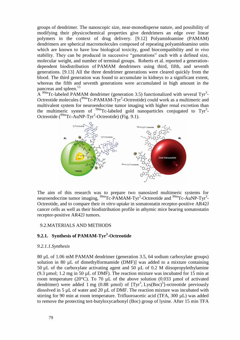
79
groups of dendrimer. The nanoscopic size, near-monodisperse nature, and possibility of
modifying their physicochemical properties give dendrimers an edge over linear
polymers in the context of drug delivery. [9.12] Polyamidoamine (PAMAM)
dendrimers are spherical macromolecules composed of repeating polyamidoamino units
which are known to have low biological toxicity, good biocompatibility and in vivo
stability. They can be produced in successive “generations” each with a defined size,
molecular weight, and number of terminal groups. Roberts et al. reported a generation-
dependent biodistribution of PAMAM dendrimers using third, fifth, and seventh
generations. [9.13] All the three dendrimer generations were cleared quickly from the
blood. The third generation was found to accumulate in kidneys to a significant extent,
whereas the fifth and seventh generations were accumulated in high amount in the
pancreas and spleen.13
A 99m
Tc-labeled PAMAM dendrimer (generation 3.5) functionalized with several Tyr3-
Octreotide molecules (99m
Tc-PAMAM-Tyr3-Octreotide) could work as a multimeric and
multivalent system for neuroendocrine tumor imaging with higher renal excretion than
the multimeric system of 99m
Tc-labeled gold nanoparticles conjugated to Tyr3-
Octreotide (99m
Tc-AuNP-Tyr3-Octreotide) (Fig. 9.1).
The aim of this research was to prepare two nanosized multimeric systems for
neuroendocrine tumor imaging, 99m
Tc-PAMAM-Tyr3-Octreotide and
99mTc-AuNP-Tyr
3-
Octreotide, and to compare their in vitro uptake in somatostatin receptor-positive AR42J
cancer cells as well as their biodistribution profile in athymic mice bearing somatostatin
receptor-positive AR42J tumors.
MATERIALS AND METHODS 9.2.
9.2.1. Synthesis of PAMAM-Tyr3-Octreotide
9.2.1.1.Synthesis
80 µL of 1.06 mM PAMAM dendrimer (generation 3.5, 64 sodium carboxylate groups)
solution in 80 µL of dimethylformamide (DMF)] was added to a mixture containing
50 µL of the carboxylate activating agent and 50 µL of 0.2 M diisopropylethylamine
(9.3 µmol; 1.2 mg in 50 µL of DMF). The reaction mixture was incubated for 15 min at
room temperature (20°C). To 70 µL of the above solution (0.033 µmol of activated
dendrimer) were added 1 mg (0.88 µmol) of [Tyr3, Lys(Boc)
5]-octreotide previously
dissolved in 5 µL of water and 20 µL of DMF. The reaction mixture was incubated with
stirring for 90 min at room temperature. Trifluoroacetic acid (TFA, 300 µL) was added
to remove the protecting tert-butyloxycarbonyl (Boc) group of lysine. After 15 min TFA

80
was removed under vacuum along with the DMF and water. The reaction mixture was
reconstituted in 0.5 mL of injectable grade water and purified using a size exclusion
high-performance liquid chromatography (HPLC) system (ProteinPak SEC 300SW, 10
µm, 7.5 mm x 300 mm Waters, 0.8 mL/min, injectable water) with a photodiode array
UV-Vis detector. The retention time (tR) for the PAMAM-Tyr3-Octreotide was 13.8
min, and for the PAMAM dendrimer and Tyr3-Octreotide it was 5.1 min and 12.2 min
respectively (Fig. 9.2). The collected fraction with a tR of 13.8 min was dried under high
vacuum, obtaining a yellowish solid.
9.2.1.2.Chemical characterization
The particle size and UV-Vis spectra of the compounds were determined in solution
(1 mg/mL). The solid samples were analyzed by medium infrared (MIR, from 4000 to
400 cm-1
) and far infrared spectroscopy and elemental analyses by combustion and
energy dispersive spectrometry. Thermogravimetric (TGA) and Differential Scanning
Calorimetric (DSC) analyses were further realized to the dried samples. Elemental
content for PAMAM-Tyr3-Octreotide. Found(%): C: 22.11, H: 2.35, N: 3.29, F: 47.0, S:
0.24, Calc.(%): C: 25.10, H: 2.73, N: 2.97, F: 38.94, S: 0.24 for the minimum formula,
C494H800N122O162Na38(C49H65N10O11S2)26(H2O)2000(C3H7NO)1120(CF3COOH)4845.
9.2.2. Labeling of PAMAM-Tyr3-Octreotide dendrimer with technetium-99m
For 99m
Tc labeling, 20 µL (20 µg) of the PAMAM-Tyr3-Octreotide solution was added
to a vial containing 10 µL of 0.1 M NaOH. To this was added 10 µL of a fresh 1 mg/mL
solution of stannous chloride in 10 mM HCl, followed by 185–370 MBq 99m
Tc-
pertechnetate in a volume of 100 µL, obtaining a final pH 9–10. The solution was
incubated at room temperature for 30 min and finally, 1 mL of 0.9% NaCl solution was
added to a final pH 6.5–7.0. For comparative studies, 99m
Tc-PAMAM dendrimer was
also prepared using the same labeling procedure.
The samples were analyzed for radiochemical purity using instant thin-layer
chromatography on silica gel (ITLC-SG, Gelman Sciences) and radio-HPLC. For the
ITLC-SG analysis 20% NaCl was used as mobile phase to determine the amount of free 99m
TcO4- (Rf=1.0), while the
99mTc-PAMAM-Tyr
3-Octreotide remains in the origin
(Rf=0.0).
In the radio-HPLC size exclusion system (ProteinPak 300SW, Waters, 1 mL/min,
injectable water), chromatographic profiles were obtained using two different detectors,
the UV-Vis detector and a radiometric detector. The sample first passed by the UV-Vis
detector (photodiode array) and after 0.5 min (0.5 mL, 1 mL/min) it passed by the
radioactivity detector (Fig. 9.2). Correspondence of retention times of the peaks of
interest (tR=10.75–11.25 min) in the chromatogram is accepted as a proof of the
chemical identity of the radiopharmaceutical. Radioactivity recovery was routinely
determined to discard the presence of hydrolyzed 99m
Tc (99m
TcO2.H2O) which remains
trapped in the column.

81
FIG. 9.2. Size-exclusion HPLC chromatogram (continuous line) and HPLC radio-chromatogram (dotted line) of the 99mTc-PAMAM-Tyr3-Octreotide conjugate (flow rate 1 mL/min).
9.2.3. 99mTc-PAMAM-Tyr
3-Octreotide Dendrimer, Stability in Human Serum
To determine the stability of 99m
Tc-PAMAM-Tyr3-Octreotide in human serum, 0.2 mL
of the radiolabeled dendrimer were diluted at a ratio of 1:5 with fresh human serum and
incubated at 37°C. The radiochemical stability was determined by sampling volumes of
0.02 mL at 10, 30 and 60 min for radio-HPLC size exclusion analysis, as described
above.
9.2.4. Preparation of 99m
Tc-AuNP-Tyr3-Octreotide
HYNIC-Tyr3-Octreotide lyophilized formulations containing EDDA (ethylenediamine-
N,N’-diacetic acid) as coligand were prepared as described by Ocampo-Garcia et al.14
The 99m
Tc-EDDA/HYNIC-Tyr3-Octreotide (
99mTc-Tyr
3-Octreotide) complex was
prepared by adding 1 mL of 0.2 M phosphate buffer, pH 7.0, followed immediately by
2.0–4.4 GBq (1 mL) of 99m
Tc-pertechnetate to the HYNIC-Tyr3-Octreotide freeze-dried
kit formulation, which was then incubated at 92°C for 20 min in a dry block heater. A
radiochemical purity >95% of 99m
Tc-Tyr3-Octreotide was verified by ITLC-SG analyses
using three different mobile phases: 2-butanone to determine the amount of free 99m
TcO4- (Rf =1), 0.1 M sodium citrate, pH 5, to determine
99mTc-EDDA and
99mTcO4
-
(Rf =1) and a 1:1 (v/v) methanol:ammonium acetate (1M) solution for 99m
TcO2.H2O (Rf
= 0). Rf values of the radiolabeled peptide in these three systems were 0.0, 0.0 and 0.7–
1.0, respectively.
To 1.5 mL of AuNP-citrate (20 nm=1.05x10¹²
particles), 100 µL (100–220 MBq) of 99m
Tc-Tyr3-Octreotide (0.25 µg of peptide; 1.29 x 10
14 molecules; 122 molecules per 20
nm-AuNP) was added with stirring for 15 min to form the 99m
Tc-AuNP-Tyr3-
Octreotide. No further purification was performed. Radiochemical purity was evaluated
using ultrafiltration (Centicron YM-30 Regenerated cellulose 30,000 MW cut off,
Millipore, Bedford, MA, USA), in which the 99m
Tc-AuNP-Tyr3-Octreotide remained in
the filter, while free 99m
Tc-Tyr3-Octreotide and
99mTcO4
− passed through the filter. In
the radio-HPLC system (ProteinPak 300SW, Waters, 1 mL/min, injectable water), the
tRs for the 99m
Tc-AuNP-Tyr3-Octreotide and
99mTcO4
− were 4.5–5 and 11 min,
respectively. 99m
Tc-AuNP-Tyr3-Octreotide prepared under these conditions is obtained

82
with high radiochemical purity (>94%) and high stability in human serum as previously
reported.14
9.2.5. In Vitro Binding studies
AR42J cells suspended in phosphate buffer saline (PBS) were diluted to 1x10⁶
cells/tube
and incubated with about 200,000 cpm of 99m
Tc-PAMAM-Tyr3-Octreotide or
99mTc-
AuNP-Tyr3-Octreotide or
99mTc-PAMAM or
99mTc-Tyr
3-Octreotide in triplicate at 37°C
for 1 h. The test tubes were centrifuged (3 min, 500g), washed twice with PBS, and the
activity of the cell pellet determined in a crystal scintillation well-type detector.
Radioactivity in the cell pellet represents both surface bound and internalized
radiopharmaceutical. An aliquot with the initial activity was taken to represent 100%,
and the cell uptake activity was then calculated. The surface bound radiopharmaceutical
was removed with 1 mL of 0.2 M acetic acid/0.5 M NaCl solution added to the
resuspended cells. The test tubes were centrifuged, washed with PBS, centrifuged, and
the pellet activity was considered as internalization.
9.2.6. In Vivo Studies
Tumor uptake and biodistribution studies in mice were carried out according to
the rules and regulations of the Official Mexican Norm 062-ZOO-1999. Athymic mice
with induced tumors received 99m
Tc-Tyr3-Octreotide or
99mTc-PAMAM or
99mTc-PAMAM-Tyr
3-Octreotide or
99mTc-AuNP-Tyr
3-Octreotide (n=3 for each one)
(0.05 mL, 1.85 MBq, i.v.). Mice were sacrificed at 2 h after radiopharmaceutical
administration. Whole heart, spleen, stomach, pancreas and kidneys and samples of
tumor, lung, liver, blood, intestines, bone and muscle were placed into pre-weighed
plastic test tubes. The activity was determined in a well-type scintillation detector along
with 3 aliquots of a diluted standard representing 100% of the injected dose. Mean
activities were used to obtain the percentage of injected activity per gram of tissue (%
ID/g). A blocking study was performed in one group of three athymic mice with
AR42J-induced tumors. One hundred microliters (1 mM) of unlabeled Tyr3-Octreotide
was intraperitoneally administered one hour before intravenous injection of 99m
Tc-PAMAM-Tyr3-Octreotide (n=3, 0.05 mL, 1.85 MBq, i.v.), and complete
dissection was carried out at 2 h after radiopharmaceutical administration as described
above.
9.2.7. Statistical analysis
Differences between 99m
Tc-PAMAM-Tyr3-Octreotide and
99mTc-PAMAM cell uptake
or between 99m
Tc-PAMAM-Tyr3-Octreotide and
99mTc-Tyr
3-Octreotide cell uptake as
well as in vivo tumor uptake between blocked and unblocked animals were evaluated
with the Student’s t-test.
RESULTS 9.3.
9.3.1. Chemical Characterization
Particle size. The average particle hydrodynamic diameter determined by light
scattering was 1.315 ± 0.132 nm for the PAMAM dendrimer and 1.649 ± 0.149 nm for
the PAMAM-Tyr3-Octreotide.

83
UV-Vis spectroscopy. UV-Vis provides the proof of synthesis as well as the
conjugation (surface modification) on dendrimers due to characteristic absorption
maximum or shift in value of lambda max (Bathochromic shift: Red shift).15
The
PAMAM spectrum did not showed any absorption main peak (Fig. 9.2A) but the
PAMAM-Tyr3-octreotide showed two main peaks at 280.5 and 329.3 nm (Fig. 9.3C),
which indicated a red-shift with respect to the bands at 278.2 and 323.4 nm found in the
[Tyr3, Lys(Boc)
5]-Octreotide spectrum (Fig. 9.3B). This suggests the conjugation of
surface modifiers (Tyr3-Octreotide peptides) to the PAMAM dendrimer.
Elemental analysis. The elemental analysis of dried PAMAM rendered the minimum
formula C494H800N122O188Na64(CH3OH)70(H2O)44.
From the elemental analysis (experimental section) the minimum formula for PAMAM-
Tyr3-Octreotide is
C494H800N122O162Na38(C49H65N10O11S2)26(H2O)2000(C3H7NO)1120(CF3COOH)4845. The
formula (C49H65N10O11S2)26 correlates with the attachment of 26 Tyr3-Octreotide
molecules to one PAMAM molecule (26 peptides: 1 dendrimer stoichiometry) and an
overall conjugation reaction yield of >90%.
FIG. 9.3. Size-exclusion HPLC chromatogram of the mixture (210 nm) A) PAMAM dendrimer B) Tyr3-Octreotide and
C) PAMAM-Tyr3-Octreotide HPLC (flow rate 0.8 mL/min). UV-Vis spectrum of the compounds A, B and C (inset).
FT-IR spectroscopy. MIR and FIR spectra of PAMAM, PAMAM-Tyr3-Octreotide
conjugate and Tyr3-Octreotide peptide are shown in Figure 9.4. and the assignations in
Table 9.1. The MIR spectrum of the PAMAM reveals the characteristic vibrations of the
-CO-NH-, COO- terminal groups and ethylene groups. The medium weak band at 1047
cm-1
is associated to vibration of methanol.

84
TABLE 9.1. MAIN INFRARED VIBRATIONAL FREQUENCIES (CM-1
) FOR PAMAM
DENDRIMER, TYR3-OCTREOTIDE AND PAMAM-TYR
3-OCTREOTIDE
Functional Group PAMAM Tyr3-Octreotide PAMAM-Tyr3-Octreotide
Amide (N‒H) 3263m
3074m 3276ms, 3061m 3031mw
Lys, amine (N‒H) --- 3500-3000, 836mw 3500-3000, 841ms
Carboxylic acid (COOH) --- --- 1798w
s (‒CH2‒) as (‒CH2‒) 2964m,2828m 2916m, 2866sh 2956sh, 2870mw
Amide I s (C=O) 1640ms 1642vs 1670vs, 1647sh
Amide II (N‒H) 1462sh,1555vs 1457ms 1445m
Amide III (C‒N) 1313sh 1358sh 1327mw, 1025m
Amide group (O=C‒N‒C) --- 1181s, 1132s 1181vs, 1123vs
Indole ring (HC‒N) --- 1515s 1475mw
Carboxylate ion
as (COO-) 1399s --- ---
Sulphur (C‒S), (S‒S) --- 722m, 517w 723s, 517m
: stretching vibration symmetric (s) or asymmetric (as); : bending vibration vs: very strong; s: strong; m: medium;
mw: medium weak; w: weak; sh: shoulder
MIR and FIR spectra of the PAMAM-Tyr3-Octreotide are characteristic of the
conjugate. The region from 3700 to 2500 cm-1
reveals –CONH, -CH2- and protonated
amide (2500 cm-1
). In the region of amide I a very intense and broad band centered at
1670 cm-1
is observed. Shoulders on the left side (1688 cm-1
) and on the right side (1647
cm-1
) of this band are seen. This band is broad because the vibrational frequencies of
carbonyls from dendrimer and Tyr3-Octreotide are located in the region of the carbonyls
from TFA present in the conjugate.19
A very broad intense band with two maximum at
1181 and 1123 cm-1
is associated with amide groups from the conjugate. However, its
feature and intensity indicate that this band has contribution from TFA since stretching
vibrations of CF3 groups (1147, 1200 cm-1
) are inside this band.19
The sharp band
centered at 1025 cm-1
corresponds to the vibration of the C-N-C bond of the secondary
amide formed between the dendrimer and the Tyr3-Octreotide. In the FIR spectrum, the
medium band at 517 cm-1
is associated to the disulfide from the Tyr3-Octreotide.
Different conformational arrangements about the disulfide bond lead to the S-S
stretching vibration bands occurring in different positions. In the PAMAM-Tyr3-
Octreotide the conformation adopted permits to observe this band with good intensity
while in the Tyr3-Octreotide spectrum is very weak. This suggests that the
conformational arrangement of the conjugate yields a highly stable macromolecule. The
very intense band at 264 cm-1
is assigned to the rocking vibration from the TFA.
Between 245 and 30 cm-1
it is revealed the hydrogen bonding between the dendrimer
and the solvent molecules as well as the strong interaction TFA-dendrimer in the
interior cavities. TFA proton transfer to the amides occurs as previously mention.17,18
The vibrational frequencies of the functional groups of the PAMAM-Tyr3-Octreotide
conjugate were satisfactorily identified.
9.3.2. Labeling
99m
Tc-PAMAM-Tyr3-Octreotide and
99mTc-AuNP-Tyr
3-Octreotide were obtained with
radiochemical purities of 96 ± 2% (n=12) (Fig. 9.2). In the direct labeling of PAMAM a
N4-chelating system is assumed to be formed which is successfully used to prepare
stable complexes with the Tc=O3+
core producing minimum alteration of the molecule
bioactivity.20

85
FIG. 9.4. Medium infrared spectra (left, from 4000 to 400 cm-1) and far infrared spectra (right, from 700 to 30 cm-1)
of A) PAMAM dendrimer, B) Tyr3-Octreotide peptide, and C) PAMAM-Tyr3-Octreotide conjugate.
9.3.3. In vitro studies
Human serum stability. 99m
Tc-PAMAM-Tyr3-Octreotide diluted in human serum was
stable over 60 min, because at this time, the radiochemical purity was 94 ± 2%.
In vitro binding studies. As shown in Table 9.2, 99m
Tc-PAMAM-Tyr3-Octreotide had
specific recognition, because the somatostatin receptor-positive cell uptake was 2 times
higher with respect to that of 99m
Tc-PAMAM (P < 0.05). The 99m
Tc-PAMAM
accumulation (16.69 %) in AR42J cells is related to a passive uptake, because
nanoparticles can be taken up by cells due to their nanometric size,6,21
but with the
conjugation of Tyr3-Octreotide to PAMAM, the tumor cell uptake was significantly
increased (32.83 %). The significant difference in cell binding results (32.83% vs
16.69%) was due to active targeting, meaning specific somatostatin receptor
recognition. Results also showed higher cell uptake (P<0.05) of the multimeric system
(99m
Tc-PAMAM-Tyr3-Octreotide) with respect to that of the monomer (
99mTc-Tyr
3-
Octreotide) due to the high capacity of nanoparticles to be internalized into the
cytoplasm (Table 9.2).21
No significant difference in cell uptake was found between 99m
Tc-PAMAM-Tyr3-Octreotide and
99mTc-AuNP-Tyr
3-Octreotide.
TABLE 9.2. AR42J CELL UPTAKE AND INTERNALIZATION OF DIFFERENT
RADIOPHARMACEUTICALS AT 1 H (% OF TOTAL ACTIVITY ± SD, N = 6).
Radiopharmaceutical Cell uptake
(mean ± SD)
Internalization
(mean ± SD) 99mTc-PAMAM-Tyr3-Octreotide
99mTc-PAMAM
99mTc-Tyr3-Octreotide
99mTc-AuNP-Tyr3-Octreotide
32.83 ± 2.22*,&
16.69 ± 3.28*
7.44 ± 1.40&
34.61 ± 1.54
26.30 ± 1.78*,&
11.64 ± 1.77*
4.71 ± 1.65&
N.D.
*Significant difference (P< 0.05) between 99mTc-PAMAM-Tyr3-Octreotide and 99mTc-PAMAM. &Significant difference (P< 0.05) between 99mTc-PAMAM-Tyr3-Octreotide and 99mTc-Tyr3-Octreotide
N.D. = not determined because of the AuNP aggregation in acidic conditions

86
9.3.4. Biodistribution studies
The tissue distribution of radioactivity 2 h after 99m
Tc-PAMAM-Tyr3-Octreotide
administration showed evident tumor uptake (4.12 ± 0.57% ID/g) and high
accumulation in the pancreas (15.08 ± 3.11% ID/g) which overexpresses somatostatin
receptors (Fig. 9.5). The in vivo tumor uptake was significantly higher (P < 0.05) than
that of 99m
Tc-PAMAM and its specificity was confirmed by the receptor blocking study,
in which the previous injection of Tyr3-Octreotide significantly diminished the activity
in the tumor (4.12% ID/g vs. 2.03% ID/g, P < 0.05) (Fig. 9.5). A high liver and spleen
uptake was observed due to the dendrimer colloidal nature. No significant difference in
blood, liver, spleen and tumor uptake was found between 99m
Tc-PAMAM-Tyr3-
Octreotide and 99m
Tc-AuNP-Tyr3-Octreotide (P > 0.05) (Fig. 9.5). However, the
99mTc-
labeled PAMAM-Tyr3-Octreotide showed a significant higher renal excretion than the
multimeric system of 99m
Tc-AuNP-Tyr3-Octreotide (8.71% ID/g vs. 4.47% ID/g,
P < 0.05) (Fig. 9.5).
FIG. 9.5. Biodistribution of 99mTc-PAMAM-Tyr3-Octreotide, 99mTc-AuNP-Tyr3-Octreotide, 99mTc-Tyr3-Octreotide
(monomer) and 99mTc-PAMAM in athymic mice with AR42J-induced tumors at 2 h after i.v. administration (n=3).
Tumor uptake was significantly higher (P < 0.05) for the multimeric system 99mTc-PAMAM-Tyr3-Octreotide with
respect to 99mTc-Tyr3-Octreotide or 99mTc-PAMAM. No significant difference in the tumor uptake was found between 99mTc-PAMAM-Tyr3-Octreotide and 99mTc-AuNP-Tyr3-Octreotide (P > 0.05).
Because of its rapid clearance from the blood and most tissues, the highest tumor-to-
blood, pancreas-to-blood and kidney-to-blood ratios were obtained with the 99m
Tc-Tyr3-
Octreotide monomer (Fig. 9.5). Nevertheless pancreas-to-blood ratios for the 99m
Tc-PAMAM-Tyr3-Octreotide and
99mTc-AuNP-Tyr
3-Octreotide multimeric systems
were as high as 9.48 and 13.38 respectively, and the tumor-to-blood ratio was 2.59 and
3.06 correspondingly. These ratio values (> 1) confirm their usefulness as radiolabeled
analogues of somatostatin for neuroendocrine tumor imaging and their tumor uptake the
potential as therapeutic agents.
CONCLUSIONS 9.4.
1. The elemental analyses and spectroscopy techniques demonstrated that 26
Tyr3-Octreotide molecules were successfully conjugated to one molecule of the
PAMAM dendrimer. 99m
Tc-PAMAM-Tyr3-Octreotide and
99mTc-AuNP-Tyr
3-
Octreotide, obtained with high radiochemical purities (>94%), are highly stable in

87
human serum and show high and specific somatostatin-positive tumor uptake in mice.
Therefore, they are potential target-specific radiopharmaceuticals for neuroendocrine
tumor imaging.
ACKNOWLEDGEMENTS
This study was supported by the International Atomic Energy Agency (Contract No.
18358/RO).
Published articles:
1. Orocio-Rodríguez, E., Ferro-Flores, G., Santos-Cuevas, C. L., Ramírez, F. D. M.,
Ocampo-García, B. E., Azorín-Vega, E., & Sánchez-García, F. M. (2015). Two Novel
Nanosized Radiolabeled Analogues of Somatostatin for Neuroendocrine Tumor
Imaging. Journal of Nanoscience and Nanotechnology,15(6), 4159-4169.
2. Ocampo García B.E., Miranda-Olvera R.M., Santos-Cuevas C.L., García-Becerra R.,
Azorín-Vega E.P., Ordaz-Rosado D., (2014). In vitro Decrease of the BCL-2 Protein
Levels in Lymphoma Cells Induced by Gold Nanoparticles and Gold Nanoparticles-Anti-
CD20, Nanoscience & Technology, 1(3): 1-6.
Poster presentation:
Ocampo-García B.E., Ferro-Flores G., Orocio- Ramírez F. de M., Santos-Cuevas C.L., Azorín-
Vega E., Isaac-Olivé K. Two Novel Nanosized Radiolabeled Analogues of Somatostatin for
Neuroendocrine Tumor Imaging, 21th International Symposium on Radiopharmaceutical
Science, University of Missouri, Missouri, USA. May 26-31, 2015.
REFERENCES TO CHAPTER 9
[9.1] DE JONG, M., BREEMAN, W.A.P., KWEKKEBOOM, D.J., VALKEMA, R.,
KRENNING, E.P., Acc. Chem. Res. 42, 873 (2009).
[9.2] PŁACHCIŃSKA, A., MIKOŁAJCZAK, R., KOZAK, J., RZESZUTEK, K.,
KUŚMIEREK, J., Eur. J. Nucl. Med. Mol. Imaging. 33, 1041 (2006).
[9.3] GONZALEZ-VAZQUEZ, A., FERRO-FLORES, G., DE MURPHY, C.A.,
GUTIERREZ-GARCIA, Z., Appl. Radiat. Isot. 64, 792 (2006).
[9.4] SEPULVEDA-MENDEZ, J., DE MURPHY, C.A., PEDRAZA-LOPEZ, M.,
MURPHY-STACK, E., ROJAS-BAUTISTA, J.C., GONZALEZ-TREVIÑO, O.,
Nucl. Med. Commun. 33, 69 (2012).
[9.5] SOWA-STASZCZAK, A., LENDA-TRACZ, W., TOMASZUK, M., GŁOWA,
B., HUBALEWSKA-DYDEJCZYK, A., Nucl. Med. Rev. Cent. East Eur. 16, 9
(2013).
[9.6] MORALES-AVILA, E., FERRO-FLORES, G., OCAMPO-GARCIA, B.E.,
LEON-RODRIGUEZ, L.M., SANTOS-CUEVAS, C.L., MEDINA, L.A.,
Bioconjugate Chem. 22, 913 (2011).
[9.7] LUNA-GUTIERREZ, M., FERRO-FLORES, G., OCAMPO-GARCIA, B.E.,
JIMENEZ-MANCILLA, N.P., MORALES-AVILA, E., DE LEON-
RODRIGUEZ, L.M., ISAAC-OLIVE, K., J. Labelled Comp. Radiopharm. 50,
140 (2012).
[9.8] MENDOZA-SANCHEZ, A.N., FERRO-FLORES, G., OCAMPO-GARCIA,
B.E., MORALES-AVILA, E., RAMIREZ, F. DE M., LEON-RODRIGUEZ,
L.M., J. Biomed. Nanotechnol. 6, 375 (2010).

88
[9.9] OCAMPO-GARCIA, B.E., RAMIREZ, F. DE M., FERRO-FLORES, G.,
LEON-RODRIGUEZ, L.M., SANTOS-CUEVAS, C.L., MORALES-AVILA,
E., Nucl. Med. Biol. 38, 1 (2011).
[9.10] JIMENEZ-MANCILLA, N.P., FERRO-FLORES, G., OCAMPO-GARCIA,
B.E., LUNA-GUTIERREZ, M.A., RAMIREZ, F. DE M., PEDRAZA-LOPEZ,
M., TORRES-GARCIA, E., Curr. Nanosci. 18, 193 (2012).
[9.11] MORALES-AVILA, E., FERRO-FLORES, G., OCAMPO-GARCIA, B.E.,
GOMEZ-OLIVAN, L., J. Biomed. Nanotechnol. 8, 991 (2012).
[9.12] AGASHE, H.B., BABBAR, A.K., JAIN, S., SHARMA, R.K., MISHRA, A.K.,
ASTHANA, A., GARG, M., DUTTA, T., JAIN, N.K., Nanomedicine. 3, 120
(2007).
[9.13] ROBERTS, J.C., BHALGAT, M.K., ZERA, R.T., J. Biomed. Mater. Res. 30, 53
(1996).
[9.14] OCAMPO-GARCIA, B.E., FERRO-FLORES, G., MORALES-AVILA, E.,
RAMIREZ, F. DE M., Nucl. Med. Commun. 32, 1095 (2011).
[9.15] GAUTAM, S.P., GUPTA, A.K., AGRAWAL, S., SUREKA, S., Int. J. Pharm.
Pharm. Sci. 4, 77 (2012).
[9.16] ASTRUC, D., BOISSELIER, E., ORNELAS, C., Chem. Rev. 110, 1857 (2010).
[9.17] V.D., MAIOROV, I.S., KISLINA, G.I., VOLOSHENKO, N.B., LIBROVICH,
Russ. Chem. Bull., Int. Ed. 49, 1526 (2000).
[9.18] I., TANIS, K., KARATASOS, Phys. Chem. Chem. Phys.11, 10017 (2009).
[9.19] VALENTI, L.E., BURGOS-PACI, M., DE PAULI, C.P., GIACOMELLI, C.E.,
Anal. Biochem. 410, 118 (2011).
[9.20] CIRSTOIU-HAPCA, A., BOSSY-NOBS, L., BUCHEGGER, F., GURNY, R.,
DELIE, F., (2007). Int J Pharm 331, 190(2011).
[9.21] MUKHERJEE, P., BHATTACHARYA, R., BONE, N., LEE, Y.K., PATRA,
C.R., WANG, S, LU, L., SECRETO, C., BANERJEE, P.C., YASZEMSKI,
M.J., KAY, N.E., MUKHOPADHYAY, D., J Nanobiotechnology, 5, 4(2007).
[9.22] BHATTACHARYA, R., MUKHERJEE, P., Adv Drug Deliv Rev. 60,1289
(2008)
[9.23] KAMINSKI, M.S., TUCK, M., ESTES, J., KOLSTAD, A., ROSS, C.W.,
ZASADNY, K., REGAN, D., KISON, P., FISHER, S., KROLL, S., WAHL,
R.L., 131I-tositumomab therapy as initial treatment for follicular lymphoma. N
Engl J Med. 352, 441(2005).

89
10. NANOSIZED DELIVERY SYSTEMS FOR RADIOPHARMACEUTICALS
IRFAN ULLAH KHAN1, RIZWANA ZAHOOR
1, ABUBAKER SHAHID
1, FOZIA IRAM
1,2
1Institute of Nuclear Medicine & Oncology (INMOL), New Campus Road, Lahore-54600, Pakistan
2Department of Chemistry, Lahore College for Women University (LCWU), Lahore-54600, Pakistan
INTRODUCTION 10.1.
The work plan for next 15 months to be performed in this CRP as finalized during First
Coordination Meeting of this CRP, held at IAEA HQr’s, Vienna, Austria (7-11 July, 2014), is
as under:
Bombesin and DOTA derivatized NPs to complex Lu-177/Ga-67 will be produced by
ARGENTINA, BRAZIL, ITALY-PALERMO, POLAND, SINGAPORE/ THAILAND.
Samples with Specs for labeling containing 2-50 mg of solid will be sent to ITALY-
MILAN and IRAN for radiolabeling and to ITALY-PADOVA, MALAYSIA, MEXICO
and PAKISTAN for in-vitro/ in-vivo evaluation or both.
Functionalized natural polymers will be provided by THAILAND to USA, EGYPT,
PAKISTAN and MALAYSIA for stabilizing radioactive Au NPs.
As it is clear from above, Pakistan is mostly involved in that experimental part of work
which is based on receiving samples from other partner labs. Being Chief Scientific
Investigator (CSI) of this CRP from Pakistan, I am working in close collaboration with partner
groups working on development of nanoconstructs, e.g., the lab in Argentina has synthesized/
characterized Albumin NP’s, coupled with DOTA-Bombesin and send the final product, e.g.,
Albumin-NP’s-DOTA-Bombesin to Pakistan for radiolabeling and complete bioevaluation
analysis.
In Pakistan, my lab has been investigating several synthetic methods for preparation of
NP’s. These include physical, chemical and biochemical techniques. The particles produced by
these methods usually are unstable and as such require a stabilizing agent to be included in the
process of synthesis. With increasing awareness about environmental impact of synthetic
methods, it is always desirable to use the so-called green chemistry approach. Therefore,
methods have been developed where the additional use of reducing and stabilizing agents can
be avoided1,2
. For application of NPs in medicine, it is more important that all the materials
used as aids in synthesis should be biocompatible. Keeping in view these requirements, we
previously used Arabinoxylan (AX) isolated from ispaghula seed husk as the reducing and
dispersing agent. AXs are gel-forming food components that are not hydrolyzed by enzymes of
the upper gastrointestinal tract, and coagulate at an acidic pH. Thus, they can deliver the
encapsulated material into the last part of the large intestine (colon). AXs from isphagula
(Plantagoovata) seed husk are abundantly available and well-characterized materials with

90
potential use as drug carriers3,4
. The AXs are composed of reducing sugars which can reduce
gold or silver ions to particles and at the same time, having a hydrogel-like network, can
disperse the particles in themselves. They have also been proved to be non-toxic for drug
delivery5. Therefore, the use of AX from ispaghula for green synthesis of gold and silver NPs
shows exceptional high stability, which may find their use in medicine.
FIG. 10.1. Structure of Araboxylan (Ax)
METHODS AND RESULTS 10.2.
10.2.1. Materials
Chloroauric acid, HAuCl4·3H2O, silver nitrate, sodium hydroxide, and hydrochloric
acid were of analytical grade and purchased from Merck, Germany. They were used without
further purification. Ispaghula (P. ovata) seed husk was obtained from local market. Nanopure®
water was used in this work.
Arabinoxylan-mediated synthesis of gold and silver nanoparticles with high
stability
We have synthesized highly stable gold and silver nanoparticles (NPs) using
arabinoxylan (AX) from ispaghula (Plantago ovata) seed husk. The NPs were synthesized by
stirring a mixture of AX and HAuCl4·H2O or AgNO3, separately, below 100◦C for less than an
hour, where AX worked as the reducing and the stabilizing agent. Characterization was done by
surface plasmon resonance (SPR) spectroscopy, transmission electron microscopy (TEM),
atomic force microscopy (AFM), and X-ray diffraction (XRD). The particle size was (silver: 5-

91
20 nm and gold: 8-30 nm) found to be dependent on pH, temperature, reaction time and
concentrations of AX and the metal salts used.
10.2.2. Synthesis of AuNP’s
To start the synthesis procedure, varying amounts (1-20 ml) of the mucilage were added
to 20 ml solutions of 1.0 mM HAuCl4·3H2O and AgNO3 separately, and total volume was made
up to 40 ml with water under constant stirring. Synthesis of NPs was marked by a change in
color, e.g., a change in color (yellow to brown to purple in case of AuNPs and colorless to
yellow to brown in case of AgNPs) was observed in 15-180 min (in case of AuNPs) and 60-240
min (in case of AgNPs) depending upon the amount of the mucilage added, pH and
temperature. The optimum conditions were determined by varying the amount of mucilage, pH
(3-12), temperature (25-100◦C) and time. The NP suspensions thus obtained were washed
several times with Nanopure®
water in order to remove any unreacted gold or silver salts.
10.2.3. Synthesis of 198
AuNPs
The radioactive gold nanoparticles (198
AuNPs) were synthesized by using the above
procedure for synthesis of cold AuNPs replacing non-radioactive gold salt with
H198
AuCl4.3H2O (1mM; 20ml).
10.2.4. Imaging Studies
10.2.4.1. Imaging Equipment
Images were recorded by using a large field-of-view (LFOV) dual head gamma camera
Infinia® GE Free-Geometry Dual-Detector Cameras 6/2006 Optional Hawkeye® Hybrid
NM/CT) fitted with a high resolution and high energy collimator. Data processing was done on
ECAM workstation using ESOFT by SYNGO™ software.
10.2.4.2. Animal Preparation
Healthy local Lahore breed rabbits (aged 12-16 weak; 900 ± 50g) were used. Before
injecting to animals, compounds were tested in in vitro assays in Hela cells. All the animals
were anesthetized by intramuscular ketamine injection (50mg/Kg). A suspension of 198
AuNPs
(140 µg ml-1
: 1 mCi ml-1
) was injected (in 1 ml volume) intravenously into the ear vein of
rabbits (n = 2). The accuracy of the dose was controlled using both the volume injected and the
radioactivity of 198
Au. Any activity remaining in the syringe was measured and used to
calculate the exact dose injected.
10.2.4.3. Imaging Protocol
DYNAMIC study acquisition comprised of 10 frames of 60 sec each. It was followed
by Anterior and posterior whole body. STATIC images acquired at 1h, 2h, and 24h, post

92
injection. To obtain clear visualization, images were acquired from various (additional)
positions, e.g., anterior, posterior, left lateral, right and lateral, etc.
FIG. 10.2. Dynamic study of rabbit for 15 min
FIG. 10.3. Static views of rabbit at various time intervals
10.2.5. Biodistribution of 198
Au-Ax in Mice
After re-anesthetization, at 2h and 24h post injection, the animals were sacrificed and
organs of interest and entire remaining carcass were collected, weighed and counted for
radioactivity in a well Beckman 8000 gamma counter (Beckman, Brea, CA). The radioactivity
in the tissue samples and in an aliquot of the injection mixture was determined and the amount
of activity in the organs was expressed as percent injected dose per gram (% IDg-1
) of the
tissue.

93
FIG. 10.4 Biodistribution of 198AuNP’s-Ax in mice model
10.2.6. Cell Culture
HeLa cells were cultured in DMEM (Gibco) high glucose with 10% FBS and 1%
Pen/Strep (Gibco) in 10,000 cells per cm2 and incubated for 24 h at 5% CO2 and 90% humidity.
After 24h medium was freshed and cells were incubated again for 24h without any additive of
NP or even gel and after incubation, cells had characteristic morphology for HeLa. All cells
were happily growing and no sign of cell stress was observed.
10.2.6.1. Treatment with Gel
HeLa cells were cultured in DMEM (Gibco) high glucose with 10% FBS and 1%
Pen/Strep (Gibco) in 10,000 cells per cm2 and incubated for 24h at 5% CO2 and 90% humidity.
After 24h filtrate from Gel was added in medium (1:1) and cells incubated again for 24h in
same conditions. Cells were swollen and some acquired a flat morphology which shows some
stress in cells but most of cells showed good shape.
10.2.6.2. Treatment with AuNP’s
Cells were cultures in same conditions for initial 24h and after that Gold NPs were
added in 1:1 ratio and cells were cultured again for more 24h. AuNPs treated cells appeared
deformed and their morphology was quite different. Only few cells had characteristic
morphology and most of them were round and some were lysed as obvious from cell debris.
10.2.6.3. Treatment with AgNP’s
Cells were treated with Silver NPs after initial 24h seeding and incubated again. As a
result cells, showed drastically different cell morphology and almost all cells were swollen and
0
0.5
1
1.5
2
2.5
3
3.5
3h
24h

94
many had prominent particles inside their cell membrane. Cells were round to oval shaped and
no debris of cells was observed.
a b c d FIG.10. 5. Shape of Hela cells a) before any treatment, b) after treatment with Ax gel, c) after treatment with AuNP’s and d)
after treatment with AgNP’s
10.2.6.4. Neutral Red Uptake Assay for Cytotoxicity and Cell Viability measurement
Neutral Red is supra-vital dye and all actively growing cells uptake the Neutral red from
surrounding and bind it to lysosomes of cells. This test is widely used to check cell viability
and also for cytotoxicity assays. Protocol devised by Repetto et al., was used to perform it.
Cells were seeded in 10,000 cells per cm2 density and cultured over night to ensure proper cell
attachment. Next day cells were treated with samples and left in incubator again for 24h. After
incubation media was removed and cells were washed with PBS (Phosphate Buffer Saline) and
Neutral red media was added and incubated again for 2h. After two hours, neutral red media
was removed and cells were again washed with PBS. Freshly prepared de-staining solution was
added and culture was incubated for 10 min. Cells released their color and its Optical Density
was taken at 570nm10
.
FIG. 10.6 Comparative measurement of OD shows that control cells had highest value for Neutral red uptake and hence,
highest cell viability as compared to Gel, Au or Ag NPs.
10.2.7. Preparation of 177
Lu-DOTA-BBN-AlbNP’s
This sample was received from collaborative group working in Argentina. In Pakistan,
we performed radiolabeling with Lu-177, quality control and imaging studies in normal rabbit
models on gamma camera. Detail of experimental procedures and findings is given below:
0
0.1
0.2
0.3
0.4
0.5
control Gel Gold NPS Silver NPs

95
FIG. 10.7 Synthesis of Albumin Nanoparticles
10.2.7.1. Materials used for Radiolabeling with 177
Lu
The following materials were used to start the procedure:
• 20µg of AlbNP’s-DOTA-bombesin
• 40µl of Ammonium acetate buffer solution(0.4mol/L, pH-5)
• 100µl of Ascorbic acid solution(100mg/L)
• 50µl of Ammonium acetate buffer solution(0.4mol/L, pH-5)
• 10µl of 177
Lu
10.2.7.2. Labeling Procedure with 177
Lu
As an experimental procedure, whole material described in 2,7.1 was vortexed in a
sealed glass vial, incubated at 85◦c in water bath for 25 min, and cooled the mixture to room
temperature, checked pH that should be 4.
10.2.7.3. Quality Control of 177
Lu-DOTA-BBN-AlbBPs
For quality control, ITLC-SG strips were used as stationery phase. For mobile phase, we
tried various solvents, e.g., citrate buffer (pH 4), ammonium acetate buffer (pH 5) and
NH4OH/EtOH/H2O = 1/5/10. In first two solvents, there was no separation and activity was
spread on whole strip but in third solvent system, there was good separation and we got
radiochemical purity (RCP) of 91.12 %.
10.2.7.4. Effect of Activity and Volume over Labeling Efficacy
The effect of activity on labeling efficacy was studied by varying the amount of activity
from 5 mCi to 30 mCi by keeping all parameters constant. Similarly, the effect of volume over
labeling efficacy was studied by varying the amount of volume from 10 to 200 µl. The
maximum labeling efficacy was found at 10 µl and beyond 50 µl, the solution became turbid.
Data of both experiments is given in Fig. 10.8.

96
FIG. 10.8. a) Effect of Activity on Labelling Efficacy, b) Effect of Volume on Labelling Efficacy
10.2.7.5. Imaging Scintigraphy of 177
Lu-DOTA-BBN-AlbNPs in Rabbit
Healthy local Lahore breed rabbits (aged 12–16 weak; 900 ± 50g) were used. All the
animals were anesthetized by intramuscular ketamine injection (50mg/Kg). A solution of 177
Lu-
AlbNPs-DOTA-Bombesin (2 mCi ml-1
) was injected intravenously into the ear vein of rabbits
(n = 2). The accuracy of the dose was controlled using both the volume injected and the
radioactivity of 177
Lu. Any activity remaining in the syringe was measured and used to
calculate the exact dose injected. DYNAMIC study acquisition comprised of 10 frames of 60
sec each. It was followed by Anterior and posterior whole body STATIC images acquired at 15
min, 24h, 48h, 72h, and 96h, post injection. To obtain clear visualization, images were acquired
from various (additional) positions, e.g., anterior, posterior, left lateral, right and lateral, etc.
Data are shown below:
0
20
40
60
80
100
0 20 40La
be
llin
g Ef
fici
en
cy(%
)
Activity of Lu-177(mCi)
-20
0
20
40
60
80
100
0 100 200 300Lab
elli
ng
Effi
cie
ncy
(%)
Volume of Lu-177(µl)

97
FIG. 10.8. Dynamic Study of Rabbit model for 15 min in Dorsal and Ventral Views
FIG. 10.9. Static Images of Rabbit model for at various time intervals in Dorsal and Ventral Views
CONCLUSION 10.3.
Silver and gold NPs synthesized by use of AX, from ispaghula seed husk, were
successfully radiolabeled and quality control procedures established. Cell viability
measurement was established through cytotoxicity assays in Hela cell models. Radiolabeling
and quality control was established for 177
Lu-Alb-NPs-DOTA-Bombesin. Imaging studies
performed with 177
Lu-Alb-NPs-DOTA-Bombesin demonstrate its ideal therapeutic potential to
be further developed as a feasible theranostic agent.
REFERENCES TO CHAPTER 10

98
[10.1] Kora, A.J., Sashidhar, R.B., Arunachalam, J., (2010). Gum kondagogu
(Cochlosper-mum gossypium): A template for the green synthesis and stabilization
of silver nanoparticles with antibacterial application. Carbohydrate Polymers, 82,
670–679.
[10.2] Raveendran, P., Fu, J., Wallen, S.L. (2003). Completely green synthesis and
stabilization of metal nanoparticles. Journal of the American Chemical Society,
125, 13940–13941.
[10.3] Akbar, J., Iqbal, M.S., Chaudhary, M.T., Yasin, T., Massey, S., (2012). A QSPR
study of drug release from an arabinoxylan using ab initio optimization and neural
networks. Carbohydrate Polymers, 88(4), 1348–1357.
[10.4] Saghir, S., Iqbal, M.S., Hussain, M.A., Koschella, A., Heinze, T., (2008). Structure
char-acterization and carboxymethylation of arabinoxylan isolated from ispaghula
(plantagoovata) seed husk. Carbohydrate Polymers, 74, 309–317.
[10.5] Iqbal, M.S., Akbar, J., Hussain, M.A., Saghir, S., Sher, M., (2011). Evaluation of
hot-water extracted arabinoxylans from ispaghula seeds as drug carriers.
Carbohydrate Polymers, 83(3), 1218–1225.
[10.6] Aggarwal, Karimullah, (1945). Chemical examination of the seeds of Mimosa
pudica, Linn. Journal of Scientific and Industrial Research, 46(4): 80.
[10.7] Farooqi, M. I. H., Kapoor, V. P., & Khan, P. S. H. (1977). Chemical investigation
on the mucilage of lajwanti seeds from Mimosa pudica Linn. Indian Drugs, 15, 39.
[10.8] Saraswat, R., & Pokharkar, R., (2012). GCMS Studies of Mimosa pudica.
International Journal of Pharm Tech Research, 4(1): 93–98.
[10.9] Amin, M., Iram, F., Iqbal, M. S., Saeed, M. Z., Raza, M., Alam, S., (2013).
Arabinoxylan-mediated synthesis of gold and silver nanoparticles having
exceptional high stability. Carbohydrate Polymers, 92(2): 1896–1900.
[10.10] Repetto, G., del Peso, A., Zurita, J.L., (2008). Neutral red uptake assay for the
estimation of cell viability/cytotoxicity. Nature protocols, 3(7), 1125–1131.

99
11. NANOSIZED DELIVERY SYSTEMS BASED RADIOPHARMACEUTICALS
IN POLAND
MICHAL MAURIN
National Centre for Nuclear research, Otwock,
Poland
Profile of POLATOM
Radioisotope Centre POLATOM (POLATOM) is the division in the National Centre
for Nuclear Research, located in Otwock near Warsaw, Poland. National Centre for
Nuclear Research is a state owned research institute and the largest research institute in
Poland with over 1000 employees. It came into existence on September 1, 2011 in the
effect of merging the former Institute of Atomic Energy POLATOM with the former
Andrzej Sołtan Institute for Nuclear Studies. National Centre for Nuclear Research
pure/applied research profile combines nuclear power – related studies with various
fields of sub-atomic physics (elementary particle, nuclear physics, hot plasma physics
etc.). It is strongly involved in developing nuclear technologies and promoting practical
applications of nuclear physics methods. National Centre for Nuclear Research is also
the only Polish research institution operating a nuclear reactor, the MARIA Research
Reactor with nominal power of 30 MW, and max neutron flux of 3x10¹⁴ n/cm²s,
suitable for production of radioactive isotopes, material science and neutron irradiation
investigations, as a neutron irradiation facility.
Scientific programs of Radioisotope Centre POLATOM are focused on the research and
development in the area of radionuclide production and radiopharmaceuticals, research
related to the application of radioactive preparations and radiolabelled compounds in
various fields in medicine, research and industry. The research activity of POLATOM
presents multidisciplinary character and combine chemistry and physics of
radionuclides, radiochemistry, analytical chemistry, metrology of ionizing radiation,
biology, pharmacy and nuclear medicine.
POLATOM is a manufacturer of radionuclide 99
Mo/99m
Tc generators and marketing
authorization holder for this medicinal product. POLATOM has GMP certified
laboratories for radiopharmaceutical manufacture and up-to-date infrastructure and
experience in radiochemical and radiopharmaceutical production processes with
appropriate radiation protection equipment, personnel trained in operations in hot-cells,
co-operation with suppliers of designated hot-cells and experienced in waste
management systems. The Laboratory of Radioactivity Standards (LRS) is accredited
(accreditation no. AP 120) for radioactivity measurements of alpha-, beta- and gamma-
emitters by absolute methods and performing calibration of standard solutions and
radioactive sources. LRS is also equipped in gas proportional counters adequate to
determining particle flux leaving the area of the source and the assessment of absorbed
dose.
The main research and development domains are:
5. Investigation of novel biomolecules as carriers for radionuclides and preliminary
assessment of their diagnostic and/or therapeutic utility
6. Development of technologies for production of high specific activity
radionuclides in nuclear reactors and accelerators, using highly enriched target materials
and modern separation techniques

100
7. Development of methods for radioactivity measurement and assessment of
radionuclidic purity (determination of , β, and impurities)
8. Chemical and pharmaceutical development and biological activity assessment
using in vitro and in vivo methods of new radiopharmaceuticals for clinical application.
Laboratories of POLATOM are well prepared to carry out this advanced research
program and are systematically upgraded. In 2011 the laboratory for in vivo studies was
launched and granted approval of Ministry of Science and Higher Education to conduct
research studies of radiopharmaceuticals in laboratory animals (registration number
0162).
Very good communication network with nuclear medicine units and other institutions
interested in radiopharmaceutical development as well as with several international
research institutions has been developed, supported by scientific and applied grants.
POLATOM participates in several research projects coordinated by International
Atomic Energy Agency (IAEA) as well as in European cooperation program of
scientific and technical cooperation COST; In recent years we participated in COST
Action D18 „Lanthanides in therapy: Particle emitting Radiolanthanides and stable
Lanthanides with radiation induced by external irradiation”, in COST Action BM0607
“Targeted Radionuclide Therapy” and COST Action D38 “Metal Based System for
Molecular Imaging”. Currently we are participating in COST Action TD1004
“Theragnostics Imaging and Therapy: An Action to Develop Novel Nanosized Systems
for Imaging-Guided Drug Delivery”.

101
The 1’st year results.
There are no outcome from the cooperation with another participants of the RCM. In the
Palatom the AGuIX nanoparticles polisiloxan core with DOTA chelators where studied.
The 90Y, 177Lu radiolabeling protocols were established and in vitro and in vivo
properties of functionalized particles with TATE peptide and native particles were
established.
The Results.
INTRODUCTION: 11.1.
AGuIX particles are the nano particles consisting of polysiloxane backbone with
attached DOTA chelator groups via amide groups. Until now particles have been
characterized for their size (3–4nm), mass (8.5kDa), imaging properties in MRI (after
inclusion of Gd atoms), fluorescence reflectance imaging and SPECT of 111
In labeled
AGuIX. Previously we labeled AGuIX with 90
Y and 177
Lu and found out that AGuIX
with 5 DOTA show favorable biodistribution pattern, suitable for planning its further
clinical application. The aim of the present work was investigation of 90
Y and 177
Lu
labeling AGuIX particles functionalized with a somatostatin analogue – Octreotate
(TATE) and the in vitro evaluation of their biological activity.
MATERIALS AND METHODS: 11.2.
AGuIX particles:
Functionalization of particles:
90Y and 177Lu:
90Y (ItraPol, specific activity ~18 TBq/mg) and 177Lu (LutaPol, specific activity >0,55
TBq/mg) produced at POLATOM were used for labelling
64Cu:
64Cu was obtained from POLATOM.
Radiolabeling
The number of DOTA molecules accessible for radiolabelling in AGuIX particles was
determined by quantification of yield of radiolabelling with reactor produced 64Cu.
Biodistribution was performed in healthy Wistar rats, 2 and 23 hours after intravenous
administration of the 90Y-labelled particles.
The radiolabelling conditions were investigated for non-functionalized and TATE
functionalized Gd-substituted AGuIX particles containing approximately one or five
non-substituted DOTA chelators. The optimal radiolabelling conditions were: 1:400
molar ratio of Y to the particles (calculated based on the their Gd substitution), ascorbic
acid/NaOH pH 4.5, and 15 min incubation at 900C. The labeled nanoparticles were
purified on AmiconUltra 3kDa cut-off filters. The radiochemical purity of labeled
particles was determined by TLC on ITLC SG plates developed in 0.2M KCl pH 2.4.

102
In vitro:
The active targeting of 90
Ylabelled particles was investigated in the AR42J rat
pancreatic tumor cells (line expressing somatostatin receptors subtype 2). for labeled
AGuIX, TATE-AGuIX and DOTA-TATE as reference. The specific binding of
functionalized particles was determined in presence of excess of cold nanoparticles or
DOTATATE. Radiolabelled particles after 3x AmiconUltra filtration where added to
AR42J cells. Cells where seeded on 6 well plates (2mln cells per well) and then
incubated for 48h. The studied AGuIX particles where added to cells (in RPMI without
FBS) in amount of 0.05, 0.25, 5, 50 and 500 pmol followed by 2h incubation at 37oC.
After incubation the medium was removed, and cells where washed with cold PBS. The
binding to the cell surface receptors was determined by measurement of radioactivity of
1ml of glycine buffer used for rinsing of cells. The Internalization of AGuIX was
determined by measurement of radioactivity of 1ml of 1M NaOH used for lysis. In
order to determine the specific biding of the AGuIX-TATE the receptors were blocked
with DOTA-TATE, cold AGuIX and AGuIX-TATE.
In Vivo
Biodistribution was performed in healthy Wistar rats, 2 and 23 hours after intravenous
administration of the 90
Y-labelled particles (~10MBq/.1mL).
The in vivo experiments (nude mice with subcutaneously implemented AR42J cells)
were performed for 177
Lu-labeled AGuIX and 177
Lu-TATE-AGuIX at 2 and 24 hours
after intravenous administration.
RESULTS: 11.3.
The AGuIX and functionalized TATE-AGuIX were labeled with 90Y with yields >75%
and 45%, respectively. The radiochemical purity after filtration was >96% and > 91%,
respectively [Fig 11.1], maintained for over 24h [Tab 11.1].

103
AGuIX-TATE after purification
AGuIX after purification
FIG. 11.1. TLC radiochromatograms of 90Y-labelled AGuIX-TATE and AGuIX particles (ITLC SG, 0.2M KCl pH
2.4).
TABLE 11.1. THE YIELD OF 90Y AND 177LU RADIOLABELLING AND RADIOCHEMICAL
PURITY OF THE PARTICLES AFTER PURIFICATION AND 24H STORAGE
90Y-
AGuIX
90Y-AGuIX-
TATE
177Lu-
AGuIX
177Lu-AGuIX-
TATE
90Y-DOTA-
TATE
After
labelling 76,9% 43,4% 95,6% 55,1% 99,9%
After
filtration 96,1% 91,6% - 92,0% -
After 24h 94,2% 90,8% - - 99,8%
Cell binding of the TATE functionalized 90Y-labelled AGuIX (ca. 2%) to AR42J cells
was over 10 times higher than the parent AGuIX particles (ca.0.2%) while for 90Y-
DOTA-TATE it was around 9%. In comparison to 90Y-DOTA-TATE, a lower cell
binding of the labeled nanoparticles can be explained by lack of internalization (AGuIX
particles due to their size probably do not internalize and may block the somatostatin
receptors). Surprisingly, the binding of 90Y-TATE-AGuIX to the cells remained
unchanged, regardless whether the blocking was performed with excess of cold DOTA-
TATE or cold functionalized particles [fig 11.2].

104
FIG. 12.2. Binding to the cell membrane and internalization of 90Y labelled nanoparticles to AR42J cells (50pmol / 2
million cells).
The biodistribution of 90Y-labeled AGuIX with 5 free DOTA in healthy rats revealed
fast pharmacokinetics with rapid renal clearance (70% ID in urine and bladder after 2h)
and low accumulation in critical organs such as liver, lungs and bone (Fig. 11.3). In
comparison the radiolabelled nanoparticles with only one free chelator, it showed >2-
times lower urine excretion and over one order of magnitude higher location in liver,
spleen and bone (Fig. 11.3).
FIG. 11.3. 2 and 23h p.i.v. biodistribution of 90Y-AGuIX with 5 and 1 DOTA (Wistar rats n=4).
The biodistribution in mouse tumor model reveeled rapid renal elimination of 177Lu
labelled AGuIX (over 90% of injected activity is excreted in urine after 2h). The TATE
functionalized particles highly accumulated in kidney (over 100% ID/g in 2h after
injection remaining over 24h) [tab 2]. Biodistribution investigations revealed significant
0
1
2
3
4
5
6
7
8
9
90Y-AGuIX90Y-AGuIX-
TATE90Y-DOTA-
TATE
90Y-AGuIX-TATE +
DOTA-TATE
90Y-AGuIX-TATE +
AGuIX-TATE
90Y-AGuIX-TATE +AGuIX
cell membrane 0.13 1.54 0.82 1.45 1.21 1.53
internalization 0.03 0.38 7.44 0.44 0.12 0.14
%
blood
heart
lung
liver
sple
an
pancr
eas
kidney
s
inte
stin
es
stom
ach
bone
musc
le
bladder
+ u
rine
[%ID
]
0.0
0.5
1.0
1.5
2.0
AGuIX, 5 DOTA, 2h piv
AGuIX, 5 DOTA, 23h piv
10
20
30
40
50
60
70
80
AGuIX, 1 DOTA, 2h piv
AGuIX, 1 DOTA, 23h piv
%ID
/g

105
differences in accumulation of radiolabelled particles in tumor tissue. The T/M ratio of
labelled TATE-AGuIX remained over time at the level of 5 and T/B ratio was rising to
around 16 at 24h p.i., while for native AGuIX particles the accumulation in tumor was
declining with the elimination of activity from the blood pool [fig 11.4].
TABLE 11.2. BIODISTRIBUTION OF 177LU LABELLED AGUIX AND AGUIX-TATE IN
TUMOR BEARING MICE (NUDE MICE WITH IMPLEMENTED AR42J CELLS); %ID/G, N=4.
177Lu-AGuIX
2h
177Lu-AGuIX-TATE 2h
177Lu-AGuIX
24h
177Lu-AGuIX-TATE 24h
blood 0.05 +/- 0.03 0.43 +/- 0.28 0.01 +/- 0.00 0.03 +/- 0.02
hart 0.06 +/- 0.01 0.32 +/- 0.04 0.02 +/- 0.02 0.11 +/- 0.02
lungs 0.13 +/- 0.03 0.51 +/- 0.16 0.03 +/- 0.01 0.24 +/- 0.08
liver 0.06 +/- 0.02 0.72 +/- 0.08 0.05 +/- 0.01 0.67 +/- 0.12
spleen 0.09 +/- 0.03 0.38 +/- 0.03 0.03 +/- 0.04 0.42 +/- 0.26
pancreas 0.03 +/- 0.01 0.20 +/- 0.04 0.04 +/- 0.03 0.14 +/- 0.02
kidney 18.58 +/- 7.11 108.81 +/- 1.69 12.49 +/- 1.95 105.62 +/- 4.45
intestines 0.11 +/- 0.01 0.43 +/- 0.07 0.32 +/- 0.09 0.42 +/- 0.04
bladder 0.04 +/- 0.00 0.68 +/- 0.37 0.06 +/- 0.01 0.44 +/- 0.13
femur 0.36 +/- 0.20 4.18 +/- 1.21 0.06 +/- 0.03 4.30 +/- 1.16
muscle 0.05 +/- 0.03 0.18 +/- 0.04 0.05 +/- 0.03 0.11 +/- 0.03
urine %ID 90.26 +/- 0.44 35.72 +/- 2.93 91.98 +/- 0.51 38.22 +/- 1.32
tumor 0.32 +/- 0.06 0.86 +/- 0.26 0.07 +/- 0.03 0.53 +/- 0.03
FIG. 11.4. Tumor to muscle and tumor to blood ratios of 177Lu-AGuIX-TATE and 177Lu-AGuIX
Plans for 2 year of RCM
Participation in the IAEA project – fields of cooperation
0
5
10
15
20
177Lu-AGuIX 2h 177Lu-TATE-AGuIX 2h 177Lu-AGuIX 24h 177Lu-TATE-AGuIX 24h
tumor/muscle tumor/blood
106
The Polatom can cooperate in field of radiopharmaceutical study.
1. Labelling of nanoparticles with 68Ga, 90Y, Lu177, 99mTc, (131I)
2. Labelling and characterization of particles with “cold” metals Ga, Y, Lu and
99Tc.
3. Stability study of labelled samples
4. Study on radiochemical purity
5. Preliminary in vivo study on healthy rats
Another filed of cooperation is the activation of gold and preparation of radioactive gold
nanoparticles (protocol from Kattesh Katti).
Plans for future years
1. Study on selected cell lines (only selected optimized particles)
2. Study of selected particles on animal tumor model (to be discussed)

107
12. POLYMER-BASED NANOCARRIERS FOR RADIOPHARMACEUTICALS
PIOTR ULANSKI
Institute of Applied Radiation Chemistry, Technical University of Lodz, Lodz, Poland
Abstract:
This report summarizes the studies performed at the Institute of Applied Radiation Chemistry (IARC),
Lodz University of Technology within the framework of the CRP project during the first year of its
realization. It consists of two parts. Part I is a report on the progress in studies on radiation synthesis of
gold nanoparticles (with the final aim of employing the elaborated protocols for synthesizing gold
nanoparticles for applications in radiotherapy).
Part II is a report regarding the first steps of the study focused at synthesizing, using radiation technique,
polymer nanogels equipped with specific functional groups (the cancer-cell-binding moiety – bombesin,
and the ion-binding moiety - DOTA) for the planned nanocarriers of radioactive isotopes possessing the
active targeting function. In this part, nanogels of poly(acrylic acid) have been obtained and the influence
of synthesis parameters on the product properties has been described. Furthermore, first tests have been
performed on covalent linking of poly(acrylic acid) with a model compound mimicking the bombesin-
DOTA conjugate.
PART I.
Radiation synthesis of polymer-stabilized gold nanoparticles
INTRODUCTION 12.1.
Gold nanoparticles (AuNPs) due to their unique optical, electrical and catalytic
properties are used in many areas of life, mainly in medicine, electronics and
technologies of manufacturing modern materials [12.1, 12.2]. Their small size, high
surface area-to-volume ratio and stability at high temperatures make them perfect tool in
medical diagnostics, photodynamic therapy as well as in the active transport of drugs,
especially for cancer treatment [12.3]. Gold nanoparticles are employed in radiotherapy,
in two modes – to increase local dose deposition in tissue during radiotherapy or as a
local emitter of gamma and beta rays. In the latter case, the nanoparticles contain
radioactive gold isotopes (198 Au) [12.4, 12.5]. Gold-198 ( 412 keV, - 0.96 MeV) has
range in tissue of about 0.38 mm and half-life 2.7 days, it can be used to imaging and
localization in biodistribution studies [12.6].
Up to now, a number of methods have been used to prepare AuNPs, mainly
involving reduction of gold salt in aqueous solution by various reducing chemical
agents such as sodium borohydride or sodium citrate [12.7]. However in these methods
unreacted substrates and byproducts are difficult to remove after synthesis and can
influence the purity of particles. Physicochemical methods such as radiation- and
sonochemical synthesis of gold nanoparticles (of partially similar mechanisms of
reduction of gold) can be classified as environmentally friendly techniques, since no
chemical reducing agents are used and no side products are formed. In radiation
synthesis metal nanoparticles are obtained by reduction of metal ions to atoms in
reactions with radiolytically generated hydrated electrons (eaq-) and hydrogen atoms (H
•)

108
resulting from radiolysis of water [12.8–12.11]. Basic reactions involved in the radiation
synthesis of AuNPs, taking chloroaurate as the substrate, can be schematically described
by eqns (1–3), with a potential participation of reactions (4–5).
H2O (radiolysis) → •OH, H
•, eaq
-, H
+, H2O2 (1)
AuCl4- + 3 eaq
- → Au(0) + 4 Cl
- (2)
AuCl4- + 3 H
• → Au(0) + 3 H
+ + 4 Cl
- (3)
•OH + (CH3)2CHOH → H2O + (CH3)2
•COH (4)
AuCl4- + 3 (CH3)2
•COH → Au(0) + 3 (CH3)2C=O + 3 H
+ + 4 Cl
- (5)
In the last step, gold atoms coalesce to form clusters and finally nanoparticles:
𝑛𝐴𝑢0 → (𝐴𝑢0)𝑛 (6)
The general scheme presented above is obviously a simplification. Processes
depicted as (2), (3) and (5) are in fact multi-step reactions, further complicated by
disproportionation reactions between gold species of various oxidation states.
Due to high surface energy of gold nanoparticles and their tendency to aggregate
during the synthesis and subsequent storage, there is a need to moderate the coalescence
process and prevent aggregation. This is usually accomplished by addition of a suitable
stabilizing agent. Polymers are used as compounds which can attach to and form a layer
around the individual nanoparticles, causing their stabilization. Actually, a complex
nanoparticle is formed in this way, with a metal core and polymer shell. Often, albeit
not always, the chosen polymer is a polyelectrolyte, which renders the particles high
surface charge. Coulombic repulsive forces between the particles bearing high charge of
the same sign prevent them from aggregation. One of the polyelectrolytes being studied
with this respect is chitosan, polysaccharide of natural origin, classified as a stabilizer
which improves the dispersion stability due to the presence of the protonated amino
groups. Chitosan is generally recognized as being non-toxic, biocompatible and
biodegradable, and it has many other interesting properties, especially valuable for the
prospective biomedical applications [12.12.–12.14]. Previous studies on stabilization of
AuNP by chitosan indicated that chitosan can act not only as a stabilizer, but also as a
reducing agent of gold salt [12.15]. Using chitosans of different molecular weight as
a stabilizer, one can obtain AuNP with different size distributions [12.16].
So far, there are few works where the influence of chitosan on the stabilization of
gold nanoparticles synthesized by irradiation or sonochemical technique was examined.
Vo et al. reported the influence of type of irradiation (e-beam vs. gamma rays) and pH
on formation of gold nanoparticles stabilized by low molecular weight chitosan [12.17,
12.18]. They tried to explain the mechanism of interaction between AuCl4- and
glucosamine moieties of chitosan molecules. They found that complexation between

109
Au(III) and monomer units of chitosan occurs only above pH 3.5 and has some
influence on size of obtained AuNPs.
In this report we describe synthesis of gold nanoparticles stabilized by chitosan using
ionizing radiation (electron beam), and we study the influence of synthesis parameters
on the basic properties and stability of the products.
MATERIALS AND METHODS 12.2.
12.2.1. Materials and chemicals
Medical grade chitosan was obtained from Heppe Medical Chitosan GmbH
(Germany). Viscosity-average molecular weight of this chitosan was 158 kDa (our own
measurement in 0.2 mol dm-3
acetic acid / 0.15 mol dm-3
ammonium acetate at 25.0 C
[12.19]) while the deacetylation degree was 88 % as stated by the manufacturer.
HAuCl4 of high purity grade was purchased from Alfa Aesar. All other chemicals were
of p.a. or equivalent quality.
High-purity water (0.055 µS cm-1
, TKA MicroPure system) was used in all experiments.
Chitosan (2×10⁻³
mol dm-3
(in terms of monomer units bearing amine groups,
equivalent to 0.322 g dm-3
) was dissolved completely in 100 ml of 5×10⁻²
mol dm-3
acetic acid for 24 h. Next day chloroauric acid was added (1×10⁻³
mol dm-3
) optionally
followed by addition of isopropanol (0.2 mol dm-3
). Aqueous solutions were made up in
ultrapure water. Solution was poured into flat-bottom glass ampoules of 10 ml capacity.
12.2.2. Radiation synthesis of gold nanoparticles
Before irradiation samples were degassed by argon saturation. Solutions in
ampoules were irradiated at room temperature with 1 kGy, 2 kGy, 5 kGy and 10 kGy
doses of 6 MeV electrons from a linear electron accelerator (ELU-6e, Russia).
Irradiation was carried out using a pulsed electron beam (pulse frequency 20 Hz, pulse
duration 4 µs). Average dose rate was 95 Gy s-1
, as determined by alanine dosimetry (e-
scan, Bruker) [12.20].
12.2.3. Characterization of gold nanoparticles
The UV-Vis spectra of the gold dispersion were recorded at a resolution of 2 nm in
the range of 190-800 nm using a Lambda40 spectrophotometer (Perkin Elmer
Instruments), in quartz cells with an optical path of 1 mm. Water was used as reference.
Hydrodynamic diameter and ζ (zeta) potential of gold nanoparticles were measured
using ZetaSizer Nano ZS (Malvern Instruments Ltd.) equipped with a 633 nm laser.
Nanoparticle size was calculated on the basis of cumulative analysis. The particle
size is defined as a Z-average diameter. On the basis of the autocorrelation curve,
polydispersity index (PDI) values were obtained, which indicate the width of the

110
particle size distributions. This parameter is low, in the order of 0.10, for particles with
a narrow size distribution and reaches 1.00 for systems with very broad distributions.
ζ potential, being a measure of the surface charge of a (nano)particle, is one of the
main factors affecting the interaction between the particles in liquid. If the value of this
potential is more than 30 mV or less than -30 mV, the repulsive forces between particles
are large enough to stabilize the dispersion (solution). Lower (in absolute terms) values
of ζ potential lead to instability of dispersion, indicating a tendency to aggregation and /
or precipitation.
RESULTS AND DISCUSSION 12.3.
12.3.1. Synthesis of gold nanoparticles and their characterization by UV–Vis
spectroscopy
Formation and stability of gold nanoparticles can be conveniently followed by UV–
visible spectroscopy. In general, AuNPs absorb light between 500 and 600 nm,
depending on the particle size. As the size of nanoparticle increases, the plasmon
absorption band shifts towards longer wavelengths [12.21].
When deoxygenated aqueous solutions containing 1×10⁻³
mol dm-3
chloroauric acid
(HAuCl4), 2×10⁻³
mol dm-3
chitosan and 0.05 mol dm-3
acetic acid (to keep the solutions
acidic which is needed for chitosan solubilization) is subjected to ionizing radiation, the
light yellow color of the initial solutions turns red or pink. Fig. 1a shows absorption
spectra of the unirradiated solution, as well as solutions irradiated by various doses of
high-energy electrons, while Fig. 1b shows corresponding samples to which 0.2 mol
dm-3
of isopropanol has been added before irradiation. The initial spectrum (before
irradiation) is dominated by
a strong absorbance band in UV with a maximum at 212 nm. This band is mainly due to
the presence of chloroaurate ions, with some contributions of chitosan and acetic acid.
A shoulder at ca. 280-300 nm may be due to the presence of carbonyl groups in chitosan
(mainly in acetylamino groups). Irradiation leads to significant changes in the spectrum.
The band at
212 nm gradually disappears – it is totally absent for doses of 5 kGy and higher. In the
low-wavelength end of the spectrum we can see the residual absorbance of acetic acid
and chitosan; the band at ca. 280 nm seems to become more apparent, which may be
due to formation of carbonyl groups in chitosan, a well-known side effect in radiolysis
of this compound [12.22]. The decay of the 212 nm band is accompanied by a formation
of a new, relatively broad band in the visible range, initially centered at ca. 530 nm.
This band increases with dose and its maximum seems to undergo a blue shift, finally
stabilizing at 518 nm at the highest doses used (510 nm for samples containing
isopropanol). It should be noted that the increase in absorbance of the visible band was
not proportional to dose in the whole tested range; rather it slowed down with dose
indicating a tendency to reach a plateau. While the presence of isopropanol had some
influence the evolution of the visible band with dose and the final location of the
maximum, it apparently did not strongly influence the final absorbance value.

111
Disappearance of the 212 nm band is due to the reduction of chloroaurate by
solvated electrons and hydrogen atoms resulting from radiolysis of water. Formation of
the broad band in the visible indicates the presence of gold nanoparticles. The maximum
of absorption between 510–540 nm is characteristic for the surface plasmon resonance
band of gold nanoparticles (AuNPs). Li et al. reported that absorbance in 522–550 nm
range corresponds to AuNPs with sizes from 7 to 45 nm [12.23]. The observed dose
influence and the actual location of the maxima observed in the final spectra shown in
Fig. 1a (510–518 nm) indicates that average size of our AuNPs slightly decreases during
the synthesis to reach finally the size in the order of ca. 5–10 nm.
Influence of isopropanol on the nanoparticles size (as evidenced by a shift in the
band location) has been observed before [12.9]. It has been postulated that the alcohol
molecules can adsorb on the surface of the gold nanoparticles and the adsorbed
molecules of isopropanol stabilize the particles at a small size. However, the main
rationale behind running the tests in the absence and presence of isopropanol was to see
if any influence on the AuNP yield is observed. Out of three main products of water
radiolysis (eqn. (1)), •OH radicals are of strongly oxidizing properties. While it is not
clear if they can reverse the reduction of chloroaurate caused by hydrated electrons and
H• atoms, in the radiation synthesis procedure it seems reasonable to scavenge hydroxyl
radicals using an alcohol, since the resulting hydroxyalkyl radicals have reducing
properties and thus are expected to form additional AuNPs besides those formed by e-aq
and H•. In the systems tested here, however, this effect seems to be weak. This may be
in part due to the fact that both acetic acid and chitosan are fairly good scavengers of •OH themselves, thus limiting the influence of isopropanol.
Chitosan seems to be a good stabilizer of AuNP at the first stages of the synthesis.
No aggregation or precipitation during or immediately after the synthesis has been
observed. Long-term stability is discussed in chapter 3.2 below.
FIG. 12.1. Absorption spectra of colloidal gold nanoparticles formed by irradiation of 1 × 10⁻³ mol dm-3 HAuCl4 in
the presence of 2 × 10⁻³ mol dm-3 chitosan in aqueous 0.05 mol dm-3 acetic acid solution at pH 2.8, a) without
isopropanol, b) with 0.2 mol dm-3 isopropanol. Spectra recorded 20 minutes after irradiation.
200 300 400 500 600 700 800
0,0
0,2
0,4
0,6
0,8
1,0
1,2
1,4 0 kGy
1 kGy
2 kGy
5 kGy
10 kGy
A
/nm
518 nm
212 nm
a
200 300 400 500 600 700 800
0,0
0,2
0,4
0,6
0,8
1,0
1,2
1,4
A
/nm
0 kGy
1 kGy
2 kGy
5 kGy
10 kGy
510 nm 540 nm
212 nm
b

112
Similar results have been obtained by Vo and co-workers [12.18]. In their work (no
alcohol present) the intensity of the plasmon resonance band increased with dose and
maximum absorption was around 520 nm.
Data shown in Fig. 12.1 may be also a basis of preliminary quantitative estimates.
Taking into account that 3 eaq- or H
• are necessary to reduce Au(III) in chloroaurate ions
to Au(0),
3×10⁻³
mol dm-3
of these species must be formed for the reaction to be complete. Since
the combined yield of these species in radiolysis of water is ca. 3.4×10⁻⁷
mol J-1
, a dose
of
8.8 kGy should be sufficient for this purpose. This corroborates with the leveling off of
the product absorbance with increasing dose in the 5–10 kGy range.
12.3.2. Long-term stability of chitosan-gold nanoparticles
One of the main factors of usability of metal nanoparticles is their stability in time.
In order to evaluate the long-term stability of radiation-synthesized colloidal gold
nanoparticles in the presence of chitosan as the stabilizing agent, absorption spectra
have been recorded for up to 3 months after synthesis. These tests have been performed
on the solutions irradiated with the dose of 2 kGy (i.e., when the reduction was not yet
complete) in the absence and presence of 0.2 mol dm-3
isopropanol (Fig. 12.2 a,b,c,d).
200 300 400 500 600 700 800
0,0
0,2
0,4
0,6
0,8
1,0
1,2
1,4 212 nm
A
/nm
non irradieted
30 min after irradiation
10 days after irradiation
16 days after irradiation
100 days after irradiation
534 nm
a
200 300 400 500 600 700 800
0,0
0,2
0,4
0,6
0,8
1,0
1,2
1,4 non irradiated
20 min after irradiation
2 days after irradiation
27 days after irradiation
60 days after irradiation
A
/nm
531 nm
212 nm
b

113
FIG. 12.2. Absorption spectra of gold nanoparticles-chitosan dispersion before irradiation and after, a,c) without
isopropanol, b,d) with 0.2 mol dm-3 isopropanol, irradiation dose was 2 kGy.
In all these cases the spectra undergo a slow time evolution. The intensity of
plasmon resonance band increases without any change in its position, while in parallel a
decrease in the substrate band at 212 nm is observed. The most important result, i.e., the
stability of location of the band in the visible range, indicates that the AuNPs do not
change their size over the tested period, so there is no apparent tendency towards
aggregation. This is a clear indication that chitosan is a very good stabilizer of radiation-
and ultrasound-synthesized gold nanoparticles.
Changes in the values of absorbance suggest that reduction process still takes
place in tested systems during the storage period, although this process slows down with
time. These data indicate that the average size of nanoparticles does not change, but
their amount increases. It has been shown before that chitosan alone is capable of slow
reduction of gold ions, and in our opinion this is the main reason of the observed
changes. The reaction slows down when the substrate (chloroaurate) is depleted. One
cannot exclude that other components of the system and/or their radiolysis products also
contribute to the slow reduction process.
12.3.3. Hydrodynamic diameter and ζ potential of gold nanodispersion
The size of the gold nanoparticles is an important aspect of their future applications
in medical and industrial applications. When discussing the size of polymer-stabilized
AuNPs, one should differentiate between the size of the gold nanoparticle itself (the
core) and the size of the whole entity consisting of the core and polymer shell. Location
of absorption band at the UV-Vis spectra yields approximate information on the core
size, while the total size of the core-shell particle is of equal importance. The latter can
be assessed by dynamic light scattering measurements of hydrodynamic radii. Results
are shown in Table 12.1. Presented values indicate that AuNPs have hydrodynamic
diameter (Rh) in the range of several dozen nanometer, depending on the parameters of
the synthesis process.
0,6
0,8
1,0
1,2
1,4
0 20 40 60 80 100
0,0
0,2
0,4
0,6
A
time/days
212nm
534 nm
c
0,4
0,6
0,8
1,0
1,2
1,4
0 20 40 60 80 100 120 140
0,0
0,2
0,4
0,6
A
time/days
531 nm
212 nm
d
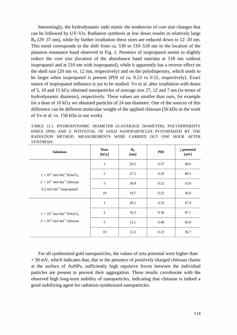
114
Interestingly, the hydrodynamic radii mimic the tendencies of core size changes that
can be followed by UV-Vis. Radiation synthesis at low doses results in relatively large
Rh (29–37 nm), while by further irradiation these sizes are reduced down to 12–20 nm.
This trend corresponds to the shift from ca. 530 to 510–518 nm in the location of the
plasmon resonance band observed in Fig. 1. Presence of isopropanol seems to slightly
reduce the core size (location of the absorbance band maxima at 518 nm without
isopropanol and at 510 nm with isopropanol), while it apparently has a reverse effect on
the shell size (20 nm vs. 12 nm, respectively) and on the polydispersity, which tends to
be larger when isopropanol is present (PDI of ca. 0.23 vs 0.52, respectively). Exact
nature of isopropanol influence is yet to be studied. Vo et al. after irradiation with doses
of 5, 10 and 15 kGy obtained nanoparticles of average size 27, 12 and 7 nm (in terms of
hydrodynamic diameter), respectively. These values are smaller than ours, for example
for a dose of 10 kGy we obtained particles of 24 nm diameter. One of the sources of this
difference can be different molecular weight of the applied chitosan (56 kDa in the work
of Vo et al. vs. 158 kDa in our work).
TABLE 12.1. HYDRODYNAMIC DIAMETER (Z-AVERAGE DIAMETER), POLYDISPERSITY
INDEX (PDI) AND Ζ POTENTIAL OF GOLD NANOPARTICLES SYNTHESIZED BY THE
RADIATION METHOD. MEASUREMENTS WERE CARRIED OUT ONE HOUR AFTER
SYNTHESIS.
For all synthesized gold nanoparticles, the values of zeta potential were higher than
+ 30 mV, which indicates that, due to the presence of positively charged chitosan chains
at the surface of AuNPs, sufficiently high repulsive forces between the individual
particles are present to prevent their aggregation. These results corroborate with the
observed high long-term stability of nanoparticles, indicating that chitosan is indeed a
good stabilizing agent for radiation-synthesized nanoparticles.
Solutions Dose
[kGy]
Rh
[nm] PDI
ζ potential
[mV]
1 × 10⁻³ mol dm-3 HAuCl4
2 × 10⁻³ mol dm-3 chitosan
0.2 mol dm-3 isopropanol
1 29.2 0.57 48.0
2 27.2 0.29 48.3
5 36.8 0.52 33.9
10 19.7 0.52 36.0
1 × 10⁻³ mol dm-3 HAuCl4
2 × 10⁻³ mol dm-3 chitosan
1 28.2 0.52 47.4
2 16.3 0.36 47.1
5 13.1 0.40 45.0
10 12.2 0.23 36.7

115
FINAL REMARKS 12.4.
Results obtained so far indicate that, in accordance with recent literature, radiation
method can be applied, in appropriate conditions, to obtain particles of gold with
nanometric size in aqueous solutions of chloroauric acid, when chitosan is used to
stabilize the obtained particles. This work will be continued and also expanded to
synthesizing gold nanoparticles by sonochemical methods and to testing various
polymers as the stabilizing agents.
PART II.
Synthesis of polymer nanocarriers for bombesin-DOTA conjugate.
INTRODUCTION 12.1.
One of the hypotheses planned to be tested in this Project is that internally cross-
linked chains of hydrophilic polymers - nanogels - could be suitable nanosized carriers
of radiopharmaceuticals. Such carriers must have proper physicochemical properties,
reasonable biocompatibility and stability. Furthermore, they should actively target
cancer cells (therefore they must possess targeting moieties) and be capable of selective
binding of ions of the radioisotopes used in the therapy (and hence the need for
chelating moieties).
The requirement of stability implies that the nanogel itself should contain
intramolecular bonds of the covalent type, and that the coupling of the required moieties
to the nanogel core should also be preferentially accomplished by covalent chemical
bonds.
Since IAEA provided the participants with small amount (20 mg) of a conjugate
compound that contains both the targeting moiety (bombesin) and the chelating moiety
(DOTA), and has a free amino group left for attachment to various nanocarriers, our aim
in this part of the Project is to elaborate the synthesis of nanocarriers (nanogels) and test
their physicochemical properties, as well as to couple the bombesin-DOTA molecules to
these nanogels and test the physicochemical properties of the whole system (nanogel
with covalently linked bombesin-DOTA molecules).
In the first year our efforts concentrated on studies on synthesizing nanogels which
would be suitable as the carriers. We have chosen poly(acrylic acid) – PAA – as the
substrate polymer. This choice was based on:
highly hydrophilic character of the polymer,
presence of carboxylic groups, which can be used for coupling with the amine
group linker of the bombesin-DOTA molecules,
some previous experience of our team in synthesizing PAA nanogels by
radiation technique.

116
According to Encyclopedia of Nanotechnology, definition of nanogels may be
directly derived from definition of polymeric gel, i.e., a two-component system
consisting of
a permanent three-dimensional network of linked polymer chains, and molecules of a
solvent filing the pores of this network [12.24]. Based on this approach polymeric
nanogels are classified as particles of polymer gels with colloidal properties, having the
dimensions in the order of nanometers. One can also distinguish bigger analogues of
micrometer size called microgels. Another way of defining polymeric nano- and
microgels is based on the specific form of these particles: all the chain segments are
linked together, thus being a part of one macromolecule. That is why nano- and
microgels can be defined as internally cross-linked polymeric chains are considered as a
distinct type of macromolecules.
There is a growing interest in synthesizing nanogels and testing them for biomedical
applications, mainly in the fields of nanoscale drug delivery systems and DNA carriers
for gene therapy. Flexibility in the chemical composition of the substrate polymers and
a variety of possible functional moieties that can be attached to the polymer backbone
leads to a broad palette of possible properties. For instance, nanogels containing
carboxylic groups are expected to be mucoadhesive and able to bind cationic drugs.
A number of synthetic routes have been developed for synthesis of nano- or
microgels of different chemical structure, architecture and properties. One of them is
radiation-induced intramolecular crosslinking of single, linear polymer chains,
developed by Rosiak and
co-workers.[12.25–12.28] Nanogels prepared by using radiation are sterile and do not
require further purification. Radiation synthesis of nanogels based on biocompatible
synthetic polymers, their derivatization and testing for biomedical applications is
currently developed in many leading radiation laboratories (Hacettepe University
[12.29–12.30], University of Maryland [12.31], Dresden University of Technology
[12.32–12.33] and University of Palermo [12.34–12.37]).
RADIATION SYNTHESIS OF POLY(ACRYLIC ACID) NANOGELS 12.2.
In this work we focused on the synthesis of nanogels from poly(acrylic acid) (PAA)
as a promising carrier of the peptide-derived drugs. First stage of the project was
devoted to analysis of changes in physicochemical parameters of PAA subjected to
ionizing radiation. Figures 3 and 4 illustrate the changes in weight-average molecular
weight and radius of gyration of PAA subjected to pulse irradiation in solutions of
various concentrations as a function of total absorbed dose. Experimental conditions are
given in the legend to Fig. 12.3. The data clearly indicate that, regardless of the polymer
concentration, at high doses we obtain strongly internally cross-linked nanogels, which,
in comparison to the starting macromolecules, have higher molecular weight but at the
same time significantly lower dimensions. While the main reason for the increase in
molecular weight is the intermolecular cross-linking occurring in the system with very

117
low yields in parallel to intramolecular recombination, the latter process is the dominant
reason for the reduction in coil dimensions.
At the first stage of irradiation (first pulse corresponding to the dose of ca. 1 kGy),
for low PAA concentrations there is some decrease in the molecular weight, which is an
evidence of the fact that the balance between intermolecular cross-linking and chain
scission is initially shifted toward the latter reaction. However for 17.5 mM solution,
this tendency is very soon reversed. The most probable reason is that internal cross-links
that have been already formed make the structure less prone to losing fragments as a
result of degradation. In other words, a chain segment of a nanogel is bound to other
segments with more than one joint; thus, if a chain break occurs in this segment, it does
not cause its detachment from the rest of the molecule (see more detailed data and
discussion below). Moreover, the nanogels, due to their reduced dimensions compared
to the initial coils, have higher translational mobility; thus, the rate of their mutual
encounters leading to intermolecular crosslinking should increase. This effect is,
however, counterbalanced to a certain extent by the reduced reaction radii, making the
necessary diffusion distance longer.
The higher the polymer concentration, the higher the yield of intermolecular cross-
linking (due to the lower number of radicals per chain and closer distance between the
coils), and therefore for 25 mM PAA there is no initial decrease in Mw. Finally, we
obtain products of molecular weight higher than the molecular weight of the parent
chains, but which have much smaller radii of gyration (Fig. 12.4). Such a tight
conformation, which is more densely packed (Fig. 12.5) than the starting coils of linear
macromolecules, is a typical feature of internally cross-linked chains.
Both for 10 mM and 17.5 mM solutions no further decrease of radius of gyration is
observed after a certain dose. It should be noted that intramolecular cross-linking is
most effective at the lowest concentration (highest number of radicals per chain at a
given dose per pulse), and perhaps a stage of tightly cross-linked structure is reached.
The possibility of further intramolecular recombination is reduced because of the
relatively rigid structure and limited mobility of the chain segments. Probably at this
stage intermolecular cross-linking starts to play a more significant role, so that some
fraction of the already formed nanogels becomes subsequently linked together.

118
FIG. 12.3. Weight-average molecular weight of PAA as a function of absorbed dose of ionizing radiation in the
course of nanogel synthesis. Ar-saturated aqueous PAA solution (pH = 2) was circulating at 1 cm3 s-1 in a gas-tight,
closed-loop system, passing through a quartz cell (0.7 cm3) that was subjected to short (2 µs) pulses of 6 MeV
electrons (pulse frequency:
0.5 Hz, dose per pulse: 1 kGy), generated by an ELU-6 linear accelerator (Eksma, Russia). Static multi-angle laser
light scattering (SLS) measurements (Brookhaven Instruments BI-200SM goniometer with a Coherent Ar-ion laser,
= 514.5 nm) were performed at
25.0 ± 0.1°C, in aqueous solution. Prior to the SLS measurement, each sample was filtered through a 0.45 µm-pore-
size filter (Minisart, Sartorius). Data were analyzed according to the Zimm algorithm,[12.38] assuming dn/dc = 0.30
cm3 g-1.[12.39] PAA concentrations (in milimol of monomer units per dm3) are given in the legend.
FIG. 12.4. Square root of the mean square radius of gyration of PAA as a function of absorbed dose in the course of
nanogel synthesis. Irradiation and SLS measurement conditions are given in Fig. 12.3.

119
FIG. 12.5. Coil density of PAA (calculated as the ratio of Mw / Rg3) as function of absorbed dose in the course of
nanogel synthesis. Irradiation and SLS measurement conditions are given in Fig. 12.3.
ELABORATION OF PEPTIDE ATTACHMENT PROCEDURE (MODEL 12.3.
STUDIES)
Second stage of the work was devoted to the elaboration of a procedure of peptide
attachment to PAA nanogels. Tryptamine has been chosen as a model compound, since
it possesses a free amino group (as the bombesin-DOTA molecule) and is a fluorescent
molecule, thus easy to detect and quantify. Two coupling procedures were tested, as
described in chapters 3.1 and 3.2 below.
12.3.1. Procedure involving carbonyl diimidazole (CDI)
As a first attempt coupling procedure based on carbonyl diimidazole (CDI) has been
made (Scheme 1). Carbonyl diimidazole (I) is a useful coupling reagent that allows one-
pot amide formation. Acyl carboxyimidazole and imidazole are initially formed but
readily react together to yield the activated species as the acylimidazole (II). Practically,
the acylimidazole is preformed and then the amine is added. This reaction, which
generates imidazole in situ, does not need an additional base and is even compatible
with HCl salts of the amine. This reagent is commonly used on a large scale in peptide
chemistry and its use can be extended to the formation of esters and thioesters. [12.40,
12.41]

120
Scheme 12.1. One-pot amide synthesis using CDI
Four different poly(acrylic acid) based nanogels (obtained by electron beam irradiation
of 10 mM Ar-saturated aqueous PAA solution (pH=2)) as well as linear (unirradiated)
PAA were used for coupling procedure. Weight-average molecular weights and sizes of
nanogels are given in Table 12.2.
TABLE 12.2. WEIGHT-AVERAGE MOLECULAR WEIGHTS AND SIZES OF PAA AND ITS
NANOGELS USED FOR COUPLING PROCEDURE WITH THE USE OF CDI.
Total absorbed dose [kGy] Mw [kDa] Rg [nm]
0 900 124
0.8 805 121
1.6 593 112
3.2 497 79
4.8 735 95
In the first step 1 ml of aqueous polymer solution (10 mM) was mixed with 1 ml of CDI
solution (60 mM in water) and stirred (2000 rpm) for 30 seconds. Next tryptamine (TA)
(5 mM, 10 ml) in 2-(N-morpholino)ethanesulfonic acid (MES) (50 mM) buffer solution
has been added. After stirring (2000 rpm, 30 seconds) solution was incubated for 10
minutes in 50 ºC. Modified polymer solutions were than dialysed (MWCO 12 kDa)

121
against water with daily water exchange and spectrophotometric detection of the
dialysis progress.
A
B
600900120015001800210024002700300033003600
T [%]
Wave number [cm-1]
TA
740cm-1 , 820cm-1 , 1010 cm-1 δN-H
1480cm-1 δC-H
1570cm-1 δN-H
2875cm-1 νN-H˅ C-H
3050cm-1, 3409cm-1 νN-H

122
C
FIG. 12.6. IR spectra of: (A) poly(acrylic acid) and its nanogels, (B) tryptamine and (C) tryptamine modified PAA.
Doses used for PAA irradiation are given in the legends.
Figure 12.6 presents IR spectra of poly(acrylic acid) and its nanogels (12.6A),
tryptamine (12.6B) and tryptamine modified poly(acrylic acid) (12.6C). Characteristic
bands for carbonyl group of PAA (1615 cm-1
and 1715 cm-1
) and amine group from
tryptamine (2875 cm-1
) have been clearly identified for polymer samples after dialysis
indicating successful coupling procedure. It is important to mention that radius of
gyration of modified nanogels was slightly affected by coupling procedure. Sizes of
nanogels before and after coupling are given in Table 12.3. Observed differences are
probably induced by changes in hydrophobicity of nanogel surface, however this is still
to be verified.
TABLE 12.3. CHANGES IN POLY(ACRYLIC ACID) RADIUS OF GYRATION BEFORE AND
AFTER COUPLING PROCEDURE.
Total absorbed dose [kGy] Rg before coupling [nm] Rg after coupling [nm]
0 124 161
0.8 121 174
1.6 112 110
4.8 95 144

123
A
B
C
FIG.12.7. Spectrofluorometric spectra of: (A) poly(acrylic acid) and exemplary PAA nanogel, (B) tryptamine and (C)
tryptamine modified PAA (ext = 280 nm, emission = 357 nm).
-0.01
0.01
300 320 340 360 380 400 420 440 460
E [-]
λ [nm]
-0.05
0
0.05
0.1
0.15
0.2
0.25
0.3
300 320 340 360 380 400 420 440 460
E [-]
λ [nm]

124
Amount of tryptamine coupled to PAA has been calculated on the basis of
spectrofluorometric analysis (ext= 80 nm, emission =357 nm). In Figure 12.7
spectrofluorometric spectra of poly(acrylic acid) and its nanogel obtained by irradiation
with 0.8 kGy (12.7A), tryptamine (12.7B) and tryptamine modified PAA nanogel
(12.7C) have been presented. Characteristic tryptamine band has been observed for
modified nanogel. Efficiency of coupling has been determined as 2.5%, which means
that 2.5% of the carboxyl groups have been modified by attachment of tryptamine.
Assuming an initial number-average molecular weight of the linear PAA as 0.5 Mw =
450 kDa and taking into account the molecular weight of the repeating unit (72 Da), this
coupling efficiency corresponds to ca. 150 tryptamine groups coupled, on average, to
each polymer chain.
12.3.2. Procedure involving N-(3-dimethylaminopropyl)-N′-ethylcarbodiimide
hydrochlo-ride (EDC)
This procedure was based on the work of Su and coworkers [12.42]. In this method,
N-(3-dimethylaminopropyl)-N′-ethylcarbodiimide hydrochloride (EDC) has been used
as the coupling agent between poly(acrylic acid) and tryptamine. Stoichiometric ratio
between PAA carboxylic groups and tryptamine was chosen to determine effectiveness
of the attachment procedure. Both coupling and dialysis of the product were performed
at 10°C.
Figure 12.8 illustrates changes in coupling efficiency (percent of carboxylic groups
with attached tryptamine) as a function of activation time with EDC for linear
(unirradiated) poly(acrylic acid). Coupling efficiency, determined by
spectrophotometric analysis of tryptamine concentration in both dialyzed (nanogel) and
dialyzing solutions, was 35–40% regardless of the reaction time, when the latter was
equal to or longer than 30 minutes.
The same procedure has been used for modification of PAA nanogels obtained from
10mM solution. Regardless of changes in the properties of macromolecules resulting
from irradiation (changes in molecular weight and radius of gyration are presented in
Figs. 12.3 and 12.4) coupling efficiency was similar (around 40%) to that obtained for
linear chains (Figure 12.9).

125
FIG. 12.8. Coupling efficiency (percent of carboxylic groups with attached tryptamine) as a function of EDC
activation time.
FIG. 12.9. Coupling efficiency (percent of carboxylic groups with attached tryptamine) for PAA nanogels obtained by
pulse-irradiation of 10 mM PAA solutions as a function of total absorbed dose. Activation time 60 minutes.
While the presented results clearly indicate that the applied procedures allow to
couple to PAA nanogels molecules bearing a free amino groups, further optimization is
0 1 2 3 4 5 6 7 8 9
0
5
10
15
20
25
30
35
40
45
50
55
60
Functionaliz
ation e
ffic
iency [%
]
Total absorbed dose [kGy]

126
definitely needed, after consultation with other CRP participants as to the desired degree
of coupling.
4. Final remarks
Nanogels of poly(acrylic) acid can be easily synthesized by intramolecular cross-
linking of polymer chains using radiation technique. By choosing synthesis parameters
one can, within some limits, adjust the molecular weight and size of the products. A
chemical procedure has been successfully tested for coupling PAA chains to a model
protein via a free amino group. After optimization of this procedure, tests will be
performed on coupling PAA nanogels with bombesin-DOTA conjugate.
REFERENCES TO CHAPTER 12
[12.1] LOUIS C., PLUCHERY O., (2012) Gold Nanoparticles for Physics,
Chemistry and Biology. Imperial College Press, London.
[12.2] CHRISTINE-DANIEL M., ASTRUC D., (2004) Gold nanoparticles:
assembly, supramolecular chemistry, quantum-size-related properties, and
applications toward biology, catalysis, and nanotechnology. Chem Rev 104,
293–346.
[12.3] MURUGAN, M., ANTHONY, K.J.P., JEYARAJ, M., RATHINAM, N.K.R.,
GURUNATHAN S,, (2014) Biofabrication of gold nanoparticles and its
biocompatibility in human breast anenocarcinoma cells (MCF-7). J Ind Eng
Chem 20, 1713–1719.
[12.4] SCHÄFFLER M., SOUSA F., WENK, A., SITIA, L., HIRN, S., SCHLEH,
C., HABERL, N., VIOLATTO, M., CANOVI, M., ANDREOZZI, P.,
SALMONA, M., BIGINI, P., KREYLING, W.G., KROL, S., (2014) Blood
protein coating of gold nanoparticles as potential tool for organ targeting.
Biomaterials 35 (10), 3455–3466.
[12.5] CUTLER, C.S., CHANDA, N., SHUKLA, R., SISAY, N., CANTORIAS,
M., ZAMBRE, A., MCLAUGHLIN, M., KELSEY, J., UPENANDRAN, A.,
ROBERTSON, D., DEUTSCHER, S., KANNAN, R., KATTI, K., (2013)
Nanoparticles and phage display selected peptides for imaging and therapy of
cancer. Recent Res Cancer Res 194, 133–147.
[12.6] CUTLER, C.S., HENNKENS, H.M., SISAY, N., HUCLIER-MARKAI, S.,
JURISSON, S.S., (2013) Radiometals for combined imaging and therapy.
Chem Rev 113, 858–883.
[12.7] DOYEN, M., BARTIK, K., BRUYLANTS, G., (2013) UV-Vis and NMR
study of the formation of gold nanoparticles by citrate reduction: Observation
of gold-citrate aggregates.
J Colloid Interface Sci 399, 1–5.
[12.8] GACHARD, E., REMITA, H., KHATOURI, J., KEITA, B., NADJO, L.,
BELLONI, J., (1998) Radiation-induced and chemical formation of gold
clusters. New J Chem 22, 1257–1265.
[12.9] CARUSO, R.A., ASHOKKUMAR, M., GRIESER, F.; (2002) Sonochemical
formation of gold sols. Langmuir 18, 7831–7836.

127
[12.10] OKITSU, K., ASHOKKUMAR, M., GRIESER, F., (2005) Sonochemical
synthesis of gold nanoparticles: effects of ultrasound frequency. J Phys Chem
B 109, 20673–20675.
[12.11] SPOTHEIM-MAURIZOT, M., MOSTAFAVI, M., DOUKI, T., RIGNY, P.,
(2008) Radiation Chemistry. From Basics to Applications in Material and
Life Sciences. EDP Sciences, Les Ulis.
[12.12] DASH, M., CHIELLINI, F., OTTENBRITE, R. M., CHIELLINI, E., (2011)
Chitosan - a versatile semi-synthetic polymer in biomedical applications.
Prog Polym Sci 36, 891–1014.
[12.13] CROISER, F., JEROME, C., (2013) Chitosan-based biomaterials for tissue
engineering. Eur Polym J 49, 780–792.
[12.14] HU, L., MAO, Z., GAO, C., (2009) Colloidal particles for cellular uptake and
delivery.
J Mater Chem 19, 3108–3115.
[12.15] WEI, D., QIAN, W.; (2008) Facile synthesis of Ag and Au nanoparticles
utilizing chitosan as a mediator agent. Colloids Surf B 62, 136–142.
[12.16] CAZACU, A., BINDAR, D., TARTAU, L., HRITCU, L., STEFAN, M.,
NITA, L., IONESCU, C., NICA, V., RUSU, G., DOBROMIR, M., MELNIG,
V. (2011) Effect on nerve structures of functionalized gold-chitosan
nanoparticles obtained by one pot synthesis. Ann Univ Iasi 12, 45–51.
[12.17] VO, K.D.N., GUILLON, E., DUPONT, L., KOWANDY, C., COQUERET,
X., (2014) Influence of Au(III) interaction with chitosan on gold
nanoparticles formation. J Phys Chem C 118, 4465–4474.
[12.18] VO, K.D.N., KOWANDY, C., DUPONT, L., COQUERET, X., HIEN, N.Q.,
(2014) Radiation synthesis of chitosan stabilized gold nanoparticles
comparison between e-beam and irradiation. Radiat Phys Chem 94, 84–87.
[12.19] ROSIAK, J.M., ULANSKI, P., AL-ASSAF, S., (2009) Protocol for
determination of intrinsic viscosity of chitosan, IAEA Co-ordinated Research
Programme: Development of Radiation-Processed Products of Natural
Polymers for Application in Agriculture, Healthcare, Industry and
Environment; International Atomic Energy Agency: Research Coordination
Meeting, Reims, France, 201–221.
[12.20] ISO/ASTM51607–13. Standard Practice for Use of the Alanine-EPR
Dosimetry System.
[12.21] OH, E., SUSUMU, K., GOSWAMI, R., MATTOUSSI, H. (2006) One-phase
synthesis of water-soluble gold nanoparticles with control over size and
surface functionalities. Langmuir 26, 7604–7613.
[12.22] ULANSKI, P., VON SONNTAG, C.; (2000) OH-radical-induced chain
scission of chitosan in the absence and presence of dioxygen. J Chem Soc
Perkin Trans 2 2000, 2022–2028.
[12.23] LI ,T., PARK, H.G., CHOI, S.H.; (2007) -Irradiation-induced preparation of
Ag and Au nanoparticles and their characterizations. Mat Chem Phys 105,
325–330.
[12.24] ULANSKI, P., ROSIAK, J.M., (2004) Polymeric nano/microgels; In:
Encyclopedia of Nanoscience and Nanotechnology. HS Nalwa (Ed.), Vol.
VIII, American Scientific Publishers, Stevenson Ranch, CA, USA, pp. 845–
871.
[12.25] ULANSKI, P., JANIK, I., ROSIAK, J.M., (1998) Radiation formation of
polymeric nanogels. Radiat Phys Chem 52, 289–294.

128
[12.26] KADLUBOWSKI, S., GROBELNY, J., OLEJNICZAK, W., CICHOMSKI,
M., ULANSKI, P., (2003) Pulses of fast electrons as a tool to synthesize
poly(acrylic acid) nanogels. Intramolecular crosslinking of linear chains in
additive-free aqueous solution. Macromolecules 36, 2484–2492.
[12.27] JESZKA, J.K., KADLUBOWSKI, S., ULANSKI, P., (2006) Monte Carlo
simulations of nanogels formation by intramolecular recombination of
radicals on polymer chain. Dispersive kinetics controlled by chain dynamics.
Macromolecules 39, 857–870.
[12.28] KADLUBOWSKI, S., ULANSKI, P., ROSIAK, J.M., (2012) Synthesis of
tailored nanogels by means of two-stage irradiation. Polymer 53, 1985–1991.
[12.29] ISIK, S.D., GÜVEN, O., (2010) Preparation of poly(vinyl pyrrolidone)
nanogels by radiation-induced crosslinking of dilute aqueous solutions.
Contributed Paper at IRaP 2010, Univ. of Maryland, College Park, USA, 25–
29 Oct. 2010.
[12.30] ISIK, S.D., (2011) Preparation of poly(vinyl pyrrolidone) nanogels by
radiation-induced cross-linking. Thesis, Hacettepe University, Ankara,
Turkey.
[12.31] AN, J.C., WEAVER A., KIM, B., BARKATT, A., POSTER, D.,
VREELAND W.N., SILVERMAN J., AL-SHEIKHLY.M., (2011) Radiation-
induced synthesis of poly(vinylpyrrolidone) nanogel. Polymer 52, 5746–
5755.
[12.32] ARNDT. K.F, SCHMIDT. T., REICHEL.T.R., (2001) Thermo-sensitive
poly(methyl vinyl ether) micro-gel formed by high energy radiation. Polymer
42, 6785–6791.
[12.33] KRAHL, F., ARNDT, K.F., (2010) Synthesis of microgels by radiation
methods. Adv Polym Sci 234, 95–128.
[12.34] DISPENZA, C., GRIMALDI N,.A., SABATINO, M.A., TODARO, S.,
BULONE, D., GIACOMAZZA, D., PRZYBYTNIAK, G., ALESSI, S.,
SPADARO, G, (2012) Studies of network organization and dynamics of e-
beam crosslinked PVPs: from macro to nano. Radiat Phys Chem 81 1349–
1353.
[12.35] DISPENZA C, SABATINO MA, GRIMALDI N, BULONE D, BONDI ML,
CASALETTO MP, RIGOGLIUSO S, ADAMO G, GHERSI G; (2012)
Minimalism in radiation synthesis of biomedical functional nanogels.
Biomacromolecules 13, 1805–1817.
[12.36] DISPENZA C, RIGOGLIUSO S, GRIMALDI N, SABATINO MA,
BULONE D, BONDÌ ML, GHERSI G; (2013) Structure and biological
evaluation of amino-functionalized PVP nanogels for fast cellular
internalization. React Funct Polym 73, 1103–1113.
[12.37] DISPENZA C, ADAMO G, SABATINO MA, GRIMALDI N, BULONE D,
BONDI ML, RIGOGLIUSO S, GHERSI G; (2014) Oligonucleotides-
decorated-poly(N-vinyl pyrrolidone) nanogels for gene delivery. J Appl
Polym Sci 131, 39774–39782.
[12.38] KRATOCHVIL P; (1987) Classical Light Scattering from Polymer Solutions.
Elsevier, Amsterdam.
[12.39] ULANSKI P, BOTHE E, HILDENBRAND K, ROSIAK JM, von Sonntag C;
(1996) Hydroxyl-radical-induced reactions of poly(acrylic acid); a pulse
radiolysis, EPR and product study. Part I. Deoxygenated aqueous solutions. J
Chem Soc Perkin Trans 2 1996, 13–22.
[12.40] MONTALBETTI CAGN, FALQUE V; (2005) Amide bond formation and
peptide coupling. Tetrahedron 61 10827–10852.

129
[12.41] Hermanson GT, Bioconjugate Techniques, Academic Press, 1996.
[12.42] SU S, WANG H, LIU X, WU Y, NIE G; (2013) iRGD-coupled responsive
fluorescent nanogel for targeted drug delivery. Biomaterials 34, 3523–3533.

130
13. DEVELOPING DELIVERY SYSTEMS FOR RADIOPHARMACEUTICALS
SAY CHYE JOACHIM LOO
Nanyang Technological University, Singapore, Singapore
Nanotechnology has found its presence in many industries and applications, and one
such promising application is in the biomedical arena. It has been exploited for the
development of nanomedicine, for drug delivery and bioimaging purposes. For
example, drug delivery in the form of nanomedicine utilizes nano-sized particles to
transport and release pharmaceutical compound into the body, to achieve the most
desirable therapeutic outcome and in the safest possible manner. Here, we review some
of the particulate drug delivery systems that have been developed in the School of
Materials Science and Engineering (MSE), Nanyang Technological University (NTU),
Singapore. Towards the end, a summary of the results obtained till date, in contribution
towards the CRP, will be reported.
MULTILAYERED PARTICULATE SYSTEMS 13.1.
Double-layered ternary-phase microparticles composed of a poly(D,L-lactide-co-
glycolide) (50:50) (PLGA) core and a poly(L-lactide) (PLLA) shell impregnated with
poly(caprolactone) (PCL) particulates were loaded with ibuprofen (IBU) and
metoclopramide HCl (MCA) through a one-step fabrication process. MCA and IBU
were localized in the PLGA core and in the shell, respectively. The aim was to study the
drug release profiles of these double-layered ternary-phase microparticles in comparison
to binary-phase PLLA (shell) / PLGA (core) microparticles and neat microparticles
(FIG. 1). The particle morphologies, configurations and drug distributions were
determined using scanning electron microscopy (SEM) and Raman mapping. The
presence of PCL in the PLLA shell gave rise to an intermediate release rate of MCA
between that of neat and binary-phase microparticles. The ternary-phase microparticles
were also shown to have better controlled release of IBU than binary-phase
microparticles. The drug release rates for MCA and IBU could be altered by changing
the polymer mass ratios [13.1].
Ternary-phase microparticles, therefore, provide more degrees of freedom in preparing
microparticles with a variety of release profiles and kinetics. This study presents a
feasible route to load radiopharmaceuticals into such particles, and would be further
explored.

131
FIG. 13.1. (a) Double-layered microparticle; (b) Fluorescent-labelled double-layered sub-micron particle; (c)
Raman mapping of drug-loaded double-layered microparticle; (d) Sequential release of two different drugs from
double-layered microparticle; (e) Different morophologies of double-layered microparticles. Ref: PCT Application
no. PCT/SG2010/000215; Acta Biomaterialia 2012; 8: 2271.
MICROENCAPSULATION OF NANOPARTICULATES 13.2.
Drug delivery is the administration of pharmaceutical agent(s) to achieve a therapeutic
effect in patients. Conventional drug-delivery strategies often require repetitive drug
administration and this can lead to strongly fluctuating drug levels that may pose undue
physiological discomfort to the recipient. Furthermore, certain drugs at high
concentrations can sometimes trigger undesirable toxicity or immunological responses
in the patient, which can be detrimental. Proper drug administration requires a fine
balance between time, dosage, and mode of delivery, and, more importantly, should aid
in improving patient compliance and clinical outcomes. To help realize a multiple-drug
delivery system, the design of a polymeric microcapsule that can house different drug-
loaded particles using a modified solid/water/oil/water (S/W/O/W) emulsion technique
was developed. A simple emulsion technique was devised to encapsulate multiple
fluorophore-loaded particles within a single polymeric microcapsule (FIG. 13.2(a)). The
largeness in size for the microcapsules had been selected to present insights into the
interior structure of the construct as precedence to miniaturization of the system. Visual
analyses (SEM and CLSM) had confirmed the feasibility of this methodology (FIG.
13.2(b)). Two drugs of different hydrophilicities were also loaded into PLLA
microparticles and successfully encapsulated in the PLGA microcapsule. Drug release
studies showed that the PLGA shell provided a means to retard burst release, while
controlling the release [13.2–13.4]. The results presented offer more opportunities to
further examine the system’s effectiveness as a drug delivery platform for
radiopharmaceuticals, which would be explored in future research undertakings.

132
HYBRID CERAMIC/POLYMER PARTICULATES FOR BONE 13.3.
TARGETING
Using sodium dodecyl sulphate micelles as template, hollow-cored calcium phosphate
nanocapsules were produced. The surfaces of the nanocapsule were subsequently
silanised by a polyethylene glycol (PEG)-based silane with an N-hydroxysuccinimide
ester end groups which permits for further attachment with bisphosphonates (BP).
Characterizations of these nanocapsules were investigated using Field Emission
Scanning Electron Microscopy (FESEM), Transmission Electron Microscopy, Fourier
Transform Infra-Red Spectroscopy, X-ray diffraction, X-ray photoelectron spectroscopy
and Dynamic Light Scattering. To further validate the bone-targeting potential, dentine
discs were incubated with these functionalised nanocapsules. FESEM analysis showed
that these surface-modified nanocapsules would bind
Strongly to dentine surfaces compared to non-functionalized nanocapsules. We envisage
that respective components would give this construct a bifunctional attribute, whereby
(1) the shell of the calcium phosphate nanocapsule would serve as biocompatible
(a) (b)
FIG. 13.2. (a) Schematic representation of simultaneously encapsulation of particles loaded with two different dyes within
a single microcapsule; (b) FESEM images of a typical microcapsule excised to disclose the cross-section region. (a–c)
Subsequent magnification had revealed the presences of smaller PLLA microparticles residing within the hollow space of
the microcapsule. Note that no microparticles were found on the outer shell region of the microcapsule. Ref: Assigned
Singapore Patent No. 190009; Advanced Healthcare Materials 2012; 1: 159.

133
coating aiding in gradual osteoconduction, while (2) surface BP moieties, acting as
targeting ligands, would provide the bone-targeting potential of these calcium phosphate
nanocapsules [13.5–13.6]. Such nanocapsules can be considered for the delivery of Sm-
153-EDTMP for bone targeting.
FIG. 13.3. (a) Schematic representation of coating drug-loaded PLGA particles with calcium phosphate layer; (b)
SEM images of PLGA particles showing calcium phosphate nanoparticles coated on its surface. Ref: USA Appl. No.:
13/885,931 and Europe Appl. No.: 11842961.2; PCT/SG2010/000214; Journal of Materials Science: Materials in
Medicine 2014 Accepted.
HYDROPHILIC-HYDROPHOBIC CORE-SHELL PARTICLES 13.4.
Current focus on particulate drug delivery entails the need for increased drug loading
and sustained release of water soluble drugs. Commonly studied biodegradable
polyesters, such as poly(lactide-coglycolide) (PLGA) and poly(L-lactide) (PLLA), are
lacking in terms of loading efficiency of these drugs and a stable encapsulation
environment for proteins. While hydrogels could enable higher loading of hydrophilic
drugs, they are limited in terms of controlled and sustained release. With this in mind,
the aim was to develop microparticles with a hydrophilic drug-loaded hydrogel core
encapsulated within a biodegradable polyester shell that can improve hydrophilic drug
loading, while providing controlled and sustained release. Herein, we report a single
step method of fabricating microparticles via a concurrent ionotropic gelation and
solvent extraction. Microparticles fabricated possess a core–shell structure of alginate,
encapsulated in a shell constructed of either PLGA or PLLA. The cross-sectional
morphology of particles was evaluated via scanning electron microscopy, calcium
alginate core dissolution, FT-IR microscopy and Raman mapping (FIG. 13.4). The
incorporation of alginate within PLGA or PLLA was shown to increase encapsulation
efficiency of a model hydrophilic drug metoclopramide HCl (MCA). The findings

134
showed that the shell served as a membrane in controlling the release of drugs [13.7].
Such gel-core hydrophobic-shell microparticles thus allow for improved loading and
release of water soluble drugs, and can be considered for the loading of fluid based
hydrophilic radiopharmaceuticals.
FIG. 13.4. Schematic diagram of alginate-PLGA/PLLA core-shell particle and SEM images of subsequent particles
prepared accordingly. Release profile from such a particle is found on the bottom right corner. Ref:
PCT/SG2013/000051; Biomaterials Science 2013; 1: 486.
RESULTS OBTAINED FOR CRP 13.5.
The key project objective is to produce polymeric (synthetic and natural) nanoparticles
for the development of new generation of nanoparticle-based tumour specific
radiopharmaceuticals. Bombesin-conjugated PLGA nanoparticles will be co-produced
by Thailand/Singapore.
The team has produced PLGA-NH2 nanoparticles of sizes ~200nm (FIG. 13.5a). The
hydrodynamic size of these nanoparticles were further validated using dynamic light
scattering (DLS) (FIG. 13.5b). These PLGA nanoparticles were subsequently sent to
Thailand to be conjugated with bombesin.

135
FIG.13.5. a: PLGA nanoparticles under FESEM of sizes ~200nm; b: DLS measurement of hydrodynamic size of
PLGA nanoparticles.
These nanoparticles were conjugated with bombesin, and results from FTIR and EDX
showed successful conjugation (FIG. 13.6). At the same time, the particle sizes after
conjugation remained unchanged at ~200nm (FIG. 13.7), while nanoparticles are now
negatively-charged upon conjugation.
a. b.

136
FIG. 13.6. a: FTIR spectra showing conjugation of Bombesin onto PLGA nanoparticles; b: EDX confirming
Bombesin conjugation.

137
FIG. 13.7. a: TEM images of Bombesin-conjugated PLGA nanoparticles; b: DLS and Zeta Potential measurements of
Bombesin-conjugated PLGA nanoparticles.
CONCLUSIONS 13.6.
Particulate drug delivery systems can be developed for the delivery of drugs. Such
delivery systems shown here include multi-layered particles, microencapsulation of
nanoparticles, ceramic-polymer hybrid particles and hydrophobic-hydrophilic core-shell
particles. Of these, PLGA nanoparticles of sizes ~200nm were prepared and
characterized. These were sent to Thailand for further bombesin conjugation. FTIR and
EDX showed successful conjugation. TEM and DLS results showed that particle sizes
remained unchanged at ~200nm, while conjugated nanoparticles are now negatively-
charged.
REFERENCES TO CHAPTER 13
[13.1] LOO, S.C.L., LEE, W.L., Multi-phase microparticles and method of manufacturing
multi-phase microparticles. NTU TD ref: TD/019/09 – US Provisional No.
61/184,559 filed 5 Jun 2009 (lapsed). PCT Application no. PCT/SG2010/000215
filed 7-Jun-2010. National phase: USA.
[13.2] LOO, S.C.L., LEE, W.L., WEE, P.X., Multi-drug-loaded gastric-floating
microcapsules. US Provisional Application no. 61/683,955 filed on 16-Aug-
2012. US PCT Application No: PCT/SG2013/000282. 8 July 2013.
[13.3] T STEELE, SCJ LOO, S VENKATRAMAN, FYC BOEY. Drug loaded
nanoparticles and microparticles encapsulated into low-melting polyester wax films
locally delivered by angioplasty balloons. PCT Application no.
PCT/SG2011/000457.
[13.4] LOO, S.C.L., KHUNG, Y.L., LEE, W.L., Method for encapsulating particles. PCT
Application No: PCT/SG2011/000414. National phase: Europe (Appl. No.: 11 842
853.1), Singapore (Appl. No.: 201303178–6)

138
[13.5] LOO, S.C.J., BASTARI, K., VENKATRAMAN, S., Method for coating particles
with calcium phosphate and particles, microparticles and nanoparticles formed
thereof. NTU TD ref: TD/076/09 – US Provisional Application No. 61/416,973
filed 24-Nov-2010 (lapsed). PCT Application no. PCT/SG2011/000414 filed 24-
Nov-2011. National phase: USA (Appl. No.: 13/885,931), Europe (Appl. No.:
11842961.2), Singapore.
[13.6] VENKATRAMAN, S., LOO, S.C.J., Targeted drug delivery to bone. NTU TD
ref: TD/024/08 – US Provisional No. 61/184,548 filed 5 Jun 2009 (lapsed).
PCT Application no. PCT/SG2010/000214 filed 7-Jun-2010. National phase:
Singapore, China, Europe, India and USA
[13.7] LOO, S.C.J., LIM, M.P. LEE, W.L., Methods of manufacturing core-shell
microparticles, and microparticles formed thereof. PCT Application
No: PCT/SG2013/000051 filed 6-Feb-2013.

139
14. GREEN SYNTHESIS OF POLYSACCHARIDE AND POLYPEPTIDE-
BASED NANOPARTICLE TOWARD THE CREATION OF STABLE AND
FUNCTIONALIZED RADIOPHARMACEUTICS FOR DIAGNOSIS AND
THERAPY
W. PASANPHANa,b*
, T. RATTANAWONGWIBOONa,b
, S.WONGKRONGSAKa,b
, T.
TANGTHONGa,b
Department of Materials Science, Faculty of Science, Kasetsart University, Bangkok 10900,
Thailand
b Center of Radiation Processing for Polymer Modification and Nanotechnology (CRPN),
Department of Materials Science, Faculty of Science, Kasetsart University, Bangkok 10900,
Thailand
Abstract
Progress in green synthesis of nanoparticle and their conjugation with DOTA-bombesin (DOTA-
BBN) for diagnosis and therapy have been developed and characterized. Chitosan (CS) and silk fibroin
(SF) have been prepared as water-soluble polysaccharide and polypeptide-based antioxidant nanoparticles
(NPs) using irradiation process. DOTA-BBN was then conjugated onto NPs for radiolabeling and for
producing and stabilizing radioactive gold nanoparticles (AuNPs). There are three types of NPs, i.e. (i)
DOTA-BBN conjugated amphiphilic core-shell water-soluble chitosan nanoparticle (WSCS NPs) (122
nm) for drug encapsulation and radiolabelling, (ii) DOTA-BBN conjugated water-soluble chitosan
nanoparticle (WSCS-DOTA-BBN NPs) (86 nm) and (iii) DOTA-BBN conjugated water-soluble silk
fibroin nanoparticle (WSSF-DOTA-BBN NPs) (60–250 nm) for AuNPs preparation and stabilization.
The conjugation of DOTA-BBN onto these NPs was carried and characterized using FT-IR, SEM-EDX,
XPS, TEM and DLS. The uses of WSCS-DOTA-BBN NPs and WSSF-DOTA-BBN NPs to create non-
radioactive AuNPs have been carried out and analyzed by UV-vis, TEM, SAXS and DLS. The NPs
samples (i, ii and iii) will be distributed to the CRP member for further studies on their stability in
biological media, radiolabelling, cell internalization, etc. Quantitative analysis of the amount of DOTA-
BBN in the NPs sample will be analyzed using SEM-EDX. The other NPs for radiopharmaceuticals will
also be continuously developed and characterized.
INTRODUCTION 14.1.
Synthesis of gold nanoparticles (AuNPs) is one of the expanding research areas in
nanotechnology. With their novel properties, they have been applied in many
applications particularly biomedicine and other bio-related applications, such as drug
delivery [14.1], photothermal therapy [14.2], diagnosis [14.3] and biosensor [14.4].
Several methods such as chemical reduction [14.5], sonochemical [14.6], and
photochemical reduction [14.7] have been developed to synthesize metal NPs including
AuNPs. In the past decade, with some hazardous chemicals remaining in the products,
the alternative green synthesis has been emerged. The green synthesis of AuNPs has
been performed using natural reducing and stabilizing agents. Biomolecules from plant
extracts have been widely proposed as green synthesis of AuNPs [14.8] as a result of
their antioxidant and reducing properties. However, small molecular natural products
still lack of the stability including chelating and stabilizing efficiencies.
Biomacromolecules, e.g., starch, chitosan, gelatin and alginate, have been the most
widely used as capping agents for the synthesis of metal NPs. Unlike the small
molecular reducing agents, the biopolymers themselves exhibit low reactivity to reduce
metal ion to metal atom because of their inherent high molecular weight, low solubility
or lack of reducing power function.
140
Chitosan (CS), a deacetylated form of chitin, makes up the second-most naturally
abundant copolysaccharides next to cellulose. It is one of the biodegradable
carbohydrate polymers consisting of pyranose ring of β-(1,4)-2-acetamido-2-deoxy-β-
D- glucose (chitin) and β-(1,4)-2-amino-2-deoxy-β-D-glucose (chitosan) linked with a
glycosidic linkage. It dictates numerous advantage properties, such as biodegradability,
biocompatibility, bioactivity and non-toxicity. The amino (–NH2) and hydroxyl (–OH)
groups of CS at C-2 and C-6 positions not only provide reactive sites for
functionalization but also exhibit strong metal ion chelation, oil absorption,
antimicrobial, and antioxidant activities. Regarding to the antioxidants, many
researchers have focused on the extraction, identification, modification and application
of alternative natural antioxidants to use in numerous purposes in order to avoid
pathogenic risk of the synthetic antioxidants [14.9]. It is also known that the most
common chemical species, which act as antioxidant or reducing agents are hydroxyl, –
OH (e.g. phenolics), sulfhydryl, –SH (e.g. cysteine and glutathione) and amino, –NH2
groups (e.g. uric acid, spermine and proteins) [14.10]. Therefore, CS has attracted much
attention to develop as natural antioxidant and reducing agent since it has plenty of –OH
and –NH2 groups as H-atom donation [14.11] have concluded that the –OH group in the
saccharide units can react with free radicals by the H-abstraction reaction and the –NH2
group of CS can react with free radicals to form additional stable macroradicals.
Although the –OH and –NH2 groups of CS exhibit many unique properties including
antioxidant and reducing power, CS has its inherent drawback of non-reactivity and
insolubility because of strong inter- and intra-molecular hydrogen bonding. The –NH2
group (pKa~6.2-7.0) is completely protonated in acids with pKa less than 6.2 making
CS soluble. Thus, CS is insoluble in water, aqueous bases and organic solvents [14.12].
This is a vastest limitation to use CS in aqueous solution at neutral pH to apply in a
wide range of applications particularly biological media. Although several water-
soluble CS (WSCS) derivatives and radiation-induced degradation of CS have been
proposed, to the best of our knowledge the strategies to prepare WSCS-NPs via
irradiation in order to be applied as a green antioxidant and reducing agent for green
synthesis of AuNPs have not yet been demonstrated.
Silk waste (SF) is a part of the cocoons of silkworms (Bombyxmori). The
composition of such silk waste is similar to that of good silk. It composes of fibroin core
polymer (75–83%) and sericin glue-like protein (17–25%), as a coating. Primary
structure of silk fibroin (SF) polymers mainly consists of amino acid sequence of [Gly-
Ala-Gly-Ala-Gly-Ser]n. existing in glycine (Gly, 43%), alanine (Ala, 30%) and serine
(Ser, 12%) units [14.13]. SF forms an arrangment of repetitive protein layers of
antiparallel β-sheet strength. SF protein from the Bombyxmori silkworm is FDA
approved and it has been used in biomedical applications. Particularly, SF has been the
most widely developed as scaffold materials and successfully used in wound healing
and in tissue engineering of bone, cartilage, tendon and ligament tissues [14.14].
Although the chemical structure of SF is similar to collagen, SF exhibits a less
inflammatory response than the collagen including poly (lactic acid) [14.15]. More
recently, it has been reported that SF produced from Bombyxmori harbor antioxidant

141
and hypolipidemic properties [14.16]. Due to plenty of hydroxyl, amine and carboxyl
groups, SF protein is also of great interest as a natural antioxidant for applying in food,
cosmetic, pharmaceutical and biomedicine [14.17]. Up to date, SF has been developed in
micro- and nanoparticles for drug delivery applications. It is the most widely used
methods for SF nanoparticles production, such as emulsion-solvent evaporation/
extraction methods, phase separation or coacervation, self-assembly, solvent
displacement, rapid expansion of supercritical solution, and spray-drying [14.18];
however, less known aspects of radiation-induced degradation and nanostructure
formation of SF.
Herein, the goal of the work for 15 months is to prepare and characterize the
water-soluble chitosan and silk fibroin nanoparticles, i.e., WSCS NPs and WSSF NPs,
respectively using high-energetic irradiation. The properties of the obtained WSCS NPs
and WSSF NPs were characterized by FT-IR, XRD and TGA. The molecular weight of
WSCS NPs and WSSF NPs were determined by GPC and gel electrophoresis. The
morphological information and nanoscaled size were also verified using SEM, TEM and
DLS. The article also assesses the antioxidant activity and reducing power of the
WSCS-NPs and WSSF-NPs using in vitro DPPH radical scavenging activity, ferric
reducing antioxidant power (FRAP). In addition, amphiphilic core-shell WSCS NPs
(PEGMA-DCWSCS) was also prepared in order to use for anticancer drug
encapsulation and radiolabelling leading to be a dual chemotherapy and radio-diagnosis.
DOTA-BBN was conjugated onto both these three types of NPs, i.e., WSCS NPs and
WSSF NPs and PEGMA-DCWSCS. The conjugation was characterized using FT-IR,
SEM-EDX, XPS, TEM and DLS. The uses of WSCS-DOTA-BBN NPs and WSSF-
DOTA-BBN NPs to create non-radioactive AuNPs have been carried out and analyzed
by UV-vis, TEM, SAXS and DLS.
EXPERIMENTAL SECTION 14.2.
14.2.1. Preparation of NPs [14.19–14.21]
Amphiphilic core-shell nanoparticles was synthesized according to Pasanphan et
al. [14.19]. Deoxylcholic acid was conjugated onto water-soluble chitosan (WSCS)
using conjugating agent (EDC/NSH). PEGMA-shell was decorated onto the
hydrophobic WSCS by radiation-induced grafting. The mixtures were irradiated with
the doses of 2, 10, 20 and 30 kGy. The samples was transferred into a dialysis bag
(MWCO = 12,00014,000 Da), which was enclosed into 2 L of distilled water and
dialyzed with renewed water for 48 h. The product was frozen and then lyophilized to
obtain PEGMA-DCWSCS. Degree of grafting (DG) of PEGMA onto DCWSCS was
calculated according to the following relationship: [(WgWo)/Wo]×100, where Wo and
Wg represent the initial weight of DCWSCS and the grafted weight of PEGMA-
DCWSCS, respectively. The samples were characterized by FTIR, EA, TGA, XRD,
TEM, AFM, SEM, GPC and DLS.
Water-soluble chitosan (WSCS NPs) was prepared according to Pasanphan et al.

142
[14.20]. Briefly, CS in aqueous acetic solution was gamma-rays irradiated in air. The
sample was neutralized using NaOH and separated by dialysis membrane for 24 h.
Water-soluble silk fibroin (WSSF NPs) was simply prepared according to
Wongkrongsak et al. [14.21]. Briefly, silk fibroin was dissolved in water and irradiated
with the doses of 10 and 25 kGy. The samples were characterized by FTIR, TGA, XRD,
TEM, GPC and DLS.
14.2.2. DOTA-BBN conjugation onto NPs
WSCS and WSSF NPs: DOTA-BBN (1 mg/ml) solution of 200 µl was gently
stirred and mixed with 2 ml of EDC (1 mol equivalent to WSCS, 22.6382 mg) and NHS
(1 mol equivalent to WSCS, 13.5911 mg) each solution. WSCS (20 mg) was dissolved
in 5 ml of distilled water and added to the mixture solution. The volume of solution was
adjusted to 10 ml. The reaction was leaved for 6h at room temperature. The sample was
purified by dialysis membrane (MWCO = 3,500 Da) with renewed water for 48 h.
Similarly, conjugation of DOTA-BBN on WSSF (20 mg) was carried out by using EDC
(1 mol equivalent to WSSF, 9.9481 mg) and NHS (1 mol equivalent to WSSF, 5.9725
mg) as the same procedure.
PEGMA-DCWSCS: DOTA-BBN (0.3 mg/ml, 100 µl) was added in 300 µl
distilled water. Each of EDC (3.5261 mg) and NHS (2.1169 mg) were dissolved in
distilled water (800 µl). EDC and NHS solution were added to DOTA-BBN solution
and stirred for 15 min. 1 ml of PEGMA-DCWSCS (2 mg/ml) was dropped in mixture
solution. The mixture solution was kept on gentle stirring for 6 h at room temperature.
Unreacted DOTA-BBN was separated from product by dialysis membrane (MWCO =
1,000 Da). The conjugated NPs were characterized by FTIR, SEM-EDX, XPS, TEM
and DLS.
14.2.3. Green synthesis of non-radioactive 197
AuNPs [14.20, 14.21]
WSCS-NPs (100 mg) were dissolved in distilled water (100 mL) and stirred
overnight to obtain homogeneous solution. The concentration at 0.0010, 0.02, 0.04,
0.10, 0.20, 0.40 and 0.60 mg/ml of 5 ml solution were mixed with HAuCl4 solution (0.2
M, 5 ml). The mixture was reacted at room temperature for 24 h before collecting UV-
vis spectra. The absorbance was measured in a 1-cm path length quartz cuvette with a
Libra S32 Spectrophot- ometer, Biochrom, UK in the range of visible light (500–700
nm). Similarly, the formations of AuNP in CS-HOAc, SF, WSSF-NPs and Glu were
carried out with the same procedure. AuNPs formation was characterized by UV-vis,
TEM, SAXS, AFM and DLS.
RESULTS AND DISCUSSION 14.3.
14.3.1. Amphiphilic core-shell PEGMA-DCWSCS NPs and its DOTA-conjugation
Chemical structure was confirmed by FTIR (Fig. 14.1A). It was found an increase
of ester peak at 1729 cm-1
due to the conjugation. In addition, significant increase of

143
amide peaks and N-H stretching due to the DOTA-BBN indicated the successful
conjugation. Based on DOTA-BBN chemical structure, there is a sulfur atom (S)
besides C H O and N elements. Thus S should be a good indicator for confirming the
conjugation. SEM-EDX technique was measured and it was found S in the conjugated
NPs when compared with the original NPs. This implied the successful conjugation of
DOTA-BBN onto the NPs. Particle size and surface charge were also determined by
DLS. Two particle size of DOTA-BBN conjugated core-shell NPs are 15% of 122 nm
and 81% of 836 nm.
FIG.14.1. (A) FTIR and (B) SEM-EDX of PEGMA-DCWSCS and PEGMA-DCWSCS-DOTA-BBN
14.3.2. Preparation and characterization of WSCS-NPs [14.20]
14.3.2.1. Chemical structure of WSCS and its conjugation with DOTA-BBN
DOTA-BBN was conjugated onto WSCS via the amino group of CS and carboxylic acid
group of DOTA. Conjugation was confirmed by FTIR. Increase of the amide bond and N-H
stretching confirmed the successful conjugation (Fig. 14.2A). Since in DOTA-BBN structure
contain sulfur (S) besides C H O and N elements. SEM-EDX and XPS were used to confirm the
(A) (B)

144
conjugation. SEM-EDX indicated 0.142% of S in the conjugated WSCS when compared with
non-conjugated WSCS (Fig. 14.2B). XPS also shows the S2p peak at 170 eV confirming
conjugation (Fig. 14.2C).
FIG. 14.2 Characterization of DOTA-BBN conjugated WSCS NPs by (A) FTIR (B) SEM-EDX and (C) XPS
The particle morphologies of WSCS and DOTA-BBN conjugated WSCS are shown in
Fig. 14.3(A). After conjugation, the particle size was larger. The particle size changed
from 174 to 192 nm (Fig 14.3(B). The surface charge was changed to be more neutral
when the DOTA-BBN was conjugated on WSCS.
FIG. 14.3 (A) TEM and (B) DLS of WSCS NPs and WSCS-DOTA-BBN
14.3.2.2. Green synthesis of AuNPs using WSCS NPs and WSCS-DOTA-BBN NPs
Physical appearances of AuNPs in WSCS and WSCS-DOTA-BBN at the initial
time and after 24 h are shown in Fig. 14.4. AuNPs remarkably produced after 24 h
NH2
S
NH
O
O
NHNH
O
O
NH
O
NHNH
O
NH
NH2
O
NH
O
NHNH
O
O
NH
NH
O
NH
NH2O
NH
O
O
O
O
N
N
N
N
OH
OH
O
O
O
NH2
NH2
O
NH2
N
H
NNH
O
OH
O
OHNH
O
OH
O+
NHCOCH3
O
(A) (B) (C)

145
particularly when incubated in WSCS-DOTA-BBN. UV-vis spectra show the
absorption peak at 546 indicated the formation of AuNPs. Spherical AuNPs were
observed in TEM image. The particle size observed by DLS increased from 49 nm to 86
nm when using the DOTA-BBN conjugated WSCS. In comparison, particle sizes of
AuNPs in WSCS-DOTA-BBN observed by SAXS, TEM and DLS are 24, 61 and 86
nm. The surface charge became more neutral close to zero when the WSCS-DOTA-
BBN was used to create AuNPs.
FIG. 14.4 Formation of AuNPs produced in WSCS-NPs WSCS-DOTA BBN NPs
14.3.3. Preparation and characterization of WSSF-NPs [20]
14.3.3.1. Chemical structure and morphology of WSSF-NPs
Chemical structures of SF and irradiated SF were characterized from the FT-IR
spectra (Fig. 14.6A). The FT-IR spectrum of SF also shows the peaks at 3280 cm
interpreted as N-H stretching spectrum of the irradiated SF indicated that all
characteristic peaks of the SF still remained after exposing to electron beam. One can be
seen that the peak at 3280 cm belonging to the N-H stretching also increased. The
result suggested that the terminal amino group (-NH2) formed after peptide bond
cleavage.
FIG. 14.5. (A) FT-IR spectra, (B) SEM of (a) SF and (b) WSSF-NPs [20].
(A) (B)

146
The morphological information of the lyophilized WSSF-NPs powder was
investigated by SEM as presented in Fig. 14.5B. The morphology of non-irradiated SF
presented in spherical particle, which closely packed at interfaces (Fig. 14.5B(a)). This
implies strong inter- and intra-molecular hydrogen bonds and the Vander Waals force in
SF structure which self-assemble into anti-parallel β-sheet. Compared with non-
irradiated SF, the representative morphology of the irradiated-SF prepared using a dose
of 30 kGy revealed distinguish spherical particles, which loosely packed (Fig.
14.5B(b)). The average particle size of the lyophilized WSSF-NPs was in the range of
295–560 nm. The morphological change observed in SEM micrograph indicated that SF
structure was destroyed after electron beam irradiation. The morphological data
supported the increase of the N-H (Amide II) implying the increase of amorphous phase
as demonstrated by FT-IR intensity ratio.
The chemical structure of the irradiated SF was characterized by FTIR. The main
chemical structure still remains after irradiation with the dose of 10 and 25 kGy. In
addition, the O-H and N-H stretching peaks increased due to the chain-end after
fragmentation by irradiation.
14.3.3.2. Conjugation of DOTA-BBN onto WSSF NPs
DOTA-BBN was conjugated onto SF under the same condition as already
described. The conjugation happens between the OH group of SF and the COOH group
of DOTA leading to the formation of an ester bond at 1720 cm-1
. SEM-EDX also shows
the S element of 0.081% in the conjugated SF implying that DOTA-BBN was
conjugated onto SF.
The morphologies of the SF and DOTA-BBN conjugated SF0 SF10 and SF25 are
revealed in Fig 14.7(A) and (B). Without conjugation, the particle morphology exhibits
only one phase. By conjugating with DOTA-BBN, the shell of DOTA-BBN occurred
on the surface of WSSF NPs. The particle size of WSSF-DOTA-BNN observed by
TEM exhibited larger than the non-conjugated one.

147
FIG. 14.6. (A) FT-IR spectra non-irradiated SF0, DOTA-BBN, SF0-DOTA-BBN, SF10-DOTA-BBN and SF25-
DOTA-BBN
FIG. 14.7. TEM images of (A) WSSF and WSSF-DOTA-BBN prepared SF0, SF10 and SF25.
(A)
(B)
NH2
S
HN
O
O
NH
HN
O
O
NH
OHN
NH
OHN
NH2
O
NH
OHN
NH
O
OHN
NH
O
NH
NH2O
HN
O
O
O
O
N
N
N
N
C
C
C
HO
HO
O
O
O
O
H2N
NH2
O
NH2
NH
NHN
CH
O
HN
NH
O CH3 HN
O
NH
O CH3
O

148
The particle size and surface charge were also determined by DLS. The particle sizes after
conjugation are larger than non-conjugated SF. Similar to the previous conjugation, the surface
charge also increase to be more neutral.
FIG. 14.8. (A) Particle size and (B) surface charge of WSSF and WSSF-DOTA-BBN prepared SF0, SF10 and SF25.
14.3.3.3. Green synthesis of AuNPs using WSSF NPs and WSSF-DOTA-BBN NPs
The effect of WSSF concentration on AuNPs production was carried out and
presented in Fig. 14.9A. By varying the concentration of SF10 and SF25, the AuNPs
were significantly created as observed by color change and UV absorption peak at 545
nm (Fig.14.9B (b) and (c)). On the other hand the SF0 is not effective to create AuNPs
(Fig.14.9B(a)). The result confirmed that irradiation of SF improves the reducing
power. It was also found that the number of AuNPs produced in SF10 and SF25 is
nearly the same (Fig. 14.9C). Therefore, 10 kGy would be enough to enhance reducing
power of SF.
(A) (B)

149
FIG. 14.9. (A) Physical appearance (B) UV-vis spectra and (C) absorption at maximum wavelength as a function of
concentration of WSSF0, WSSF10 and WSSF25.
The efficiency of DOTA-BBN with (Fig. 14.10A(a)) and without (Fig. 14.10A(b))
conjugated SF0, SF10 and SF25 to create and stabilize AuNPs are shown in Fig. 14.10.
SF0 is not effective to create AuNPs both with and without DOTA-BBN conjugation.
After conjugation, the creation of AuNPs decreased as seen in UV-vis (Fig. 14.10B).
The AuNPs in WSSF and WSSF-DOTA-BBN exhibited both spherical and triangle
shape with the size of 18 and 24 nm, respectively. The hydrodynamic diameters were 73
and 253 nm with the surface of -13.6 and -12.7 mV for AuNPs-WSSF and AuNPs-
WSSF-DOTA-BBN.
(C) (A) (a)
(b)
(c)
(B) (a) (b) (c)

150
FIG. 14.10. (A) Physical appearance (B) UV-vis spectra and (C) TEM images of AuNPs prepared in (a) WSSF and
(b) DOTA-BBN conjugated WSSF from difference doses.
CONCLUSIONS 14.4.
The present experiments proved that irradiation would be efficient to produce
WSCS-NPs and WSSF-NP including amphiphilic core-shell PEGMA-DCWSCS NPs in
terms of hydrophilic shell decoration. The WSCS-NPs and WSSF-NPs exhibited
excellent antioxidant activities for the AuNPs synthesis. DOTA-BBN was successfully
conjugated onto PEGMA-DCWSCS NPs, WSCS NPs, WSSF NPs. The amphiphilic
core-shell PEGMA-DCWSCS NPs would be used for anticancer drug encapsulation as
dual chemotherapy and radiolabelling for radio-diagnosis. Both WSCS-NPs and WSSF-
NPs are effective polysaccharide and polypeptide-based antioxidant, reducing and
stabilizing agents for the green synthesis of AuNPs and for further development of
radioactive 198
AuNPs for diagnosis and therapy.
ACKNOWLEDGEMENTS
The authors gratefully acknowledge Coordinated Research Project (CRP) (no.
18316), International Atomic Energy Agency (IAEA) and the Kasetsart University
Research and Development Institute (KURDI); Science Research Fund (ScRF), Faculty
of Science, Kasetsart University, Thailand for financial support. The authors also thank
the Office of Atoms for Peace (OAP), the Thailand Institute of Nuclear Technology
(TINT), Ministry of Science and Technology and Prof. Dr. Suwabun Chirachanchai, the
Petroleum and Petro- chemical College, Chulalongkorn University, for providing
gamma irradiator, electron beam accelerator and characterization facilities.
(a)
(b)

151
REFERENCES TO CHAPTER 14
[14.1] AGASTI, S.S., CHOMPOOSOR, A., YOU, C.C., GHOSH, P., KIM, C.K.,
ROTELLO, V.M., “Photoregulated release of caged anticancer drugs from gold
nanoparticles”, J. Am. Chem. Soc. 131 (2009) 5728–5729.
[14.2] HAINFELD, J.F., DILMANIAN, F.A., ZHONG, Z., SLATKIN, D.N., KALEF-
EZRA, J.A., SMILOWITZ, H.M., “Gold nanoparticles enhance the radiation therapy
of a murine squamous cell carcinoma”. Phys. Med. Biol. 55 (2010) 3045–3059.
[14.3] McMAHON, S.J., MENDENHALL, M.H., JAIN, S., CURRELL, F., “Radiotherapy
in the presence of contrast agents: a general figure of merit and its application to gold
nanoparticles”, Phys. Med. Biol. 53 (2008) 5635–5651.
[14.4] CARLO, G.D., CURULLI, A., TORO, R.G., BIANCHINI, C., CARO, T.D.,
PADELETTI, G., ZANE, D., INGO, G.M., “Green synthesis of gold-chitosan
nanocomposites for caffeic acid sensing”, Langmuir 28 (2012) 5471–5479.
[14.5] MURPHY, C.J., GOLE, A.M., STONE, J.W., SISCO, P.N., ALKILANY, A.M.,
GOLDSMITH, E.C., BAXTER, S.C.,”Gold nanoparticles in biology: beyond toxicity
to cellular imaging”. Acc. Chem. Res. 41 (2008) 1721–1730.
[14.6] NAEINI, A.T., ADELI, M., VOSSOUGHI, M., “Synthesis of gold nanoparticle
necklaces using linear-dendritic copolymers”, J. Eur. Polym. 46 (2010) 165–170.
[14.7] STERN, J.M., STANFIELD, J., KABBANI, W., HSIEH, J.T., CADEDDU, J.A.,
“Selective prostate cancer thermal ablation with laser activated gold nanoshells”, J.
Urol. 179 (2008) 748–753.
[14.8] MITTAL, A.K., CHISTI, Y., BANERJEE, U.C., “Synthesis of metallic nanoparticles
using plant extracts”, Biotechnol. Adv. 31 (2013) 346–356.
[14.9] WU, Z., MING, J., GAO, R., WANG, Y., LIANG, Q., YU, H., “Characterization and
antioxidant activity of the complex of tea polyphenols and oat β-glucan”, J. Agric.
Food Chem. 59 (2011) 10737–10746.
[14.10] DECKER, E.A., “Strategies for manipulating the prooxidative/antioxidative balance
of foods to maximize oxidative stability”, Trends Food Sci. Technol. 9 (1998) 241–
248.
[14.11] CASTANGNINO, E., OTTAVIANI, M.F., CANGIOTTI, M., MORELLI, M.,
CASETTARI, L., MUZZARELLI, R.A.A., “Radical scavenging activity of 5-
methylpyrrolidinone chitosan and dibutyryl chitin”, Carbohydr. Polym. 74 (2008)
640–647.
[14.12] RINAUDO, M., “Chitin and chitosan: properties and applications” Prog. Polym.Sci.
31 (2006) 603–632.
[14.13] KOJTHUNG, A., MEESILPA, P., SUDATIS, B., TREERATANAPIBOON, L.,
UDOMSANGPETCH, R., OONKHANOND, B., “Effects of gamma radiation on
biodegradation of Bombyx mori silk fibroin”, Int. Biodeter. Biodegr. 62 (2008) 487–
490.
[14.14] UDE, A.U., ESHKOOR, R.A., ZULKIFILI, R., ARIFFIN, A.K., DZURAIDAH,
A.W., AZHARI, C.H., “Bombyx mori silk fibre and its composite: A review of
contemporary developments”, Mater. Design. 57 (2014) 298–305.
[14.15] SHE, Z., HUANG, Z., ZHANG, B., JIN, C., FENG, Q., XU, Y., “Preparation and in
vitro degradation of porous three-dimensional silk fibroin/chitosan scaffold”, Polym.
Degrad. Stabil. 93 (2008) 1316–1322.
[14.16] ZHANG, X., GENG, X., JIANG, H., LI, J., HUANG, J., “Synthesis and
characteristics of chitin and chitosan with the (2-hydroxy-3-trimethylammonium)
propyl functionality, and evaluation of their antioxidant activity in vitro”, Carbohydr.
Polym. 89 (2012) 486–491.
[14.17] MOURE, A., CRUZ, J.M., FRANCO, D., DOMINGUEZ, J.M., SINEIRO, J.,
DOMINGUEZ, H., NUNEZ, M.J, PARAJO, J.C., “Natural antioxidants from residual
sources. Food. Chem. 72 (2001) 145–171.

152
[14.18] WANG, X., YUCEL, T., LU, Q., HU, X., KAPLAN, D.L., Silk Nanospheres and
Microspheres from Silk/PVA Blend Films for Drug Delivery. Biomaterials. 31 (2010)
1025–1035.
[14.19] PASANPHAN, W., RATTANAWONGWIBOON, T., RIMDUSIT, P., PIROONPAN,
T., “Radiation-induced graft copolymerization of poly(ethylene glycol)
monomethacrylate onto deoxycholate-chitosan nanoparticles as a drug carrier”,
Radiat. Phys. Chem. 94 (2014) 199–204.
[14.20] PASANPHAN, W., RATTANAWONGWIBOON, T., CHOOFONG, S., GUVEN, O.,
KATTI, K., “Irradiated chitosan nanoparticle as a water-based antioxidant and
reducing agent for a green synthesis of gold nanoplatforms”, Radiat. Phys. Chem. 106
(2015) 360–370.
[14.21] WONGKRONSAK, S., TANGTHONG, T., PASANPHAN, W., “Electron beam
induced water-soluble silk fibroin nanoparticles as a natural antioxidant and reducing
agent for a green synthesis of gold nanocolloid”, Radiat. Phys. Chem. Article In Press.

153
15. IN VIVO STUDIES AND PRODUCTION OF NANOSIZED DELIVERY
SYSTEMS FOR RADIOPHARMACEUTICALS
KATTESH V. KATTI
University of Missouri, Columbia,
USA
(i) Detailed In vivo toxicity profiles of Gum-Arabic functionalized Au-198
nanoparticles fully established:
Introduction: Gum arabic-coated radioactive gold nanoparticles (GA-198AuNPs) offer
several advantages over traditional brachytherapy in the treatment of prostate cancer,
including homogenous dose distribution and higher dose-rate irradiation. Our objective
was to determine the short-term safety profile of GA-198AuNPs injected intralesionally.
We proposed that a single treatment of GA-198AuNPs would be safe with minimal-to-no
evidence of systemic or local toxicity.
Methods: Nine dogs with spontaneously occurring prostatic cancer were treated.
Injections were performed with ultrasound or computerized tomography guidance.
Complete blood counts, chemistry panels, and urinalyses were performed at weekly
intervals for 1 month and imaging was repeated 4 weeks postinjection. Planar
scintigraphic images were obtained within 30 minutes of injection.
Results: No statistically significant difference was found in any hematologic or
biochemical parameter studied, nor was any evidence of tumor swelling or abscessation
found in eight dogs with repeat imaging; one dog died secondary to urethral obstruction
12 days following injection. At 30 minutes postinjection, an average of 53% of injected
dose in seven dogs was retained in the prostate, with loss of remaining activity in the
bladder and urethra; no systemic update was detected.
GA-198
AuNP production
University of Missouri Research Reactor irradiation facilities were used for the
production of 198Au and the GA-198AuNPs in the volume and dose requested by
previously published methods with slight modifications.10 Briefly, gold foil was irradi-
ated at a flux of 8×10¹³ neutrons/cm2/s; the radioactive foil was then dissolved with
aquaregia, dried, and reconstituted in hydrochloric acid to yield H198AuCl4. GA-198AuNPs
were produced by the addition of the reducing agent tris hydroxymethyl phosine-alanine
(P(CH2NHCH(CH3)-COOH)3 in the presence of gum arabic. The final solution was
brought to a pH of 7 and sterile filtered. The gold concentration in the final
nanoparticulate suspension was 0.325 mg/mL (1.6 mM; 9.026×10¹⁵ NPs). Production of
GA-198AuNPs was confirmed by quality control tests including radio thin layer
chromatography and ultraviolet–visible spectroscopy. Radio thin layer chromatography
198AuCl4 was in the nanoparticulate form. The
physicochemical parameters were established using the nonradioactive analogue (GA-
AuNP). Particles had a hydrodynamic diameter of 85 nm (±20 nm) and zeta potential of
24.5±1.5 mV as determined by dynamic light scattering measurements. The core size of
the particles was 12–15 nm as determined by transmission electron microscopy.

154
Conclusions:
Although traditional brachytherapy using 103Pd or 125I seeds are successful in treating
men with low grade prostatic cancer, significant numbers of men with intermediate and
high grade disease relapse following treatment.2,4,6–9 Highlighting the importance of
successful irradiation, evidence in the literature suggests that achieving early local
control in intermediate and high Gleason score prostatic carcinoma can decrease the risk
of disease metastasis.21,22 Therefore, treatment options that provide excellent control
of local disease while sparing normal tissues can improve both the quality and quantity
of life in men with PC. It is hypothesized that due to size, GA-198AuNPs have more
homogenous dose distribution and higher emission of energy compared to current
brachytherapy seeds used to treat PC. The gum arabic glycoprotein provides a nontoxic
coating on NPs with a hydrodynamic diameter of 85 nm and is highly stable in vivo.16
The ease of production combined with the properties of 198Au and the safety of GA-
198AuNP make this new treatment an exciting advancement in prostatic cancer therapy.
While this study was not powered to evaluate outcome, ongoing studies will evaluate
long-term efficacy and optimize dosimetry.
This study provides evidence that intralesional injection of GA-198AuNP is safe with
minimal short-term systemic toxicity in the naturally occurring large animal model of
prostatic cancer. The favorable safety profile of GA-198AuNP in this group of dogs
suggests that further study for dosimetry and therapeutic efficacy in dogs with prostate
cancer and subsequent Phase I clinical trials in men are warranted.
(ii) Theranostic efficacy studies of EGCG-Au-198 nanoparticles for treating
prostate cancer (in dogs):
Synthesis, Characterization of 198
AuNP-EGCG: 198
AuNP-EGCG has been synthesized
by stirring an aqueous solution of EGCG with reactor produced 198
AuCl4-. An instant
reaction results in the formation of EGCG conjugated radioactive gold nanoparticles
with over 99% yield of 198
AuNP-EGCG in deionized water without addition of an
external reducing agent. EGCG serves as both reducing and stabilizing agent. We have
optimized two different analytical techniques as quality control for examining the purity
of nanotherapeutic agents. Analytical Method 1: UV-Visible Spectrometry: The
formation of nanoparticles (198
AuNP-EGCG) is confirmed by UV-Visible absorption
measurements. The homogeneous distribution of nanoparticles is ascertained from the
plasmon absorption band λmax, and plasmon line width, Δλ. This data is cross
correlated with TEM and Disc centrifuge analysis techniques using non-radioactive
surrogates. Analytical Method 2: Thin Layer Chromatography: In order to determine
complete conversion of 198
AuCl4- to nanoparticles, standard thin layer chromatography
technique is used. Radioactive counting methods are used to differentiate the starting 198
AuCl4-material from the
198AuNP product. A radiochemical purity and percent
labeling is tested by TLC on Whatman cellulose paper. Briefly a few µL of the solution
is spotted onto Whatman cellulose plates and developed in methanol (2 mL) with 1 drop
of concentrated HCl (~10 M). The paper strip is scanned on a Radio-TLC-BioScan to
determine the percent radiolabeling yield. The nanoparticles remain at the origin Rf =
0, while H198
AuCl4 migrates with an Rf = 0.9. The radiolabeling yield is >99 %.
In Vitro Stability and Hemocompatibility Studies: In vitro stability and
hemocompatibility on the nanoconstruct have been studied in detail using non-
radioactive surrogates. AuNP-EGCG has met FDA standards on in vivo stability and

155
Figure 2 Biodistribution of 198
AuNP-EGCG
Figure 3 Therapeutic Efficacy of 198
AuNP-EGCG
hemocompatibility characteristics (full details on product specifications will be
provided at the time of pilot clinical trials). Our results clearly indicate that these
nanoconstructs are biocompatible and stable, in vivo, for clinical applications in the
treatment of cancer and Osteoarthritis.
Biodistribution of Nanotherapeutic Agent in Mice: The refractory features found in
prostate tumor patients’ tumor membranes can be easily compared to those refractory
features of prostate tumors dogs. Prior to performing studies in dogs, extensive
investigation was undertaken in mice to understand efficacy and dosimetry. The data
obtained from the prostate tumor bearing mice model, can be corroborated to provide a
detailed understanding of the in vivo retention and
clearance of 198
AuNP-EGCG. We have performed
in vivo studies of intratumoral administration of 198
AuNP-EGCG (3.5Ci/tumor) to groups (n=5) of
SCID mice bearing human prostate cancer
xenografts (Figure 15.2). The 198
AuNP-EGCG
nanoparticles exhibited slow clearance (leakage)
minimal/no leakage into the blood with only 0.06 %
ID/g at 24 h as shown in Figure 15.2. Lungs (0.33
% ID/g) and pancreas (0.22 % ID/g) showed very
low uptake. The liver had 0.51 % ID/g after 30 min
that increased to 6.13 % ID/g after 24 h. The
gastrointestinal uptake contributed to the feces having 1.71% ID after 24 h. The uptake
in other non-target organs was very low. These data revealed excellent (93%) tumor
retention of nanoconstruct over a period of 24 hr. The pharmacokinetic data validate the
retention of therapeutic payloads of 198
AuNP-EGCG nanoparticles within synovial
membrane, which closely mimic prostate tumors.
Therapeutic Efficacy Data of EGCG-198
AuNP Nanotherapeutic Agent: Refractory
tumor mass often exhibit features similar in morphology found in refractory synovial
mass. Therefore, we have used the data obtained from the SCID mice bearing a flank
model of human prostate cancer derived from a subcutaneous implant of 10 million PC-
3 cells to demonstrate the therapeutic efficacy of 198
AuNP-EGCG. The overall reduction
in tumor volume was 80% three weeks after a single dose intratumoral administration of 198
AuNP-EGCG (136µCi) as shown in Figure 15.3. There was no uptake of therapeutic
payload in non-target organs as the small amount (2–5%) of the injected dose released
from the tumor was subsequently cleared
through the renal pathway. The end-of-study
biodistribution on Day 42 showed that 37.4.±
8.1%ID (mean ± sem; n=5) remained in the
residual tumor, while 17.8.±6.1%ID was noted
for carcass and 2.5.±1.7 %ID was observed for
the liver. Retention in other tissues was
negligible, with radioactivity near background
levels for blood, heart, lung, spleen, intestines,
stomach, bone, brain and skeletal muscle.
The biodistribution and therapeutic data suggests that 198
AuNP-EGCG will have
excellent retention properties within the refractory regions of the tumor which are
rich in laminin receptors and are favorable for localization within synovial
membranes after intra-articular injections.

156
Toxicity of 198
AuNP-EGCG: The overall health status and blood measures of the 198
AuNP-EGCG treated animals indicate that the radiotherapy was not only effective,
but also well tolerated. The detailed toxicity studies further provide concomitant
evidence for in vivo tolerance of 198
AuNP-EGCG. Based on the successful outcome of
therapeutic efficacy of 198
AuNP-EGCG nanoparticles in mice, recently, one dog with
large prostate tumor was injected with 198
AuNP-EGCG and monitored through
scintigraphic imaging for the retention of the injected dose. The following figure
provides conclusive proof that over 85.5% of injected activity of EGCG-Au-198-NP
administered intratumorally is retained within the tumor.
The above are ventral abdominal views of a dog injected with EGCG-Au-198-NP
intratumorally into an advanced, naturally-occurring prostate carcinoma. The injection
sites into the prostate are marked with a blue arrow. The bladder in the 0 h scan is
marked with a yellow arrow. In the 0 h scan 14.2% of the activity is present in the
bladder. No activity is visualized outside of the prostate at any time-point after that. Of
the total injected activity, 85.8% remains within the prostate, a marked increase from
the gumarabic coated Au-198-NP. Note also that the activity disperses within the
prostate over the 24 hours period as seen in the preliminary studies in a mouse xenograft
model. The increased retention and intratumoral dispersion would be expected to
improve both radiation dose and dose-distribution as described in our proposal. These
images strongly suggest that the proposed nanoparticle platform is a significant
enhancement over the prior platform investigated and warrant future Phase 1 clinical
trials in human patients.
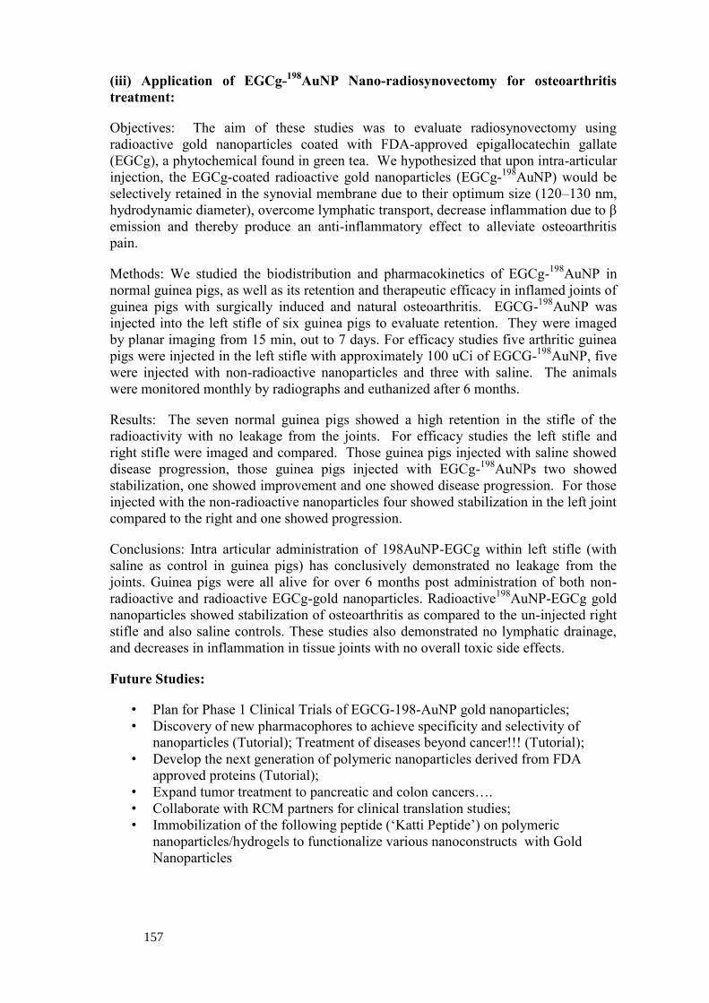
157
(iii) Application of EGCg-198
AuNP Nano-radiosynovectomy for osteoarthritis
treatment:
Objectives: The aim of these studies was to evaluate radiosynovectomy using
radioactive gold nanoparticles coated with FDA-approved epigallocatechin gallate
(EGCg), a phytochemical found in green tea. We hypothesized that upon intra-articular
injection, the EGCg-coated radioactive gold nanoparticles (EGCg-198
AuNP) would be
selectively retained in the synovial membrane due to their optimum size (120–130 nm,
hydrodynamic diameter), overcome lymphatic transport, decrease inflammation due to β
emission and thereby produce an anti-inflammatory effect to alleviate osteoarthritis
pain.
Methods: We studied the biodistribution and pharmacokinetics of EGCg-198
AuNP in
normal guinea pigs, as well as its retention and therapeutic efficacy in inflamed joints of
guinea pigs with surgically induced and natural osteoarthritis. EGCG-198
AuNP was
injected into the left stifle of six guinea pigs to evaluate retention. They were imaged
by planar imaging from 15 min, out to 7 days. For efficacy studies five arthritic guinea
pigs were injected in the left stifle with approximately 100 uCi of EGCG-198
AuNP, five
were injected with non-radioactive nanoparticles and three with saline. The animals
were monitored monthly by radiographs and euthanized after 6 months.
Results: The seven normal guinea pigs showed a high retention in the stifle of the
radioactivity with no leakage from the joints. For efficacy studies the left stifle and
right stifle were imaged and compared. Those guinea pigs injected with saline showed
disease progression, those guinea pigs injected with EGCg-198
AuNPs two showed
stabilization, one showed improvement and one showed disease progression. For those
injected with the non-radioactive nanoparticles four showed stabilization in the left joint
compared to the right and one showed progression.
Conclusions: Intra articular administration of 198AuNP-EGCg within left stifle (with
saline as control in guinea pigs) has conclusively demonstrated no leakage from the
joints. Guinea pigs were all alive for over 6 months post administration of both non-
radioactive and radioactive EGCg-gold nanoparticles. Radioactive198
AuNP-EGCg gold
nanoparticles showed stabilization of osteoarthritis as compared to the un-injected right
stifle and also saline controls. These studies also demonstrated no lymphatic drainage,
and decreases in inflammation in tissue joints with no overall toxic side effects.
Future Studies:
• Plan for Phase 1 Clinical Trials of EGCG-198-AuNP gold nanoparticles;
• Discovery of new pharmacophores to achieve specificity and selectivity of
nanoparticles (Tutorial); Treatment of diseases beyond cancer!!! (Tutorial);
• Develop the next generation of polymeric nanoparticles derived from FDA
approved proteins (Tutorial);
• Expand tumor treatment to pancreatic and colon cancers….
• Collaborate with RCM partners for clinical translation studies;
• Immobilization of the following peptide (‘Katti Peptide’) on polymeric
nanoparticles/hydrogels to functionalize various nanoconstructs with Gold
Nanoparticles