Nanobody-Based Targeting of the Macrophage Mannose...
14
Therapeutics, Targets, and Chemical Biology Nanobody-Based Targeting of the Macrophage Mannose Receptor for Effective In Vivo Imaging of Tumor-Associated Macrophages Kiavash Movahedi 1,2 , Steve Schoonooghe 1,2 , Damya Laoui 1,2 , Isabelle Houbracken 3 , Wim Waelput 6 , Karine Breckpot 4 , Luc Bouwens 3 , Tony Lahoutte 5,7 , Patrick De Baetselier 1,2 , Geert Raes 1,2 , Nick Devoogdt 5 , and Jo A. Van Ginderachter 1,2 Abstract Tumor-associated macrophages (TAM) are an important component of the tumor stroma and exert several tumor-promoting activities. Strongly pro-angiogenic TAMs that reside in hypoxic tumor areas highly express macrophage mannose receptor (MMR, CD206). In this study, we targeted MMR þ TAMs using nanobodies, which are single-domain antigen-binding fragments derived from Camelidae heavy-chain antibodies. MMR-specific nanobodies stained TAMs in lung and breast tumor single-cell suspensions in vitro, and intravenous injection of 99m Tc-labeled anti-MMR nanobodies successfully targeted tumor in vivo. Retention of the nanobody was receptor-specific and absent in MMR-deficient mice. Importantly, co-injection of excess unlabeled, bivalent anti-MMR nanobodies reduced nanobody accumulation in extratumoral organs to background levels, without compromising tumor uptake. Within tumors, the 99m Tc-labeled nanobodies specifically labeled MMR þ TAMs, as CCR2-deficient mice that contain fewer TAMs showed significantly reduced tumor uptake. Further, anti-MMR nanobodies accumulated in hypoxic regions, thus targeting pro-angiogenic MMR þ TAMs. Taken together, our findings provide preclinical proof of concept that anti-MMR nanobodies can be used to selectively target and image TAM subpopulations in vivo. Cancer Res; 72(16); 4165–77. Ó2012 AACR. Introduction Tumors harbor dynamic microenvironments in which can- cer cells are intimately associated with nontransformed host cells. The tumor-associated stroma is considered to play an important role during tumor growth, influencing phenomena such as angiogenesis, metastasis, and immune suppression (1). As such, the stroma forms an attractive target for diagnostic and therapeutic applications (2). Different myeloid cell types are important components of the tumor stroma (3, 4). In particular, macrophages are often found to infiltrate tumors in high numbers (5–7). We previ- ously characterized tumor-associated macrophages (TAM) in different preclinical tumor models (8). Extensive gene and protein expression analysis led us to identify distinct TAM subsets, termed MHC II hi and MHC II low TAMs. Interestingly, these subsets reside in different intratumoral microenviron- ments and differentially express molecules involved in inflam- mation, chemotaxis, and angiogenesis. MHC II low TAMs are mainly located in hypoxic tumor areas and are strongly pro- angiogenic. In contrast, MHC II hi TAMs are found in normoxic/ perivascular regions and are significantly less pro-angiogenic. Besides MHC II, we identified several membrane markers that can distinguish between these TAM subpopulations. This included the macrophage mannose receptor (MMR, CD206), an endocytic C-type lectin receptor known for its prominent expression on alternatively activated macrophages, which is consistently upregulated on MHC II low TAMs in all tumor models studied (8). These observations make MMR an attrac- tive marker for targeting the MHC II low hypoxic TAM subset in vivo. Antibody-based tumor-targeting strategies are widely explored (9, 10). Antibodies can be used for tumor imaging or delivering therapeutic agents to tumor cells. However, limitations of conventional antibodies include a poor pene- tration of solid tumors and high Fc-mediated aspecific binding, highlighting the need for smaller and more specific binding Authors' Affiliations: 1 VIB Laboratory of Myeloid Cell Immunology, 2 Laboratory of Cellular and Molecular Immunology, 3 Cell Differentiation Unit, Diabetes Research Centre, 4 Laboratory of Molecular and Cellular Therapy, Department of Immunology-Physiology, 5 In vivo Cellular and Molecular Imaging Laboratory (ICMI), Vrije Universiteit Brussel; 6 Ana- tomo-Pathology Department, and 7 Nuclear Medicine Department, Univer- sitair Ziekenhuis (UZ) Brussel, Brussels, Belgium Note: Supplementary data for this article are available at Cancer Research Online (http://cancerres.aacrjournals.org/). K. Movahedi, S. Schoonooghe, and D. Laoui contributed equally to this work. G. Raes, N. Devoogdt, and J.A. Van Ginderachter share senior authorship. Corresponding Authors: Jo A. Van Ginderachter, Laboratory of Cellular and Molecular Immunology, Vrije Universiteit Brussel, Building E8, Plein- laan 2, Brussels B-1050, Belgium. Phone: 32-2-6291978; Fax: 32-2- 6291981; E-mail: [email protected]; and Nick Devoogdt, In Vivo Cellular and Molecular Imaging Laboratory (ICMI), Vrije Universiteit Brussel, Laar- beeklaan 103, Brussels B-1090, Belgium. E-mail: [email protected] doi: 10.1158/0008-5472.CAN-11-2994 Ó2012 American Association for Cancer Research. Cancer Research www.aacrjournals.org 4165 on May 10, 2019. © 2012 American Association for Cancer Research. cancerres.aacrjournals.org Downloaded from Published OnlineFirst June 19, 2012; DOI: 10.1158/0008-5472.CAN-11-2994
Transcript of Nanobody-Based Targeting of the Macrophage Mannose...

Therapeutics, Targets, and Chemical Biology
Nanobody-Based Targeting of the Macrophage MannoseReceptor for Effective In Vivo Imaging of Tumor-AssociatedMacrophages
Kiavash Movahedi1,2, Steve Schoonooghe1,2, Damya Laoui1,2, Isabelle Houbracken3, Wim Waelput6,Karine Breckpot4, Luc Bouwens3, Tony Lahoutte5,7, Patrick De Baetselier1,2, Geert Raes1,2,Nick Devoogdt5, and Jo A. Van Ginderachter1,2
AbstractTumor-associated macrophages (TAM) are an important component of the tumor stroma and exert several
tumor-promoting activities. Strongly pro-angiogenic TAMs that reside in hypoxic tumor areas highly expressmacrophage mannose receptor (MMR, CD206). In this study, we targeted MMRþ TAMs using nanobodies, whichare single-domain antigen-binding fragments derived from Camelidae heavy-chain antibodies. MMR-specificnanobodies stained TAMs in lung and breast tumor single-cell suspensions in vitro, and intravenous injection of99mTc-labeled anti-MMR nanobodies successfully targeted tumor in vivo. Retention of the nanobody wasreceptor-specific and absent in MMR-deficient mice. Importantly, co-injection of excess unlabeled, bivalentanti-MMR nanobodies reduced nanobody accumulation in extratumoral organs to background levels, withoutcompromising tumor uptake. Within tumors, the 99mTc-labeled nanobodies specifically labeled MMRþ TAMs, asCCR2-deficient mice that contain fewer TAMs showed significantly reduced tumor uptake. Further, anti-MMRnanobodies accumulated in hypoxic regions, thus targeting pro-angiogenic MMRþ TAMs. Taken together, ourfindings provide preclinical proof of concept that anti-MMR nanobodies can be used to selectively target andimage TAM subpopulations in vivo. Cancer Res; 72(16); 4165–77. �2012 AACR.
IntroductionTumors harbor dynamic microenvironments in which can-
cer cells are intimately associated with nontransformed hostcells. The tumor-associated stroma is considered to play animportant role during tumor growth, influencing phenomenasuch as angiogenesis, metastasis, and immune suppression (1).As such, the stroma forms an attractive target for diagnosticand therapeutic applications (2).
Different myeloid cell types are important components ofthe tumor stroma (3, 4). In particular, macrophages are oftenfound to infiltrate tumors in high numbers (5–7). We previ-ously characterized tumor-associated macrophages (TAM) indifferent preclinical tumor models (8). Extensive gene andprotein expression analysis led us to identify distinct TAMsubsets, termed MHC IIhi and MHC IIlow TAMs. Interestingly,these subsets reside in different intratumoral microenviron-ments and differentially express molecules involved in inflam-mation, chemotaxis, and angiogenesis. MHC IIlow TAMs aremainly located in hypoxic tumor areas and are strongly pro-angiogenic. In contrast,MHC IIhi TAMs are found in normoxic/perivascular regions and are significantly less pro-angiogenic.Besides MHC II, we identified several membrane markers thatcan distinguish between these TAM subpopulations. Thisincluded the macrophage mannose receptor (MMR, CD206),an endocytic C-type lectin receptor known for its prominentexpression on alternatively activated macrophages, which isconsistently upregulated on MHC IIlow TAMs in all tumormodels studied (8). These observations make MMR an attrac-tive marker for targeting the MHC IIlow hypoxic TAM subsetin vivo.
Antibody-based tumor-targeting strategies are widelyexplored (9, 10). Antibodies can be used for tumor imagingor delivering therapeutic agents to tumor cells. However,limitations of conventional antibodies include a poor pene-tration of solid tumors and high Fc-mediated aspecific binding,highlighting the need for smaller and more specific binding
Authors' Affiliations: 1VIB Laboratory of Myeloid Cell Immunology,2Laboratory of Cellular and Molecular Immunology, 3Cell DifferentiationUnit, Diabetes Research Centre, 4Laboratory of Molecular and CellularTherapy, Department of Immunology-Physiology, 5In vivo Cellular andMolecular Imaging Laboratory (ICMI), Vrije Universiteit Brussel; 6Ana-tomo-Pathology Department, and 7Nuclear Medicine Department, Univer-sitair Ziekenhuis (UZ) Brussel, Brussels, Belgium
Note: Supplementary data for this article are available at Cancer ResearchOnline (http://cancerres.aacrjournals.org/).
K. Movahedi, S. Schoonooghe, and D. Laoui contributed equally to thiswork.
G. Raes, N. Devoogdt, and J.A. Van Ginderachter share senior authorship.
Corresponding Authors: Jo A. Van Ginderachter, Laboratory of Cellularand Molecular Immunology, Vrije Universiteit Brussel, Building E8, Plein-laan 2, Brussels B-1050, Belgium. Phone: 32-2-6291978; Fax: 32-2-6291981; E-mail: [email protected]; and Nick Devoogdt, In Vivo Cellularand Molecular Imaging Laboratory (ICMI), Vrije Universiteit Brussel, Laar-beeklaan 103, Brussels B-1090, Belgium. E-mail: [email protected]
doi: 10.1158/0008-5472.CAN-11-2994
�2012 American Association for Cancer Research.
CancerResearch
www.aacrjournals.org 4165
on May 10, 2019. © 2012 American Association for Cancer Research. cancerres.aacrjournals.org Downloaded from
Published OnlineFirst June 19, 2012; DOI: 10.1158/0008-5472.CAN-11-2994

units. Nanobodies (Nb) are the smallest available antigen-binding fragments (15 kDa) derived from Camelid heavy–chain-only antibodies (11, 12). Nbs are stable, soluble, havea high affinity, and show an increased tissue penetration,making them particularly suitable for tumor targeting (13,14). Previous work showed that a Nb-conjugate can efficientlytarget and kill cancer cells harboring a model antigen (15, 16).In addition, employing pinhole SPECT/micro-CT technology,99mTc-labeled Nbs have been successfully used as probes forcancer cell markers in tumor imaging (17–20) and for dendriticcell markers to image their in vivo biodistribution (21). Indeed,because of their small size, unboundNbs are rapidly eliminatedby renal clearance, resulting in high signal-to-noise ratios. As aresult, imaging can be carried out as early as 1 hour postin-jection of the nanobody probe, enabling the use of short-livedradioisotopes with a clear benefit for the patient (22).
In this article, we describe the production, selection, andcharacterization of Nbs against MMR. We show that 99mTc-labeled anti-MMR Nbs allow fast and specific targeting ofMMRþ cells in tissues, including a strong labeling of tumoralstromal cells, as shown by pinhole SPECT/micro-CT imaging.Importantly, excess administration of unlabeled bivalentanti-MMR Nbs provides a novel strategy for eliminating extra-tumoral signals while maintaining the targeting of tumor-associated MMRþ cells, a major part of which is constituted byMMRþ TAM. Altogether, we preclinically validated 99mTc-labeled MMR-targeting tracers suitable for molecular imagingof MMRþ stromal cells using SPECT cameras, which mightpave the way for novel approaches in diagnostic imaging andtherapeutic targeting of the tumor stroma.
Materials and MethodsMice and cell lines
Animal studies followed the guidelines of the institutionalreview board. Female Balb/c and C57BL/6 mice were fromHarlan. C57BL/6 MMR-deficient, CCR2-deficient, and MMTV-PyMTmice were provided by Etienne Pays (Universit�e Libre deBruxelles), Frank Tacke (AachenUniversity), andMassimilianoMazzone (KU Leuven), respectively. The Balb/c mammaryadenocarcinoma TS/A and 3LL-R clone of the C57BL/6 LewisLung carcinoma (8) were injected subcutaneously (s.c.) in theflank or in the fat pads (3� 106 cells). Within 12 to 14 days afterinoculation, TS/A and 3LL-R tumor–bearing mice were sub-jected to imaging. MMTV-PyMT mice bearing macroscopictumors were consecutively imaged with distinct tracers 48 to72 hours apart. Tumor dissection and flow cytometry werecarried out 96 hours after the last scan.
Tumor preparation and flow cytometryPreparation of tumor single-cell suspensions has been
described previously (8). Antibodies used for stainingswere anti-CD11b(M1/70)/PE-Cy7, anti-Ly6G(1A8)/FITC(Becton Dickinson Biosciences), anti-IA/IE(M5/114.15.2)/PerCPCy5.5 (Biolegend), anti-Ly6C(ER-MP20)/AF647, anti-MMR(MR5D3)/PE, and anti-F4/80(CI:A3-1)/PE (Serotec). Toprevent aspecific binding, rat anti-mouse CD16/CD32(2.4G2; Becton Dickinson Biosciences) was used. Nanobo-dies were labeled using the Alexafluor488 or Alexafluor647
Protein Labeling Kit (Invitrogen) according to the manu-facturers' instructions.
Immunofluorescence stainingsMice were injected intravenously with 500 mg Alexa-flu-
or647-labeled Nbs and intraperitoneally with 80 mg/kg pimo-nidazole [hypoxyprobe-1, HPI, Inc.] for hypoxia stainings. Twohours later, tumors were fixed in 4% paraformaldehyde, rehy-drated overnight (20% sucrose), and sectioned (5-mm-thickslices). Antibodies usedwere: rat anti-F4/80/alexa-fluor488 (CI:A3-1, Serotec), F(ab')2 donkey anti-rabbit/Cy3 JacksonIm-muno). Pictures were acquired with a Plan-Neofluar 10�/0.30 or 20�/0,50 (Carl Zeiss) objective on a Zeiss Axioplan2microscope with an Orca-R2 camera (Hamamatsu) and Smart-capture 3 software (Digital Scientific, United Kingdom).
Generation of mono- and bivalent nanobodiesThe anti-MMR Nb clones 1 and 3 were isolated from an
immune Nb phage-display library (23, 24). An alpaca (Vicugnapacos) was immunized weekly with 100 mg MMR extracellulardomain (R&D Systems) 6 times. Peripheral blood lymphocytemRNA was converted to cDNA, from which Nb-codingsequences were amplified and ligated onto the pHEN4 pha-gemid vector (25). Using M13K07 helper phages, the Nb librarywas expressed on phages, and specific Nb-phages wereenriched by 3 rounds of selection on microtiter plates (Nunc)coated with recombinant MMR. Individual colonies werescreened in ELISA for antigen recognition and sequenced. TheNb genes of clones 1 and 3were recloned into the vector pHEN6to encode a C-terminal His6 tag (25). Nanobody Nb BCII10 (25)was used as the negative control.
Bivalent Nbs were generated by attaching a linker sequence30 of the anti-MMR Nb clone1 VHH sequence using primerbiNbF and primers biNbG4SR, biNbg2cR, and biNbIgAR (Sup-plementary Table S1), which code for a (G4S)3 (GGGGSGG-GGSGGGGS), llama IgG2 hinge (AHHSEDPSSKAPKAPMA), orhuman IgA hinge (SPSTPPTPSPSTPPAS) linker, respectively.PCR fragments were inserted 50 of the a-MMR cl1 gene or theBCII10 gene in the pHEN6 vector.
Periplasmic expression and purification of mono- and biva-lent Nbs was carried out as described previously (19).
99mTc-Nanobody labeling, pinhole SPECT-micro-CTimaging, and biodistribution analysis
Nanobodies were labeled with 99mTc at their hexahistidinetail and subjected to quality assurance, as described previously(17, 19, 21).Micewere intravenously injectedwith 100 to 200mLof 45 to 155MBqof 99mTc-labeledNb, with orwithout an excessof bivalent unlabeled Nanobody. At 60 or 180 minutes postin-jection, anesthesia, micro-CT, and pinhole SPECT-imagingwere carried out as described previously (19). Image viewingwas conducted using AMIDE Medical Image Data Examinersoftware. High-resolution image 3-dimensional (3D)-recon-structions were generated using OsiriX Imaging Software. At30 minutes after initiating micro-CT/SPECT acquisition,organs were removed and weighed, and radioactivity wasmeasured using an automated g-counter (Cobra II Inspector5003; Canberra-Packard). Tissue and organ uptake was
Movahedi et al.
Cancer Res; 72(16) August 15, 2012 Cancer Research4166
on May 10, 2019. © 2012 American Association for Cancer Research. cancerres.aacrjournals.org Downloaded from
Published OnlineFirst June 19, 2012; DOI: 10.1158/0008-5472.CAN-11-2994
calculated as the percentage of injected activity per gram tissue(%IA/g), corrected for decay.
StatisticsStatistical analyses were conducted using the Student's t
test assuming unequal variances. Because multiple compar-isons are made (9–10 different organs), the P values of theStudent's t-test were adjusted by Holm's procedure (26). The Renvironment (27) and the multtest package (28) were used forstatistical analyses and creation of graphs. The significance ofthe Student's t-test and corrections for multiple testing was setto 0.05.
ResultsMMR as a potential marker for the differential targetingof TAM subsets in vivoPreviously, we showed that, in tumor single-cell suspen-
sions, MMRwas differentially expressed betweenMHC IIhi andMHC IIlow TAMs, as assessed by flow cytometry using anti-MMR monoclonal antibodies (8). Here, we show in TS/Amammary carcinoma and 3LL-R lung carcinoma subcutane-ous tumor single-cell suspensions that MMR was either not orpoorly expressed on CD11b� cells, granulocytes, monocytes,and Ly6Cint TAMs (Supplementary Fig. S1). Next, we investi-gatedMMR expression patterns in TS/A tumor sections triple-stained for MMR, CD11b, and MHC II (Supplementary Fig. S2).MMR and CD11b staining were almost completely co-local-ized, showing that MMRþ cells were indeed TAMs. Inter-estingly, MMR expression poorly co-localized with CD11bþ
MHC IIþ cells (mostly corresponding to MHC IIhi TAMs),indicating that MMR staining was mainly restricted to MHCIIlow TAMs. Therefore, MMR can be used for differentiallylabeling MHC IIhi and MHC IIlow TAMs on tumor sections.Together, these results indicate that MMR could be aninteresting marker for specifically targeting the M2-like/hypoxic MHC IIlow TAMs in vivo.
Generation and characterization of a-MMR nanobodiesNanobodies were raised against the recombinant extracel-
lular domain of MMR and, after screening of an immune phagelibrary, 2MMR-specific Nb clones were isolated: Nb cl1 and cl3.The binding characteristics of the anti-MMR Nbs were com-pared using surface plasmon resonance (SPR) measurements(Supplementary Table S2 and Supplementary Fig. S3). Nb cl1showed an 8-fold higher apparent affinity for immobilizedrecombinant MMR compared with Nb cl3 (KD ¼ 2.31 �10�8 vs. 1.91 � 10�7 mol/L, respectively). In addition, SPRcompetition studies showed that pretreatment with cl1 doesnot preclude cl3 binding and vice versa, indicating that anti-MMR Nbs cl1 and cl3 bind to non-overlapping epitopes (Sup-plementary Fig. S3).First, we investigated whether thea-MMRNb cl1 could bind
surface-expressed MMR on TAMs ex vivo. In this regard, flowcytometric analyses were carried out using fluorescentlylabeled Nb cl1 on subcutaneous TS/A and 3LL-R tumor sin-gle-cell suspensions (Fig. 1). As a negative control Nb, weconsistently used Nb BCII10 (25). The a-MMR Nb cl1 wasfound to bind to a subset of CD11bþ cells, but not to CD11b�
cells (Fig. 1A and 1B). Within the CD11bþ fraction of TS/Atumors, the a-MMR Nb did not bind to monocytes (Fig. 1C,gate 1), granulocytes (gate 5), and only very weakly to Ly6Cint
TAMs (gate 2). Staining was, therefore, restricted to MHC IIhi
(gate 3) and MHC IIlow TAMs (gate 4), with the latter subsetbinding a-MMR Nb to a much greater extent. For 3LL-Rtumors, a-MMR Nb binding was restricted to MHC IIlow TAMs(Fig. 1D, gate 5) and was not recorded on MHC IIhi TAMs (Fig.1D, gate 4) nor CD11bþ MHC IIhi tumor-associated DCs(TADC; Fig. 1D, gate 3), expressing high levels of CD11c andcostimulatory molecules (data not shown). These results are,therefore, in line with our observations using a-MMR mono-clonal antibodies (Supplementary Fig. S1).
Finally, a-MMR Nb cl1 bound to myeloid subsets in healthyorgans of tumor-bearingmice, an important example being theliver, wherein distinct macrophage subpopulations werestained in single-cell suspensions (Supplementary Fig. S4).
Assessment of the biodistribution and specificity ofa-MMR nanobodies in naive mice using pinhole SPECT/micro-CT analysis and ex vivo dissection
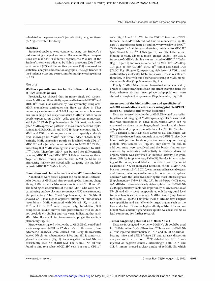
Next, we assessedwhether thea-MMRNbs could be used fortargeting and imaging of MMR-expressing cells in vivo. First,this was investigated in naive mice, where MMR can beexpressed on tissue macrophages, dendritic cells, and subsetsof hepatic and lymphatic endothelial cells (29, 30). Therefore,99mTc-labeled a-MMR Nb cl1, a-MMR Nb cl3, and control NbBCII10were injected intravenously in naive C57BL/6mice. Onehour postinjection, total-body scans were acquired usingpinhole SPECT/micro-CT (Fig. 2A; only shown for cl1). Inaddition, mice were sacrificed and the biodistribution wasassessed by measuring radioactivity in various dissectedorgans, which was expressed as injected activity per gramtissue (%IA/g; Supplementary Table S3). Besides intense stain-ing of the kidneys and bladder, consistent with the rapidclearance of Nb, an increased retention of the a-MMR Nb,but not the control Nb BCII10, was measured in several organsand tissues, including cardiac muscle, bone marrow, spleen,and liver, with the latter two showing the most intense signals(Supplementary Table S3; Fig. 2A). In wild-type (WT) mice,a-MMRNb cl1 showed a clearly higher uptake thana-MMRNbcl3 (Supplementary Table S3). Importantly, in vivo retention ofNb cl1 and cl3 is receptor-specific as only background-leveltracer uptake is seen in organs of MMR-KO mice (Supplemen-tary Table S3, Fig. 2A). Therefore, thea-MMRNbshave ahigh invivo specificity and can efficiently target organs such as theliver and spleen. Given the higher affinity of Nb cl1 for recom-binant MMR and the higher in vivo uptake, we chose this Nb aslead compound for further research.
Tumor-targeting potential of a-MMR Nb cl1Next, we investigated whether a-MMR Nb cl1 could be used
for TAM-targeting in vivo. Therefore, 99mTc-labeleda-MMRNbcl1 was injected intravenously in TS/A and 3LL-R s.c. tumor–bearing mice and SPECT/micro-CT and ex vivo dissectionanalyses were carried out. 99mTc-labeled Nb BCII10 wasinjected as negative control. Interestingly, both TS/A and3LL-R tumors showed a clear uptake of a-MMR Nb, which
MMR-Specific Nanobody for TAM Targeting and Imaging
www.aacrjournals.org Cancer Res; 72(16) August 15, 2012 4167
on May 10, 2019. © 2012 American Association for Cancer Research. cancerres.aacrjournals.org Downloaded from
Published OnlineFirst June 19, 2012; DOI: 10.1158/0008-5472.CAN-11-2994

Figure 1. a-MMR Nb cl1 differentially binds to TAM subsets in tumor single-cell suspensions. Single-cell suspensions of TS/A (A) or 3LL-R tumors (B) werestained with the indicated markers. Staining of a-MMR Nb was examined on gated myeloid subsets of TS/A (C) or 3LL-R tumors (D). Shaded histogramsrepresent Nb BCII10 staining.
Movahedi et al.
Cancer Res; 72(16) August 15, 2012 Cancer Research4168
on May 10, 2019. © 2012 American Association for Cancer Research. cancerres.aacrjournals.org Downloaded from
Published OnlineFirst June 19, 2012; DOI: 10.1158/0008-5472.CAN-11-2994
was significantly higher than tumor uptake of Nb BCII10 (Fig.2B–C). These findings were confirmed through ex vivo dissec-tion analysis: TS/A tumor uptake was 3.02 � 0.10%IA/g fora-MMR Nb and 0.40 � 0.03%IA/g for Nb BCII10; 3LL-R tumor
uptake was 3.02 � 0.19%IA/g for a-MMR Nb and 0.74 �0.03%IA/g for Nb BCII10 (Table 1). To further ascertain thespecificity of tumor uptake, 3LL-R tumorswere grown inMMR-KO mice. In these animals, 3LL-R tumors grew progressively
Figure 2. Fused pinhole SPECT/micro-CTimages of mice injected with 99mTc-labeleda-MMRNbcl1. A, naiveWTorMMR-KOmicewere injected with 99mTc-labeled a-MMR Nbcl1 and, 1 hour postinjection, images wereacquired. Coronal and sagittal views areshown. B, coronal and transverse views ofWT s.c. TS/A-bearing mice 3 hours after99mTc-labeled Nb BCII10 or a-MMR Nbinjection. C, coronal and transverse views ofs.c. 3LL-R–bearing mice.
MMR-Specific Nanobody for TAM Targeting and Imaging
www.aacrjournals.org Cancer Res; 72(16) August 15, 2012 4169
on May 10, 2019. © 2012 American Association for Cancer Research. cancerres.aacrjournals.org Downloaded from
Published OnlineFirst June 19, 2012; DOI: 10.1158/0008-5472.CAN-11-2994

and the distinct TAMsubsets remained present andwereMMRnegative, as assessed by flow cytometry (Supplementary Fig.S5). Importantly, tumor uptake of a-MMR Nb was reduced by10-fold (0.33� 0.03%IA/g; Table 1), showing its dependence onMMR expression by host cells.
a-MMR Nb cl1 targets hypoxic TAMs in vivoHaving established that a-MMR Nb cl1 specifically targeted
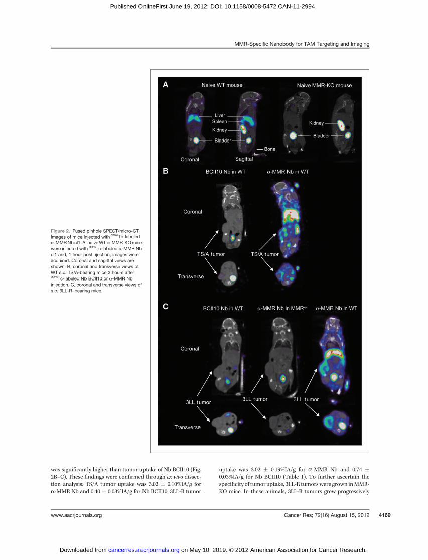
MMRþ cells in tumors, we aimed to ascertain whether this wasdue to TAM targeting. Previous work showed that CCR2-deficiency can result in a significant decrease in TAM infiltra-tion with only a minimal effect on tumor growth, resultingfrom the compensatory influx of tumor-promoting neutrophils(31, 32). To investigate whether CCR2-deficiency affected thenumbers of TAMs and, in particular, MHC IIlow TAMs in ourmodel, flow cytometric analyses were carried out on single-cellsuspensions of equally sized s.c. 3LL-R tumors grown inWT orCCR2-KO mice. This showed that CCR2-deficiency led to adramatic reduction in the number of MHC IIlow TAMs, whileinfiltration of Ly6GþMMR� neutrophils was significantlyincreased (Fig. 3A; Supplementary Fig. S6). Next, we comparedthe tumor-uptake of 99mTc-labeled a-MMR Nb cl1 injected inWTwith those in CCR2-KO 3LL-R tumor–bearingmice. 99mTc-
labeled a-MMR Nb showed a similar biodistribution in theorgans/tissues of CCR2-KO compared with WT tumor bearers(Supplementary Table S4). Importantly however, uptake of99mTc-labeled a-MMR Nb was significantly reduced inCCR2-KO tumors: 2.97 � 0.22%IA/g in WT compared with1.83 � 0.1%IA/g in CCR2-KO tumors (Fig. 3B). This indicatesthat TAMs residing in solid tumors are indeed targets ofa-MMR Nbs in vivo.
Because MHC IIlowMMRþ TAM have been reported toassociate with hypoxic regions (8), we next assessed whethera-MMR Nbs preferentially label hypoxic TAM in vivo. In thisregard, AF647-coupleda-MMRNbswere injected intravenous-ly in s.c. 3LL-R WT or MMR-KO tumor–bearing mice. Twohours later, tumors were collected, sectioned, and stained forthe hypoxia marker pimonidazole (hypoxyprobe) and themacrophage marker F4/80. Interestingly, AF647 fluorescencealmost completely co-localized with F4/80 staining in WTtumors, but was absent from MMR-KO tumors (Fig. 3C). Inaddition, the majority of AF647(bright) cells were located inhypoxic areas and stained with pimonidazole (Fig. 3C–D).These results convincingly show that a-MMR Nbs can targethypoxic tumor regions in vivo, where they bind to the residingMMRþ macrophages.
Table 1. Uptake values of 99mTc-labeled a-MMR or BCII10 Nb in TS/A and 3LL-R tumor–bearing WTmicebased on dissection at 3 hours postinjection.
Organs/tissues a-MMR Nb in WT (%IA/g) Nb BcII10 in WT (%IA/g) a-MMR Nb in MMR-KO (%IA/g)
TS/A tumor–bearing miceHeart 1.45 � 0.12 0.10 � 0.01a
Lungs 1.55 � 0.36 0.98 � 0.12Liver 12.6 � 0.54 0.59 � 0.02a
Spleen 8.95 � 0.60 0.24 � 0.01a
Kidney left 79.7 � 2.32 273 �14.8a
Kidney right 80.8 � 3.62 261 � 11.4a
Muscle 0.52 � 0.03 0.05 � 0.01a
Bone 1.33 � 0.10 0.08 � 0.01a
Blood 0.13 � 0.02 0.14 � 0.01Tumor 3.02 � 0.10 0.40 � 0.03a
3LL-R tumor–bearing miceHeart 2.02 � 0.11 0.17 � 0.01a 0.06 � 0.01a
Lungs 1.46 � 0.05 0.58 � 0.04a 1.02 � 0.70Liver 9.55 � 1.02 1.03 � 0.06b 1.36 � 1.06b
Spleen 4.61 � 0.50 0.41 � 0.03b 0.17 � 0.02b
Kidney left 108 � 16.1 368 � 10.10b 153 � 27.2Kidney right 88.6 � 21.7 305 � 54.7b 155 � 20.7Muscle 0.61 � 0.05 0.08 � 0.02a 0.05 � 0.02a
Bone 1.69 � 0.10 0.13 � 0.01a 0.06 � 0.01a
Blood 0.10 � 0.01 0.24 � 0.01a 0.09 � 0.01Tumor 3.02 � 0.19 0.74 � 0.03a 0.33 � 0.03a
NOTE: Tracer uptake is expressed as injected activity per gram (%IA/g). Data aremean�SEM (n¼ 6). Significancewas testedbetweena-MMR Nb versus Nb BCII10 and a-MMR Nb injected in WT vs. MMR-KO (3LL-R) for the indicated organ.aP < 0.0001.bP < 0.01.
Movahedi et al.
Cancer Res; 72(16) August 15, 2012 Cancer Research4170
on May 10, 2019. © 2012 American Association for Cancer Research. cancerres.aacrjournals.org Downloaded from
Published OnlineFirst June 19, 2012; DOI: 10.1158/0008-5472.CAN-11-2994
Strategies for increasing the tumor-to-tissue ratioof 99mTc-labeled a-MMR Nb cl1Amethodology for the specific in vivo targeting of a tracer to
TAMs, but not to other sites in the body, could be of important
diagnostic and therapeutic significance. However, both in theTS/A and 3LL-R models, 99mTc-labeled anti-MMR Nb accu-mulates to a higher extent in liver and spleen as comparedwiththe tumor. Therefore, we aimed tominimize binding of labeled
Figure 3. a-MMR Nb targeting in WT andCCR2-KO tumor-bearingmice. A, percentagesof MHC IIlow TAMs and Ly6Gþ neutrophils intumor single-cell suspensions of WT andCCR2-KO tumors. Mean � SEM (n ¼ 4). B,uptake values of 99mTc-labeled a-MMR Nb cl1or Nb BCII10 in WT or CCR2-KOmice 12-dayspost 3LL-R injection. ���, P < 0.001. C, AF647-labeled a-MMR Nb cl1 and pimonidazole wereinjected i.v. in 3LL-R WT or MMR-KO tumorbearers. Two hours later, tumors werecollected and stained for F4/80 andhypoxyprobe. D, overlays of a-MMR Nb-AF647, hypoxyprobe, and F4/80 signals in WT3LL-R tumors.
MMR-Specific Nanobody for TAM Targeting and Imaging
www.aacrjournals.org Cancer Res; 72(16) August 15, 2012 4171
on May 10, 2019. © 2012 American Association for Cancer Research. cancerres.aacrjournals.org Downloaded from
Published OnlineFirst June 19, 2012; DOI: 10.1158/0008-5472.CAN-11-2994
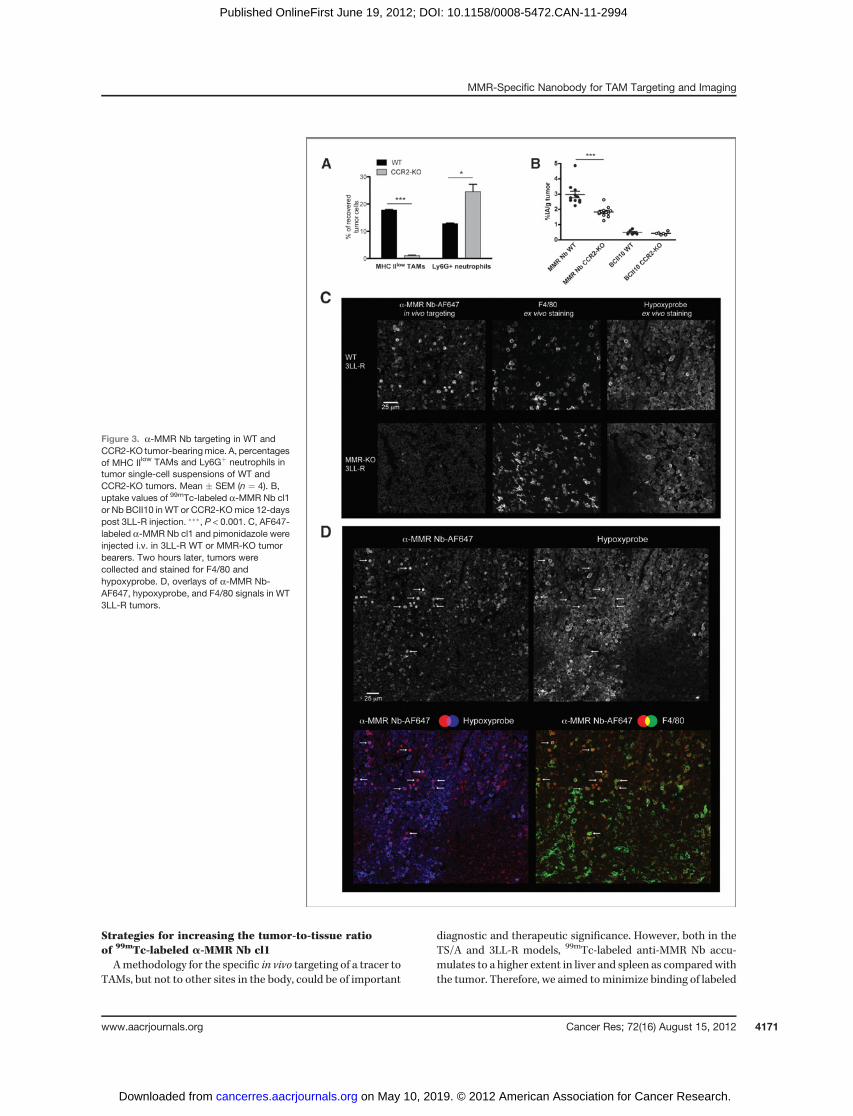
tracer in these extratumoral sites, while preserving tumortargeting. The efficient tumor-targeting potential of nanobo-dies is hypothesized to be a direct result of their small size. Toinvestigate this hypothesis, a series of larger bivalent Nbs werecreated (Fig. 4A). First,aMMR-aMMRbivalent Nbs weremadeby cloning 3 different peptide linkers with increasing prolinecontent (glycine-serine linker, part of the llama IgG2c hinge orpart of the human IgA hinge) between 2Nb cl1 sequences. All ofthese bivalent Nbs showed a 5-fold higher avidity comparedwith the monovalent Nb cl1, which can be largely attributed toa 3-fold increase inKD (Supplementary Table S2), and displayeda very similar in vivo biodistribution (Supplementary Table S5).In addition, using the llama IgG2c linker, aMMR-BCII10 bis-pecific Nbs and BCII10-BCII10 bivalent Nbs were generated
and their in vivo biodistribution was evaluated in TS/A and3LL-R tumor–bearing mice. Interestingly, aMMR-BCII10 andespeciallyaMMR-aMMRNbs showed a significantly enhancedtargeting of liver and spleen, but a dramatically reducedtargeting of tumor, compared with monovalent a-MMR Nbs(Fig. 4B). Therefore, these bivalent Nbs seem to possess desir-able features to efficiently block extratumoral binding siteswhile preserving intratumoral binding sites. To test thishypothesis, we coinjected 99mTc-labeled monovalent a-MMRNb with a 20-fold molar excess of unlabeled bivalent aMMR-aMMR Nb and assessed the specific uptake of labeled Nb indistinct organs. While the retention of monovalent 99mTc-labeled a-MMR Nb is reduced in all organs to the aspecificbackground level seen with Nb BCII10, the uptake in tumors is
Figure 4. Increasing tumor-to-tissue ratios of 99mTc-a-MMR Nbtracer uptake by excess unlabeledbivalent a-MMR Nb cl1. A,overview of different Nbconstructs. B, mono- and bivalent99mTc-labeled Nbs were injected ins.c. TS/A or 3LL-R tumor–bearingmice and uptake values werecalculated 3 hours postinjection viaorgan dissection. C, s.c. TS/Atumor–bearing mice were injectedwith 99mTc-labeled NbBCII10,99mTc-labeled a-MMR Nbcl1, or 99mTc-labeled a-MMR Nbþ20-fold molar excess of unlabeledbivalenta-MMRNbcl1. C1: uptakevalues of 99mTc-a-MMR Nb[expressed as injected activity pergram (%IA/g)] at 3 hourspostinjection. Mean� SEM (n¼ 6).C2: a-MMR Nb-to-backgroundratio, calculated as 99mTc-a-MMRNb uptake values/99mTc-NbBCII10. C3: tumor-to-tissue ratio of99mTc-a-MMR Nb, calculated astracer uptake in the tumor/traceruptake in the organ. Statisticalsignificance was tested between99mTc-a-MMR Nb and 99mTc-a�MMR Nb þ cold Nb �, P < 0.05;��, P < 0.01; ���, P < 0.001.LN, lymph node.
Movahedi et al.
Cancer Res; 72(16) August 15, 2012 Cancer Research4172
on May 10, 2019. © 2012 American Association for Cancer Research. cancerres.aacrjournals.org Downloaded from
Published OnlineFirst June 19, 2012; DOI: 10.1158/0008-5472.CAN-11-2994

only slightly diminished (Fig. 4C). As a result, the tumor-to-tissue ratio of labeleda-MMRNb is dramatically increased andtracer uptake is highest in the tumor. This allowed the tumor tobe clearly distinguishable in SPECT/micro-CT imaging of micebearing subcutaneous tumors (Fig. 5A, B, SupplementaryVideo S1). Importantly, very similar imaging data wereobtained when TS/A tumors were grown orthotopically in themammary fat pad (Fig. 5C–D; Supplementary Video S2), forwhich the presence of the 2 main TAM subsets was reportedbefore (8). Finally, imaging studies were carried out in trans-genic MMTV-PyMTmice, which spontaneously develop mam-mary tumors (33). In this regard, a mouse bearing multiplemacroscopic tumors was consecutively imaged (48-hour inter-
vals to allow complete elimination and decay of the 99mTctracer) with either 99mTc-labeled a-MMR Nb, 99mTc-labeledBCII10 Nb, or 99mTc-labeled a-MMR Nb coinjected with unla-beled bivalentaMMR-aMMRNb.When 99mTc-labeleda-MMRNb was injected alone, tumors were not easily distinguish-able due to high extratumoral uptake (Fig. 6A). However, co-injecting unlabeled bivalent aMMR-aMMR Nb minimalisedextratumoral Nb retention and resulted in tracer uptake inthe most prominent macroscopic nodules as seen via high-resolution 3D CT reconstructions (Fig. 6B; SupplementaryVideo S3). Notably, fluorescence-activated cell-sorting(FACS) analysis showed that, for all 3 selected tumorshighlighted in Fig. 6B, distinct TAM subpopulations were
Figure 5. Fused pinhole SPECT/micro-CT images of mice coinjectedwith 99mTc-labeled a-MMR Nbwith excess unlabeled bivalenta-MMR Nb. A, coronal views ofsubcutaneous TS/A-bearing mice3 hours after injection of99mTc-labeled a-MMR Nb cl1,99mTc-labeled a-MMR Nb cl1 þ20-fold molar excess of unlabeledbivalent a-MMR Nb cl1, or99mTc-labeled Nb BCII10. B, 3Dreconstruction of SPECT/CT imagesof a subcutaneous TS/A-bearingmouse injected with indicated tracer,3 hours postinjection (planar view,Supplementary Video S1 for 3D view)C, coronal and sagittal views of micebearing orthotopic TS/A tumors inthe mammary gland 3 hours afterinjection with indicated tracers. D,high-resolution 3D reconstruction ofCT and SPECT/CT images of anorthotopic TS/A-bearing mouseinjected with indicated tracer, 3hours postinjection (planar view,Supplementary Video S2 for 3Dview).
MMR-Specific Nanobody for TAM Targeting and Imaging
www.aacrjournals.org Cancer Res; 72(16) August 15, 2012 4173
on May 10, 2019. © 2012 American Association for Cancer Research. cancerres.aacrjournals.org Downloaded from
Published OnlineFirst June 19, 2012; DOI: 10.1158/0008-5472.CAN-11-2994

present, whereby MMR expression was highest on the MHCIIlow TAMs (Fig. 6C).
Effect of mono- and bivalent a-MMR Nb cl1 on immunecell activation
Monoclonal anti-MMR antibodies are known to potentiallyactivate macrophages and DCs (34). To assess whether mono-or bivalenta-MMRNb cl1 elicits a response, Nbswere added in
varying concentrations to bone-marrow–derived DCs (BMDC)or macrophages (BMDM) in vitro or were injected at a highdose in vivo. Monovalent a-MMR Nbs did not alter cytokine/chemokine production by BMDCs nor BMDMs in vitro, with orwithout LPS stimulation (Supplementary Fig. S7). With thehighest concentration of bivalent Nb (40 mg/mL), we observeda small, but significant, increase in TNF production by DCsand TNF and IL1Ra production by macrophages in vitro.
Figure 6. a-MMR Nb-basedimaging and TAM targeting inMMTV-PyMT mice. A, a MMTV-PyMT mouse with multiplemacroscopic nodules wasconsecutively (48- to 76-hourintervals) injected with indicatedtracers; images were taken 3 hourspostinjection. Coronal views areshown. n¼3. B, high-resolution 3Dreconstruction of CT and SPECT/CT images of the samemouse afterinjection of 99mTc-labeled a-MMRNb and blocking bivalent a-MMRNb. Out of multiple nodules, thenumbers indicate those tumorsthat were chosen for dissection.C, FACS analysis of single-cellsuspensions from the tumorsindicated in B.
Movahedi et al.
Cancer Res; 72(16) August 15, 2012 Cancer Research4174
on May 10, 2019. © 2012 American Association for Cancer Research. cancerres.aacrjournals.org Downloaded from
Published OnlineFirst June 19, 2012; DOI: 10.1158/0008-5472.CAN-11-2994

Importantly however, the highest in vivo dose of Nb used in thisstudy (5 mg monovalent Nb þ 200 mg bivalent Nb) did notinduce any significant increase in the serum cytokine levels,both for naive and tumor-bearing mice (Supplementary Fig.S8). Overall, we conclude that anti-MMR Nbs are innovativetools for the targeting and imaging of hypoxic MMRþ TAMswithout the risk of inducing overt innate immune responsesin vivo.
DiscussionUntil now, antibody-based tumor-targeting approaches
have mostly been directed against antigens expressed oncancer cells (10). However, the antigenic profile of cancer cellscan be unstable and depends on the cancer type. In addition,tumors contain a large stromal compartment, which includesmyeloid cells such as macrophages (35). Stromal cells mightprovide a good alternative for tumor-targeting, because theirantigenic profile is more stable and might be similar acrossdifferent cancer types. Our previouswork indicatedMMRas aninteresting marker for targeting the most M2-oriented (andpotentially most tumor-promoting) macrophage subset intumors (8). In this article, we describe the production ofMMR-specific nanobodies and show that they can be used forefficient in vivo targeting and imaging of TAMs in solid tumors.We describe 2 a-MMRNbs, which bind to different epitopes
and have distinct affinities and in vivo targeting efficiencies.The a-MMR Nb cl1 was selected as the lead compound and itstumor-targeting potential was first examined in mice bearingsubcutaneous 3LL-R lung or TS/A breast carcinoma tumors.For bothmodels, ex vivodissection showed that thea-MMRNbhad a tumor uptake of approximately 3%IA/g. Previous studiesusing nanobodies for the targeting of antigens (CEA, HER-2) ontumor xenografts, in which all cancer cells ectopically expressthe antigen, showed tumor uptake levels in the same range (15,16). Further, these amounts were sufficient to almostcompletely eradicate tumors in an antibody-dependentenzyme prodrug therapy approach (16). We, thus, concludethat a-MMR Nb efficiently targeted solid tumors.To investigate whether nanobody targeting was receptor-
specific, we compared the tumor uptake of a-MMR Nb withthat of Nb BCII10, and, more stringently, we compared theuptake of a-MMR Nb in tumors fromWT with those in MMR-KOmice. Together, these data convincingly showed that in vivotargeting of the a-MMR Nb is MMR-specific. In tumor single-cell suspensions, a-MMR Nbs primarily bound to TAMs,indicating that this was also one of their targets in vivo.However, we could not exclude the potential targeting of otherMMRþ cells in intact tumors. Therefore, we compared thetumor retention of a-MMR Nb injected in WT or CCR2-KOtumor–bearing mice. The 3LL-R tumors in CCR2-KO micecontained significantly lower TAM numbers as compared withWT tumors, and showed a significant reduction in a-MMR Nbuptake, which recommends TAM targeting. Moreover, AF647-labeled a-MMR Nb injected in tumor-bearing mice mainlystained MMRþF4/80þ macrophages in hypoxic regions.Together, these results indicate that tumor accumulation ofa-MMR Nb is mainly due to its penetration in hypoxic envir-onments and targeting of MHC IIlow TAMs residing there.
However, CCR2-KO tumors still showed some a-MMR Nbretention. This could indicate that, besides residual targetingof the remaining TAMs, other MMRþ cells are targeted. Forexample, MMR can be expressed on lymphatic vessels where itaffects leukocyte trafficking and contributes to cancer cellmetastasis (36), indicating the potential value of a-MMR Nbfor targeting tumor lymphatics. Future investigations will haveto address this issue.
Finding tumor-specific markers for antibody-based target-ing remains a daunting task. This is especially true whentargeting the tumor stroma, because stromal antigens aretypically not restricted to tumors. In this regard, a-MMR Nbstargeted, to a higher extent, the liver and spleen ofmice bearingsubcutaneous tumors. This may hamper the usefulness ofthese tools in both diagnostic and therapeutic applications.Importantly however, we describe a novel approach to reducethe targeting of tracers to healthy organs to background levels,while preserving an efficient targeting of the tumor. Indeed, co-injecting an excess of unlabeled bivalenta-MMRNbblocked allextratumoral sites, while only slightly affecting tumor-specifictracer uptake. This is a result of a bivalent Nb's higher uptake inextratumoral organs such as the liver and spleen (probablyexplained by a higher avidity for aMMR-aMMR Nb and/orincreased in vivo retention due to larger dimensions foraMMR-aMMR and aMMR-BCII10), coupled to a low accu-mulation in the tumor (probably due to poor tumor penetra-tion). Interestingly, modeling studies have indicated thatintermediate-sized targeting agents (�25 to 30 kDa) have thelowest tumor uptake levels among a spectrum of tumor-targeting polypeptides of various sizes (37). Bivalent Nbs,which are 30 kDa, therefore seem to follow this rule and havea low tumor uptake. Similar observations were made withDARPins, which are similar in size to Nbs (15 kDa), and forwhich fusion of 2 DARPins results in a significantly lowertumor uptake (38).
We believe that the strategy of co-injecting bivalent colda-MMR Nb to reduce extratumoral tracer uptake could betranslatable to the clinical setting. Preloading therapies, wherean excess of cold antibody is injected in patients, are alreadybeing carried out. In antibody-based radioimmunotherapy ofnon-Hodgkin lymphoma, excess amounts of unlabeled anti-CD20 antibody is predosed to patients before injection of 90Y-or 131I-conjugated anti-CD20 antibody (39), resulting inincreased tracer uptake in tumors and reduced uptake inextratumoral organs such as the spleen. However, determiningthe optimal cold dose for individual patients is not straight-forward because the cold antibody can compete with labeledantibody for free antigen sites in the tumor (40, 41). Themonovalent-labeled–bivalent-cold Nb approach describedhere seems an attractive alternative, because bivalent Nbs donot efficiently compete for free binding sites in the tumor,while they block extratumoral sites much more efficiently.
Because TAMs are found to be a major stromal componentinmany cancer types,a-MMRNbs could potentially be used fortargeting a variety of unrelated tumors. We have successfullyused this approach for the subcutaneous 3LL-R lung carcino-ma model, the subcutaneous and orthotopic TS/A breastcarcinoma model, and for the spontaneous MMTV-PyMT
MMR-Specific Nanobody for TAM Targeting and Imaging
www.aacrjournals.org Cancer Res; 72(16) August 15, 2012 4175
on May 10, 2019. © 2012 American Association for Cancer Research. cancerres.aacrjournals.org Downloaded from
Published OnlineFirst June 19, 2012; DOI: 10.1158/0008-5472.CAN-11-2994

breast carcinoma model. Coupled to our methodology ofrestricting extratumoral tracer uptake, this could now providenovel and attractive diagnostic or therapeutic opportunities.Clear examples would be diagnostic tumor imaging and thenoninvasive quantification of TAMs or specific TAM subsetsinside any given tumor, which could be of prognostic value.Further, as a-MMR Nbs can penetrate hypoxic areas wherethe majority of MHC IIlow/MMRþ TAMs reside, this mightprovide a new avenue for visualizing hypoxic regions withinthe tumor, and may be potentially relevant for guidedradiotherapy (42). In addition, radioimmunotherapy mightbe the most promising therapeutic application for these Nbs,because coupling of Nbs to proteins (e.g., toxins or prodrug-converting enzymes; ref. 43), might reduce the tumor-target-ing efficiency due to a size increase. As a cautionary note,engagement of MMR could potentially trigger cytokine/chemokine release by DCs and macrophages (34, 44). How-ever, our results did not show an overt cytokine/chemokineresponse after in vivo administration of high doses of mono-and bivalent a-MMR Nb.
In addition, MMR is a widely used marker for human M2macrophages (45–47), which is expressed on TAMs fromhuman tumors. Co-culture of human macrophages and ovar-ian cancer cells induces a strong upregulation of MMR expres-sion (48). Further, Allavena and colleagues have shown thatMMR is widely expressed on TAMs isolated from ovariancancer patients, and that its engagement by tumor mucinscan induce an immune-suppressive phenotype (44). In addi-tion, our ongoing preliminary studies show that, in humanbreast cancer samples, MMRþ TAMs are clearly detected and,interestingly, are enriched in fibrotic foci, which are known tobe amarker for intratumoral hypoxia and correlate with a poorprognosis (data not shown; ref. 49). However, it remains to betested whether MRC1þ TAMs carry out the tumor-promoting,pro-angiogenic functions in human tumors as reported pre-viously in murine tumors.
In conclusion, our work indicates that, in preclinical models,TAM subsets can be efficiently targeted in vivo using nano-bodies against MMR. In addition, we provide amethodology to
restrict tracer uptake to the tumor. This could form the basisfor developing novel imaging and therapeutic applications forthe diagnosis and treatment of cancer.
Disclosure of Potential Conflicts of InterestK. Movahedi has ownership interest (including patents). S. Schoonooghe has
ownership interest (including patents) with patent on MMR Nb discussed inarticle. D. Laoui has ownership interest (including patents) for US 20110262348.T. Lahoutte, P. De Baetselier, G. Raes, and N. Devoogdt have ownership interest(including patents). No potential conflicts of interest were disclosed by the otherauthors.
Authors' ContributionsConception and design: K. Movahedi, S. Schoonooghe, D. Laoui, P. DeBaetselier, G. Raes, N. Devoogdt, J.A. Van GinderachterDevelopment of methodology: K. Movahedi, S. Schoonooghe, D. Laoui, I.Houbracken, L. Bouwens, T. Lahoutte, N. DevoogdtAcquisition of data (provided animals, acquired and managed patients,provided facilities, etc.): K. Movahedi, S. Schoonooghe, I. Houbracken, W.Waelput, L. Bouwens, T. Lahoutte, N. DevoogdtAnalysis and interpretation of data (e.g., statistical analysis, biostatistics,computational analysis): K. Movahedi, S. Schoonooghe, D. Laoui, I. Hou-bracken, W. Waelput, T. Lahoutte, G. Raes, N. Devoogdt, J.A. Van GinderachterWriting, review, and/or revision of the manuscript: K. Movahedi, S.Schoonooghe, D. Laoui, W. Waelput, K. Breckpot, L. Bouwens, T. Lahoutte, P.De Baetselier, G. Raes, N. Devoogdt, J.A. Van GinderachterAdministrative, technical, or material support (i.e., reporting or orga-nizing data, constructing databases): K. Movahedi, S. Schoonooghe, P. DeBaetselier, G. Raes, N. DevoogdtStudy supervision: L. Bouwens, P. De Baetselier, G. Raes, N. Devoogdt, J.A. VanGinderachter
AcknowledgmentsThe authors thank Cindy Peleman, Ella Omasta, Marie-Th�er�ese Detobel, and
Maria Slazak for technical assistance, Dr. Evelien Vaes for help with statisticalanalysis, and Dr. Yannick Morias for help with experiments.
Grant SupportThis work was supported by a doctoral grant from FWO-Vlaanderen to K.
Moyahedi, a scholarship from "Stichting Emmanuel van der Schueren" to K.Moyahedi, a doctoral grant from IWT-Vlaanderen to D. Laoui, a grant from"Stichting tegen Kanker" to P. De Baetselier and J.A. Van Ginderachter, and anSBO grant from IWT-Vlaanderen to P. De Baetselier, S. Schoonooghe, G. Raes, N.Devoogdt, and T. Lahoutte.
The costs of publication of this article were defrayed in part by the payment ofpage charges. This article must therefore be hereby marked advertisement inaccordance with 18 U.S.C. Section 1734 solely to indicate this fact.
Received September 9, 2011; revised May 26, 2012; accepted June 14, 2012;published OnlineFirst June 19, 2012.
References1. Grivennikov SI, Greten FR, Karin M. Immunity, inflammation, and
cancer. Cell 2010;140:883–99.2. Hofmeister V, Schrama D, Becker JC. Anti-cancer therapies targeting
the tumor stroma. Cancer Immunol Immunother 2008;57:1–17.3. Laoui D, VanOvermeire E,Movahedi K, Van denBossche J, Schouppe
E, Mommer C, et al. Mononuclear phagocyte heterogeneity in cancer:different subsets and activation states reaching out at the tumor site.Immunobiology 2011;216:1192–202.
4. Coffelt SB, Lewis CE, Naldini L, Brown JM, Ferrara N, De Palma M.Elusive identities and overlapping phenotypes of proangiogenic mye-loid cells in tumors. Am J Pathol 2010;176:1564–76.
5. Pollard JW. Tumour-educated macrophages promote tumour pro-gression and metastasis. Nat Rev Cancer 2004;4:71–8.
6. Mantovani A, Sica A. Macrophages, innate immunity and cancer:balance, tolerance, and diversity. Curr Opin Immunol 2010;22:231–7.
7. Van Ginderachter JA, Movahedi K, Hassanzadeh Ghassabeh G,Meerschaut S, Beschin A, Raes G, et al. Classical and alternativeactivation ofmononuclear phagocytes: picking the best of bothworldsfor tumor promotion. Immunobiology 2006;211:487–501.
8. Movahedi K, Laoui D, Gysemans C, Baeten M, Stange G, Van denBossche J, et al. Different tumor microenvironments contain function-ally distinct subsets of macrophages derived from Ly6C(high) mono-cytes. Cancer Res 2010;70:5728–39.
9. Carter P. Improving the efficacy of antibody-based cancer therapies.Nat Rev Cancer 2001;1:118–29.
10. Zafir-Lavie I, Michaeli Y, Reiter Y. Novel antibodies as anticanceragents. Oncogene 2007;26:3714–33.
11. Hamers-Casterman C, Atarhouch T, Muyldermans S, Robinson G,Hamers C, Songa EB, et al. Naturally occurring antibodies devoid oflight chains. Nature 1993;363:446–8.
12. Arbabi Ghahroudi M, Desmyter A, Wyns L, Hamers R,Muyldermans S.Selection and identification of single domain antibody fragments fromcamel heavy-chain antibodies. FEBS Lett 1997;414:521–6.
13. Van Bockstaele F, Holz JB, Revets H. The development of nanobodiesfor therapeutic applications. Curr Opin Investig Drugs 2009;10:1212–24.
14. Revets H, De Baetselier P, Muyldermans S. Nanobodies as novelagents for cancer therapy. Expert Opin Biol Ther 2005;5:111–24.
Movahedi et al.
Cancer Res; 72(16) August 15, 2012 Cancer Research4176
on May 10, 2019. © 2012 American Association for Cancer Research. cancerres.aacrjournals.org Downloaded from
Published OnlineFirst June 19, 2012; DOI: 10.1158/0008-5472.CAN-11-2994

15. Cortez-Retamozo V, Lauwereys M, Hassanzadeh Gh G, Gobert M,Conrath K, Muyldermans S, et al. Efficient tumor targeting by single-domain antibody fragments of camels. Int J Cancer 2002;98:456–62.
16. Cortez-Retamozo V, Backmann N, Senter PD, Wernery U, De Baet-selier P, Muyldermans S, et al. Efficient cancer therapy with a nano-body-based conjugate. Cancer Res 2004;64:2853–7.
17. GainkamLO,Huang L,Caveliers V, KeyaertsM,HernotS, Vaneycken I,et al. Comparison of the biodistribution and tumor targeting of two99mTc-labeled anti-EGFR nanobodies in mice, using pinhole SPECT/micro-CT. J Nucl Med 2008;49:788–95.
18. Huang L, Gainkam LO, Caveliers V, Vanhove C, Keyaerts M, DeBaetselier P, et al. SPECT imaging with 99mTc-labeled EGFR-specificnanobody for in vivomonitoring of EGFR expression. Mol Imaging Biol2008;10:167–75.
19. Vaneycken I, Govaert J, Vincke C, Caveliers V, Lahoutte T, De Baet-selier P, et al. In vitro analysis and in vivo tumor targeting of ahumanized, grafted nanobody in mice using pinhole SPECT/micro-CT. J Nucl Med 2010;51:1099–106.
20. Vaneycken I, Devoogdt N, Van Gassen N, Vincke C, Xavier C, WerneryU, et al. Preclinical screening of anti-HER2 nanobodies for molecularimaging of breast cancer. FASEB J 2011;25:2433–46.
21. De Groeve K, Deschacht N, De Koninck C, Caveliers V, Lahoutte T,Devoogdt N, et al. Nanobodies as tools for in vivo imaging of specificimmune cell types. J Nucl Med 2010;51:782–9.
22. Vaneycken I, D'HuyvetterM,Hernot S, DeVos J, Xavier C,DevoogdtN,et al. Immuno-imaging using nanobodies. Curr Opin Biotechnol2011;22:877–81.
23. Saerens D, Kinne J, Bosmans E, Wernery U, Muyldermans S, ConrathK. Single domain antibodies derived from dromedary lymph node andperipheral blood lymphocytes sensing conformational variants ofprostate-specific antigen. J Biol Chem 2004;279:51965–72.
24. Saerens D, Stijlemans B, Baral TN, Nguyen Thi GT, Wernery U, MagezS, et al. Parallel selection of multiple anti-infectome nanobodieswithout access to purified antigens. J Immunol Methods 2008;329:138–50.
25. Conrath KE, Lauwereys M, Galleni M, Matagne A, Frere JM, Kinne J,et al. Beta-lactamase inhibitors derived from single-domain antibodyfragments elicited in the camelidae. Antimicrob Agents Chemother2001;45:2807–12.
26. Holm S. A Simple sequentially rejectivemultiple test procedure. ScandJ Stat 1979;6:65–70.
27. Ihaka R, Gentleman R. R: a language for data analysis and graphics. JComput Graph Stat 1996;5:299–314.
28. Pollard KS, Ge Y, Taylor S, Dudoit S. Resampling-based multiplehypothesis testing. [Cited 2007 Jul 16]. Available from: www.bioconductor.org/packages/release/bioc/html/multtest/html.
29. Linehan SA, Martinez-Pomares L, Stahl PD, Gordon S. Mannosereceptor and its putative ligands in normal murine lymphoid andnonlymphoid organs: in situ expression of mannose receptor byselected macrophages, endothelial cells, perivascular microglia,and mesangial cells, but not dendritic cells. J Exp Med 1999;189:1961–72.
30. McKenzieEJ, Taylor PR,StillionRJ, LucasAD,Harris J,GordonS, et al.Mannose receptor expression and function define a new population ofmurine dendritic cells. J Immunol 2007;178:4975–83.
31. Sawanobori Y, Ueha S, Kurachi M, Shimaoka T, Talmadge JE, Abe J,et al. Chemokine-mediated rapid turnover of myeloid-derived sup-pressor cells in tumor-bearing mice. Blood 2008;111:5457–66.
32. Pahler JC, Tazzyman S, Erez N, Chen YY,Murdoch C, NozawaH, et al.Plasticity in tumor-promoting inflammation: impairment of macro-
phage recruitment evokes a compensatory neutrophil response. Neo-plasia 2008;10:329–40.
33. Guy CT, Cardiff RD, Muller WJ. Induction of mammary tumors byexpression of polyomavirus middle T oncogene: a transgenic mousemodel for metastatic disease. Mol Cell Biol 1992;12:954–61.
34. Chieppa M, Bianchi G, Doni A, Del Prete A, Sironi M, Laskarin G, et al.Cross-linking of themannose receptor onmonocyte-derived dendriticcells activates an anti-inflammatory immunosuppressive program. JImmunol 2003;171:4552–60.
35. Condeelis J, Pollard JW.Macrophages: obligate partners for tumor cellmigration, invasion, and metastasis. Cell 2006;124:263–6.
36. Marttila-Ichihara F, Turja R, Miiluniemi M, Karikoski M, Maksimow M,Niemela J, et al. Macrophage mannose receptor on lymphatics con-trols cell trafficking. Blood 2008;112:64–72.
37. Schmidt MM, Wittrup KD. A modeling analysis of the effects ofmolecular size and binding affinity on tumor targeting. Mol CancerTher 2009;8:2861–71.
38. Zahnd C, Kawe M, Stumpp MT, de Pasquale C, Tamaskovic R, Nagy-DavidescuG, et al. Efficient tumor targetingwith high-affinity designedankyrin repeat proteins: effects of affinity and molecular size. CancerRes 2010;70:1595–605.
39. Sharkey RM, Press OW, Goldenberg DM. A re-examination of radio-immunotherapy in the treatment of non-Hodgkin lymphoma: pro-spects for dual-targeted antibody/radioantibody therapy. Blood2009;113:3891–5.
40. Gopal AK, Press OW, Wilbur SM, Maloney DG, Pagel JM. Rituximabblocks binding of radiolabeled anti-CD20 antibodies (Ab) but notradiolabeled anti-CD45 Ab. Blood 2008;112:830–5.
41. Kletting P, Meyer C, Reske SN, Glatting G. Potential of optimalpreloading in anti-CD20 antibody radioimmunotherapy: an investiga-tion based on pharmacokinetic modeling. Cancer Biother Radiopharm2010;25:279–87.
42. Bentzen SM. Theragnostic imaging for radiation oncology: dose-painting by numbers. Lancet Oncol 2005;6:112–7.
43. Kreitman RJ. Recombinant immunotoxins containing truncated bac-terial toxins for the treatment of hematologic malignancies. BioDrugs2009;23:1–13.
44. Allavena P, Chieppa M, Bianchi G, Solinas G, Fabbri M, Laskarin G,et al. Engagement of the mannose receptor by tumoral mucins acti-vates an immune suppressive phenotype in human tumor-associatedmacrophages. Clin Dev Immunol 2010;2010:547179.
45. Raes G, Van den Bergh R, De Baetselier P, GhassabehGH, Scotton C,Locati M, et al. Arginase-1 and Ym1 are markers for murine, but nothuman, alternatively activated myeloid cells. J Immunol 2005;174:6561; author reply 6562.
46. O'Brien J, Lyons T, Monks J, Lucia MS, Wilson RS, Hines L, et al.Alternatively activated macrophages and collagen remodeling char-acterize the postpartum involuting mammary gland across species.Am J Pathol 2010;176:1241–55.
47. Lovren F, Pan Y, Quan A, Szmitko PE, Singh KK, Shukla PC, et al.Adiponectin primes human monocytes into alternative anti-inflamma-tory M2 macrophages. Am J Physiol Heart Circ Physiol 2010;299:H656–63.
48. Hagemann T, Wilson J, Burke F, Kulbe H, Li NF, Pluddemann A, et al.Ovarian cancer cells polarize macrophages toward a tumor-associat-ed phenotype. J Immunol 2006;176:5023–32.
49. Colpaert CG, Vermeulen PB, Fox SB, Harris AL, Dirix LY, Van MarckEA. The presence of a fibrotic focus in invasive breast carcinomacorrelateswith the expression of carbonic anhydrase IX and is amarkerof hypoxia and poor prognosis. Breast Cancer Res Treat 2003;81:137–47.
MMR-Specific Nanobody for TAM Targeting and Imaging
www.aacrjournals.org Cancer Res; 72(16) August 15, 2012 4177
on May 10, 2019. © 2012 American Association for Cancer Research. cancerres.aacrjournals.org Downloaded from
Published OnlineFirst June 19, 2012; DOI: 10.1158/0008-5472.CAN-11-2994

2012;72:4165-4177. Published OnlineFirst June 19, 2012.Cancer Res Kiavash Movahedi, Steve Schoonooghe, Damya Laoui, et al.
Imaging of Tumor-Associated MacrophagesIn Vivofor Effective Nanobody-Based Targeting of the Macrophage Mannose Receptor
Updated version
10.1158/0008-5472.CAN-11-2994doi:
Access the most recent version of this article at:
Material
Supplementary
http://cancerres.aacrjournals.org/content/suppl/2012/06/19/0008-5472.CAN-11-2994.DC1
Access the most recent supplemental material at:
Cited articles
http://cancerres.aacrjournals.org/content/72/16/4165.full#ref-list-1
This article cites 48 articles, 19 of which you can access for free at:
Citing articles
http://cancerres.aacrjournals.org/content/72/16/4165.full#related-urls
This article has been cited by 14 HighWire-hosted articles. Access the articles at:
E-mail alerts related to this article or journal.Sign up to receive free email-alerts
Subscriptions
Reprints and
To order reprints of this article or to subscribe to the journal, contact the AACR Publications Department at
Permissions
Rightslink site. Click on "Request Permissions" which will take you to the Copyright Clearance Center's (CCC)
.http://cancerres.aacrjournals.org/content/72/16/4165To request permission to re-use all or part of this article, use this link
on May 10, 2019. © 2012 American Association for Cancer Research. cancerres.aacrjournals.org Downloaded from
Published OnlineFirst June 19, 2012; DOI: 10.1158/0008-5472.CAN-11-2994