Name the Condition - Dr. Scott Croes' Website - Home Page · Which is made of connective tissue?...
69
Name the Condition
Transcript of Name the Condition - Dr. Scott Croes' Website - Home Page · Which is made of connective tissue?...

Name the
Condition

Ringworm

Name the Condition

Staphylococcus aureus infection

Name the Condition

Lyme disease
• bacteria carried by
ticks.

Identify the following: Epidermis, sebaceous (oil) gland,
hair follicle, sweat gland, and subcutaneous layer

Identify the following: Epidermis, sebaceous (oil) gland,
hair follicle, sweat gland, and subcutaneous layer
(Sebaceous)

Identify the epidermis and the dermis!
Which is made of connective tissue? What
type?
Identify the epidermis, dermis and determine the
type of tissue in each

Identify the epidermis and the dermis!
Which is made of connective tissue? What
type?
Identify the epidermis, dermis and determine the
type of tissue in each
Epidermis
(keratinized
stratified squamous
epithelium)
Dermis
(dense
irregular
connective
tissue

What type of cell are the arrows indicating?

Melanocytes

Name the cells indicated by the red arrows

Langerhan’s Cells

Identify the layers, cells and structures as indicated


Name the 4 layers of thin skin in both
the cartoon and the photomicrograph.

Name the 4 layers of thin skin in both
the cartoon and the photomicrograph.

• Name the
Layers of skin
and label the
dermal papilla
and dermis

• Name the
Layers of skin
and label the
dermal papilla
and dermis

Name the layer of skin shown

Stratum Spinosum

Name the specific layers of skin indicated by the
boxes and arrows

Name the specific layers of skin indicated by the arrows
• SC: Stratum corneum SS: Stratum spinosum
• SL: Stratum lucidum SB: Stratum basale
• SG: Stratum granulosum
SG

Identify the thick and thin skin.
Can you name the layers?

Identify the thick and thin skin.
Can you name the layers?
Thin Skin
Thick Skin

Identify the type of fibers stained in this section

Histology: Normal skin with stain for elastic fibers
Identify the type of fibers stained in this section

Identify the papillary and reticular regions

Identify the papillary and reticular layers

Name the structure indicated by the arrows
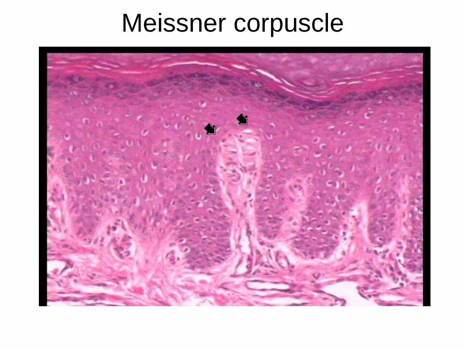
Meissner corpuscle

Name the structure

Meissner
corpuscle

Name the Condition?

Stretch Marks (Striae)

Skin Condition Involving Melanocytes
• Condition:
• Condition:

Skin Condition Involving Melanocytes
• Condition: Albinism
• Condition: Vitiligo

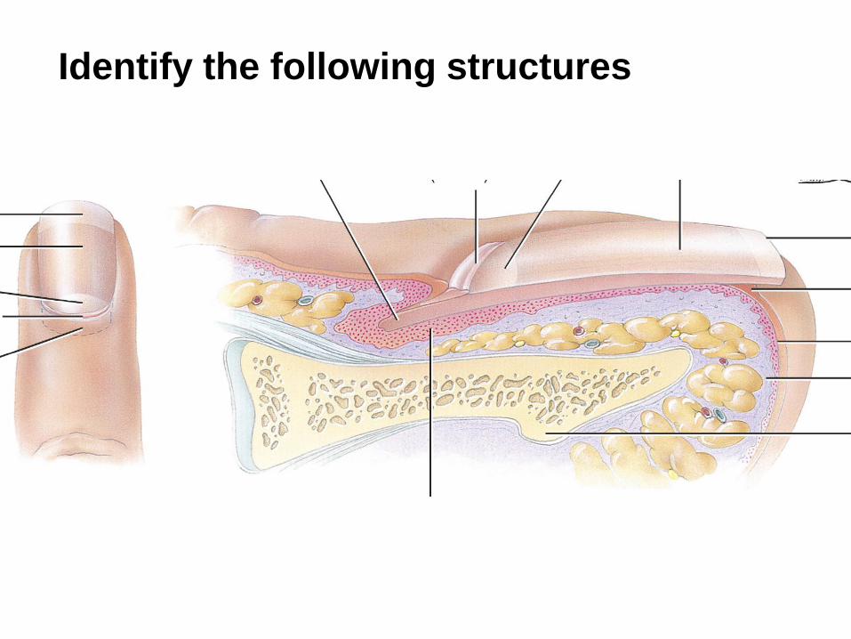
Name the
following
structures

Name the
following
structures

• Name the following structures

• Name the following
structures

• Identify the following: Epidermis; Dermis; Hair follicle; Hair
bulb; External root sheath; Dermal papilla; Arrector pili
muscle; Sebaceous gland


• Identify the following: Dermal papilla; Melanocyte;
Melanin granules

• Identify the following: Dermal papilla; Melanocyte;
Melanin granules

Identify the following: Epidermis; Dermis; Hair follicle; Hair
bulb; Sweat Gland and subcutaneous layer


Identify the following structures

Identify the following structures

Identify the structure indicated by the arrow.

The sebaceous gland is indicated by the arrow. Note how its
duct is unbranched and how it empties into a hair follicle.

Name this white substance

Vernix Caseosa

Name This Condition?

Seborrhea (cradle cap)

• Identify the sebaceous glands and
Eccrine (merocrine) Sweat Glands:

• Identify the sebaceous glands and
Eccrine (merocrine) Sweat Glands:

Identify the following structures: Epidermis, Hair cortex, Hair
medulla, Hair follicle, Hair matrix, Papilla, Hair Bulb, Arrector pili,
Dermal papillary region, Dermal reticular region

Identify the following structures: Epidermis, Hair cortex, Hair
medulla, Hair follicle, Hair matrix, Papilla, Hair Bulb, Arrector pili,
Dermal papillary region, Dermal reticular region

Identify the structure
indicated by the arrow
Identify the following structures

Identify the following structures

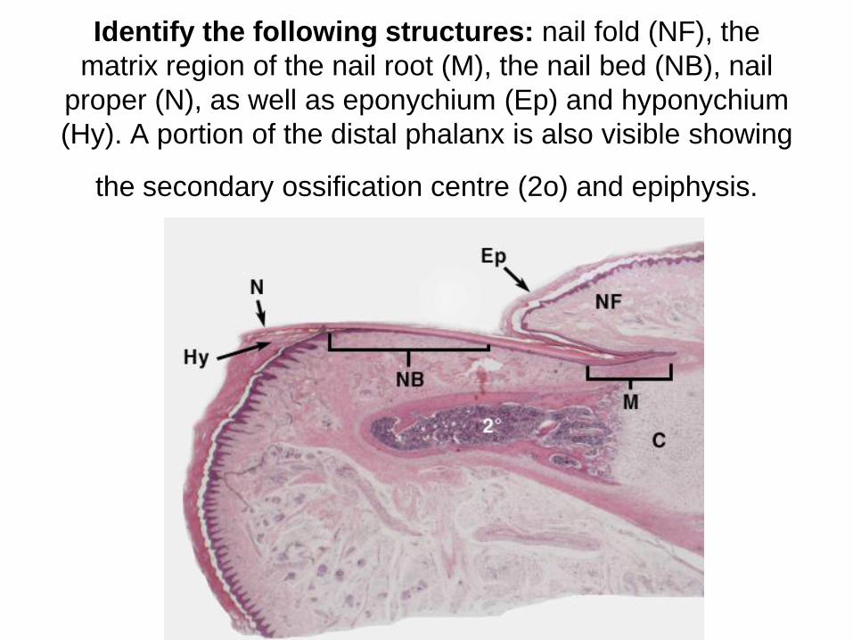
Identify the following structures: nail fold (NF), the
matrix region of the nail root (M), the nail bed (NB), nail
proper (N), as well as eponychium (Ep) and hyponychium
(Hy). A portion of the distal phalanx is also visible showing
the secondary ossification centre (2o) and epiphysis.
Identify the following structures: nail fold (NF), the
matrix region of the nail root (M), the nail bed (NB), nail
proper (N), as well as eponychium (Ep) and hyponychium
(Hy). A portion of the distal phalanx is also visible showing
the secondary ossification centre (2o) and epiphysis.
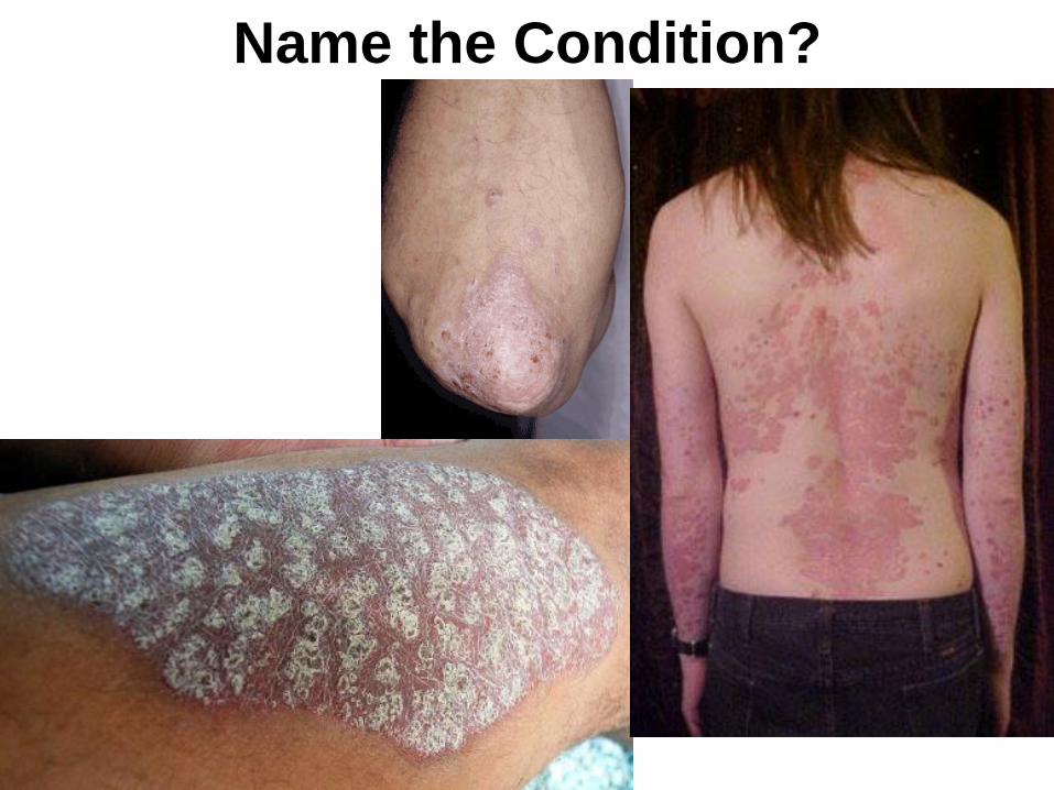
Name the Condition?

Psoriasis

How can I know if
my moles are
suspicious?