
NAME OF THE ACTIVITY: NATIONAL WORKSHOP ON …
57
NAME OF THE ACTIVITY: “NATIONAL WORKSHOP ON TECHNIQUES IN PLANT SCIENCES” DATE FACULTY DEPARTMENT/COMMITTEE COORDINATORS NAME 4-5 dec. 2019 Dr. Sunila Khurana (Convener) Dr. Amit Vashishtha (Co- Convener) Dr Neeti Mehla (Co- Convener) Dr Aditi Kothari- Chhajer (Co- Convener) Botany Dr. Sunila Khurana TIME VENUE NUMBER OF PARTICIPANTS NATURE: Outdoor/Indoor 9.30 am to 5.30 pm Sri Venkateswara College, Delhi University 25 Indoor SUPPORT/ASSISTANCE: UGC BRIEF INFORMATION ABOUT THE ACTIVITY (CRITERION NO. - ): TOPIC/SUBJECT OF THE ACTIVITY TECHNIQUES IN PLANT SCIENCES OBJECTIVES The workshop aimed at providing hands-on-training in the field of in-vitro morphogenesis of medicinal plants and study of genetic fidelity through molecular markers. A session on practical exposure to HP-TLC technique was conducted along with the brains storming session on question paper setting for the faculty. METHODOLOGY Interactive sessions were facilitated by subject experts. The methodology included discussion, demonstration and hands-on-training. OUTCOMES The workshop aimed at providing hands-on training in the field on in-vitro morphogenesis of medicinal plants and study of genetic fidelity through molecular markers. The workshop aimed to create awareness about the conservation of biodiversity and medicinal plants. Moreover, hands-on training enabled the participants to be able to perform in-vitro studies in various plant systems. Genetic integrity of the plants derived through micro FACULTY: DEPARTMENT/ COMMITTEE IQAC ACTIVITY No:
Transcript of NAME OF THE ACTIVITY: NATIONAL WORKSHOP ON …

NAME OF THE ACTIVITY: “NATIONAL WORKSHOP ON TECHNIQUES IN PLANT
SCIENCES”
DATE FACULTY DEPARTMENT/COMMITTEE COORDINATORS
NAME
4-5 dec. 2019 Dr. Sunila
Khurana
(Convener)
Dr. Amit
Vashishtha
(Co-
Convener)
Dr Neeti
Mehla (Co-
Convener)
Dr Aditi
Kothari-
Chhajer (Co-
Convener)
Botany Dr. Sunila Khurana
TIME VENUE NUMBER OF PARTICIPANTS NATURE:
Outdoor/Indoor
9.30 am to 5.30 pm Sri
Venkateswara
College,
Delhi
University
25 Indoor
SUPPORT/ASSISTANCE:
UGC
BRIEF INFORMATION ABOUT THE ACTIVITY (CRITERION NO. - ):
TOPIC/SUBJECT
OF THE ACTIVITY
TECHNIQUES IN PLANT SCIENCES
OBJECTIVES The workshop aimed at providing hands-on-training in the field of in-vitro
morphogenesis of medicinal plants and study of genetic fidelity through molecular
markers. A session on practical exposure to HP-TLC technique was conducted
along with the brains storming session on question paper setting for the faculty.
METHODOLOGY Interactive sessions were facilitated by subject experts. The methodology
included discussion, demonstration and hands-on-training.
OUTCOMES
The workshop aimed at providing hands-on training in the field on in-vitro
morphogenesis of medicinal plants and study of genetic fidelity through
molecular markers. The workshop aimed to create awareness about the
conservation of biodiversity and medicinal plants. Moreover, hands-on
training enabled the participants to be able to perform in-vitro studies in
various plant systems. Genetic integrity of the plants derived through micro
FACULTY: DEPARTMENT/ COMMITTEE IQAC ACTIVITY No:

propagation becomes crucial if genetic transformation studies have to be
carried out and hence, hands-on training in this field hugely benefited the
faculty by providing an overall understanding of this area of plant
biotechnology. Phytochemical analysis of extracts derived from Medicinal
plants can be qualitatively studied using HP-TLC. A session on practical
exposure to this technique was highly beneficial in expanding the horizons
of the teachers attending the workshop. A brain storming session on
question paper setting helped teachers create challenging and concept-
based exercises.
PROOFS & DOCUMENTS ATTACHED (Tick mark the proofs attached):
Notice & Letters
Student list of participation
Activity
report
(Workshop
Mannual)
Photos
Feedback form
Feedback analysis News clip with details Certificate Any other
IQAC Document No: Criterion No: Metric No:
Departmental file no IQAC file No;
NAME OF
TEACHER &
SIGNATURE
NAME OF HEAD/ COMMITTEE
INCHARGE & SIGNATURE
IQAC COORDINATOR (SEAL &
SIGNATURE)
Dr. Sunila Khurana
Dr Amit
Vashishtha
Dr Neeti Mehla
Dr. Aditi Kothari
Dr. Sunila Khurana
For Reference
Criterion I Curricular Aspects (planning & Implementation)
Criterion V Student Support & Progression
Criterion II Teaching Learning & Evaluation Criterion VI Governance
Criterion III Research, Innovations & Extension Criterion VII Institutional Values & Best Practices
Criterion IV Learning Resources and Infrastructure
ACTIVITY 1 : NATIONAL WORKSHOP ON TECHNIQUES IN PLANT SCIENCES
Date: 4-5 Dec. 2019 Time: 9.30am-5.30 pm Venue: Sri Venkateswara
College Criterion No: I/ II/ III/ V/ VII
LIST OF PARTICIPANTS
1. Dr. Mayuresh Joshi, Ramnarain Ruia College, Mumbai
2. Ms. Yugandhara Patil, Ramnarain Ruia College, Mumbai
3. Ms. Sushma Bhosale, Ramnarain Ruia College, Mumbai
4. Ms. Swati Singh, Ramnarain Ruia College, Mumbai
5. Ms. Vimal Temkar, Ramnarain Ruia College, Mumbai
6. Dr. Prabhavati, Shivaji College, Delhi
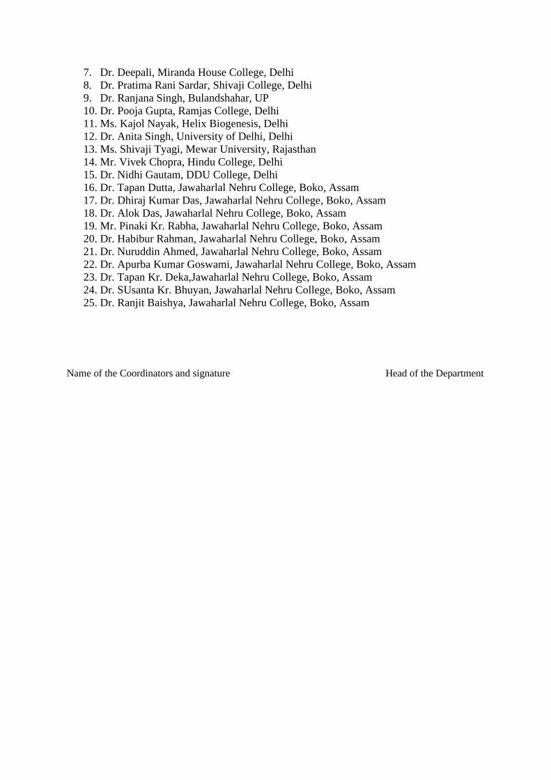
7. Dr. Deepali, Miranda House College, Delhi
8. Dr. Pratima Rani Sardar, Shivaji College, Delhi
9. Dr. Ranjana Singh, Bulandshahar, UP
10. Dr. Pooja Gupta, Ramjas College, Delhi
11. Ms. Kajol Nayak, Helix Biogenesis, Delhi
12. Dr. Anita Singh, University of Delhi, Delhi
13. Ms. Shivaji Tyagi, Mewar University, Rajasthan
14. Mr. Vivek Chopra, Hindu College, Delhi
15. Dr. Nidhi Gautam, DDU College, Delhi
16. Dr. Tapan Dutta, Jawaharlal Nehru College, Boko, Assam
17. Dr. Dhiraj Kumar Das, Jawaharlal Nehru College, Boko, Assam
18. Dr. Alok Das, Jawaharlal Nehru College, Boko, Assam
19. Mr. Pinaki Kr. Rabha, Jawaharlal Nehru College, Boko, Assam
20. Dr. Habibur Rahman, Jawaharlal Nehru College, Boko, Assam
21. Dr. Nuruddin Ahmed, Jawaharlal Nehru College, Boko, Assam
22. Dr. Apurba Kumar Goswami, Jawaharlal Nehru College, Boko, Assam
23. Dr. Tapan Kr. Deka,Jawaharlal Nehru College, Boko, Assam
24. Dr. SUsanta Kr. Bhuyan, Jawaharlal Nehru College, Boko, Assam
25. Dr. Ranjit Baishya, Jawaharlal Nehru College, Boko, Assam
Name of the Coordinators and signature Head of the Department

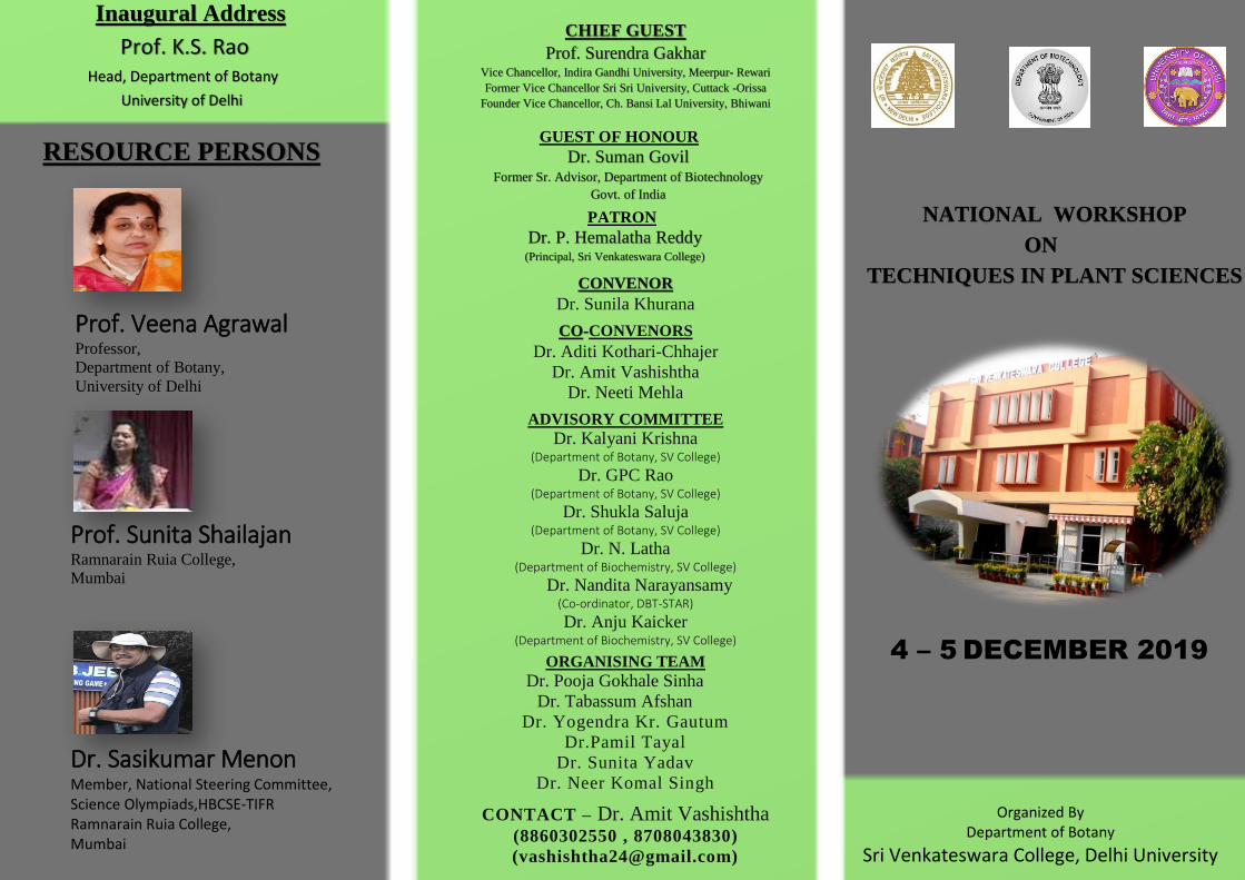
NATIONAL WORKSHOP
ORGANIZED BY
TECHNIQUES IN PLANT SCIENCES
ON
(Accredited by NAAC with 'A' Grade)UNIVERSITY OF DELHI
SRI VENKATESWARA COLLEGE
RESOURCE PERSONS
Chief GuestProf. S. K. Gakhar
Vice Chancellor,Indira Gandhi UniversityMeerpur-Rewari
Former Vice Chancellor Sri Sri University Cuttack-Orissa
Founder Vice Chancellor, Ch. Bansi LalUniversity,Bhiwani
Prof. Veena AggarwalDepartment of Botany
University of Delhi
Prof. Sunita ShailajanDean-Research Consultancy
and Innovation
Ruia College
University of Bombay
Dr. Sasikumar MenonMember, National Steering
Committee
Science Olympiads,
HBCSE-TIFR,
DAE, GOI
PATRONDr. P. Hemalatha Reddy
PRINCIPAL
Sri Venkateswara College
CONVENORDr. Sunila Khurana
CO-CONVENORSDr. Aditi Kothari-Chhajer
Dr. Amit Vashishtha
Dr. Neeti Mehla
ADVISORY COMMITTEEDr. Kalyani Krishna
(Department of Botany, SV College)
Dr. GPC Rao
(Department of Botany, SV College)
Dr. Shukla Saluja
(Department of Botany, SV College)
Dr. N. Latha
(Department of Biochemistry, SV College)
Dr. Anju Kaicker
(Department of Biochemisty, SV College)
ORGANISING TEAM
Dr. Tabassum Afshan
Dr. Pooja Gokhale Sinha
Dr. Yogendra Kr. Gautam
Dr. Pamil Tayal
Dr. Neer Komal Singh
Dr. Sunita Yadav
4-5 DECEMBER,2019
Inaugural address Prof. K.S Rao
Head, Department of BotanyUniversity Of Delhi
Guest of HonourDr. Suman GovilAdvisor, Department of
BiotechnologyGovt. of India

THEME OF WORKSHOP
The workshop aims at providing hands-on training in the field of in-vitro morphogenesis of medicinalplants and study of genetic fidelity through Molecular Markers. Plant tissue culture is a part of PlantBiotech which is the collection of many techniques that are used to maintain and grow plants undercontrolled aseptic conditions. Genetic integrity of the plants derived through micropropogationbecomes crucial if genetic transformation studies have to be carried out and hence hands-on trainingin this field shall provide an overall understanding of this area of plant biotechnology. With thechanging phase of education it is imperative for teachers to stay abreast with the latest trends inhigher education to meet the emerging challenges & opportunities. In order to enhance this facet ofteaching -learning process, this workshop provides an opportunity to interact with leading faculty forsome brainstorming sessions for the development of questions with higher order thinking skills.
WORKSHOP AT A GLANCEHands on trainingIn-vitro morphogenesis in Medicinal Plants.Genetic fidelity using Molecular markers.HP-TLC.Brainstorming session on question paper setting involvinghigher order thinking skills.
Who can attend the workshop– Faculty from Colleges/Universities.
Last date For Registeration- 1st December,2019Registration Link – https://docs.google.com/forms/d/e/1FAIpQLScmxs5LtvK9n79AehnC72m8RS4X2AKLc3QuxWWf_mPxSlV9fA/viewform?vc=0&c=0&w=1
Registration Fees - Rs. 750/-
For any query,Contact:
Amit Vashishtha- 8860302550
8708043830
E-mail :[email protected]
(Double tap to open the link)
Prof. Veena Agrawal Professor, Department of Botany, University of Delhi
NATIONAL WORKSHOP
ON
TECHNIQUES IN PLANT SCIENCES
Prof. Sunita Shailajan Ramnarain Ruia College,
Mumbai
Inaugural Address
Prof. K.S. Rao Head, Department of Botany
University of Delhi
RESOURCE PERSONS
CHIEF GUEST
Prof. Surendra Gakhar Vice Chancellor, Indira Gandhi University, Meerpur- Rewari
Former Vice Chancellor Sri Sri University, Cuttack -Orissa
Founder Vice Chancellor, Ch. Bansi Lal University, Bhiwani
GUEST OF HONOUR
Dr. Suman Govil Former Sr. Advisor, Department of Biotechnology
Govt. of India
PATRON
Dr. P. Hemalatha Reddy (Principal, Sri Venkateswara College)
CONVENOR
Dr. Sunila Khurana
CO-CONVENORS
Dr. Aditi Kothari-Chhajer
Dr. Amit Vashishtha
Dr. Neeti Mehla
ADVISORY COMMITTEE
Dr. Kalyani Krishna (Department of Botany, SV College)
Dr. GPC Rao (Department of Botany, SV College)
Dr. Shukla Saluja (Department of Botany, SV College)
Dr. N. Latha (Department of Biochemistry, SV College)
Dr. Nandita Narayansamy
(Co-ordinator, DBT-STAR)
Dr. Anju Kaicker (Department of Biochemistry, SV College)
ORGANISING TEAM
Dr. Pooja Gokhale Sinha
Dr. Tabassum Afshan
Dr. Yogendra Kr. Gautum
Dr.Pamil Tayal
Dr. Sunita Yadav
Dr. Neer Komal Singh
CONTACT – Dr. Amit Vashishtha (8860302550 , 8708043830)
4 – 5 DECEMBER 2019
Dr. Sasikumar Menon Member, National Steering Committee, Science Olympiads,HBCSE-TIFR Ramnarain Ruia College, Mumbai
Organized By Department of Botany
Sri Venkateswara College, Delhi University
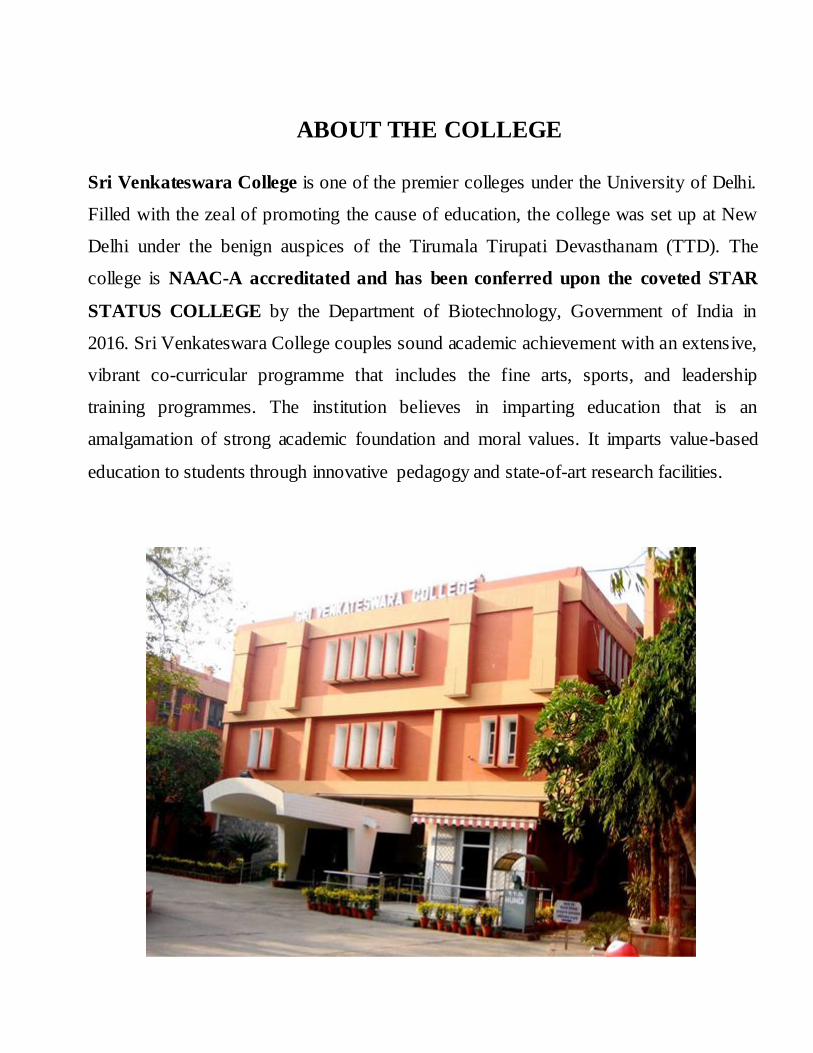
ABOUT THE COLLEGE
Sri Venkateswara College is one of the
premier colleges under the University of
Delhi. Filled with the zeal of promoting the
cause of education, the college was set up at
New Delhi under the benign auspices of the
Tirumala Tirupati Devasthanam (TTD). The
college is NAAC-A accreditated and has
been conferred upon the coveted STAR
STATUS COLLEGE by the Department of
Biotechnology, Government of India in 2016.
Sri Venkateswara College couples sound
academic achievement with an extensive,
vibrant co-curricular programme that includes
the fine arts, sports, and leadership training
programmes. The institution believes in
imparting education that is an amalgamation
of strong academic foundation and moral
values. It imparts value-based education to
students through innovative pedagogy and
state-of-art research facilities.
WORKSHOP AT A GLANCE
Hands-on training
In-Vitro morphogenesis in Medicinal
Plants.
Genetic Fidelity Using Molecular
Markers.
HP-TLC.
Brainstroming session on question
paper setting involving higher order
thinking skills.
WHO CAN ATTEND THE
WORKSHOP – Faculty from Colleges/Universities.
Last date For Registeration –
1st December 2019
Registration Link – https://docs.google.com/forms/d/e/1FAIpQLScmxs5LtvK9n
79AehnC72m8RS4X2AKLc3QuxWWf_mPxSlV9fA/viewform?vc=0&c=0&w=1
Registration Fees - Rs. 750/-
“THE SCIENCE OF TODAY IS THE
TECHNOLOGY OF TOMORROW”
- EDWARD TELLER
THEME OF WORKSHOP
The workshop aims at providing hands-on
training in the field of in vitro morphogenesis
of medicinal plants and study of genetic
fidelity through molecular markers. Plant
tissue culture is a part of plant biotechnology
which is the collection of many techniques
that is used to maintain and grow plants
under controlled aseptic conditions. Genetic
integrity of the plants derived through
micropropogation becomes crucial if genetic
transformation studies have to be carried out
and hence hands-on training in this field shall
provide an overall understanding of this area
of plant biotechnology.
With the changing face of education it is
impertive for teachers to stay abreast with the
latest trends in higher education to meet the
emerging challenges & opportunities. In
order to enhance this facet of teaching -
learning process, this workshop provides an
opportunity to interact with leading faculty
for some brainstorming sessions for the
development of questions with higher order
thinking skills.
Registration Link – https://docs.google.com/forms/d/e/1FAIpQLScmxs5LtvK9n79Aeh
nC72m8RS4X2AKLc3QuxWWf_mPxSlV9fA/viewform?vc=0&c=0&w=1

Organised by
SRI VENKATESWARA COLLEGE
(Accredited by NAAC with „A‟ grade)
University of Delhi
4 – 5
DECEMBER 2019
Sri Venkateswara College is one of the premier colleges under the University of Delhi.
Filled with the zeal of promoting the cause of education, the college was set up at New
Delhi under the benign auspices of the Tirumala Tirupati Devasthanam (TTD). The
college is NAAC-A accreditated and has been conferred upon the coveted STAR
STATUS COLLEGE by the Department of Biotechnology, Government of India in
2016. Sri Venkateswara College couples sound academic achievement with an extensive,
vibrant co-curricular programme that includes the fine arts, sports, and leadership
training programmes. The institution believes in imparting education that is an
amalgamation of strong academic foundation and moral values. It imparts value-based
education to students through innovative pedagogy and state-of-art research facilities.
ABOUT THE COLLEGE

PATRON
Dr. P. Hemalatha Reddy
CONVENOR
Dr. Sunila Khurana
CONVENORS CO-
Dr. Aditi Kothari-Chhajer Dr. Amit Vashishtha
Dr. Neeti Mehla
ADVISORY COMMITTEE
Dr. Kalyani Krishna Dr. GPC Rao
Dr. Shukla Saluja Dr. N. Latha
Dr. Anju Kaicker
ORGANISING MEMBERS Dr. Pooja Gokhale Sinha
Dr. Yogendra Kr. Gautum Dr. Tabassum Afshan
Dr.Pamil Tayal Dr. Sunita Yadav Dr. Neer Komal Singh
THEME OF WORKSHOP
The workshop aims at providing hands-on training in the field of in vitro morphogenesis of
medicinal plants and study of genetic fidelity through molecular markers. Plant tissue culture
is a part of Plant Biotech which is the collection of many techniques that is used to maintain
and grow plants under controlled aseptic conditions. Genetic integrity of the plants derived
through micropropogation becomes crucial if genetic transformation studies have to be
carried out and hence hands-on training in this field shall provide an overall understanding of
this area of plant biotechnology.
With the changing demography of education it is impertive for teachers to stay abreast with
the latest trends in higher education to meet the emerging challenges & opportunities. In
order to enhance this facet of teaching -learning process, this workshop provides an
opportunity to interact with leading faculty for some brainstorming sessions for the
development of questions with higher order thinking skills.
PATRON Dr. P. Hemalatha Reddy
CONVENOR
Dr. Sunila Khurana
CO-CONVENORS
Dr. Aditi Kothari-Chhajer Dr. Amit Vashishtha
Dr. Neeti Mehla
ADVISORY COMMITTEE
Dr. Kalyani Krishna Dr. GPC Rao
Dr. Shukla Saluja Dr. N. Latha
Dr. Anju Kaicker
ORGANISING MEMBERS Dr. Pooja Gokhale Sinha Dr. Yogendra Kr. Gautum
Dr. Tabassum Afshan Dr.Pamil Tayal
Dr. Sunita Yadav Dr. Neer Komal Singh

Programe Schedule
4th December 2019
9.30 am Invocation and Lighting of Lamp
9.40 am Address by DBT-STAR College Status Co-ordinator, Dr. Nandita
Narayansamy
9.45 am
Principal‟s address, Dr. P. Hemalatha Reddy
9.55 am Address by the Chief Guest
Prof. S.K. Ghakhar
Vice Chancellor
Indira Gandhi University
Meerpur - Rewari, Haryana
10.10 am Inaugural address by
Prof. K.S. Rao
Head
Department of Botany
University of Delhi, Delhi
10: 30 – 11:00 am High tea and Interaction with faculty
Session –I
11:00 -11:45 am
Session Chair: Dr. Suman Govil, Former Senior Advisor,
Department of Biotechnology, Government of India
Talk on “Applications of HP-TLC in Medicinal plants” by Prof.
Sunita Shailajan, Ramnarain Ruia Autonomous College, Mumbai
11:45-1:30 pm Hands on Training in HP-TLC by Prof. Sunita Shailajan
1:30-2:00 pm Lunch
2:00-3:00 pm HP-TLC Contd….
3:00-5:00 pm Session on Question Paper setting by Dr. Sasikumar Menon, ,
Ramnarain Ruia Autonomous College, Mumbai
5th December 2019
Session – II
9:30 am Introductory Talk on Plant Tissue culture by Prof. Veena Agrawal
Department of Botany
University of Delhi
10:30 am Tea break

10:45-2:00 pm Hands-on training in Plant Tissue Culture Techniques by Prof. V.
Agrawal
2:00-2:30 pm Lunch
Session -III
2:30- 4:30 pm Hands-on training on Genetic Fidelity using Molecular Markers by Prof. Veena Agrawal
4:30 pm Distribution of Certificates
5:00 pm Tea

SESSION – I
Hands on training on HP-TLC
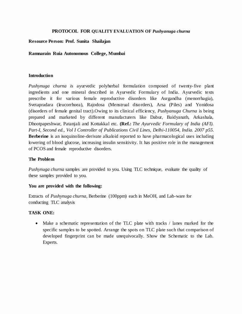
PROTOCOL FOR QUALITY EVALUATION OF Pushyanuga churna
Resource Person: Prof. Sunita Shailajan
Ramnarain Ruia Autonomous College, Mumbai
Introduction
Pushynuga churna is ayurvedic polyherbal formulation composed of twenty-five plant
ingredients and one mineral described in Ayurvedic Formulary of India. Ayurvedic texts
prescribe it for various female reproductive disorders like Asrgandha (menorrhagia),
Svetapradara (leucorrhoea), Rajodosa (Menstrual disorders), Arsa (Piles) and Yonidosa
(disorders of female genital tract).Owing to its clinical efficiency, Pushyanuga Churna is being
prepared and marketed by different manufacturers like Dabur, Baidyanath, Arkashala,
Dhootpapeshwar, Patanjali and Kottakkal etc. (Ref.: The Ayurvedic Formulary of India (AFI).
Part-I, Second ed., Vol I Controller of Publications Civil Lines, Delhi-110054, India. 2007 p55.
Berberine is an isoquinoline-derivate alkaloid reported to have pharmacological uses including
lowering of blood glucose, increasing insulin sensitivity. It has positive role in the management
of PCOS and female reproductive disorders.
The Problem
Pushynuga churna samples are provided to you. Using TLC technique, evaluate the quality of
these samples provided to you.
You are provided with the following:
Extracts of Pushynuga churna, Berberine (100ppm) each in MeOH, and Lab-ware for
conducting TLC analysis
TASK ONE:
Make a schematic representation of the TLC plate with tracks / lanes marked for the
specific samples to be spotted. Arrange the spots on TLC plate such that comparison of
developed fingerprint can be made unequivocally. Show the Schematic to the Lab.
Experts.

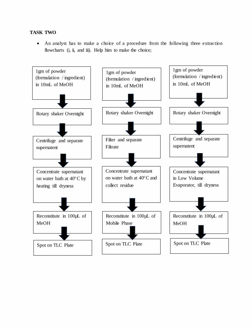
1gm of powder
(formulation / ingredient)
in 10mL of MeOH
1gm of powder
(formulation / ingredient)
in 10mL of MeOH
Rotary shaker Overnight Rotary shaker Overnight Rotary shaker Overnight
Centrifuge and separate
supernatent
Centrifuge and separate
supernatent Filter and separate Filtrate
Concentrate supernatant
on water bath at 40C by
heating till dryness
Concentrate supernatant
on water bath at 40C and
collect residue
Concentrate supernatant
in Low Volume
Evaporator, till dryness
Reconstitute in 100µL of
MeOH
Reconstitute in 100µL of
MeOH
Reconstitute in 100µL of
Mobile Phase
Spot on TLC Plate Spot on TLC Plate Spot on TLC Plate
1gm of powder
(formulation / ingredient)
in 10mL of MeOH
TASK TWO
An analyst has to make a choice of a procedure from the following three extraction
flowcharts (i, ii, and iii). Help him to make the choice;

Show your answer to the Lab- Expert.
Now ask for Task Three.
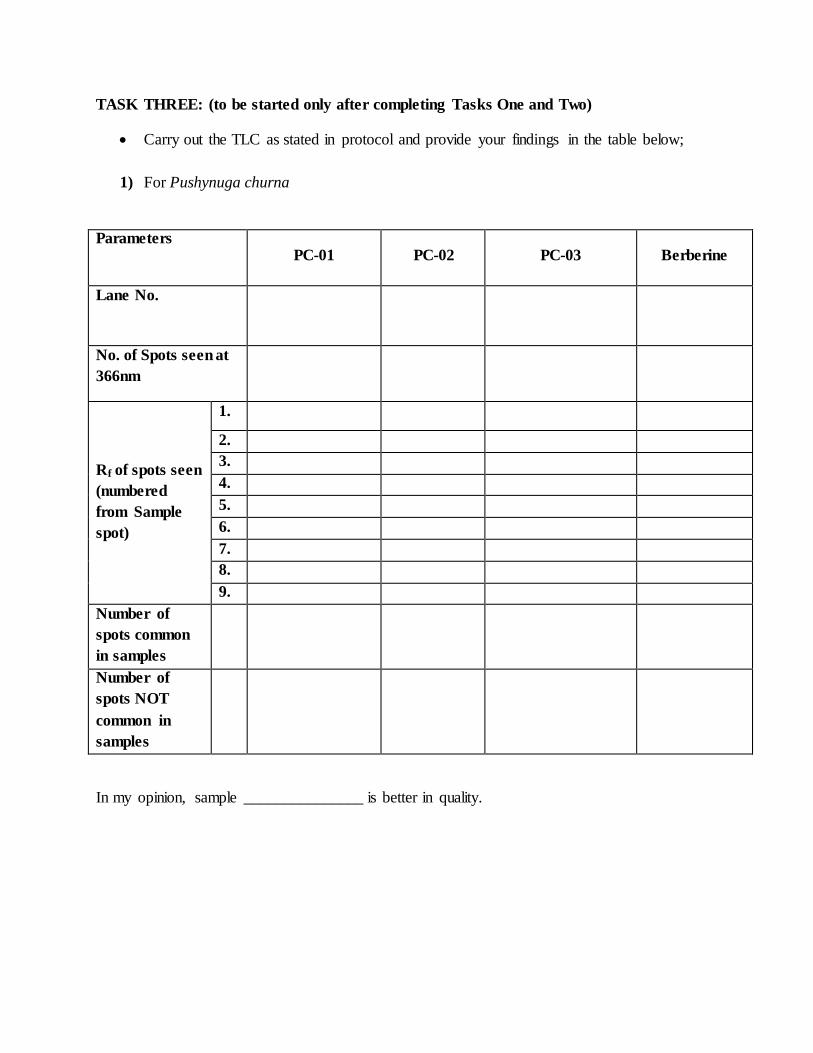
TASK THREE: (to be started only after completing Tasks One and Two)
Carry out the TLC as stated in protocol and provide your findings in the table below;
1) For Pushynuga churna
Parameters PC-01 PC-02 PC-03 Berberine
Lane No.
No. of Spots seen at
366nm
Rf of spots seen
(numbered
from Sample
spot)
1.
2.
3.
4.
5.
6.
7.
8.
9.
Number of
spots common
in samples
Number of
spots NOT
common in
samples
In my opinion, sample _______________ is better in quality.

Protocol for TLC
Measure the dimension of the plate and cut the plate of the required size (i.e 5x9cm for
Pushyanuga churna ).
Leave 1cm space from bottom and 0.5cm from both sides, mark 7.5cm as solvent front
and label the spots accordingly (Note: Use only pencil for markings on plate)
Load the sample with the help of fused capillary on to the TLC plate in form of
Spot/band (Note: Use separate fused capillary for every sample to avoid contamination.
load the sample at least five times, dry the spot before next loading).
Prepare the mobile Phase, toluene: ethyl acetate: methanol: formic acid (6:6:2:1v/v/v/v)
for quantitation of Berberin from Pushyanuga churna and vortex it.
Pour the mobile phase in the beaker and cover it with glass lid, allow it to saturate it for
20 minutes.
Gently place the TLC plate into the mobile phase of the saturated beaker, with the spotted
end of the plate towards the bottom close to the solvent. Submerge the bottom edge of the
plate into the solvent, allowing the solvent to run upto the marked solvent front (7.5cm).
Remove the plate and allow it to air dry (Note: Allow the solvent to evaporate from the
plate.)
Visualize the plate at 366nm in UV-Visible visualizing chamber and calculate the
Retardation Factor (Rf) and note them is the results sheet.
1.0cm
7.5cm
1 2 3 4
0.5cm
Plate1: 5x9cm
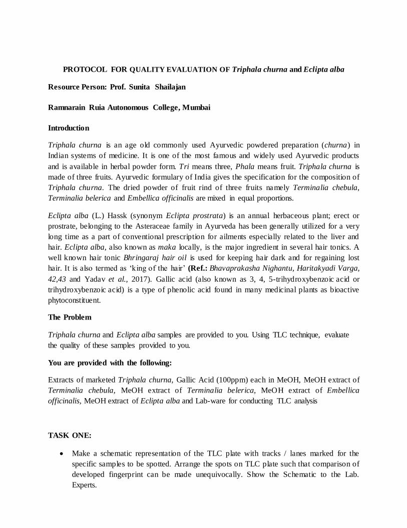
PROTOCOL FOR QUALITY EVALUATION OF Triphala churna and Eclipta alba
Resource Person: Prof. Sunita Shailajan
Ramnarain Ruia Autonomous College, Mumbai
Introduction
Triphala churna is an age old commonly used Ayurvedic powdered preparation (churna) in
Indian systems of medicine. It is one of the most famous and widely used Ayurvedic products
and is available in herbal powder form. Tri means three, Phala means fruit. Triphala churna is
made of three fruits. Ayurvedic formulary of India gives the specification for the composition of
Triphala churna. The dried powder of fruit rind of three fruits namely Terminalia chebula,
Terminalia belerica and Embellica officinalis are mixed in equal proportions.
Eclipta alba (L.) Hassk (synonym Eclipta prostrata) is an annual herbaceous plant; erect or
prostrate, belonging to the Asteraceae family in Ayurveda has been generally utilized for a very
long time as a part of conventional prescription for ailments especially related to the liver and
hair. Eclipta alba, also known as maka locally, is the major ingredient in several hair tonics. A
well known hair tonic Bhringaraj hair oil is used for keeping hair dark and for regaining lost
hair. It is also termed as „king of the hair‟ (Ref.: Bhavaprakasha Nighantu, Haritakyadi Varga,
42,43 and Yadav et al., 2017). Gallic acid (also known as 3, 4, 5-trihydroxybenzoic acid or
trihydroxybenzoic acid) is a type of phenolic acid found in many medicinal plants as bioactive
phytoconstituent.
The Problem
Triphala churna and Eclipta alba samples are provided to you. Using TLC technique, evaluate
the quality of these samples provided to you.
You are provided with the following:
Extracts of marketed Triphala churna, Gallic Acid (100ppm) each in MeOH, MeOH extract of
Terminalia chebula, MeOH extract of Terminalia belerica, MeOH extract of Embellica
officinalis, MeOH extract of Eclipta alba and Lab-ware for conducting TLC analysis
TASK ONE:
Make a schematic representation of the TLC plate with tracks / lanes marked for the
specific samples to be spotted. Arrange the spots on TLC plate such that comparison of
developed fingerprint can be made unequivocally. Show the Schematic to the Lab.
Experts.
1gm of powder
(formulation / ingredient)
in 10mL of MeOH
1gm of powder
(formulation / ingredient)
in 10mL of MeOH
Rotary shaker Overnight Rotary shaker Overnight Rotary shaker Overnight
Centrifuge and separate
supernatent
Centrifuge and separate
supernatent
Filter and separate
Filtrate
Concentrate supernatant
on water bath at 40C by
heating till dryness
Concentrate supernatant
on water bath at 40C and
collect residue
Concentrate supernatant
in Low Volume
Evaporator, till dryness
Reconstitute in 100µL of
MeOH
Reconstitute in 100µL of
MeOH
Reconstitute in 100µL of
Mobile Phase
Spot on TLC Plate Spot on TLC Plate Spot on TLC Plate
1gm of powder
(formulation / ingredient)
in 10mL of MeOH
TASK TWO
An analyst has to make a choice of a procedure from the following three extraction
flowcharts (i, ii, and iii). Help him to make the choice;

Show your answer to the Lab- Expert.
Now ask for Task Three.

TASK THREE: (to be started only after completing Tasks One and Two)
Carry out the TLC as stated in protocol and provide your findings in the table below;
1) For Triphala churna
Parameters Terminali
a chebula
Terminali
a belerica
Embellica
officinalis
Triphala
Churna (A)
Triphala
Churna (B)
Gallic
acid
Lane No.
No. of Spots seen in
visible
No. of Spots seen at
366nm
No. of Spots seen at
254`nm
Rf of spots seen
(numbered
from Sample
spot)
1.
2.
3.
4.
5.
6.
7.
8.
9.
Number of
spots common
in samples and
formulations
Number of
spots NOT
common in
samples and
formulation
Number of
spots from
ingredients of
the
formulation
In my opinion sample _______________ is better in quality than sample ______________
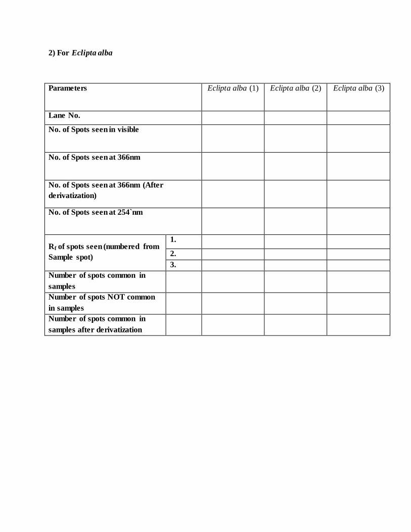
2) For Eclipta alba
Parameters Eclipta alba (1) Eclipta alba (2) Eclipta alba (3)
Lane No.
No. of Spots seen in visible
No. of Spots seen at 366nm
No. of Spots seen at 366nm (After
derivatization)
No. of Spots seen at 254`nm
Rf of spots seen (numbered from
Sample spot)
1.
2.
3.
Number of spots common in
samples
Number of spots NOT common
in samples
Number of spots common in
samples after derivatization

Protocol for TLC
Measure the dimension of the plate and cut the plate of the required size (i.e 6x9cm for Triphala
churna and 3x9 cm for Eclipta alba).
Leave 1cm space from bottom and 0.5cm from both sides, mark 7.5cm as solvent front and
label the spots accordingly (Note: Use only pencil for markings on plate)
Load the sample with the help of fused capillary on to the TLC plate in form of Spot/band
(Note: Use separate fused capillary for every sample to avoid contamination. load the sample at
least five times, dry the spot before next loading).
Prepare the mobile Phase, toluene: ethyl acetate: formic acid (2:7:1v/v/v) for quantitation of
gallic acid from Triphala churna and toluene: n hexane (9:1.2 v/v) for fingerprint of Eclipta
alba and vortex it.
Pour the mobile phase in the beaker and cover it with glass lid, allow it to saturate it for 20
minutes.
Gently place the TLC plate into the mobile phase of the saturated beaker, with the spotted end
of the plate towards the bottom close to the solvent. Submerge the bottom edge of the plate into
the solvent, allowing the solvent to run upto the marked solvent front (7.5cm).
Remove the plate and allow it to air dry (Note: Allow the solvent to evaporate from the plate.)
Visualize the plate at 254nm and 366nm in UV-Visible visualizing chamber and calculate the
Retardation Factor (Rf) and note them is the results sheet.
Derivatize the (E. alba) plate by Dipping the plate in 10% Methanolic sulphuric acid reagent for
50 seconds, heat the plate at 110°C using hot air oven for 5minutes).
Visualize the plate at 366nm in UV-Visible visualizing chamber and note them in the results
sheet.
Plate1: 6x9cm Plate2: 3x9cm
7.5cm
7.5cm
1 2 3 4 5 6 1 2 3 0.5cm
0.5cm
1.0cm
1.0cm

SESSION – II
Hands on training on In-vitro
morphogenesis in medicinal plants

PROTOCOL FOR IN VITRO CLONAL PROPAGATION OF ECONOMICALLY
IMPORTANT PLANT TAXA
Resource Person: Prof. Veena Agrawal
Plant biotech. Lab Department of Botany, University of Delhi, Delhi - 110 007
Requirements:
Plant Material: Any plant system (herb, shrub or tree) of economic value such as
medicinal, neutraceutical, crop or commercial importance.
Chemicals: Teepol, bavistin, cefotaxime, citric acid, mercuric chloride, salts of macro- and
micro elements of Murashige and Skoog's (1962) and Knop‟s (1865) basal media, sucrose,
agar,1N NaOH, 1N HCL, plant growth regulators [2,4-dichlorophenoxyacetic acid (2,4-D),
α-naphthaleneacetic acid (NAA), indole-3-acetic acid (IAA), indole-3-butyric acid (IBA), 6-
benzyladenine (BA), thidiazuron (TDZ), kinetin (kn), 2- iP [6-(gamma, gamma-
Dimethylallylamino) purine] and absolute alcohol. All the chemicals should be of Sigma-
Aldrich, Merck./Hi- Media, SRL. and of standard companies.
Glass wares: Beakers (500, 250, 1000 ml), measuring cylinders (10, 50, 100, 500, 1000 ml
capacity), conical flasks.
Equipments: Laminar air flow cabinet, pH meter, oven/ heater, stirrer, autoclave, pipettes,
tips, Petri plates, forceps, scalpel, blades.
Miscellaneous: Aluminum foil, tissue paper, butter paper, cotton plugs, spirit lamp, discard,
dip, match box, marker, rectified spirit, autoclaved distilled water and glass plates/ Petri-
plates for inoculation.

Methods:
Preparation of Stock Solutions:
Stock solutions were prepared according to the salt composition given below:
Table 1. Composition of Murashige and Skoog (1962) and Knop's (1865) medium
S. No. Nutrients MS (1962) Knops
(1865)
Major (g/1000 mL)
1 KNO3 38 3
2 NH4NO3 33 -
3 KH2PO4 3.4 3
4 MgSO4.7H2O 7.4 3
5 CaCl2.2H2O 8.8 -
Minor (mg/1000 mL)
6 MnSO4.H2O 446 500
7 H3BO3 124 200
8 ZNSO4.7H2O 172 10
9 KI 16.6 -
10 Na2MoO4.2H2O 5 -
11 CuSO4 0.5 -
12 CoCL2.6H2O 0.5 -
Organic (mg/100 mL)
13 Glycine 20 -
14 Meso-inositol 100 100
15 Nicotinic acid 5 -
16 Pyridoxine HCl 5 -
17 Thiamine HCl 1 10
Iron (mg/100 mL)
18 FeSO4.7H2O 278 55.6
19 Na2EDTA 373 74.4
Preparation of stock solution of growth regulators:
Weigh 10 mg of the plant growth regulator in an Eppendorf tube or other glass container.
Add 1-2 drops of solvent (ethanol/NaOH) to dissolve the powder. Once completely
dissolved, make volume up to 10 ml with autoclaved distilled water. Store the stock solution
in the fridge for further use.
Table 2. List of some Important plant growth regulators and their storage
S.N.
Plant growth regulators
Molecular weight
(g/mol)
Solvent Stock Storage
1 2,4-D
(2, 4- Dichlorophenoxyacetic acid)
221 Ethanol
(Pure)
2-8 ºC
2 NAA
(α-Naphthaleneacetic acid)
186.2 Ethanol /
1N NaOH
2-8 ºC
3 IAA
(Indole-3-acetic acid)
175.2 Ethanol /
1N NaOH
0 ºC
4 IBA
(Indole-3-butyric acid)
203.2 Ethanol /
1N NaOH
0 ºC
5 BAP
(6-Benzylaminopurine)
225.3 1N NaOH 2-8 ºC
6 Kinetin 215.2 1N NaOH 0 ºC
7 2-iP
[6-(gamma,gamma-
Dimethylallylamino] purine
203.2 1N NaOH 0 ºC
8 TDZ
(Thidiazuron)
220.25 1N NaOH 0 ºC
Preparation of Culture media:

Semi-solid MS basal medium was prepared using above mentioned stock solutions
containing 0.8 % (w/v) agar-agar (bacteriological grade, Thermo Fisher Scientific®) and 3 %
(w/v) sucrose (DCM, Daurala, India) as given hereunder:
1. Weigh 8 g of agar and 30 g of sucrose and put these in the conical flask of 1L containing 300
ml of distilled water. Melt the agar and sucrose by keeping the flask on the hot plate/ heater.
2. In a separate measuring cylinder of 1/ 2L add 50 ml stock solutions each of micro and macro
salts and 10 ml each of iron and organic prepared above (Table 1).
3. Add the mixture of agar and sucrose (prepared at step 1) to this measuring cylinder
containing MS salts and make the volume 1L by adding hot distilled water. Shake gently till
homogenous solution is prepared. Transfer the medium to conical flask to set the pH.
4. Add growth regulator if required prior setting pH.
5. The pH of the medium was adjusted to 5.8 with 1 N NaOH and 1 N HCl (Thermo Fisher
Scientific®) prior to autoclaving at 121°C and 15 lbs psi for 15 min.
6. Take out the test-tubes from the autoclave and wait till the medium temperature come down
to around 60°C.
7. Make slants of the medium by tilting the test tubes and place these in the culture room for inoculation.
Surface sterilization of explants:
1. Nodal explants (0.5-1.0 cm in length) were cut from the fresh twigs.
2. These explants were washed thoroughly under running tap water for 15-20 min, followed by
rinsing with 5% v/v Teepol (Reckitt and Colman, Mumbai, India) for 10-15 min.
3. Subsequently, the explants were treated with 2% (w/v) citric acid to minimize browning
effect from the cut ends of nodal explants.
4. To remove any fungal and bacterial contaminants adhered to the surface, explants were
treated with 0.5% (w/v) Bavistin® fungicide (Bavistin®, Carbendazim 50% WP, BASF
India Ltd., Mumbai, India) + 150 mg/L cefotaxime (Alkem, Sikkim, India) for 10 min and
then rinsed three times with distilled water.
5. Final surface sterilization is to be done with 0.1% (w/v) HgCl2 for 2 min in laminar air flow
cabinet followed by rinsing 3 or 4 times with autoclaved double distilled water.

Induction of morphogenesis and multiple shoots:
In vitro regeneration(micropropagation) can be achieved through either organogenesis
or somatic embryogenesis:
1. Direct regeneration: Cotyledonary nodes, stem nodal segments and shoot tip explants are
the ideal material for direct induction of multiple shoots.For indirect organogenesis root
hypocotyle, cotyledon and leaf, explants are used.
2. Initially, there is a bud break and elongation of axillary shoot takes place on MS basal
medium.
3. For multiple shoot induction MS medium supplemented with different plant growth
regulators (BAP, KN, TDZ, or 2-iP) at concentrations ranging from 0.1, 1, 5, 10 & 20 µM is
beneficial. (See Figs 1&2).
4. The cultures are daily observed for 28 days and data pertaining to shoot number and shoot
length recorded weekly on observation book. Average number of shoot and shoot length are
calculated and statistically analyzed.
Induction of roots in the excised shoots (Rhizogenesis):
1. For root induction, in vitro raised shoots were excised and placed on MS medium
supplemented with different concentrations (0.1, 1, 5, 10 µM) of IAA, IBA, or NAA.
Emergence of direct roots is initiated with in one weeks from the basal cut ends. However,
if there is an intervening callus then some additives such as PVP or activated charcoal may
be added to the rooting medium.
2. Average number of roots per shoot and average root length were recorded after 4 weeks of
culture.
Indirect organogenesis: Explants such as root, hypocotyle, cotyledonary leaf, stem leaf are
cultured on MS /B5 basal medium augmented with auxin (IAA,NAA,IBA or 2,4-D) in
different concentration ranging from 0.1-20 µM to induce callus. Such callus pieces are then
sub cultured on cytokinin ( BA,Kin.,2iP,Zeatin) supplemented basal medium for induction
and proliferation of shoots. These micro shoots are subsequently excised and transferred to
rooting medium given above for induction of roots. Finally these are hardened and
acclimatized to soil and fields.(See Fig.1)
Somatic embryogenesis: Somatic embryos are embryo like structures derived from somatic
tissues and mimic all the stages of zygotic embryos. These are also considered as best

material for clonal propagation as the embryo directly develops in to shoot and root in one
step. Explants are first cultured on auxin supplemented MS/B5 medium to induce callus.
The callus either organize embryos or shoot buds. The embryogenic calluses are
microscopically identified with the embryo- like structures and are further sub cultured on
maturation medium (abscisic acid/proline/sucrose/PEG) for proper developments. Such
mature embryos are then transferred to basal medium for germination in to complete
plantlets.(See Figs 1&3).
In vitro hardening and field acclimatization:
1. After rhizogenesis, healthy plantlets with well developed roots were removed from medium
and washed under running tap water to remove the adhering medium.
2. They were treated with 1 % bavistin (BASF, Mumbai, India) solution to prevent any fungal
infection, before being transferred to plastic pots (5 cm diameter) containing autoclaved soil.
3. The plantlets were irrigated with one fourth MS salts for one week and one-tenth for next one
month.
4. The plantlets were covered with polythene bags having, 3–5 holes to maintain the humidity.
5. The potted plants were maintained inside the glass house at controlled conditions till they are
established well in the soil.
6. After 45 days, the plantlets were transplanted to earthen pots (25 cm diameter) containing
garden soil and were maintained there for 2-3 months.
7. The plantlets were finally transferred to soil-bed for field acclimatization till maturity.
Recording of data and statistical analysis:
1. For in vitro regeneration, the average number of shoots per explant, the average shoot length,
the average number of roots per shoot and the average root length has been represented as
mean values along with standard error (Mean ± SE).
2. The mean values were calculated on the basis of a minimum of 24 replicates in each
experiment and repeated at least for once or twice.
3. The data expressed as mean ± SE have been statistically analyzed using ANOVA (Analysis
of Variance) through SPSS (Statistical Package for Social Sciences) version 16.0. The
differences between means were tested for significance by Duncan‟s multiple range test
(DMRT) at P ≤ 0.05.

Fig.1. An overview of Micropropagation revealing direct and indirect in vitro regeneration

Fig. 2. Direct organogenesis through nodal explants of Spilanthes calva DC. after 6 weeks of
culture. (a) Field raised mature plant (4-month-old) of Spilanthes calva (inset shows nodal explant at the time of inoculation); (b) Single axillary shoot after 6 weeks of culture on MS basal
medium; (c) Differentiation of multiple shoots from nodal explants within 6 weeks of culture on MS+10 μM BA; (d) Culture showing stunted shoots and formation of callus at the basal end of the nodal explant on MS+20 μM BA; (e) Excised in vitro shoot inducing root on MS (1/2)+ 0.1
μM IBA; (f) Tissue culture-derived plant acclimatized to soil in glass house; (g) Three month old tissue culture raised plants in garden.
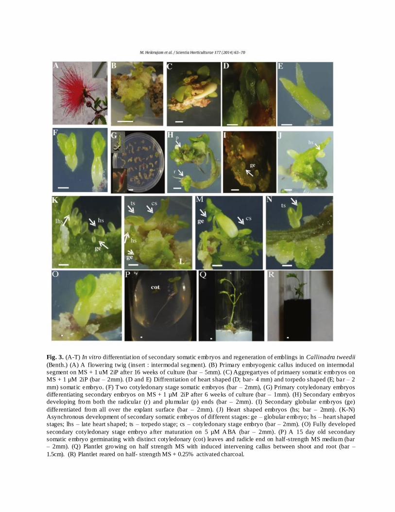
Fig. 3. (A-T) In vitro differentiat ion of secondary somatic embryos and regeneration of emblings in Callinadra tweedii
(Benth.) (A) A flowering twig (insert : intermodal segment). (B) Primary embryogenic callus induced on intermodal
segment on MS + 1 uM 2iP after 16 weeks of culture (bar – 5mm). (C) Aggregartyes of primaery somat ic embryos on
MS + 1 µM 2iP (bar – 2mm). (D and E) Diffrentiation of heart shaped (D; bar- 4 mm) and torpedo shaped (E; bar – 2
mm) somat ic embryo. (F) Two cotyledonary stage somatic embryos (bar – 2mm), (G) Primary cotyledonary embryos
differentiating secondary embryos on MS + 1 µM 2iP after 6 weeks of culture (bar – 1mm). (H) Secondary embryos
developing from both the radicular (r) and plumular (p) ends (bar – 2mm). (I) Secondary globular embryos (ge)
differentiated from all over the explant surface (bar – 2mm). (J) Heart shaped embryos (hs; bar – 2mm). (K-N)
Asynchronous development of secondary somatic embryos of different stages: ge – globular embryo; hs – heart shaped
stages; lhs – late heart shaped; ts – torpedo stage; cs – cotyledonary stage embryo (bar – 2mm). (O) Fully developed
secondary cotyledonary stage embryo after maturation on 5 µM ABA (bar – 2mm). (P) A 15 day old secondary
somatic embryo germinating with distinct cotyledonary (cot) leaves and radicle end on half-strength MS medium (bar
– 2mm). (Q) Plantlet growing on half strength MS with induced intervening callus between shoot and root (bar –
1.5cm). (R) Plantlet reared on half- strength MS + 0.25% activated charcoal.

References:
1. George, E.F. 2008. Plant Propagation by Tissue Culture. Vol. 1, Dordrecht, Springer.
2. Gamborg O.L.; Miller, R.A. & Ojima, K. 1968. Nutrient requirements of suspension
cultures of soybean root cells. Exp. Cell Res. 50: 151-158.
3. Heikrujam, M.; Kumar, D.; Kumar, S.; Gupta, S.C. & Agrawal, Veena. 2014. High
efficiency cyclic production of secondary somatic embryos and ISSR based assessment
of genetic fidelity among the emblings in Calliandra tweedii- an ornamental woody
legume. Sci. Hortic. 177: 63–70. Elsevier, Netherlands.
4. Knop, N., 1865. Quantitative untersuchungan uber die Ernahrungsprozesse der pflangen
Land Wortsch. Vers. Skn. 7: 93-109.
5. Murashige, T., and Skoog, F., 1962. A revised medium for rapid growth and bio assays with
tobacco tissue cultures. Physiol. Plant. 15: 473-497.
6. Pandey, Vibha & Agrawal, Veena. 2009. Efficient micropropagation protocol of Spilanthes
acmella L. possessing strong antimalarial activity. In Vitro Cell Dev. Biol.Pl. 45: 491-
499. Springer, USA. (I.F. 1.057).
7. Razaq, M.; Heikrujam, M.; Chetri, S. K. & Agrawal, Veena. 2012. In vitro clonal
propagation and genetic fidelity of the regenerants of Spilanthes calva DC. using RAPD
and ISSR marker. Physiol. Mol. Biol. Plant 19: 251-260. (IF: 1.539), Springer, India.

SESSION – III
Hands on training on Genetic fidelity
using molecular markers

EVALUATION OF GENETIC FIDELITY OF In-vitro REGENERATED PLANTS
Resource Person: Prof. Veena Agrawal
Plant biotech. Lab Department of Botany, University of Delhi, Delhi - 110 007
Requirements:
Plant material: Fresh leaves of tissue cultured plants and mother plant.
Chemicals: Liquid nitrogen, CTAB, EDTA, TRIS, PVP, β-mercaptoethanol, isopropanol
(chilled), chloroform: isoamylalcohol (24:1 v/v),TE buffer, ethanol (70%), HCl (conc.) NaOH
pellets and double distilled water, DNA universal markers SSR, SRAP, SCoT, RAPD, etc),
agarose powder, TBE buffer, DNA ladders, gel loading dye (Bromophenol blue), sterile water.
CTAB buffer components (Stock Solutions) Final conc. in CTAB buffer CTAB buffer(100 ml)
5% CTAB
5 M NaCl *
1 M Tris-HCl (pH 8.0) *
0.5 M EDTA (pH 8.0) *
2-mercaptoethanol
2% CTAB
1.4 M NaCl
100 mM Tris-HCl
20 mM EDTA
0.2% 2-mercaptoethanol
40 ml
28 ml
10 ml
4 ml
200 µl
*To be autoclaved
TE Buffer: 10 ml of 1 M Tris HCl pH 8.0 and 2 ml of 0.5 M EDTA. Make the final volume to
1 L with ddH2O.
1 M Tris HCl pH 8.0: Dissolve 121.1 g of Tris in about 700 ml of H2O. Adjust the pH to 8.0.
Make the final volume to 1 L with ddH2O.
0.5 M EDTA: Dissolve186.12 g of EDTA in about 700 ml of H2O. Add 16-18 g of NaOH
pellets. Adjust pH to 8.0. Make the final volume to 1 L with ddH2O.
5 M NaCl: Dissolve 292.2 g of NaCl in about 700 ml of H2O. Make the final volume to 1 L.
Instruments: Water bath, refrigerated centrifuge, ph meter, spectrophotometer, gel
electrophoresis unit, PCR machine, GEL-DOC.
Miscellaneous: Micro-pestle, water bath maintained at 60°C, centrifuge, micropipettes, vortex
mixer, 2 ml microcentrifuge tubes, weigh boats or equivalents, spatulas, weighing balance, loops,
rack for microcentrifuge tubes, optional: vacuum desiccator to dry DNA pellets, mortar and
pestle (autoclaved), spatula, forceps, pipettes with tip box, ice-box, eppendorfs with stands,
butter paper, tissue papers, aluminium foil, gloves, markers.

I. Genomic DNA Isolation
Principle: Lysis, extraction and precipitation
Plant cells can be lysed with the ionic detergent cetyltrimethylammonium bromide (CTAB),
which forms an insoluble complex with nucleic acids in a low-salt environment. Under these
conditions, polysaccharides, phenolic compounds and other contaminants remain in the
supernatant and can be washed away. The DNA complex is solubilized by raising the salt
concentration and precipitated with isopropanol.
a. Lysis of the cell membrane: The first step of DNA extraction is the rupture of the cell
wall and nuclear membrane. For this purpose, the homogenised sample is first treated
with the extraction buffer containing EDTA, Tris/HCl and CTAB. All biological
membranes have a common structure comprising lipid and protein molecules held
together by non-covalent interactions (Fig. 1).
Figure 1: Simplified representation of the cell membranes
The lipid molecules are arranged as a continuous double layer in which the p rotein molecules are
embedded or remain superficial. The lipid molecules are constituted by hydrophilic ends called
“heads” and hydrophobic ends called “tails”. Because of the similar composition of both the
lipids and the detergent, the CTAB component of the extraction buffer has the function of
capturing the lipids constituting the cell and nuclear membrane (Fig. 2).

Figure 2: Mechanism of lipid solubilisation using CTAB
During the lysis phase, the detergent captures the lipids and the proteins allowing the release of
the genomic DNA (Fig. 3). In a specific salt (NaCl) concentration, the detergent forms an
insoluble complex with the nucleic acids. EDTA, a chelating component, has high affinity
towards divalent ions like Ca2+, Mn2+, Mg2+. Magnesium is a cofactor for DNase. By binding
Mg with EDTA, the activity of present DNase is reduced. The broken cell is thus, treated with
EDTA to chelate the Mg2+ ions so that DNase and other nucleases lose their function and we are
able to get good yield of DNA. NaCl provides Na+ ions that will block negative charge from
phosphates on DNA. DNA molecules repel one another because of the preponderance of
negatively charged phosphate groups. The Na+ ions will form an ionic bond with the negatively
charged phosphates on the DNA, neutralizing the negative charges and allowing the DNA
molecules to come together. Tris/HCl gives the solution a pH buffering capacity (a low or high
pH damages DNA). It is important to notice that since nucleic acids can easily undergo
degradation at this step of extraction, the time between homogenization of the sample and
addition of CTAB buffer should be minimal.
Figure 3: Disruption of the cellular membrane and extraction of genomic DNA
b. Extraction: In this step, polysaccharides, phenolic compounds, proteins and other cell
lysates dissolved in the aqueous solution are separated from the CTAB nucleic acid

complex. The elimination of the polysaccharides as well as phenolic compounds is
particularly important because of their capacity to inhibit a great number of enzymatic
reactions. Under low salt concentration (< 0.5 M NaCl), the contaminants of the nucleic
acid complex do not precipitate and can be removed by extraction of the aqueous solution
with chloroform. The chloroform denatures the proteins and facilitates the separation of
the aqueous and organic phases. Normally, the aqueous phase forms the upper phase.
However, if the aqueous phase is dense because of salt concentration (> 0.5 M), it will
form the lower phase. In addition, the nucleic acid will tend to partition into the organic
phase if the pH of the aqueous solution has not been adequately equilibrated to a value of
pH 7.8 - 8.0. If needed, the extraction with chloroform is performed two or three times in
order to completely remove the impurities from the aqueous layer. To achieve the best
recovery of nucleic acid, the organic phase may be back-extracted with an aqueous
solution that is then added to the prior extract.
c. Precipitation: Once the nucleic acid complex has been purified, the last step of the
procedure, precipitation, can be performed. In this final stage, the nucleic acid is liberated
from the detergent. For this, the aqueous solution is first treated with a precipitation
solution comprising a mixture of CTAB and NaCl at elevated concentration (> 0.8 M
NaCl). The salt is needed for the formation of a nucleic acid precipitate. Sodium acetate
maybe preferred over NaCl for its buffering capacity. Under these conditions, the
detergent, which is more soluble in alcohol than in water, can be washed out, while the
nucleic acid precipitates. The successive treatment with 70% ethanol allows an additional
purification, or wash, of the nucleic acid from the remaining salt.
Procedure:
The procedure requires sterile conditions. Contamination may be avoided during sample preparation by using single-use equipment, decontamination solutions and avoiding dust. The genomic DNA extraction steps are carried out as follows:
1. Place a small amount of Cauliflower curd (Brassica oleracea) material (~ 200 mg) in a sterile
2 ml micro centrifuge tube and add 1000 µl of warmed 2 X CTAB extraction buffer. Grind to a fine powder with a sterile plastic micro-pestle.
2. Incubate the micro centrifuge tube at 65oC for ~ 30 min (in a water bath, or on a controlled
heating block), inverting tubes occasionally to mix contents.
3. Cool briefly and then add 750 µl of chloroform-isoamyl alcohol (24:1). Mix the contents for
about 2 min by inverting the tubes.
4. Centrifuge at ~ 10,000 rpm for 5 min. Carefully (without disturbing the bottom layer) pipette
out the aqueous (top) layer into a new sterile 2 ml micro-centrifuge tube that already contains ~ 2/3rds of a volume of ice-cold isopropanol. Gently mix the contents by a few inversions. White threads of DNA will probably become evident. Place samples in a –20oC freezer for 30 min or
longer.

5. Spool ot the DNA, wash with 70% ethanol and dry under vacuum. The DNA is finally suspended in 2 ml of TE (10 mM Tris-HCl and 1 mM EDTA) buffer.
Precautions:
a. Sterilize bottles, tools and solutions with an autoclave at 15 psi for 15 minutes at 121°C.
Do not sterilize DNA or plant material by autoclaving or heating.
b. Do not autoclave CTAB and 2-mercaptoethanol.
c. Do not inhale fumes of chloroform, isoamyl alcohal and phenol.
d. Avoid physical contact with above mentioned reagents.
e. Tip of Cauliflower (Cauliflower curd) has to be taken as tissue.
f. While incubation, water bath has to be maintained at 60°C.
g. At the time of incubation, 2ml tubes are to be inverted at regular interval so as to mix the
content.
Reference:
a. Saghai-Maroof M. A., Soliman K. M., Jorgensen R. A. and Allard R. W. (1984) Ribosomal DNA spacer- length polymorphism in barley: Mendelian inheritance, chromosomal location, and population dynamics. Proc. Natl. Acad. Sci. USA. 81: 8014–
8019.
II. DNA Quantification and Quality Analysis
After isolation of DNA, quantification and analysis of quality are necessary to ascertain the
approximate quantity of DNA obtained and the suitability of DNA sample for further analysis.
This is important for many applications including digestion of DNA by restriction enzymes or
PCR amplification of target DNA. The most commonly used methodologies for quantifying the
amount of nucleic acid in a preparation are: (i) gel electrophoresis; and (ii) spectrophotometric
analysis. If the sample amount is less, the former method is usually preferred.
A. Agarose gel electrophoresis
Agarose gel electrophoresis is a powerful separation method frequently used to analyze DNA
fragments generated by restriction enzymes, and it is a convenient analytical method for
separating DNA fragments of varying sizes ranging from 100 bp to 25 kb. Agarose gel
electrophoresis can also be used to separate other charged biomolecules such as RNA and
proteins. This method of quantification is based on the ethidium bromide fluorescent staining of
DNA. Ethidium bromide is a fluorescent dye, which intercalates between the stacked bases. The
fluorescent yield of the dye: DNA complex is much greater than the unbound dye. UV irradiation
at 254nm is absorbed by the DNA and transmitted to the dye and the bound dye itself absorbs
radiation at 302nm and 366nm. This energy is retransmitted at 590nm, the reddish-orange region

of the visible spectrum. In case of genomic DNA, the nucleic acids are electrophoretically
separated on a 1% agarose gel containing ethidium bromide at a final concentration of 0.5 g/ml.
The quantity of DNA can be estimated by comparing the fluorescent yield of the samples with a
series of standards, for instance, lambda () DNA at varying known concentrations. This
provides a very rapid and sensitive means of estimating the nucleic acid concentration. A large
number of samples with as little as 1-5ng of DNA can be quantified. Besides quantification, it
also allows provides the advantage of analyzing the quality of the DNA preparation. Native
DNA, which migrates as a tight band of high molecular weight (40 kb), presence of RNA, and
degraded/sheared DNA, if any, can be visually identified on the gel.
Principle:
DNA has a distinct chemical structure, in which the nucleobases, the letters of the DNA code,
are joined by a backbone of a sugar, deoxyribose, and a phosphate group. DNA contains a
negative charge for every nucleobase present, making the mass-to-charge ratio of DNA the same
across different fragment sizes. Because of this negative charge, when we apply an electrical
current (anodal, negative) to a solution containing DNA, the DNA molecules will migrate
towards the positively charged electrode (cathodal).
The Gel Matrix
In agarose gel electrophoresis we introduce a gel matrix, imagine several layers of sieves or
netting, which the DNA migrates through along the voltage gradient towards the positive
electrode. This matrix creates resistance and means that smaller molecules migrate more quickly
while larger molecules migrate more slowly. The difference in migration rate is how we separate
the different sizes of DNA molecule to determine their length. The gel matrix is created by
dissolving a natural polysaccharide called agarose, derived from a type of seaweed, in a
conductive buffer typically at around 1% agarose, and allowing it to set into a gel. The pore size
in this gel matrix is well suited to the separation of DNA and the speed of migration can be
influenced by increasing or decreasing the percentage of agarose in the mixture.
Agarose is a polysaccharide extracted from the seaweed. The long chain of
(polymer) agarobiose creates the agarose sugar. Agaropectin is removed from the agar to
form agarose.A linear agarose polysaccharide is made up of the monomeric unit of D-galactose
and 3, 6-anhydro-L-galactopyranose disaccharide.The agarose powder is only soluble in the
water on boiling. After cooling, it undergoes hydrogen bonding (cross-linking) which
consequence in polymerization. By forming the hydrogen between an adjacent molecule, it
creates a three-dimensional matrix of pores.The size of the pores varies as the concentration
of agarose in the gel varies. The pores create a channel in the gel from where the DNA can
migrates.
The melting temperature of the agarose is nearby the boiling temperature (~ 95ºC) whereas the
gelling temperature is ~ 37ºC – 43ºC.
Running the gel
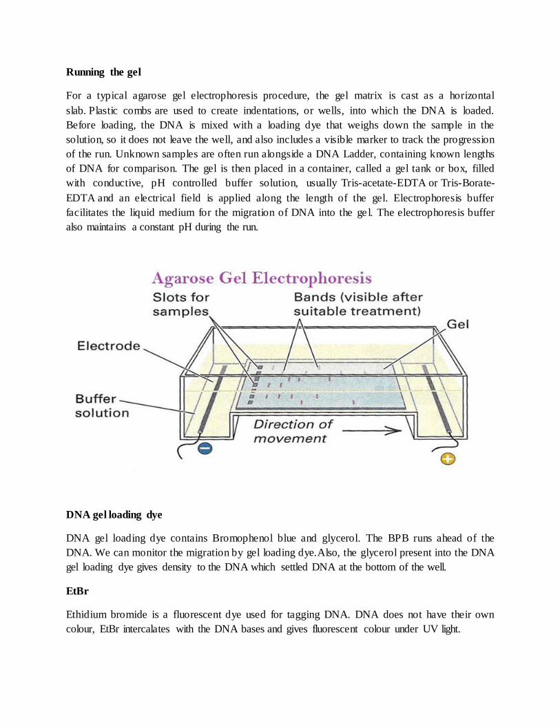
For a typical agarose gel electrophoresis procedure, the gel matrix is cast as a horizontal
slab. Plastic combs are used to create indentations, or wells, into which the DNA is loaded.
Before loading, the DNA is mixed with a loading dye that weighs down the sample in the
solution, so it does not leave the well, and also includes a visible marker to track the progression
of the run. Unknown samples are often run alongside a DNA Ladder, containing known lengths
of DNA for comparison. The gel is then placed in a container, called a gel tank or box, filled
with conductive, pH controlled buffer solution, usually Tris-acetate-EDTA or Tris-Borate-
EDTA and an electrical field is applied along the length of the gel. Electrophoresis buffer
facilitates the liquid medium for the migration of DNA into the ge l. The electrophoresis buffer
also maintains a constant pH during the run.
DNA gel loading dye
DNA gel loading dye contains Bromophenol blue and glycerol. The BPB runs ahead of the
DNA. We can monitor the migration by gel loading dye.Also, the glycerol present into the DNA
gel loading dye gives density to the DNA which settled DNA at the bottom of the well.
EtBr
Ethidium bromide is a fluorescent dye used for tagging DNA. DNA does not have their own
colour, EtBr intercalates with the DNA bases and gives fluorescent colour under UV light.

Precautions:
Agarose powder is hazardous hence always wear gloves, face mask and goggles while
preparing of an agarose gel.
During the boiling of agarose wear oven-gloves (heat resistant). Do not use plastic
gloves, it will burn your hand.
EtBr is a carcinogenic and mutagenic therefore take necessary precautions.
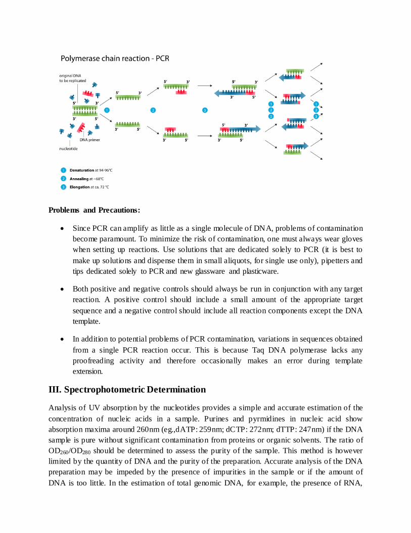
B. Polymerase Chain Reaction (PCR)
Polymerase chain reaction (PCR) is a method widely used in molecular biology to make several
copies of a specific DNA segment. Using PCR, copies of DNA sequences are
exponentially amplified to generate thousands to millions of more copies of that particular DNA
segment. The vast majority of PCR methods rely on thermal cycling. Thermal cycling exposes
reactants to repeated cycles of heating and cooling to permit different temperature-dependent
reactions – specifically, DNA melting and enzyme-driven DNA replication.
Principle:
PCR uses the enzyme DNA polymerase that directs the synthesis of DNA from deoxynucleotide
substrates on a single-stranded DNA template. DNA polymerase adds nucleotides to the 3` end
of a custom-designed oligonucleotide when it is annealed to a longer template DNA. Thus, if a
synthetic oligonucleotide is annealed to a single-stranded template that contains a region
complementary to the oligonucleotide, DNA polymerase can use the oligonucleotide as a primer
and elongate its 3` end to generate an extended region of double stranded DNA.
Components:
DNA template is the sample DNA that contains the target sequence. At the beginning of
the reaction, high temperature is applied to the original double-stranded DNA molecule
to separate the strands from each other.
DNA polymerase is a type of enzyme that synthesizes new strands of DNA
complementary to the target sequence. The first and most commonly used of these
enzymes isTaqDNA polymerase (fromThermisaquaticus), whereasPfuDNA polymerase
(fromPyrococcusfuriosus) is used widely because of its higher fidelity when copying
DNA. Although these enzymes are subtly different, they both have two capabilities that
make them suitable for PCR: 1) they can generate new strands of DNA using a DNA
template and primers, and 2) they are heat resistant.
Primers are short pieces of single-stranded DNA that are complementary to the target
sequence. The polymerase begins synthesizing new DNA from the end of the primer.
Nucleotides (dNTPs or deoxynucleotide triphosphates) are single units of the bases A,
T, G, and C, which are essentially "building blocks" for new DNA strands.
Procedure:
Typically, PCR consists of a series of 20–40 repeated temperature changes, called thermal
cycles, with each cycle commonly consisting of two or three discrete temperature steps.
The individual steps common to most PCR methods are as follows:
1. Initial Denaturation for 2 minutes at 94°C: This initiation step heats the double
stranded DNA template strand to the point where the strands start denaturing and the
hydrogen bonds are broken between the nucleotide base pairs.
2. Denature 30 seconds at 94°C: Continued denaturation of double stranded DNA.
3. Anneal primers for 30 seconds at 55°C: The forward and reverse primers are stable
within this temperature range to anneal to each of the single stranded DNA template
strands. The DNA polymerase is also stable enough to now bind to the primer DNA
sequence.
4. Extend DNA for 1 minute at 74°C: The Taq polymerase has an optimal temperature
around 70-75°C so this step enables the DNA polymerase to synthesize and elongate the
new target DNA strand accurately and rapidly.
5. Repeat steps 2-5 25-30 times.
6. Final Extension for 5 minutes at 74°C: A final extension to fill- in any protruding ends
of the newly synthesized strands.
7. Final hold: The final step cools the reaction chamber to 4–15 °C (39–59 °F) for an
indefinite time, and may be employed for short-term storage of the PCR products.
Problems and Precautions:
Since PCR can amplify as little as a single molecule of DNA, problems of contamination
become paramount. To minimize the risk of contamination, one must always wear gloves
when setting up reactions. Use solutions that are dedicated solely to PCR (it is best to
make up solutions and dispense them in small aliquots, for single use only), pipetters and
tips dedicated solely to PCR and new glassware and plasticware.
Both positive and negative controls should always be run in conjunction with any target
reaction. A positive control should include a small amount of the appropriate target
sequence and a negative control should include all reaction components except the DNA
template.
In addition to potential problems of PCR contamination, variations in sequences obtained
from a single PCR reaction occur. This is because Taq DNA polymerase lacks any
proofreading activity and therefore occasionally makes an error during template
extension.
III. Spectrophotometric Determination
Analysis of UV absorption by the nucleotides provides a simple and accurate estimation of the
concentration of nucleic acids in a sample. Purines and pyrmidines in nucleic acid show
absorption maxima around 260nm (eg.,dATP: 259nm; dCTP: 272nm; dTTP: 247nm) if the DNA
sample is pure without significant contamination from proteins or organic solvents. The ratio of
OD260/OD280 should be determined to assess the purity of the sample. This method is however
limited by the quantity of DNA and the purity of the preparation. Accurate analysis of the DNA
preparation may be impeded by the presence of impurities in the sample or if the amount of
DNA is too little. In the estimation of total genomic DNA, for example, the presence of RNA,

sheared DNA etc. could interfere with the accurate estimation of total high molecular weight
genomic DNA.
Principle:
The Beer-Lambert Law relates the absorption of light to the properties of the material through
which the light travels. This law states that there is a logarithmic dependence between the
transmission of light through a substance and the product of the absorption coefficient of the
substance and the path length. For DNA and RNA, the heterocyclic rings of nucleotides
(adenine, guanine, cytosine and thymine/uracil) result in nucleic acid molecules absorbing
ultraviolet (UV) light maximally at 260nm (λmax = 260nm).
A = absorbance at a particular wavelength ε = extinction coefficient
b = path length of the spectrophotometer c = concentration of sample
The Beer-Lambert law can be used with the appropriate extinction coefficients to determine
nucleic acid concentration.
For most commercial instruments, reliable spectrophotometric quantification (A260) readings
lie between 0.1 and 1.5. While nucleic acids absorb maximally at 260nm, other common
contaminants absorb strongly at wavelengths near 260nm. Solvents such as guanidine and
phenol, as well as salts, have maximal absorbance around 230nm, while proteins contribute at
230nm and 280nm. These neighbouring peaks, if present in a sample, will inflate the reading at
260nm.
Procedure:
1. Take 1 ml TE buffer/ dd H2O in a cuvette and calibrate the spectrophotometer at 260nm as
well as 280nm.
2. Add 10 l of each DNA sample to 990l TE (Tris-EDTA buffer)/ / dd H2O and mix well.
3. Use TE buffer// dd H2O as a blank in the other cuvette of the spectrophotometer.
4. Note the OD260 and OD280 values on spectrophotometer.
5. Calculate the OD260/OD280 ratio.
A ratio between 1.8- 2.0 denotes that the absorption in the UV range is due to nucleic
acids.
A ratio lower than 1.8 indicates the presence of proteins and/or other UV absorbers.
A ratio higher than 2.0 indicates that the samples may be contaminated with chloroform
or phenol. In either case (<1.8 or >2.0) it is advisable to re-precipitate the DNA.
6. The amount of DNA can be quantified using the formula:
DNA concentration (mg/ml) = OD260 x dilution factor x 50 g/ml
Example of Calculation
A sample of dsDNA was diluted 50X. The diluted sample gave a reading of 0.65 on a
spectrophotometer at OD260. To determine the concentration of DNA in the original sample,
perform the following calculation:
dsDNA concentration = 50 μg/mL × OD260 × dilution factor
dsDNA concentration = 50 μg/mL × 0.65 × 50
dsDNA concentration = 1.63 mg/mL
Spectrophotomteric Conversions for Nucleic Acids:
1 A 260 of ds DNA = 50 g/ml
1 A 260 of ss oligonucleotides = 33 g/ml
1 A 260 of ss RNA = 40 g/ml

NOTES
………………………………………………………………………………………
………………………………………………………………………………………
………………………………………………………………………………………
………………………………………………………………………………………
………………………………………………………………………………………
………………………………………………………………………………………
………………………………………………………………………………………
………………………………………………………………………………………
………………………………………………………………………………………
………………………………………………………………………………………
………………………………………………………………………………………
………………………………………………………………………………………
………………………………………………………………………………………
………………………………………………………………………………………
………………………………………………………………………………………
………………………………………………………………………………………
………………………………………………………………………………………
………………………………………………………………………………………
………………………………………………………………………………………
………………………………………………………………………………………
………………………………………………………………………………………
………………………………………………………………………………………
………………………………………………………………………………………
………………………………………………………………………………………
………………………………………………………………………………………
………………………………………………………………………………………

National Workshop on Techniques in Plant Sciences
LIST OF PARTICIPANTS
1. Dr. Mayuresh Joshi, Ramnarain Ruia College, Mumbai 2. Ms. Yugandhara Patil, Ramnarain Ruia College, Mumbai 3. Ms. Sushma Bhosale, Ramnarain Ruia College, Mumbai 4. Ms. Swati Singh, Ramnarain Ruia College, Mumbai 5. Ms. Vimal Temkar, Ramnarain Ruia College, Mumbai 6. Dr. Prabhavati, Shivaji College, Delhi 7. Dr. Deepali, Miranda House College, Delhi 8. Dr. Pratima Rani Sardar, Shivaji College, Delhi 9. Dr. Ranjana Singh, Bulandshahar, UP 10. Dr. Pooja Gupta, Ramjas College, Delhi 11. Ms. Kajol Nayak, Helix Biogenesis, Delhi 12. Dr. Anita Singh, University of Delhi, Delhi 13. Ms. Shivaji Tyagi, Mewar University, Rajasthan 14. Mr. Vivek Chopra, Hindu College, Delhi 15. Dr. Nidhi Gautam, DDU College, Delhi 16. Dr. Tapan Dutta, Jawaharlal Nehru College, Boko, Assam 17. Dr. Dhiraj Kumar Das, Jawaharlal Nehru College, Boko, Assam 18. Dr. Alok Das, Jawaharlal Nehru College, Boko, Assam 19. Mr. Pinaki Kr. Rabha, Jawaharlal Nehru College, Boko, Assam 20. Dr. Habibur Rahman, Jawaharlal Nehru College, Boko, Assam 21. Dr. Nuruddin Ahmed, Jawaharlal Nehru College, Boko, Assam 22. Dr. Apurba Kumar Goswami, Jawaharlal Nehru College, Boko, Assam 23. Dr. Tapan Kr. Deka,Jawaharlal Nehru College, Boko, Assam 24. Dr. SUsanta Kr. Bhuyan, Jawaharlal Nehru College, Boko, Assam 25. Dr. Ranjit Baishya, Jawaharlal Nehru College, Boko, Assam
PHOTOS


