n zo on oti c a g e n t s i n b e g u - AFSCA-FAVV Toxoplasmosis Trichinellosis Prion diseases...
164
t r e n d s a n d s o u r c e s r e p o r t o n z o o n o t i c a g e n t s i n b e l g i u m i n 2 0 0 7 working group on foodborne infections and intoxications 2007
Transcript of n zo on oti c a g e n t s i n b e g u - AFSCA-FAVV Toxoplasmosis Trichinellosis Prion diseases...

trends and sources
report on zoonotic agents in belgium in 2007
working group on foodborne infections and intoxications
2007

trends and sourcesreport on zoonotic agents in belgium in 2007

3
Executive summary
Zoonoses are diseases or infections that are transmissible from animals to humans. The in-
fection of humans can be acquired directly from animals or indirectly through the ingestion
of contaminated foodstuffs. It’s important that humans in contact with animals are aware of
possible transmission of a zoonotic disease and that consumers are informed about poten-
tial zoonotic pathogens which can cause food borne illness.
Surveillance of zoonoses remains an enormous task as well as an opportunity for all compe-
tent authorities. Measures and systems of disease surveillance, diagnosis and control must
be implemented on a national level and have to be based on a suitable regulatory frame-
work and an appropriate level of funding. Active collaboration between all actors of the
food chain, stakeholders, industry, scientists, experts of the national reference laboratories
and other laboratories, specialists of the competent authorities, technical committees have
to bring together their expertise, experiences, methods and findings. Only a collaborative
approach and effective partnership at all levels will achieve success to control zoonoses and
to improve food safety.
The most commonly reported zoonotic infections in humans are those caused by bacte-
rial zoonotic agents that can be shed by asymptomatic farm animals. Campylobacteriosis
remained for the third consecutive year the most frequently reported zoonotic disease in
humans. Broiler and other poultry meat are an important source of foodborne Campylo-
bacter infections. Salmonellosis is the second most frequently reported zoonosis. Salmonella
was for the first time only the second most important cause of foodborne outbreaks after
norovirus outbreaks. The major sources of Salmonella in foodborne outbreaks are table
eggs, poultry meat and pig meat. Salmonella reduction remains an important task. 2007 was
the first year of implementation of the new Salmonella control programmes in breeding
flocks of Gallus gallus by the Member States to met the Salmonella reduction target to 1%
set down by the Community legislation before the end of 2009.
Bacterial diseasesBrucellosis
CampylobacteriosisEscherichia coli (VTEC)
LeptospirosisListeriosis
MRSAQ-fever
SalmonellosisTuberculosis
YersiniosisViral diseases
Avian influenzaHantaviruses
RabiesWest Nile virus
Parasitic diseasesCysticercosis
EchinococcosisSarcosporidiosis
ToxoplasmosisTrichinellosis
Prion diseasesFoodborne outbreaks

4
Table of contents
Executive summary 3Preface 7Introduction 8Belgian reference laboratories for zoonotic agents 11Acronyms, abbreviations and special terms 14
General information 16Susceptible human population 17Susceptible animal populations 18Animals slaughtered 2003 – 2007 21
Bacterial diseases 24Brucellosis 25Zoonotic brucellosis 25Brucellosis in cattle 26Brucellosis in sheep and goats 27Brucellosis in pigs 27Brucellosis in wildlife 28Brucellosis in humans 28Campylobacteriosis 29Campylobacteriosis 29Campylobacter in food 30Antimicrobial resistance 32Campylobacter in humans 35Escherichia coli (VTEC) infections 41Verotoxin producing Escherichia coli 41Verotoxin producing Escherichia coli in cattle 42Escherichia coli O157 in food 43Verotoxinogenic Escherichia coli in humans 44Leptospirosis 45Leptospirosis 45
Leptospirosis in animals 46Leptospirosis in humans 46Conclusions 49Listeriosis 51Listeriosis 51Listeria monocytogenes in food 52Listeria monocytogenes in humans 54MRSA 57MRSA 57MRSA in pigs 58MRSA in humans 60Conclusions. 61Q-fever 63Coxiella burnetii 63Q-fever in animals 64Q-fever in humans 64Salmonellosis 67Salmonella 67Salmonella in animal feed 68Salmonella in poultry 69Salmonella in pigs 74Salmonella in cattle 76Salmonella in food (meat and meat products) 77Salmonella in humans 80Antimicrobial resistance 83Tuberculosis 91Zoonotic tuberculosis 91Mycobacterium bovis in cattle 92Mycobacterium bovis in wildlife 93Mycobacterium bovis in humans 94Human tuberculosis 94

5
Yersiniosis 95Yersinia enterocolitica 95Yersinia enterocolitica in food 96Yersiniosis in humans 96
Viral diseases 100Avian influenza 101Avian influenza 101Monitoring in birds 102Influenza in humans: monitoring 104Hantaviruses 107Hanta disease 107Hantaviruses in animals 108Hantaviruses in humans 108Rabies 111Rabies 111Rabies in animals 112. West Nile virus 115West Nile virus 115West Nile virus in animals 116West Nile virus in humans 116
Parasitic diseases 118Cryptosporidiose 119Cryptosporidiosis 119Cryptosporidiosis in animals 120Cryptosporidiosis in humans 120Cysticercosis 125Cysticercosis 125Cysticercosis in cattle 126Echinococcosis 127
Echinococcosis 127Echinococcus in food animals 128Echinococcus in wildlife (foxes) 129Echinococcus in humans 129Sarcosporidiosis 131Sarcosporidiosis 131Sarcosporidiosis in animals 131Toxoplasmosis 133Toxoplasma 133Toxoplasmosis in animals 134Toxoplasmosis in humans 134Trichinellosis 137Trichinella 137Trichinella in food animals 138Trichinella in wildlife 138
Prion diseases 140TSE 141Transmissible spongiform encephalopathies 141TSE in animals 142Humans 145
Foodborne outbreaks 148Foodborne outbreaks 149Foodborne outbreaks in humans 150Major etiological agents 150Foodborne outbreaks in 2007 153Working group on foodborne outbreaks 157

trends and sources

7
Preface
All European member states have the obligation to yearly submit an official report
on the monitoring of zoonoses and zoonotic agents to the European Food Safety
Authority (EFSA) based on article 9 of Directive 2003/99/EC of the European Parlia-
ment and the Council. In that report all the relevant official monitoring programmes
on animals in primary production as well as on feed and food are presented. The
report specifies all available data from monitoring and research activities, as well as
laboratory findings from the previous year and includes results from antimicrobial
susceptibility testing and FBOs.
Similarly, based on article 1 of Council Decision 2119/98/EC, data on zoonotic infec-
tions in humans are officially reported each year to the European Centre for Disease
prevention and Control (ECDC).
Based on these two official reports, the FASFC, together with the federal scientific
institutions CODA-CERVA and WIV-ISP agreed to yearly publish a booklet which contains
this same information combined with data of previous years to indicate some trends of
diseases or sources of infection. The aim of this booklet is to inform professional readers
as well as persons who have a general interest in animal and human infections and in
the safety of our food and at last but not least to inform the consumer.
We hope that the reader will enjoy this sixth edition of the Belgian trends and sources
report on zoonotic agents.
Luc Vanholme
FAVV – AFSCA - FASFC
Hein Imberechts
CODA – CERVA - VAR
Geneviève Ducoffre
WIV – ISP - IPH
Katelijne Dierick
WIV – ISP - IPH
Preface
Introduction
Belgian reference laboratories for zoonotic agents
Acronyms, Abbreviations and special terms
General information

8
Introduction
This report compiles the available data for 2007 on zoon-
oses and zoonotic agents, and is derived from the official
documents reported to EFSA and ECDC. For this reason, it
is a unique document in which laboratory results from the
primary production, from food, from feed and from clini-
cal public health sources are combined. In addition to the
compulsory reporting on zoonoses and zoonotic agents as
listed in the European Directive 2003/99/EC, this document
contains data on other foodborne agents that may be of
interest to the reader, e.g. on avian influenza, transmissible
spongiform encephalopathies (TSE, e.g. mad cow disease) or
norovirus infections. For the first time, the parasitic infection
cryptosporidiosis and the emerging disease West Nile Fever
are described.
Together with the general descriptive information on the dis-
eases or the infections themselves, their evolution over time,
some recommendations on prevention of the infection are
provided. This booklet should meet the expectations of those
concerned with the possible (micro)biological contamination
of our food.
The FASFC organises diverse monitoring and eradication
programmes in, among others, the primary production and
in the transformation and distribution sectors. From their de-
scription follows that much effort is being paid to control the
contamination of foodstuffs with pathogens. Some infectious
diseases have successfully been reduced or even eliminated
(for instance brucellosis, mad cow disease) and for others (for
instance campylobacteriosis) further programmes should be
developed. In addition to the continuous effort from the au-
thorities, the consumer plays also an important role. Indeed,
respect for the cold chain and simple hygiene measures in
the kitchen may be very efficient in preventing foodborne
contaminations and illness.
Most of the data in this report are from the following sources:
The Federal Agency for the Safety of the Food Chain (FAVV-•
AFSCA-FASFC);
The Scientific Institute of Public Health (WIV-ISP-IPH);•
The Veterinary and Agrochemical Research Centre (CODA-•
CERVA-VAR).
This report was coordinated by L. Vanholme (FAVV-AFSCA),
H. Imberechts (CODA-CERVA), K. Dierick and G. Ducoffre
(WIV-ISP). Obviously, the professional help of many other
experts was needed to assemble all available analytical data
and information. The authors wish to thank all those who
collaborated actively in any way to this Trends and Sources
report, and especially (in alphabetical order):
S. Bertrand, National Reference Laboratory for Salmonella •
and Shigella, Bacteriology Section, Scientific Institute of
Public Health;

9
N. Botteldoorn, National Reference Laboratory for food-•
borne outbreaks and antimicrobial resistance, Bacteriology
Section, Scientific Institute of Public Health;
P. Butaye, Department of Bacteriology and Immunology, •
Veterinary and Agrochemical Research Centre;
L. Claes and P. Dorny, National Trichinella and Cysticercus •
Reference Centre, Veterinary Department, Institute of Tropi-
cal Medicine Antwerp;
P. Cras, TSE humans, Department Neurology and Neuropa-•
thology, Faculty of Medicine, University of Antwerp;
S. Decraeye, National Reference Laboratory for Toxoplas-•
mose, Pasteur Institute Department, Scientific Institute of
Public Health;
M. Delmée, UCL St-Luc and J. Verhaegen, UZ Leuven, Na-•
tional Reference Laboratory for Yersinia enterocolitica;
K. De Schrijver, Department of Control of Infectious Dis-•
eases, Flemisch Public Health Authorities;
L. De Zutter, Department of Veterinary Public Health and •
Food Safety, Faculty of Veterinary Medicine, University of
Ghent;
J.-J. Dubois, Scientific secretariat of the Superior Health •
Council;
M. Fauville-Dufaux, National Reference Laboratory for •
Tuberculosis and Mycobacterium, Pasteur Institute Depart-
ment, Scientific Institute of Public Health;
D. Fretin and M. Govaerts, National Reference Laboratory •
for Brucellosis, Laboratory of Bacterial diseases and Immu-
nology, Veterinary and Agrochemical Research Centre;
E. Goossens, Department of Bacteriology and Immunology, •
Veterinary and Agrochemical Research Centre;
M. Govaerts, Department of Bacteriology and Immunology, •
Veterinary and Agrochemical Research Centre;
P. Heyman, Research Laboratory for Vector-borne diseases, •
Belgian Ministry of Defence, Queen Astrid Military Hospital,
Brussels;
Ph. Houdart, Crisis prevention and crisis management divi-•
sion, Federal Agency for the Safety of the Food Chain;
M. Lambert, A. Sevenants, K. Mennens, M. Delhalle and Ph. •
Dodion, Control Directorate, Federal Agency for the Safety
of the Food Chain;
I. Le Roux and S. Van Gucht, National Reference Laboratory •
for Rabies, Pasteur Institute Department, Scientific Institute
of Public Health;
C. Letellier, Department of Virology, Veterinary and Agro-•
chemical Research Centre;

10
A. Linden, Bacteriology and Pathology of Bacterial diseases •
Department, Faculty of Veterinary Medicine, University of
Liège;
J.-Y. Michelet, National Reference Laboratory for Biotoxins, •
Food Section, Scientific Institute of Public Health;
D. Pierard, National Reference Laboratory for Enterohemor-•
rhagic Escherichia coli, Public Health, Microbiology Section,
UZ Brussels;
B. Pochet, Laboratories Directorate, Federal Agency for the •
Safety of the Food Chain;
S. Quoilin and S. Maes, Epidemiology Section, Scientific •
Institute of Public Health;
M. Struelens, National Reference Laboratory for Staphylo-•
coccus aureus, Hôpital Erasme;
E. Thiry and A. Scipioni, Department of Virology and Pathol-•
ogy of viral animal diseases, Faculty of Veterinary Medicine,
University of Liège;
C. Truyens, National Reference Laboratory for Echinococ-•
cus, Laboratory of Parasitology, Faculty of medicine, Univer-
sité Libre de Bruxelles;
T. van den Berg, B. Lambrecht, S. Marché and S. Van Borm, •
Department of Small Stock Pathology, Veterinary and Agro-
chemical Research Centre;
M. Van Esbroeck, National Reference Centre for Coxiella •
burnetii, Cryptosporidium, Leptospira and West Nile virus,
Institute of Tropical Medicine, Antwerp;
D. Vangeluwe and O. Poncin, Royal Institute of Natural •
Science;
X. Van Huffel, Control Policy Directorate, Secretariat of the •
Scientific Committee, Federal Agency for the Safety of the
Food Chain;
O. Vandenberg, National Reference Laboratory for Campy-•
lobacter, Department of Microbiology, University Medical
Center St. Peter;
E. Vanopdenbosch and S. Roels, Department of Biocontrol, •
Veterinary and Agrochemical Research Centre;
K. Vereecken, J. Hooyberghs, K. Vermeersch, J. Wits, Ph. •
Heinen, P. Poels and J-P. Maudoux, Control Policy Directo-
rate, Federal Agency for the Safety of the Food Chain;
M. Wanlin, Fondation contre les Affections Respiratoires et •
pour l’Education à la Santé, FARES – VRGT;
C. Wildemauwe, National Phage Typing Centre, Pasteur •
Institute Department, Scientific Institute of Public Health;
M. Yde, National Reference Laboratory for Listeria, Bacteriol-•
ogy Section, Scientific Institute of Public Health.

11
Belgian reference laboratories for zoonotic agents
Zoonotic agent or domain Contact Address E-mail address / Web site
Avian Influenza T. van den Berg CODA-CERVA
Groeselenberg 99
1180 Brussels
http://www.var.fgov.be/
Biotoxins J.-Y. Michelet WIV-ISP, Food Section
J. Wytsmanstraat 14
1050 Brussels
http://www.iph.fgov.be/
Brucella, public and animal health D. Fretin CODA-CERVA
Groeselenberg 99
1180 Brussels
http://www.var.fgov.be/
BSE / TSE S. Roels CODA-CERVA
Groeselenberg 99
1180 Brussels
http://www.var.fgov.be/
Campylobacter O. Vandenberg CHU St-Pierre, Microbiology
Rue Haute, 322
1000 Brussels
http://www.stpierre-bru.be/
Clostridium botulinum Y. Fikri WIV-ISP, Pasteur Institute Dpt
Rue Engeland, 642
1180 Brussels
http://www.pasteur.be/pasteur_en/index.html
Cryptosporidium M. Van Esbroeck ITG-IMT, Klinische Biologie
Kronenburgstraat, 43/3
2000 Antwerpen
http://www.itg.be/itg/GeneralSite/General-
page.asp
Echinococcus multilocularis Y. Carlier Fac. de Médecine U.L.B. Parasitologie
Route de Lennik, 808
1070 Brussels
http://www.ulb.ac.be/
Escherichia coli VTEC and EHEC (public
health)
D. Pierard UZ Brussel, Microbiology
Laarbeeklaan, 101
1090 Brussels
http://www.uzbrussel.be

12
Zoonotic agent or domain Contact Address E-mail address / Web site
Escherichia coli VTEC and EHEC (animal
health)
H. Imberechts CODA-CERVA
Groeselenberg, 99
1180 Brussels
http://www.var.fgov.be/
Foodborne outbreaks and Food Microbiol-
ogy
K. Dierick
N. Botteldoorn
WIV-ISP, Bacteriology Section
J. Wytsmanstraat, 14
1050 Brussels
http://www.iph.fgov.be/
Hantavirus P. Heyman Queen Astrid Military Hospital
Bruynstraat, 2
1120 Brussels
http://www.smd.be/rlvbd
Human influenza I. Thomas WIV-ISP, Virology Section
J. Wytsmanstraat, 14
1050 Brussels
http://www.iph.fgov.be/flu
Leptospira M. Van Esbroeck ITG-IMT, Klinische Biologie
Kronenburgstraat, 43/3
2000 Antwerpen
http://www.itg.be/itg/GeneralSite/General-
page.asp
Listeria monocytogenes M. Yde WIV-ISP, Bacteriology Section
J. Wytsmanstraat, 14
1050 Brussels
http://www.iph.fgov.be/
Mycobacterium M. Fauville-Dufaux
(public health)
F. Portaels
(public health)
M. Govaerts
(animal health)
WIV-ISP, Pasteur Institute Dpt
Rue Engeland, 642
1180 Brussels
ITG-IMT, Mycobacteriology
Nationalestraat, 155
2000 Antwerpen
CODA-CERVA
Groeselenberg, 99
1180 Brussels
http://www.pasteur.be/pasteur_en/index.html
http://www.itg.be/itg/GeneralSite/General-
page.asp
http://www.var.fgov.be/
Phage typing centre (Salmonella,
Staphylococcus)
C. Wildemauwe WIV-ISP, Pasteur Institute Dpt
Rue Engeland, 642
1180 Brussels
http://www.pasteur.be/pasteur_en/index.html

13
Zoonotic agent or domain Contact Address E-mail address / Web site
Q-Fever (Coxiella burnetii) M. Van Esbroeck
D. Fretin
ITG-IMT, Klinische Biologie
Kronenburgstraat, 43/3
2000 Antwerpen
CODA-CERVA
Groeselenberg, 99
1180 Brussels
http://www.itg.be/itg/GeneralSite/General-
page.asp
http://www.var.fgov.be
Rabies I. Le Roux
S. Van Gucht
WIV-ISP, Pasteur Institute Dpt
Rue Engeland, 642
1180 Brussels
http://www.pasteur.be/pasteur_en/index.html
Salmonella,
public health
S. Bertrand WIV-ISP, Bacteriology Section
Rue J. Wytsman, 14
1050 Brussels
http://www.iph.fgov.be/bacterio/
Salmonella,
animal health
H. Imberechts CODA-CERVA
Groeselenberg, 99
1180 Brussels
http://www.var.fgov.be/
Staphylococcus aureus M. Struelens Hôpital Erasme, Microbiologie
Route de Lennik, 808
1070 Bruxelles
http://www.erasme.ulb.ac.be
http://www.mrsa.be
Toxoplasmosis S. Decraeye WIV-ISP, Pasteur Institute Dpt
Rue Engeland, 642
1180 Brussels
http://www.pasteur.be/pasteur_en/index.html
Trichinella and other zoonotic parasites L. Claes
P. Dorny
ITG-IMT, Diergeneeskunde
Nationalestraat, 155
2000 Antwerpen
http://www.itg.be/itg/GeneralSite/General-
page.asp
Yersinia enterocolitica J. Verhaegen
M. Delmée
UZ Leuven, Microbiology
Herestraat, 49
3000 Leuven
UCL St-Luc , Microbiology Unit
Av. Hippocrate, 54.92
1200 Brussels
http://www.uzleuven.be/
http://www.saintluc.be/english/index.html
West Nile Virus M. Van Esbroeck ITG-IMT, Klinische Biologie
Kronenburgstraat, 43/3
2000 Antwerp
http://www.itg.be/itg/GeneralSite/General-
page.asp

14
Acronyms, abbreviations and special terms
BAPCOC Belgian Antibiotic Policy Coordination Committee
CODA – CERVA - VAR Veterinary and Agrochemical Research Centre
Centrum voor Onderzoek in Diergeneeskunde en Agrochemie
Centre d’Etude et de Recherches Vétérinaires et Agrochimiques
CRL Community Reference Laboratory
DGZ Vlaanderen Dierengezondheidzorg Vlaanderen, Flanders Regional Animal Health Association
ELISA Enzyme Linked Immuno Sorbent Assay
FAVV – AFSCA - FASFC Federal Agency for the Safety of the Food Chain
FAVV Federaal Agentschap voor de Veiligheid van de Voedselketen
AFSCA Agence Fédérale pour la Sécurité de la Chaîne alimentaire
FBO Foodborne outbreak
FPS Federal Public Service (Ministry) – Public Health, Food Chain Security and Environment
ITG – IMT - ITM Institute of Tropical Medicine
Instituut voor Tropische Geneeskunde
Institut de Médecine Tropicale
NRL National Reference Laboratory
Sanitel National Bovine Database
WIV - ISP - IPH Scientific Institute of Public Health
Wetenschappelijk Instituut Volksgezondheid
Institut Scientifique de Santé Publique

15

general information

17
General information
Susceptible human population
The Belgian human population remains fairly constant over the years.
The evolution of the total human population in Belgium categorised per age, sex and region
from 2002 to 2007 is shown in table 1.
Evolution in the total human population 2002-2007 Table1.
(Source: National Institute for Statistics http://statbel.fgov.be/)
2002 2003 2004 2005 2006 2007
Total 10 309 725 10 355 844 10 396 421 10 445 852 10 511 382 10 584 534
0-19 2 408 943 2 407 368 2 408 456 2 414 041 2 428 706 2 441 129
20-64 6 154 390 6 186 086 6 207 845 6 232 311 6 273 659 6 333 343
65+ 1 746 392 1 762 390 1 780 120 1 799 500 1 809 017 1 810 062
Male 5 042 288 5 066 885 5 087 176 5 111 325 5 143 821 5 181 408
0-19 1 231 221 1 230 382 1 230 570 1 233 688 1 241 251 1 246 988
20-64 3 094 653 3 110 779 3 120 599 3 131 390 3 150 333 3 180 037
65+ 716 414 725 724 736 007 746 247 752 237 754 383
Female 5 267 437 5 288 959 5 309 245 5 334 527 5 367 561 5 403 126
0-19 1 177 722 1 176 986 1 177 886 1 180 353 1 187 455 1 194 141
20-64 3 059 737 3 075 307 3 087 246 3 100 921 3 123 326 3 153 306
65+ 1 029 978 1 036 666 1 044 113 1 053 253 1 056 780 1 055 679
Brussels 978 384 992 041 999 899 1 006 749 1 018 804 1 031 215
Flanders 5 972 781 5 995 553 6 016 024 6 043 161 6 078 600 6 117 440
Wallonia 3 358 560 3 368 250 3 380 498 3 413 978 3 413 978 3 435 879
Foreigners 846 734 850 077 860 287 870 862 900 473 932 161

18
The total number of immigrants in Belgium is important
(932.161 in 2007). Foreigners are found in four typical areas:
former coal basins, cities, border regions and some con-
centrations in the triangle between Brussels, Antwerp and
Ghent, or in the triangle between Brussels, Mons and Namur.
Immigration and tourism may play an important role in the
introduction and the transmission of zoonotic or other infec-
tious diseases.
Figure 1 indicates a slight increase in all categories of human
population during last years.
Susceptible animal populations
Ruminants and pigs
The figures in the table 2 are originating from the database SANITEL, the computerised registration and identification database
of farm animals of the FASFC
Total number of herds and animals in the period 2004 – 2007Table2.
2004 2005 2006 2007
herds animals herds animals herds animals herds animals
Cattle 44.555 2.781.676 42.204 2.492.757 40.640 2.697.824 38.690 2.699.258
Pigs 10.614 10.792 10.631 9.950
Breeding sows1 664.316 657.998 653.358 632.360
Fattening pigs2 4.998.124 4.989.016 4.850.501 5.007.614
Sheep 31.405 214.612 32.323 219.274 30.924 220.600 31.523 220.611
Goats 13.736 37.666 14.247 43.727 13.025 46.950 13.381 46.950
Deer 2.965 13.427 3.093 14.655 2.021 12.805 2.907 12.648
1 Total number of available places for sows and gilts in all herds2 Total number of available places for fattening pigs in all herds
0
2.000.000
4.000.000
6.000.000
8.000.000
10.000.000
12.000.000
2002 2003 2004 2005 2006 2007
total
0-19
20-64
65+
Evolution of human population 2002 – 2007Figure1.

19
Figures 2 to 5 represent the evolution of total number of respectively bovine and porcine herds and animals over last years.
Evolution of the total number of cattle herds, period 2004 – 2007Figure2. Evolution of the total number of bovines, period 2004 – 2007Figure3.
Evolution of the total number of pig herds, period 2004 – 2007Figure4. Evolution of the total number of porcines, period 2004 – 2007Figure5.
3500036000370003800039000400004100042000430004400045000
2004 2005 2006 2007
year
herds
230000023500002400000245000025000002550000260000026500002700000275000028000002850000
2004 2005 2006 2007
year
animals
9400
9600
9800
10000
10200
10400
10600
10800
11000
2004 2005 2006 2007
year
herds
0
1000000
2000000
3000000
4000000
5000000
6000000
2004 2005 2006 2007
year
Breeding pigs
Fattening pigs

20
Poultry
Total number of herds and animals of poultry in the period 2004 – 2007Table3.
2004 2005 2006 2007
herds animals herds animals herds animals herds animals
Gallus gallus
Layers 529 14.364.922 386 10.562.160 472 13.377.548 347 9.878.202
Broilers 1.097 27.873.988 1.024 26.754.817 978 25.894.597 1.036 25.311.775
Elite, Parent, Breeding 193 2.255.085 156 2.144.874 232 3.170.815 221 2.089.933
Total 2.284 50.947.719 1.566 39.461.851 1.682 42.442.960 1.604 37.279.910
Ducks 31 33.949 17 45.140 27 77.140 17 37.880
Geese 8 4.843 5 3.800 6 4.900 3 1.800
Turkeys 63 498.146 37 246.076 41 248.006 46 267.855
Guinea fowl 27 87.440 16 71.400 12 39.200 12 39.200
Partridges 2 123.300 4 129.000 4 136.000 5 136.000
Pheasants 14 206.649 16 226.049 25 268.000 23 268.000
Pigeons 4 1.520 2 1.300 2 2.500 3 2.500
Quails 7 56.020 1 1.700 13 13
Figures 6 and 7 represent the evolution of the total number and the number per category of poultry herds and animals over
the last years.
Gallus gallus, evolution number of poultry herds, Figure6.
period 2004 – 2007
Gallus gallus, evolution total number of poultry, Figure7.
period 2004 – 2007
0
500
1.000
1.500
2.000
2.500
2004 2005 2006 2007
year
Layers
Broilers
Elite, Parent,Breeding
Total
0
10.000.000
20.000.000
30.000.000
40.000.000
50.000.000
60.000.000
2004 2005 2006 2007
year
Layers
Broilers
Elite, Parent,Breeding
Total

21
Animals slaughtered 2003 – 2007
Number of slaughtered animals in the period 2003 – 2007Table4.
2003 2004 2005 2006 2007
Cattle 570.000 564.266 523.795 496.181 495.492
Calves 317.000 317.269 313.115 327.467 306.961
Pigs 11.609.933 11.229.149 10.861.234 10.794.757 11.536.172
Solipeds 12.304 11.655 11.542 10.728 10.064
Sheep & Goats 85.626 90.933 115.356 151.803 137.492
Broilers & Layers 242.038.535 272.641.500 267.578.340 279.986.675 274.505.734
Figures 8 to 11 represent the evolution of the total number of slaughtered bovines, porcines, solipeds, sheep & goats and broil-
ers & layers over the last years.
Evolution of slaughtered bovines 2003 – 2007Figure8. Evolution of slaughtered pigs 2003 – 2007Figure9.
0
100.000
200.000
300.000
400.000
500.000
600.000
2003 2004 2005 2006 2007
year
Cattle
Calves
10.200.000
10.400.000
10.600.000
10.800.000
11.000.000
11.200.000
11.400.000
11.600.000
11.800.000
2003 2004 2005 2006 2007
year
Pigs

22
Evolution of slaughtered sheep & goats 2003 – 2007Figure10. Evolution of slaughtered broilers and layers 2003 – 2007Figure11.
0
20.000
40.000
60.000
80.000
100.000
120.000
140.000
160.000
2003 2004 2005 2006 2007
year
Sheep & Goats
220.000.000
230.000.000
240.000.000
250.000.000
260.000.000
270.000.000
280.000.000
290.000.000
2003 2004 2005 2006 2007
year
Broilers & Layers

23

bacterial diseases

25
Zoonotic brucellosis
Brucellosis in cattle
Brucellosis in sheep and goats
Brucellosis in pigs
Brucellosis in wildlife
Brucellosis in humans
BrucellosisDavid Fretin, Annick Linden, Luc Vanholme
Zoonotic brucellosisBrucellosis is an infectious disease caused by bacterial species
of the genus Brucella. Most species have a specific animal
reservoir that can cause human disease: B. abortus in cattle,
B. melitensis in sheep and goats, B. suis in pigs and B. canis
in dogs. Transmission occurs through contact with infected
animals, contaminated animal tissue or through ingestion of
contaminated products.
In humans brucellosis is characterised by flu-like symptoms
such as fever, headache, back pains and physical weakness.
Nocturnal sweats are frequently observed. Infections of the
central nervous systems or lining of the heart may occur.
• In the non-”officially brucellosis free” Mediterranean
countries, the consumption of raw milk or raw cheese
from sheep and goats is thought to be the major source of
contamination (B. melitensis).
• In Northern Europe, besides some occupational human
cases of B. abortus infections, the majority of brucellosis
cases are imported and are mainly caused by B. melitensis.
26
Brucellosis in cattleBelgium is officially free from bovine brucellosis since
the 25th of June 2003 (Commission Decision 2003/467/
EC establishing the official tuberculosis, brucellosis and
enzootic-bovine-leucosis-free status of certain Member
states and regions of Member states as regards bovine
herds).
Vaccination has been prohibited in Belgium since 1992.
Surveillance programme and methods used
Since the official brucellosis free status, the eradica-
tion programme has been changed in a surveillance
programme.
Dairy cattle are checked at least 4 times a year via tank
milk. Tank milk is examined by means of the milk ring test.
If tank milk is positive, all animals of the herd older than
2 years are tested by individual serological samples. Beef
cattle older than 2 years are serologically monitored once
every three years. The herds are selected on the basis of
geographical localisation. Furthermore, all female animals
older than 1 year and breeding bulls are serologically
tested at purchase. Each abortion or premature birth in
animals at risk is subject to compulsory notification to the
FASFC and testing for brucellosis is obligatory. Aborting
females should be kept in isolation until the results of the
investigation exclude Brucella infections.
Blood sera are analysed by micro-agglutination as screen-
ing test; in case of a positive result, an indirect ELISA test is
performed as confirmatory test. Bacteriological examination
is done in case of serological and/or epidemiological suspi-
cion. An animal is legally suspected of brucellosis in case of
a positive ELISA. If, according to the epidemiology an animal
or herd is found to be at risk, a bacteriological investigation
always takes place. Hence, a brucellosis animal is defined as
an animal in which Brucella has been isolated and a cattle
herd is considered as infected if one of its animals is positive
for brucellosis by culture.
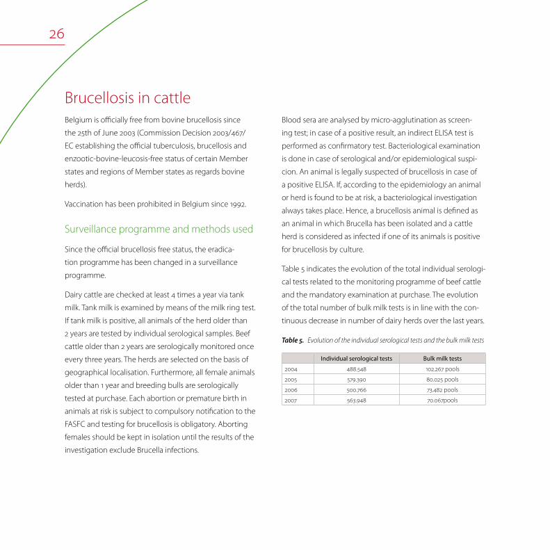
Table 5 indicates the evolution of the total individual serologi-
cal tests related to the monitoring programme of beef cattle
and the mandatory examination at purchase. The evolution
of the total number of bulk milk tests is in line with the con-
tinuous decrease in number of dairy herds over the last years.
Evolution of the individual serological tests and the bulk milk testsTable5.
Individual serological tests Bulk milk tests
2004 488.548 102.267 pools
2005 579.390 80.025 pools
2006 500.766 73.482 pools
2007 563.948 70.067pools

27
Epidemiological investigations and results of 2007 surveillance
An intensified bovine brucellosis eradication programme
started in Belgium in 1988. In case of active brucellosis, i.e.
excretion of Brucella, the plan consisted in the culling of all
animals of the infected herd (total depopulation), the slaugh-
tered animals were compensated based on the replacement
value.
The annual herd prevalence notified at the end of the year
was 1.13% in 1988 and has fallen below 0.01% since 1998. On
27th March 2000, the last case of bovine brucellosis was iden-
tified. No infected herd was detected in Belgium since then.
In the surveillance programme, animals are slaughtered for
additional testing in case of serological and/or epidemiologi-
cal suspicion. In 2007, the FASFC didn’t have to instruct any
test slaughter of animals, since no repeated positive serologi-
cal tests were found.
Brucellosis in sheep and goatsBelgium is official free for sheep and goat brucellosis (B.
melitensis) since 29 March 2001 (Commission Decision
2001/292/EC amending Decision 93/52/EEC recording the
compliance by certain Member States or regions with the
requirements relating to brucellosis (Brucella melitensis) and
according them the status of a Member State or region of-
ficially free of the disease).
Surveillance programme
Serum samples taken in the framework of national monitoring for
Visna-Maedi and at export were examined for Brucella melitensis
specific antibodies by means of ELISA (5% of the total popula-
tion). Positive samples were subsequently tested with Rose
Bengal test and Complement Fixation test. A sample is classified
as positive for brucellosis only if it is positive in all three tests.
Since 2001, yearly serum samples from about 5% of the sheep
and goats populations were tested at the National Reference
Laboratory. In addition, serum samples from sheep for export
were analysed. In 2007, 7.243 samples were tested. Serologi-
cal positive reacting animals after serial and repeated testing
were finally negative. The NRL has confirmed infections of
Yersinia enterocolitica 0:9 in sheep. Those infections are as-
sociated with false positive serology in the tests ELISA, Rose
Bengal and possibly CFT of brucellosis. The phenomenon of
FPSR (false positive serological reactors) as known for bovines
is also observed in sheep.
Brucellosis in pigs
Surveillance programme in pigs and epidemiological investigations
Serological screening for Brucella is done in breeding pigs
that are brought together (e.g. at a fair), at artificial insemina-
tion centres or in animals intended for trade. The methods
used are Rose Bengal test (RBT), Slow Agglutination test (SAT)
according to Wright, complement fixation test (CFT) and

ELISA. Bacteriological examination for Brucella and Yersinia is
done in case of positive serology.
Sometimes, false positive serological reactions are reported.
These are due to a Yersinia enterocolitica O9 infection and are
confirmed by Yersinia spp. isolation in the absence of Brucella
spp. isolation.
The domestic pig population is free of brucellosis (last
Brucella isolation in pigs in Belgium was in 1969). In 2007, all
samples were negative
Brucellosis in wildlife
Regional control programme
Since 2002, an annual surveillance programme is organised
by the Network of Wildlife Disease Surveillance (Faculty of
Veterinary Medicine, Liège) in collaboration with the NRL
(CODA - CERVA, Uccle) with the aim to analyse brucellosis
in wild boars (Sus scrofa) and lagomorphs in the South of
Belgium. Blood samples and organs of hunted or found
dead animals are analysed in order to follow seroprevalence
and identify isolates of Brucella in these species. In 2007, 259
hunted wild boars were sampled and the apparent seroprev-
alence (ELISA) was 65.64% (IC95 = 59.86 – 71.42). Brucella suis
biovar 2 was isolated from spleen and tonsil of wild boars. In
2007, 11.31% of seropositive wild boars were positive for cul-
ture on spleen and/or tonsil. In hares, no Brucella was isolated
from 154 spleen analysed between 2003 and 2006.
Recommendation
Further attention should be given to brucellosis in wild
species, as the potential for contact with B. suis can be high,
particularly for people handling and/or slaughtering game
animals. The species to be considered should include at least
wild boar, deer and other wild ruminants as well as hares.
Brucellosis in humansThe last indigenous case of Brucella was reported in 1997. It
is helpful to note that B. suis biovar 2, the only known biovar
circulating in Belgium among wild boars, shows only limited
pathogenicity for humans, if pathogenic at all.
In 2007, the NRL confirmed two cases of Brucella melitensis
biovar 2 and one case of Brucella melitensis biovar 3. The
country of origin of these three imported cases was not
known.

29
CampylobacteriosisKatelijne Dierick, Geneviève Ducoffre, Olivier Vandenberg, Luc Vanholme, Karen Vereecken
Campylobacteriosis Campylobacteriosis continued to be the most commonly
reported gastrointestinal bacterial pathogen in humans in
Belgium as in the previous three years. Campylobacteriosis
in humans is caused by thermotolerant Campylobacter spp.
Typically, the infective dose of these bacteria is low. The spe-
cies most commonly associated with human infection are C.
jejuni followed by C. coli and C. lari, but other Campylobacter
species are also known to cause human infections.
The incubation period in humans averages from two to five
days. Patients may experience mild to severe illness, with
general clinical symptoms including watery, often bloody di-
arrhea, abdominal pain, fever, headache and nausea. Usually,
infections are self-limiting and last only a few days. Infre-
quently, complications as reactive arthritis and neurological
disorders occur. C. jejuni has become the most recognised
cause of Guillain-Barré syndrome, a polio-like form of paralysis
that can result in respiratory and severe neurological dysfunc-
tion and even death.
Campylobacteriosis
Campylobacter in food
Antimicrobial resistance in strains isolated from meat and meat products
Campylobacter in humans
30
Thermotolerant Campylobacter spp. are widespread in na-
ture. The principal reservoirs are the alimentary tracts of wild
and domesticated birds and mammals. They are prevalent in
food animals such as poultry, cattle, pigs and sheep; in pets,
including cats and dogs; in wild birds and in environmental
water sources. Animals are mostly asymptomatic carriers.
The bacteria can contaminate various foodstuffs, including
meat, raw milk and dairy products, and less frequently fish,
fishery products and fresh vegetables. Contact with live
poultry, consumption of undercooked poultry meat, drinking
water from untreated water sources, and contact with pets
have been identified as important sources of infection.
The contamination of poultry carcasses and meat with
Campylobacter are monitored by the FASFC since 2000. The
incidence of positive poultry samples is high and remains
stable. Poultry meat has to be well cooked before consump-
tion and cross-contamination should be avoided during
preparation.
Campylobacter in food
Monitoring programme
In 2007, a monitoring programme in Belgian slaughterhouses,
meat cutting plants, processing plants and retail trades
representative of the Belgian production of poultry carcasses
and meat, pork carcasses and minced meat of all species
was realised by the FASFC. In addition, samples from raw
milk cheese and live bivalve molluscs were also analysed for
Campylobacter.
Specially trained staff of the FASFC performed the sampling.
Different contamination levels (25g, 0.01g and 600 cm²) were
analysed and in some cases an enumeration was per-
formed. For broiler carcasses at slaughter and cutting meat
at processing plants, independent samples were taken per
matrix in order to detect a minimal contamination rate of 1%
with 95% confidence.
The results of the 2007 monitoring of the FASFC are shown in
table 6.

31
Zoonosis monitoring programme – Campylobacter in foodTable6.
Sample Quantity of sample analysed Percentage of positive samples
Broiler
Carcasses at slaughter (n=236) 25g (caeca) 51.3%
Carcasses at slaughter (n=235) 0.01g 22.5%
Carcasses at retail (n=144) 0.01g 19.4%
Meat cuts (skinned or with skin) at processing plant (n=257) 0.01g 9.3%
Layer
Carcasses at slaughter (n=74) 25g (caeca) 98.6%
Carcasses at slaughter (n=149) 0.01g 34.7%
Carcasses at retail (n=113) 0.01g 18.6%
Poultry
Meat cuts (with skin) at retail (n=131) 0.01g 9.9%
Meat cuts (without skin) at retail (n=140) 0.01g 3.6%
Minced meat at retail (n=159) Enumeration (M=100 cfu/g) 0.0%
Meat preparation at processing plant (n=275) 0.01g 2.9%
Meat preparation at retail (n=420) Enumeration (M=100 cfu/g) 0.2%
Pork
Carcasses at slaughter (n=213) 600 cm² 12.2%
Minced meat (intended to be eaten raw) at retail (all species) (n=128) Enumeration (M=10 cfu/g) 0.0%
Minced meat (intended to be eaten cooked) at retail (all species) ( n=127) Enumeration (M=100 cfu/g) 0.0%
Raw milk cheese at retail (n=46) 25g 0.0%
Raw milk cheese at farm (n=23) 25g 0.0%
Live bivalve molluscs at retail (n=60) 25g 0.0%
The contamination rate of pig carcasses raised until 2006;
in 2007, the contamination decreased a bit. From 2008, the
Campylobacter spp. contamination will be enumerated.
Evolution of the pork Campylobacter prevalence 2004-2007Table7.
Sampling level 2004 2005 2006 2007
Pork Carcasses 600 cm2 4.9% 7.2% 13.4% 12.2%

32
Antimicrobial resistance in strains isolated from meat and meat products
Campylobacter in meat and meat products: list of antimicrobi-Table8.
als tested and breakpoints used
Antimicrobial Breakpoints (µg / ml)
Ampicillin 8 – 32
Tetracycline 4 – 16
Nalidixic acid 16 – 32
Ciprofloxacin 1 – 4
Erytromycin 1 – 8
Gentamicin 4 – 16
The percentage of resistant strains of Campylobacter in meat
is reported in the next table
Antimicrobial susceptibility testing of Campylobacter in food: Table9.
Percentage of resistant strains
Poultry meat Pork
C. jejuni
(n=111)
C. coli
(n=50)
C. coli
(n=15)
Tetracycline 34% 70% 80%
Ciprofloxacin 34% 60% 20%
Nalidixic acid 34% 60% 20%
Gentamicin 0% 0% 0%
Erythromycin 0% 8% 7%
Ampicillin 27% 20% 7%
Surveillance programme and method used
In 2007, 205 Campylobacter strains isolated in the zoon-
oses monitoring programme and originating from poultry,
(carcasses of broilers, filets, meat preparations, turkey and
carcasses of spent hens) and pork were examined for antimi-
crobial susceptibility by the NRL.
Twenty strains were isolated from pork meat or carcasses, 185
strains were isolated from poultry (broiler meat or carcasses,
spent hens and turkey). C. coli was the most prevalent strain
isolated from pork carcasses (75%), while for poultry meat C.
jejuni was the most isolated Campylobacter strain (60%) and
C. coli represented 19% of the isolates. C. lari was determined
in 4% of the isolates and was exclusively isolated from spent
hens.
Minimum Inhibitory Concentrations (MIC) were determined
by using E®-test on blood agar plates. The antimicrobials
tested and the breakpoints (following the CLSI standards)
used are listed in the following table.

33
Antimicrobial resistance in Campylobacter from poultry meat
the C. coli strains. The ampicillin resistance was much higher
in strains isolated from broiler meat and carcasses than
in strains isolated from pork meat. Eleven Campylobacter
strains were isolated from turkey, 55% of the strains were fully
sensitive and no resistance against ampicillin and gentamicin
was measured. Resistance was observed for erythromycin
(18%) ciprofloxacin (36%), nalidixic acid (36%) and tetracycline
(27%).
Compared to the results observed in previous years no
remarkable differences were noticed in the resistance pattern
of the Campylobacter strains isolated in poultry.
185 Campylobacter strains were isolated in poultry meat
and carcasses and tested for antimicrobial susceptibility (111
Campylobacter jejuni and 50 Campylobacter coli strains). In
total 39% of the C. jejuni strains were sensitive for all tested
antibiotics. Tetracycline, ciprofloxacin and nalidixic acid
resistance were present in 34% and ampicillin resistance
was noticed in 27% of the C. jejuni strains. No resistance
was detected against erythromycin and gentamicin. Overall
the antibiotic resistance within C. coli was higher than in C.
jejuni, with a much higher percentage of resistance against
ciprofloxacin (60%), nalidixic acid (60%) and tetracycline
(70%). Resistance against erythromycin was found in 8% of
Evolution of the percentage resistant Campylobacter jejuni and Campylobacter coli strains in poultryFigure12.
0
20
40
60
80
100
120
2004-J 2005-J 2006-J 2007-J 2004-C 2005-C 2006-C 2007-C
AMP TET NAL CIP ERY GEN

34
Antimicrobial resistance in Campylobacter from pork
Evolution of the percentage resistant Campylobacter coli strains isolated in porkFigure13.
0
20
40
60
80
100
AMP TET NAL CIP E RY G EN
2004 2005 2006 2007
In the C. coli isolates (15) from pork, resistance was observed
for all antibiotics except for gentamicin. Only 2 strains were
sensitive to all tested antibiotics.
The resistance against tetracycline (81%) was high followed
by ciprofloxacin (20%) and nalidixic acid (20%). Compared to
previous years a decrease in the resistance is noticed except
for tetracycline where the resistance stays very high.

35
Campylobacter in humansIn 2007, the Belgian Sentinel Laboratory Network consisted
of 110 laboratories reporting Campylobacter. 5,906 strains
of Campylobacter were isolated which represent at country
level an isolation rate of 56 per 100.000 inhabitants (in 2006:
N=5.771). The number of Campylobacter infections shows a
significant decreasing trend since 2000 at national and re-
gional level (p<0.05; Figure 14). Since 2005 Campylobacterio-
sis remains the most frequently reported zoonosis in humans.
19861987
19881989
19901991
19921993
19941995
19961997
19981999
20002001
20022003
20042005
20062007
0
2500
5000
7500
10000N / year
Total number of Campylobacter infections in humans by year (1986-2007). (Source: Sentinel Laboratory Network)Figure14.

36
Cases are reported during the entire year, with a peak in the
summertime (Figure 15).
Weekly number of Campylobacter infections in humans, 2007. (Source: Sentinel Laboratory Network)Figure15.
N / week
J F M A M J AJ S O N D
IPH-K21
0
100
200
250
150
50
Campylobacter isolation rates are slightly higher in men (53%)
than in women (47%); 21% of cases are diagnosed in children
of 1-4 year old and 20% in adults of 25-44 year old. This distri-
bution is observed since many years. There is no explanation
for this observation (Table 10).

37
Number of Campylobacter infections in humans by sex and by Table10.
age groups, 2007. (Source: Sentinel Laboratory Network)
Age groups (year) Males Females Total
N % N % N %
< 1 192 6,3 145 5,3 337 5,8
1 - 4 682 22,3 555 20,2 1237 21,3
5 –14 463 15,1 343 12,5 806 13,9
15 –24 308 10,1 369 13,4 677 11,7
25 -44 584 19,1 588 21,4 1172 20,2
45 -64 498 16,3 403 14,7 901 15,5
65 336 11,0 344 12,5 680 11,7
Total 3063 100,0 2747 100,0 5810 100,0
IPH -Epidemiologie k21_t5
Since the beginning of the registration (1983), the incidence
in Flanders is higher than in Wallonia. This was confirmed in
2007 with an estimated incidence of 66/100.000 inhabitants
in Flanders, 41/100.000 inhabitants in Wallonia and 34/100.000
inhabitants in Brussels-Capital Region. The incidence is very
high in a few districts since many years and also in 2007:
140/100.000 inhabitants in Mouscron, 128/100.000 inhabitants
in Eeklo, 122/100.000 inhabitants in Mechelen and 115/100.000
inhabitants in Leuven (Figure 16). It would be useful to make
a study to explain the reason(s) of those high numbers of
infections in certain districts of the country.
Incidence of Campylobacter infections in humans by district (N/10Figure16. 5 inhab., 2007). (Source: Sentinel Laboratory Network)
ALARAT
AW B
BGBSCRDKDMDNEKGTHSHVHYIP
KRLGLV
MCMHMNMRMSNCNMNVODOSPVRSSGSNTGTHTLTNTRVRVTVV
WR
: Aalst : Arlon : Ath : Antwerpen : Brussels : Brugge : Bastogne : Charleroi : Diksmuide : Dendermonde : Dinant : Eeklo : Gent : Hasselt : Halle-Vilvoorde : Huy : Ieper : Kortrijk : Liège : Leuven : Mouscron : Mechelen : Mons : Marche-en-Fam. : Maaseik : Neufchâteau : Namur : Nivelles : Oudenaarde : Oostende : Philippeville : Roeselare : Soignies : St.-Niklaas : Tongeren : Turnhout : Tielt : Thuin : Tournai : Veurne : Virton : Verviers : Waremme
IPH-K21
VR
BG
DK TLGT
SN
ALOD
MH
HV
LV
AWTH
MS
HS
TG
NV
MRDN
CR
NC
AR
MC TR
KR
SG
IP
TN PV
VT
BS
VVLG
HY
WR
TG
MN NM
B
DM
EK
RS
AT
OS
> 0.00 - 25.00
incidence/100,000 inhabitants[5.65 - 139.99]
> 25.00 - 50.00
> 50.00 - 75.00
> 75.00
55.80

38
Epidemiology Enteric Campylobacter
Data were obtained from the NRL for Human Enteric Campy-
lobacter. Since clinical laboratories are not obliged to send
human isolates to confirm the presence of Campylobacter, a
correct epidemiology of Campylobacter in human popula-
tions cannot be estimated.
Therefore, data about human Campylobacter cases were
obtained from the clinical laboratories of Brugmann, Queen
Fabiola, Bordet and Saint-Pierre University Hospitals located
in Brussels from January 2007 to December 2007.
During this period, a total of 5.136 stool specimens from 4.987
patients were routinely examined for Campylobacter spp.
using one selective medium and a filtration method.
As a result, Campylobacter was isolated in 446 patients.
Among these, 77.2% were C. jejuni, 11.4% were C. coli and
4.5% were C. upsaliensis. The reported prevalence of non-
jejuni / coli Campylobacter of 0.53% is higher than reported
from other European countries and is probably due to the
comprehensive isolation procedure used.
Antimicrobial resistance of human isolates
The Campylobacter isolates from the above mentioned
study (Epidemiology Enteric Campylobacter) were examined
for their resistance against three antibiotics of therapeutic
interest. The testing method was disk diffusion according to
Kirby-Bauer, following SFM recommendations.
Resistance was mostly found against ampicillin (57.4%) and
ciprofloxacin (49.8%). Most Campylobacter were susceptible
to erythromycin (84.8%). However, an increasing resistance
against erythromycin and ciprofloxacin was recorded. These
results underscore the need to monitor antibiotic resistance
in Campylobacter from patients (Table 11).
Resistance of Campylobacter in Belgium fecal isolates, trend from 2001 till 2007. Table11.
(Source: NRL (Laboratory for Microbiology, Saint-Pierre University Hospital, Brussels)
Antimicrobial Agent
2001 2002 2003 2004 2005 2006 2007
N = 280 N = 266 N = 212 N = 291 N = 260 N = 246 N = 263
% Resistant % Resistant % Resistant % Resistant % Resistant % Resistant % Resistant
Ampicillin 13,9 15,8 16,0 29,2 32,3 51,6 57,4
Erythromycin. 3,2 0,0 9,4 6,5 6,9 8,1 5,2
Ciprofloxacin 18,9 22,6 24,5 28,1 33,1 50,4 49,8

39
Invasive Campylobacter in Humans
In 2007, the NRL for Campylobacter confirmed 13 invasive
Campylobacter isolates. Among these, C. fetus was recovered
in 7 patients. The remainders were C. jejuni and C. coli in 4
and 2 cases respectively (Table 12).
Repartition by biotype of invasive Campylobacter in humans, Table12.
2007. (Source: NRL)
N Campylobacter jejuni C. coli C. fetus
I II III IV I II subsp.fetus
13 3 1 2 7


41
Escherichia coli (VTEC) infectionsHein Imberechts, Denis Pierard, Luc Vanholme, Karen Vereecken
Verotoxin producing Escherichia coliVerotoxigenic Escherichia coli (VTEC) is a group of E. coli that
are characterised by the ability to produce ‘verocytotoxins’ or
‘shiga like toxins’. Human pathogenic VTEC usually have ad-
ditional virulence factors that are important for the develop-
ment of disease in man and are called EHEC (enterohemor-
rhagic E.coli). EHEC infections are associated with a minor
number of O:H serogroups. Of these, the O157:H7 or the
O157:H- serogroup (EHEC O157) are the ones most frequently
reported to be associated with the human disease. Some
other pathogenic serotypes of E. coli, e.g. O26, O91, O103, O111
and O145 may also be involved.
Reported human EHEC infections are mostly sporadic. Hu-
man infection may occur after consumption of contaminated
food or water, after contact with contaminated water, or by
direct transmission from person to person or through contact
with infected animals.
Verotoxin producing Escherichia coli
Verotoxin producing Escherichia coli in cattle
Escherichia coli O157 in food
Verotoxinogenic Escherichia coli in humans

42
The clinical symptoms range from mild to bloody diarrhoea
through haemorraghic colitis, which is often accompanied by
abdominal cramps, usually without fever. VTEC infections can
result in haemolytic uremic syndrome (HUS), characterised
by acute renal failure, anaemia and lowered platelet counts.
HUS develops in up to 10% of patients infected with E. coli
O157 and is the leading cause of acute renal failure in young
children.
Animals are a reservoir for VTEC, and VTEC have been isolated
from many different animal species. The gastrointestinal tract
of healthy ruminants seems to be the foremost important
reservoir for VTEC. Cattle are the principal reservoir of VTEC .
The organism is excreted in the faeces. Food of bovine origin
are frequently reported as a source for human VTEC infec-
tions. Other important food sources include faecally contami-
nated vegetables and drinking water.
Prevention mainly relies on bio-security measures at farm-
level and hygienic measures at the level of the slaughter-
houses. Since August 2005, the sampling of cattle at farms
that had sent E. coli O157 positive animals to the abattoir is
not compulsory any more. In previous years, epidemiologi-
cal investigations and additional examinations at the farm of
origin, excretion of VTEC strains by other bovines was never
detected.
In Belgium, approximately 40 sporadic human cases are
registered per year.
Verotoxin producing Escherichia coli in cattle
Surveillance programme, measures and methods used
The surveillance starts when an E. coli O157 (stx1, stx2, eaeA,
enterohemolytic) is isolated from a carcass at the slaugh-
terhouse. In such case, the farm of origin is traced back via
Sanitel, the computerised registration and identification
database for farm animals, managed by the FASFC. FASFC of-
ficials inform the owner that E. coli O157 circulate on his farm
and encourage the implementation of hygienic measures,
i.e. cleaning and disinfection of milk reservoirs and milking
equipment, and cleaning of animals before transport to the
slaughterhouse.
Carcasses contaminated with E. coli O157 should be de-
stroyed or may be heat treated. In all other cases, no specific
measures are taken.
The method used for isolation of E. coli O157 was ISO
16654:2001. Briefly, the samples were enriched in mTSB with
novobiocin and treated by immunomagnetic separation.
Subsequently, the suspected colonies on CT-SMAC were
latex agglutinated for the detection of E. coli O157. Confirma-
tion of serotype (O group) was done by means of slow tube
agglutination after heating of the bacterial cultures. Virulence
factors were determined by PCR for toxin genes stx1 and stx2
and for eae (intimin). Enterohemolysis was done on appropri-
ate culture media.

43
Epidemiological investigations and results of 2007 surveillance
In 2007, no cattle farms were sampled to identify the source
of an E. coli O157 outbreak. In addition, not a single animal
VTEC strain was sent to the NRL (animal health) for typing.
Escherichia coli O157 in food
Monitoring programme
E. coli O157 was analysed in diverse beef and dairy products.
Notification is mandatory since March 2004 (Ministerial
Decree on mandatory notification in the food chain). For en-
terohemorrhagic E. coli, absence in 25g in ready-to-eat food
products put on the market is compulsory.
Results of the 2007 monitoring
The results of the monitoring by the FASFC are shown in the
following table.
Zoonosis monitoring programme - E. coli O157, 2007Table13.
Sample Prevalence
Beef Carcasses (n=1 611) 0.3%
Fresh meat at cutting plant (n=286) 0.0%
Minced meat (steak tartare) at retail
(n=152)0.0%
Meat preparations (steak tartare with
herbs and sauce) at retail (n=150)0.0%
Milk Raw cow’s milk, at farm (n=52) 1.9%
Raw goat’s, sheep’s or horse’s milk at
farm (n=25)0.0%
Cheese From raw milk, at farm (n=24) 0.0%
From raw milk, at processing (n=48) 0.0%
From raw milk, at retail (n=83) 0.0%
From raw sheep’s milk, at retail (n=25) 0.0%
From raw goat’s milk, at retail (n=25) 0.0%
From raw sheep’s or goat’s milk, at farm
(n=17)0.0%
Butter At farm (n=111) 0.0%
From raw milk, at retail (n=25) 0.0%
Cream At farm (n=45) 0.0%

Verotoxinogenic Escherichia coli in humansOnly few clinical laboratories examine human stools for the
presence of E. coli O157. Therefore, a correct incidence of
VTEC in human populations cannot be given.
In 2007, the NRL confirmed 47 verotoxigenic E coli isolated
from 46 patients. Among these:
• 40 typical VTEC isolates, positive for two factors of addition-
al virulence: the presence of the gene eae (intimin) gene
and enterohemolysin (EHEC virulence plasmid) gene.
• 7 atypical VTEC isolates, negative for intimin and enterohe-
molysin.
The number of isolates analysed annually by the NRL has
been rather constant, corresponding to a large rate of under-
diagnosis (Table 14).
E. coli: evolution in number of isolates in humans, 1998-2007. Table14.
Source: NRL
1998
1999
2000
2001
2002
2003
2004
2005
2006
2007
Number of isolates 48 53 47 46 46 47 45 47 46 47
Number of typical isolates 38 46 33 36 37 40 36 36 36 40
Number of O157 isolates 25 33 26 29 26 21 29 27 21 25
In 2007, 10 strains (6 from serotype O157:H7, 2 O145, one
O26, and one O111) were associated with haemolytic uremic
syndrome (HUS). Five patients were less then 5 years old (of
which one was infected with two strains, see below), two
were older children (8 and 12 years old) and two were adults
(54 and 81 years old).
In addition, five children with HUS were diagnosed by
serology alone: three with antibodies against O157 and two
against O145.
According to the information available at the NRL, all but
three of these cases were not related. In three related HUS
cases, a double infection with VTEC O26 and O145 was
confirmed in one case, while in the other two, evidence was
found only for O145 infection. The infection source was found
to be ice cream produced locally in a farm, where the same
VTEC O26 and O145 strains were also found in animals and
in the environment. Two more children suffered from HUS
and seven of uncomplicated diarrhea, but no samples were
obtained for laboratory investigation.

45
LeptospirosisJean-Jacques Dubois, Geneviève Ducoffre, Els Goossens, Marjan Van Esbroeck
Leptospirosis
Leptospirosis or Weill’s disease is a disease caused by Lept-
ospira interrogans sensu lato, which is divided into more
than 26 serogroups and more than 230 serovars. This aerobic
mobile spirochete is able to survive for short times outside
the host in a warm and humid environment (stagnant water,
muddy soils). It endures mostly and for longer periods in host
reservoirs, mainly rodents. After infection, a short bacteraemia
is followed by the invasion of mainly kidneys and liver, in
which leptospires can survive for years and can be intermit-
tently excreted. Accidental hosts, infected through contact
with contaminated water or soil (or by contact with infected
animals) can develop mild flu-like symptoms. Fatal subacute
kidney and or liver failure can also occur.
Leptospirosis
Leptospirosis in animals
Leptospirosis in humans

46
0,00%
2,00%
4,00%
6,00%
8,00%
10,00%
2000 2001 2002 2003 2004 2005 2006 2007
Cattle/S heepPigsPets
Laboratory tests
The standard serological test to detect leptospirosis is the
microscopic agglutination test, which is sensitive and specific
and allows a first identification at serogroup level. This test
requires however the maintenance of a panel of reference
strains, so only a limited number of laboratories are able
to perform it. Other serological tests used are rapid ag-
glutination tests and ELISA’s, which perform well for a rapid
detection, but are less specific. Isolation of the antigen is
very difficult and laborious. Antigen detection is possible by
immunofluorescence techniques and molecular techniques,
but do not allow a typing at serogroup or serovar level.
Leptospirosis in animalsIn Belgium, the number of human cases remains so far limited
(see below). In animals, leptospirosis was considered widely
present in cattle and pigs before 2001, resulting in abortions
and reproductive disorders, leading to frequent infections
in animal handlers (known as milker’s fever). Since 2001,
leptospirosis is seldom found in cattle and pigs. Surprisingly,
a sudden increase in clinical cases was noted in Belgian dogs
since 2006 (Figure 17) with a mortality ranging from 15 to 20%.
Prophylaxis with the available inactivated leptospirosis vaccines
containing serovar canicola and icterohemorhagiae is ineffec-
tive, as other serovars are involved. Also in horses, the number
of clinical cases has increased since 2006, with fatal cases in
foals younger than 3 months. The same observations are made
in Germany, France and Italy. This increase in mainly canine
leptospiroses requires further attention.
Seropositive laboratory confirmed cases in animals at the Figure17.
NRL, animal health between 2000 and 2007. (Total number of samples
received: mean of 1700 yearly (range 1390-2090))
Leptospirosis in humansLeptospirosis occurs worldwide but is most common in tropical
and subtropical areas with high rainfall. The disease is found
mainly wherever humans come into contact with the urine of
infected animals or a urine-polluted environment.
Animal-human transmission occurs through direct contact
with urine of a natural host via a wound, the mucous mem-
brane of the mouth, the nose or the eyes or indirectly by con-
taminated water or food. The longer the exposure the higher
the risk of infection. Human to human transmission is possible
but extremely rare. The incubation period is usually 6 to 12 days
with a range of 2 to 30 days.

47
The professions at highest risk of acquiring the disease are
sewage workers, but also farmers, veterinarians, slaughterhouse
staff, garbage collectors, etc. Certain hobbies can also lead to
contamination: persons involved in water sports such as swim-
ming, kayaking, diving, surfing, fishing and (wind)surfing are at
risk. This disease is mainly observed at the end of summer and
during autumn.
Clinical manifestations
Leptospirosis may induce a wide variety of clinical manifesta-
tions. Symptoms are divided into 4 main clinical categories:
• moderate influenza-like complaints
• Weil syndrome with jaundice and renal failure
• meningitis, encephalitis
• difficult breathing, including coughs and breathlessness.
Classically this disease has two phases with an abrupt begin-
ning marked by a high fever (≥ 40°C), shivers and muscle ache
for approximately one week. After a recovery period of one
to three days without symptoms a second phase follows with
multiple problems with internal organs. Generally the disease
has a good prognosis.
Diagnostics
Diagnosis is based on clinical symptoms, risk factors and
laboratory analyses. Bacterial culture is difficult and takes a
long time. Since it is useless for diagnostic purposes, serology
is done. Five to ten days after onset of symptoms, antibodies
against the leptospires can be detected in the blood.
A negative result at the beginning of an infection does not
exclude a diagnosis of leptospirosis.
Requests for analysis or confirmation of a screening result can
be sent to the reference laboratory. It is best to analyse two
samples taken within one or two weeks of each other.
Treatment
Leptospirosis is treated by antibiotics. The earlier the treatment,
the fewer symptoms and complications the patient will have.
Therefore, it is recommended not to wait for laboratory results
before starting treatment. With appropriate antibiotics a full
recovery is expected about a month after treatment begin.
Prevention
No vaccine to protect against leptospirosis is available in Bel-
gium. Professionals carrying out activities associated with risk
of leptospirosis are recommended to wear watertight glasses,
gloves, boots and clothing.
The following measures could be used in the prevention and
control of leptospirosis:
• to increase awareness of the disease among the population,
risk groups and health care providers;
• to avoid contact with animal urine, infected animals or an
infected environment;
• to wear protective clothing;
• to wash the hands after any contact with a contaminated
animal or object;

48
• to cover all injuries and wounds with waterproof dressings
before contact with contaminated freshwater or humid
environment;
• not to swim or do any water sports in contaminated water;
• to stop access of rodents into housing by obstructing pos-
sible entrances;
• to remove all rubbish and to keep areas around human
habitations clean;
• not to leave food around, especially in recreational areas
where rats may be present.
Results of the 2007 surveillance
At the NRL, a total of 631 human sera have been examined for
the presence of antibodies to Leptospira by the microscopic
agglutination technique (MAT).
Eight confirmed or probable cases have been diagnosed in
2007. All patients were men. The age of the patients ranged
from 25 to 58 years with a median age of 39.5 years. Five
patients were exposed in Belgium, 3 patients contracted the
disease during or after a stay in a (sub)tropical region (Togo,
South East Asia, Cambodja). Five patients were exposed to wa-
ter during recreational or professional activities, 3 patients were
exposed in a garden or at a farm. The infections with exposure
in Belgium were diagnosed between June and October.
Study performed by the NRL, public health
A study was carried out by the Epidemiology Unit of the IPH
using the sentinel laboratory network in collaboration with
the NRL. The aim of the study was to find out if the sentinel
laboratories were capable of diagnosing leptospirosis and if
yes, the number of cases diagnosed between 2002 and 2007. In
addition, the study aimed to find out if these laboratories sent
their samples for confirmation and if yes, to which laboratory.
Ninety-four of 110 contacted sentinel labs responded to the
survey. With the exception of seven laboratories, all sent their
samples to a larger centre for leptospirosis diagnosis. Six of the
seven laboratories that carried out their own analyses sent their
positive samples to a reference laboratory for confirmation. The
number of cases diagnosed by the reference laboratory be-
tween 2003 and 2007 varied between 3 and 14 and the number
of cases that were contaminated in Belgium was seven in 2006
and five in 2007 (Table 15). Among the 22 cases diagnosed in
2006 and 2007, 15 (68%) were contaminated by water and the
others by contact with domestic animals or by working in the
garden or on a farm.
The 2005 European Centre for Disease Control and Preven-
tion annual report mentioned that leptospirosis incidence in
Belgium was estimated in 2005 at 0.1 per 105 inhabitants (N=12).
This is comparable to the European incidence which varied be-
tween 0.1 and 0.22 per 105 inhabitants between 1995 and 2004.
49
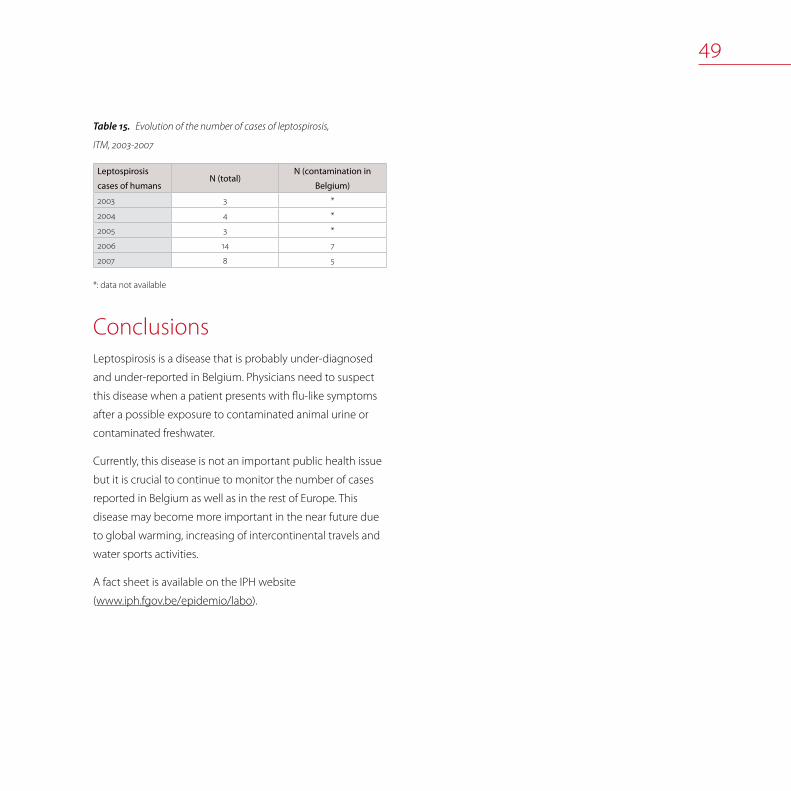
Evolution of the number of cases of leptospirosis, Table15.
ITM, 2003-2007
Leptospirosis
cases of humansN (total)
N (contamination in
Belgium)
2003 3 *
2004 4 *
2005 3 *
2006 14 7
2007 8 5
*: data not available
ConclusionsLeptospirosis is a disease that is probably under-diagnosed
and under-reported in Belgium. Physicians need to suspect
this disease when a patient presents with flu-like symptoms
after a possible exposure to contaminated animal urine or
contaminated freshwater.
Currently, this disease is not an important public health issue
but it is crucial to continue to monitor the number of cases
reported in Belgium as well as in the rest of Europe. This
disease may become more important in the near future due
to global warming, increasing of intercontinental travels and
water sports activities.
A fact sheet is available on the IPH website
(www.iph.fgov.be/epidemio/labo).


51
ListeriosisGeneviève Ducoffre, Karen Vereecken, Marc Yde
Listeriosis
The bacterial genus Listeria currently comprises six spe-
cies, but human cases of listeriosis are almost exclusively
caused by the species Listeria monocytogenes. Listeriae
are ubiquitous organisms that are widely distributed in the
environment, especially in plant matter and soil. The principal
reservoirs of Listeria are soil, forage and water. Other reser-
voirs include infected domestic and wild animals. The main
route of transmission to both humans and animals is believed
to be through consumption of contaminated food or feed.
However, infection can also be transmitted directly from
infected animals to humans as well as between humans.
Cooking kills Listeria, but the bacteria are known to multiply
at temperatures down to 4°C, which makes the occurrence in
ready-to-eat foods with a relatively long shelf life of particular
concern.
Listeriosis
Listeria monocytogenes in food
Listeria monocytogenes in humans

52
In humans severe illness mainly occurs in the unborn child,
infants, the elderly and those with compromised immune
systems. Symptoms vary, ranging from mild flu-like symp-
toms and diarrhea to life threatening infections character-
ized by septicemia and meningoencephalitis. In pregnant
women the infection can spread to the foetus, which may
either be born severely ill or die in the uterus and result in
abortion. Illness is often severe and mortality is high. Human
infections are rare yet important given the high mortality
rate associated with them. These organisms are among the
most important causes of death from foodborne infections in
industrialized countries.
In domestic animals, especially cattle, sheep and goats,
clinical symptoms of listeriosis are usually encephalitis, abor-
tion, mastitis or septicaemia. However, animals may also be
asymptomatic intestinal carriers and shed the organism in
significant numbers, contaminating the surroundings.
General food hygiene rules are essential for the prevention of
human listeriosis. As some persons are at high risk (pregnant
women, the eldery, immuno-compromised people), they
are advised not to eat certain categories of food with proven
elevated risk of L. monocytogenes contamination, such as un-
pasteurized milk and butter, soft cheeses and ice cream made
from unpasteurized milk, any soft cheese crust, smoked fish,
pâté, cooked ham, ‘rillettes’, salami, cooked meat in jelly, raw
minced meat from beef, pork and poultry, steak tartar, raw fish
and shellfish (oysters, mussels, shrimps), fish, meat and surimi
salads, insufficiently rinsed raw vegetables, and unpeeled fruit.
People should be made aware of the considerable risk of infec-
tion by consuming ready-to-eat food products.
Listeria monocytogenes in food
Monitoring programme
The matrices analysed for Listeria monocytogenes were
diverse products of beef, pork, dairy products, fish and ready-
to-eat products. Notification is mandatory since March 2004
(Ministerial Decree on mandatory notification in the food
chain). For Listeria monocytogenes in ready-to-eat products
put on the market, a maximum limit of 100 cfu/g is set.
Results of the 2007 monitoring
The results of the national monitoring program of Listeria
monocytogenes in other foods of animal origin are as follows:
• at retail: meat salad (n=48), crustacean salad (n=48), chicken
salad (n=51), dried follow-on formula (n=47), nursing bottles
(n=119), bakery products with cream (n=158), salty prepara-
tions based on raw eggs (n=55), desserts based on raw
eggs (n=119 positive)
• at processing plant: sandwich spreads (meat, chicken, crus-
tacean) (n=182), bakery products with cream (n=78).
All results were negative, except for desserts based on raw
eggs at retail (8.4% positive), sandwich spreads at processing
plants (10.4% positive) and bakery products with cream at
processing plant (3.8% positive).

53
Zoonosis monitoring programme - Listeria monocytogenes in food, 2007.Table16.
Sample Quantity analysedPercentage of
positive samples
Beef
Minced meat at retail intended to be eaten raw (steak tartare) (n=159) Enumeration (M=100 cfu/g) 0.0%
Meat preparation at retail intended to be eaten raw (steak tartare with
herbs and sauce) (n=157)Enumeration (M=100 cfu/g) 3.2%
Pork
Cooked ham at processing plant (n=59) 25g 1.7%
Cooked ham at retail (n=56) Enumeration (M=100 cfu/g) 0.0%
Raw ham at processing plant (n=78) 25g or enumeration (M=100 cfu/g) (*) 1.3%
Raw ham at retail (n=33) Enumeration (M=100 cfu/g) 0.0%
Pâté at processing plant (n=56) 25g 0.0%
Pâté at retail (n=58) Enumeration (M=100 cfu/g) 0.0%
White pudding at processing plant (n=49) 25g 8.2%
White pudding at retail (n=72) Enumeration (M=100 cfu/g) 0.0%
Sausages at processing plant (n=95) 25g or enumeration (M=100 cfu/g) (*) 6.3%
Sausages at retail (n=35) Enumeration (M=100 cfu/g) 0.0%
Meat (unspecified) Minced meat intended to be eaten raw, at retail (n=157) Enumeration (M=100 cfu/g) 1.9%
Cheeses
Cheeses made from raw milk at retail (n=83) Enumeration (M=100 cfu/g) 0.0%
Cheeses made from pasteurised milk at retail (n=123) Enumeration (M=100 cfu/g) 0.0%
Cheese made from raw sheep’s or goat’s milk at farm (n=16) Enumertation (M=100 cfu/g) 0.0%
Cheese made from raw sheep’s milk at retail (n=25) Enumertation (M=100 cfu/g) 0.0%
Cheese made from raw goat’s milk at retail (n=25) Enumeration (M=100 cfu/g) 0.0%
Cheeses made from raw milk at farm (n=24) Enumeration (M=100 cfu/g) 0.0%
Cheeses made from pasteurised sheep’s milk at retail (n=19) Enumeration (M=100 cfu/g) 0.0%
Cheeses made from pasteurised goat’s milk at retail (n=20) Enumeration (M=100 cfu/g) 0.0%
Cheeses made from raw milk at processing plant (n=48) 25g 0.0%
Cheeses made from pasteurised milk at processing plant (n=140) 25g 0.0%

54
Sample Quantity analysedPercentage of
positive samples
Dairy products
Butter at farm (n=158) Enumeration (M=100 cfu/g) 0.0%
Butter made from raw milk at retail (n=39) Enumeration (M=100 cfu/g) 0.0%
Cream at farm (n=19) Enumeration (M=100 cfu/g) 0.0%
Ice cream at farm (n=43) Enumeration (M=100 cfu/g) 0.0%
Ice cream at retail (n=77) Enumeration (M=100 cfu/g) 0.0%
Milk desserts at processing plant (n=44) 25g 0.0%
Milk desserts at retail (n=60) Enumeration (M=100 cfu/g) 0.0%
Yoghurt at retail (n=50) Enumeration (M=100 cfu/g) 0.0%
Yoghurt at farm (n=27) Enumeration (M=100 cfu/g) 0.0%
Milk powder at processing plant (n=20) Enumeration (M=100 cfu/g) 0.0%
Fish
Smoked salmon at retail (n=150) Enumeration (M=100 cfu/g) 1.3%
Fresh fish at processing plant (n=49) 25g or enumeration (M=100 cfu/g) (*) 8.2%
Fresh fish at retail (n=78) Enumeration (M=100 cfu/g) 1.3%
(*) Depending on the values of the pH and the water activity (Cf Regulation (EC) No 2073/2005 of 15 November 2005 on microbiological criteria for foodstuffs)
Listeria monocytogenes in humans
In 2007, the Sentinel Laboratory Network and the NRL re-
ported 57 cases of listeriosis. This number is less than in 2003
(N=76) and 2004 (N=89), when particularly high numbers of
listeriosis cases were recorded. For the period 1994-2007, the
annual number of cases reported is depicted in Figure 18, cor-
responding to an annual mean number of 55 cases.
Cases are reported all over the year (Figure 19).
Listeria monocytogenes isolation rates are higher in men
(54%) than in women (46%); 28% of cases are diagnosed in
adults older than 64 year (Table 17 on page 56). Old people
and those with a low immunity are more at risk to contract
this disease. Listeria monocytogenes can cause a transpla-
cental infection or an infection during childbirth resulting in
miscarriage, premature delivery or infection of the newborn.
The newborn can develop septicaemia after birth or menin-
gitis after the first week of live. Geographic distribution of the
cases in 2007 is as follows: one case was reported in Brussels,
37 cases were reported in Flanders and 17 in Wallonia (2 from
unknown geographic origin) (Figure 20 on page 56).

55
1994 1995 1996 1997 1998 1999 2000 2001 2002 2003 2004 2005 2006 20070
20
40
60
80
100N / year
N / week
J F M A M J AJ S O N D
IPH-K6
0
4
8
2
6
Number of Listeria monocytogenes infections in humans by year (1994-2007). (Sources: Sentinel Laboratory Network and NRL)Figure18.
Weekly number of Listeria monocytogenes infections in humans, 2007. (Sources: Sentinel Laboratory Network and NRL)Figure19.

Number of Listeria monocytogenes infections in humans by sex Table17.
and by age groups, 2007. Sources: Sentinel Laboratory Network and NRL
Age groups (year) Males Females Total
N % N % N %
< 1 1 3,3 3 11,5 4 7,1
1 - 4 0 0,0 1 3,8 1 1,8
5 –14 0 0,0 0 0,0 0 0,0
15 –24 0 0,0 0 0,0 0 0,0
25 -44 2 6,7 6 23,1 8 14,3
45 -64 10 33,3 5 19,2 15 28,8
65 17 56,7 11 42,3 28 50,0
Total 30 100,0 26 100,0 56 100,0
Incidence of Listeria monocytogenes infections in humans by district (N/10Figure20. 5 inhab., 2007. Sources: Sentinel Laboratory Network and NRL
ALARAT
AW B
BGBSCRDKDMDNEKGTHSHVHYIP
KRLGLV
MCMHMNMRMSNCNMNVODOSPVRSSGSNTGTHTLTNTRVRVTVV
WR
: Aalst : Arlon : Ath : Antwerpen : Brussels : Brugge : Bastogne : Charleroi : Diksmuide : Dendermonde : Dinant : Eeklo : Gent : Hasselt : Halle-Vilvoorde : Huy : Ieper : Kortrijk : Liège : Leuven : Mouscron : Mechelen : Mons : Marche-en-Fam. : Maaseik : Neufchâteau : Namur : Nivelles : Oudenaarde : Oostende : Philippeville : Roeselare : Soignies : St.-Niklaas : Tongeren : Turnhout : Tielt : Thuin : Tournai : Veurne : Virton : Verviers : Waremme
IPH-K6
0.00
0.54
incidence/100,000 inhabitants[0.00 - 3.50]
> 0.00 - 0.75
> 0.75 - 1.50
> 1.50
VR
BG
DK TLGT
SN
ALOD
MH
HV
LV
AWTH
MS
HS
TG
NV
MRDN
CR
NC
AR
MC TR
KR
SG
IP
TN PV
VT
BS
VVLG
HY
WR
TG
MN NM
B
DM
EK
RS
AT
OS
In 2007 the NRL serotyped 52 clinical strains of Listeria monocy-
togenes. Contrary to 2006, no strain of Listeria ivanovii of human ori-
gin was received. The serovar 4b and 1/2a were the most prevalent
(63% and 29% respectively). One clinical strain presented with an
unusual serovar (4e). Six strains were related to pregnancy (3 strains
isolated from the mother and 3 strains from the newborn). Six strains
were isolated from patients with a meningo-encephalitis form of
listeriosis; 38 strains were isolated from blood and 2 strains from pus.
In 2007 the NRL still received strains from two outbreaks of listeriosis
that peaked in 2006. Two new outbreaks of listeriosis (serovar 4b)
emerged with 4 and 2 cases respectively. In any of these episodes
no source of contamination could be determined

57
MRSAPatrick Butaye, Marc Struelens, Luc Vanholme
MRSAStaphylococcus aureus is an important pathogen both for
humans and for animals. S. aureus are bacteria commonly
carried on the skin or in the nose of healthy people. Last
decades, resistance of this pathogen to methicillin and all
other beta-lactam antimicrobial agents has been of special
concern, especially since these so-called methicillin-resistant
Staphylococcus aureus (MRSA) also tend to accumulate
resistance genes to most other (broad-spectrum) antibiotics
as well, making it difficult to treat infections by these strains.
MRSA is a well recognised pathogen in humans, causing
mainly septicaemia, pneumonia and skin infections. Some
clones of these MRSA strains have been spread among hos-
pitals and rest homes (HA [Hospital acquired] MRSA) causing
major outbreaks. In addition, MRSA clones related to com-
munities were identified (CA [Community Acquired] MRSA).
These latter strains were found different from the nosoco-
mial isolates and are generally less resistant to antimicrobial
agents other than beta-lactams.
MRSA
MRSA in animals
MRSA in humans

58
MRSA have also been identified in association with infection
in horses, cattle and dogs. Transfer of these bacteria from
animals to man through direct contact have been demon-
strated. Recently, a new type of MRSA (i.e. ST398) was found
in pigs. The strain was also isolated from the pig owner, his
family, a patient and the nurse in the hospital. Further studies
confirmed that MRSA ST398 was present in various countries
in pigs, but also in other species, including dogs.
In 2007, a study was undertaken to estimate the prevalence
of MRSA in pigs in Belgium.
MRSA in pigs
MRSA in pigs
A survey was performed in 2007 to determine the preva-
lence of MRSA colonization among the pig farms and in the
individual pigs. Fifty farms were sampled and on each farm,
faecal samples of 30 animals of different age groups were
investigated.
The samples were analysed by DGZ Vlaanderen who placed
them in enrichment broth medium made of BHI broth sup-
plemented with NaCl 7.5%. After 24h incubation, broth was
sub-cultured for another 16 h onto a selective chromogenic
agar for MRSA (MRSA-ID, BioMérieux, France). Subsequently
the plates were further analysed at CODA-CERVA. Suspected
colonies were picked up and subcultured on a blood agar
plate, demonstrating the typical characteristics of S. au-
reus. The strains were further identified by PCR, including
confirmation of the presence of the gene causing methicillin
resistance. A subset of strains was typed by spa typing.
Of the 1500 samples analysed 663 were positive for MRSA,
originating from 34 farms. The number of positive farms and
the number of positive animals amongst the different age
groups sampled are shown in Table 18. The data dealing with
farm type and with age group are shown in Figure 21 and
Figure 22.
Approximately 70% of the farms were positive for MRSA, with
the majority of the fattening farms being positive.
At the animal level, the prevalence in weaned piglets was
significantly higher than in sows or in fattening pigs. When
comparing differences in fattening pigs between farm types,
significantly more fattening pigs were positive in the open
farms.
In the open farms, clearly more farms are affected and more
animals are positive. The regrouping of animals from different
origins may be the cause of this, but also other factors should
be taken into account since fattening pigs from closed farms
are expected to be less positive compared to piglets. The rea-
son for this discrepancy needs further investigation, looking
at the factors causing colonisation differences.
The MRSA strains were as expected untypable by PFGE. All
strains were of sequence type ST398. Spa-typing revealed in
all but one farm t011. In one farm a combination of t011 and
t034 was seen. Here 1 strain was t011 and four t034.

59
Fattening & negative
Breeding & positive
Fattening & positive
Breeding & negative
Distribution of tested farms according to farm typeFigure21.
posi
tive
posi
tivepo
sitiv
e
posi
tive
nega
tive
nega
tive
nega
tivene
gativ
e
0
10203040
506070
8090
100
sows weaned �piglets
fattening pigs -closed
fattening pigs -open
% a
nim
als
Percentage animals positive and negative for the different Figure22.
age groups
MRSA prevalence data in pigsTable18.
MRSA Non MRSA Total % MRSA
Farms 34 16 50 68
Closed farms 19 15 34 56
Open farms (fattening farms) 15 1 16 94
Animals
Sows 88 252 340 26
Weaned piglets 141 199 340 41
Fattening pigs (closed farms) 87 245 332 26
Fattening pigs (open farms) 347 141 488 71
These results show that MRSA are highly prevalent in Belgian
pig farms. There is clearly an age effect with piglets being
more positive than sows or fattening pigs. There is also a
difference in MRSA prevalence between closed and open
farms, indicating that, differences in farm management are
important for colonisation.

60
MRSA in humansThe NRL of Staphylocci – MRSA of the Université Libre de
Bruxelles provided specialized analysis:
Identification, characterisation and antimicrobial resistance •
pattern of atypical Staphylococci strains by means of
- Phenotypic methods such as colony morphology, bio-
chemical tests, minimal inhibition concentrations (MIC
determination), phenotyping of glycopeptides suscepti-
bility (population studies)
- Genotypic methods: identification of different Sta-
phylococci species by sequence analysis of gene rpoB,
detection of gene nuc (to identify S. aureus), resistance
gene mecA (coding for oxacillin resistance), resistance
gene mupA (coding for mupirocine resistance) and of
resistance genes against macrolides-lincosamides-strep-
togramines (MLS), tetracycline and aminoglycoside.
Detection of genes coding for toxins: PCRs for exfoliative •
toxins A and B, Panton Valentine Leucocidin (PVL), Toxic
Shock Syndrome Toxin-1 (TSST-1) and 18 enterotoxins
among which sea, seb, sec, sed, see, seg, seh, sei, selj, selk,
sell, selm, seln, selo, selp, selq, selr.
Molecular typing, genomic micro restriction by Pulsed •
Field Gel Electrophoresis (PFGE), by spa sequence and
typing of the Staphylococcal Chromosome Cassette mec
(SCCmec typing), detection of the agr group, detection to
the presence of the arcA gene as marker of the pathogenic
locus of the Arginine Catabolic Mobile Element (ACME).
The NRL especially analysed clinical strains of patients with
wound, skin or other clinical infections (arthritis, peritonitis,
respiratory tract infections) or Staphylococcal strains originat-
ing from local epidemiological studies.
In the period 2006 – 2007 the NRL participated at a survey
on the clonal diversity of S. aureus isolates (MRSA and MSSA)
originating from invasive infections. This survey, as part of
the EARSS programme (European Antimicrobial Resistance
Surveillance System), was in collaboration with the Epidemi-
ology Division of the IPH. The aim of this study was to identify
the spread of predominant clones of S. aureus in Europe and
to visualize this spread by mapping. Detailed information
on the results of this EARSS programme is available on the
website http://www.earss.rivm.nl/.
In 2007, the NRL participated at the first national prevalence
study of the specific MRSA ST398 clonal lineage in swine and
their caretakers. This study was executed in collaboration with
IPH, VAR, DGZ Vlaanderen and the BAPCOC working group to
determine the prevalence and the risk factors of MRSA type
ST398 carriage by professionals and their environment and to
compare human and animal isolates based on the polymor-
phism of the gene coding for protein A (spa typing). Some
results of this study are mentioned in MRSA in pigs.

61
ConclusionsThe number of reported PVL-positive MRSA strains, 25 à 30 a
year, remain stable since 2005. These strains belonged prin-
cipally to clone MRSA ST80-SCCmec IV, a predominant strain
in Europe. These PVL-positive MRSA-strains caused mainly
infections of the skin and some non-lethal invasive infections
(bacteraemia, osteomyelitis).
In 2007, the resistance percentage of S. aureus against oxacil-
lin, isolated from patients with bacteraemia hospitalised in
Belgium, stabilised at about 23% as in 2006 and this after a
decrease of the important prevalence peak in 2004 (33%).
This information corresponded to the results of a national
surveillance programme realized by IPH. Since 2004 a decline
of almost 50% of MRSA strains between all clinical isolates of
S. aureus (from 34 hospitals in 2007) was found.
Within the framework of the European survey on clonal
diversity of the S. aureus strains responsible for invasive infec-
tions, the combined genotyping method spa typing/SCCmec
indicated the presence of 4 important genotypes within the
MRSA-strains, which represented more than 90% of all strains.
These genotypes are: spa CC008-ST8-SCCmec IV (pulsotype
A20), spa CC740-ST45-SCCmec IV (pulsotype B2), spa CC002-
ST5-SCCmec IV (pulsotype C) and spa CC002-ST5-SCCmec II
(pulsotype G). MSSA-strains displayed a more extensive clonal
diversity.
Concerning MRSA-strains of health care centers, genotyping
of these strains coming from local epidemiological surveys
and associated to MRSA-strains of epidemic/clustered cases
in hospitals, mainly belonged to three epidemic nosocomial
clones A20, B2 and G10.
Since 2003, MRSA clone ST398 strains of animal origin are
regularly isolated in human patients. This MRSA-strain spread
from animals to humans. This spread occurred particularly in
the Flemish Region where an intensive farming of pigs takes
place. The swine population appeared to be highly colo-
nized by the porcine-related clone ST398 (see MRSA in pigs).
Not only spread from animals to humans but also between
humans may occur. This ST398 clone was also isolated from
a number of other animal species such as bovine, equine,
poultry and pets.


63
Q-feverDavid Fretin, Marjan Van Esbroeck, Luc Vanholme
Coxiella burnetii Q-fever is a zoonotic disease caused by Coxiella burnetii. Q-
fever (Q for query) is a systemic disease caused by an obligate
intracellular bacterium Coxiella burnetii that is highly resistant
to heat, drying and many common chemical and physical
agents. This resistance enables the bacteria to survive for
a long period in the environment. Coxiella burnetii occurs
worldwide except in New Zealand.
Natural reservoirs are more than 40 species of ticks and
free-living vertebrates, primarily rodents. Ticks or their excreta
spread the disease to domestic animals, e.g. sheep, goats, cat-
tle and dogs. These animals may display a cycle that does not
involve ticks since Coxiellae can multiply in the trophoblast of
the placenta. The placentas and amniotic fluids of these ani-
mals contain large numbers of bacteria which contaminate
pastures and soil. Once animal secreta or excreta have dried,
infectious dust is created.
Coxiella burnetii
Q-fever in animals
Q-fever in humans

64
Q-fever in animalsCattle, sheep, and goats are the main reservoirs but a wide
variety of other animals can be contaminated, including
domesticated pets. Coxiella burnetii does not usually cause
clinical disease in these animals, although an increased
abortion rate and fertility problems in cattle, sheep and goats
are observed. The emergence of these common symptoms
over a longer period of time leads finally to the diagnosis of
Q-fever.
Organisms are excreted in milk, urine and faeces by infected
animals. Animals shed the organisms especially during par-
turition within the amniotic fluids and the placenta. Airborne
transmission can occur in premises contaminated by pla-
cental material, birth fluids or excreta from infected animals.
Airborne inhalation is the most important transmission route
of infection.
In 2007, the NRL analyzed 220 sera of bovine animals, 1 serum
of a sheep and 1 serum of a dog. In total 73 bovine sera were
positive. Also the serum of the dog tested positive. The
clinical suspicion of the sheep could not be confirmed by
analysis.
Recommendations for prevention and control in case of
detection of Q-fever:
Public education and information on sources of infection•
Advice to persons ‘at risk’, especially persons with pre-•
existing cardiac valvular disease or individuals with vascular
grafts and pregnant women
Restrict access to barns and laboratories used in housing •
potentially infected animals
Put aborted animals in quarantine•
Analysis of placenta and aborted foetuses in case of any •
abortion
Appropriate disposal of placenta, birth products, foetal •
membranes and aborted foetuses
Use only pasteurized milk and milk products•
Infected holding facilities should be located away from •
populated areas. Measures should be implemented to
prevent airflow to other occupied areas.
Q-fever in humansTransmission in people is either airborne or results from
direct or indirect contact with infected animals or their dried
excreta. Consumption of infected food such as unpasteurised
milk or dairy products leads to infection and seroconversion
but rarely to clinical symptoms.
Infection with Coxiella burnetii is either inapparent, acute, or
chronic. The incubation period of acute Q-fever ranges from
2 to 4 weeks. The infection has an abrupt onset and patients
present usually with high fever, hepatitis or pneumonia. The
spontaneous evolution is usually a complete recovery but in
immunocompromised hosts a chronic infection can develop
with endocarditis as the major clinical form.

65
Consumption of pasteurized milk or raw milk only from
Q-fever free herds as well as proper hygiene when in contact
with infected animals are the best preventive measures.
In the ITM (NRL), a total of 1656 human sera have been exam-
ined for the presence of phase I and II IgM and IgG antibodies
to Coxiella burnetii by IFAT (Focus Technologies).
Fourteen probable cases have been detected in 2007. Ad-
ditionally 51 possible cases have been found merely on the
basis of one serological result (due to the lack of follow-up
samples) and without clinical information.
The age of the 14 patients with a probable infection ranged
from 20 to 68 years with a median age of 44.7 years. Ten
patients (71%) were male. Four patients stayed abroad before
the start of their illness (Egypt (2), Democratic Republic
of Congo, Senegal). The travel history of the remaining 10
patients is not known.


67
SalmonellosisSophie Bertrand, Patrick Butaye, Katelijne Dierick, Hein Imberechts, Luc Vanholme, Karen Vereecken, Katie Vermeersch
SalmonellaSalmonella is an important zoonotic pathogen and a major
cause of registered bacterial foodborne infections, both in
individuals and in communities. The genus Salmonella is
currently divided into two species: S. enterica and S. bongori.
S. enterica is further divided into six sub-species and most
Salmonella belong to the subspecies S. enterica subsp. en-
terica. Members of this subspecies have usually been named
based on where the serovar or serotype was first isolated.
Mostly, the organisms are identified by genus followed by
serovar, e.g. S. Virchow. More than 2.500 serovars of zoonotic
Salmonella exist and the prevalence of the different serovars
change over time.
Human salmonellosis is usually characterized by the acute
onset of fever, abdominal pain, nausea, and sometimes
vomiting. Symptoms are often mild and most infections are
self-limiting, lasting a few days. However, in some patients,
the infection may be more serious and the associated
dehydration can be life threatening. In these cases, as well as
when Salmonella causes septicaemia, effective antimicrobi-
als are essential for treatment. Salmonellosis has also been
associated with long-term and sometimes chronic sequelae
e.g. reactive arthritis.
Salmonellosis
Salmonella in animal feed
Salmonella in poultry
Salmonella in pigs
Salmonella in cattle
Salmonella in food
Salmonella in humans
Antimicrobial resistance

68
The common reservoir of Salmonella is the intestinal tract of
a wide range of domestic and wild animals which result in a
variety of foodstuffs covering both food of animal and plant
origin as sources of infections. Transmission often occurs
when organisms are introduced in food preparation areas
and are allowed to multiply in food, e.g. due to inadequate
storage temperatures, inadequate cooking or cross con-
tamination of ready-to-eat food. The organism may also be
transmitted through direct contact with infected animals or
faecally contaminated environments and humans.
Food prepared with contaminated raw eggs, egg products
or insufficiently heated poultry meat or pork are the source
of the human Salmonella infection. Therefore, surveillance
programmes that in time detect Salmonella contaminations
early in the whole food chain (feed, living animals, slaughter-
houses, cutting plants, retail sector, restaurants) together with
sanitary measures to reduce contamination are essential. In
addition, good hygiene practices during food preparation
in the kitchen, adequate refrigeration and adequate heating
also help to prevent Salmonella infections.
In animals, sub-clinical infections are common. The organ-
ism may rapidly spread between animals in a herd or flock
without detection and animals may become intermittent
or persistent carriers. Infected cows may succumb to fever,
diarrhea and abortion. Within calf herds, Salmonella may
cause outbreaks of diarrhea with high mortality. Fever and
diarrhea are less common in pigs than in cattle. Sheep, goats
and poultry usually show no signs of infection.
Salmonella in animal feedEach year, an official monitoring for the detection of Sal-
monella in compound feedingstuffs and in raw materials is
organised by the Federal Agency for the Security of the Food
Chain. Microbiological testing on 25g samples is done in the
FASFC laboratories. In case of isolation of Salmonella in official
samples no certification is provided.
Out of 68 samples of feed materials of animal origin (e.g. dairy
products, meat and bone meal, poultry offal meal, feather
meal, blood meal, animal fat and egg powder), none was
found positive. Sixty nine fish meal and fish oil samples were
analysed in 2007, all results were negative.
One out of 104 samples from feed materials of oil seed origin
was found contaminated (with S. typhimurium).
In addition, 405 compound feedingstuffs were tested. Only
two compound feed for poultry were found contaminated
with Salmonella (S. Agona was identified).

69
Salmonella in poultry
Salmonella in breeders and hatcheries
Surveillance programme in breeding flock
The regional animal health associations (i.e. “Association Ré-
gionale de Santé et d’Identification Animales” [ARSIA (http://
www.arsia.be/)] and DGZ Vlaanderen (http://www.dgz.be/)])
organise the official sampling in the framework of the Belgian
Salmonella control programme in breeding flock.
All breeder flocks are routinely examined for Salmonella at
delivery as day-old birds (imported and domestic flocks). At
the farm, pieces (5 by 5 cm) of the inner linings of the delivery
boxes of the day-old chickens are taken by the owner, i.e.
one sample for the hen-chicks and one for the cock-chicks.
Each sample consists of 20 pieces of inner linings. The two
samples are analysed separately. In addition, 20 living hen-
chicks and 20 living cock-chicks are tested serologically. The
samples have to be taken the day of the delivery and have
to reach the lab within 24h of sampling. Breeding chicks
during the rearing period are sampled at the age of 16 weeks
by technicians of DGZ and ARSIA. For this purpose, a pooled
faecal sample of 60 x 1g or, alternatively, 2 pairs of overshoes
is taken. Technicians of DGZ and ARSIA also officially sample
all breeding hens in production; i.e. a pooled faeces sample
of 60 x 1g, or 2 pairs of overshoes every six weeks. In addition,
every two weeks each flock is sampled on mandatory basis
with 2 pairs of overshoes by the owner.
All samples are immediately analysed in the laboratories of
DGZ or ARSIA according to ISO 6579:2002 FDAM.
The official programme also controls the hygiene level of
hatcheries 4 times a year. This is done during visits of the
technician at non-hatching days and comprise various sites
of the hatchery, including hatching drawers. Rodac samples
are taken and both total bacteria and moulds are counted.
After appropriate incubation, an index or code is given to
the number of colonies per surface of approximately 22
cm² in order to facilitate comparisons. In addition, a specific
Salmonella control is done 4 times a year, on pooled samples
from dead-in-shell chicks and on fluff and meconium. These
samples are sent to the laboratory by the owner.
In 1999 the royal and ministerial decrees concerning the
sanitary qualification (Gezondheidskwalificatie - Qualification
sanitaire, Royal Decree of 10 August 1998, Ministerial Decree
of 19 August 1998) came into force. They prescribe minimal
requirements for infrastructure and general hygienic meas-
ures including specific sampling for Salmonella detection
on farms with more than 5 000 birds. Thus, all poultry flocks
before arrival at the slaughterhouse (i.e. breeders, layers and
broilers) undergo a bacteriological examination.

70
Case definition, notification, sanitary measures
and vaccination
A poultry breeding flock is considered Salmonella positive
when S. Enteritidis, S. Typhimurium, S. Virchow, S. Hadar or
S. Infantis is isolated from one-day-old chickens, at 16 weeks
(rearing) or at the occasion of one of the official samplings
during production. If at least one sample in a flock is positive,
the whole flock is considered as positive.
Confirmatory samples (5 faeces samples and 2 dust sam-
ples) during rearing or production may be requested by the
farmer, and are taken by the competent authority. The results
of these analysis are binding.
The isolation of zoonotic Salmonella is notifiable since Janu-
ary 2004 and should be reported to the FASFC.
Several measures are taken on the positive breeder flock: the
hatching eggs are no longer incubated, but are removed
and destroyed, and not yet incubated hatching eggs may
be pasteurised. In addition, positive flocks are logistically
slaughtered within the month . After removal the houses are
thoroughly cleaned and disinfected and a Salmonella control
is performed using 2 samples of 25 swabs.
Vaccination against S. Enteritidis is mandatory, vaccination
against S. Typhimurium is strongly recommended for parent
flocks. Both attenuated and inactivated vaccines are available.
Epidemiological investigations and results
of 2007 surveillance
In 2007, 206 rearing flocks and 496 production flocks were
tested. 2 rearing flocks were positive for Salmonella (1 for S.
Enteritidis, 1 for S. O3,10) and 19 flocks in production were
positive for Salmonella of which 1 for S. Enteritidis, 3 for S.
Typhimurium, 2 for S. Hadar, 4 for S Worthington and 1 flock
for each of the following serotype: S. Braenderup, S. Coeln, S.
Havana, S. Mbandaka, S. Meleagridis, S. Montevideo, S. Rissen,
S. Senftenberg and S. O4:t.
Salmonella in layers and broilers
Surveillance programme in commercial poultry flocks
The national control programme for Salmonella in layers and
broilers is performed according to the sanitary qualification
act, which is applicable to farms with more than 5 000 birds.
Sampling is done by the farmer and consists of an exit sam-
ple for Salmonella, within 3 weeks of slaughter. The owner
can sample in 3 ways: (1) pooled faeces (60 x 1g) taken with
swabs, (2) a pooled faeces (60 x 1g) taken by hand, or (3) two
pairs of overshoes. All samples have to be examined by an
accredited laboratory within 48h.
In addition, layer and broiler flocks may be sampled as day-
old chicks at the farm (entry control). In this purpose, the
owner samples pieces of inner linings of the delivery boxes in
the same way as is done for breeder flocks. After transport of
reared layers to the production unit, a 60 x 1g faecal sample
may be taken from the delivery boxes. Every flock is sampled
taking into account the different origins of rearing.

71
As of July 2007, all layer flocks on farms with more than 199
laying hens are sampled every 15 week during production with
two pairs of overshoes (pooled) and at the end of rearing.
Case definition, notification, sanitary measures
and vaccination
A poultry layer flock is declared positive if S. Enteritidis is
isolated at one day of age, during rearing or during produc-
tion. In addition, the flock is positive if Salmonella belonging
to any serotype is isolated within 3 weeks before slaughter. As
for broilers, a flock is declared positive if in one of the samples
Salmonella is isolated. Salmonella is notifiable to the FASFC
since January 2004.
In case of positive findings in layers, the poultry house must
be cleaned and disinfected after removal of the positive flock
and eggs are placed on the market as B-eggs for heat treat-
ment. If Salmonella was detected in a broiler flock at 3 weeks
before slaughter, the birds were slaughtered at the end of the
day (logistic/sanitary slaughter).
Vaccination against S. Enteritidis is mandatory for layers. Both
attenuated and inactivated vaccines are available.
Epidemiological investigations and results
of 2007 surveillance
In laying hen flocks within 3 weeks before slaughter, 17 out of
340 samples were positive for Salmonella, corresponding to
17 out of 307 flocks (5.54%) and 16 out of 175 farms. Serotype
data were not available; see results from NRL. As of July 2007,
109 flocks were sampled at 16 weeks, 4 were positive for
Salmonella spp, of which 2 for S. Enteritidis, 1 for S. Mbandaka,
1 not typable and 1 for Salmonella vaccine strain. During
production, 378 flocks were sampled, 21 were positive for Sal-
monella spp (S. Enteritidis 11, S. Braenderup 2, S. Livingstone
1, S. Mbandaka 1, S. Senftenberg 1, S. Virchow 1, S. Infantis 1, S.
Typhimurium 2, S. O6,7 1, not typable 2).
As for broilers, three weeks before slaughter 275 (3,12%) of
8809 flocks were found to be contaminated with Salmonella,
corresponding to 123 out of 978 farms. Serotype data were
not available; see results from NRL.
Laboratory findings of the NRL show that in total 745 Salmo-
nella strains from poultry origin were analysed in 2007, which
is 24% less than in 2006, when the European coordinated
monitoring programme in broilers was organised.
The proportion of serotype Enteritidis (38.5%), Infantis (10.5%)
and to a lesser extent Typhimurium isolates (7.9%) increased
as compared to 2006 (27.7%, 3.3% and 5.2%, respectively);
that of S. Paratyphi B decreased from 23.2% in 2006 to 11.3%
in 2007.
The origin of 336 Salmonella poultry isolates was known in
more detail. Only 15 strains were from breeders, and serotype
Enteritidis was once identified. Three isolates belonged to
serotype Typhimurium. The majority of layer isolates (n=36)
were S. Enteritidis (44.4%), but also serotypes Braenderup
(11.1%) and Typhimurium (8.3%) were found. In addition, 3 S.
Gallinarum strains were typed. The majority of broiler isolates
were S. Enteritidis (64.2%), and also serotype Infantis (9.8%)
was frequently identified. Other frequent serotypes were
Paratyphi B (all var. Java) (7.7%) and Typhimurium (6.7%).

72
Yearly, the number of poultry isolates sent to the NRL was ap-
proximately 700 to 1 100, except for 2005 when the European
coordinated monitoring among layers caused a significant
rise of isolates (almost 1 500 in total). Among poultry, S.
Enteritidis is still the most prevalent serotype in Belgium, and
its proportion is unmistakably raising, reaching its highest
0%
5%
10%
15%
20%
25%
30%
35%
40%
45%
50%
1996 1997 1998 1999 2000 2001 2002 2003 2004 2005 2006 20070
200
400
600
800
1000
1200
1400
1600
TotalEnteritidisInfantisParatyphi BTyphimuriumVirchow
value in 2007. The proportion of Typhimurium strains fluctu-
ates between 5.2% and 13.0%. The raise of S. Virchow strains
in 2000-2003 has come to an end, whereas S. Paratyphi B and
eventually S. Infantis may become more important, especially
in broilers (Figure 23).
Evolution of the percentages of the principal Salmonella serotypes isolated from poultry between 1996 and 2007 - The bars represent the Figure23.
total number of poultry isolates per year, and refer to the right axis; the lines represent the percentage of each serotype per year and refer to the left axis

73
Salmonella in turkeys, geese, ducks, pigeons and other poultry
Surveillance programme and sampling
The national control programme for Salmonella in turkeys,
geese, ducks and guinea fowl is performed according to the
sanitary qualification act (see before). Sanitary Qualification
A is mandatory for all commercial breeding flocks. Flocks are
at least sampled as day-old chickens, at the age of 26 weeks
when entering the production unit if this is on a different
farm than the rearing unit, and within the last 3 weeks before
slaughter. Meat production flocks are sampled within three
weeks of slaughter if the holding has a capacity of more than
5 000 birds (Sanitary Qualification B). On a voluntary basis,
one-day-old birds may be sampled also.
Samples for day-old-birds are taken at the farm, and consist
of pieces (5 by 5 cm) of the inner linings of delivery boxes.
Two samples, each composed of 20 pieces of inner linings,
are taken for each flock, one for the hen chicks and one for
the cock chicks. The two samples are analyzed separately ac-
cording to ISO 6579:2002.
At 26 weeks, 60 blood samples were taken of each breeder
flock. If one or more blood sample are positive, faecal sam-
ples are taken to confirm the results. The owner takes faeces
samples from the delivery boxes at time of delivery. A sample
consists of 60 x 5 to 10g sub-samples taken from every flock
with different origin of rearing. The samples have to be exam-
ined by an accredited laboratory within 48 hours.
Within 3 weeks before slaughter, the owner takes a pooled
faecal sample consisting of 60 x 1g sub-samples of each flock.
Alternatively, the sampling may consist of a pooled faecal
sample of 60 x 1g taken by hand, or recovered from two pair
of overshoes that were pooled for analysis.
From October 2006 to September 2007, the European coordi-
nated monitoring of turkey flocks was undertaken according
to article 5 of Directive 2003/99/EC. Details of a report of this
baseline study on the prevalence of Salmonella in turkey hen
flocks can be found at the website of the European Food
Safety Authority) (http://www.efsa.europa.eu).
Case definition, sanitary measures and vaccination policy
A turkey flock is considered positive if zoonotic Salmonella
serotypes were isolated. Measures are taken only at time of
slaughter: if the flock is Salmonella positive, it is slaughtered
at the end of the day (logistic/sanitary slaughter).
There is no vaccination policy for breeding flocks, nor for
meat production flocks.
Notification of zoonotic Salmonella to the FASFC is compul-
sory since January 2004.
Results of the investigation in 2007
For meat turkey flocks following results were found which
were not a part of the baseline study: 51 flocks were sam-
pled at the end of production of which 4 were positive for
Salmonella spp.
12 flocks of guinea fowl were tested. One was positive for
Salmonella spp.
74
Data from the NRL Salmonella indicate a high number of
turkey strains (n=74) due to the European coordinated moni-
toring. Of these, 41.9% were S. Paratyphi B strains, of which
only 1 isolate was tartrate positive (var. Java) and 30 tartrate
negative. Another 41.9% belonged to serotype Kottbus, a
serotype only identified among birds and poultry.
Salmonella in ducks and partridges
The surveillance programme for breeder animals of ducks,
and for meat producing ducks is similar to that of turkeys
(sanitary qualification A for breeders and B for meat produc-
tion).
One meat production flock of ducks was tested, and found
negative.
Salmonella in pigs
Serology
Surveillance programme in fattening pigs
Similar to former years, in 2007 the blood samples from fat-
tening and growing pigs that were taken in the framework
of the monitoring of Aujeszky’s disease were also analysed
for Salmonella. Blood samples from pigs were taken every 4
months. Depending on the number of pigs in the farm, 10
to 12 blood samples were taken. The analysis for Salmonella-
specific antibodies was done in the veterinary laboratories
ARSIA and DGZ by means of the same commercially available
ELISA kit, following the manufacturer’s instructions.
The aim of the current mandatory surveillance programme is
to identify maximum 10% of pig farms with the highest Sal-
monella prevalence and the identification Salmonella-specific
risk factors in these herds. Indeed, it is likely that those herds,
when participating in the supportive control programme,
will benefit the best results in terms of decreasing the risk for
Salmonella infections. A herd is considered as a ‘Salmonella
risk herd’ if 3 consecutive mean S/P ratio’s are above 0,6. The
S/P ratio gives a value to the presence of antibodies against
Salmonella spp.
Pigs were not vaccinated in 2007, since no vaccine was
authorised in Belgium.
Results in 2007
A total of 200.697 serological analyses were performed. Of these,
33.516 samples (16,7%) had a S/P ratio above 0,6. 31% of the 6978
farms monitored had at least once a mean S/P ratio above 0,6. .
Bacteriology
There was no surveillance system for Salmonella in pigs
based on bacteriology. However, several samples were taken
for research activities.
Laboratory findings from the NRL showed that S. Typhimu-
rium continues to be the most prevalent serotype among pig
isolates, representing more than 60% of the total number of
pig Salmonella. Serotype Derby is the second most important
serotype, but represents less than 10% of the strains (Figure
24). As compared to 2006, about 20% less pig strains were
analysed in 2007 (n=391; n=481 in 2006).

75
0%
10%
20%
30%
40%
50%
60%
70%
80%
90%
100%
1996 1997 1998 1999 2000 2001 2002 2003 2004 2005 2006 20070
100
200
300
400
500
600
700
800
TotalDerbyTyphimurium
Evolution of the percentages of the principal Salmonella serotypes isolated from pigs between 1996 and 2007 - The bars represent the total Figure24.
number of pig isolates per year, and refer to the right axis; the lines represent the percentage of each serotype per year and refer to the left axis

76
0%
10%
20%
30%
40%
50%
60%
70%
80%
90%
100%
1996 1997 1998 1999 2000 2001 2002 2003 2004 2005 2006 20070
20
40
60
80
100
120
140
160
TotalDublinTyphimurium
Evolution of the percentages of the principal Salmonella serotypes isolated from cattle between 1996 and 2007 - The bars represent the total Figure25.
number of cattle isolates per year, and refer to the right axis; the lines represent the percentage of each serotype per year and refer to the left axis
Salmonella in cattleThere was no official monitoring programme for Salmonella
in cattle in 2007. Salmonella isolates were sent on a voluntary
basis to the NRL for serotyping.
In Belgium no Salmonella vaccine is authorised in cattle.
According to the NRL, S. Dublin continues to be the principal
serotype isolated in cattle since 2002, and reaching a propor-
tion of more than 60%. S. Typhimurium (about 30%) is the
second most important (Figure 25). In 2007, the number of
Salmonella isolates from cattle (n=80) has almost doubled as
compared to 2006 (n=46).

77
Salmonella in food (meat and meat products)
Monitoring programme
In 2007, Salmonella food monitoring was done on the fol-
lowing matrices: carcasses, trimmings and raw ham of pork,
minced meat and meat preparations of beef, carcasses, meat
cuts and meat preparations of broilers, carcasses of spent
hens and minced meat and meat preparations of diverse
species. Sampling of pork carcasses was done by means of
swabs. The carcass samples of broilers and spent hens at
slaughter consisted of neck skin. Different contamination
levels were analysed: 25g, 600 cm², 1g and 0.1g. Sampling was
done by specially trained staff.
Notification is mandatory since March 2004 (Ministerial
Decree on mandatory notification in the food chain). For
Salmonella, absence in 25g in ready-to eat food is requested.
Epidemiological investigations and results of 2007 surveillance
The results of the monitoring 2007 – Salmonella in meat and meat productsTable19.
SpeciesQuantity of sample
analysedPrevalence Predominant serotype
Other serotypes
(in decreasing order)
Beef
Minced meat (steak tartare) at retail
(n=128)25g 1.6%
Meat preparation (steak tartare with
herbs and sauce) at retail (n=132)25g 2.3%
Pork
Carcasses at slaughter (n=293) 600cm² 16.0% Typhimurium Derby, Anatum, Rissen, London
Trimmings at processing plant
(n=411)25g 4.1% Typhimurium Derby, Livingstone, London
Raw ham at retail (n=31) 25g 0.0%
Raw ham at processing plant (n=65) 25g 1.5%
Broilers
Carcasses at slaughter (n=58) 1g 10.3% Livingstone Indiana, Mbandaka
Carcasses at slaughter (n=59) 25g (caeca) 13.6% Enteritidis Typhimurium O5+, Paratyphi var. Java
Carcasses at retail (n=145) 25g 6.9%

78
SpeciesQuantity of sample
analysedPrevalence Predominant serotype
Other serotypes
(in decreasing order)
Meat cuts (skinned or with skin) at
processing plant (n=170)1g 6.5% Typhimurium
Virchow, Indiana, Hadar, Montevideo,
Paratyphi B
Spent hens
Carcasses at slaughter (n=176) 0.1g 45.4% Enteritidis Typhimurium, Infantis, Agona
Carcasses at slaughter (n=142) 25g (caeca) 46.5% Enteritidis Typhimurium O5+, Paratyphi var. Java
Carcasses at retail (n=113) 25g 10.6%
Poultry
Meat cuts (with skin) at retail (n= 131) 25g 9.2%
Meat cuts (without skin) at retail
(n= 140)25g 4.3%
Minced meat at retail (n=70) 10g 13.0%
Meat preparation at retail (n=27) 10g 11.0%
Meat preparation at processing plant
(n=53)10g 17.0%
Meat products intended to be eaten
cooked at processing plant (n=32)10g 0.0%
Meat products intended to be eaten
cooked at retail (n=86) 10g 5.8%
Minced meat intended to be eaten
raw (all species) at retail (n=129)25g 6.2%
Minced meat intended to be eaten
cooked at retail (all species) (n=136)10g 7.4%
Meat preparation intended to be
eaten raw (all species) at retail (n=47)25g 2.1%
Meat preparation intended to be
eaten cooked (all species) at retail
(n=40)
10g 2.5%
Meat preparation intended to be
eaten raw (all species) at processing
plant (n=88)
25g 11.4%
Meat preparation intended to be
eaten cooked (all species) at process-
ing plant (n=80)
10g 0.0%
Mechanically separated meat (n=126) 10g 23.8%

79
The contamination of pig carcasses and trimmings increased
in 2007. The contamination of spent hen carcasses raised
again in 2007. The contamination of broiler cutting meat
decreased in 2007.
Evolution of the food Salmonella prevalence, 2004-2007Table20.
SamplesSampling
level2004 2005 2006 2007
Pork Carcasses 600cm2 12.3% 9.3% 7.1% 16%
Trimmings 25g 10.4% 7.3% 2.4% 4.1%
Broilers Carcasses 1g 7.9% 5.7% 1.4% 10.3%
Fillets 25g 20.6% 14.2% 13.3% 7.4%
Spent hens Carcasses 1g 19.6% 22.6% 35.6% 45.4%
Salmonella in other food of animal origin
The results of the national monitoring program of milk and
dairy products are as follows:
at retail: dried follow-on formula (n=38), nursing bottles •
(n=119), cheese of pasteurised milk (n=122), cheese of
pasteurised sheep’s milk (n=19), cheese of pasteurised
goat’s milk (n=20), cheese of raw milk (n=81), cheese of raw
sheep’s milk (n=10), cheese of raw goat’s milk (n=10), butter
of raw milk (n=16), milk desserts (n=40), ice cream (n=58)
at processing plant: cheese of raw milk (n=78), cheese of •
pasteurised milk (n=118), milk desserts (n=35), milk powder
(n=20)
at farm: cheese of raw milk (n=76), goat’s or sheep’s cheese •
of raw milk (n=17), butter (n=71), ice cream (n=29), raw
goat’s, sheep’s or horse’s milk (n=34), raw cow’s milk (n=59),
cream (n=19)
All results were negative.
The results of the national monitoring program in other foods
of animal origin are as follows:
at retail: fresh fish (n=62, 1.6%), cooked molluscs (n=60), •
cooked crustaceans (n=29), meat salad (n=44), chicken
salad (n=47), crustacean salad (n=49), bakery products with
cream (n=82), live bivalve molluscs (n=60), raw shrimps
(n=31), chocolate (n=30), confectionary with chocolate
(n=30), table eggs (n=117), liquid egg products (n=76),
powdered egg products (n=24), salty preparation based on
raw eggs (n=58), dessert based on raw eggs (n=115)
at processing plant: gelatine (n=10), cooked crustaceans •
and molluscs (n=67), raw shrimps (n=32), sandwich spreads
(crustacean, meat, chicken) (n=159), fresh fish (n=30),
chocolate (n=15) and confectionery (n=24), bakery prod-
ucts with cream (n=44), egg products (n=82)
All results were negative, except for fresh fish at retail (1.6%
positive), crustacean salad at retail (2% positive), live bivalve
molluscs at retail (1.7%), liquid egg products at retail (1.3%
positive), salty preparation bases on raw eggs at retail (1.7%
positive).

80
Salmonella in humans
Surveillance programme and methods used
Data about human salmonellosis cases and human isolates
were obtained from 160 clinical laboratories. All isolates were
serotyped by slide agglutination with commercial antisera
following the Kauffmann-White scheme. When necessary,
additional biochemical tests were performed to confirm
the identification or to differentiate between the subspe-
cies. Phage typing and antimicrobial susceptibility testing
were performed on isolates randomly sampled from the
four serotypes Enteritidis, Typhimurium, Hadar and Virchow.
Two additional serotypes (Brandenburg and Derby) were
also randomly sampled, all isolates of S. Infantis, S. Newport,
S. Typhi and S. Paratyphi were selected and tested for their
antimicrobial susceptibility.
The objective of the national surveillance programme is to
document the occurrence and trends of serotypes, to detect
local, regional, national or even international outbreaks, to
find and eliminate the source and to suggest preventive
actions to the FASFC. This national Salmonella surveillance
also intended to rapidly interact at the international level via
electronic communication (with the Enter-net international
surveillance network) and helped detecting outbreaks and
targeting preventive strategies.
Epidemiological investigations and results of 2007 surveillance
From 1987 on, a remarkable increase in the number of human
salmonellosis cases was registered, consecutively to the rise
of the serotype Enteritidis, leading to a peak of 15.774 cases
in 1999 (Figure 26, Table 21). In that year, exceptionally high
numbers of S. Enteritidis. Between 2000 and 2007, the total
number of laboratory-confirmed cases varied between 14.088
and 3.975 (Table 21). In 2003, the high number of salmonel-
losis cases mainly resulted from the increase of the serotype
Enteritidis. These isolates exceeded for the first time 70% of
the total number of Salmonella strains analysed. From 2005 a
substantial decrease of Salmonella Enteritidis infections com-
pared with the annual number of cases in the period 2000-
2004 was recorded. This decrease persisted in 2007 where the
total number of cases caused by Salmonella spp. and by S.
Enteritidis decreased to 3693 and 987 cases, respectively.
In recent years, the number of S. Typhimurium isolates
remained at a level of about 2 500 strains per year, but started
to decrease from 2005 (Table 21). After decreasing over the
last years, S. Infantis increased in 2004 up to more than 100
cases to become the third serotype in human cases in 2004,
but decreased to 37 and 38 cases in 2006 and 2007, respec-
tively. Regarding S. Virchow, about 140 to 150 isolates were
annually registered from 2000 to 2003, whereas from 2004
less than 100 strains were yearly reported. A remarkable drop
of S. Brandenburg (322 in 2000 vs about 29 from 2003 to 2007)
cases was noted over the last years. Similarly, the number of
S. Derby cases is shrinking since the beginning of 2000 but
remained stable over the period 2004-2007.

81
Trends for the most prevalent Salmonella serotypes from 1986 to 2007Table21.
1986 1987 1988 1989 1990 1991 1992 1993 1994 1995 1996 1997 1998 1999 2000 2001 2002 2003 2004 2005 2006 2007
Total 6092 6360 8247 9752 11695 10891 10391 10840 11294 10754 12008 14239 14514 15774 14088 11065 10075 12792 9543 4916 3693 3975
Enteritidis 298 320 1163 2236 3382 4721 4084 5260 5700 5138 6145 8284 9003 10492 9503 7112 6398 9118 6075 2226 1052 987
Typhimurium 3512 3233 3699 4018 4756 3652 3835 3528 3418 3623 3522 3347 3221 3348 2799 2370 2438 2486 2459 1659 1826 2233
Others 2282 2807 3385 3498 3557 2518 2472 2052 2176 1993 2341 2608 2290 1934 1786 1583 1239 1188 549 691 815 596
Derby 131 169 168 177 161 134 139 103 113 107 118 157 162 138 169 158 92 100 64 67 52 64
Brandenburg 167 151 159 255 302 176 161 147 204 241 214 296 274 279 322 200 148 66 63 76 47 29
Virchow 152 170 235 293 302 224 295 273 308 245 178 114 115 86 147 143 132 152 91 65 46 28
Infantis 168 173 168 275 249 224 225 160 150 174 267 263 180 169 120 126 74 52 107 58 37 38
0
2000
4000
6000
8000
10000
12000
14000
16000
18000
1980
198119
8219
8319
8419
8519
8619
8719
8819
8919
9019
9119
9219
9319
9419
9519
9619
9719
9819
9920
0020
0120
0220
0320
0420
0520
0620
07
Labo
rato
ry-c
onfir
med
cas
es
Total Enteritidis Typhimurium Others
Trend of the human Salmonella isolates and of the two major serotypes Enteritidis and Typhimurium over the last twenty seven years in Figure26.
Belgium: number of laboratory confirmed cases

82
Age and seasonal distribution
Most cases of salmonellosis were reported in children less than 5 years old (48.4% of cases), with no significant gender difference
Human cases of Salmonella: Age and gender distribution 2007. Note that the gender of all salmonellosis cases is not known. M: male; F: Table22.
female; SR: sex ratio
Salmonella Salmonella Enteritidis Salmonella Typhimurium
Age Total M F SR Total M F SR Total M F SR
< 1 year 330 162 163 1.0 58 28 27 1.0 168 75 91 0.8
1 to 4 y 1594 788 780 1.0 313 163 147 1.1 1114 541 551 1.0
5 to 14 y 685 367 314 1.2 215 128 86 1.5 409 212 195 1.0
15 to 24 y 176 80 87 0.9 38 21 17 1.2 89 42 43 1.0
25 to 44 y 306 146 155 0.9 109 45 62 0.7 79 42 35 1.2
45 to 64 y 291 155 132 1.2 101 55 43 1.3 96 58 37 1.6
≥ 65 y 374 155 208 0.7 99 35 59 0.6 159 77 78 1.0
unknown 291 84 94 0.9 54 20 29 0.7 119 47 45 1.0
Total 3975 1937 1933 1.0 987 495 470 1.0 2233 1094 1075 1.0
Regarding the seasonal distribution (Figure 27), about 200 to
400 cases were monthly reported between January and July
2007. From August until September, the monthly number of
isolates increased, to reach about 500 isolates. From Octo-
ber to December, the monthly number of isolates gradually
decreased.
Seasonal distribution (2000-2007)
0
500
1000
1500
2000
2500
Jan
Feb Mar Apr MayJu
ne July
Aug Sep OctNov Dec
Nb
of c
ases
(S
alm
onel
la s
p.)
2000 2001 2002 2003 20042005 2006 2007
Seasonal distribution 2002 – 2007Figure27.

83
Antimicrobial resistance
Antimicrobial resistance in isolates from living animals
Methods used
Antibiotic susceptibility data of Salmonella strains from
livestock came from the NRL. Tests were performed by the
disk diffusion test, using Neo-Sensitabs (Rosco). Tests and
interpretation were done according to the manufacturers
guidelines using an inoculum and breakpoints as described
by CLSI (Kirby-Bauer). Internal control was performed with
quality control strain E. coli ATCC25922. Results were accepted
when results with the QC strain were within the limits as
proposed by Rosco.
Animal Salmonella: list of antimicrobials tested - For all Table23.
susceptibility tests Neo-Sensitabs from Rosco were used according to the
providers instructions
Antimicrobial Amount of antimicrobial Breakpoints (mm)
Ampicillin 33μg 17 - 19
Ceftiofur 30μg 20 – 22
Streptomycin 100μg 23 – 25
Neomycin 120μg 20 – 22
Gentamicin 40μg 20 – 22
Tetracycline 80μg 20 – 22
Sulfonamides 240μg 20 – 22
Trimethoprim -
Sulfonamides
5.2μg + 240μg 27 – 31
Nalidixic acidid 130μg 21 – 24
Enrofloxacin 10μg 20 – 22
Chloramphenicol 60μg 21 – 24
Florfenicol 30μg 15 - 18
Epidemiological investigations and results of 2007 surveillance
The susceptibility of 891 Salmonella isolates was tested in 2007.
Within the same LIMS dossier only one isolate belonging to
the same serotype was selected for susceptibility testing, and
therefore strains were likely to be independent from each other.
The antimicrobials used are mentioned in Table 23.
A total of 550 Salmonella isolates (61.7%) were fully suscep-
tible to all antimicrobial drugs tested. Most resistance was
found against Ap (27.6%), Su (26.8%), Tc (24.8%), St (24.5%),
but also against TSu (16.6%) and Nal (11.6%). Seventy-two
strains were found resistant against Cm (8.1%); about 57%
of these isolates were also resistant against Ff. Moreover,
twenty-nine isolates were found Cef resistant (3.3%). Most of
the cephalosporin resistant strains originated from poultry
(n=26) (14 S. Infantis, 6 S. Paratyphi B var. Java) and two from
pigs. In addition, five Enr resistant strains (0.6%) (two bovine
S. Typhimurium, two S. Paratyphi B from poultry and one S.
Dublin) were detected. Finally, four strains were resistant to
neomycin and one to gentamicin.
Most (88.6%) S. Agona isolates (n=35) were fully susceptible
for all antimicrobials tested. Three strains were multiresistant
and had the profile Ap St Tc Su Cm Ff.
Most of S. Derby strains (n=21) were sensitive (71.4%),
although some resistance against Tc (23.8%), Su (19.0%), St
(14.3%) and Ap (14.3%) was noticed.
As for S. Dublin isolates (n=31; most from cattle), 41.9% were
found completely susceptible. Resistance against Su (48.43%),
Cm and St (both 45.2%) and Nal (32.3%) was noticed.

84
Most S. Enteritidis isolates (n=105) were susceptible (95.2%).
Resistance was only found against Ap (3.8%; 4 isolates) and
against Nal (1.0%).
All S. Hadar (n=19) strains were found resistant against Nal
(100%). In addition, Tc (94.7%) and St (63.2%) were frequently
found, and to a lesser extent against Ap (26.3%). Twelve iso-
lates (63.2%; 12 isolates) were resistant to Nal St and Tc.
Less than 10% of the S. Indiana strains (n=31) were fully
susceptible. Twenty-six (83.9%) of these strains had the profile
Ap St Tc Su TSu.
About three quarter (76.3%) of the S. Infantis strains (n=59)
were susceptible. All resistant strains (n=14) originated from
poultry and were Ap and Cef resistant. Few strains showed
co-resistance to St, TSu, Su and Tc.
As for S. Paratyphi B (n=33) almost only tartrate positive (i.e.
var. Java; n=30) strains were tested. Only 10% of variety Java
were sensitive. Resistance was mainly observed against Su
(76.7%), TSu (73.3%), St (70.0%) and Nal (66.7%). Also resistance
against Ap was frequently observed (56.7%), and six strains
(all from poultry) were Cef resistant. All three S. Paratyphi B,
tartrate negative isolates (2 from poultry, one from turkeys)
had the profile Ap Su TSu Nal.
Fourty-four percent of S. Typhimurium isolates (n=248) were
found susceptible; classic variant (O5+) strains were found
slightly more often susceptible (45.3%) than Copenhagen
variant (O5-) isolates (41.6%). Pentaresistance Ap St Tc Su Cm
was encountered in 13.8% and 20.2% of O5+ and O5- isolates,
respectively. Cef resistance was found in one poultry O5+ and
one pig O5- isolate.
Only 18.2% of the S. Virchow isolates (n=11) were suscepti-
ble to the antimicrobials tested. Most resistance was found
against Nal (72.7%) and Ap (54.5%).
Some strains belonging to other serotypes were also tested,
but to a lesser extent. Most of these isolates were fully sensi-
tive for all the antimicrobials tested.
Antimicrobial resistance in strains isolated from meat and meat products
During 2007, all 1009 strains of Salmonella enterica isolated
during the zoonosis monitoring program from different
matrices like poultry (broiler meat and carcasses from spent
hens) pork, beef and other foods (shrimps, milk, RTE food,
e.g.) were sent to the IPH for serotyping and determination
of antimicrobial resistance. Minimum Inhibitory Concentra-
tions (MIC) were determined by the use of E-test (n=638) for
most isolates. In view of the harmonisation of the testing at
European level sensititre (n= 371) was used for the second
part of the strains as recommended by the CRL antibiotic re-
sistance. The antimicrobials tested were ampicillin, ceftriaxon,
chloramphenicol, ciprofloxacin, kanamycin, nalidixic acid,
streptomycin, sulfamethoxazole, tetracycline and trimetho-
prim and gentamicin. Interpretation of the results was
according to CLSI. Quality control was performed by using an
Escherichia coli ATCC 25922 strain. Breakpoints used are listed
in the following table.

85
Salmonella from food: list of antimicrobials tested with their Table24.
breakpoints
Antimicrobial Breakpoints (µg / ml)
Ampicillin 16
Ceftriaxon 2
Streptomycin 32
Kanamycin 64
Tetracycline 16
Sulfamethoxazol 256
Trimethoprim 16
Nalidixic acid 32
Ciprofloxacin 4
Chloramphenicol 32
Gentamicin 4
The level of resistance of Salmonella isolates from broilers
and pork is influenced by the serotype distribution in the cor-
responding matrix. The presence of highly resistant serotypes
as Hadar, Virchow, Paratyphi B and Typhimurium contributed
mainly to the high resistance levels in some matrices. The
results for poultry (broiler and carcasses from spent hens),
pork and beef are summarized in the next table. Other matri-
ces where Salmonella was isolated were frog’s legs, shrimps,
ready to eat dishes, mussels, milk powder, eggs and animal
feed. In total about 60 different serotypes were identified,
with S. Enteritidis being the most prevalent serotype mainly
isolated from carcasses of spent hens. For broiler meat and
poultry meat a large variation in serotypes were identi-
fied with Typhimurium, Paratyphi B and Virchow as most
prevalent. Typhimurium and Derby are the most prevalent
Salmonella serotypes isolated from pork samples.
Antimicrobial susceptibility testing of Salmonella spp. isolated Table25.
from different food matrices: percentage of resistant strains
Ant
imic
rob
ial t
este
d
Poul
try
mea
t Bro
iler (
n=34
1)
Poul
try
mea
t Sp
ent h
ens
(n=
118
)
Pork
(n=
343)
Beef
(n=
22)
Ampicillin 52 19 42 14
Ceftriaxon 16 3 3 0
Streptomycin 39 12 36 18
Kanamycin 3 0 1 0
Tetracycline 26 10 45 23
Sulfamethoxazol 42 14 45 23
Trimethoprim 45 10 22 14
Nalidixic Acid 33 8 3 0
Ciprofloxacin 0.3 0 0 0
Chloramphenicol 5 8 14 4
Antimicrobial resistance in strains isolated from poultry meat
In 2007, 459 Salmonella enterica isolates from poultry meat
were tested for their antimicrobial susceptibility. Of all
tested strains 35% were sensitive for all tested antibiotics.
Most resistance was found to ampicillin (44%), streptomycin
(36%), trimethoprim (36%), sulfamethoxazole (35%), nalidixic
acid (32%) and tetracycline (22%). Chloramphenicol resist-
ance was observed in 6% of the Salmonella strains isolated
from poultry meat. The resistance against the 3rd genera-
tion cephalosporin ceftriaxon was very high with 57 (12%)

86
resistant Salmonella strains. Especially the serotype Paratyphi
B expresses resistance against ceftriaxon. For ciprofloxacin
only one strain was resistant and kanamycin resistance was
observed in 2% of the strains. In comparison with 2006 the
percentage of resistance increased considerably for almost
all the antibiotics tested except for chloramphenicol where a
slight decrease in the resistance was noticed.
For 2007, 80 S. Enteritidis isolates from poultry meat were
tested for their susceptibility to all antimicrobials. The resist-
ance in this serotype is very low as was found in previous
years. Only two strains showed resistance, one against ampi-
cillin and the other strain against streptomycin and nalidixic
acid.
From the 52 S. Typhimurium isolates from poultry meat 31%
were sensitive to the tested antibiotics. A high degree of
multi-resistance was observed with AmpStrSulTetTrmp (14%)
and AmpStrSulTet (11%) as most prevalent antibiotic resist-
ance profiles.
0 10 20 30 40 50 60
AMP CEF KAN TET SUL TMP SXT NAL CIP CHL
2001-B 2002-B 2003-B 2004-P 2005-P 2006-P 2007-P
Percentage resistant Salmonella strains in broiler meat (2001-2003) and poultry meat (2004-2007)Figure28.

87
Antimicrobial resistance in strains isolated from pork
In total 343 Salmonella strains from pork were tested for their
susceptibility. S. Typhimurium (178) and S. Derby (69) were
the two most frequently isolated serotypes from pork. In total
57% of the strains were sensitive to all tested antibiotics. A
high degree of resistance was determined for tetracycline
45%, sulfamethoxazole 45%, ampicillin 42% and streptomycin
36%. No resistance was noticed to ciprofloxacin. Chloram-
phenicol resistance was observed in 14% of the strains.
Low percentages of resistance were observed for kanamycin
2%, ceftriaxon 3.5% and nalidixic acid (3%). Multiresistance
was observed in 22% of the strains (> 4 antimicrobials). Com-
pared to 2006 a general increase in antimicrobial resistance
was observed. 2007 was also the first year that resistance
against the third generation cephalosporins was observed
in the serotypes Paratyphi B (n=8), Typhimurium (n=2), Gold-
coast and Derby.
0
10
20
30
40
50
60
AMP CEF KAN TET SUL TMP SXT NAL CIP CHL
2001 2002 2003 2004 2005 2006 2007
Percentage resistant Salmonella strains in pork (2001-2007)Figure29.

88
S. Typhimurium: very high resistance percentages were
observed for ampicillin (63%) sulphonamides (63%), strep-
tomycin (55%) and tetracycline (57%). Four strains resistant
to kanamycin were detected but these strains were multi
resistant with the following resistance profile was observed
AmpChlKanStrSulTetTrmp.
S. Derby: In general a lower percentage of antibiotic resist-
ance is observed in S. Derby strains compared to S. Typhimu-
rium strains isolated in pork. Resistance against tetracycline
(26%) sulphonamide (20%), streptomycin (19%) and ampicil-
lin (9%) were the most common observed. Only two strains
were multi resistant with the profile AmpStrSulTetTrmp and
AmpCefStrSulTet.
In 2007 also a European survey about the prevalence of
Salmonella in slaughter pigs was performed to harmonise
the sampling and analysis methods in the different member
states. In total 137 Salmonella strains isolated from ileo-caecal
lymph nodes (n= 72) and carcasses (n= 65) were analysed for
the antibiotic resistance. S. Typhimurium , with 61% of the
strains was the most prevalent serotype. Within the serotype
Typhimurium, 40% belonged to the variant Copenhagen. In
total 41% of the strains were sensitive to all tested antibiot-
ics. Resistance against ciprofloxacin and gentamicin was
not observed. High resistance percentages ware observed
for tetracycline (49%) sulphonamide (50%), ampicillin (45%)
and streptomycin (39%). Resistance against cefotaxime, a 3rd
generation cephalosporin, was observed in two strains: one
S. Anatum strain with following resistance profile AmpCef,
and one S. Typhimurium strain that was multi resistant to the
following antibiotics AmpCefChlStrSulTetFlor.
Antimicrobial resistance of human isolates
Methods used
A total of 1039 human Salmonella isolates randomly selected
from the six most important serotypes in 2007 (Enteritidis,
Typhimurium, Hadar, Virchow, Brandenburg and Derby),
comprising as well all isolates of the serotypes Infantis, New-
port, Typhi and Paratyphi, were examined for their resistance.
Thirteen antibiotics of therapeutic or epidemiological interest
were tested in disk diffusion according to Kirby-Bauer, follow-
ing CLSI procedures (Table 26 ).
List of antimicrobials used for susceptibility testing of SalmonellaTable26.
AntimicrobialAmount of
antimicrobial
Breakpoints
(mm)
Ampicillin 10 µg 14 - 16
Amoxicillin + clavulanic acid 20/10 µg 14 - 17
Cefotaxime 30 µg 15 - 22
Streptomycin 10 UI 12 - 14
Kanamycin 30 UI 14 - 17
Spectinomycin (excepted for Typhi,
Paratyphi A and B)100µg 19 - 25
Azithromycine (only for Typhi,
Partyphi A and B)15µg ND
Gentamicin 10 µg 13 - 14
Tetracycline 30 µg 12 - 14
Sulfonamides 300 µg 16 - 13
Trimethoprim 5 µg 15 - 11
Trimethoprim + Sulfamethoxazole 1,25/ 23,75 µg 11 - 15
Nalidixic acid 30 µg 14 - 18
Ciprofloxacin 5 µg 16 - 20
Chloramphenicol 30 µg 13 - 17

89
Epidemiological history and results of 2007 surveillance
Resistance was mostly found to tetracycline (25.8%), sulfona-
mides (25.7%), ampicillin (26.5%), streptomycin (24.9%), and to
a lesser extent to trimethoprim (7.9%).
The vast majority (87.2%) of human S. Enteritidis isolates
(n=476) was fully sensitive to all antimicrobials tested.
S. Typhimurium (n=308) showed a high level of resistance;
especially resistances to ampicillin (68.8%), sulfonamides
(66.2%), tetracycline (63.3%) and streptomycin (61.7%) are
striking. More than the half of the isolates (58.7%) was found
resistant to four or more antimicrobial agents. In addition,
almost 18.1% of the isolates showed multi-resistance to at
least ampicillin, chloramphenicol, streptomycin, sulfonamides
and tetracycline. About 76.7% of these multi-resistant isolates
(ACSSuT) were of phage type DT104.
Except two strains, all S. Hadar isolates (n=33) were resistant
to at least one antibiotic. Resistance to tetracycline, nalidixic
acid, ampicillin and streptomycin reached values from 54.5%
up to 87.9%. Simultaneous resistance to these four antibiotics
was observed in 45.4% of these isolates. The isolates from this
serotype remained fully sensitive to cefotaxime, ciprofloxacin,
chloramphenicol and gentamicin.
In S. Virchow (n=29), multi-resistance was less common as
compared to 2003 (34.5% of the strains in 2007 instead of
60% of the 2003 isolates). The highest incidence of resist-
ance was observed for nalidixic acid (75.9%). Resistances
to ampicillin, tetracycline, sulfonamides, trimethoprim and
trimethoprim+sulfonamides were common (approximately
30%). One strain of S. Virchow showed resistance to cefo-
taxime.
In contrast, the vast majority of S. Brandenburg (n=29) and S.
Derby (n=65) isolates remained sensitive to the vast majority
of tested antibiotics: 72.4% and 83% sensitive or resistant to
one antibiotic, respectively.
S. Infantis (N= 38) displayed in general a low level of multi-
resistance.
The vast majority of S. Paratyphi B (n=26) and S. Newport
(n=21) isolates remained sensitive to the vast majority of
tested antibiotics: respectively 64% and 71.4% were fully
sensitive to all antimicrobials tested. However, two isolates of
S. Newport displayed resistance to at least 8 antibiotics but
remained sensitive to cefotaxime and ciprofloxacin.
No tendency could be highlighted from the results on S.
Typhi. That could be due to the fact that most of isolates are
travel-associated and that the origins (country/region) of the
isolates were different.
In general, resistance patterns and levels of Salmonella iso-
lated in 2007 were comparable to those from 2002-2006.
Phagetyping of human isolates
A total of 476 human S. Enteritidis isolates were phage typed.
Of these, 17.2% were PT 21 and 14.7% were PT 4. In addition,
308 S. Typhimurium isolates were phage typed and most
prevalent types were DT104 (23.6%), DT120 (16.2%), DT12
(13.6%), DT193 (7.4%), and U302 (7.4%).

Antimicrobial resistance in human Salmonella of serotypes Enteritidis, Typhimurium, Brandenburg, Derby, Hadar, Virchow, Infantis, Typhi, Table27.
Newport, Paratyphi B and A isolated in 2007.
Abbreviations antimicrobial; AMP, ampicillin; AMC, amoxicillin + clavulanic acid; CTX, cefotaxime; TET, tetracycline; CIP, ciprofloxacin; TMP, trimetho-
prim; SPE, Spectinomycin; AZY: Azythromycin; NAL, nalidixic acid; CHL, chloramphenicol; GEN, gentamicin; KAN, kanamycin; STR, streptomycin; SUL,
sulfonamides.(ND: not determined).
Total N Amp Amx Ctx Tet Cip Tmp Azy Spe Nal Chl Gen Kan Stp Sul Stx
Enteritidis 987 476 5,0 0,0 0,8 3,2 0,0 1,9 ND 2,3 6,9 1,1 0,0 0,2 2,9 3,2 1,9
Typhimurium 2233 308 68,8 2,9 0,0 63,3 1,0 13,6 ND 42,5 4,5 32,8 0,0 2,3 61,7 66,2 12,7
Derby 65 65 6,2 0,0 0,0 15,4 0,0 12,3 ND 4,6 3,1 0,0 0,0 1,5 9,2 16,9 12,3
Hadar 41 33 54,5 0,0 0,0 72,7 0,0 3,0 ND 0,0 87,9 0,0 0,0 0,0 78,8 3,0 3,0
Infantis 38 38 13,2 0,0 2,6 13,2 0,0 10,5 ND 13,2 13,2 0,0 0,0 5,3 13,2 23,7 10,5
Virchow 29 29 31,0 0,0 3,4 27,6 0,0 27,6 ND 17,2 75,9 6,9 6,9 3,4 13,8 34,5 27,6
Brandenburg 29 29 3,4 0,0 0,0 20,7 0,0 13,8 ND 3,4 6,9 0,0 0,0 0,0 3,4 24,1 13,8
Newport 21 21 4,8 4,8 0,0 14,3 0,0 9,5 ND 14,3 28,6 4,8 14,3 4,8 14,3 14,3 9,5
Paratyphi B 26 25 12 0 0 12 0 20 0 ND 16 0 0 0 24 12 20
Typhi 11 9 11,1 0,0 0,0 11,1 0,0 0,0 0,0 ND 33,3 11,1 0,0 0,0 11,1 11,1 0,0
Dublin 9 9 0 0 0 0 0 0 ND 55,6 22,2 55,6 0 0 55,6 55,6 11,1
Paratyphi A 6 6 0 0 0 0 0 0 0 ND 66,7 0 0 0 0 0 0

91
TuberculosisMaryse Fauville-Dufaux, Marc Govaerts, Luc Vanholme, Maryse Wanlin
Zoonotic tuberculosis (Mycobacterium bovis)Tuberculosis in humans caused by Mycobacterium bovis is rare.
In regions where M. bovis infections in cattle are largely
eliminated, only few residual cases occur among elderly
persons as a result of the reactivation of dormant M. bovis
within old lesions and among migrants from high-prevalence
countries. Agricultural workers may acquire infection of M.
bovis by inhaling aerosols from coughing infected cattle and
may subsequently develop typical pulmonary or genito-
urinary tuberculosis. Such patients may infect cattle through
cough or urine. Evidence for human-to-human transmission
of zoonotic tuberculosis is only rarely reported.
In developing countries, where M. bovis is largely prevalent
among cattle, some studies reported that 3-6% of all tubercu-
losis cases are due to M. bovis and that mostly young people
get infected through the ingestion of contaminated raw milk.
Also occupational contacts should be regarded as a risk fac-
tor for transmission to humans, although companion animals
can provide a less common indirect route of infection.
Zoonotic tuberculosis (Mycobacterium bovis)
Mycobacterium bovis in cattle
Mycobacterium bovis in wildlife
Mycobacterium bovis in humans
Human tuberculosis (Mycobacterium tuberculosis)

92
In humans, the disease caused by M. bovis is clinically indis-
tinguishable from that caused by M. tuberculosis. Pulmonary
tuberculosis is frequently observed and cervical lymphaden-
opathy, intestinal lesions, chronic skin tuberculosis and other
non pulmonary forms are particularly common.
Mycobacterium bovis in cattleBelgium is officially free from bovine tuberculosis (Myco-
bacterium bovis) since 25 June 2003 (Commission Decision
2003/467/EC establishing the official tuberculosis, brucellosis
and enzootic bovine leucosis free status of certain Member
States and regions of Member States as regards bovine
herds).
Surveillance programme
The control of tuberculosis is based on Council Directive
64/432/EEC, which is implemented and adapted in the
national legislation since 1963 and was last amended by Royal
Decree of 17 October 2002.
The control implies:
Skin testing of animals at purchase (mandatory),•
In case of a positive reactor, skin testing of all the animals •
of the holding and skin testing of all contact animals (trac-
ing on and tracing back),
Systematic post mortem examinations at the slaughter-•
house. In case a suspected lesion is identified, a sample is
sent to the NRL for analysis.
The FASFC is informed about any doubtful or positive result
of the skin test and may decide to re-examine (additional
tests) the animals or to kill them (test slaughter, additional
tests). If M. bovis is isolated as a consequence of post mortem
examinations or of mandatory test-slaughter, all animals in
the herd of origin are skin tested and a complete epidemio-
logical investigation is performed.
An animal is defined as infected with bovine tuberculosis if
the skin testing is positive or if M. bovis is isolated by culture
or confirmed by laboratory testing (PCR). A holding is defined
as infected if M. bovis was isolated or detected by PCR from
an animal of the holding.
Isolation of M. bovis and biochemical testing is exclusively
performed in the NRL where also IFN-gamma and molecular
typing by means of IS6110 RFLP, spoligotyping and MIRU-
VNTR are done.
In Belgium, vaccination against tuberculosis is prohibited.
Epidemiological investigations and results of 2007 surveillance
At the slaughterhouse, 444 tissue samples from individual
animals were taken. The samples originated from animals
suspected of being infected with M. bovis, i.e. skin test reac-
tors, animals that had been in contact with M. bovis infected
animals or animals that showed suspicious lesions at meat
inspection. The samples were submitted to the NRL where
culture, PCR and confirmatory tests were done. M. bovis was
detected in 89 animals all belonging to the outbreak herds.

93
Evolution of bovine tuberculosis outbreaks in cattle herds in Table28.
Belgium
2000 2001 2002 2003 2004 2005 2006 2007
24 23 13 7 8 5 8 5
The NRL performs routine IS6110 RFLP typing and spoligotyp-
ing of M. bovis field isolates. Since 1995, the strains of 96% of
the outbreak herds are typed by both methods. More recent-
ly, all strains typed by RFLP and spoligotyping were addition-
ally analysed by MIRU-VNTR, which is done in collaboration
with Pasteur Institute Department (IPH). As a consequence,
a comprehensive database of the vast majority of M. bovis
types isolated in Belgium since 1995 is maintained.
Out of the 89 M. bovis isolates originating form the outbreak
herds in 2007, 41 were typed by spoligotyping, 37 of which
0
5
10
15
20
25
30
2000 2001 2002 2003 2004 2005 2006 2007
year
Evolution of bovine tuberculosis outbreaks in cattle herds in Figure30.
Belgium
were additionally typed by IS6110 RFLP. They belonged to
3 combined molecular types based on three distinct RFLP
profiles and spoligotypes SB0162 and SB0121. One outbreak
herd displayed a totally new genotype, novel to the historical
1995-2006 collection. The second genotype shared by two
outbreak herds had been previously isolated (102 times) in
1995-96 and 1999-2004 from 39 breakdown herds in various
parts of the country. The last genotypic profile was shared by
the remaining two, geographically clustered outbreak herds,
and similarly known (23 times) from 4 identically localized
outbreaks in 1999-2000 and 2003.
Mycobacterium bovis in wildlifeA number of wild life animal species (deer, wild boars, badg-
ers) might contribute to the spread and/or maintenance of
M. bovis infection in cattle.
In the South of Belgium, the control of wild animal diseases is
carried out by “the Network of Wildlife Disease Surveillance” of
the Faculty of Veterinary Medicine (University of Liège).
In 2007, targeted organs of 385 hunted wild cervids (Cervus
elaphus and Capreolus capreolus), 134 found dead/shot for
sanitary reason cervids, 184 hunted wild boars (Sus scrofa), 17
found dead/shot for sanitary reason wild boars and 4 found
dead badgers (Meles meles) were checked for suggestive
lesions of tuberculosis. Samples from 14 wild boars showing
suspect lesions were sent to the NRL for analysis but none
proved culture positive for Mycobacterium bovis.

One tigon (Panthera tigris x Panthera leo), one leopard
(Panthera pardus) and one African Grey Parrot (Psittacus
erithacus) suspect of tuberculosis were received in 2007 from
wildlife centers and proved negative for M. bovis in culture.
Mycobacterium bovis in humansMycobacterium bovis is capable of infecting humans. In
humans, infection with M. bovis causes a disease similar to
infections with M. tuberculosis, which is the primary agent of
tuberculosis in humans.
The main transmission routes of M. bovis to humans are con-
taminated food (especially raw milk and raw milk products)
or through direct contact with animals.
In 2007, 3 human cases of bovine tuberculosis were reported
to the Belgian Register and identified by molecular tech-
niques in the NRL. No link between these patients and bovine
tuberculosis in a Belgian herd could be detected.
One patient had a pulmonary disease and the two other
ones (born in Morocco) had an extra- pulmonary form of the
disease. Among them, one patient already detected in 2005
(abdominal tuberculosis), was infected by a multidrug resist-
ant isolate. The MIRU-VNTR profile and spoligotype of this
isolate were identical to the genetic profiles observed in 2005
and 2006, but the strain acquired resistance to isoniazid and
to rifampicin in 2007.
As previously emphasized, the number of M. bovis detected
in humans could be underestimated as some clinical labo-
ratories identify mycobacterial cultures with molecular tests
specific for the entire complex M. tuberculosis, unable to
distinguish M. bovis from M. tuberculosis.
Human tuberculosis (Mycobacterium tuberculosis)The incidence of human tuberculosis shows little variation
over the last years. In 2001, 2002, 2003, 2004, 2005, 2006 and
2007 respectively 1321, 1309, 1128, 1226, 1144, 1127 and 1030 new
notified cases of active human tuberculosis were detected.
Over the 60% were male patients. In 2007, 48% of the tuber-
culosis cases were foreigners.
Groups at risk are persons with a marginal existence, asylum
seekers and refugees. Alcoholism and a co-infection with HIV
are known as specific risk factors. Human tuberculosis cases
are mainly concentrated in urban populations.

95
YersiniosisMichel Delmée, Geneviève Ducoffre, Luc Vanholme, Karen Vereecken
Yersinia enterocoliticaThe bacterial genus Yersinia comprises three main species
that are known to cause human infections: Yersinia entero-
colitica, Y. pseudotuberculosis and Y. pestis (plague which
is believed to no longer exist in Europe). Y. pseudotubercu-
losis and specific types of Y. enterocolitica cause foodborne
enteric infections in humans. This chapter deals mainly with Y.
enterocolitica infections.
Only certain biotypes of strains of Y. enterocolitica cause
illness in humans. Infection with Y. enterocolitica most often
causes diarrhoea and abdominal pain and occurs most
often in young children. Common symptoms in children are
fever, abdominal pain and diarrhoea, which is often bloody.
Symptoms typically develop 4 to 7 days after exposure and
may last 1 to 3 weeks or longer. In older children and adults,
right-sided abdominal pain and fever may be the predomi-
nant symptoms and may be confused with appendicitis. In
a small proportion of cases, complications such as skin rash,
joint pains, and/or bacteraemia may occur.
Yersinia enterocolitica
Yersinia enterocolitica in food
Yersiniosis in humans

96
Pigs are considered as the primary reservoir for the human
pathogenic Y. enterocolitica types. In infected pigs, the bac-
teria are most likely to be found in the tonsils. However other
animal species, e.g. cattle, sheep, deer, small rodents, cats and
dogs may also carry pathogenic serotypes. Clinical disease in
animals is uncommon.
Infection is most often acquired by eating contaminated
food, particularly raw or undercooked pork. The ability of the
organism to grow at + 4°C makes refrigerated food with a rel-
atively long shelf life a probable source of infection. Drinking
contaminated unpasteurised milk or untreated water can also
transmit the organism. On rare occasions, transmission may
occur by direct contact with infected animals or humans.
Within Y. enterocolitica, the majority of isolates from food
and environmental sources are non-pathogenic types. It is,
therefore, most important that investigations discriminate
between which strains are pathogenic for humans. Biotyp-
ing of the isolates is essential to determine whether or not
isolates are pathogenic to humans, and this method is ideally
complemented by serotyping.
Infection caused by Y. pseudotuberculosis shows many
similarities with the disease pattern of Y. enterocolitica. Infec-
tions are caused by the ingestion of the bacteria from raw
vegetables, fruit, other foodstuffs or via contaminated water
or by direct contact with infected animals.
Yersinia enterocolitica in food
Monitoring programme
The Competent Authority organised a monitoring of meat
since 1997, which showed a very low prevalence of Yersinia
enterocolitica in pork, beef and poultry. In 2007, the moni-
toring programme concentrated on one matrix, i.e. minced
meat containing pork meat.
Monitoring Yersinia enterocolitica in pork meat.Table29.
SampleQuantity
analysed
Percentage
of positive
samples
Minced meat (containing pig meat)
intended to be eaten raw at retail
(n=129)
1g 0.0%
Minced meat (containing pig meat)
intended to be eaten cooked at retail
(n=131)
1g 0.0%
Yersiniosis in humansIn 2007, the Sentinel Laboratory Network registered 248
cases, corresponding to a national incidence estimated at 2.5
per 100.000 inhabitants.
Since 1986, when 1.514 cases were reported by this network,
the number of human infections in Belgium significantly
decreased (Figure 31).

97
19861987
19881989
19901991
19921993
19941995
19961997
19981999
20002001
20022003
20042005
20062007
0
500
1000
1500
2000N / year
Total number of Yersinia enterocolitica infections in humans by year (1986-2007). Source: Sentinel Laboratory NetworkFigure31.
Cases were observed all over the year (Figure 32.
Weekly number of Yersinia enterocolitica infections in humans, 2007. (Source: Sentinel Laboratory Network(Figure32.
N / week
J F M A M J AJ S O N D
IPH-K26
0
15
5
10

98
Forty percent of cases were 0 to 4 year old children (Table 30).
Number of Yersinia enterocolitica infections in humans by sex Table30.
and by age groups, 2007. (Source: Sentinel Laboratory Network)
Age groups (year) Males Females Total
N % N % N %
< 1 1 0,8 5 4,1 6 2,5
1 - 4 57 47,9 36 29,3 93 38,4
5 –14 30 25,2 27 21,0 57 23,6
15 –24 9 7,6 13 10,6 22 9,1
25 -44 8 6,7 16 13,0 24 9,9
45 -64 12 10,1 15 12,2 27 11,2
65 2 1,7 11 8,9 13 5,4
Total 119 100,0 123 100,0 242 100,0
As already reported in former years, the incidence in Flanders
is higher than in Wallonia. In 2007, the incidence was 2.9 per
100,000 inhabitants in Flanders, 1.6 per 100,000 inhabitants in
Wallonia and 0.6 per 100,000 inhabitants in Brussels-Capital
Region (Figure 33).
Bio-serotyping was performed by the National Refer-
ence Laboratories. In 2007, 72.6% of the 376 isolates tested
belonged to the pathogenic bio-serotypes (including 5
Yersinia pseudotuberculosis) with serotype O:3 / biotype 4
accounting for 68.62 % of the total. The remaining 103 strains
(27.4%) belonged to non-pathogenic bio-serotypes and their
number did not vary markedly during the last years, in con-
trast to the obvious decrease of the pathogenic strains.

99
AL AR AT
AW B
BG BS CR DK DM DN EK GT HS HV HY IP
KR LG LV
MC MH MN MR MS NC NM NV OD OS PV RS SG SN TG TH TL TN TR VR VT VV
WR
: Aalst : Arlon : Ath : Antwerpen : Brussels : Brugge : Bastogne : Charleroi : Diksmuide : Dendermonde : Dinant : Eeklo : Gent : Hasselt : Halle-Vilvoorde : Huy : Ieper : Kortrijk : Liège : Leuven : Mouscron : Mechelen : Mons : Marche-en-Fam. : Maaseik : Neufchâteau : Namur : Nivelles : Oudenaarde : Oostende : Philippeville : Roeselare : Soignies : St.-Niklaas : Tongeren : Turnhout : Tielt : Thuin : Tournai : Veurne : Virton : Verviers : Waremme
IPH-K26
0.00
2.34
incidence/100,000 inhabitants[0.00 - 7.91]
> 0.00 - 2.50
> 2.50 - 5.00
> 5.00
VR
BG
DK TLGT
SN
ALOD
MH
HV
LV
AWTH
MS
HS
TG
NV
MRDN
CR
NC
AR
MC TR
KR
SG
IP
TN PV
VT
BS
VVLG
HY
WR
TG
MN NM
B
DM
EK
RS
AT
OS
Incidence of Yersinia enterocolitica infections in humans by district (N/10Figure33. 5 inhab., 2007).
(Source: Sentinel Laboratory Network)

viral diseases

101
Avian influenzaSophie Quoilin, Thierry van den Berg
Avian influenza
A total of 307 wild birds have been tested positive in the EU
during 2007. The vast majority was found in Germany (298),
with small numbers in France (7) the Czech Republic (1) and
Hungary (1). Almost all these cases were reported in a seven-
week period between mid-June and beginning of August.
This is in contrast to 2006, when 14 Member States reported
a total of 748 positive cases of A/H5N1 in wild birds between
February and August, indicating a broader distribution of
the virus in the EU, mostly in a wave that rose and fell in the
first half of the year. Grebes grabbed the attention in 2007 as
compared to swans in 2006. The apparent lower level of virus
circulation in wild birds during 2007 has not been reflected
in the reported number of domestic poultry outbreaks in
the EU. Indeed, 14 outbreaks in domestic poultry have been
reported in six Member States: Hungary, Germany, the Czech
Republic, Poland, Romania and UK. Although the 3 first
countries have also reported cases in wild birds, the timing
and location of cases in domestic poultry offer no obvious
epidemiological link to wild bird infection in the majority of
cases. This is in contrast to 2006, when most of the 33 domes-
tic poultry outbreaks reported in the five affected Member
States (Hungary (29), Sweden (1), German (1), Denmark (1)
and France (1)) were preceded by the positive identification
of virus in wild birds in the vicinity of the index case, giving a
strong epidemiological link to the source of infection.
General
Monitoring in birds
Influenza in humans: monitoring

102
It seems, therefore, that unlike 2006, where proactive surveil-
lance in wild birds established the presence of virus in several
locations prior to the virus being identified in poultry, epide-
miological data from 2007 did not detect infection in local
wild birds before infection in domestic flocks. This is open to
various interpretations, one being that EU wild bird surveil-
lance, although extensive, has not been sufficient to trace
infection in wild birds. However, in those areas where do-
mestic flocks have been affected, even increased retroactive
wild bird surveillance around cases in domestic poultry has,
in most cases, failed to identify H5N1 infection in wild bird
populations. This points to the possibility of other routes of
entry into domestic poultry, such as through trade in poultry
products, or via fomites. The latter has been suggested in the
linked outbreaks in Hungary and the United Kingdom (UK) in
February 2007 when an outbreak of HPAIV H5N1 occurred in
a single turkey farm in Suffolk and trade in poultry meat was
implicated as the most likely source of infection. Indeed, the
source of virus was not clearly evident but epidemiological
investigation, including sequence analysis, indicated that the
introduction likely originated from importation of fresh turkey
meat from a subclinically infected goose flock in Hungary.
In October 2007, a supermarket bargain action for frozen
ducks in Brandenburg cities was at the origin of a silent
spread of H5N1 virus in Germany. This insidious incursion of
HPAIV H5N1 into the food chain was possible by the absence
of clinical signs in infected ducks, making syndromic surveil-
lance useless. These findings call for a regular virological
monitoring of duck flocks as serology is often less indicative
in this species. As some countries in eastern Europe also have
dense populations of domestic ducks and geese, which are in
contact with migrating birds, they could lurk in these domes-
tic fowl, causing periodic outbreaks of bird flu across Europe.
This year after year changing epidemiological situation warns
for a constant and sustained surveillance of avian influenza.
Monitoring in birdsIn Belgium, like in other EU Member-States, a large survey
has been implemented since autumn 2005 including passive
(dead birds) and active wild birds surveillance (swabs), exclu-
sion diagnosis in the professional sector (upon abnormal
mortality rate or treatment set-up) and active serological
surveillance in poultry (H5 and H7 specific HI tests). This
important monitoring is organised by the FASFC in close
coordination with VAR. The active wild bird surveillance is
a close cooperation between the Royal Belgian Institute of
Natural Science, the Veterinary Faculty of Liège and VAR. All
tests are performed at VAR.
Passive monitoring of dead wild birds
An expert group had determined the criteria for passive
monitoring and further analysis of dead birds. These criteria
are related to the number of dead birds, the finding place
and the conditions in which the dead birds are found, in
order to avoid an overload of samples to be sent to the labo-
ratory. Species included each single dead swan, 5 waterfowls,
20 gulls or starlings. According to the findings during the
spring and summer in Germany, grebes (single) were added
to the initial list.

103
During 2007, a total of 30 suspicions complying with these
criteria (and corresponding to more than 100 wild birds) were
analysed by Real Time RT-PCR and/or viral isolation, all with
negative results.
0
2
4
6
8
10
12
14
16
1 2 3 4
Number of suspicion dossier / trimester of 2007Figure34.
Active monitoring of wild birds
The active monitoring of wild birds was increased to 4.113
samples in 2007. Cloacal swabs were taken systematically but
oral swabs were also taken on waterfowl during the high risk
season (autumn migration period), as it is now well accepted
that H5N1 is mostly excreted by the respiratory track. As in
2006, the bird species taken into consideration were mallard,
common teal, common shelduck, northern pintail, Canada
goose, Egyptian Goose, coot, golden plover, lapwing, black-
headed gull, herring gull, terns and raptors.
Sampling was organised in the whole territory of Belgium
with a logically greater pressure on areas where waterfowl
density is the highest. Three groups were targeted: birds
wintering in or migrating through Belgium and potentially
originating from regions where H5N1 occurs, birds-eating rap-
tors susceptible to be good indicators of virus contamination
and feral waterfowl representing a very important part of the
biomass of Anatidae in Belgium, particularly during breeding
season.
In total, 977 samples were taken from hunted waterfowls and
3136 swabs were taken during ringing activities of other wild
birds. No HP H5N1 was detected during this active surveil-
lance program but only low pathogenic avian influenza with
an overall rate of about 2%. These included LPAI H1N1, H4N6
and H12N2 from mallards, H11N9 from northern pintails, H13N2
from black-headed gulls and H4N6 from a common shelduck.
Interestingly, several APMVs were also isolated at this occa-
sion from a goshawk (PPMV-1) or mallards.
Surveillance of professional poultry flocks (syndromic surveillance)
In case of any abnormal symptoms in a domesticated poultry
flock, the owner has to inform his veterinarian who is obliged
to examine clinical symptoms and evaluate a possible suspi-
cion. In case of suspicion, samples are taken for further analy-
sis. In 2007, 110 possible cases were recorded and examined
(Figure 35). All results were negative for H5 and H7 but a LPAI
H6N8 was isolated from a turkey. The peak of sampling was
consistent with the outbreak in UK in early 2007.

104
Serological screening of housed poultry flocks
Serological screening for antibodies against H5 and H7 viral
antigens of the avian influenza virus in poultry holdings was
performed during the 3 last trimesters of 2007 like in 2006. In
total, sera from 8500 birds were analysed by HI test, following
standard procedures. One chicken, one duck and one goose
flocks were found positive for H5 and 2 chicken flocks for H7.
Cloacal swabs were taken from these flocks to identify pos-
sible active shedders but were all negative.
0
10
20
30
40
50
60
1 2 3 4
Number of exclusion diagnosis dossiers / trimester of 2007.Figure35.
Influenza in humans: monitoring The surveillance of suspected cases of a virus infection by In-
fluenza A/H5N1 is based on a standard operational procedure
as made available for all clinicians.
(http://www.influenza.be/nl/document/Procedure_H5N1_
voor_artsen_NL.pdf )
(http://www.influenza.be/fr/document/Version%2029%20
mars%202006%20Procédure_suspicion_d_influenza_pour_
Medecin.pdf )
In 2007, the epidemiologist on guard duty received 7 calls for
suspicion of Influenza A/H5N1 infection. Among these calls,
one involved two patients, this makes 8 cases for 7 calls of
which 5 were classified ‘No case’ and 3 as ‘Suspects’ (Figure 37).
0
500
1000
1500
2000
2500
3000
3500
1 2 3 4
Serological screening of poultry / trimester of 2007Figure36.

105
J F M A M J J A S O N D
Months
Number of cases
Suspect
No case
Number of cases for suspicion of infection with Influenza A/Figure37.
H5N1, represented per month and classified by case definition, January –
December, 2007. (Source: Report of Influenza A/H5N1 surveillance 2007,
IPH (Epidemiology Unit))
Each time the epidemiologist on duty receives a call about
suspicion of Influenza A/H5N1 infection, the symptoms and
risk factors are discussed with the medical doctor and the
health inspector of the Community in order to determine the
corresponding case definition level. When a case is classi-
fied as ‘no case’, tests for Influenza A/H5N1 are usually not
performed. In 2007 we did one test for a ‘no case’.
Following the procedure, tests for suspected cases and one
‘no case’ were not performed in emergency except for one
case due to the severity of the medical history of the patient.
This patient was diagnosed as an Influenza A/H3N2 infection
(Table 31).
Results of lab-tests carried out on 4 samples of suspect cases Table31.
for virus infection by Influenza A/H5N1, represented per case definition,
January – December 2007. (Source: Surveillance of flu in Belgium, 2006-
2007, IPH (Virology Unit))
Case definition
Lab
resu
lts
No
case
Susp
ect
Poss
ible
Prob
able
Tota
l
A H3N2 0 1 0 0 1
Negative 1 2 0 0 3
Total 1 3 0 0 4
Among risk factors, three patients had a travel history (Table 32).
Distribution of the suspected cases of virus infection by Influ-Table32.
enza A/H5N1 per destination, January – December 2007.
Source: Report of Influenza A/H5N1 surveillance, 2007, IPH (Epidemiology
Unit)
Country Frequency Lab results
China 1 Neg
Thailand 1 A/H3N2
Kuwait 1 Neg
No travel history 1 Neg
Total 4
The patient who did not travel started pulmonary symptoms
a few weeks after her chickens suddenly died.
The surveillance system for the suspected cases of virus infec-
tion by Influenza A/H5N1 was effective. A sharp decrease is
observed in comparison to the 29 calls reported in 2006.
In Belgium, no human case of virus infection by Influenza A/
H5N1 has been identified in 2007.


107
Hantaviruses Geneviève Ducoffre, Paul Heyman, Luc Vanholme
Hanta disease
Wild (or laboratory) rodents are the reservoir for hantaviruses
worldwide; humans are accidental hosts. The infection is
chronic and apparently asymptomatic in host animals. A
hantavirus serotype is hosted by a specific rodent species.
According to the infectious agent and its region, hanta-viral
diseases present with different level of severity, from mild
infections to severe hemorrhagic fever with renal syndrome
(HFRS). HFRS shows as an acute onset of fever, lower back
pain, hemorrhagic manifestations and renal involvement.
Hantavirus pulmonary syndrome (HPS) was also described as
an infection predominantly involving the respiratory system.
Outbreaks of HFRS and HPS are generally observed during
years with dense rodent populations resulting from favorable
climatic and environmental conditions and when this popu-
lation is heavily infected by the virus. Human activities, such
as rodent trapping, farming, cleaning rodent-infested areas,
camping and hunting, are also associated with the occur-
rence of hantavirus disease.
Hanta disease
Hantaviruses in animals
Hantaviruses in humans

108
Hantavirus is excreted through urine, faeces or saliva of
rodents. The transmission of hantaviruses to humans mainly
occurs via inhalation of infected excretions. Person-to-person
transmission is rare. The virus can survive hours or days in the
environment.
Strategies to prevent hanta-viral infections consist in control-
ling rodents in and around the houses, and cleaning houses
with bleach. Preventive measures in endemic areas rely es-
sentially on information campaigns and rodent control.
Hantaviruses in animalsRodents are the only known reservoirs of hantaviruses. Other
small mammals (e.g. rabbits) can be infected as well, but are
1996 1997 1998 1999 2000 2001 2002 2003 2004 2005 2006 20070
100
200
300
400N / year
less likely to transmit the virus to other animals or humans.
While antibodies to hantaviruses are found in numerous
other species of rodents and their predators, no evidence
supports the transmission of infection to other animals or to
humans from the “dead-end” hosts.
A scientific research program to screen the seroprevalence of
some small animal species is still running.
Hantaviruses in humansIn 2007, the Sentinel Laboratory Network and the NRL report-
ed 298 cases of hantavirus. This report indicates an increase of
cases as compared to 2006 (N=163; Figure 38).
Total number of Hantavirus infections in humans by year (1996-2007). (Sources: Sentinel Laboratory Network and NRL)Figure38.

109
Classically, hantavirus infections in Belgium display a seasonal peak in spring and summer and a periodic resurgence every 2 to
3 years. High seasonal peaks were reported in Belgium during the springs-summers of 1996, 1999, 2001, 2003 and specially 2005
(Figure 39).
In 2007, the majority of cases are adults over 19 years (90%) and 73% are males (Table 33).
0
25
50
75N /4 weeks
IPH-K53
1996 1997 1998 1999 2000 2001 2002 2003 2004 2005 2006 2007
Distribution of Hantavirus infections in humans (N/4 weeks), 1996 –2007. (Sources: Sentinel Laboratory Network and NRL)Figure39.
Number of cases of Hantavirus infections in humans by sex and age groups, 2007. (Sources: Sentinel Laboratory Network and NRL)Table33.
Age groups (year) Males Females Total
N % N % N %
< 1 1 0,5 1 1,2 2 0,7
1 - 4 0 0,0 2 2,5 2 0,7
5 –14 9 4,1 4 4,9 13 4,4
15 –24 29 13,4 10 12,3 39 13,1
25 -44 87 40,1 31 38,3 118 39,6
45 -64 68 31,3 24 29,6 92 30,9
65 23 10,6 9 11,1 32 10,7
Total 217 100,0 81 100,0 298 100,0

Among the cases reported in 2007, 81% (N=234) resided in
Wallonia, 16% (N=45) in Flanders and 3% (N=10) in Brussels
(9 unknown). The highest incidence rates are reported in the
districts of Thuin (N=58), Liège (N=40), Philippeville (N=24)
and Mons (N=24). Most of these areas are known to be
endemic for the disease, but cases in the district of Liège are
only reported since 2003 on (Figure 40).
Part of the increase observed since 2005 could be due to
a greater awareness among health professionals and to a
higher hantavirus testing. However, under-diagnosing of
hantavirus infections remains a problem.
AL AR AT
AW B
BG BS CR DK DM DN EK GT HS HV HY IP
KR LG LV
MC MH MN MR MS NC NM NV OD OS PV RS SG SN TG TH TL TN TR VR VT VV
WR
: Aalst : Arlon : Ath : Antwerpen : Brussels : Brugge : Bastogne : Charleroi : Diksmuide : Dendermonde : Dinant : Eeklo : Gent : Hasselt : Halle-Vilvoorde : Huy : Ieper : Kortrijk : Liège : Leuven : Mouscron : Mechelen : Mons : Marche-en-Fam. : Maaseik : Neufchâteau : Namur : Nivelles : Oudenaarde : Oostende : Philippeville : Roeselare : Soignies : St.-Niklaas : Tongeren : Turnhout : Tielt : Thuin : Tournai : Veurne : Virton : Verviers : Waremme
IPH-K53
0.00
2.82
incidence/100,000 inhabitants[0.00 - 39.33]
> 0.00 - 2.50
> 2.50 - 5.00
> 5.00
VR
BG
DK TLGT
SN
ALOD
MH
HV
LV
AWTH
MS
HS
TG
NV
MRDN
CR
NC
AR
MC TR
KR
SG
IP
TN PV
VT
BS
VVLG
HY
WR
TG
MN NM
B
DM
EK
RS
AT
OS
Incidence of Hantavirus infections in humans by district (N/10Figure40. 5 inhab., 2007). (Sources: Sentinel Laboratory Network and NRL)

111
RabiesIngrid Leroux, Steven Van Gucht, Luc Vanholme
Rabies
Rabies is a zoonotic viral disease caused by a rhabdovirus
of the genus Lyssavirus. The animal reservoir are carnivores
(typically foxes) and bats. Other animals may be infected also,
but do not play a role in the maintenance of the disease.
The genus Lyssavirus, within the Rhabdoviridae family, is sub-
divided into several genotypes based on RNA sequencing:
genotype 1 – ‘Classic’ rabies virus, worldwide spread
genotype 2 – Lagos bat virus, Africa
genotype 3 – Mokola virus, Africa
genotype 4 – Duvenhage virus, Africa
genotype 5 – European bat lyssavirus 1 (EBLV-1), Europe
genotype 6 – European bat lyssavirus 2 (EBLV-2), Europe
genotype 7 – Australian bat lyssavirus, Australia.
Rabies
Rabies in animals
112
‘Classic’ rabies virus (RABV), genotype 1, causes an acute
viral encephalomyelitis of warm blooded animals (e.g. foxes,
dogs, cats, wildlife) and humans.
Rabies is transmitted to other animals and humans through
close contacts with saliva from infected animals, especially via
bites or scratches, or less frequently via licks on injured skin
or on mucous membranes. The incubation period is usually
from 4 to 8 weeks, but may range from 10 days to as long as
one year or more. Once symptoms of the disease develop,
rabies is fatal to both animals and humans. In humans, initial
symptoms may include anxiety, headaches and fever. In
a later phase, the effects of the encephalitis intensify. The
inability to swallow liquids has given the disease the name
of hydrophobia. Respiratory failure finally leads to death.
Therefore it is important for any person who has been bitten
by a ‘suspected’ animal (abnormal behaviour) to seek medical
attention and start the necessary treatment consisting of
wound treatment, passive immunization and vaccination.
Some people may die despite post-exposure treatment
using modern vaccines and/or rabies immunoglobulins.
Pre-exposure vaccination should be offered to persons at risk,
such as laboratory workers, veterinarians, animal handlers,
international travellers. Currently available vaccines are safe
and effective against both the classic rabies virus and the bat
lyssaviruses.
Lyssaviruses and rabies in European bat species.
Over one thousand species of bats are known worldwide.
Bats are listed as endangered and protected animals across
Europe. Rabies that may be detected in bats in some
European countries is caused by two independent Lyssa
virus genotypes 5 and 6 (EBL-1 and EBL-2) that are related to
the Classical rabies virus. Some but not all the bat species
carry the viruses. Bat rabies is a public health concern: after
infection e.g. due to a bat bite, the disease is fatal in humans.
Post-exposure vaccination and treatment following a bat bite
or after exposure to bats is highly recommended. Education
and recommendations should be given to travellers in order
to reduce the risk of infection. Although dogs represent a
more serious threat in many countries, the risk of rabies infec-
tion by bat bites should not be underestimated.
In July 2001, Belgium has obtained the official status of
rabies-free country according to the OIE guidelines and the
WHO recommendations. The last indigenously acquired
case of rabies occurred in Belgium in a bovine in July 1999.
Unfortunately, the official rabies-free status of Belgium was
suspended at the end of October 2007 due to the detection
of a rabid dog illegally imported from Morocco.
No indigenous cases of human rabies have been reported
since 1923 although imported cases are diagnosed from time
to time.
Rabies in animals
Surveillance programme and methods used
Food animals with nervous symptoms are suspect for rabies
and therefore should be notified to the FASFC. Affected ani-
mals are killed and their brain is examined by immunofluo-

113
rescence and virus cultivation in neuroblastoma cells at the
NRL. The remaining nervous tissue of rabies-negative animals
is afterwards transmitted to the NRL for TSE diagnosis.
Wildlife found dead or shot is transferred to the clinical veteri-
nary laboratories for autopsy. In case of suspected behaviour
or lesions, brain samples are examined at the NRL.
Vaccination policy
Since there were no more cases of rabies for the last years,
vaccination of foxes by baits was stopped by the end of 2003.
Vaccination started in 1989. Vaccine baits (Raboral, Rhône-
Mérieux) were dispersed for the oral vaccination of foxes.
Twice a year, in April and October, a zone of approximately
1.800 km2 along the German border was covered by spread-
ing 32 000 baits by means of a helicopter (17.78 baits per
km2).
In the south of the country, below the rivers Sambre and
Meuse, vaccination of dogs is compulsory.
Epidemiological investigations and results of 2007 surveillance
Passive surveillance of rabies
A total of 602 brain samples were examined for rabies virus
at the NRL. The majority of samples originated from wild-
life (n=219), especially foxes (n=141) and deer (n=41), cattle
(n=196) and sheep and goats (n=160). Twenty-three dead-
found bats were also examined. The high number of cattle
and small ruminants is the consequence of the surveillance
system for transmissible spongiform encephalopathy (TSE).
In these species: all suspected cases were first examined for
rabies. Rabies must be considered in the differential diagnosis
of TSE, although the course of the disease is usually shorter.
None of the samples was found positive.
In October 2007, a suspicion of rabies on clinical symptoms
in a dog illegally imported from Morocco was confirmed.
The clinical diagnosis was proved by laboratory testing after
euthanasia of the animal. Finally 32 persons and 18 pet own-
ers with possible contact with the rabic animal were traced.
Medical information and follow-up by experts of the Institute
of Public Health – Pasteur Department of all ‘contact’ persons
was realised.
Due to the detection of this rabid dog, Belgium was no
longer officially rabies-free since the end of October 2007. The
officially free status was regained on 28 October 2008.
Surveillance of wildlife
Wildlife found dead or shot for signs of illness and/or aggres-
sion are autopsied by the network of wildlife surveillance. In
addition, brain samples are transmitted to the NRL. In 2007,
the network has transmitted 219 samples of wild animals
(foxes, wild cervids, badgers, mink and raccoon) to the NRL.
All cases were negative (immunofluorescence and virus
cultivation).

Seroprevalence of bat lyssaviruses
In 2006, a preliminary study was undertaken to estimate the
seroprevalence of EBL-1 and -2 in Belgian bats. Antibodies
against EBL-1 were found in blood of 9 out of 58 bats cap-
tured in the South of Belgium. No antibodies against EBL-2
were found. Bats appeared in good health, indicating that
EBL-1 circulates in Belgian bats without causing lethal disease.

115
West Nile virusCarine Letellier, Marjan Van Esbroeck, Luc Vanholme
West Nile virus
The West Nile virus (WNV) is a zoonotic mosquito-transmitted
arbovirus of the genus Flavivirus in the family Flaviviridae.
WNV has a wide geographical range. It occurs throughout
Africa, the Middle East, southern Europe, Russia, India and
Indonesia and it was introduced into North America in 1999
in New York and has rapidly spread westwards across the
continental Unites States and into Canada.
Transmission occurs mainly through the bite of ornithophilic
mosquitoes of the Culex genus but the virus has been oc-
casionally isolated from other arthropods, such as ticks.
Migratory birds are involved in the transmission cycle of this
virus as reservoir and amplifying hosts of the virus. Humans
and horses are considered to be accidental dead-end hosts.
Migratory birds wintering in or passing through WNV-
endemic areas could allow the dissemination of the virus
from Africa to the temperate zones of Europe and Asia dur-
ing spring migrations. Infected mammals like humans and
horses are incidental hosts, unable to transmit the virus, the
viremia being weak and of short duration. These hosts do not
contribute to the transmission cycle.
West Nile virus
West Nile virus in animals
West Nile virus in humans

116
In humans, the majority of WNV infections cause a non-symp-
tomatic or a mild flu-like illness. However some infections
can cause encephalitis which may lead to death, particu-
larly in elder patients. The incubation period is 3-6 days. In
human infections, transmission by direct contact does not
occur. However, virus transmission by transfusion of blood
and blood products as well as by organ transplantation and
breastfeeding has been observed.
Most equine infections are subclinical or unapparent, as
approximately 10% of the infected horses develop clinical
neuro-invasive disease. The fatality rate of clinically affected
horses can reach 40%.
Based on the recent spread of the disease in North America
and Europe (cases in France, Hungary, Spain and Italy) it can
not ruled out that the disease will show up in our country,
knowing that the vector mosquitoes and the ecological con-
ditions are present. In this context, there is an urgent need for
the improvement of diagnostic tools that are able to detect
the 5 lineages of WNV.
West Nile virus in animals
Early warning system
Better analytical methods are essential to be used in an early
warning system for the timely detection of WNV. A system
based on 2 levels of surveillance (in collaboration with the
Royal Belgian Institute of Natural Sciences, http://www.natu-
ralsciences.be/index_html) will be evaluated:
a passive surveillance based on the investigation of cases •
of abnormal mortality of birds and\or of equine encephali-
tis cases, and
an active serosurveillance based on the serological follow-•
up of poultry sentinels (domestic birds like ducks, goose)
and wild birds (in particular Corvidae trapped in specific
baited cages).
The surveillance also should target the circulation of WNV in
migratory birds, by virological investigation of Silvidae, cap-
tured during spring when they come back from sub-Saharan
Africa.
Horses should be monitored in case of WNV circulation in
Belgium.
West Nile virus in humansIn the ITM (NRL), a total of 115 human sera have been ex-
amined for the presence of antibodies to West Nile virus by
ELISA in 2007.
No confirmed, probable or possible case have been detected.

117

parasitic diseases

119
CryptosporidiosisLeen Claes, Geneviève Ducoffre, Marjan Van Esbroeck, Luc Vanholme
CryptosporidiosisCryptosporidiosis is a parasitic disease caused by the
protozoan Cryptosporidium spp.. Many species of Crypt-
osporidium can infect humans and a wide range of animals.
Cryptosporidium parvum and C. hominis are the most preva-
lent species causing disease in humans. Infections by C. felis,
C. meleagridis, C. canis and C. muris have also been reported.
This parasite can affect the intestines of all mammals. It is
spread by the fecal-oral route. It is probably one of the most
common waterborne diseases caused by recreational and
drinking water worldwide.
People get infected after ingestion of the parasite. It is
typically an acute, self-limiting short-term infection of the
intestines in persons with intact immune systems. Symptoms
appear from two to ten days after infection and last up to two
weeks. The most common symptom is watery diarrhea. Other
symptoms are stomach pains or cramps, nausea, vomiting,
dehydration, weight loss and a low fever. Treatment by oral or
intravenous fluid therapy is primarily supportive to rehydrate
infected persons. Some individuals are asymptomatic after
infection but are nevertheless active shedders of sporulated
oocysts of the parasite. Even after symptoms have subsided, a
person may still be infective for some weeks.
Cryptosporidiosis
Cryptosporidiosis in animals
Cryptosporidiosis in humans

120
In immunocompromised persons, such as AIDS patients, in-
fection frequently causes a particularly severe and permanent
form of watery diarrhea coupled with a greatly decreased
ability to absorb key nutrients through the intestinal tract.
This results in a progressively severe dehydration, electrolyte
imbalance, malnutrition, wasting and in some cases will lead
up to death within 3 to 6 months.
The most important zoonotic reservoirs are cattle, sheep and
goats. Also contaminated pets or exotic animals (e.g. snakes,
tortoises) can spread the disease.
Prevention can be done through good personal hygiene,
avoiding unsafe water sources, washing hands carefully after
going to the toilet or contacting stool, and before eating or
preparing food. Wash and peel all raw fruits and vegetables
before eating is recommended. If possible, avoid contact with
infected humans or infected animals. Do not drink water from
lakes, rivers, springs, ponds or streams. If safety of the drinking
water is questionable, water should be boiled. Simply bring-
ing water to the boil will kill any cryptosporidium oocysts in
it. Suspect water supplies can also be carefully filtered before
drinking.
Cryptosporidiosis in animalsNo official monitoring program of cryptosporidiosis in ani-
mals was organized. Laboratory diagnosis confirmed some
clinical suspicions of cryptosporidiosis.
Cryptosporidiosis in humansInfections with Cryptosporidium parvum/hominis occur
worldwide. Most reports describe endemic outbreaks caused
by contaminated water and food. The infectious oocysts may
survive in the environment for months.
In immunocompetent persons, Cryptosporidium infection
is frequently asymptomatic. The most severe courses of diar-
rhoea are observed in immunocompromised patients.
In 2007, the Sentinel Laboratory Network reported 259 cases
of cryptosporidiosis, corresponding to a national incidence
estimated at 2.5 per 100.000 inhabitants (Figure 41). The
incidence was very high in 1998 and 1999 (8.2 per 100.000
inhabitants).
Cases were observed all over the year (Figure 42)
One quarter of cases is reported in 0 to 4 year old children
and one quarter in 25-44 year old adults (Table 34).

121
19921993
19941995
19961997
19981999
20002001
20022003
20042005
20062007
0
250
500
750
1000N / year
Total number of Cryptosporidium infections in humans by year, 1992-2007. (Source: Sentinel Laboratory Network)Figure41.
N / week
J F M A M J AJ S O N D
IPH-K15
0
5
10
15
20
Weekly number of Cryptosporidium infections in humans, 2007. (Source: Sentinel Laboratory Network)Figure42.

122
Number of Cryptosporidium infections in humans by sex and Table34.
by age groups, 2007. (Source: Sentinel Laboratory Network)
Age groups (year) Males Females Total
N % N % N %
< 1 5 3,9 5 3,9 10 3,9
1 - 4 34 26,8 26 20,3 60 22,5
5 –14 32 25,2 18 14,1 50 19,6
15 –24 12 9,4 21 16,4 33 12,9
25 -44 28 22,0 39 30,5 67 26,3
45 -64 11 8,7 10 7,8 21 8,2
65 5 3,9 9 7,0 14 5,5
Total 127 100,0 128 100,0 255 100,0
The incidence in Flanders is higher than in Wallonia. In 2007,
the incidence was 3.2 per 100.000 inhabitants in Flanders,
1.6 per 100.000 inhabitants in Wallonia and 0.6 per 100.000
inhabitants in Brussels-Capital Region (Figure 43)
In the ITM (NRL), a total of 1851 human faecal samples have
been examined for the presence of Cryptosporidium after
concentration and staining according to Heine.
Cryptosporidium has been detected in 16 samples. Nine
samples were from patients consulting the outpatient clinic
of the ITM and 6 samples have been sent to the reference
laboratory by external laboratories. Eleven patients (69%)
were male. The age of the patients ranged from 0 to 66 years,
with a median age at 27 years. No travel history is known
from the patients whose sample was sent to the reference
laboratory by external laboratories. Seven of the 9 patients
who consulted at the outpatient clinic of the ITM, travelled
to Africa before the start of their illness. Two patients did not
travel abroad.

123
AL AR AT
AW B
BG BS CR DK DM DN EK GT HS HV HY IP
KR LG LV
MC MH MN MR MS NC NM NV OD OS PV RS SG SN TG TH TL TN TR VR VT VV
WR
: Aalst : Arlon : Ath : Antwerpen : Brussels : Brugge : Bastogne : Charleroi : Diksmuide : Dendermonde : Dinant : Eeklo : Gent : Hasselt : Halle-Vilvoorde : Huy : Ieper : Kortrijk : Liège : Leuven : Mouscron : Mechelen : Mons : Marche-en-Fam. : Maaseik : Neufchâteau : Namur : Nivelles : Oudenaarde : Oostende : Philippeville : Roeselare : Soignies : St.-Niklaas : Tongeren : Turnhout : Tielt : Thuin : Tournai : Veurne : Virton : Verviers : Waremme
IPH-K15
0.00
2.45
incidence/100,000 inhabitants[0.00 - 9.93]
> 0.00 - 2.50
> 2.50 - 5.00
> 5.00
VR
BG
DK TLGT
SN
ALOD
MH
HV
LV
AWTH
MS
HS
TG
NV
MRDN
CR
NC
AR
MC TR
KR
SG
IP
TN PV
VT
BS
VVLG
HY
WR
TG
MN NM
B
DM
EK
RS
AT
OS
Incidence of Cryptosporidium infections in humans by district (N/10Figure43. 5 inhab., 2007).
(Source: Sentinel Laboratory Network)


125
CysticercosisLeen Claes, Luc Vanholme
CysticercosisCysticercus bovis in muscular tissue of cattle is the larval
stage of the tapeworm, Taenia saginata, a parasitic cestode of
the human gut (taeniasis). The risk factor for bovine cysticer-
cosis infection in cattle is the ingestion of feed contaminated
with T. saginata eggs shed in human faeces. Cattle can
become infected when grazing contaminated pastures in or
around the farm. Free access of cattle to surface water, the
flooding of pastures and the proximity of wastewater effluent
have been identified as risk factors for bovine cysticercosis.
Humans contaminate themselves by the ingestion of raw
or undercooked beef containing the larval form (cysticerci).
Usually the pathogenicity for humans is low. The tapeworm
eggs contaminate the environment directly or through
surface waters. Human carriers should be treated promptly.
Strict rules for the hygienic disposal or sanitation of human
faeces with a method that inactivates T. saginata eggs should
be developed. The spreading of human excrement on land
should not be allowed.
Cysticercosis
Cysticercosis in cattle

Macroscopic examination is routinely done in adult cattle as
well as in calves and sheep in the slaughterhouse. Serological
examination is possible and confirmation of the lesions by
PCR can be done. The introduction of serological techniques
for the detection of cysticerci antigens in the serum of cattle
should be developed. This would allow the detection of more
cases than visual inspection of carcasses at the slaughter-
house, which has a low sensitivity.
Although Cysticercus ovis in sheep is not transmissible to
humans, its presence causes total rejection of the carcass. No
sheep were found to be infected in 2006 and 2007.
The Belgian pig population is free of Cysticercus cellulosae.
Taenia solium is not autochthonous in Belgium.
Cysticercosis in cattlePost-mortem, macroscopic examination of carcasses is
routinely done in the slaughterhouse. In 2007, 495 492 adult
cattle and 306 961 veal calves were examined.
Figures 44 and 45 from the FASFC show that in 2007, 10 car-
casses of adult cattle were rejected for generalised cysticerco-
sis. In addition, the meat of 1.517 adult cattle was treated by a
10 days freezing before human consumption.
0
500
1.000
1.500
2.000
2.500
3.000
3.500
4.000
4.500
2002 2003 2004 2005 2006 2007
lightly contaminatedcarcass
Cysticercosis: detection of lightly contaminated bovine Figure44.
carcasses at slaughterhouse
0
5
10
15
20
25
30
2002 2003 2004 2005 2006 2007
heavily contaminated carcass
Cysticercosis: detection of heavily contaminated bovine Figure45.
carcasses at slaughterhouse

127
EchinococcosisLeen Claes, Carine Truyens, Luc Vanholme
EchinococcosisHuman echinococcosis, known as hydatid disease, is caused
by the larval stages of the small tapeworms of the species
Echinococcus granulosus or Echinococcus multilocularis of
the genus Echinococcus.
Echinococcus granulosus, the agent of cystic echinococ-
cosis, produces unilocular human hydatidosis. E. granulosis is
a small tapeworm (6 mm) that lives in the small intestine of
dogs, foxes and other canids which are the definitive hosts.
The adult tapeworm releases eggs that are passed in the
faeces. Sheep, goats, pigs, cattle and wild boar serve as inter-
mediate hosts in which ingested eggs hatch and release the
larval stage (oncosphere) of the parasite. Humans also can
acquire infection by accidental ingestion of typical taeniid
eggs, which are excreted in the faeces of infected dogs and
foxes. When eggs are ingested by the intermediate hosts or
by humans, the larval stages (oncospheres) liberated from
the eggs migrate via the bloodstream to the liver, lungs and
other tissues to develop hydatid cysts.
Echinococcosis
Echinococcus in food animals
Echinococcus in wildlife (foxes)
Echinococcus in humans

128
These cysts may develop unnoticed over many years, and ul-
timately rupture. Clinical symptoms and signs of the disease
depend on the location of the cysts and are often similar to
those induced by slow growing tumours. Within these cysts
brood capsules and protoscoleces develop. Each protoscolex
is a potentially infective organism for canids. The definite
hosts become infected by ingestion of these cyst-containing
organs (of the infected intermediate hosts).
Indigenous unilocular hydatidosis in man has been sporadi-
cally reported in Belgium. Recommendations for basic risk-
mitigation actions are destruction of contaminated viscera
found at the slaughterhouse in order to avoid the infection
of dogs.
Echinococcus multilocularis has a similar life cycle as E.
granulosus. Foxes and to a lesser extent dogs are the defini-
tive hosts of this parasite and small rodents and voles the
intermediate hosts. E. multilocularis is the agent of highly
pathogenic alveolar (multilocular) echinococcosis in humans.
Alveolar echinococcosis is of particular public health rele-
vance as it results in a chronic cancer-like liver disease. Inges-
tion of the eggs by humans can result in the development of
invasive cysts in the liver. Most untreated cases in humans are
fatal. In the intermediate hosts, the larval form of the parasite
remains in the invasive proliferative stage in the liver, thus
invading the surrounding tissues. With regards to domestic
animals, cats have been ruled out as hosts of E. multilocularis,
since the parasite does not fully develop in their intestine.
Possible risk factors include contact with dogs hunting for
game, and ingestion of contaminated water or contaminated
unwashed fresh products (in particular raspberries and straw-
berries) and vegetables. Chewing grass is another practice to
be associated with alveolar echinococcosis. Contamination of
the hands during gardening, through contact with contami-
nated soil, may also carry some risk.
Recommendations to improve the protection of public
health are the use of good general hygiene practices such as
washing fruit and vegetables before consumption, cooking
berries or mushrooms (washing alone is not sufficient, nei-
ther does freezing at -18°C!), hand-washing after gardening
and before the consumption of meals. Also hand-washing
after contact with dogs, especially if they have direct contact
with wildlife or if they live in areas where wildlife, in particular,
foxes, rodents or voles, is abundant. Planned treatment of
dogs with taenicides and subsequent hygienic disposal of
their faeces in endemic areas is recommended.
Echinococcus in food animals
Surveillance programme and results
Post mortem macroscopic examination is done at the slaugh-
terhouse in the Echinococcus domestic intermediate hosts:
cattle, sheep, horses and pigs.
Whole carcasses or parts are rejected in case cysts are found.

129
Echinococcus in wildlife (foxes) In Belgium, the percentage of infested foxes varies according
to the region, with an increasing rate from the North-West
to the South-East (2% in the Flanders region, 33% in the Wal-
loon region). The endemic region is situated under the river
Meuse, on the heights of the Ardennes. As the population of
foxes increased in the last few years, the opportunity for con-
tact between humans and this wild carnivore, even in urban
areas, has consequently increased. In 2007, no budget was
available to perform analyses on Echinococcus in wildlife
.
Echinococcus in humansTill the end of 2003, 8 autochthonous cases of alveolar
echinococcosis have been diagnosed. It concerned patients
between 36 and 90 years old, 4 women and 4 men. Two cases
presented a rapid evolution due to immunosuppression. Two
other cases were lethal due to chirurgical complications.
In 2004, a serological study among 115 forest guards working
in Belgium did not identify any suspected case of echinococ-
cosis in this specific risk group.
Since 2004, the NRL performs on average 250 serological
analyses / year for antibodies against Echinococcus multiloc-
ularis. Interestingly, a slight tendency to an increased demand
is observed. In screening tests using a crude antigenic ex-
tract, 3 to 7% of the samples are positive, i.e. 7 to 12 patients/
year. More specific tests (in collaboration with the Institute for
Parasitology of the Faculty of Medicine, Berne, Switzerland)
show that most of these positive cases correspond in fact
to cross reactions between Echinococcus multilocularis and
Echinococcus granulosus, i.e. most of these patients suffer
from hydatidosis.
In 2006 and 2007, 2 new autochthonous cases of alveolar
echinococcosis were confirmed in patients of 64 and 82 years
old.


131
Sarcosporidiosis Leen Claes, Luc Vanholme
Sarcosporidiosis The following species are of zoonotic importance: Sarcocystis
bovihominis (man final host, bovine intermediate host),
Sarcocystis suihominis (man final host, pig intermediate host).
Man is infected with Sarcocystis spp. by ingesting under-
cooked infected meat. Sarcocystis spp. infections are mostly
asymptomatic but may cause mild a-specific gastrointestinal
symptoms like nausea and diarrhoea.
Sarcosporidiosis in animals
Surveillance programme in food animals
Carcasses are partially or entirely condemned when myositis
eosinophilica (green colouring spots of the carcass) is seen
at post-mortem examination in the slaughterhouse. Myositis
eosinophilica may be linked with sarcosporidiosis, although
the association is not unequivocally proven.
Sarcosporidiosis
Sarcosporidiosis in animals


133
ToxoplasmosisLeen Claes, Stephan Decraeye
Toxoplasma
Toxoplasma gondii is an obligate intracellular organism that
can be found worldwide. The final hosts are the felidea (more
commonly cats); humans and almost all warm-blooded
animals are intermediate hosts. The sexual cycle takes place
exclusively in the intestines of felidea. As a result, millions of
oocysts are shed into the environment with the cat’s faeces
within the first two weeks after infection. These oocysts
sporulate and are very resistant to environmental damage
and can persist for several years. Oral ingestion of oocysts
by a seronegative host leads to toxoplasmosis. The infection
has an acute and a chronic phase. The latter characterised by
the persistent presence of tissue cysts in the host (in muscle,
brain, heart …). Carnivorous ingestion of infected tissues by a
seronegative host (final or intermediate) will lead to develop-
ment of the disease.
Toxoplasmosis
Toxoplasmosis in animals
Toxoplasmosis in humans

134
Toxoplasmosis in animalsThe majority of grazing animals are indiscernible carriers of
tissue cysts. There is a need for suitable microscopic, serologi-
cal and molecular biological methods for both indirect and
direct detection of T. gondii in animals and food. Veterinary
samples can be screened serologically to see if the animals
have been infected. The presence of tissue cysts can be
detected by PCR or bio-assay. But this is not routinely done
and there is no data on the status of toxoplasmosis in the
livestock in Belgium. Recently, the European Food Safety
Authority has recommended the initiation of monitoring
programmes in the pre-harvest sector on sheep, goats, pigs
and game.
The consequences of veterinary toxoplasmosis depend on
the animal species. In ovine and goats, primary infection dur-
ing gestation leads to abortion or stillbirth, with important
economic losses. In general, about 70% of sheep and goats
are infected. The outcome of toxoplasmosis in cattle is less
clear: although 30-50% is seropositive, there is less association
with transmission to humans. The prevalence in pigs (and
other animals) depends on the production system, with a
higher infection rate in animal friendly farms (“free run”). In
general we can state that the more the animals have outdoor
access, the higher the prevalence of toxoplasmosis. The
presence of cats and rodents on the farm will also increase
the infection rate. Limitation of these factors is important as
preventive measures. The seroprevalence in cats is around
25 to 70%, depending on the cat’s way of life. For dogs the
prevalence is around 15-20%.
Toxoplasmosis in humansToxoplasma gondii infects humans mostly by oral route: by
ingestion of oocysts excreted by cats (e.g. in litter trays), by
ingestion of cysts present in inadequately cooked meat or
by eating not properly washed raw vegetables (e.g. salad)
contaminated with cat faeces.
The prevalence of toxoplasmosis increases with age as the
risk that an individual is exposed to the parasite increases in
time. About 50% of the Belgian population is seropositive,
the majority of adult persons have acquired immunity to
re-infection, but remain carriers. More than 80% of infections
with T. gondii are asymptomatic. However mild (flu-like) to
moderate clinical symptoms are possible (lymphadenopathy,
chronic fatigue, retinochoroiditis) .
In immunocompromised patients (HIV, organ transplanta-
tion) severe disease may occur, like pulmonary toxoplasmosis
and encephalitis, with fatal consequences in 37% of the cases.
In transplant patients (due to immunosuppressive drugs) the
parasite reactivates or can be transmitted via infected donor
tissues and results in high morbidity and mortality.
T. gondii is able to cross the placental barrier and cause foetal
infection. An infection in seronegative, pregnant woman
can develop into congenital toxoplasmosis, with pathologi-
cal consequences for the foetus that depends on the time
of the infection during pregnancy. In Belgium, congenital
toxoplasmosis occurs in 9/10,000 pregnancies. The earlier
the infection (first trimester), the higher the risk of severe
complications (ocular disorders, hydrocephalus and severe

135
mental retardations, intra-uterine death). Infections at the
end of the pregnancy are often without direct consequences.
However, the child can be carrier of latent tissue cysts that
can re-activate later in life and cause symptomatic toxoplas-
mosis (e.g. ocular disease). Costs associated with congenital
toxoplasma infection have been estimated at 1.26 million US$
per case. The disease burden of toxoplasmosis is comparable
to that of other foodborne diseases such as salmonellosis or
campylobacteriosis.
There is a whole battery of tests available to diagnose
toxoplasmosis. As the disease is generally asymptomatic,
diagnosis relies mostly on serological tests. In case of immu-
nocompromised patients or congenital toxoplasmosis, more
direct tests like PCR and bio-assay are needed to evaluate the
severity of the illness.
Only a very limited number of drugs can be used to control
the infection: macrolides (mainly spiramycine) and inhibitors
of folate metabolism (pyrimethamine and sulfamides). In ad-
dition, these are only active on the free form of the parasite,
not on the cysts formed in the tissues. The treatment takes a
long time, does not prevent the formation of new tissue cysts
and is not without adverse effects. However, the effectiveness
of antibiotic treatment in the case of congenital toxoplasmo-
sis has been questioned. For this reason preventive measures
are very important for high-risk patients.
Efforts are made for primary prevention of toxoplasmosis dur-
ing pregnancy. The mode of acquiring toxoplasmosis from
meat, cat faeces and contaminated soil is so circumscribed
that simple but effective measures should be recommended
during pregnancy: regular hand-washing, especially after
contact with cats, meat, soil and water. Freezing meat (at
-20°C for 48 hours) before consumption or adequate heating
of meat during preparation are other effective measures.
Cleaning the cat litter should be avoided.


137
TrichinellosisLeen Claes, Luc Vanholme
TrichinellaTrichinellosis is a zoonotic disease caused by parasitic nema-
todes of the genus Trichinella. Human infections are mainly
caused by the species T. spiralis, T. nativa, T. britovi and in a
few cases by T. pseudospiralis. It is transferred to humans by
the consumption of contaminated raw or undercooked meat
or meat products from an infested animal contaminated with
infectious larvae.
The clinical signs of acute trichinellosis in humans are charac-
terised in a first phase by nausea, vomiting, diarrhoea, fatigue,
fever and abdominal cramps. In a second phase, symptoms
may include pains, headaches, fevers, eye swelling or facial
oedema, aching joints, chills, cough, itchy skin, diarrhoea or
constipation. In more severe cases, difficulties with coordina-
tion of movements as well as heart and breathing problems
may occur.
Trichinella
Trichinella in food animals
Trichinella in wildlife

138
The parasite has a wide range of host species, mostly mam-
mals. Trichinella undergo all stages of the life cycle, from larva
to adult, in the body of a single host.
Trichinella is an intestinal parasite whose larvae can be
present in the muscles of different animal species. Particularly,
the following animals represent a risk for humans:
game, in particular wild boar and carnivorous hosts such as •
the bear and fox;
backyard pigs and pigs with extensive outdoor access •
including pigs from organic farms;
horses.•
Therefore, pork, wild boar and horse meat should always be
examined before marketing. Carcasses found positive for the
presence of Trichinella are declared unfit for consumption.
Commission Regulation (EC) N° 2075/2005 imposes system-
atic Trichinella examination of all pig carcasses intended for
export and all horses, wild boar and other susceptible wildlife
animals.
Trichinella has not been detected in carcasses of pigs and
horses destined for human consumption in Belgium for many
years. Improvements in the monitoring and the reporting of
Trichinella in wildlife should be considered.
It is recommended to travellers not to import raw meat of
susceptible animals, e.g. sausages or bear meat. Also the
consumption abroad of meat of unknown quality should be
avoided.
Trichinella in food animals
Surveillance programme and methods used
Pig carcasses intended for intra community trade or export,
except when frozen, all locally slaughtered horses and wild
boars placed on the market were checked for Trichinella. The
analysis is done by artificial digestion: the magnetic stirrer
method of pooled 100 gram sample as described in Commis-
sion Regulation (EC) N° 2075/2005, 1 gram per fattening pig,
2 grams per breeding sow or boar and 5 grams per horse or
wild boar. Serology may be done in live pigs and for epide-
miological studies on wildlife.
Notification to the FASFC is compulsory.
Results of the 2007 surveillance
A total of 11.512.404 pigs, 10.064 solipeds (mainly horses) and
13.731 wild boars were examined. All samples from pigs and
solipeds were negative. 1 pooled sample, from 3 wild boar
piglets, revealed a suspected Trichinella larva. This was mor-
phologically confirmed by the NRL. Further species identifica-
tion by the CRL failed.
Trichinella in wildlifeIn 2007, 35 badgers and 62 foxes were analyzed for Trichinella,
all tested negative. An important measure to avoid spreading
of trichinellosis among wildlife is not to leave offal of animal
carcasses in the field after skinning of hunted animals.

139

prion diseases

141
TSEPatrick Cras, Sophie Quoilin, Stefan Roels
Transmissible spongiform encephalopathiesTransmissible spongiform encephalopathies (TSEs) known
as ‘prion’ diseases, are caused by an infectious agent whose
molecular properties have not been fully determined. The
animal TSEs include the archetype – scrapie in domestic
sheep and goats – and animal diseases much more recently
recognized, including transmissible mink encephalopathy
(TME) and feline spongiform encephalopathy (FSE); chronic
wasting disease (CWD) of deer and elk; and bovine spongi-
form encephalopathy (BSE).
Public health protection is covered by the testing of all
animals in the slaughterhouse of 30 months of age and more,
suspected animals (in farm and slaughterhouse) and fallen
stock of 24 months and more. Additionally, all organs and
tissues that can be infectious are routinely removed at the
slaughterhouse, destroyed and therefore excluded from the
(human and animal) food chain. This last measure is better
known as the removal of specific risk material (SRM).
TSE
TSE in animals
Humans

142
TSE in animalsBSE became a notifiable disease in Belgium in 1990. In 1997, a
Royal Decree described the regulations for the epidemiologi-
cal surveillance for ruminant TSE in Belgium, including the
herd slaughter and compensation policy. In the beginning of
2001, this ‘passive’ surveillance was supplemented with an ‘ac-
tive’ surveillance (based on EU Regulation (EC) N° 999/2001)
controlling slaughtered animals and the fallen stock. For
the moment the NRL uses 5 tests for diagnosis, i.e. the ‘rapid’
ELISA test, histopathology, immunohistochemistry, electron-
microscopic detection of scrapie associated fibrils (SAFs) and
western blotting.
In Belgium, all 19 private labo-
ratories (primary ‘active’ screen-
ing) and the NRL are accredited
(ISO 17025:2005) and the whole
epidemiological surveillance is
coordinated by the FASFC.
Number of animals controlled in Belgium (2001 - 2007)Table35.
Year SlaughterhouseSuspected Animals:
Herd screening / farm, slaughter, autopsiesFallen stock
2001Cattle
Small ruminants
360.948
0
3.522 / 379
11 / 45
13.060
0
2002Cattle
Small ruminants
410.379
2.195
3.277 / 377
428 / 85
36.386
780
2003Cattle
Small ruminants
357.389
2 447
1.126 / 250
205 / 52
33.691
499
2004Cattle
Small ruminants
358.120
39
172 / 254
333 / 170
35.322
1.650
2005Cattle
Small ruminants
325.302
703
15 / 234
8 / 86
41.729
1.588
2006Cattle
Small ruminants
320.541
8 076
8 / 185
81 / 246
44.066
3.064
2007Cattle
Small ruminants
313.570
7.291
0 / 240
111 / 155
46.099
2.584
TotalCattle
Small ruminants
2.455.258
20.751
8.120 / 1.947
1.177 / 755
250.353
10.165

143
Positive TSE cases in cattle and sheep in Belgium (First case until 2007)Table36.
Year Cattle Sheep
1992 0 5 (First case) / 5C
1993 0 0
1994 0 0
1995 0 0
1996 0 0
1997 1 (First case) / C 2 / C
1998 6 / C 11 / 3C – 8Sc
1999 3 / C 12 / 2C – 10Sc
2000 9 / C 0
2001 46 / 28S – 10C – 7F – 1Sc 0
2002 38 / 17S – 5C – 16F 25 (1 atypical case) / 1S - 2C - 2F – 20Sc
2003 15 / 10S – 5F 2 / F
2004 11 / 6S – 3C – 2F 13 (1 atypical case) / 1S – 3F – 9Sc
2005 2 / 1S – 1C 2 (2 atypical cases) / F
2006 2 / S 3 (3 atypical cases) / 2F – 1S
2007 0 3 (2 atypical cases) / S
Total
133
(64 slaughterhouse / 38 clinical cases /
30 fallen stock / 1 second case in a farm)
78 cases
(25 primary cases - 9 atypical cases) /
(6 slaughterhouse / 14 clinical cases / 11 fallen stock / 47 Sc)
S = slaughterhouse control / C = suspected clinical / F = fallen stock / Sc = additional case in a herd
Laboratory and epidemiologic studies provided strong cir-
cumstantial evidence for a causal link between vCJD and the
bovine spongiform encephalopathy (BSE) epizootic in cattle
with the most likely route of primary human infection being
through dietary exposure to highly infected bovine tissues.
Using the standard diagnostic techniques (see above) for
diagnosis of TSE in small ruminants, it is not possible to dif-
ferentiate between scrapie and BSE, i.e. TSE associated with
the variant of Creutzfeldt-Jakob disease in man. Following
the first confirmed BSE case in a French goat in 2002 (Vet Rec
2005; 156(16): 523-524), the need to implement a rapid test
for discrimination between scrapie (no risk for public health)
and BSE (threat for public health) in small ruminants became
stringent. In the past, only strain typing in mice was available.
This technique is very time consuming (up to 3 years before
final evaluation) and needs specialist expertise. For these
reasons, only a limited number of results are available and
not all small ruminants positive for TSE could be tested. With
the introduction in 2005 of 3 new (EC) validated tests based
on western blotting, discriminative tests became available.
According to EU regulation 999/2001, amendment 36/2005,

144
Low/high = concentration of protease ; Control antibody = antibody for the core of the PrPres;
Test Antibody = antibody for the N-Terminal of the PrPres; Nor98 = atypical scrapie
each Member State had to implement one of these 3 tests
or to subcontract the testing to another Member State. In
Belgium, the choice of the discriminatory test was based on
the experience with an earlier, similar ELISA test. The principle
of this new CEA (Bio-Rad) test is based on the difference in
resistance against proteolysis of the N-terminal of the resist-
ant PrP protein (PrPres) in scrapie (more resistant) and in BSE
(less). In the presence of high concentrations of the enzyme
(proteinase K), PrPres is destroyed in BSE and not in scrapie.
Thus, revelation of the protease products by means of two
antibodies (one against the N-terminal and one against the
core of the PrPres) differentiates between scrapie and BSE.
Both antisera will detect scrapie PrP products in the pres-
ence of both the low and high concentration of the enzyme,
whereas only the antiserum against the PrPres core will
detect BSE PrPres in high concentration of protease. Both an-
tisera will detect BSE PrPres in low concentration of enzyme.
Additionally, this test can also identify atypical scrapie strains,
based on the principle that in these strains the complete
PrPres will be destroyed in combination with high enzyme
concentrations, resulting in the total absence of a signal for
both the N-Terminal and core. (Figure 46).
Final result of the (CEA) discriminatory test in small ruminantsFigure46.

145
The main objective to introduce these rapid discriminatory
tests is to limit the cases submitted for mouse strain typing,
as explained above.
Another development in 2007 in the field of TSE typing was
the description and typing of atypical cases of BSE. In the
past, BSE was considered an infection caused by one strain
that was present all over Europe. This hypothesis changed
when Italian researchers found cases of BASE or Bovine
Amyloid Spongiform Encephalopathies based on western
blotting that differentiate between classical and atypical BSE
strains. In literature, two atypical BSE strains are described, i.e.
H & L types, L type being the synonym of the former BASE
cases. The H type could also be linked to the cases of TSE in
mink (Transmissible Mink Encephalopathy).
HumansThe disease of Creutzfeldt-Jakob (CJD) is a transmissible
spongiform encephalopathy (TSE), also called prion dis-
eases, which is characterized by the fast deterioration of the
cerebral functions. Prion diseases are rare diseases compared
with other neurodegenerative diseases such as Alzheimer’s
disease but they are unique because transmissible, coming
on the international scientific scene when the first case of a
new expression of CJD (variant CJD or vCJD) was diagnosed
and linked to the consumption of beef contaminated by BSE
(bovine spongiform encephalopathy).
Subsequently a Belgian CJD surveillance network was created
in 1998 in order to follow the disease trends and the potential
occurrence of variant CJD cases. The network is built on the
collaboration of the seven academic reference centres of
neurology/neuropathology and the IPH . Academic reference
centres offer a support for diagnosis of CJD communicating
data on patients with a probable or neuropathological con-
firmed diagnosis to IPH. Regarding the importance of having
a confirmation of a clinical suspicion, a financial support facili-
tates an autopsy for suspected CJD patient. Data are analysed
and interpreted by IPH allowing an epidemiological follow up
of the disease in the country. A procedure was also set up in
order to deal with possible public health consequences if a
variant CJD was to be diagnosed. Incidence data are shared
with other European countries in a Collaborative Study Group
(EuroCJD) and figures communicated to the European Centre
for Diseases Control.
Several forms of CJD are described according to their
transmission way: sporadic, genetic and acquired, this one
including the iatrogenic form and variant. The diagnosis
of CJD is based on clinical presentation, evolution of the
symptoms and laboratory tests but can only be confirmed by
neuropathological investigation. Added to the age of onset,
these elements differ from a CJD form to another neu-
ropathological disorder allowing a differential diagnosis.
In Europe, the estimated incidence of sporadic CJD is around
1 to 2 cases per million-year. After 10 years surveillance, the
observed incidence rate in Belgium is 1.53 cases by 1.000.000
inhabitants per year. The median number of cases per year is
15.5 (range 9-22) with a total 159 reported cases on a 10 years
period. Among them 97% are sporadic, 2.5% genetic and only
one is classified as iatrogenic (0.5%). There is no preponder-

146
ance in the sex ratio although large year-to-year variations are
observed. The median age at onset is 66 year (range 31-88).
The mean disease duration is 6 month.
Since 1995, 203 patients died of variant CJD in 11 countries
of which 7 are European countries. UK is the most affected
country with 80% of the patients deceased of this disease. In
Belgium no case of variant CJD has ever been diagnosed. Al-
though the population has been exposed to BSE and consid-
ering that border countries are affected (France, the second
most affected country and The Netherlands), the probability
to diagnose a case in Belgium can not be excluded. It is
also illustrated by the proportion of cases occurring outside
UK since 2005. After a peak observed in 2002, a continuous
decline in the number of vCJD cases is observed in UK unlike
others countries where the majority of the new cases are
now diagnosed.
0
5
10
15
20
25
30
1995 1996 1997 1998 1999 2000 2001 2002 2003 2004 2005 2006 2007 Year
Number of cases
Outside UK
UK
Number of vCJD in UK and outside UK, 1995 to 2007 - Report of the surveillance network, IPH, 2007Figure47.
Despite the efficacy of control measures on BSE epidemic,
vCJD remains a major public health concern because prions
could be present in the population highlighting today the
risk of secondary transmission by medical acts (e.g.: blood
transfusion, surgery digestive, ocular, ORL, dental care).
Introduced as a foodborne disease 15 years ago, vCJD has the
potential to become a more significant public health issue
due to new transmission ways associated with medical care!

147

foodborne outbreaks

149
Foodborne outbreaksNadine Botteldoorn, Katelijne Dierick, Jean-Yves Michelet
In Belgium different authorities have competences
related to FBOs.
The FASFC deals with safety of foodstuffs, epidemiological
investigation of FBOs and related animal health issues.
The Communities (Flemisch, French and German speaking
Community) which deal with person related matters as hu-
man health, can start an epidemiological investigation by its
Public Health medical inspectors in case of a FBO.
The IPH (NRL on FBOs) analyses all suspected food samples,
collects all data on FBOs and gives scientific support to the
FASFC officers and the Public Health Inspectors.
A national “Platform FBOs”, approved by the National Confer-
ence of Ministers of Public Health, was created to advance
data exchange between different competent authorities on
food safety, animal health and public health. Furthermore in
2007, for a better communication, a protected web applica-
tion was made available to exchange outbreak data and
laboratory results in “real time” between the different authori-
ties dealing with FBO. In this web-application a common
file is created for each individual outbreak, and the data and
laboratory results are shared between food inspectors and
human health inspectors.
Foodborne outbreaks
Major etiological agents
Foodborne outbreaks 2007
Working group on foodborne outbreaks

150
Foodborne outbreaks in humansA foodborne outbreak is defined as an incident, observed
under given circumstances, of two or more human cases of
the same disease and/or infection, or a situation in which
the observed number of human cases exceeds the expected
number and where the cases are linked, or are probably
linked, to the same food source (Directive 2003/99/EC, Article
2(d)). Data are collected from FASFC, the Flemish Community,
the French Community, the Brussels Common Community
Committee, the sentinel laboratories network for human
clinical microbiology, and the Federal Reference Centres for
FBOs, Salmonella and Shigella , Listeria and C. botulinum.
The reporting includes both general and household out-
breaks.
The causative agents covered are Salmonella spp., Shigella
spp., Campylobacter spp., Verotoxinogenic E.coli, Listeria
monocytogenes, Clostridium botulinum, Staphylococcus
aureus, Bacillus cereus, Clostridium perfringens, Giardia, No-
rovirus, toxins of Staphylococcus aureus and Bacillus cereus,
histamine and shellfish biotoxins.
Major etiological agents
Foodborne bacteria
Salmonella enterica
Although the number of human salmonellosis is drastically
decreased since 2005 in Belgium, it remains an important
reported pathogen in FBOs. The onset time varies between
6 and 48 hours after ingestion of the contaminated food.
Nausea, vomiting, abdominal cramps, diarrhoea, fever and
headache are the symptoms in an acute outbreak and last
for 1-2 days or longer. In case of an outbreak human samples
(stool) and suspected food samples are tested for Salmonella.
If Salmonella is detected, PFGE typing can confirm the clonal
relationship between the human isolates and those isolated
from food products. Raw or undercooked meat, poultry
meat, eggs, shrimps, cream-filled desserts and chocolate are
frequently associated with foodborne Salmonella outbreaks.
The food can be the origin of contamination or transmit the
infection from a contaminated food handler.
Campylobacter jejuni and C. coli
Since 2005, Campylobacter is the most frequently reported
illness of foodborn origin in humans in Belgium. Campylo-
bacter jejuni and C. coli infections cause diarrhoea, which
may be watery or sticky and can contain blood. Other symp-
toms often observed are fever, abdominal pain, constipation,
nausea, headache and muscle pain. The illness usually occurs
2-5 days after ingestion of the contaminated food or water
and generally lasts 7-10 days, but relapses are not uncommon

151
(about 25% of cases). Campylobacter frequently contami-
nates raw poultry meat and raw pork. Raw milk and cheeses
made from raw milk are also sources of infections.
Yersinia enterocolitica
Yersiniosis is frequently characterized by symptoms as gastro-
enteritis with diarrhoea and/or vomiting; however, fever and
abdominal pain are typical symptoms. Yersinia infections can
also cause pseudo-appendicitis and arthritis. Illness onset is
usually between 24 and 48 hours after ingestion of food or
water, which are the usual vehicle of infection. Contaminated
and undercooked pork is a common source of infection, but
also ice-cream has been reported as the source of infection.
Clostridium perfringens
The common form of Clostridium perfringens poisoning is
characterized by intense abdominal cramps and diarrhea
which begin 8-22 hours after consumption of food con-
taminated with large numbers of vegetative C perfringens
cells capable of producing the food poisoning toxin. Toxin
production in the human digestive tract is associated with
sporulation. The illness is usually self-limited within 24 hours
but less severe symptoms may persist in some individuals for
1 or 2 weeks. In most instances, the actual cause of poisoning
by C. perfringens is temperature abuse of prepared foods.
Small numbers of the organisms are often present after cook-
ing and multiply to food poisoning levels during cooling and
storage of prepared foods under favorable anaerobic condi-
tions (e.g. fat layer on stock). Meat, meat products, and gravy
are the foods most frequently implicated.
Staphylococcus aureus
Some Staphylococcus strains are capable of producing a
highly heat-stable enterotoxin in the food, that causes illness
in humans. The onset of symptoms in staphylococcal food
poisoning is usually rapid and in many cases acute, depend-
ing on individual susceptibility to the toxin, the amount
of contaminated food eaten, the amount of toxin in the
ingested food, and the general health status of the victim.
The most common symptoms are nausea, vomiting and
abdominal cramping. Recovery generally takes two days.
Foodstuffs at risk for staphylococcal food poisoning are those
that require considerable handling during preparation and
that are kept at slightly elevated temperatures after prepara-
tion. Contamination occurs by infected food handler or by
the food itself (e.g. milk).
Bacillus cereus
Although Bacillus cereus is a well-known cause of foodborne
illness it is not commonly reported because the pathogen
usually causes only mild symptoms. Two types of food poi-
soning, an emetic and a diarrhoeal type, can be observed. For
the emetic type, a heat-stable emetic toxin named cereulide,
preformed in the food, is responsible for the symptoms
similar to those of Staphylococcus aureus intoxication, and
is characterized by a short incubation period. This type is
probably the most dangerous since it has been associated
with life-threatening acute conditions like acute liver failure.
Heat-unstable enterotoxins, produced in the gut by vegeta-
tive cells cause the diarrhoeal type, with symptoms similar to
those of the C. perfringens food poisoning, with a 6 to 24h

152
incubation period. The emetic type is frequently associated
with the consumption of food rich in carbohydrates such as
rice and pasta whereas the diarrhoeal type is often associated
with cooked meat and meat products.
Foodborne viruses
Foodborne and waterborne viral infections are increasingly
recognized as causes of illness in humans. This increase is
partly explained by changes in food processing, consump-
tion patterns, and globalisation of the food trade. Bivalve
molluscan shellfish, especially oysters that are consumed raw,
are notorious as a source of foodborne viral infections (filter-
feeding shellfish can concentrate viruses up to 100-fold from
large volumes faecally contaminated water). Several other
foodstuffs however, have also been implicated as vehicles of
transmission (fruits, berries, vegetables, salads, sandwiches).
Raw and minimally processed fruits and vegetables are high
risk food products.
Viruses cannot grow in or on food but may be present on
fresh products by contact with polluted water in the grow-
ing area or during processing. Unhygienic handling during
distribution or final preparation is also reported as a cause of
contamination. People can become infected without show-
ing symptoms. Person to person transmission is common
and the high frequency of secondary cases following a FBO
results in amplification of the problem. It is often difficult to
identify whether the food is contaminated at the source, as is
common with oysters, or whether the food is contaminated
by a sick food handler, or whether person to person transmis-
sion occurred.
Although numerous faecal-orally transmitted viruses exists,
the risk of foodborne transmission is highest for hepatitis A
virus and norovirus. European data show that oysters are fre-
quently reported as a main source of contamination, but wa-
ter, fruits and food handler contamination are also reported.
Increased awareness towards viral infections and improved
detection methods due to advances in molecular techniques,
especially real-time RT-PCR which allow quantification, has
made diagnosis and outbreak management easier.
Noroviruses are among the most important causes of gastro-
enteritis in adults and often occur as outbreaks which may be
foodborne. They are the most common cause of non-bacte-
rial FBOs recognised in Europe and United States and have
been diagnosed worldwide. Noroviruses can be transmitted
from person to person, or indirectly via food or water con-
taminated with faeces or vomit. They are responsible of mild,
self-limited gastro-enteritis but attack rates are high.
Marine biotoxins
Marine biotoxin poisoning in humans is caused by inges-
tion of shellfish containing algae toxins. Bivalve molluscs like
mussels, oysters and scallops feed with phytoplankton. Some
kinds of phytoplankton produce, under favorable climatic
and hydrographic circumstances, natural toxins which are
consequently absorbed by the bivalve molluscs. According to
the effects that they cause, biotoxins are classified in different
groups, the 3 main groups being the paralytic shellfish poi-
soning toxins (PSP), the diarrhoeic shellfish poisoning toxins
(DSP) and the amnesic shellfish poisoning toxins (ASP).

153
The effects of these toxins are generally observed as acute in-
toxications: PSP are causing paralysis in man, in extreme case
resulting in death. These toxins are accumulated by shellfish
grazing on algae producing these toxins. Symptoms of hu-
man PSP intoxication vary from a slight tingling or numbness
to complete respiratory paralysis. In fatal cases, respiratory
paralysis occurs within 2 to 12 hours of consumption of the
PSP contaminated food. The responsible toxins are produced
by worldwide present dinoflagellates.
DSP are characterised by the diarrhoea they produce in man,
unpleasant but not lethal. Symptoms include diarrhoea,
nausea, vomiting and abdominal pain starting 30 minutes to
a few hours after ingestion. Complete recovery occurs within
three days. Here too, worldwide present dinoflagellates are
responsible for the production of the toxins. Europe and
Japan seem to be the most affected areas.
ASP have been detected at the American and Canadian east-
coast. The symptoms of the intoxication include abdominal
cramps, vomiting, disorientation and memory loss (amnesia).
A permanent loss of memory is possible. In extreme cases, for
older people, a lethal result has been reported. These toxins
are produced by a diatom and the main ASP toxin is domoic
acid.
Parasites
Giardia lamblia
Giardia lamblia is a protozoan that may cause diarrhea within
1 week of ingestion of the cyst, which is the environmental
survival form and infective stage of the organism. Often
illness lasts for 1 to 2 weeks, but there are cases of chronic
infections lasting months to years. Illness is most frequently
associated with the consumption of contaminated water,
contaminated vegetables that are eaten raw or food con-
tamination by infected or infested food handlers. Cool moist
conditions favor the survival of the organism.
Foodborne outbreaks in 2007
Prevention of foodborne outbreaks
Since the most frequent causes of FBOs are disruption of cold
chain, insufficient heating of the food, lack of personal hy-
giene, bad hygiene in the kitchen, long delay between prepa-
ration and consumption and raw materials of poor microbio-
logical quality, outbreaks can be prevented by the application
of simple hygienic rules like adequate refrigeration of the
food, hand washing before and during preparation, clean
surfaces and materials in the kitchen, separation of raw and
cooked food and sufficient heating during preparation.
Reported outbreaks in 2007
In 2007, a total of 75 outbreaks of foodborne infections and
intoxications were recorded in Belgium. More than 846
people were ill and at least 67 persons were hospitalized.
Until 2006, all listeriosis cases with a possible food link were
included in the total number of outbreaks, even if only one
human case was reported, together with perinatal cases
affecting a mother and a newborn baby. However, according
to the instructions of the European Food Safety Authority,

154
those were not considered as FBOs any more in 2007. This is
partially the reason for the decrease in the number of FBOs in
Belgium in 2007.
The geographic distribution of all foodborne outbreaks is
shown in figure 48.
Geographical distribution of FBOs in Belgium – 2007Figure48.
Causative agents in 2007
For the first time Norovirus became the most frequently
detected foodborne pathogen in FBOs: 10 outbreaks were
reported. The virus was detected in the food and human
samples (n=2), in the food (n=3) only or in human samples
only (n=5).
In 11% of the outbreaks Salmonella was the causative agent
(n=8), 99 persons were affected and 20 hospitalised. This
confirms the decrease in importance of Salmonella as causa-
tive agent noticed in 2004 (53%), 2005 (20%) and 2006 (12%).
S. Enteritidis was still the most dominant serotype and was
detected in 87.5% of the Salmonella outbreaks. In 4 of the 7
Enteritidis outbreaks the link could be made with desserts

155
made with fresh eggs (Tiramisu (n=3) and chocolate mousse
(n=1)). The person who prepared the chocolate mousse how-
ever, was carrier of the same strain, which makes it difficult to
conclude whether the eggs or the food handler that contam-
inated the chocolate mousse, since the contamination was
only found at the outside of the egg-shell. One Salmonella
outbreak was linked with minced meat from a contaminated
cutter and one was linked to travelling to Croatia. The only
other serovar isolated in FBOs was S.Typhimurium var. Copen-
hagen and was linked with consumption of pitta meat.
Coagulase positive Staphylococcus spp caused 7% of the
outbreaks in 2007 (n=5). Toxine A was produced by most of
the strains.
Thermotolerant Campylobacter was responsible for 3% of the
outbreaks.
B. cereus was the causative agent in seven outbreaks (9% of
the outbreaks) and 57 persons became ill. Only in one out-
break the emetic type was detected, the six other ones were
caused by enterotoxin producing strains.
Verotoxinogenic E. coli were detected in 2 outbreaks. O157
was the causative agent in the first one but the food source
was unknown. Both E. coli O145 and O26 were detected in
the second outbreak due to farm-made ice-cream birthday
cakes, where 12 people were affected including 5 human
cases of HUS.
In 53% of the outbreaks no causative agent could be identi-
fied. An important reason for this was the absence of lefto-
vers of the meal in many of those outbreaks.
FBOs in humans in Belgium in 2007Table37.
Causative agent
Out
bre
aks
Ill Die
d
Hos
pita
lised
Sources
Bacillus cereus 7 57 0 0 Mixed meals, meat
Campylobacter 2 10 0 1 Grilled meat
Clostridium 0 0 0 0
Verotoxinogenic
E. coli2 15 0 6 Ice-cream, unknown
Norovirus 10 348 0 37 Sandwiches, mixed meals
Listeria 0 0 0 0
Other agents 0 0 0 0
Parasites 0 0 0 0
Salmonella 8 98 0 14Preparations with raw eggs,
minced meat
Staphylococcus 5 69 0 5 Mixed meals, hamburgers
Histamine 0 0 0 0
Unknown 40 220 0 4
Yersinia 1 17 0 0 Unknown
Total 75 835 0 67
Source of the foodborne outbreaks
Most FBOs (56%) were due to the consumption of meals
composed of different ingredients. Meat and meat based
products were responsible for 19% of the outbreaks. Bakery
products, including preparations with raw eggs such as ti-
ramisu and chocolate mousse were responsible for 5% of the
outbreaks. These preparations were the only egg related out-
breaks in 2007, all with S. Enteritidis, and count for 4% of the
total outbreaks. In 2006, 2005 and 2004 this was respectively
4%, 8% and 36%. This shows that the decrease in egg-related
illness is maintained. Sandwiches were the vehicle in 4 out of
10 norovirus outbreaks.

156
Setting of the foodborne outbreaks
In more than 90%, FBOs could be traced back to the place
of exposure. Restaurants (38%) were the most frequently
associated with FBOs, followed by shops (butchers, bakeries)
representing 21% and institutional catering services (13%).
Other locations of exposure were camping (3%) and one farm
with an outbreak of E.coli O145 and O26 was detected.
Place of exposure
at homeat workrestaurantcanteenfarmInstitutional catering mass gatheringrecreation placeunknown
Place of exposure in FBOs 2007Figure49.
Description of two outbreaks of special interest
A FBO caused by Staphylococcus aureus in frozen
hamburgers in the summer of 2007 at a Scouts’ camp in the
south of Belgium
At least 15 children and adults became ill, showing severe
nausea, vomiting and diarrhoea shortly after eating lunch.
The FASFC sampled all leftovers of the suspected food (milk,
hamburgers, cheese, ketchup and pasta) as well as frozen
hamburgers of the same production date, sampled at the su-
permarket. The samples were submitted to the NRL for FBOs
for microbiological analysis and detection of toxins. Since the
production site was in the Netherlands, an inspection was
performed at the hamburger production plant by the Dutch
Authorities. An extensive review of production and handling
procedures and laboratory testing of different lots of snacks
from the production plant was performed.
The hamburgers served at the camp were contaminated
with high levels of S. aureus, and tested positive for S. aureus
enterotoxin type A. The hamburgers sampled at the super-
market as well as the hamburgers sampled at the production
plant contained varying levels of S. aureus, but no enterotox-
ins could be detected. The S. aureus isolates were subjected
to molecular typing by PFGE and MLST. All food isolates and
isolates from the production plant belonged to the same
PFGE type indicating a common source of contamination.
The inspection revealed that the cooling system used to
rapidly cool the cooked hamburgers was contaminated with
S. aureus and could not be properly cleaned.
An outbreak of verocytotoxin-producing E. coli O145
and O26 infections associated with the consumption of ice
cream produced at a farm
In October 2007, an outbreak of verocytotoxin-producing E.
coli (VTEC) O145 and E. coli O26 occurred among consumers
of ice cream produced and sold in September 2007 at a farm
in the province of Antwerp. The ice cream was consumed
at two birthday parties and was at the farm. Five children,

157
between two and 11 years, developed haemolytic uraemic
syndrome (HUS), and seven other co-exposed persons con-
tracted severe diarrhoea. In three of the five HUS cases VTEC
O145 infections were laboratory confirmed, one in association
with VTEC O26. Identical isolates of E. coli O145 and O26 were
detected with PCR and PFGE in faecal samples of patients
and in ice cream leftovers from one of the birthday parties,
in faecal samples taken from calves, and in samples of soiled
straw from the farm at which the ice cream was produced.
Ice cream was made from pasteurized milk and most likely
contaminated by one of the food handlers.
De Schrijver K. et al, Euro Surveill. 2008 Feb 28; 13 (9)
Working group on foodborne outbreaksThe working group was created in 1995 by the Institute of
Public Health and brings together, on a voluntary basis, the
main actors in the field of foodborne infections and intoxica-
tions in Belgium.
Since its final reform in 1993, Belgium consists of Communi-
ties and Regions, each with their specific responsibilities
and competences. Since food and food hygiene is a federal
matter and matters related to persons such as illness are the
competence of the Flemish, French or German community,
data on FBOs are dispersed. As a consequence, there was a
need for a working group that assures the coordination, the
streamlining of policy and the harmonization of the approach
between the different partners implicated in outbreaks.
The group is composed of delegates representing
the FPS - Public Health, Food Chain Safety •
and Environment,
the FASFC, •
the Health Inspection Services of the Communities, •
the Brussels Community Coordination Commission, •
the Anti-poison Centre, •
the Food microbiology laboratory of the University •
of Ghent,
the NRL for food microbiology at the University of Liège, •
the VAR.•
The IPH houses the working group and is represented by the
Epidemiology section, the Reference centres for Salmonella
and Shigella, for Listeria and for foodborne outbreaks.
The main goals of the working group are to exchange field
information on detection, epidemiological investigation,
controlling and reporting of outbreaks and eventually of spo-
radic cases of foodborne infections in the country. Significant
effort has been put on the improvement of outbreak data
collections and case-control studies. The working group also
provides scientific support to the mandatory annual Belgian
Trends and Sources Report to the European Food Safety
Agency (EFSA).
In 2004, the Belgian authorities recognized the working
group as ‘Platform for foodborne infections and intoxications
and food related zoonoses’ reporting to the Conference of
Ministers of Public Health.

158
TablesTable 1. Evolution in the total human population 2002-2007 17
Table 2. Total number of herds and animals in the period 2004 – 2007 18
Table 3. Total number of herds and animals of poultry in the period 2004 – 2007. 20
Table 4. Number of slaughtered animals in the period 2003 – 2007 21
Table 5. Evolution of the individual serological tests and the bulk milk tests 26
Table 6. Zoonosis monitoring programme – Campylobacter in food 31
Table 7. Evolution of the pork Campylobacter prevalence 2004-2007 31
Table 8. Campylobacter in meat and meat products: list of antimicrobials tested and breakpoints used 32
Table 9. Antimicrobial susceptibility testing of Campylobacter in food: Percentage of resistant strains 32
Table 10. Number of Campylobacter infections in humans by sex and by age groups, 2007. 37
Table 11. Resistance of Campylobacter in Belgium fecal isolates, trend from 2001 till 2007. 38
Table 12. Repartition by biotype of invasive Campylobacter in humans, 2007 39
Table 13. Zoonosis monitoring programme - E. coli O157, 2007 43
Table 14. E. coli : evolution in number of isolates in humans, 1998-2007 44
Table 15. Evolution of the number of cases of leptospirosis, ITM, 2003-2007 49
Table 16. Zoonosis monitoring programme - Listeria monocytogenes in food, 2007. 53
Table 17. Number of Listeria monocytogenes infections in humans by sex and by age groups, 2007 56
Table 18. MRSA prevalence data in pigs 59
Table 19. The results of the monitoring 2007 – Salmonella in meat and meat products 77

159
Table 20. Evolution of the food Salmonella prevalence, 2004-2007 79
Table 21. Trends for the most prevalent Salmonella serotypes from 1986 to 2007 81
Table 22. Human cases of Salmonella: Age and gender distribution 2007 82
Table 23. Animal Salmonella: list of antimicrobials tested 83
Table 24. Salmonella from food: list of antimicrobials tested with their breakpoints 85
Table 25. Antimicrobial susceptibility testing of Salmonella spp. isolated from different food matrices: 85
Table 26. List of antimicrobials used for susceptibility testing of Salmonella 88
Table 27. Antimicrobial resistance in human Salmonella isolated in 2007. 90
Table 28. Evolution of bovine tuberculosis outbreaks in cattle herds in Belgium 93
Table 29. Monitoring Yersinia enterocolitica in pork meat. 96
Table 30. Number of Yersinia enterocolitica infections in humans by sex and by age groups, 2007. 98
Table 31. Results of lab-tests carried out on 4 samples of suspect cases for virus infection by Influenza A/H5N1 105
Table 32. Distribution of the suspected cases of virus infection by Influenza A/H5N1 per destination, 2007 105
Table 33. Number of cases of Hantavirus infections in humans by sex and age groups, 2007 109
Table 34. Number of Cryptosporidium infections in humans by sex and by age groups, 2007 122
Table 35. Number of animals controlled in Belgium (2001 - 2007) 142
Table 36. Positive TSE cases in cattle and sheep in Belgium 143
Table 37. FBOs in humans in Belgium in 2007 155

160
FiguresFigure 1. Evolution of human population 2002 – 2007 18
Figure 2. Evolution of the total number of cattle herds, period 2004 – 2007 19
Figure 4. Evolution of the total number of pig herds, period 2004 – 2007 19
Figure 3. Evolution of the total number of bovines, period 2004 – 2007 19
Figure 5. Evolution of the total number of porcines, period 2004 – 2007 19
Figure 6. Gallus gallus, evolution number of poultry herds, period 2004 – 2007 20
Figure 7. Gallus gallus, evolution total number of poultry, period 2004 – 2007 20
Figure 8. Evolution of slaughtered bovines 2003 – 2007 21
Figure 9. Evolution of slaughtered pigs 2003 – 2007 21
Figure 10. Evolution of slaughtered sheep & goats 2003 – 2007 22
Figure 11. Evolution of slaughtered broilers and layers 2003 – 2007 22
Figure 12. Evolution of the percentage resistant Campylobacter jejuni and Campylobacter coli strains in poultry. 33
Figure 13. Evolution of the percentage resistant Campylobacter coli strains isolated in pork 34
Figure 14. Total number of Campylobacter infections in humans by year (1986-2007) 35
Figure 15. Weekly number of Campylobacter infections in humans, 2007 36
Figure 16. Incidence of Campylobacter infections in humans by district (N/105 inhab., 2007) 37
Figure 17. Seropositive laboratory confirmed cases in animals at the NRL, animal health between 2000 and 2007 46
Figure 18. Number of Listeria monocytogenes infections in humans by year (1994-2007) 55
Figure 19. Weekly number of Listeria monocytogenes infections in humans, 2007 55

161
Figure 20. Incidence of Listeria monocytogenes infections in humans by district (N/105 inhab., 2007 56
Figure 21. Distribution of tested farms according to farm type 59
Figure 22. Percentage animals positive and negative for the different age groups 59
Figure 23. Evolution of the percentages of the principal Salmonella serotypes isolated from poultry, 1996 – 2007 72
Figure 24. Evolution of the percentages of the principal Salmonella serotypes isolated from pigs, 1996 – 2007 75
Figure 25. Evolution of the percentages of the principal Salmonella serotypes isolated from cattle, 1996 – 2007 76
Figure 26. Trend of the human Salmonella isolates over the last twenty seven years in Belgium 81
Figure 27. Seasonal distribution 2002 – 2007 82
Figure 28. Percentage resistant Salmonella strains in broiler meat (2001-2003) and poultry meat (2004-2007). 86
Figure 29. Percentage resistant Salmonella strains in pork (2001-2007). 87
Figure 30. Evolution of bovine tuberculosis outbreaks in cattle herds in Belgium 93
Figure 31. Total number of Yersinia enterocolitica infections in humans by year (1986-2007) 97
Figure 32. Weekly number of Yersinia enterocolitica infections in humans, 2007 97
Figure 33. Incidence of Yersinia enterocolitica infections in humans by district (N/105 inhab., 2007) 99
Figure 34. Number of suspicion dossier / trimester of 2007 103
Figure 35. Number of exclusion diagnosis dossiers / trimester of 2007. 104
Figure 36. Serological screening of poultry / trimester of 2007 104
Figure 37. Number of cases for suspicion of infection with Influenza A/H5N1, January – December, 2007 105
Figure 38. Total number of Hantavirus infections in humans by year (1996-2007) 108

162
Figure 39. Distribution of Hantavirus infections in humans (N/4 weeks), 1996 –2007 109
Figure 40. Incidence of Hantavirus infections in humans by district (N/105 inhab., 2007) 110
Figure 41. Total number of Cryptosporidium infections in humans by year, 1992-2007 121
Figure 42. Weekly number of Cryptosporidium infections in humans, 2007 121
Figure 43. Incidence of Cryptosporidium infections in humans by district (N/105 inhab., 2007) 123
Figure 44. Cysticercosis, detection of lightly contaminated bovine carcasses at slaughterhouse 126
Figure 45. Cysticercosis: detection of heavily contaminated bovine carcasses at slaughterhouse 126
Figure 46. Final result of the (CEA) Discriminatory test in small ruminants 144
Figure 47. Number of vCJD in UK and outside UK, 1995 to 2007 146
Figure 48. Geographical distribution of FBOs in Belgium – 2007 154
Figure 49. Place of exposure in FBOs 2007 156
Editor in charge:
Gil Houins, FAVV-AFSCA
AC-Kruidtuin - Food Safety Center
Kruidtuinlaan 55 - 1000 Brussel
© FAVV-AFSCA 2008
Graphic design: FAVV Communication service
Print: Cartim, Gentbrugge
Printed on FSC compliant paper
D/2008/10.413/1
Cert no. CU-COC-809718-CX


• Federal Agency for the Safety of the Food Chain (FAVV-AFSCA)
• Scientific Institute of Public Health (WIV-ISP)
• Veterinary and Agrochemical Research Centre (CODA-CERVA)
2007