n g o l o g y: O r y A O Otolaryngology: Open Access Report OMCS nternationalOpen Access O...
3
OMICS International Case Report Otolaryngology: Open Access O t o l a r y n g o l o g y : O p e n A c c e s s ISSN: 2161-119X Elarbi et al., Otolaryngol (Sunnyvale) 2017, 7:1 DOI: 10.4172/2161-119X.1000287 Volume 7 • Issue 1 • 1000287 Otolaryngol (Sunnyvale), an open access journal ISSN: 2161-119X *Corresponding author: Mohamed saleh Elarbi, M,medSC, FFDRCSI, Head of Department, Department of Maxillofacial surgery, AOA Neurosurgery university hospital, Tripoli, West Tripoli, Libyan Arab Jamahiriya, Tel: 00218913163322; E-mail: [email protected] Received December 22, 2016; Accepted January 30, 2017; Published February 06, 2017 Citation: Elarbi MS, Alshawish H, Khalifa O (2017) Large Sublingual Dermoid Cyst in the Floor of Mouth and Submental Space. Otolaryngol (Sunnyvale) 7: 287. doi: 10.4172/2161-119X.1000287 Copyright: © 2017 Elarbi MS, et al. This is an open-access article distributed under the terms of the Creative Commons Attribution License, which permits unrestricted use, distribution, and reproduction in any medium, provided the original author and source are credited. Large Sublingual Dermoid Cyst in the Floor of Mouth and Submental Space Mohamed Saleh Elarbi*, Hamza Alshawish and Osama Khalifa Department of Maxillofacial surgery, AOA Neurosurgery university hospital, Tripoli, West Tripoli, Libyan Arab Jamahiriya Keywords: Dermoid cyst; Floor of the mouth; Congenital lesions; Submental; Sublingual space Case Report A 13 year old boy was referred to the oral and maxillofacial Clinic for an 8 month history of sublingual swelling. e father of the boy stated that the swelling noticed for few months as painless slowly growing mass under his tongue and below his chin. In meanwhile, the patient denied any other symptoms. Physical examination showed a soſt, compressible midline sublingual and submental swelling, slightly elevating the tongue (Figures 1A and 1B). e overlying floor of mouth mucosa normal without alteration in salivary flow, submental swelling ill-defined with normal color skin. e computed tomography showed a large, midline lesion in the floor of the mouth, which appeared to be located in both sublingual and submental perforating the mylohyoid muscle (Figures 2A and 2B). e ultrasonic scan of the area demonstrated an infrasonic formation with distinct boundaries (Figure 3). Differential diagnosis includes plunging ranula, Lipoma, dermoid cyst, lymphoma and vascular malformation. On clinical, ultrasonography, CT scan of the area, decision made to remove the lesion under general anaesthesia. Aſter discussion with parents, full explanation about the procedure, considering age of this patient and scar in the submental area a decision made to enucleate this lesion via an intra-oral approach under general anaesthesia, preparation for surgery done including consent from parents. Enucleation of the cyst was performed under general anesthesia using intraoral approach with a midline vertical and mucosal incision along the ventral surface of the tongue, dissection slowly and protecting the submandibular gland thee cyst was sent to the pathology (Figures 4A-4C). Histopathology report confirms clinical diagnosis as dermoid cyst. Initial postoperative recovery uneventful and the boy discharged from the hospital the second day with oral antibiotics for 5 days and to be reviewed in the clinic regularly. On review no sign of recurrence or infection. Abstract A dermoid cysts are benign congenital lesions of ectodermal origin not commonly seen in the mouth, however when presents as a swelling in the middle of the mouth floor and its developmental lesion usually due to retention of germinal epithelium during growth of branchial arches and Lower jaw. Usually diagnosed during the 2 nd and 3 rd decade of life and rarely seen in children. This is a case of 13 years old boy was presented with firm painless swelling in the floor of mouth extending into the submental space, dermoid cyst should be considered in the differential diagnosis as one of the lesions presented in any midline swelling. The main treatment is surgical excision using an intraoral approach to avoid scar extra-orally. A B Figure 1: A) Fluctuant midline sublingual swelling, B) Midline submental swelling. Figure 2: Left, coronal section of computed tomography (CT) showing a sizeable cystic formation with well-demarcated boundaries. Right, sagittal section of CT showing anteroposterior extension of the cyst through the mylohyoid muscle.
Transcript of n g o l o g y: O r y A O Otolaryngology: Open Access Report OMCS nternationalOpen Access O...
Open AccessOMICS InternationalCase Report
Otolaryngology: Open AccessOto
lary
ngology: Open Access
ISSN: 2161-119X
Elarbi et al., Otolaryngol (Sunnyvale) 2017, 7:1DOI: 10.4172/2161-119X.1000287
Volume 7 • Issue 1 • 1000287Otolaryngol (Sunnyvale), an open access journalISSN: 2161-119X
*Corresponding author: Mohamed saleh Elarbi, M,medSC, FFDRCSI, Head of Department, Department of Maxillofacial surgery, AOA Neurosurgery university hospital, Tripoli, West Tripoli, Libyan Arab Jamahiriya, Tel: 00218913163322; E-mail: [email protected]
Received December 22, 2016; Accepted January 30, 2017; Published February 06, 2017
Citation: Elarbi MS, Alshawish H, Khalifa O (2017) Large Sublingual Dermoid Cyst in the Floor of Mouth and Submental Space. Otolaryngol (Sunnyvale) 7: 287. doi: 10.4172/2161-119X.1000287
Copyright: © 2017 Elarbi MS, et al. This is an open-access article distributed under the terms of the Creative Commons Attribution License, which permits unrestricted use, distribution, and reproduction in any medium, provided the original author and source are credited.
Large Sublingual Dermoid Cyst in the Floor of Mouth and Submental SpaceMohamed Saleh Elarbi*, Hamza Alshawish and Osama Khalifa
Department of Maxillofacial surgery, AOA Neurosurgery university hospital, Tripoli, West Tripoli, Libyan Arab Jamahiriya
Keywords: Dermoid cyst; Floor of the mouth; Congenital lesions; Submental; Sublingual space
Case Report A 13 year old boy was referred to the oral and maxillofacial Clinic for
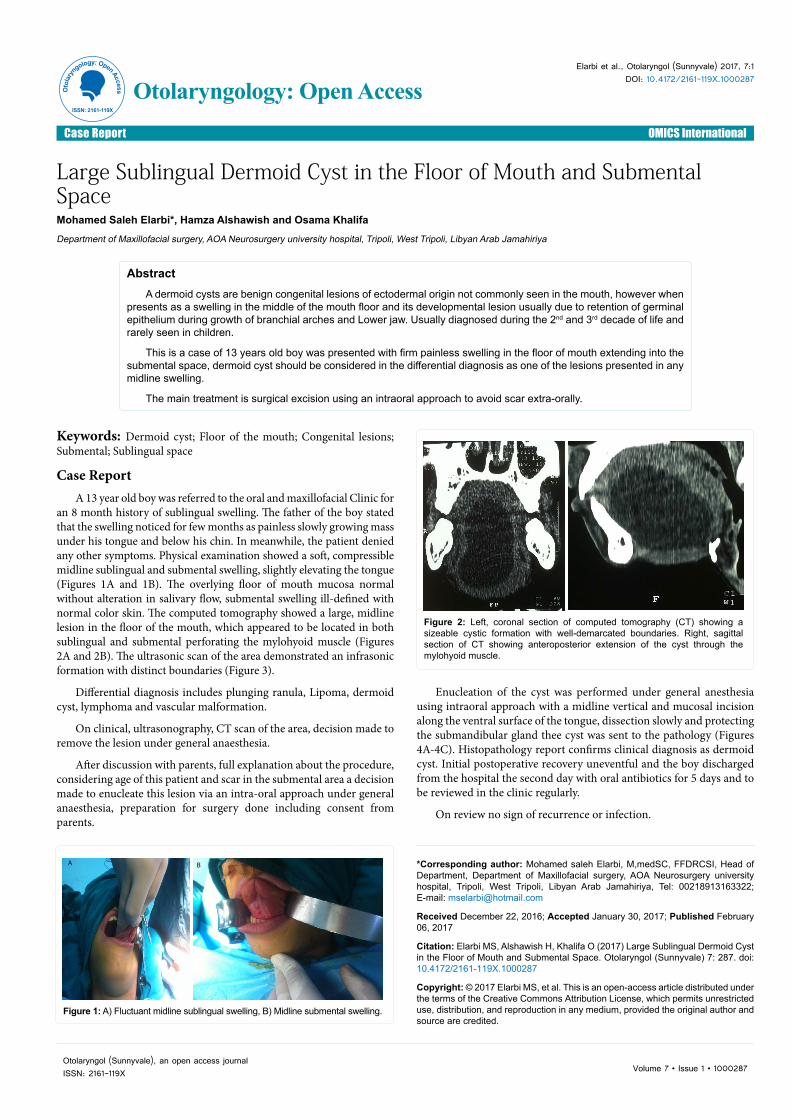
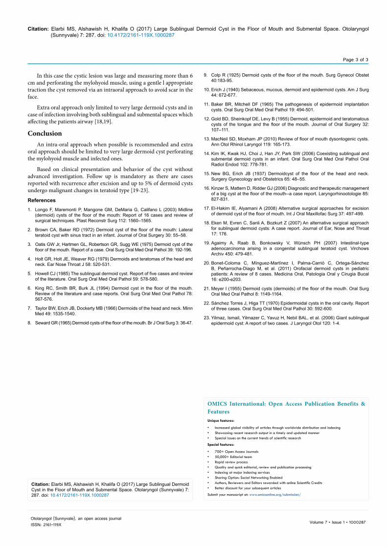
an 8 month history of sublingual swelling. The father of the boy stated that the swelling noticed for few months as painless slowly growing mass under his tongue and below his chin. In meanwhile, the patient denied any other symptoms. Physical examination showed a soft, compressible midline sublingual and submental swelling, slightly elevating the tongue (Figures 1A and 1B). The overlying floor of mouth mucosa normal without alteration in salivary flow, submental swelling ill-defined with normal color skin. The computed tomography showed a large, midline lesion in the floor of the mouth, which appeared to be located in both sublingual and submental perforating the mylohyoid muscle (Figures 2A and 2B). The ultrasonic scan of the area demonstrated an infrasonic formation with distinct boundaries (Figure 3).
Differential diagnosis includes plunging ranula, Lipoma, dermoid cyst, lymphoma and vascular malformation.
On clinical, ultrasonography, CT scan of the area, decision made to remove the lesion under general anaesthesia.
After discussion with parents, full explanation about the procedure, considering age of this patient and scar in the submental area a decision made to enucleate this lesion via an intra-oral approach under general anaesthesia, preparation for surgery done including consent from parents.
Enucleation of the cyst was performed under general anesthesia using intraoral approach with a midline vertical and mucosal incision along the ventral surface of the tongue, dissection slowly and protecting the submandibular gland thee cyst was sent to the pathology (Figures 4A-4C). Histopathology report confirms clinical diagnosis as dermoid cyst. Initial postoperative recovery uneventful and the boy discharged from the hospital the second day with oral antibiotics for 5 days and to be reviewed in the clinic regularly.
On review no sign of recurrence or infection.
AbstractA dermoid cysts are benign congenital lesions of ectodermal origin not commonly seen in the mouth, however when
presents as a swelling in the middle of the mouth floor and its developmental lesion usually due to retention of germinal epithelium during growth of branchial arches and Lower jaw. Usually diagnosed during the 2nd and 3rd decade of life and rarely seen in children.
This is a case of 13 years old boy was presented with firm painless swelling in the floor of mouth extending into the submental space, dermoid cyst should be considered in the differential diagnosis as one of the lesions presented in any midline swelling.
The main treatment is surgical excision using an intraoral approach to avoid scar extra-orally.
A B
Figure 1: A) Fluctuant midline sublingual swelling, B) Midline submental swelling.
Figure 2: Left, coronal section of computed tomography (CT) showing a sizeable cystic formation with well-demarcated boundaries. Right, sagittal section of CT showing anteroposterior extension of the cyst through the mylohyoid muscle.

Citation: Elarbi MS, Alshawish H, Khalifa O (2017) Large Sublingual Dermoid Cyst in the Floor of Mouth and Submental Space. Otolaryngol (Sunnyvale) 7: 287. doi: 10.4172/2161-119X.1000287
Page 2 of 3
Volume 7 • Issue 1 • 1000287Otolaryngol (Sunnyvale), an open access journalISSN: 2161-119X
DiscussionDermoid cysts are developmental benign conditions that have
potential to be found anywhere in the body, however majority of the cases reported in the midline of the body commonly reported in ovaries and testes, the head and neck area much less involved.
In the oral cavity it represents less than 0.01% of all oral cavity cysts [1].
Out of 1945 Dermoid cysts in the body only 103 cases in the head and neck region which accounted 6.9% studied in Mayo clinic, of which only 24 cases involving the floor of mouth 1, 6% only [2-6].
In another study carried out of 541 dermoid cysts of the body, 184 (34%) seen in the head and neck region and only 35 (6.5%) in the floor of mouth [7].
The suggested hypothesis for the origin of the dermoid cyst in the floor of mouth falls in the categories: congenital and acquired [7], the most widely accepted theory describes that the cyst results from entrapped midline ectodermal tissue during fusion in the third and fourth weeks in intra uterine life.
The arches thought to arise from first or post-traumatic implantation deep tissues injuries demonstrated as a result of accidental or surgical, whereas the lateral dermoid cyst is from the first branchial cleft [8,9].
Post-traumatic implantation deep tissue injuries demonstrated as a result of accidental or surgical [4,10]. Baker and Mitchell this tissue cavities theory by implanting skin in subcutaneous tissue of experimental rats, in which cystic cavities filled with keratin and hair [11].
Meyer classified the congenital floor of mouth cysts in to three different categories:
1. Ectodermal origin, which is lined by stratified squamous epithelial lined cyst
2. Dermoid variant mesodermal structure is sebaceous connective tissues or sweat gland
3. Teratoid is an epithelial lined cyst containing both epithelial and non-epithelial elements such as bone, muscle and gastrointestinal tissues [12,13].
According to the anatomical relationship between the floor of the mouth and the Dermoid cyst the cyst can be classified in relation to the mylohyoid muscle [7].
Clinically the picture of dermoid cysts usually present as painless slowly growing cystic lesion in the sublingual and or submental spaces with a doughy consistency often present where as those below the mylohyoid muscle in the submental area presents as asymptomatic swelling. In some cases they may cause dysphagia and dyspnea and respiratory distress, which may require Tracheostomy [6].
Clinically dermoid cyst may mimic plunging ranula, lipoma, vascular and lymphatic malformation, salivary gland neoplasm, and thyroglossal duct cyst.
Diagnostic aids such as ultra sound, magnetic resonance imaging and computed tomography with or without contrast will help defining the size, extent and nature of the lesion [1,14].
Dermoid cyst usually seen as well defined, unilocular cystic lesion have an intermediate to low signals intensity on T1-weighted MRI image and ultrasound shows typically unilocular cystic mass depending on the dermal appendages within the cyst wall [7,15].
Enucleation of the cyst through an intraoral or extra oral approach is the most commonly recommended treatment in the literature. However, marsuplization has also been proposed as a treatment alternative in cases of large cysts [16].
Depending on size, site of cysts usually intra-oral approach for small and moderate cyst (those of 6 cm and less) above the mylohyoid, where as those of large size (more than 6 cm) and below the mylohyoid and involving the submental and submandibular spaces.
In this case the lesion involving sublingual and submental space perforating the mylohyoid muscle and intraoral approach to get good cosmetic and functional effect, reported cases using intraoral approach in large dermoid cyst [13,17].
Figure 3: The (Echo) ultrasonic scan of the area demonstrated an infrasonic formation with clear boundaries.
A B
C
Figure 4: A) Intra oral midline incision from the base of the tongue to the floor of the mouth providing exposure of the lesion, B) The surgical site after complete removal of the cyst, C) The cyst measured approximately 6.5 × 4 cm sent as specimen to pathology.
Citation: Elarbi MS, Alshawish H, Khalifa O (2017) Large Sublingual Dermoid Cyst in the Floor of Mouth and Submental Space. Otolaryngol (Sunnyvale) 7: 287. doi: 10.4172/2161-119X.1000287
Page 3 of 3
Volume 7 • Issue 1 • 1000287Otolaryngol (Sunnyvale), an open access journalISSN: 2161-119X
In this case the cystic lesion was large and measuring more than 6 cm and perforating the mylohyoid muscle, using a gentle l appropriate traction the cyst removed via an intraoral approach to avoid scar in the face.
Extra oral approach only limited to very large dermoid cysts and in case of infection involving both sublingual and submental spaces which affecting the patients airway [18,19].
ConclusionAn intra-oral approach when possible is recommended and extra
oral approach should be limited to very large dermoid cyst perforating the mylohyoid muscle and infected ones.
Based on clinical presentation and behavior of the cyst without advanced investigation. Follow up is mandatory as there are cases reported with recurrence after excision and up to 5% of dermoid cysts undergo malignant changes in teratoid type [19-23].
References
1. Longo F, Maremonti P, Mangone GM, DeMaria G, Califano L (2003) Midline (dermoid) cysts of the floor of the mouth: Report of 16 cases and review of surgical techniques. Plast Reconstr Surg 112: 1560–1565.
2. Brown CA, Baker RD (1972) Dermoid cyst of the floor of the mouth: Lateral teratoid cyst with sinus tract in an infant. Journal of Oral Surgery 30: 55–58.
3. Oatis GW Jr, Hartmen GL, Robertson GR, Sugg WE (1975) Dermoid cyst of the floor of the mouth. Report of a case. Oral Surg Oral Med Oral Pathol 39: 192-196.
4. Holt GR, Holt JE, Weaver RG (1979) Dermoids and teratomas of the head and neck. Ear Nose Throat J 58: 520-531.
5. Howell CJ (1985) The sublingual dermoid cyst. Report of five cases and review of the literature. Oral Surg Oral Med Oral Pathol 59: 578-580.
6. King RC, Smith BR, Burk JL (1994) Dermoid cyst in the floor of the mouth. Review of the literature and case reports. Oral Surg Oral Med Oral Pathol 78: 567-576.
7. Taylor BW, Erich JB, Dockerty MB (1966) Dermoids of the head and neck. Minn Med 49: 1535-1540.
8. Seward GR (1965) Dermoid cysts of the floor of the mouth. Br J Oral Surg 3: 36-47.
9. Colp R (1925) Dermoid cysts of the floor of the mouth. Surg Gynecol Obstet 40:183-95.
10. Erich J (1940) Sebaceous, mucous, dermoid and epidermoid cysts. Am J Surg 44: 672-677.
11. Baker BR, Mitchell DF (1965) The pathogenesis of epidermoid implantation cysts. Oral Surg Oral Med Oral Pathol 19: 494-501.
12. Gold BD, Sheinkopf DE, Levy B (1955) Dermoid, epidermoid and teratomatous cysts of the tongue and the floor of the mouth. Journal of Oral Surgery 32: 107–111.
13. MacNeil SD, Moxham JP (2010) Review of floor of mouth dysontogenic cysts. Ann Otol Rhinol Laryngol 119: 165-173.
14. Kim IK, Kwak HJ, Choi J, Han JY, Park SW (2006) Coexisting sublingual and submental dermoid cysts in an infant. Oral Surg Oral Med Oral Pathol Oral Radiol Endod 102: 778-781.
15. New BG, Erich JB (1937) Dermoidcyst of the floor of the head and neck. Surgery Gynecology and Obstetrics 65: 48–55.
16. Kinzer S, Mattern D, Ridder GJ (2006) Diagnostic and therapeutic management of a big cyst at the floor of the mouth--a case report. Laryngorhinootologie 85: 827-831.
17. El-Hakim IE, Alyamani A (2008) Alternative surgical approaches for excision of dermoid cyst of the floor of mouth. Int J Oral Maxillofac Surg 37: 497-499.
18. Eken M, Evren C, Sanli A, Bozkurt Z (2007) An alternative surgical approach for sublingual dermoid cysts: A case report. Journal of Ear, Nose and Throat 17: 176.
19. Agaimy A, Raab B, Bonkowsky V, Wünsch PH (2007) Intestinal-type adenocarcinoma arising in a congenital sublingual teratoid cyst. Virchows Archiv 450: 479-481.
20. Bonet-Coloma C, Mínguez-Martínez I, Palma-Carrió C, Ortega-Sánchez B, Peñarrocha-Diago M, et al. (2011) Orofacial dermoid cysts in pediatric patients: A review of 8 cases. Medicina Oral, Patologia Oral y Cirugia Bucal 16: e200-e203.
21. Meyer I (1955) Dermoid cysts (dermoids) of the floor of the mouth. Oral Surg Oral Med Oral Pathol 8: 1149-1164.
22. Sánchez Torres J, Higa TT (1970) Epidermoidal cysts in the oral cavity. Report of three cases. Oral Surg Oral Med Oral Pathol 30: 592-600.
23. Yilmaz, Ismail, Yilmazer C, Yavuz H, Nebil BAL, et al. (2006) Giant sublingual epidermoid cyst: A report of two cases. J Laryngol Otol 120: 1-4.
OMICS International: Open Access Publication Benefits & FeaturesUnique features:
• Increased global visibility of articles through worldwide distribution and indexing• Showcasing recent research output in a timely and updated manner• Special issues on the current trends of scientific research
Special features:
• 700+ Open Access Journals• 50,000+ Editorial team• Rapid review process• Quality and quick editorial, review and publication processing• Indexing at major indexing services• Sharing Option: Social Networking Enabled• Authors, Reviewers and Editors rewarded with online Scientific Credits• Better discount for your subsequent articles
Submit your manuscript at: www.omicsonline.org/submission/
Citation: Elarbi MS, Alshawish H, Khalifa O (2017) Large Sublingual Dermoid Cyst in the Floor of Mouth and Submental Space. Otolaryngol (Sunnyvale) 7: 287. doi: 10.4172/2161-119X.1000287